Embed Size (px)
Citation preview
T&ah&on Vol. 48. No. 41. pp. 9033-9072.1992 Priited in Great Britain
0040402or92 s5.00+.00 @ 1992 kJ8Mum hesS Ltd
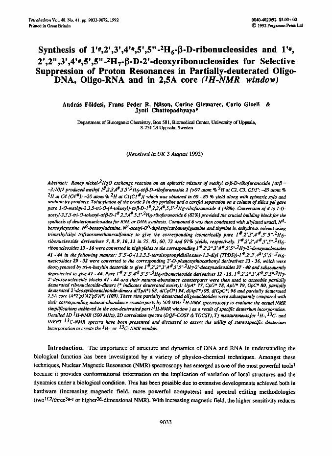
Synthesis of 1’#,2’,3’,4’#,5’,5 “-2H,-~-D-ribonucleosides and l’#, 2’,2”,3’,4’#,5’,5”-2H,~~-D-2’-deoxyribonucleosides for Selective Suppression of Proton Resonances in Partially-deuterated Oligo-
DNA, Oligo-RNA and in 2,5A core (JH-NMR window)
And&s Fbldesi, Frans Peder R. Nilson, Corine Glemarec, Carlo Gioeli & Jyoti Chattopadhyaya*
Department of Bioorganic Chemistry, Box 581, Biomedical Center, University of Uppda, S-751 23 Uppsala, Sweden
(Received in UK 3 August 1992)
Abstract: Raney nickeL2H20 exchange reaction on an epimeric mixture of methyl a@-D-ribofuranoside [a@ =
-3:lOJl produced methyl 1#,23.4#Ss’2H6-~~-D-ri~f~anosi& 2 [>97 atom % 2H at C2. C3. CSl5’: -8.5 atom %
2H at C4 (C4%); -20 atom 46 2H at Cl(C1 #)I which was obtained in 60 - 80 % yield along with epimeric xylo and arabitw by-products. Toluoylation of the crude 2 in dry pyridine and a careful separadon on a wlumn of silica gel gave pure l-O-methyl-235-~i-0-(4-toluoyl)-~~-D-l~2~.4~JJ’-2Hs_~bof~anosi& 4 (48%). Conversion of 4 to I-O-
acetyl-23~-ni-O-tolrroyI-a/BD-l~~~,~SS~-2H6-~~~~si~ 6 (82%) provided the crucial btdlding block for the
synthesis of deuterionucleosi&sfor RNA or DNA synthesis. Compound 6 was then condensed with silylated uracil, N4- benwylcytosine, N6-bensoyladetdne, N2-acetyl-06-diphenylcarbamoylguanine and thymine in anhydrous solvent using trimethylsilyl trifuoromethanesulfonate to give the corresponding isomerically pure 1 ~,2’,3’.4”#,5’5”-2H6-
ribonucleoside derivatives 7,8,9,X0, I1 in 75, 85, 60, 73 and 91% yields, respectively. 14.2’,3’,4QS’J”-2H6-
ribonucleosides 13 - I6 were converted in high yields to the corresponding 1~~‘~“3~,4~~‘~‘~~H7-2’-deoxynucleosides
41 - 44 in the following manner: 3’s’-O-(1 .I.3,3-tetraisopropyldisiloxane-1.3~diyl (TPDS))-11#2’~‘.4#‘5’5”-2H6- nucleosides 29 - 32 were converted to the corresponding 2’-O-phenoxythiocarbonyl derivatives 33 - 36, which were deoxygenated by tri-n-butyltin deuteri& to give l”2’,2”3’,4RSS”-2H7-2’-&o~nucleosi&s 37 - 40 and subsequently
&protected to give 41 - 44. Pure 1#2’3’,41g’S”2H6-ribonucleosidc derivatives 12 - 15, l~,2’.2”3*.4BJ’J”2H7- 2’-deo~nucleoside blocks 41 - 44 and their natural-abundance counterparts were then used to assemble partially deuterated ribonucleotide-dimers (* indicates Luterated moiety): UpA* 77, CpC* 78, ApU* 79. GpC* 80. partially deuterated 2’-&oxyribonucleotide-dimers d(TpA*) 93, d(CpG+) 94, d(ApT+) 95. d(GpC*) 96 and partially dettterated 2.5A core (A*2p5*A2pSAA*) (109). These nine partially deuterated oligonucleotides were subsequently compared with their corresponding natural-abundance counterparts by 500 MHz IH-NMR spectroscopy to evaluate the actual NMR simplifications achieved in the non-dcuteratedpart (IH-NMR window ) as a result of spect~c dcuterium incorporation. Detailed ID IH-NMR (500 MHz). 20 correlation spectra (DQp-COSY di TOCSY), TI measurements for lH-, l3C- and
INEPT T3C-NMR spectra have been presented and discussed to assess the utility of stereospecific deuterium incorporation to create the IH- or 13C- NMR window.
Introduction. The importance of structure and dynamics of DNA and RNA in understanding the biological function has been investigated by a variety of physico-chemical techniques. Amongst these techniques, Nuclear Magnetic Resonance (NMR) spectroscopy has emerged as one of the most powerful tools1 because it provides conformational information on the implication of variation of local structures and the dynamics under a biological condition. This has been possible due to extensive developments achieved both in hardware (increasing magnetic field, more powerful computers) and spectral editing methodologies (twotf.2/thme3a-e or higher%limensional NMR). With increasing magnetic field, the higher sensitivity reduces
9033

9034 A. FOLDESI et al.
the amount of an oligomer needed to obtain a good quality spectrum, and increases the dispersion of resonance signals reducing the spectral complexity due to resonance overlap (from second order J couplings to first order). Homonuclear two-dimensional (2D) correlated spectroscopy (COSY) provides a direct proof of the existence of
resolved scalar couplings (3JEE). and correlate the chemical shifts of coupling partner through the single or multiple coherence transfer of nuclear spins from one transition to another (as in DQF-COSY) or by the migration of cohemnces in an oscillatory manner through the entire spin system (TOCSY) which visualize the structure of the spin system in a most direct and informative mannerIc~2. On the other hand, 2D nuclear Overhauser enhancement (NOESy)5 result from the transfer of magnetization due to motional processes causing cross relaxation of dipolar-coupled spins which fall off with the sixth power of the distance between two relaxing protons [< r(t)-6 >-J/6], where r(f) = ensemble of distances due to interconversions of conformations when the NMR measurements were being made) 5b-e. Thus NMR has the capabilities of yielding both interproton distances and bond torsion angles which in conjunction with various computational methods4 (e.g. distance geometry, energy minimization and molecular dynamics) can give the solution structure of oligonucleotides (i.e.
conformations of sugars, glycosidic bonds, phosphate backbone, H-bonding, stackings etc).ld In these efforts to collect conformational informations, it is ideal that each resonance line and cross-peak due to two interacting nuclei is clearly separated in homonuclear proton-proton, heteronuclear proton-carbon, proton-phosphorus, carbon-phosphorus, NOESY and ROESY experiments. Although such fiit order informations ate possible to extract from the 2D and 3D NMR experiments of a smaller oligonucleotide, it is simply impossible to collect all of these informations in a non-prejudicial manner from a large molecule bigger than 14-16 mer duplex DNA and
8-12 mer single stranded RNA. These problems are associated with spectral overlap which becomes more and more complex due to overcrowding of resonances particularly from the repeating pentose moieties with increasing chain length. It is clear that any technique that simplifies spectral complexities would have a considerable impact in future structural studies on larger DNA or RNA molecules that represent specific biological function. The problem due to severe spectral overlap of proton resonances in absorption assignments and nOe volume measurements could partly be solved by chemical means by selective or complete suppression of absorptions arising from a chosen domain of an oligomer by substituting proton (IH) with deuteron (2H) while extracting necessary information arising from the non-deuterated part of the molecule. By incremental shift of the non-deuterated site (IH-NMR window) in an oligo-DNA or RNA (see Scheme l), one should be able to
put together the total structural information of a much larger oligonucleotide than what is possible today. What is important in this concept that two IH-NiUR windows in two different oligomers should have at least an overlap of a nucleotide residue with specific chemical shifts in order to be able to correlate protons from both windows with respect to the same nucleotide reference point (i.e. same proton resonances in both NMR-windows).
The use of deuterium exchange for the spectral assignment of nucleosides and oligonucleotides is a well established technique6*162527tj. The deuteration of the nucleobase residue has been described (e.g. exchange of protons at CI-purine and CS-cytosine with deuterioammonium bisulfite at pD 7.8 in deoxyoligomers~ gave 90 - 95 atom % a incorporation, and platinum-catalyzed exchange at CS-methyl of thymidine in 2H206b afforded 94 atom % 2H incorporation) and its effect on 1D and 2D IH-NMR spectra was studied. In general, most attention has been however given to the possible simplification of the most crowded sugar part of the lVMR spectra which holds important informations regarding the dynamics of both local and global conformation of the molecule. A large variety of enzymatic20l25 and chemical methods7-19,2t-23,26-2* h ave been devised for deuterium incorporation at both sugar8@22,27i-j~2* or nucleoside7,9-14*25 levels which have the potential to provide

Synthesis of 1’#,2’,3’,4’#,5’,5”-zH~-~-D-ribonucleosides 9035
monomeric building blocks deuterated at specific carbon center(s)7-**.*7t-ja or perdeuterated*5. Enzyme
promoted incorporation of deuterium has been found to be rather low (- 90%)20 and unsatisfactory for NMR work because of tire stray resonances arising as a consequence of the low level of deuteratior&*5 and cumbersome isolation of deuterated mononucleotide blocks=. 5’.5”-*Hz-Adenosine was prepared from 2’,3’-O- isopropylideneadenosine-S-carboxylic acid7 or from methyl-2.3~isopropylidene-PD-ribofuranosiduronic acid*.
. . : : : : : : : : . :
“NMR-window” = Non-deuterated
DNA/RNA DeUtSTllti DNA/RNA Signifies shift of “NMR-window” across the whole length of the molecule. Note that the design of NMR-windows with a slight overlap enables one to walk on the molecule by a common nOe or a chemical shift on two adjacent “NMR-windows”
Succesive shift of “NMR-window” in a DNA or RNA molecule allows the incremental assemblage of I,,,.,,_ cot$omatwnal lt$ormtton such as rorswnal angles or noes using the techniques of Multidimensional
Scheme 1
The mixtures of diastereoisomers of 5’-deuterioadenosineg and S’(R/S)-deuteriothymidine (98 atom % 2H
incorporation)10 were obtained via the reduction of the appropriate 5’aldehydes by sodium borodeuteride or lithium aluminium deuteride. The 5’aldehyde derivative of 2’-deoxyguanosine was converted to 5’ or 4’- deuterio-2’-deoxyguanosinet t by heating the aldehyde in *H2O/pyridine mixture (1: 1) followed by reduction of the aldehyde with NaBH4.4’~Deuterium labeled uridine and thymidine (98 atom % *H) was obtained upon
NaBD4 addition to the protected 4’,5’-unsaturated nucleoside followed by oxidationt*. Deuterium was incorporated at C3’ (97 atom % *H) of adenosine at sugar level upon stereoselective reduction of 1,2:5,6-di-O- isopropylidene-a-D-hexofuranos-3-ulose to 1,2:5,6-di-O-isopropylidene-3-deute~~a-D-~bohexof~anoset3
using sodium borodeuteride. Recently, more than 95 atom % *H incorporation has been accomplished at C3’ of adenosine with virtually complete stereoselectivity upon reduction of the 2’-O-terr-butyldimethylsilyl(TBDMS)- 3’-ketonucleoside by sodium borodeuteride in acetic acid 14. Specifically 2’-monodeuterated (R or S)-2’- deoxycytidines were synthesized15 from specifically 2-monodeuterated-2-deoxy-D-ribosesteobtained upon stereospecific reduction of a 2,3dehydro-hexopyranose with lithium aluminium deuteride and oxidation of the resulting glycal. Syntheses of 2’-deoxy-2’(S)-deuterio-uridine and cytidine were carried out by the use of l- methyl-2deoxy-2(S)-deuterioribofuranoside 17.2-Deoxy-1-deuterio-D-erythro-pentose., 2-deoxy Z(S)-deuterio- D-erythro-pentose and 2-deoxy- 1,2(S)-dideuterio-D-erythro-pentose were obtained from D-arabinose by a reaction sequence involving the formation and LiAlD4 reduction of ketene dithioacetal derivative@. Detailed method was published by us about the stereospecific synthesis of all eight 2’ or 2”deuterio-2’-deoxynucleosides by reductive opening of appropriate methyl 2.3~anhydro-a-D-rib0 or B-D-lyxofuranosides with LiAlD419.
2’,2”-Dideuterio2’deoxyguanosine and thymidine were prepared from 2-deoxyribose 5-phosphate using 2- deoxyribose 5-phosphate aldolase enzyme in *Hz0 achieving some 90 atom 96 deuterationm. The synthesis of
9036 A. FOLDESI etal.
all 2’.2”-dideuteri&‘-deoxynucleosides was achieved in this laboratory with deuterium incorporation at both nucleoside and sugar levels (oxidation of C2’, subsequent reduction with NaBD4 or LiAlD4 followed by
deoxygenation by tributyltin deuteride) *l. More than 99 atom % *H incorporation at Cl of 2’-deoxyribose has been reporteda recently by reduction of 3,5-bis-0-TBDMS-2-deoxyribonolactone by Dibal-D. On the other hand, the same reaction performed on the 2Jdideuterated ribonolactone obtained upon base-catalyzed H/D exchange at C2 of 2deoxyribonolactone (-95% and -85% deuteration at 2 and 2’-position, respectively) gave 1,2,2’-trideuterio-2’-deoxyxibose**.
Clearly, each position of the sugar residue can be selectively labeled, and some of these selectively deuterated nucleosides have indeed found their use in solid-state *H-NMR studies on the internal motions of nucleosides23 and oligonucleotides 24. In the temperature dependent lineshape analysis in solid-state *H-NMR
spectroscopy. the stereoselectivity of 2’ versus 2” labeling 24~ or the level of deuteration do not play a significant role. The use of specifically deuterium labeled nucleotides for the simplification of 1D and 2D tH-NMR spectra in solution studies has not however attracted much attention. An early and so far most extensive use of deuteration in the 1D NMR studies was perfom& by Danyluk et al. These workers isolated perdeuterated *H- labeled mononucleotides (-90 atom 96 *H incorporation)20 in a tedious manner from RNA digest of blue-green algae grown in a20. These perdeuterated nucleoside blocks were then used to obtain a wide variety of partially deuterated dimers and trimers*s for the purpose of resonance assignments in ID *H-NMR spectra (200 - 300
MHz). Synthesis of 4’,5’,5”-*H3-adenosine26 was accomplished and coupled to appropriately blocked adenosine 3’-phosphite to give ApA* (pA* = 4’,5’,5”-*H3-PA). This dimer allowed the unequivocal measurement of nOe difference between phosphorus and H-3’26.
A powerful alternative method of stereospecific deuteration to give polydeuterated sugars employs exchange of hydrogen with deuterium at the hydroxyl bearing carbon (i.e. methylene and methine protons of hydroxyl bearing carbon) using deuterated Raney nickel catalyst in *H2G7. Detailed studies revealed structure dependent difference in exchange rates *7e*h, high level of epimerization 27d& significantly lower extent of deoxygenation27h and difficulties in the reproducibility of the level of deuteration *7h. Despite these inherent
problems in the deuterated Raney nickel-*Hz0 exchange reaction with sugars, a number of deuterated nucleosides specifically labeled at 2’,3’ and 4’ positions were prepared for the first time by us taking advantages of this method. Our procedure28 consisted of deuteration at 2. 3 and 4 positions of methyl P-D-
arabinopyranoside by Raney nickel-*Hz0 exchange reaction 27 followed by reductive elimination of 2-hydmxyl group by tributyltin deuteride*g to give methyl P-D-2,2’,3,4-*I+-2-deoxtibopyranoside which was converted to methyl a/P-D-2,2’,3,4-*H4-2-deoxyribofuranoside and glycosylated to give various 2,2’,3,4-*H4-
nucleosides (>97 atom 96 *H incorporation for H3’ & H4’; -94 atom % *H incorporation for Ii2 and IQ’). Recently methyl P-D-erythrofuranoside was treated with deuterated Raney Ni to afford after purification methyl PD-2,3,4(S)-*H3+rythrofuranoside (-75 atom % *H incorporation at C2 and C4(S) positions and 100 atom %
*H incorporation at C3) *Ti. This sugar was converted to D-3,4,5(S)-*H3-ribose from which the four ribonucleoside were prepared. These nucleosides were subsequently reduced to the corresponding 3’,4’,5’(S)- *H3-2’-deoxynucleosides*7j.
Results and Discussions. During our 1D and/or 2D NMR studies on conformation of trimeric, tetrameric, pentameric, heptameric, nonameric and decameric branched RNAs3h and DNA duplexesmb we realized, that, though the assignment of given protons could be facilitated by selective deuteration at a specific site of short oligomers, the substantial simplification of the crowded sugar domain to extract unambiguously J

Synthesis of 1 ‘#,2’,3’,4’#.5’,5”-*H6-p-D-ribonucleosides 9037
1 2 3:R=Ac 4:R=Tol
5:RrAc 6:R=Tol
HO OH m, OH AC0 OAc
7: B=U 12: B=U 17: B=U, 21: B=U;
8: B=C& 13: B=CBZ 18: B=CBI 22: B=cB’
9: B=ABZ 14: B=ABZ 19: B=ABZ 23: B=A” lo: B=GAy 11: B=T
15: B-GDpc - AC 16: B=T
20: B=GF 24: B=GF
25: B=U, Q- it -0
26: B=? 29: B=C’= 33: B=CB= 37: B=cB’
27: B = ABZ 30: B=Ak 34: B-A& 38: B=ABI
28: B=Gr 31: B=G;r 35: B =Giy 39: B = Gr
32: B=T 36. B=T 40: B=T
41: B=CB” 45: B=C?’ 49: B =CBz 53: B=CB=
42: B=A& 46: B=ABL 50: B=A” 54: B=A&
43: B=Gz 47: B =G;r 51: B=Gr 5% B =G”F
44: B=T 48: B=T 52: B=T 56: B=T
57: B’ =UTd 61: B=UTo’ 65: B’=T 69: B=T
58: B’=C+ 62: B=C& 66: B’=@ 70: B= CBz
59: B'r A*= 63: B=ABL 67: B’=AL 71: B=A& 60: B’=GmB 64: B=GmB 68: B’=GmB 72: B=GmB
scheme2

9038 A. FOLDESI~~CZ~.
couplings or nOe volumes could not be achieved in this manner.Thisprompted us to reconsider the IH-NMR
window concept (Scheme 1) which in its most simple form has been proposed by Danyluk er ul.25. In their work, they achieved only a low and unsatisfactory level of deuteration (-90 atom % 2H incorporation)20~25 which suffices for the purpose of assignment of resonances only. In the present concept of IH-NiUR window, we have set out to achieve a highest possible level of deuteration of nucleoside moieties for the substantial
simplification of the crowded sugar domain. Clearly, the highest possible level of deuteration would suppress stray proton resonances from these deuterated moieties, and will help in the unambiguous extraction of conformational informations from the IH-NMR window (creation of really lH-NMR invisible domain due to deuteration, see Scheme 1) with modem multidimensional NMR techniques. We realized that the goal for high level of deuteriation at as many carbons as possible can be achieved only by synthetic chemistry since the published enzymatic method is known to give low level of deuterium incorporation from deuterated blue green algae20125 unacceptable for the present concept of IH-NMR window.
We envisioned at the outset that the preparation of a simple 2’,3’,5’,5”-2H4-nucleoside with ~97 atom % 2H incorporations should serve well for the purpose of creating IH-NMR window because of the fact that it would have Hl’ vicinal to 2H-2, and H4’ would be flanked by vicinal 2&3’,5’,5” which should block the
propagation of J relays in the 2D spin-spin correlated spectroscopy such as DQF-COSY or TOCSY experiments. This means that although Hl’ and H4’ would be detectable in 1D lH-NIvlR experiments, they would be completely undetectable in 2D COSY, DQF-COSY or TOCSY experiments thus simplifying the spectra for assignment purposes allowing the extraction of coupling constants and nOe volume informations from the protonated part of the molecule. It was clear to us at this stage that the H4’ of 2’(2”),3’,5’,5”-2I-I4(5)-nucleotide would show coupling with the S-phosphorous in 1D 1H-NMR experiments but this would be undetectable in 2D experiments because of the reasons said above.
Since the glycosylation reaction of a nucleobase with the a/p mixture of sugar derivative 5 or 6 gives p-
D-nucleoside in a stereospecific manner owing to the intermediacy of carboxonium ion formed with participation of the 2’-0-acyl protecting group3la, we decided to start our Raney nickel-‘&l20 exchange reaction on a crude mixture of methyl a@-D-ribofuranoside (Scheme 2). A mixture of methyl a,$-D-ribofuranoside (-3:lO) 1 was obtained in 98% yield upon treatment of D-ribose with concentrated sulfuric acid in dry methanol32 at -4 ‘C for
24 h, followed by neutralization upon a passage through a column of Amberlyst A-21 (OH- form) ion exchange resin. This epimeric mixture of sugars 1,2H20 and deuterated Raney nickel were heated under reflux with exclusion of atmospheric moisture (see experimenatal). The site specific exchange (rates C5 > C3 => C2 >> C4) competes with slower epimerization reactions at C2 and C3 27. It has been shown that complete deuteration at C5 and 30-50% deuteration at C2 and C3 could be achieved within 6 h upon boiling methyl c1 or BD-ribofuranoside
in 2H20 with deuterated Raney nickel. In the early stage of oiu work, the generally applied W-2 type Raney nickel33 was used with moderate reaction time (3 - 4 days). In our experiments, however, we wished to drive the deuteration exchange reactions to as high deuterium atom content as possible for suppressing all stray resonances (monitored by 500 MHz lH-NMR). An acceptable level of deuterium incorporation (>97 atom % 2H) even at the relatively slowly exchanged C2 required at least 4 - 7 days of boiling under reflux (see experimental) giving a higher level of deuterium exchange at the expense of lower yield (60 - 80 %) of the desired methyl 1#,2,3,4#,5,5’-2I-I6-a#-D-ribofuranoside 2 (>97 atom % 2H at C2, C>, C5/5’; -85 atom % 2H
at C4 (C4”t); -20 atom 8 2H at Cl(Cl’#)). The lH-NhlR spectra of this crude sugar derivative 2 at 500 MHz revealed the presence of products due to side reactions 27d@ (see anomeric region in Figure 1 A). Subsequent

Synthesis of 1’#,2’,3’.4’#,5’,5”-*H6-P_Dribonuc1eosides 9039
I I l-/I I I I 5.0 ppm 4.8 4.2 3.9 3.8 3.5
i (C) A
I 13, 1 1 , I’ ’ 1 v 1 8 , 1 8 n I
6.5 ppm 6.3 6.0 5.6 4.9 4.4
1~~‘~~“1’~~~/~“““1”“1”” ~~‘~1”~~‘~~1~‘111’1~~“~~~1~~ 8.00 ppm 7.80 7.20 6.40 5.60 4.60
Figure 1: 500 MHz IH-NMR spectra of deuterated D-ribofuranoses (a97 atom % 2H at C2. C3. C5/5’; -85 atom % 2H at C4; -20 atom % 2H at Cl) and their natural-abundance counterparts (99.985 atom % 1~). (A) shows the reaction mixture of deuterated Raney-Ni exchanged methyl-(a@)-D-ribofuranoside (2) & (B) shows natural-abundance methyl-(cslp: -3/10)-D-ribofuranoside (1). (C) shows I-O-acetyl-2.3.5~tri-0-(4-toluoyi)-P_D- ribofuranoside (6) and (D) shows its natural-abundance counterpart. Note that only sugarproronr are shown in the above subspccua. (E) shows 2’,3’,5’-O-tri-(4-to~uoy~)-1”,2’,3’.4’W,5’.5”-2H6-uridine (7); (F) shows natural- abundance counterpart

9040 A. FOLDESI et al.
acetylation of this crude product 2 with acetic anhydride in dry pytidine resulted in l-0-methyl-2,3,5-u-i-O- acetyl-a,@-D-1#,2,3,4#.5,5’-2I-I6-ribofuranoside 3 in 82% yield, which was converted to 1,2,3,5-tetra-O-acetyl- a@-D-1#,2,3,4#,5,5’-2H@bofuranoside 5 by a treatment with sulfuric acid in acetic acid-acetic anhydride for
12 h at room temperature (RT) (97%). Neither compound 3 nor 5 were possible to purify by chromatographic means. Compound 5 was therefore coupled with N-protected nucleobases31 and purified chromatographically. The examination of lH-NMR spectra of the chromatograpically pure reaction product revealed that the the required P-D-nucleoside was the major component (90-9596) which was contaminated with inseparable by-
products. This impurity content slowly diminished during the run through the synthetic sequences and purification steps giving pure 5’-hydroxy blocks 25 - 28 and 53 - 56. Another striking finding was the highly variable level of deuterium exchange (-98% down to -70 96) depending upon the given batch of the deuterated Raney-Ni catalyst used in the isotope exchange reaction. This problem could partially be. solved by repetition of the deuteration reaction one more. time without any further loss of yield of 2 due to isomerization reactions. This is because of the fact that the first cycle of deuteration exchange for 4-7 days gave the thermodynamic mixture of epimers which did not alter in the second cycle of deuteration exchange. In the course of synthesis of RNA dimers it turned out, that 95-962 2H incorporation is sufficient for the suppression of cross peaks in DQF- COSY and TOCSY spectra. On the contrary, the same level of deuteration was not sufficient to suppress the cross peaks in the DNA dimers because of more effective J relays through the 2’-deoxy protons (i.e. H2’ and ID”). These findings clearly indicated the need for (i) a reliable deuterium exchange method to attain a consistent deuterium enrichment in the sugar in different batches, and (ii) since such deuterium exchange reaction will inherently produce epimeric xylo and arabino sugars as main by-products 27a, therefore the purification of the methyl a/P-D-1#,2,3,4#,5,5’-2H6-ribofuranoside 2 from the exchange reaction should be achieved.
In the pursuit for a more active catalyst, we tried to use W-5 type Raney nicke1s4, which is prepared at
higher temperature (50 f4 ‘C). With this modification and by the use of at least 20 ml 2H2O/g of sugar, it was possible to achieve a reproducible >97 atom % deuterium incorporation at c2, C3 and C5 positions in 2. It was also noted that Cl and C4 could be consistently deuterium enriched by -20 and -85 %, respectively. As it tums out from Table 1 (compound 9 vs others), the extent of exchange at Cl and C4 increases slowly depending on the length of reflux (-10 atom % 2H at Cl and -78 atom % 2H at C4 after boiling at reflux for 4 days, and -17
atom % 2H at Cl and -85 atom % 2H at C4 after boiling at reflux for 7 days) whereas the deuteration levels at
C2, C3 and C4 practically do not change. In order to obtain pure starting material for the coupling reactions with the N-protected nucleobases, we decided to apply 4-toluoyl protection for the hydroxyls, which made the UV detection possible facilitating the separation procedure. The toluoylation was carried out in dry pyridine (5 ml/mm01 sugar) at ambient temperature overnight35 to give after a careful separation on a column of silica gel the desired practically pure 1-0-methyl-2,3,5-tri-O-(4-toluoyl)-a/P-D-1~,2,3,4~,5,5’-2H~-~ibofuranoside 4 in 48%
yield. The conversion of l-O-methyl 4 to 1-0-acetyl 6 provided a second opportuinity for purification. The reaction, performed in acetic acid-acetic anhydride mixture by adding sulfuric acid, gave the l-0-acetyl-2,3,5-u+ O-toluoyl-o/~-D-l#,2,3,4#,5,5’-2H6-ribofuranoside 6 (Figure 1C) in a substantially higher yield (82%) after
purification than the previous one, indicating that the most important separation was really achieved after the toluoylation reaction (1 + 4).
Compound 6 was condensed with silylated uracil, N4benzoylcytosine36, N6-benzoyladenine36, @- acetyl-Oe-diphenylcarbamoylguanine j7 and thymine in anhydrous 1,2-dichloroethane (in case of the protected
Synthesis of 1 ‘#,2’,3’,4’#,5’,5”-2Hs-P-D-ribonuclesi~s 9041
guanine dry toluene) using trimethylsilyl trifluoromethanesulfonate as Lewis acid catalyst31$37 to prepare the corresponding 1”,2’,3’,4’#,5’,5”-2Hg-B-D-ribonucleoside derivatives 7,8,9,10, 11 in 75, 85,60,73 and
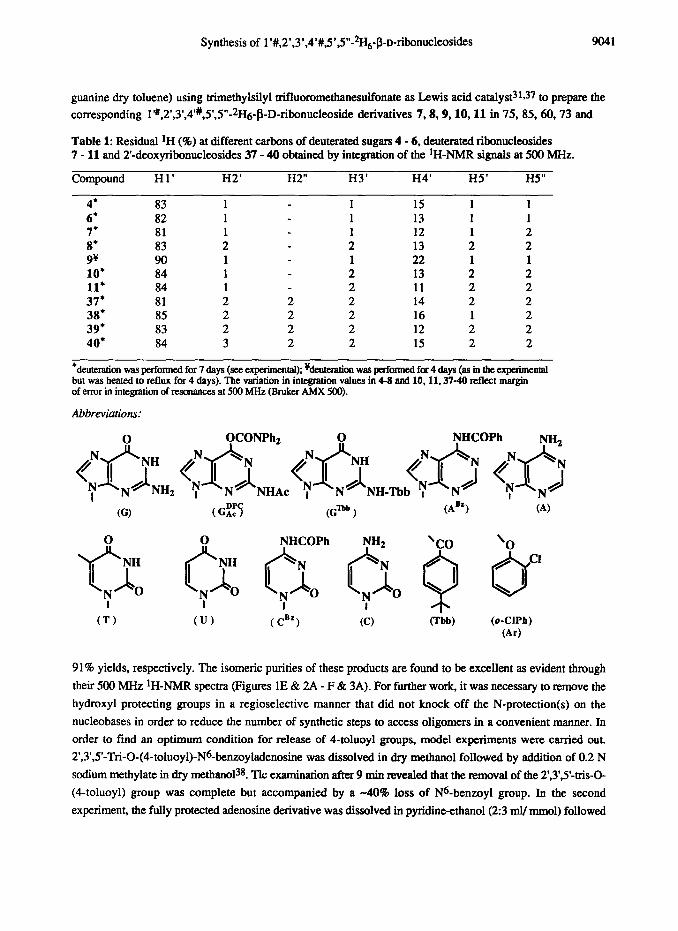
Table 1: Residual lH (96) at different carbons of deuterated sugars 4 - 6, deuterated ribonucleosides 7 - 11 and 2’-deoxyribonucleosides 37 - 40 obtained by integration of the IH-NMR signals at 500 MHz.
Compound H 1’ H2’ H2” H3’ H4’ H5’ H5”
4’ 83 1 1 15 1 1 6* 82 1 1 13 1 1 7* 81 1 1 12 1 2 8* 83 2 2 13 2 2 9v 90 1 1 22 1 1 10’ 84 1 2 13 2 2 11, 84 1 2 11 2 2 37* 81 2 2 2 14 2 2 38’ 85 2 2 2 16 1 2 39* 83 2 2 2 12 2 2 40, 84 3 2 2 15 2 2
*deUeration was performed for 7 days (see experimental); %euteration was performed for 4 days (as in the experimental but was heated to reflux for 4 days). The variation in integration values in 4-8 and 10,11.37-40 reflect margin of error in integration of resonances at 500 MHz (Bruker AMX 500).
Abbreviations:
I
e-3 (AB") ’ (A)
CT) &W (0.ClPh) (Ar)
91% yields, respectively. The isomeric purities of these products are found to he excellent as evident through their 500 MHz ‘H-NMR spectra (Figures 1E & 2A - F & 3A). For further work, it was necessary to remove the hydroxyl protecting groups in a regioselective manner that did not knock off the N-protection(s) on the nucleobases in order to reduce the number of synthetic steps to access oligomers in a convenient manner. In order to find an optimum condition for release of 4-toluoyl groups, model experiments were carried out. 2’,3’,5’-Tri-O-(4-toluoyl)-Na-benzoyladee was dissolved in dry methanol followed by addition of 0.2 N
sodium methylate in dry methar101~. Tic examination after 9 mitt revealed that the removal of the 2’,3’,S-tris-O- (6toluoyl) group was complete but accompanied by a -40% loss of N6-benzoyl group. In the second experiment, the fully protected adenosine derivative was dissolved in pyridineethanol(2:3 ml/ mrnol) followed

9042 A. FOLDESI et al.
WI L
H5. H4. H5”
8.00 ppm
Figure 2: 500 MHz ‘H-NMR spectra of deuterated-P-D-nucleosides (a97 atom % *H at C2’. C3’. W/5”; -85 atom % 2H at 0% (C4’3; -20 atom % 2H at Cl’ (Clr)) and their natural-abundance counteqarts (99.985 atom % 1~). (A) shows 2’,3’.S-O-~i-(4-toluoyl)-l’Y,2’,3’,4’1,5’,5”-2H~-N4-benzoylcytidine (8); (B) shows natural- abundance counterpart: (C) shows 2’.3’.5’-O-tri-(4-toluoyl)-1”,2’.3’.4’1,5’,5”-2H~-N6-benzoyladenosine (9): (D) shows natural-abundance countem; (E) shows 2’,3’,5’-0-ui-(4-toluoyl)-1”.2’.3’,4’1.5’,5”-2H~-N2-acetyl-06- diphenylcarbamoylguanosine (10); (F) shows natural-abundance counterpart;

7%
B1r
lJTd
; B
-A&
74; B'=+,
B=GDpc
77: B'=U; B=A
78:
B=G
B'=A&, B=U
AC
B'=C;
75:
79: B'=A; B=U
76: B1=GTBB;B=CBz
801 B'=G; B=C
89: B'=T;
B=ABz
93: B'=T; B=A
90: B'=C%; B=G"F
94: B'=C; B=G
91: B'=A$
B=T
95: B'=A, BIT
92: B1=GTBB;B=p
96: B'=G; B=C
H
81.
B’=
UTo
’; B
=A-
82; B’
=C”;
B=
GTB
B
83:
B1=
Am
; B
=u’“”
84
: B
’=G
mB
; B
=p H
97: B'=T;
B=A"
98: B'=C"; B=GTBB
99: B'=A"; B=T
loo: B1=GmB; B=CB*
Sch
eme 3
H
H
85: B'=U; B=A
86: B'=C; B=G
87: B'=A; B=U
88: B'=O; B=C
101: B'=T; B=A
102: B'=C; B=G
103: B'=A; B=T
104: B'=G: B=C

A. FOLDESI et al.
(A ! h.
(B
J&j* 1
-I- 7.80 ppm 7.60 730 6.00 n
rrrrF - _rl -T- IT”““““““““““‘I”“““I1 -,*mI,-I,I,II 8.32 ppm 1.54 6.10 4.50 4.00 2.40
l-~-----l---f-l---~~~--T-~ 8.80 ppm 8.16 8.00 7.50 6.40 4.96 4.00 3.9cl 2.70
Figure 3: 500 MHz ‘H-NMR spectra of deuterated-P-D-nucleosides (>97 atom % *H at C2’. C3’. W/S”; -85 atom % *H at C4’ (C43; -20 atom % *H at Cl’ (Cl’#)) and their natural-abundance counterparts (99.985 atom % IH). (A) shows ~-(2’,3’,5’-~-ui-(4-tOlUOyl)-~‘~,2’,3’,4’u,5’,5”-2H6-~-D-ribofaranOSy~)-thymine (11); (B) shows natural-abundance counterp$; (C) shows 3’.5’-0-(1.l,3,3-~traisopropyldisilo~~e-l,3-diyi)-l~~,2~,2~~,~,4~,5~5~~- 2H7-2’-deoxy-N4-benzoylcyudme (37); (D) shows natural-abundance counterpart; (E) shows 3’,5’-O-(1 .1,3,3- tetraisopropyldisiloxane-1 ,3-diyl)-l’~.2’.2”,3’,4’~.5’.5”-*H7-2’-deoxy-N6-bn~oyladenosine (38); (F) shows natural-abundance counterpart.

Synthesis of 1’#,2’,3’,4’#,5’,5”-*H6_B-D-ribonucleosides 9045
by addition of ethanolic NaOH (6.4 ml of ethanol and 6.4 ml of 2N NaOH / mmol)39, complete deprotection was found to take place within 5 min without any loss of base protection. The latter method was used to obtain the crude deuterated nucleosides 12,13,14,15 and 16 in 99, 98, 105, 79 and 99% yields, respectively, after neutralization by Dowex cation exchange resin (H+ form) and processing as specified in the experimental part for the different nucleobase protected nucleosides. Though these nucleosides were contaminated by toluoyl derivative (6toluic acid) as evidenced by 1H-NMR, this impurity did not however interfere in any respect with the subsequent reaction sequences. Crude 12 - 15 were treated with 4-methoxyniphenyhnethyl (MMTr) chloride in dry pyridine overnight to give the S-O-MMTr-1’#,2’,3’,4#‘,5’,5”-2Hg-nucleosides 17,18, 19 and 20 in
53,78,72 and 78 % yields, respectively, after column chromatography. These derivatives were acetylated by acetic anhydride treatment in dry pyridine to afford the 5’-O-MMTr-2’,3’-di-O-acetyl-1’~,2’,3’,4’#,5’,5”-~H~- nucleosides 21.22,23 and 24 in 85, 97, 92 and 78% yields, respectively. After removal of the 5’-0-MMTr group by a treatment with 80% aqueous acetic acid at ambient temperature overnight, the 5’-hydroxy blocks 25, 26, 27 and 28 were obtained in 95, 81, 94 and 58 % yields, respectively, which were used for dimer
syntheses. For the conversion of our deuterated ribonucleosides 13 - 16 to the corresponding 2’-deoxynucleosides
41- 44, the convenient route devised by Robins et al. was chosen 3&2tv 28. Treatment of compounds 13 - 16 with 1,3-dichloro- 1,1,3,3-tetraisopropyldisiloxane 40 in dry pyridine resulted in 3’,5’-O-(1,1,3,3-tetraiso- propyldisiloxane- 1,3-diyl(TPDS))- 1’#,2’,3’,4’#,5’,5”-2Hg-nucleosides 29, 30,31 and 32 in 85, 77, 83 and 66 % yields, respectively, obtained as white foams. The reaction of 2’-hydroxyls in 3’,5’-O-(TPDS)- nucleosides with phenoxythiocarbonyl chloride in dry acetonitrile using N,N-dimethylaminopyridiie as catalyst
did not proceed smoothly. In a set of control experiments nondeuterated counterparts of 33 and 34 were obtained in -60% yield and complete loss of compound occured in case of the nondeuterated guanosine derivative 31. Since the guanosine derivative proved to be the most sensitive under the above condition, we took the nondeuterated counterpart of 31 as a starting material for further model experiments to devise condition for the introduction of phenoxythiocarbonyl group to the 2’-OH. Overnight treatment of nondeuterated 31 with
phenoxythiocarbonyl chloride in dry pyridine gave nondeuterated 35 (61 %), after a work-up with saturated
sodium bicarbonate and column chromatography, but the reaction mixture as well as the product had a strong brown colour. Treatment of the same compound in the same manner but in dry dichloromethane using l-
methylimidazole as catalyst proved to be more successful to give nondeuterated 35 in 77 % yield. Application of
this method for introduction of phenoxythiocarbonyl group in deuterated 29 - 32 gave the corresponding 2’- phenoxythiocarbonate derivatives 33, 34, 35 and 36 in 87, 88, 89 and 89 % yields, respectively. 2’- Deoxygenation of these compounds by tributyltin deuteride in presence of 2,2’-azobis(2-methyl-propionitrile) (AfBN) in dry toluene at 75 Y! proceeded without any problem to give compounds 37,39 and 40 in 75,96 and
95 % yields respectively, except for the adenosine derivative 34, in which case simultaneous partial loss of benzoyl group had occured as it was revealed by the *H-NMR spectra. The re-benzoylation of the 3’,5’-O- TPDS-1’#,2’,2”,3’,4’#,5’,5”-2H7-2’-deoxyadenosine was done easily to give an additional crop of the desired derivative increasing the original 77% yield to 92%. The deuterium incorporation at the 2” was >97 % as evidenced by integration of lH-NMR spectra at 500 MHz (Figures 3C - F & 4A - D) of the residual proton resonance, which is consistent with the isotope content of LiAlD4 (98 atom % 2H) used in the reduction of tributyltin chloride4t to tributyltin deuteride which was used as the reagent. After removal of TPDS protection by a treatment with tetrabutylammonium fluoride in dry tetrahydrofuran, the 1’#,2’,2”,3’,4’#,5’,5”-2H7-2’-

9046 A. FOLDESI et al.
(A)
8.18 ppm 7.40 7.20 6.32 4.70 4.00 2.64
11 (Cl -L H6
Figure 4: 500 MHz IH-NMR spectra of deuterated-P-D-nucleosides (>97 atom % 2H at C2’. C3’. W/5”; -85 atom 96 2H at C4’ (C4”): -20 atom 46 2H at Cl’ (Clr)) and their natural-abundance counterparts (99.985 atom % 1~). (A) shows 3’.S-0-(1.1.3,3-tetraisopropyldisiloxane-1,3-diyl)-1’1,2’,2”.3’.4’W,S.5”-2H7-2’-deoxy-N2-acetyl- 06-diphenylcarbamoylguanosine (39); (B) shows natural-abundance counterpart; (C) 3’,5’-O-(1.1,3.3- tetraisopropyldisiloxane-1 ,3-diyl)-1’P,2’,2”.3’,4’Y,5’,5”-2H7-rhymidine (40); (D) shows natural-abundance counterpart. lH-NMR spectra of partially deuterated and natural diibonucleoside-(3’+53-monophosphates in D20 at 298 K. (E): UpA* where the 1*,2’,3’,4’# and 5’/5” protons of the adenosine @A*) residue are exchanged with 2H, (FJ natural UpA.

Synthesis of 1’#,2’,3’,4’#,5’,5”-*H6-P_Dribonuc1eosides 9047
deoxynucleosides 41,42,43 and 44 were obtained in 88, 93, 80 and 83 % yields, respectively. These compounds were further transformed to the S-0-MMTr derivatives 45,46,47 and 48 (87,90,84 and 88%, respectively), followed by acetylation of the 3’-hydroxyls as documented above for the ribo compounds to afford the fully protected heptadeuterio nucleosides 49,50,51 and 52 in 89, 77, 94 and 83 % yields, respectively. It should be noted here that acetylation of 3’-hydroxyl group of 2’deoxyadenosine derivative 46 had to be carried out at low temperature (4 “C) with small excess of acetic anhydride (1.3 equiv) in order to
avoid the formation of bis-Ne-protected adenine (Ne-benzoyl, Ne-acetyl) derivative. This diminished regioselectivity of 3’-0-acetylation reaction in 46 is presumably owing to the lack of the inductive effect of the missing 2’-OH group4*, which also enhances the basic@ of N6. Removal of S-0-MMTr group was achieved by a short treatment with 2 % benzenesulfonic acid in dichloromethane-methanol(7:3, v/v) mixture to give the S-hydroxy blocks 53,54,55 and 56 in 86, 90, 78 and 69 % yields, respectively. Though the harsher acid
treatment in case of 2’deoxypurine nucleosides (especially N6-benzoyladenosine43) can be potentially harmful despite the fact that the glycosyl bonds in 50 and 51 are stabilized by 3’-0-acetyl group4*, the shortened treatment gave 5’-hydroxy blocks with yields comparable to those obtained for ribo compounds using milder acid treatment (with 80% acetic acid).
Preparation of Partially Deuterated Dinucleotides & Trinucleotide. In order to investigate the effect of deuteration on 1D and 2D lH-NMR such as DQF-COSY, HOHAHA (TOCSY) and NOESY, two sets of dimers were synthesized using phosphotriester chemistry 43. The 5’-OH group of 1’#,2’,3’,4’#,5’,5”-*H6-
ribonucleoside blocks 25,26,27 and 28 as well as the nondeuterated 61,62,63 and 64 were coupled to the triethylammonium salt of 2’-O-(3-methoxy-l,5-dicarbomethoxypentane-3-yl(MDMP))-S-O-~r-~~nucl~-
side 3’-(2-chlorophenyl)-phosphates 43-46 57,58,59 and 60 according to the reaction Schemes 2 & 3 in dry pyridine in the presence of l-mesitylenesulfonyl-3-nitro-1,2,4-triazole (MSNT)45 to give the fully protected partially-deuterated dimers UpA* (* denotes for deuterated nucleoside moiety) 73 (72%), CpG* 74 (95%), ApU* 75 (79%), GpC* 76 (400/o), and their natural counterparts UpA 81 (91%), CpG 82 (84%), ApU 83 (65%) and GpC 84 (84%) after saturated sodium hydrogen carbonate work-up and column chromatography. The deprotection of the dimers above was carried out using a well established literature procedure44a,46 and subsequent purification on Sephadex A-25 culumn using a linear gradient of ammonium bicarbonate to obtain the
deprotected partially deuterated dimers UpA* 77 (530/o), CpG* 78 (49%), ApU* 79 (77%), GpC* 80 (49%),
and their nondeuterated natural counterparts UpA 85 (76%), CpG 86 (76%), ApU 87 (56%) and GpC 88
(85%), which were lyophylised from *Hz0 before they were subjected to NMR studies. The 5’-OH group of 1’#,2’,2”,3’,4”,5’,5”-*H7-2’-deoxyribonucleoside blocks 53 - 56 and the nondeuterated 69 - 72 were coupled with the 3’-phosphotriester blocks 65 - 68 as described above434 to give the fully protected partially deuterated di-(2’-deoxynucleoside)monophosphates d(TpA*) 89 (86%), d(CpG*) 90 (75%), d(ApT*) 91 (77%),
d(GpC*) 92 (80%), and their natural counterparts d(TpA) 97 (860/o), d(CpG) 98 (83%), d(ApT) 99 (90%) and d(GpC) 100 (89%). Subsequently, the depmtected partially deuterated 2’-deoxyribonucleotide dimers d(TpA*) 93 (62%), d(CpG*) 94 (92%), d(ApT*) 95 (78%), d(GpC*) 96 (80%), and their natural counterparts d(TpA) IO1 (80%), d(CpG) 102 (90%), d(ApT) 103 (89%) and d(GpC) 104 (71%) were obtained after deprotection and purification pmcedures reported for the diribonucleoside-monophosphates dimers43-46.
In order to further evaluate the actual NMR simplification that has taken place in the IH-NMR window part as a result of specific deuterium incorporation we decided to prepare the shortest oligomer in which a central nondeuterated unit (‘H-NMR window ) is sandwiched between two deutetated units, i.e. a trinucleotide. An

9048
(B) H5C H2C
H6C Hl’G Hl’C H2’G H3’G
HS/H5"C
7.0 7.6 4.4 4.2 4.0 3.6
6.0 4.0 2.5
e--T, ’ 1 a ’ ’ ’ r / t ’ I r ’ 9 ’ ’ I PPm 6.0 40
Figure 5: IH-NMR specUa of natural and partially deuterated dil-ibonucleoside-(3’~5’)-monophosphates, di(Z’- deoxyribonucleoside)-(3’-t5’)-monophosphates and 2.5A core in D20 a: 298 K. (A): natural CpG, (B): CpG*
where the 11.2’.2”,3’,4’# and 5’/5” protons of the guanosine (JIG*) residue are exchanged with 2H. (C): natural
d(CpG), (D): d(CpG*) where the 1”,2’,2”.3’,4’# and S/5” protons of the guanosine (pG*) residue are exchanged.
(E) A1(2’+5’)A2(2’+5’)A3, (F): A1*(2’+5’)A2(2’+5’)A3* where the 1’#,2’.3’,4’# and 5’/5” protons of the 5’-
terminal At and 3’-terminal A3 residues are exchanged with 2H. The HIJI of At* and A3* singlet. The H4’# of At*
residues appear as appears as a singlet while the H4# of A3’
phosphorus of the A2(3’+5’)A3* phosphate linkage. appears as a doublet due to its coupling 10 the

Synthesis of 1’#,2’,3’,4’#,5’,5”-%6-P_Pribonucleosides 9049

9050 A. FOLDESI et al.
obvious choice was the 2.5A core (A2’pS’A2’pSA) 114 and its partially deuterated counterpart (A*2’pSA2’p5’A*) 110 because it mimics the antiviral properties of interferon47, and the detailed structnral studies of the 2,5A core 114 have been performed by lH-NMR spectroscopy4*. In the present synthesis of A2’p5’A2’p5’A 114 and its partially deuterated counterpart A+2’p5’A2’pSA* 110. we have employed the same strategy which was devised earlier@b in this laboratory, and based on the use of simultaneous 5’ & 3’ protection by the l,3-dichloto-1,1.3,3-tetraisopropyl-disiloxane4~h (Scheme 4). The eight partially deuterated dinucleotides and the trinucleotide were subsequently compared with the corresponding natural counterparts to evaluate the actual NiUR sim.plt#hatiot~ achieved in the IH-NMR winabw part as a result of specific a%xterkun
incorporation 69. A few 1 and 2D lH-NMR spectra at 500 MI-Ix (UpA*, CpG*, d(CpG*), A*2’p5’A2’p5’A* and their natural abundance comerpar&) are shown in Figures 4E - F & 5 - 12 as representative examples.
The properties and application of deuterated nucleoside blocks in the NMR
spectroscopy. The main source of information used to solve 3D structures of nucleic acids by NMR spectroscopy resides in short (dA) interproton distance data. To obtain these distances, the lH-NMR spectrum must first be assigned using the two-dimensional correlation and nOe experiments which show through bond and through space connectivities respectively. Correlation experiments serve to group together protons belonging to the same sugar residue, while the detection of nOes serve to connect one residue with its immediate neighbours dependending upon their dipolar relaxation rates which are inversely proportional to the sixth power of their distance. The simplest experiment to delineate the spin systems via scalar correlation is the COSY experiment55 which shows direct through bond connectivities. COSY can however show some serious limitations due to spectral overlap in the 1’. 2, 3’. 4’ and 5’/5” region. The coupling network is then best identified by means of Hartmann -Hahn (HOI-MI-IA) spectroscopy~ which shows both the direct and the relayed through bond connectivities along the Hl’-I-Z-H3’-H4’-HS’/lW’ pathway in each sugar unit. In the 2D COSY or HOHAHA spectrum, the intensity of a cross peak depends on the magnitude of the J-coupling constant57. A
sequential assignment can also be performed using nOe spectroscopy 58.59. The base and sugar moieties of the
same nucleotide are connected via the intraresidue Hl’/IXZ’/I-I3’(i)-H8/I-I6(i) cross relaxation pathway. The through space connectivities along the Hl’/H2’(i-l)-H8/I-I6(i)-Hl’PI2’(i) and H8/H6(i)-H5(i+l) pathway will give the sequential assignment along one strand
In addition to the interproton distances derived from the nOe data, the vicinal 3J spin-spin coupling
constants [i.e. 3Jt*,z, 3J1*,2”, 3J2’,3@, ~JY,Y, 3J3:4: 3J4’,51, 3J4,5**] provide useful information regarding the
phase angle of the sugar pucker and the conformation about the C!4’-C5’ bond. Various proton-proton torsion angles in the nucleotide can be estimated from the magnitude of these 3Ecouplings which are easily obtained by
analyzing the multiplet pattern in COSY-like spectra such as DQF-COSY@ or E-COSY61. However, the assignments by HOHAHA or NOESY experiments, or the determination of vicinal coupling
constants by DQF-COSY are seriously restricted to cases where the spectral overlap of the proton resonances is not too critical and where the linewidths are not significantly larger than the magnitude of the J-couplings. In the NMR spectra with overlapping resonances, it becomes increasingly difficult to assign the chemical shifts, and to extract data from the nOe experiments, or to identify the proton spin system to extract the torsional informations, and thus, it becomes impossible to solve the solution structure of biologically functional oligonucleotides as the sire of the molecule increases. The problem of spectral overlap is particularly critical in the proton NMR spectra of RNA as seen from the few reported RNA structural studies’j2 compared to DNA studiesle. The exchange of some of the sugar hydrogens with deuterium in selected nucleotides indeed simplifies the NMR spectrum by

(A)
(B)
WA
-I
Figu
re
6: 2
D h
omon
ucle
ar
Har
tman
n H
ahn
(HO
HA
HA
) sp
ectra
of
the
nat
ural
an
d pa
rtial
ly
deut
erat
ed
Urid
ylyl
-(3’
+5’)
-ade
nosi
ne
(UpA
) in
D20
at
298
K.
Pane
l A
re
pres
ents
th
e 2D
spe
ctru
m
for
the
nond
eute
rate
d U
pA.
Pane
l B
rep
rese
nts
the
2D
spec
trum
of
the
par
tially
de
uter
ated
U
pA*
whe
re t
he I
’#, 2
’,3’,
4’#
and
S/5”
pro
tons
of
the
aden
osin
e @
A*)
resi
due
are
exch
ange
d.
In th
e 1D
spe
ctru
m,
the
Hl’
appe
ars
as
a si
ngle
t at
6.0
5 pp
m
whi
le
the
H4’
app
ears
as
a d
oubl
et
at 4
.29
ppm
due
to
its
coup
ling
to t
he p
hosp
horu
s of
the
3’+
5’
phos
phat
e lin
kage
. In
the
2D
spe
ctru
m,
the
J-ne
twor
k fo
r th
e 3’
-term
inal
res
idue
@A
*) h
as v
anis
hed.
H3'
C
“4’C
W
IHS
’C
H2’
C
I I
I T
ff
Iwo
0 II
II II
H?x
w
’c
W‘H
SG
H
2C
“2-G
I /
(B)
I&
I I\
A ,i
A__
i/ --
--It
H’-
-=l
ik
Figu
re
7:
HO
HA
HA
sp
ectra
of
th
e na
tura
l an
d pa
rtial
ly
deut
erat
ed
2’-
deox
ycyt
idyl
yl-(
3’+5
’)-2
’-de
oxyg
uano
sine
[d
(CpG
)]
in D
20
at 2
98K
. Pa
nel
A
repr
esen
ts
the
2D s
pect
rum
fo
r th
e no
n-de
uter
ated
d(
CpG
). Pa
nel
B r
epre
sent
s th
e 2D
spe
ctru
m
of t
he p
artia
lly
deut
erat
ed
d(C
pG*)
w
here
th
e lJ
t, 2’
,2”,
3’
. qJy
and
57
5” p
roto
ns
of t
he g
uano
sine
(p
G*)
res
idue
are
exc
hang
ed.
In th
e 1D
spe
ctru
m,
the
Hl’
appe
ars
as a
sin
glet
at
6.19
ppm
whi
le t
he H
4’ a
ppea
rs
as a
dou
blet
at 4
.10
ppm
du
e to
its
cou
plin
g to
the
pho
spho
rus
of t
he 3
’+5’
ph
osph
ate
linka
ge.
In t
he 2
D
spec
trum
, th
e J-
netw
ork
for
the
3’-te
rmin
al r
esid
ue h
as v
anis
hed.

9052 A. FOLDESI et al.
significantly reducing the spectral overlap. This work has shown that it is possible to suppress the proton resonances of some selected sugar residues by specific deuteration in an oligo-DNA or oligo-RNA through the creation of IH-NMR window. Such specific deuteration of sugar moieties in a large DNA or RNA molecule should create an tH-NMR invisible part, and allows the study of structurally functional region(s) in a large RNA or DNA molecule in the tH-NMR window part. Clearly, the reduced number of protons makes it possible to assign the protons of the residual sugar units in the tH-NMR window, which, in turn allows the measutement of both the vicinal J-couplings necessary to derive the torsion angles and the nOe volumes for distance measurements. We have substituted the 2, 2”, 3’ and S/S protons in 2’-deoxytibonucleosides, or the 2, 3’ and 575” protons of the sugar residues in ribonucleosides with deuterons (>97 % 2H). Note that the Hl’ and
H4’ in these 2’deoxyribonucleosides and ribonucleosides are also -20 96 and -85 % deuterated (Hl”, H4’#), respectively. In these deuterated nucleosides, the Hlr appears as a clear singlet in the 1D lH-NMR spectra. The H4’# however appears as a doublet in the 1D spectrum due to its coupling with the phosphorus of the S- phosphate linkage. To show the efficiency of the methods for simplification of the tH-NMR spectra in the tH- NMR window, several natural abundance and partially-deuterated di-[ribonucleoside](3’-+5’)monophosphates (UpA* 73, CpG* 74, ApU* 75, GpC* 76) and di-[2’-deoxyribonucleoside](3’-+5’)monophosphates (d(TpA*) 89, d(CpG*) 90, d(ApT*) 91, d(GpC*) 92), and trimer (A*2’p5’A2’p5’A*) 110 have been
synthesized (vide supru) and their lH 1D NMR , 2D HOHAHA and 2D DQF-COSY spectra have been studied@. A few representative examples of 2D NMR simplifications are shown in Figures 4E - F, 5 A - E, 6 - 9 and 10 - 12. In 2D HOHAHA experiments, the number of nuclei to which the magnetization is distributed is
controlled through the duration of the spin lock period. Large mixing times give rise to relayed and multiple relay peaks. The transfer of magnetization is therefore possible across the total spin system. Since the 2’, (2”). 3’, 5’ and 5” protons are >97 8 exchanged with deuterium, the relay between the Hl’# and H4’# is interrupted. In the 2D HOHAHA spectra, no Hl’-H4’ cross peak is visible and the network for the deuterated sugar is totally absent. In Figures 6 - 8, the HOHAHA spectra of natural and partially deuterated dimers are represented where the Hl’#, H2’ (I-D”), H3’, H4’# and H5’/H5” of the 3’-terminal residue are exchanged with deuterium. In the 1D spectrum of these partially-deuterated dimers and trimers, the Hl’ from the deuterated sugar moiety appears
as a singlet while the H4’ (H4’ of 3’ terminal) appears as a doublet due to its coupling with the phosphorus of the phosphate linkage (Figs. 4E,F and 5A-E). In the 2D spectrum, however, the network for the 3’-terminal residue (and 5’ terminal residue of trimer) is totally absent making the assignment of the pmtonated sugar residue easier. In the DQF-COSY experiment, the magnetization is transferred from one proton to the other proton which
is coupled to it. A cross-peak is generated between these two coupled spins. Therefore, in the DQF-COSY spectra, despite the fact that the Hl’# and H4’# are not fully deuterated, all the cross peaks originating from the
deuterated sugar residue have vanished (Figs. 10 - 12). In Figures 10 - 12 are represented the DQF-COSY spectra of natural and partially deuterated dimers where the Hl’#, I-D (H2”), H3’, H4’# and H5’/H5” of the 3’- terminal residue ate deuterated. It can be seen that all cross peaks originating from the 3’-terminal deuterated sugar residue are absent in the off diagonal region. The crowded Hl’#, I-K?’ (H2”), H3’, H4’# and H5’/H5” region close to the diagonal in panel A is simplified in panel B and it becomes possible to extract the residual cross peaks and to measure the vicinal J-couplings. An example is shown in Figure 10, Panel B which represents the cross peaks present in the box 2. The box 2 contains four cross peaks namely the H2’-H3’ and H3’-H4’ cross peaks for the uridine residue and the H4’-H5’ and H4’-H5” cross peaks for the adenosine residue. A vertical slice through these pattern is shown in Figure 10, panel C. The overlap of the cross peaks

Synthesis of 1’#,2’,3’,4’#.5’,5”-2H,&D-ribonucleosides 9053
r- 5-
J I 5
_i
1 1

9054 A. FOLDESI er al.
I 6
6
I I
(A)
5
-t ,I
5
ifm
: WI
-5
-6
_ mm
,I I,,,,,,,, ,,,,,,(,, ,,),
rwm 5 4
Figure 10: DQF-COSY spectra of the natural and partially deuterated Uridylyl-(3+5’)-adenosine (UpA) in 40 at 298K. Panel A: 2D spectrum of the natural UpA. The cross peaks used for the determination of the vicinal 3JEE coupling constants am shown in the numbered boxes: (1) Hl’U-HP’U. (2): H2’U-H3’U, H3%H4’U cross peaks. and H4’A-H5’A. H4’A-H5”A. (3) H4%H5’U. H4’U-H5”U, (4) Hl’A-H2’A. (5) H3’A-H4’A. (6) H2’A-H3’A. Panel B; Expansion of the cross peak in box 2. Panel C: Vertical slice through the cross peak at the site indicated by an arrow. The determination of the J-couplings is complicated due to the overlap of the H2’U-H3’U with the H4’A-HSA cross peaks. Similarly, The H3%H4’U cross peak overlap with the H4’A-H5”A cross peak. Panel D: 2D spectrum of UpA* where the 1’#.2’. 3’. 4# and 5/S” of pA* have been exchanged with deuterium. The empty boxes show that all cross peaks involving the adenosine residue have vanished. Panel E: Expansion of the cross peak in box 2. Panel F: Vertical slice through the cross peak at the site indicated by an arrow, which now contains only the HZ’U-H3’U and H3’U-H4’U allowing an easy extraction of the coupling constants.

Synthesis of 1’#,2’,3’,4’#,5’,5”-2H6-P-D_ribonucleosides 9055
makes the measurement of the J-coupling difficult. Panel E and F represent the same region but for the deuterated dimer. Only the IX?‘-H3’ and H3’-H4’ cross peaks of the uridine residue am present which makes the
determination of the J-couplings easy and straightforward. Dipolar coupling which operates through space is responsible for the dominating mechanism of relaxation
in solution. It appears in the 1D NMR spectrum as line broadening. It also generates the mutual relaxation between spatially close nuclei, the cross relaxation which gives rise to the nOe. Upon random deuteration, each of the remaining protons in the molecule will be surrounded by fewer other protons and will have fewer pathway for cross relaxation. This will result in longer relaxation times and therebye narrower line widths. Upon selective deuteration of the protons of some sugars, the overlap problem can be overcome, but the relaxation time of the remaining protons in other non-deuterated sugar moiety(ies) will not be much affected due to cross relaxation. For example, upon selective deuteration of the I-I2’, H2”, H3’, H4’ and HS/HS of the cytidine residue (C*) in the dimer d(GpC*), the relaxation time of the Hl’ of G is little affected [TI = 1.7s in the nature1 d(GpC) and Tt = 1.8s in the partially deuterated d(GpC*)]. On the other hand, the relaxation time Tl of the HIJt of the C* changes from 2s in the natural dimer to 3.9s in the partially-deuterated d(GpC*). In the natural trimer AI2’pSA22’pSA3, the Tt for the Hl’ are as follows: 2.1s for A 1, 2.0s for A2 and 2.5s for A3. In the partially
deuterated trimer A*t2’p5’A22’p5’A*3, where the I-K?‘, H3’, H4’ and H5’/I-I5” of the A1 and A3 residues have
been >97% exchanged with deuterium and Hlq and H4’# are respectively -20 and 80 % deutered, the TI for HI* in AI and A3 are 3s and 4s, respectively, but the Tt for Hl’ in A2 is 2.1s which is basically unaltered from
the natural counterpart. Information on the conformation of the sugar ring can also be obtained from 13C-NMR chemical shifts and
coupling constants63. The conformation about the C3’-03’ bond (E) and C5’-OS bond (p) in nucleic acids can be monitored by carbon-phosphorus coupling constants, 3Jc4’p3~,3Jc~p~ for E and 3Jcep5* for p. In large
oligomers, the assignment of the carbon resonances becomes more complicated as the size of the molecule increases. Also, the measurement of the carbon-phosphorus coupling constants from the DC{ IH) spectrum is difficult due to the overlap of the carbon resonances. In the t3C( IH) spectrum with nGe., the carbon bonded to a deuterium atom should have a much lower intensity than a carbon bonded to a proton because of the fact that nGe effect does not arise through deuterium, and the intensity is also considerably decreased because of the splitting due to the 1J coupling with deuterium. The Tt relaxation time for the carbons of four natural and partially
deuterated 2’-deoxynucleosides dA, dG, dC and T have been measured using inversion recovery experiments. The carbons bonded to a deuterium atom C2’, C3’ and C5’ have longer relaxation time than the carbons bonded to hydrogen. (In natural 2’-deoxyadenosine, the Tt of the sugar carbons are: Cl’ = 0.65s, C2’ = 0.39s, C3’ =
0.71s, C4’ = 0.65s and C5’ = 0.33s. In deurerated 2’-deoxyudenosine, the TI of the sugar carbons are: Cl’ = 0.6s C2’ = 1.76s, C3’ = 1.5s, C4’ = 0.6s and C5’ = 2.1s. In natural 2’-deoxycytidine, the Tl of the sugar
carbons are: Cl’ = 0.65 s, C2’ = 0.41s, C3’ = 0.68s, C4’ = 0.62s and C5’ = 0.35s. In deuterured 2’- deoxycytidine, the Tt of the sugar carbons are: Cl’ = 0.61s, C2’ = 1.1s. C3’ = 1.2s, C4’ = 0.6s and C5’ = 2.5s. In natural thymidine, the TI of the sugar carbons are: Cl’ = 0.7 s. C2’ = 0.41s. C3’ = 0.71s, C4’ = 0.65s and C5’ = 0.36s. In deurerured zhymidine, the Tl of the sugar carbons are: Cl’ = 0.7s C2’ = 1.82s, C3’ = Is, C4’ = 0.66s and CS = 1.58s. In natural 2’-deoxyguanosine, the T1 of the sugar carbons are: Cl’ = 0.61s, C2 = 0.4s, C3’ = 0.62s, C4’ = 0.62s and C5’ = 0.31s. In deuterured 2’-deoxguanosine, the Tl of the sugar carbons are: Cl’ = 0.6s. C2’ = 1.3s, C3’ = l.ls, C4’ = 0.6s and CS = 1.6s.) Consequently, it should be possible to

9056 A. FOLDESI et al.
(A)
(B)
i ill y
-4
-6
I pm
Figure 11: DQF-COSY spectra of natural and partially deuterated 2’-deoxycytidylyl-(3’-‘5?-2’-deoxyguanosine [d(CpG)l in D20
at 298K. Panel A: 2D spectrum of the natural d(CpG). The cross peaks used for the determination of the vicinal 3J~~ coupling constants are shown in the numbered boxes: (1) HI’-H2’C, (2) HI’-H2”C. (3) H2’-H2”C. (4) H2’-H3’C, (5) H2”-H3%, (6) H4’-W’. H4’-H5”C. (7) H3’-H4’C, (8) HI’-HZ’G, (9) HI’-H2”G, (10) H2’-H2”G, (11) HZ’-H3’G. (12) H2”-H3’G, (13) H3’-H4’G. Panel B: 2D spectrum of d(CpG*) where the I@, 2’. 2”. 3’. 4* and 5’/5” of G* have been exchanged with deuterium. The empty boxes show that all cross peaks involving the pG* residue have vanished.

Synthesis of 1’#,2’,3’,4’#,5’,5”-2H&~ribonucleosides
invert or suppress particular carbon resonances selectively by the appropriate choice of the relaxation delay, and
thereby to distinguish between the carbons which am bonded to a hydrogen or to a deuterium. A straightforward and convenient means to observe the carbon chemical shifts and to measure the carbon-
phosphorus coupling constants in oligo-DNA or oligo-RNA is to use a t3C( ‘H) INEPT experiment64*65.66
where the proton spin polarization is transferred to carbon. This experiment acts as a filter eliminating effectively the resonances of the deuterated carbons. 13C-NMR INEPT experiments, with and without proton decoupling, have been performed on natural and partially deuterated 2’-deoxynucleotides, dA, dG, dC and T. In the INEPT spectra of deuterated nucleosides @A*, dG*, dC* and T* where the 2’, 2”, 3’, S, 5” are exchanged with >97% deuterium, and 1” and 4’# are respectively -20 and 80 % deuterated), the transfer of polarization from lH to t3C arises only for the Cl’ and C4’ carbons. The resonances of the carbons bonded to deuterium, C2’, C3’ and CS are effectively eliminated (Figures 13 and 14). One should also try to get the maximum possible information from the deuterated part of the molecule. The sensitivity of the carbon bearing deuterium can be selectively enhanced by using a 13C12H) INEPT experiment where the *H spin polarization is selectively transferred to l3C nucleus67@. The sensitivity of this experiment is comparable to the normal proton decoupled t3C experiment with nOe. The quadrupolar *H nucleus relaxes faster than the lH nucleus and since the rate of repetition of the INEPT is determined by the relaxation Tt of *H, faster repetition rates can be used. Hence, depending upon
whether the spin polarization to carbon nucleus is transfered through proron [‘3C( ‘H) INEPT experiment] or deuterium [ 13C(*H) INEPT experiment] in partially deuterated DNA or RNA (such as 77-80,93-96 or llO),
one can visualize either *H-bonded carbon or *H-bonded carbon selectively thus creating effective I%?-Nh4R
window.
Experimental Section
Materials and Methods. Nickel-aluminium alloy powder @O/50%), 2,2’-azobis(2-methyl-propionitrile) (AIBN), ammonia solution 328, chlorotrimethylsilane, acetic anhydride, Amberlyst A-21 ion exchanger, 1,1,3,3_tetramethylguanidine, 1-methylimidazcle and toluoyl chloride were purchased from Merck. D-Ribose, 4-methoxytriphenylmethyl chloride, diphenylcarbamyl chloride, benzoyl chloride, lithium aluminium deuteride (98 atom % *H), tri-n-butyltin chloride, 1,1,1,3,3,3-hexamethyklisilazane and phenyl chlorothionofotmate were purchased from Aldrich. 2H20 (99.9 atom % D) for the deuteration experiments was purchased from Goss Sci. Inst. Ltd. Sodium hydroxide was purchased from Eka Nobel AB, Sweden, ammonium hydrogen carbonate from BDH. 1,3-Dichloro-1,1,3,3-tetraisopropyldisiloxane (TPDSC12)40, ochlorophenylphosphoro-bis-(1,2,4- triazolide)‘t‘tb, ryn-4-nitrobenzaldoxime 45 and l-(2-mesitylenesulfonyl)-3-nitro-1,2,4-triazole (MS-NT)45 were prepared using literature procedures. Pyridine and toluene were distilled after being refluxed over calcium hydride for 3 - 4 h, 1,Zdichloroethane and dichloromethane was stirred with phosphorus pentoxide overnight followed by distillation under nitrogen. TLC was carried out using Merck pre-coated silica gel 60 F254 plates in the following solvent systems: (A) methanol-dichloromethane (199, vb), (B) methanol-dichlommethane (5:9.5, v/v), (C) methanol-dichloromethane (10:90, v/v), (D) acetonitrile-water (70:30, v/v). The short column chromatographic separations were done using Merck G60 silica gel. DEAE-Sephadex A-25 (Pharmacia) was used for the anion excange chromatography. After purification on DEAE-Sephadex column, the ammonium counterions in dimers and trimers were replaced with Na+ by passing the compounds through a Dowex 50 WX 8 (C. Roth Gmbh) (Na+ form) column, then they were repeatedly freeze-dried from 2H20. tH-NMR spectra were recorded with a Jeol FX 90 Q and Bntker AMX 500 spectrometer at 90 and 500 MHz, respectively, using TMS (0.0 ppm) or acetonitrile peak for 2H20 solutions (set at 6 = 2.0 ppm) as the internal standards. t3C-NMR spectra were taken with a Jeol FX 90 Q spectrometer at 27.7 MHz with TMS as internal reference for solutions other than *Hz0 in case of which CH3CN (set at 6 = 1.3 ppm) was used as internal reference. 3lP-NMR spectra were recorded at 36 MHz and 202 MHz in the same solvent as for tH-NMR spectra using 85 4% phosphoric acid (0.0 ppm) or CAMP (-2.1 ppm) as external standard. Chemical shifts are reported in ppm (6 scale). The two- dimensional NMR experiments were performed on a Bruker AMX-500 MHz spectrometer. The DQF-CGSY and Hartmann-Hahn spectra were recorded in pure-phase absorption mode with the time proportional incrementation

9058
(A)
(B)
I!
I
: II
x *’ 07
cl2
11
0
Figure 12: DQF-COSY spectra of natural and partially deuterated 2.5A core At(2’+5’)A2(2’+5’)A3 in D20 at 308 K. Panel A represents the 2D spectrum for the non-deuterated A1(2’+S’)A2(2’+5’)A3. The cross peaks used for the determination of the vicinat 3JHH coupling constants are shown in the numbered boxes: (1) HI’-H2’At. (2) H2’-WAl. (3) H3’-H4’A1, (4) H4’-H5’A1. (5) H4’-H5”A’, (6) HI’-H2’A2, (7) H2’-H3’A2, (8) H3’-H4’A2, (9) H4’-H5’A2, (10) H4’-H5”A2, (11) HI’-H2’A3, (12) H2’-H3’A3, (13) H3’-H4’A3, (14) H4’-HS’, H4’-H5”A3. Panel B represents the 2D spectrum of the partially deuterated A1’(2’-+5’)A2(2’+5’)A3* where the I’#, 2’. 3’. 4’# and S/5” of At* and A3* have been exchanged with deuterium. The empty boxes show that aU cross peaks involving the Al* and A3* residues have vanished.

Synthesis of 1’#,2’,3’,4’#,5’,5”-2Hs-~-D-ribonucleosides 9059
method (TPPI) and with low power preirradiation of the residual HDO peak during the relaxation delay. The DQF-COSY52 spectra were acquired with 4096 complex data points in t2 and 256 points in tl. The data were zero filled to give a 4096 x 1024 point matrix and a sine-square bell window was applied in both directions before Fourier transformation. The HartmannHahnS3 spectra were acquired with 2048 complex data points in t2 and 256 points in tl. The data were zero filled to give a 2048 x 1024 point matrix and a sine-square bell window was applied in both directions before Fourier transformation. The 1 H- 3tP chemical shift cornelation experiments was performed in the absolute magnitude mode. A 1024 x 128 matrix data set was zero filled to 1024 x 512 data points a sine-square bell multiplication was applied in both directions before Fourier transformation. The 13C NMR INEPT experiments with and without proton decoupling and the l3C NMR inversion recovery experiments were performed on a Jeol GX 270 MHz spectrometer operating at 67.8 MHz for carbon. IR absorption spectra were recorded with a Perkin-Elmer 298 spectrometer. The chemical shifs of all deuterated compounds have been compared to the IH- and z~C-iVMR spectra of corresponding natural-abundance counterpart in or&r to delineate (i) site and level of deuteration, and (ii) isomeric purity. Tri-n-butyltin deuteride was prepared via a modified method 41. Lithium aluminium deuteride (1 g. 23.8 mmol) was suspended in dry diethyl ether (75 ml) and freshly distilled u-i-n-butyltin chloride (16 ml) was added dropwise (30 min) at RT under argon. After an additional stirring for 3 h, the mixture was cooled in an ice-bath and water (-50 ml) was slowly added. The etheral phase was washed with water (2 x 50 ml), dried over MgSO4 and evaporated. Residue was subjected to vacuum distillation and fractions collected up to 75 “C were used in deuteration reactions. 1%~NMR (neat): 30.2 C-p (taken as reference50); 27.38 C-y, 13.79 C-6; 8.15 C- a. IR (neat): vSa_D= 1298 cm-l (litr.sl: vsa_D= 1300 cm-l). Methyl a@-D-ribofuranoside (1). A solution of D-r&se (8 g, 53.3 mol) in dry methanol (120 ml) was treated at 0 ‘C with concentrated sulfuric acid (0.5 ml), dissolved in 3-4 ml ice-cold dry methanol and added in a portionwise manner, then stored in refrigerator at -4 ‘C for 24 h. The solution was neutralized by passage through a bed of Amberlyst A-21 (OH- form) (300 g) resin pre-washed with distilled methanol. After eluting the column with 1 L distilled methanol, eluant was evaporated and residue coevaporated with deionized water followed by drying on oilpump to give the mixture of title compound 1 [a/p = -3: lo] as a syrup (8.57 g, 98%).
lH-NMR (D20): 4.92 (d. J t.2 = 3.2 Hz, 1H) H-l (a); 4.83 (s, 1H) H-l (p); 4.04 - 3.28 (m. 5H) H-2, H-3, H- 4 & H-5 (a+P); 3.36 (s, 3H) OMe (a); 3.33 (s, 3H) OMe (p). l3C-NMR (a): 103.5 Cl; 84.9 C4; 71.5 C2; 70.0 C3; 61.9 C5; 55.8 OCH3; (p) 108.3 Cl; 83.2 C4; 74.6 C2; 71.2 C3; 63.2 C5; 55.5 OCH3. Preoaration of the Raney-nickel catalyst (W-S). A one-liter Erlenmever flask containing a five- cent&neter long teflon-coated-magnet. was placed in a plastic beaker on top of a magnetic stirrer. A th&kometer was fixed alone the inner wall of the flask. The flask was filled with deionized-water (192 ml) and when slowlv stirring, NaOH$ellets (51.2 g) were added in one portion. After all pellets had dissolved, some ice-water wa’s filled into the plastic beaker letting the temperature decrease to 50 ‘C. Addition Ni-Al-alloy was started keeping the temperature ar 50 f4 ‘C. The total amount of Ni-Al-alloy (40.0 g) should be added within approximately 30 min starting with small portions and ending with up to 1 ml per portionThe temperature is adjusted by the cooling-rate and by the addition speed. After completion of the addition, the flask was heated in an oil-bath at 50 ‘C for approximately 60 min. The flask was left for 1 h to cool down to mom temperature. Deionized-water (3x 1L) was added and decanted. The particles were transferred to a 250 ml filtering flask with hose connection. A 20-cm long PVC-tube was connected on the hose of the flask. The flask was placed on a magnetic stirrer and the tube ended freely over a one liter beaker (for safety if to many particles are washed out). While stirring slowly, water (totally 4 L) was added slowly,while liquid containing-v&y small particles went into the beaker, and the flask could be left overnieht. for continued washing the next dav. Washing was continued for a whole dav (using totally 20 L of wat&, during the last 10 L, a-funnel was put into the-filtering flask). In the beginning stirring was used between the washings while in the end stirring was used continously. After the washing the pH was checked in the following manner: most of the liquid was decanted from the flask, the particles were standing in a small volume of water for 10 min, then the pH of the remaining water in the flask was measured. If the pH was 6.5 - 7.0 and the liquid was almost clear, then the deuteration ofihe catalyst could be started. Preparation of the deuterated Raner-nickel catalyst. The catalvst oarticles were transferred to a 50 ml serum bottle containing a two-centimct~long tefloncoated magnet. The bottle was sealed using a rubber stopper and the suspension was stirred for one minute, then after settling of the particles, water was removed with a pasteutpipette. After repeating this procedure a few times, deuteriumoxide (approximately 1.5 ml) was added, the bottle was flushed with nitrogen whereafter it was stoppered and sealed with parafilm. Stirring was maintained for 30 min (“1st wash”). The liquid was removed as before. 2H20 was added (same amount), the bottle was flushed again with nitrogen, sealed with parafilm and stirted for half an hour. (At this stage the bottle could be left without stirring ovemight,“2nd wash”). This washing procedure was repeated a few times. From

(Al)
(B I)
-k
----
----
C
D11
I c2
Cl’
c2
C4'
C3'
PPH
90 85
80
75
JO
65
60
55
50
45
40
35
30
PPM
90 85
80
75
JO
65
60
55
50
45
40
35
30
Figu
re 1
3: 1
3C IN
EPT
NM
R sp
ectra
of n
atur
al an
d pa
rtial
ly d
eute
rate
d de
oxyn
ucle
osid
es a
t 298
K in
CD
C13
. (A2)
: Nat
ural
2’-d
eoxy
cytid
me [
dCl.
(Al):
Deu
lera
ted 2
’-
deox
ycyt
ine
[dC
*], (
Bz)
: Nat
ural
P’-
deox
yade
nosi
ne [d
A],
(By)
: Deu
tera
ted
2’-d
eoxy
aden
osin
e id
A*]
. (C
2): N
atur
al 2
’4eo
xygu
anos
ine
[dG
], (C
l): D
eute
rate
d 2’
- de
oxyg
uano
sine
[dG
*], @
2): N
atur
al th
ymid
ine (
T), (
Dl):
Deu
tera
ted t
hym
idin
e F].
In d
C*,
dA
*. d
G*
and
P,
the 2
’, 2”
. 3’. 5
’ and
5” p
roto
ns h
ave b
een
exch
ange
d w
ith
deut
eriu
m (~
97%
2H).
The H
l’ is
- 20
% de
uter
ated
whi
le th
e H
4’ is
- 85
% d
eute
mtc
d. In
dC
*, d
A*.
dG
+ an
d P,
th
e tra
nsfe
r of p
olar
izat
ion
from
lH to
l3C
aris
es o
nly
for t
he C
l’and
C4’
carb
ons.
The r
eson
ance
s of t
he ca
rbon
s cov
alen
tly bo
nded
to d
eute
rium
, W,
C3’
and
CS’
, are e
ffec
tivel
y el
imin
ated
.
. I..
. .“. ”
--
“..
. “.
“. .
~.
-” .
.I ~
“”
,.” “
.. “.
_ _.
. .-
.“I..
.“...”
.
” I
I”..
,_,

PPM
PPM
90 85
80
75
70
65
60
55
50
45
40
35
30
90
85
80
75
70
65
60
55
50
45
40
35
30
Figu
re
14:
13C
(1~
) IN
EPT
NM
R e
xper
imen
ts
of n
atur
al
and
parti
ally
de
uter
ated
de
oxyn
ucle
osid
es
at 2
98K
in
CD
C13
. (A
Z):
Nat
ural
2’
deox
ycyt
idin
e [d
C],
(Al):
D
eute
mte
d 2’
deox
ycyt
ine
[dC
*].
(IQ
): N
atur
al 2
’deo
xyad
enos
ine
[dA
]. (I
%])
: Deu
tera
ted
2’-d
coxy
aden
osin
e [d
A*l
. (C
z):
Nat
ural
2’-d
eoxy
guan
csin
e [f
fil,
(Cl):
Deu
tera
ted
2’-
deox
ygua
nosi
ne
[dG
*],
(D2)
: N
atur
al
thym
idin
e [T
j, (D
l):
Deu
tera
ted
thym
idin
e (T
*].
In d
C*.
dA
*, d
G+
and
T*,
the
2’,2
”, 3
’, 5’
and
5” p
roto
ns h
ave
been
exc
hang
ed
with
de
uter
ium
(>
97%
2H
). Th
e H
l’ is
- 20
% d
eute
rate
d w
hile
the
H4’
is -
85%
deu
teta
ted.
In
dC
*, d
A*.
dG
* an
d P.
th
e tra
nsfe
r of
pol
ariz
atio
n fro
m 1
~ to
13C
aris
es o
nly
for
the
Cl’
and
C4’
car
bons
. Th
e re
sona
nces
of
the
car
bons
cov
alen
tly
bond
ed t
o de
uter
ium
, C
2’. C
3’ a
nd C
5’, a
m e
ffec
tivel
y el
imin
ated
.

A. FOLDESI et al.
the 4th wash to the 16th wash a twofold amount of 2H20 was used with stirring for at least one hour if at daytime, or add a threefold amount without stirring if overnight. Usually 3-4 overnight washes were used. Gas evolved during the washes and during the last washes a strong white color appeared. After the 16th wash the catalyst was ready for use (total 2H20 used -50 ml). Methyl a/P-D-ribofuranoside-1#,2,3,4s,5,5’-2H~ (2) (# denotes partial deuteration). To a mixture of methyl u&ibofuranoside l(l.4 g, 8.5 mmol) coevaporated two times with deuterium oxide (99.8 % atom D, - 6 ml each) and dissolved in the same solvent (28 ml), deuterated Raney-Ni (settled volume: 14.5 ml) was added using a graduated pipette and after bubbling nitrogen through the mixture for 10 min, it was boiled to reflux with stirring, at 110 ‘C oil bath, for 7 days in an atmosphere of nitrogen. The mixture was filtered through a bed of celite, washed with water, and the combined aqueous phases were evaporated to give a syrup (0.84 g. 82%). IH-NMR (D20) of main components 2. tH-NMR (D20) (a): 4.92 (s, 1H) H-l; 4.02 (s, 0.3H) H-4; 3.37 (s, 3H) 0CH3; (p) 4.83 (s, 1H) H-l; 3.93 (s, 0.7H) H-4; 3.33 ( s, 3H ) 0CH3. 13C -NMR (D20) (a): 103.5 Cl; 84.9 C4; 55.8 0CX-Q; (p): 108.3 Cl; 83.1 C4; 55.52 0CH3. 1-0-Methyl-2,3,5-tri-O-acetyl-a/~-D-ribofuranoside-l~,2,3,4~,S,S’-~H~ (3). To a solution of methyl a@-D-ribofuranoside-1#,2,3,4#,5’,5”-2H6 (2) (6.3 g, 37.2 mmol) in dry pyridine (62.5 ml), acetic anhydride (25 ml) was added with cooling in an ice-bath and stirring, the stirring was continued for 2 days at room temperature. Usual work-up and drying on oil pump gave a thick syrup (9 g, 82%). tH-NMR (CDC13) of main components 3: (p): 4.87 (s, 1H) H-l; 4.09 (s) H-4#; 3.40 (s, 3H) GCH3; (a): 5.10 (s, 1H ) H-l; 4.29 (s) H-4#; 3.37 (s, 3H) OC!H3; 2.11 - 2.06 (3xs, 9H) 3xOAc (a+P). l-O-Methyl-2,3,5-tri-O-(4-toluoyl)-a/P_D~,2,3,4~,S,S’-~H~ (4). Methyl a/p- D-ribofuranoside-1#,2,3,4#,5’,5”-2% (2) (1.49 g, 8.8 mmol) was coevaporated with dry pyridine 5 times to give a dry thick syrup which subsequently was dissolved in dry pyridine (44 ml). This solution was cooled to 0 ‘C in an ice-bath, then toluoyl chloride (7 ml, 53 mmol) was added in one portion with stirring. Stirring was maintained for 20 min at bath temperature, then at room temperature overnight, when Tic showed complete consumption of starting sugar. After addition of methanol and additional stirring for 5 min. usual work-up resulted in an oily residue which was subjected to short column chromatography. First column (120 g silica) was packed, loaded and washed with 75 8 petroleum ether in CH2Cl2 until complete removal of reagent residue then the polarity was increased. Mixed fractions were re-chromatographed starting with 20 96 petroleum ether in dichlommethane followed by sequential increase of polarity (a series of similar colums was necessary on mixed fractions). After four columns, sufficiently pure compound 4 was obtained as a thick syrup (2.19 g, 48%). Rf: 0.50 (System A). tH-NMR (CDC13): 8.0 - 7.1 (m, 9H) toluoyl (a+P); (p): 5.13 (s, 1H) H-l; 4.70 (s) H-4+ 3.40 (s, 3H) OCH,; (a): 5.37 (s, 1H ) H-l; 4.61 (s) H-4#; 3.47 (s, 3H) 0CH3; 2.39 & 2.36 (~xs, 9H) 3~ toluoyl-CH3 (a+P ). 1,2,3,5-Tetra-O-acetyl-a/~-D-ribofuranoside-l~,2,3,4~,5,5’-2H~ (5). l-O-Methyl-2,3,5-tri-O- acetyl-a/P-D-ribofuranoside-2,3,4,5.5’-2H4 (3) (9 g, 30.5 mmol) was dissolved in acetic acid (75 ml) and acetic anhydride (17.5 ml) and treated with sulfuric acid (3.75 ml) with ice-cooling. After 12 h at room temperature the solution was stirred with ice-water, extracted with dichloromethane, washed with water, then aqueous sodium hydrogen carbonate, dried over MgS04 and evaporated to give compound 5 as pale yellow syrup (9.6 g, 97 %). IH-NMR of main components (CDC13): (p): 6.16 (s, 1H) H-l; 4.36 (s) H-4$ (a): 6.42 (s, 1H) H-l; 4.44 (s) H-4#; 2.2 -2.1 (4xs, 12H) 4xOAc (a+p)_ l-O-Acetyl-2,3,5-tri-O-(4-toluoyl)-a/P-D-ribofuranoside-l~,2,3,4~,5,5’-2H~ (6). l-O-Methyl- 2,3,5-ai-O-toluoyl-a/P-D-ribofuranoside-2,3,4#,5,5’-2H4 (4) (1.57 g, 3 mmol) was dissolved in acetic acid (4.3 ml) and acetic anhydride (1.0 ml) and treated with sulfuric acid (0.23 ml) with ice-cooling. Resulting mixture was stirred ovemigt at room temperature, then ice-water was added followed by usual work-up. Short column chromatography as described for compound 4 gave 6 as a white foam (1.36 g, 82 8). Rf: 0.47 (System A). ‘H-NMR (CDC13): 8.1 - 7.1 (m, 9H) toluoyl (a+P ); (p): 6.41 (s, 1H) H-l; 4.75 (s) H-4#; 2.01 (s, 3H) acetyl; (a) 6.68 (s, 1H ) H-l; 2.13 (s, 3H) acetyl; 2.41 & 2.37 (2xs, 9H) 3x toluoyl-CH3 (a+P). 2’,3’,5’-Tri-O-(4-toluoyl)-1’#,2’,3’,4’#,5’,5 “-“He-uridine (7). Uracil (73 mg, 0.65 mmol) was coevaporated with dry toluene (2x) then suspended in 1,1,1,3,3,3-hexamethyldisilazane (3.9 ml) followed by addition of chlorotrimethylsilane (0.39 ml) and the heterogeneous mixture was heated at reflux at 120 “C under an atmosphere of nitrogen for 60 min. The clear solution resulted was evaporated, then coevaporated with dry

Synthesis of 1’#,2’,3’,4’#,5’,5”-2Hb_P-D-ribonucleosides 9063
toluene (2x) to remove traces of hexamethyldisilazane. 1-0-Acetyl-2,3,5-tri-O-toluoyl-a/BD-ribofuranoside- 1#,2.3.4#,5,5’-*He 6 (276 mg, 0.5 mmol) was coevaporated three times with dry toluene, dissolved in 1,2- dichloroethane (6.5 ml) and added to the oily persilylated uracil followed by addition of trimethylsilyl trifluoromethanesulfonate (0.125 ml). The mixture was heated at 70 ‘C in nitrogen atmosphere for 4 h. Usual work-up and short column chromatography yielded compound 7 (0.23 g, 75%). Rf: 0.45 (System B). *H-NMR (CDCl$: 8.81 (br. s, 1H) N-H ; 8.0 -7.1 (m, 12H) toluoyl; 7.42 (d, J5.6 = 8.1 Hz, 1H) H-6; 6.35 (s, 1H) H- l’#; 5.59 (dd, JNB,~ = 2.2 Hz, 1H) H-5; 4.68 (s) H-4”; 2.42,2,41 8r 2.38 (~xs, 9H) 3x tohoyl-CH3. 2’,3’,5’-Tri-0-(4-toluoyl)-l’~,2’,3’,4’#,5’,5”-2H 6-N4-benzoylcytidine (8). N4-Benzoyl- cytosine (140 mg, 0.65 mmol) was condensed with sugar 6 (276 mg, 0.5 mmol) as described for compound 7 (0.42 g, 85%). Rf: 0.56 (System B). IH-NMR (CDC13): 8.71 (br. s, 1H) N-H ; 8.1 -7.1 (m, 19H) toluoyl, benzoyl, H-5 & H-6; 6.50 (s, 1H) H-l*; 4.75 (s) H-4*; 2.44 & 2.20 (2~s. 9H) 3x toluoylCH3. 2’,3’,5’-Tri-O-(4-toluoyl)-1’#,2’,3’,4’#,5’,5”-2H6- N6-benzoyladenosine (9). Ne-Benzoyl- adenine (650 mg, 2.71 mmol) and deuterated sugar 6 (1.36 g, 2.47 mmol) were condensed as described for compound 7 (1.2 g, 60%). Rf: 0.53 (System B). lH-NMR (CDC13): 8.98 (br. s, 1H) N-H ; 8.72 (s. 1H) H-8; 8.17 (s, 1H) H-2; 8.1 -7.1 (m, 17H) toluoyl, benzoyl; 6.50 (s, 1H) H-I’#; 4.82 (s) H-4’#; 2.42 & 2.38 (2xs, 9H) 3x toluoyl-CH3. 2’,3’,5’-Tri-O-(4-toluoyl)-1’#,2’,3’,4’#,5’,5 “-2H,-N2-acetyl-Oh-diphenylcarbamoyl guanosine (10). N*-acetyl-Oh-diphenylcarbamoylguanine (253 mg, 0.65 mmol) and deuterated sugar derivative 6 (276 mg. 0.5 mmol) were condensed by the method described by Robins37 (0.32 g, 73%). Rf: 0.65 (System B). IH-NMR (CDC13): 8.10 (br. s, 1H) N-H ; 8.05 (s. 1H) H-8; 8.0 -7.1 (m, 22H) toluoyl, phenyls; 6.33 (s, IH) H-l’#; 4.82 (s) H-4$ 2.48 (s, 3H) acetyl; 2.42 & 2.38 (2xs, 9H) 3x toluoyl-CHg. 1-(2’,3’,5’-Tri-0-(4-toluoyI)-1’#,2’,3’,4’~,5’,5”-2H6-~ -D-ribofuranosyl)-thymine (11). Thymine (82 mg, 0.65 mmol) was condensed with deuterated sugar derivative 6 (276 mg, 0.5 mmol) as described for compound 7 (0.28 g, 91% ). Rt: 0.48 (System B). IH-NMR (CDCl3): 8.35 (br.s, 1H) N-H; 8.05 (s, 1H) H-8; 7.37 - 7.30 (m, 1OH) Ar.; 6.09 (s, 1H) H-l’#; 4.45 (s) H-4’#; 2.46 (s, 3H) AC-@; 2.15 - 1.88 (3xs, 9H) 3xOAc. 1’#,2’,3’,4’#,5’,5”-zH&Jridine (12). Compound 7 (264 mg, 0.43 mmol) was treated as written for compound 14, but partition between water and dichloromethane followed by dichloromethane wash of water (3x) phase was used for purification. Evaporation of water phase gave compound 12 (87 mg, 99%). lH-NMR (D20): 7.82 (d, Jg e= 8.0 Hz, 1H) H-6; 5.84 (s, 1H) H-l’#; 5.83 (d, 1H) H-5; 4.05 (s) H-4’#. 1’#,2’,3’,4’#,5’,5”-slls-N4-Benzoylcytidine (13). Compound 8 (0.77 g, 1.1 mmol) was treated as written for compound 14 to give crude compound 13 with minor impurities (0.375 g, 98%). tH-NMR (CDCl$methanol-d4/DMSO&): 8.53 (d, J5,e = 7.7 Hz, 1H) H-6; 8.1 - 7.4 ( m, 5H) benzoyl; 7.44 (d, J5.6 = 7.7 Hz, IH) H-5; 5.88 (s, 1H) H-l”; 4.03 (s) H-4’#. 1’#,2’,3’,4’#,5’,5”-2H6-N6-Benzoyladenosine (14). Compound 9 (1.17 g, 1.6 mmol) was dissolved in a mixture of pyridine (3.2 ml, 2.0 ml/mmol) and ethanol (4.8 ml, 3.0 ml/mmol), and ethanolic sodium hydroxide solution (prepared from 2 N aqueous sodium hydroxide (6.4 ml) and 6.4 ml ethanol) was added. After stirring for 5 min at room temperature, the solution was neutralized by addition of Dowex ion exchange resin (H+ form). Resin was filtered off, then washed with ethanol (200 ml), the combined filtrate and washings were evaporated. Repeated coevaporation with toluene gave a powder which was trimrated with diethylether, filtered then rinsed with diethylether again. The dry crude 14 (0.63 g, 105%) was sufficiently pure for being taken in the next step. lH-NMR (CDC13/methanol-d4): 8.73 (s, 1H) H-8; 8.37 (s, 1H) H-2; 8.1 - 7.4 (m, 5H) benzoyl; 6.01 (s, 1H) H-l’#; 4.25 (s) H-4’#. 1’~,2’,3’,4’~,5’,5”-2H~-N2-Acetyl-0~-diphenylcarbamoylguanosine (15). Compound 10 (316 mg, 0.36 mmol) was treated as described for compound 14, but instead of trituration, a short column of silica was run to afford compound 15 (150 mg, 79%). IH-NMR (CDC&/methanol-d4): 8.58 (s, IH) H-8; 7.4 - 7.2 (m, 10H) phenyls; 6.08 (s, 1H) H-1”; 4.13 (s) H-4$ 2.28 (s, 3H) N*-OAc. l-(1’#,2’,3’,4’#,5’,5”-2H &3-D-RibofuranosyH-thymine (16). Compound 11 (264 mg, 0.43 mmol) was treated as written for compound 12 to obtain crude compound 16 (112 mg. 99%). IH-NMR (D20): 7.61 (d, JscB3,6= 1.2 Hz, 1H) H-6; 5.83 (s, 1H) H-l*; 4.03 (s) H#, 1.81 (d, 3H) 5-CH3. Procedure A: 5’-O-MMTr-1’#,2’,3’,4’#,5’,5”-2H 6-uridine (17). To a solution of compound 12 (110 mg, 0.44 mmol), in dry pyridine (5 ml), MMTr-Cl(178 mg. 0.575 mmol) was added and stirred overnight at room temperature. After usual-work up and purification on short silica gel column compound 17 (121 mg, 53
9064 A. FOLDESI er al.
%) was obtained. IH-NMR (CDC13): 8.0 (d, J5.6 = 8.06 Hz, 1H) H-6; 7.5 - 6.8 (m, 14 H) MMTr; 5.88 (s, IH) H-l’#; 5.33 (d, 1H) H-5; 4.15 (s) H-4’#; 3.77 (s, 3H) OCH3. 5’-O-MMTr-l’#,2’,3’,4’~,5’,5”-2H,-N~-benzoylcytidine (18). Compound 13 (116 mg, 0.33 mmol) was treated with MMTr-Cl(l22 mg, 0.39 mmol.) to give 18 (161 mg, 78 96) according to Procedure A. ‘H-NMR (CDC13) : 8.84 (br.s, 1H) N-H, 8.26 (d, J 5,~ = 7.57 Hz, 1H) H-6; 8.0 - 6.8 (m, 19H) MMTr + benzoyl; 5.87 (s, 1H) H-1”; 4.39 (s) H-4’#; 3.78 (s, 3H) OCH3. 5’-0-MMTr-1’#,2’,3’,4’#,5’,5”-2Hs-N6-benzoyladenosine (19). Compound 14 (150 mg, 0.4 mmol) was treated with MMTr-CI(l85 mg, 0.6 mmol) to give 19 (186 mg, 72 96) as in Procedure A. IH-NMR (CDC13): 9.28 (br. s, 1H) NH; 8.66 (s, 1H) H-8; 8.24 (s, 1H) H-2; 8.1 - 6.7 (m, 19H) MMTr + benzoyl; 6.07 (s, 1H) H-l*; 4.38 (s) H-4’#; 3.75 (s, 3H) 0CH3. 5’-O-MMTr-1’~,2’,3’,4’~,5’,5”-2H6-N2-acetyl-06-diphenylcarbamoylguanosine (20). Procedure A was applied to compound 15 (145 mg, 0.275 mmol) and MMTr-Cl(lO2 mg, 0.33 mmol) to give compound 20 (172 mg, 78 %). IH-NMR (CDC13): 8.54 (s, 1H) N-H, 8.19 (s. 1H) H-8; 7.5 - 6.7 (m, 24H) MMTr + phenyls; 5.92 (s, 1H) H-l’#; 4.49 (s) H-4”; 3.75 (s, 3H) 0CH3; 2.23 (s, 3H) @-OAc. Procedure B: 5’-0-MMTr-2’,3’-di-O-acetyl-l’#,2’,3’,4’#,5’,5”-2H,-uridine (21). Compound 17 (108 mg, 0.207 mmol) was coevaporated with dry pyridine (3x) then re-dissolved in dry pyridine (4 ml) and treated with acetic anhydride (63.5 mg, 2.5 mol eq.) and stirred at RT overnight. After usual work-up and purification on silica gel column, compound 21(106 mg, 85 %). Yield: 106 mg (85 %). tH-NMR (CDCl,): 8.25 (br. s, 1H) N-H; 7.69 (d, Js6 = 8.3 Hz, 1H) H-6; 7.4 - 6.8( m, 14 H) MMTr; 6.23 (s, 1H ) H-l? 5.32 (d, 1H) H-5 ; 4.22 (s) H-4”; 3.81 (s, 3H) 0CH3; 1.60 ( s,6H ) OAc. 5’-0-MMTr-2’,3’-di-O-acetyl-1’#,2’,3’,4’~,5’,5”-2H~-N4-benzoylcy~idine (22). Compound 18 (142 mg, 0.227 mmol) was treated as described in Procedure B to yield compound 22 (157 mg, 97 %). IH- NMR (CDC13): 8.25 (d, 1H) H-6; 8.0 - 6.8 (m, 19H) MMTr + benzoyl.; 6.32 (s, 1H) H-l’#; 4.30 (s, 1H) H- 4’; 3.82 (s, 3H) OMe; 2.12, 2.07 (2xs, 6H) 2xOAc. 5’-0-MMTr-2’,3’-di-0-acetyl-1’#,2’,3’,4’#,5’,5 “-2Hs-N6-benzoyladenosine (23). Treatment of compound 19 (200 mg, 0.3 mmol) as described in Procedure B afforded compound 23 (208 mg, 92 %). 1H- NMR (CDCl3): 8.59 (s, 1H) H-8; 8.25 ( s, 1H) H-2; 7.7 - 6.8 (m, 19H) MMTr + benzoyl; 6.30 (s, 1H) H-l’#; 4.33 (s) H-4@; 3.78 (s, 3H) OMe; 2.12, 2.05 (2xs, 6H ) OAc. 5’-O-MMTr-2’,3’-di-O-acetyl-1’#,2’,3’,4’#,5’,5 “-2H~-N2-acetyl-06-diphenylcarbamoyl- guanosine (24). Compound 20 (60 mg, 0.075mmol) was acetylated according to Procedure B to obtain compound 24 (52 mg, 78 %). 1H-NMR (CDClj): 8.06 (s, 1H) H-8; 7.4 - 6.8 (m, 24H) MMTr + phenyls; 6.17 (s, 1H) H-l’#; 4.32 (s) H-4’#; 3.75 (s, 3H) OMe; 2.36 - 2.05 (3xs, 9H) 3x AC. Procedure C: 2’,3’-Di-0-acetyl-1’*,2’,3’,4’#,5’,5”-2Hs -uridine (25). Compound 21 (100 mg, 0.165 mmol ) was dissolved in 80 % aqueous acetic acid (8 ml) at room temperature and stirred overnight then volatile materials were evaporated and residue coevaporated with toluene twice. Purification on silica gel column gave compound 25 (52 mg. 95%). IH-NMR (CDC13): 8.62 (br.s, 1H) N-H, 7.72 (d, J5.6 = 8.06 Hz, 1H) H-6; 6.04 (s, 1H) Hl’; 5.78 (d, 1H) H-5; 4.21 (s) H-4’#; 2.14, 2.09 (2xs, 6H) 2x OAc. 2’,3’-Di-0-acetyl-1’#,2’,3’,4’#,5’,5”-2H 6-N4-benzoylcytidine (26). Compound 22 (140 mg, 0.198 mmol) was subjected to Procedure C to obtain compound 26 (70 mg, 81%).*H-NMR (CDC13): 8,29 (d, 1H) H-6; 7.9 -7.5 (m, 6H) benzoyl + H-5; 6.12 (s, 1H) H-1$ 4.27 (s) H-4’#; 2.11, 2.08 ( 2xs, 6H ) 2x OAc. 2’,3’-Di-O-acetyl-l’#,2’,3’,4’#,5’,5”-2H~-N6-benzoyladenosine (27). Procedure C was performed on compound 23 (215 mg, 0.28 mmol) to give compound 27 (132 mg, 94 %). IH-NMR (CDC13): 9.27 (br. s, 1H) N-H, 8.76 (s, 1H) H-8; 8.13 (s, 1H ) H-2; 8.1 - 7.5 (m, 5H) benzoyl; 6.13 (s, 1H) H-1”; 4.36 (s) H-4’#; 2.17 -2.02 (2xs, 6H ) 2x OAc. 2’,3’-Di-0-acetyl-1’#,2’,3’,4’#,5’,5”-2H6- N2-acetyl-Oh-diphenylcarbamoylguanosine (28). Compound 24 (100 mg, 0.113mmol) was treated according to Procedure C to give compound 28 (40 mg, 58%). 1H-NMR (CDC13): 8.05 (s, 1H) H-8; 7.4 -7.3 (m, 1OH) phenyls; 6.03 (s, 1H) H-l’#; 4.31 (s) H-4’#; 2.39 - 2.02 (3xs, 9H) 3xAc. Procedure D: l-(3’,5’-O-(TPDS)-l’#,2’,3’,4’#,5’,5”-2H ,-P-D-ribofuranosyl)-thymine (32). Compound 16 (0.53 g, 1.5 mmol) was repeatedly coevaporated with dry pyridine and re-dissolved in the same solvent (15 ml). After addition of 1,3-dichloro-1,1,3,3-tetraisopropyldisiloxane (0.62 ml, 1.95 mmol, 1.3 mol eq.), the mixture was stirred under dry condition for lh, followed by normal work-up. The resulting syrup was

Synthesis of 1’#,2’,3’,4’#,5’,5”-2H&ti-ribonucleosides 9065
chromatographed on a short column of silica gel to give compound 32 (0.46 g, 61 %) as a foam. lH-NMR (CDC13): 9.31 (br. s, IH) N-H; 7.44 (d, JscE3,6= 1.2 Hz, 1I-I) H-6; 5.71 (s, 1I-I) H-1+$ 3.99 (s) H-4’#; 1.90 (br. s, 3H) 5-CH3; 1.1 - 1.0 (m, 24H) methyls of TPDS. 3’,5’-0-(TPDS)-1’#,2’,3’,4’#,5’,5”-2H6-N4-benzoylcytidine (29). Procedure D was applied to compound 18 (0.66 g, 1.88 mmol) to get compound 29 (0.96 g, 85%). IH-NMR (CDQ): 8.28 (d, J5,6= 7.6 Hz, 1H) H-6; 8.0 - 7.4 (m, 5I-I) benzoyl; 5.86 (s, 1H) H-l’#; TPDS.
4.23 (s) H-49; 1.1 - 1.0 (m, 24H) methyls of
3’,5’-0-(TPDS)-1’#,2’,3’,4’#,5’,5”-2H,-N6-benzoyladenosine (30). Procedure D was carried out on compound 19 (0.57 g, 1.52 mmol) to afford compound 30 as a foam (0.72 g, 77%). IH-NMR (CDCl3): 9.26 (br. s., 1H) N-H, 8.74 (s, 1H) H-8; 8.15 (s, 1H) H-2; 8.1 - 7.4 (m, 5H) Bz; 6.03 (s, 1H) H-1$ 4.11 (s) H-4’#; 1.1 - 1.0 (m, 24H) methyls of TPDS. 3’,5’-0-(TPDS)-1’~,2’,3’,4’~,5’,5”-2H6-N2-acetyl-06-diphenylcarbamoylguanosine (31). Treatment of compound 20 (152 mg, 0.29 mmol) according to Procedure D gave compound 31 (185 mg, 83%). tH-NMR (CDC13): 8.29 (br. s., 1H) N-H, 8.18 (s, 1H) H-8; 7.5 - 7.2 (m, 1OH) phenyls; 6.01 (s, 1H) H-l’#; 4.12 (s) H-4’#; 2.49 (s, 3H) AC; 1.1 - 1.0 (m, 24H) methyls of TPDS. Procedure E: l-(2’-O-Phenoxythiocarbonyl-3’,5’-O-(TPDS)-l’~,2’,3’,4’~,5’,5”-2H6-~-D- ribofuranosyl)-thymine (36). To the mixture of vacuum-dried 32 (0.46 g, 0.92 mmol) and l- methylimidazole (0.147 ml, 1.84 mmol, 2.0 mol eq.) in dry dichloromethane (10 ml), phenyl chlorothionoformate (0.179 ml, 1.29 mmol, 1.4 mol eq) was added and stirred in nitrogen atmosphere at room temperature overnight. After dilution with dichloromethane, the reaction mixture was poured into saturated sodium hydrogen carbonate solution and extracted with dichloromethane. Pooled organic phases were washed with saturated citric acid solution (2 x 30 ml), dried with MgS04. evaporated and the residue was purilkd on a short column of silica gel to afford compound 32 as a foam (0.58 g. 89 %). Rf: 0.51 (System B). IH-NMR (CDCl3): 8.66 (br. s., 1H) N-H; 7.6 - 7.0 (m, 6H) H-6 + phenoxy; 5.92 (s, 1H) H-1$ 4.05 (s) H-4’a; 1.92 (d, JscB3,6=1.2 Hz, 3H) 5-CH3; 1.1 - 1.0 (m, 24H) methyls of TPDS. 2’-O-Phenoxythiocarbonyl-3’,5’-O-(TPDS)-1’#,2’,3’,4’#,5’,5”-2HL-N4-benzoylcytidine (33). Compound 29 (0.92 g, 1.55 mmol) was converted to compound 33 (0.98 g, 87%) using Procedure E. Rf: 0.60 (System B). lH-NMR (CDCl3): 8.88 (br. s., 1H) N-H; 8.27 (d, J5,6= 7.6 Hz, 1H) H-6; 8.0-7.1 (m, 11H) phenoxy + benzoyl + H-5; 6.08 (s, 1H) H-1”; 4.19 (s) H-4’#; 1.1 - 1.0 (m, 24H) CH3 of TPDS. 2’-O-Phenoxythiocarbonyl-3’,5’-O-(TPDS)-l’*,2’,3’,4’~,5’,5”-2H6N6-benzoyladenosine (34). Compound 30 (0.72 g, 1.16 mmol) was subjected to Procedure E to get compound 34 (0.77 g. 88%). Rt: 0.59 (System B). tH-NMR (CDC13): 9.21 (br. s., 1H) N-H, 8.78 (s, 1H) H-8; 8.19 (s, 1H) H-2; 8.1 - 7.1 (m, 1OH) phenoxy + benzoyl; 6.24 (s, 1H) H-l##; 4.14 (s) H-4”; 1.1 - 1.0 (m, 24H) methyls of TPDS. 2’-O-Phenoxythiocarbonyl-3’,5’-O-(TPDS)-l’~,2’,3’,4’~,5’,5”-~H6-N~-acetyl-O~-diphenyl- carbamoylguanosine (35) Treatment of compound 32 (0.28 g. 0.37 mmol) as described in Procedure E gave compound 35 (0.30 g, 89%). Rf: 0.68 (System B). IH-NMR (CDCl3: 8.12 (s, IH) H-8; 7.94 (br. s, l- H) N-H; 7.6 - 7.0 (m, 15H) phenoxy; 6.20 (s, IH) H-l’#; methyls of TPDS.
4.17 (s) H-4’#; 2.58 (s, 3H) AC; 1.1 - 1.0 (m, 24H)
Procedure F: 3’,5’-0-(TPDS)-1’#,2’,2”,3’,4’#,5’,5*’-2H 7-thymidine (40). After coevaporation with dry toluene, compound 36 (0.70 g, 1.12 mmol) was dissolved in the same solvent (20 ml), AIBN (37 mg, 0.23 mmol, 0.2 mol eq.) and tri-n-butyltin deuteride (0.46 ml, 1.71 mmol, 1.5 mol eq.) were added. The solution was degassed by nitrogen (20 min), followed by heating at 75 ‘C under nitrogen atmosphere for 3.5 h. Volatile materials were evaporated, the residual oil was subjected to column chromatography to afford compound 40 (0.52 g, 95 %). Rf: 0.44 (System B). lH-NMR (CDCl3): 9.04 (br. s., 1H) N-H; 7.42 (d, J5cE3,6= 1.2 Hz, 1H) H-6; 6.08 (s, 1H) H-l*; 3.74 (s) H-4’#; 1.92 (d, 3H) 5-CH3; 1.1-1.0 (m, 24H) methyls of TPDS. 3’,5’-O-(TPDS)-1’#,2’,2”,,4’#,5’,5 “-2H7-N4-benzoyl-2’-deoxycytidine (37). Procedure F was used for 14 h to convert compound 33 (0.29 g, 0.39 mmol) to compound 37 (0.17 g, 75%). Rf: 0.41 (System B). IH-NMR (CDC13): 8.78 (br. s., IH) N-H, 8.33 (d, J5,6= 7.7 Hz, 1H) H-6; 8.0 - 7.4 (m, 6H) benzoyl + H- 5; 6.08 (s, 1H) H-l’#; 3.83 (s) H-4$ 1.1 - 1.0 (m, 24H) methyls of TPDS. 3’,5’-0-(TPDS)-1’#,2’,2”,3’,4’#,5’,5”-2H7-N6-benzoyl-2’-deoxyadenosine (38) Treatment of compound 34 (0.76 g, 1.01 mmol) according to Procedure F gave compound 38 (0.47 + 0.07 g; 77 I 15%). Rf: 0.45 + 0.28 (System B). *H-NMR (CDC13): 9.13 (br. s., 1H) N-H, 8.77 (s, 1H) H-8; 8.17 (s, IH) H-2; 8.1 - 7.4 (m, 5H) benzoyl; 6.04 (s, 1H) H-1”; 4.12 (s) H-46; 1.1 - 1.0 (m, 24H) methyls of TPDS.

9066 A. FOLDESI et al.
3’,5’-0-(TPDS)-1’~,2’,2”,3’,4’~,5’,5”-~H~-N~-acetyl-O~-diphenylcarbamoyl-2’-deoxy- guanosine (39). Compound 35 (0.66 g, 0.73 mmol) was treated as in Procedure F to afford compound 39 (0.53 g, 96%). Rf: 0.50 (System B). IH-NMR (CDC13): 8.21 (s. IH) H-8; 8.03 (br. s., 1H) N-H; 7.6 - 7.1 (m. 1OH) phenyls; 6.30 (s, HI) H-l*; 3.89 (s) H-4*; 2.55 (s, 3H) AC; 1.1 - 1.0 (m. 24H) methyls of TPDS. Procedure G: 1’#,2’,2”,3’,4’#,5’,5”-2H7-Thymidine (44). Compound 40 (0.49 g, 1.0 mmol) was dissolved in dry tetrahydrofurane (10 ml) and 1.0 M TBAF solution in dry THF (1 ml, 1.0 mmol, 1.0 mol. q.) was added. After stirring for 5 min, volatile materials were evaporated and the residue was purified on silica gel to give compound 44 (0.22 g, 83 96). IH-NMR (CDCl@leOH-&): 7.70 (d, Jsc.J~,~= 1.2 Hz, 1H) H-6; 6.26 (s, 1H) H-lq; 3.92 (s) H-4’#; 1.92 (d. 3H) 5-CH3. 1’*,2’,2”,3’,4’~,5’,5”-2H7-N4-Benzoyl-2’-deoxycytidine (41) When compound 37 (153 mg, 0.26 mmol) was treated according to Procedure G, compound 41 (77 mg, 88%) was obtained. IH-NMR (CDC13/MeOH-d.t): 8.53 (d. J5,6= 7.6 Hz, 1H) H-6; 8.1 - 7.5 (m, 6H) benzoyl + H-5; 6.23 (s, 1H) H-1”; 4.03 (s) H-4*. 1’#,2’,2”,3’,4’~,5’,5”-2H7-N6-Benzoyl-2’-deoxyadenosine (42). Compound 38 (0.51g, 0.84 mmol) was subjected to the treatment described in Procedure G to give compound 42 (0.28 g, 93%). lH-NMR (CDCl$MeOH-d4): 8.75 (s, 1H) H-8; 8.34 (s, 1H) H-2; 8.1 - 7.4 (m, 5H) benzoyl; 6.46 (s, 1H) H-l*; 4.17 (s) H-4’#. 1’~,2’,2”,3’,4’~,5’,5”-2H7-N2-Acetyl-06-diphenylcarbamoyl-2’-deoxyguanosine (43). Upon treatment of compound 39 (0.21 g. 0.27 mmol) with TBAF as described in Procedure G compound 43 (0.22 g, 80%) was obtained. tH-NMR (CDC13): 8.89 (br. s.. 1H) N-H, 8.20 (s, 1H) H-8; 7.5 - 7.1 (m, 10H) phenyls; 6.26 (s, 1H) H-1”; 3.97 (s) H-4*; 2.30 (s, 3H) @-acetyl. 5’-O-MMTr-1’#,2’,2”,3’,4’#,5’,5 “-2H7-N4-benzoyl-2’-deoxycytidine (45) Upon subjecting compound 41(0.15 g, 0.44 mmol) to a treatment described in Procedure A compound 45 (0.23 g, 87%) was obtained. tH-NMR (CDC13): 8.82 (br. s., 1H) N-H, 8.28 (d, Jg,e= 7.3 Hz, 1H) H-6; 8.1 - 6.7 (m, 20H) MMTr + benzoyl + H-5; 6.30 (s, 1H) H-l’#; 4.18 (s) H-4’$3.79 (s, 3H) OCH3. 5’-0-MMTr-1’#,2’,2”,3’,4’#,5’,5”-2H7-N6-benzoyl-2’-deoxyadenosine (46). Compound 42 (0.27 g, 0.75 mmol) was treated according to Procedure A to obtain compound 46 (0.43 g, 90%). lH-NMR (CDC13): 9.14 (br. s., 1H) N-H, 8.71 (s, 1H) H-8; 8.14 (s, 1H) H-2; 8.1 - 6.7 (m, 19H) MMTr + benzoyl; 6.46 (s, 1H) H-l+; 4.15 (s) H-4’++ 3.76 (s, 3H) 0CH3. 5’-O-MMTr-1’#,2’,2”,3’,4’#,5’,5 “-2H7-N2-acetyl-06-diphenylcarbamoyl-2’-deoxyguanosine (47). Compound 43 (0.11 g, 0.22 mmol) was converted into compound 47 (0.15 g, 84%) according to Procedure A. tH-NMR (CDC13): 8.20 (br. s., 1H) N-H, 8.08 (s, 1H) H-8; 7.5 - 6.7 (m, 24H) MMTr + phenyls; 6.42 (s, 1H) H-14; 4.14 (s) H-4*; 3.73 (s. 3H) GCH3; 2.36 (s, 3H) @-AC. 5’-0-MMTr-1’#,2’,2”,3’,4’#,5’,5”-2H7-thymidine (48). Compound 44 (0.22 g, 0.88 mmol) was treated as described in Procedure A to obtain compound 48 (0.4 g, 88 %) as a foam. tH-NMR (CDC13): 9.45 (br. s., 1H) N-H, 7.60 (d, Jsc.J~,~= 1.2 Hz, 1H) H-6; 7.5-6.78 (m, 14H) MMTr; 6.42 (s, 1H) H-l’#; 4.07 (s) H-4’+ 3.78 (s, 3H) GCH3; 1.45 (d, 3H) 5-CH3. 3’-O-Acetyl-5’-O-MMTr-1’~,2’,2”,3’,4’u,5’,5”-~H~-N~-benzoyl-2’-deoxycytidine (49) Upon treating compound 45 (0.23 g, 0.38 mmol) as described in Procedure B, compound 49 was obtained (0.22 g, 89%). IH-NMR (CDC13): 8.75 (br. s., 1H) N-H, 8.16 (d, Jg,e= 7.6 Hz, 1H) H-6; 8.0 - 6.8 (m, 20H) MMTr + benzoyl + H-5; 6.31 (s, 1H) H-l’s; 4.26 (s) H-4’#; 3.8 (s, 3H) OCH3; 2.08 (s, 3H) OAc. 3’-0-Acetyl-5’-0-MMTr-1’~,2’,2”,3’,4’*,5’,5”-~H~-N~-benzoyl-2’-deoxyadenosine (50). Treatment of compound 46 (0.28 g, 0.45 mmol) as described in Procedure B with 1.2 eq. of acetic anhydride at - 4 ‘C for 24 h afforded compound 50 (0.23 g, 77%). 1H-NMR (CDC13): 9.12 (br. s., 1H) N-H; 8.74 (s, 1H) H-8; 8.18 (s, 1H) H-2; 8.1 - 6.7 (m, 19H) MMTr + benzoyl; 6.52 (s, 1H) H-l’#; 4.30 (s) H-4”; 3.78 (s, 3H) OCH3; 2.12 (s, 3H) OAc. 3’-0.Acetyl-5’.O-MMTr-1’#,2’,2”,3’,4’#,5’,5 “-2H7-N2-acetyl-06-diphenylcarbamoyl-2’- deoxyguanosine (51). Compound 47 (0.25 g, 0.32 mmol) was reacted according to Procedure B to obtain compound 51 (0.25 g, 94%). IH-NMR (CDC13): 8.10 (s, 1H) H-8; 7.94 (br. s., 1H) N-H, 7.5 - 6.7 (m, 24H) MMTr + phenyls; 6.37 (s, 1H) H-l’#; 4.25 (s) H-4’#; 3.75 (s, 3H) GCH3; 2.42 & 2.11 (ds, 2x3H) N2 & OAc. 3’-0.Acetyl-5’-0.MMTr-1’#,2’,2”,3’,4’#,5’,5 “-2H7-thymidine (52). Procedure B was applied to compound 48 (0.4 g, 0.77 mmol) to give compound 52 (0.36 g, 83 %) as a foam. IH-NMR (CDC13): 9.15

Synthesis of 1’#,2’,3’,4’#,5’,5”-ZH6_B_D_ribonucleoss 9067
(br. s., 1H) N-H, 7.61 (d, J5cB3,6= 1.2 Hz, 1H) H-6; 7.4 - 6.8 (m, 14H) MMTK 5.29 (s, 1H) H-1”; 4.12 (s) H-4”; 3.80 (s, 3H) GCH3; 2.08 (s, 3H) AC; 1.41 (d, 3H) 5-CH3. Procedure H: 3’-O-Acetyl-1’#,2’,2”,3’,4’#,5’,5 “-2H7-tbymidine (56). Compound 52 (0.34 g, 0.6 mmol) was dissolved in 12 ml (2Oml/mmol) of 2 % benzenesulfonic acid in dichloromethane-methanol (70/30 v/v) at room temperature. After 6 min this solution was neutralized with triethylamine, volatile matters were evaporated and the residue was subjected to column chromatography to give compound 56 (0.12 g, 69 8) as a foam. IH-NMR (CDCl$vleOH-d4): 7.75 (d, J 4’#; 2.12 (s, 3H) OAc; 1.92 (d, 3H) 5-CH3.
5CH3,6= 1.2 Hz, IH) H-6; 6.31 (s. 1H) H-l’#; 4.08 (s) H-
3’-O-Acetyl-1’#,2’,2”,3’,4’#,5’,5 “-2H7-N4-benzoyl-2’-deoxycytidine (53). Procedure H was carried out on compound 49 (0.21 g, 0.32 mmol) to yield compound 53 (104 mg, 86%). IH-NMR (CDCl3): 8.27 (d, 55.6’ 7.3 Hz, 1H) H-6; 8.0 - 7.4 (m, 6H) benroyl + H-5; 6.27 (s, 1H) H-l*; 4.19 (s) H-4”; 2.11 (s, 3H) OAc. 3’-O-Acetyl-1’#,2’,2”,3’,4’#,5’,5 “-2H7-N6-benzoyl-2’-deoxyadenosine (54) Upon treatment of compound 50 (0.17 g, 0.25 mmol) as described in Procedure H, compound 54 was obtained (90 mg, 90%). lH-NMR (CDCl3): 9.11 (br. s., 1H) N-H, 8.78 (s, 1H) H-8; 8.11 (s, 1H) H-2; 8.1 - 7.4 (m, 5H) benzoyl; 6.36 (s, 1H) H-l’#; 4.28 (s) H-4’#; 2.14 (s, 3H) OAc. 3’.0-Acetyl-1’#,2’,2”,3’,4’#,5’,5 “-2H~-N2-acetyl-06-diphenylcarbamoyl-2’-deoxyguanosine (55). Compound 51 (0.21 g, 0.26 mmol) was treated as described in Procedure H to obtain compound 55 (112 mg, 78 %). IH-NMR (CDCl3): 8.18 (br. s., 1H) N-H; 8.10 (s, 1H) H-8; 7.5 - 7.2 (m, 1OH) Ph; 6.26 (s, IH) H-l’#; 4.19 (s) H-4’#; 2.40 & 2.12 (ds, 2x3H) @ & OAc. General procedure I: Fully protected UpA* (73) (* denotes for deuterated nucleoside). Diester 57 (120 mg, 0.13mmol) and 5’-hyroxy block 23 (50 mg. O.lmmol) were coevaporated with dry pyridine, redissolved in the same solvent (2 ml) then MSNT (88mg, 0.3 mmol, 3 mol eq.) was added and the mixture was stirred under dry condition for 60 min. Usual work-up and short column chromatography gave dimer 73 (89 mg, 72%). lH-NMR (CD(&): 9.29 (br.s, 1H) N-H A; 8.78 (s. 1H) H-8 A; 8.26 & 8.21 (s, 1H) H-2 A; 8.04 - 7.07 (m, 14H) toluoyl, o-chlorophenyl, benzoyl + H-6 U, 6.27 & 6.22 (2x s, 1H ) H-l’#A; 6.04 (d, 1H) H- 1’U; 5.62 (d, J 5,e = 8.0 Hz, 1H) H-5 U; 5.24 - 3.57 (m, 8H) H-2’& H-3’& H-4, H-5’& H-5”U + H-4’sA + Thp; 2.39 ( s, 6H ) Me-Tol; 2.12 & 2.04 (2xs,6H) acetyls; 1.54 (m, 6H) Thp. 3lP-NMR (CDCl$: -7.47, -7.71. Fully protected CpG* (74). Compound 58 (47 mg. 0.041 mmol ) and compound 24 (21 mg, 0.034 mmol) were condensed according to Procedure I to give compound 74 (52 mg, 95 %I,). IH-NMR (CDCl3) : 8.85 - 8.67 (2xbr. s, 2H) N-H G; 8.14 - 6.81 (m, 35H) MMTr, benzoyl, o-chlorophenyl, phenyls + H-6 & H-5 C, 6.42 (d, 1H) H-1’C; 6.08 & 5.96, (2~s. 1H ) H-l’#G; 5.13 - 4.43 (m, 3H) H-2’& H-3’& H-4’C; 4.11 (s, 1H) H-4’#G; 3.79 (s, 3H) OMe (MMTr); 3.54 - 3.52 (s,s,m, 8H) MDMP-methyl esters, H-S& H-5°C; 3.06 (s, 3H) OMe (MDMP); 2.37 - 1.83 (m, 8H) acetyls, methylenes (MDMP). 3lP-NMR (CDCl3 ): -7.15, -7.69. Fully protected ApU* (75). Condensation of compound 59 (208 mg,O.l8 mmol) and compound 21 (50 mg, 0.15 mmol) was carried out in the same manner as described in Procedure I using MSNT (133 mg, 0.45 mmol) to give compound 75 (163 mg, 79%). ‘H-NMR (CDCl$DzO): 8.72 (s, IH) H-8 A, 8.24 (s, 1H) H-2 A; 8.05 - 6.78 (m, 24H) Arom.+ H-6 U; 6.27 (d, 1H) H-1’A; 5.94 (s, 1H) H-l’#U; 5.72 (d, J5.6 = 8.OHz, 1H) H-5’U; 5.6 - 4.6 (m, 3H) H-2’& H-3’& H-4’A; 4.27 (d, 1H) H-4’#U; 3.78 (s, 3H) OMe(MMTr); 3.67 - 3.53 (s, s, m, 8 H) MDMP-methyl esters, H-5’& H-5”A; 3.16 (s, 3H) OMe (MDMP); 2.50 - 1.60 (m, 8H) methylenes (MDMP); 2.11 & 2.08 (2xs, 6H ) acetyls U. 31P-NMR (CDCl3 ): -6.30, -7.35. Fully protected GpC* (76). A mixture of compound 60 (145 mg, 0.12 mmol) and compound 22 (50 mg, 0.11 mmol) was treated in the same manner as described in Procedure J to give dimer 76 (70 mg, 40% ). lH- NMR (CDC13): 8.01 (s, 1H) H-8 G; 7.87 - 6.68 (m, 3OH) Arom.+ H-2 G + H-6 & H-5 C; 6.21 - 4.20 (m, 6H) H-l’#& H-4’#C + H-l’& H-2’& H-3’& H-4’ G; 3.79 (s, 3H) OMe (MMTr); 3.64 - 3.48 (m s, 8H) MDMP- methyl esters & H-5’& H-5” G; 3.05 (s, 3H) OMe (MDMP); 2.78 - 1.1 (s, m, 23H) methylenes (MDMP)+ acetyls C+TBB. 3lP-NMR (CDC13 ): -6.29, -7.25. Procedure J: Dimer UpA* (77). Compound 73 (50 mg, 0.04 mmol) was dissolved in a mixture of dioxane : water (1:l v/v , 5 ml). Syn-4-nitrobenzaldoxime (46.5 mg, 0.4 mmol) was added, followed by 1,1,3,3-tetramethylguanidine (50 ul, 0.4 mmol) and stirred at room temperature for 24 h. Aqueous ammonia (32%, 15 ml) was added and the stirring was continued for 2 days at 50 “C. After evaporation and coevaporation with distilled water, the residue was treated with 80 % aqueous acetic acid (10 ml) overnight, evaporated, coevaporated with dist.water, re-dissolved in distilled water and extracted with dichloromethane. The water phase was evaporated, taken up in distilled water (5 - 6 ml) and applied on a DEAE-Sephadex A-25 column (2 x

9068 A. FOLDESI~~U~.
20 cm) using ammonium hydrogen carbonate (0.001 M - 0.12 M, linear gradient) as eluant. The center part of the product peak was collected, concentrated on a rotavapor, and co-evaporated with redistilled water until !‘?ee of salt, exchanged into it’s sodium form and dried. Yield: 353 A 260 units (53%). 3lP-NMR (D20): -0.61. Dimer CpG* (78). Compound 74 (52 mg, 0.03 mmol) was treated as described in Procedure J to give compound 78 (282 Am units, 49%). 3*P-NMR (D20): -0.68. Dimer ApU* (79). Compound 75 (51 mg. 0.037 mmol) was treated according to Procedure J to afford dimer 79 (672 Am units, 77%). 3tP-NMR @O): -0.73. Dimer GpC* (80). Compound 76 (52 mg, 0.032 mmol) was deprotected as described in Procedure J to give dimer SO (282 Am units, 49 %). 3lP-NMR (D20): -0.85. Fully protected UpA (81). 3’-Phosphodiester block 57 (0.21 g. 0.18 mmol) and 5’-OH block 63 (76 mg, 0.17 mmol) were condensed as described in Pmcedure I to get dimer gl(O.225 g, 91%). IH-NMR (CDC13): 9.11 & 9.04 (2s, 1H) NH, 8.78 & 8.76 (2s, 1H) H-8; 8.22 & 8.05 (2s, 1H) H-2; 8.1 - 6.8 (m, 28H) MMTr, o-chlorophenyl, toluoyl, benzoyl and H-6; 6.3 - 6.1 (m, 2H) H-1’A & U; 6.0 - 5.5 (m, 2H) H-2’ & 3’A, 5.38 & 5.34 (2d, 9.00, 1H) H-5; 5.2-4.7 (m, 2H); H-2’ & 3’U. 4.6 - 4.3 (m. 4H) H4’-A & U, H-5’A; 3.80 (s, 3H) MMTr methoxy; 3.60 & 3.58,3.54 & 3.51,3.26 & 3.22 (6s, 9H) MDMP methyl esterS and methoxy; 3.33 (m, 2H) H-5’U ; 2.41 (s, 3H) toluoyl; 2.5 - 1.6 (m, 8H) MDMP methylenes; 2.10 & 2.08, 2.04 & 2.05 (4s, 6H) acetyls. 3lP-NMR (CDCl3): -6.86, -7.35. Fully protected CpG (82). Condensation of diester block 58 (202 mg, 0.18 mmol) with 5’-OH block 64 (79 mg, 0.15 mmol) was carried out as described in Procedure I to get dimer 82 (192 mg, 84%). *H-NMR (CDC13): 8.2 - 6.6 (m, 30H) MMTr, benzoyl, H-5, H-6, H-8, Tbb, o-chlorophenyl; 6.48 (m, 1H) H-1’C; 6.17 & 6.09 (2d, 5.4, 1H) Hl’G; 6.0 - 5.6 (m, 2H) H-2’& 3’G; 5.0 - 4.3 (m, 6H) H-2’& 3’ & 4’C, H-4’& 5’G; 3.77 (s, 3H) MMTr methoxy; 3.58 & 3.54 & 3.41 (3s, 6H) MDMP methyl ester; 3.5 - 3.3 (m, 2H) H-SC; 3.01 & 2.98 (2s, 3H) MDMP methoxy; 2.4 - 1.6 (m, 6H) MDMP methylenes; 2.17 & 2.08 & 2.00 (3s, 6H) acetyls; 1.24 & 1.19 (2s, 9H) Tbb. 3lP-NMR (CDCl3): -6.96, -8.57. Fully protected ApU (83). Condensation of diester block 59 (320 mg, 0.28 mmol) with 5’-OH block 61 (89 mg, 0.20 mmol) was carried out using MSNT (207 mg, 0.71 mmol) as described in Procedure I to get dimer 83 (191 mg, 65%). IH-NMR (CDC13): 9.01 (s, 1H) NH, 8.67 & 8.62 (2s, 1H) H-8; 8.27 & 8.25 (2s, 1H) H-2; 8.1 - 6.8 (m, 28H) MMTr, o-chlorophenyl, toluoyl, benzoyl and H-6; 6.3 - 6.0 (m, 2H) H-1’A & U; 5.86 & 5.76 (2d, 8.3, 1H) H-5; 5.7 - 5.2 (m, 4H) H-2’& 327, H-2’& 3’A; 4.7 - 4.2 (m, 4H) H-4’A & U, H- 5’A; 3.78 (s, 3H) MMTr methoxy; 3.55 & 3.53 (2s, 6H) MDMP methyl esters; 3.8 - 3.2 (m, 2H) H-5’A, 2.76 & 2.64 (2s, 3H) MDMP methoxy; 2.41 (s, 3H) toluoyl; 2.5 - 1.4 (m, 8H) MDMP methylenes; 2.13 & 2.09 & 2.07 & 2.05 (4s, 6H) acetyls. SIP-NMR (CDC13): -6.30, -7.20. Fully protected GpC (84). Diester block 60 (189 mg, 0.15 mmol) was coupled to the 5’-OH block 62 (49 mg, 0.12 mmol) as described in Procedure I to afford dimer 84 (153 mg, 84%). IH-NMR (CDCl,): 8.01 (s, 1H) H-8; 7.9 - 6.6 (m, 29H) MMTr, benzoyl, H-5, H-6, Tbb, o-chlorophenyl; 6.30 & 6.11 (2d, 4.15, 1H) H- 1’G; 5.96 (d, 7.2, 1H) Hl’C; 5.8 - 5.6 (m, 4H) H-2’& 3’G. H-2’& 3’C, 4.6 - 4.1 (m, 4H) H-4’& 5’C, H- 4’G; 3.64 (s, 3H) MMTr methoxy; 3.58 & 3.56 & 3.53 & 3.48 (3s, 6H) MDMP methyl ester, 3.5 - 3.1 (m, 2H) H-5%, 2.81 & 2.78 (2s, 3H) MDMP methoxy; 2.4 - 1.6 (m, 6H) MDMP methylenes; 2.11 & 2.10 & 2.08 (3s, 6H) acetyls; 1.26 (s, 9H) Tbb. 3lP-NMR (CDCl3): -6.18, -7.18. Dimer UpA (85). Deprotection of dimer 81 (100 mg, 67 pmol) was done according to Procedure J to give dimer 85. (1206 A260 o.d. units, 76 %). 3tP-NMR (D20): -0.68. Dimer CpG (86) Upon treatment of dimer 82 (107 mg, 74 pmol) as described in Procedure J, compopnd 86 (1017 A260 o.d. units, 76 %) was obtained. 3*P-NMR (D20): -0.71. Dimer ApU (87). Procedure J was applied to dimer 83 (107 mg, 73 pmol) to afford dimer 87 (954 A260 o.d. units, 56 %). 3*P-NMR (D20): -0.43. Dimer GpC (88). Dimer 84 (111 mg, 73.2 pmol) was treated as described in Procedure J to give dimer 88 (1116 A260 o.d. units, 85 96). 3lP-NMR (D20): -0.54. Fully protected d(TpA*) dimer 89. Phosphodiester block 65 (0.21 g, 0.26 mmol) and 5’-hyroxy block 54 (80 mg, 0.20 mmol) were condensed using MSNT (191 mg, 0.65 mmol) as described in Procedure I to obtain dimer 89 (0.14 g, 86 %). tH-NMR (CDC13): 9.62 & 9.23 (2xs, 2H) NH, 8.81 (s, 1H) H-8; 8.33 & 8.24 (2xs, 1H) H-2; 8.1 - 6.8 (m, 24H) MMTr, o-chlorophenyl, benzoyl and H-6; 6.53 & 6.50 (2xs, 1H) H- l’#A, 6.5 - 6.2 (m, 1H) H-lT, 5.3 - 5.1 (m, 2H) H-3’T, 4.4 - 4.1 (m, 1H) H-4’T 8~ 4’#A; 3.76 (s, 3H) MMTr methoxy; 3.5 - 3.2 (m, 2H) H-5’T ; 2.6 - 2.3 (m, 2H) H-2’2”T, 2.12 & 1.90 (2xs, 3H) acetyl; 1.34 & 1.25 (2xs, 3H) 5CH3. 3tP-NMR (CDC13): -7.25, -7.32. Fully protected dimer d(CpG*) (90). Phosphodiester block 66 (161 mg, 0.18 mmol) and 5’-hydroxy block 55 (83 mg, 0.15 mmol) were treated with MSNT (133 mg, 0.45 mmol) as reported in Procedure I to give

Synthesis of l’#,2’,3’,4’#,5’,5”-ZH6-B-~ribonucleosides 9069
dimer 90 (149 mg, 75%). 1H-NMR (CDCl,): 8.75 & 8.71 (2xbr. s. 2H) NH; 8.2 - 6.8 (m. 36H) MMTr, benzoyl, H-5, H-6, H-8, phenyls, o-chlorophenyl; 6.3 - 6.1 (m, 1H) H-l’C, 6.33 & 6.27 (2~s. 1H) Hl’#G; 5.3 - 5.0 (m, 1H) H-3’C, 4.5 - 4.1 (m, 1H) H-4’C & G, 3.77 (s, 3H) MMTr methoxy; 3.5 - 3.3 (m. 2H) H- 5’C, 3.2 - 2.0 (m, 2H) H-2’,2”C; 2.38,2.32, 2.13 & 2.11 (4xs, 6H) acetyls. SIP-NMR (CDCl3): -7.10, -7.69. Fully protected dimer d(ApT*) (91). Phosphodiester block 67 (239 mg, 0.26 mmol) and 5’-hydroxy block 56 (57 mg, 0.2 mmol) were. treated as reported in Procedure I to give compound 91(168 mg, 77%). tH- NMR (CDCl3): 9.88, 9.60, 9.47 & 9.47 (4xbr. s. 2H) NH; 8.72 (s, 1H) H-8; 8.17 (s, 1H) H-2; 8.1 - 6.7 (m, 24H) MMTr, o-chlorophenyl, benzoyl and H-6; 6.6 - 6.2 (m. 1H) H-1’A; 6.29 (s, 1H) H-l’#T; 5.5 - 5.2 (m, 1H) H-3’A; 4.7 - 4.1 (m, 1H) H-4’A & 49T; 3.76 (s, 3H) MMTr methoxy; 3.6 - 2.4 (m, 4H) H-5’A, H- 2’,2”A; 2.10 (s, 3H) acetyl; 1.82 (s. 3H) 5CH3.31P-NMR (CDCl3): -7.15, -7.71. Fully protected dimer d(GpC*) (92). Phosphodiester block 67 (233 mg, 0.24 mmol) and S-hydroxy block 56 (69 mg, 0.18 mmol) were treated with MSNT (174 mg, 0.59 mmol) in the same manner as described in Procedure J to afford dimer 92 (182 mg, 80%). 1H-NMR (CDCl3: 10.50, 9.88 & 8.81 (3xbr. s, 2H) NH; 8.25 & 8.13 (2xd, J5,6= 7.7 Hz, 1H) H-6; 7.9 - 6.7 (m, 29H) MMTr, benzoyl, H-5, H-8, Tbb, o- chlorophenyk 6.32 & 6.25 (2~s. 1H) H-l’#C, 6.1 - 5.9 (m, 1H) H-1’G; 5.5 - 5.3 (m, 1H) H-3%; 4.5 - 4.2 (m, 1H) H-4’#C & HXG, 3.72 & 3.71 (2xs, 3H) MMTr metboxy; 3.4 - 2.1 (m, 4H) H-5%, H-2’.2”G; 2.08 & 2.06 (2xs, 3H) acetyl; 1.32 (s, 9H) Tbb. xlP-NMR (CDCl3): -6.89, -7.42. General procedure K: Dimer d(TpA*) (93). Dimer 89 (65 mg, 60 pmol) was treated as described in procedure K. After extraction with dichloromethane, the aqueous phase was evaporated and the residue was purified on two preparative TLC! plates (System D). Appropriate band was cut out and eluted and washed with water. After evaporation of the solvent, the residue was applied on a DEAE-Sephadex A-25 column using a linear gradient of ammonium bicarbonate buffer as eluant (0.001 M - 0.12 M, 500 ml each). Appropriate fractions were collected and evaporated then desalted by repeated coevaporation with water giving dimer 93 (859 A260 o.d. units, 62%). 3*P-NMR @2G): -0.98. Dimer d(CpG*) (94). Dimer 90 (75 mg, 55 pmol) was treated according to Procedure K to get compound 94 (910 A260 o.d. units, 92%) 3*P-NMR i&O): -0.95. Dimer d(ApT*) (95). Treatment of dimer 91 (86 mg. 79 pmol) as described in Procedure K afforded compound 95 (1418 AZM) o.d. units, 78%) 3lP-NMR (D20): -0.93. Dimer d(GpC*) (96). Dimer 92 (69 mg, 55 pmol) was deprotected and purified as written in Procedure K to give dimer 96 (570 A260 o.d. units, 58%) SIP-NMR (D20): -0.76. Fully protected d(TpA) dimer (97). Phosphodiester block 65 (157 mg, 0.2 mmol) and 5’-hydroxy block 71 (60 mg, 0.15 mmol) were condensed as described in Procedure I to give dimer 97 (140 mg, 86%). 1H- NMR (CDCl3): 9.68 & 9.57 (2xbr. s, 2H) NH, 8.83 (s, 1H) H-8; 8.35 & 8.26 (2xs, IH) H-2; 8.1 - 6.8 (m, 24H) MMTr, o-chlorophenyl, benzoyl and H-6; 6.6 - 6.2 (m, 2H) H-I’A & T; 5.6 - 5.1 (m, 2H) H-3’A & T; 4.6 - 4.1 (m, 4H); H-4’A & T, H-5’A; 3.77 (s, 3H) MMTr methoxy; 3.6 - 2.1 (m, 6H) H-5’A , H-2’,2”T & A; 2.14 & 2.13 (2xs, 3H) acetyl; 1.35 (s, 3H) 5CH3. 31P-NMR (CDCl3): -7.30, -7.40. Dimer d(CpG) (98). Phosphodiester block 66 (220 mg, 0.24 mmol) and 5’-hydroxy block 72 (93 mg, 0.2 mmol) were treated as reported in Procedure I to obtain dimer 98 (182 mg, 83%). tH-NMR (CDCl3): 12.46, 10.38, 10.22 (3xbr. s, 2H) NH, 8.2 - 6.7 (m, 30H) MMTr, benzoyl, H-5, H-6, H-8, Tbb, a-chlorophenyl; 6.5 - 6.0 (m, 2H) H-1’C & G; 5.6 -4.8 (m, 2H) H-3’G & C; 4.7 - 3.8 (m, 4H) H-4’C & G, H-5’G, 3.7 & 3.75 (2xs, 3H) MMTr methoxy; 3.5 - 1.4 (m, 6H) H-5’C, H-2’,2”C & G; 2.14 & 2.13 (2xs, 3H) acetyl; 1.30 & 1.25 (2xs, 9H) Tbb. SIP-NMR (CDC13): -7.74, -8.52. Dimer d(ApT) (99). Phosphodiester block 67 (239 mg, 0.26 mmol) and 5’-hydroxy block 69 (57 mg, 0.2 mmol) were treated in the same manner as reported in Procedure I to afford dimer 99 (196 mg, 90%). IH-NMR (CDCl3): 10.12, 9.98,9.58 (3xbr. s, 2H) MI, 8.70 (s, 1H) H-8; 8.18 (s, 1H) H-2; 8.1 - 6.7 (m, 24H) MMTr, o-chlorophenyl, benzoyl and H-6; 6.6 - 6.2 (m, 2H) H-1’A & T, 5.6 - 5.1 (m, 2H) H-3’A & T; 4.6 - 4.0 (m, 4H) H-4’A & T, H-5T, 3.76 (s, 3H) MMTr methoxy; 3.6 - 3.2 (m, 2H) H-5’A; 3.2 - 2.1 (m, 4H) H-2’,2”A 8r T; 2.11 (s, 3H) acetyl; 1.83 (s, 3H) 5CH3. 3lP-NMR (CDC13): -7.15. -7.69. Dimer d(GpC) (100). Diester block 68 (258 mg, 0.26 mmol) and S-hydroxy block 69 (75 mg, 0.2 mmol) were coupled according to Procedure I to give dimer 100 (219 mg, 89%). tH-NMR (CDCl,): 10.25, 9.84 (2xbr. s, 2H) NH; 8.26 & 8.12 (2xd, J5,6= 7.6 Hz, 1H) H-6; 7.9 - 6.7 (m, 29H) MMTr, benzoyl, H-5, H-8, Tbb, o-chlorophenyl; 6.4 - 5.9 (m, 2H) H-1’G & C; 5.5 - 5.2 (m, 2H) H-3’G & C; 4.6 - 4.2 (m, 4H) H-4’& 5’C, H-4’G; 3.72 & 3.71 (2xs, 3H) MMTr methoxy; 3.5 - 2.1 (m. 6H) H-5’G, H-2’,2”C & G; 2.09 & 2.06 (2xs, 3H) acetyl; 1.32 (s, 9H) Tbb. 3tP-NMR (CDCl3): -6.74, -7.45.

9070 A. FOLDESI et al.
Dimer d(TpA) (101). Procedure K was applied to dimer 97 (86 mg, 79 pmol) to afford dimer 101 (1461 Ax0 o. d. units, 80%). 3IP-NMR (D20): - 1.00. Dimer d(CpG) (102). Upon treatment of dimer 98 (98 mg, 79 mmol) as described in Procedure K, compound 102 (1422 Am o. d. units, 90%) was obtained. 3lP-NMR (D20): -0.88. Dimer d(ApT) (103). Dimer 99 (86 mg, 79 pmol) was deprotected according to Procedure K to give dimer 103 (1616 A260 o. d. units, 89%). 3lP-NMR (D20): -0.95. Dimer d(GpC) (104). Dimer 100 (99 mg, 79 pmol) was deprotected as described in Procedure K to give compound 104 (1011 Am o. d. units, 71%). 3lPNMR (D20): -0.90. Conversion of (3’,J’-O-(TPDS)-1’,2’,3’,4~‘,5’,5”-ZH~-N6-benzoyladenosine (30) into the triethylammonium salt of its 3’-(o-chlorophenyl)-phosphate (106). After repeated coevaporation with dry pyridine, compound 30 (99 mg, 0.16 mmol) was dissolved in the same solvent (2 ml) and o- chlorophenylphosphorobis-( !,2.4-triaxolide) (1.3 ml, 0.25 M in acetonittile) was added. After 90 min excess of reagent was hydrolysed by methylamine-water mixture and worked up in the usual manner to give compound 106 (142 mg, 97%) as a foam. lH-NMR (CDC13): 8.68 (s, 1H) H-8; 8.17 (s, 1H) H-2; 8.1 - 7.1 (m, 9H) benzoyl, o-chlorophenyl; 6.34 (s, 1H) H-l *; 4.15 (s) H-4’#; 3.2-2.8 (m, 6H) CH2CH3; 1.23 (t. J= 7.3 Hz, 9H) CH2CH3; 1.08, 1.01 & 0.99 (3~s. 24H) methyls of TIPSI. 3lP-NMR: -5.42. Fully protected dimer (A2’pSA*) (107). Diester block 105 (270 mg, 0.30 mmol) and 5’-hydroxy block 27 (105 mg, 0.23 mmol) were condensed according to Procedure I to get compound 107 (210 mg, 74%). lH- NMR (CDC13): 9.08 (br. s, 2H) N-H; 8.78, 8.75, 8.69 & 8.68 (4~s. 2H) H-8; 8.27,8.24 8r 8.19 (3xs, 2H) H- 2; 8.1 - 7.4 (m, 1OH) Bz; 7.27 & 7.23 (2xs, 4H) o-chlorophenyl; 6.30 & 6.09 (2~s. 2H) H-1’A & H-l’aA*; 5.6-5.3 (m, 1H) H-3’A; 5.1 - 4.9 (m, 1H) H-2’A, 4.3 - 3.9 (m, 3H) H-4’ & H-5’A. 2.16, 2.13, 2.07 & 2.02 (4xs, 6H) acetyls; 1.08, 1.03.0.97 & 0.94 (4xs, 24H) methyls of TIPSI. 3lP-NMR: -6.66, -6.93. S-OH-(A2’pSA*) (108). Fully protected dimer 107 (203 mg, 0.16 mmol) was dissolved in 0.2 M HCl solution in dioxane-water (41) (1.67 ml) and the resulting clear solution was stirred for 2 h at RT, when Tic showed complete conversion of starting material to a compound of lower Rf. Usual work-up and short column chromatography gave dimer 108 (171 mg, 85%) as a foam. lH-NMR (CDCl3): 9.36 & 9.17 (2xbr. s. 2H) N- H, 8.79, 8.76, 8.68 (3~s. 2H) H-8; 8.36, 8.30. 8.22 & 8.13 (4xs, 2H) H-2; 8.1 - 7.4 (m, 1OH) benzoyls; 7.3 - 6.8 (m, 4H) o-chlorophenyl; 6.26 & 6.17 (2xs, 2H) H-1’A & H-lsA*; 5.8 - 5.5 (m, 1H) H-2’A; 5.0 - 4.9 (m, 1H) H-3’A; 4.3-4.2 (m, 1H) H-4’A; 4.0 - 3.7 (m, 2H) H-5’& 2.12, 2.09,2.06 & 2.03 (4xs, 6H) acetyls; 1.03 & 1.00 (2~s. 24H) methyls of TIPSI. 3lP-NMR: -7.25, -7.42. Fully protected trimer (A*2’pJ’A2’pSA*) (109). Condensation of diester block 106 (115 mg, 0.13 mmol) and 5’-hydroxy block 108 (145 mg, 0.13 mmol) was carried out as described in Procedure I to give compound 109 (211 mg, 90%). lH-NMR (CDCl3): 9.33,9.17,9.14 & 9.00 (4x br. s, 3H) N-H, 8.75, 8.72, 8.70, 8.61, 8.60 & 8.58 (6xs, 3H) H-8; 8.26, 8.22, 8.19 &c 8.17 (4xs, 3H) H-2; 8.1 - 6.8 (m, 19H) benxoyls, o-chlorophenyl; 6.3 - 6.1 (m, 3H) H-l#A* & A & A*; 5.8 -5.4 (m, 1H) H-3’A, 5.2 - 4.9 (m, 1H) H-2’A; 4.7 - 4.2 (m, 3H) H-4’ & H-SA; 2.11, 2.09, 2.04 & 1.87 (4xs, 6H) acetyls; 1.05, 1.00, 0.95, 0.91 & 0.88 (5xs, 36H) methyls of TIPSI. 3lP-NMR: -6.76, -6.81, -6.88, -7.08, -7.13. Deprotection of fully protected trimer (109) to trimer (A*2’pSA2’p5’A*) (110). Trimer 109 (113 mg, 55 pmol) and syn-4-nitrobenxaldoxime (183 mg 1.1 mmol) were dissolved in dioxane (4 ml) then water was added (2 ml) followed by 1,1,3,3-tetramethylguanidine (137 ml, 1,l mmol) and the mixture was stirred for 20 h at room temperature. Ammonia solution (32%) (20 ml) was added and stirring was maintained for additional 51 h. Volatile matters were evaporated and after three coevaporations with water, 0.3 M TBAP (THF:pyridine 4:l) (1.1 ml) was added. After stirring for 6 h, solvent was evaporated, residue was coevaporated with water (2x), dissolved in water (25 ml) and extracted with dichloromethane. Water phase was concentrated and applied on a Sephadex A-25 column eluted with ammonium hydrogen carbonate buffer (0.001 - 0.15 & 0.15 - 0.30 M, 500 ml each). After evaporation of appropriate fractions and desalting with coevaporations with water trimer 110 (1871 A260 o.d. units, 78%) was obtained. 3lP-NMR (D20): -1.27, -1.63. Fully protected dimer (A2’p5’A) (111). Condensation of diester block 105 (0.59 g, 0.65 mmol) and 5’- hydroxy block 63 (0.23 g, 0.50 mmol) was carried out as described in Procedure I to give compound 111 (0.56 mg, 90%). 3lP-NMR: -6.66, -6.96. 5’-OH-(A2’p5’A*) (112). Dimer 107 (0.54 g, 0.43 mmol) was treated as described in case of compound 108 to give dimer 112 (0.49 g, 91%) as a foam. 3lP-NMR: -7.25, -7.47. Fully protected trimer (A2’pS1A2’p5’A) (113). Condensation of diester block 105 (152 mg, 0.17 mmol) and 5’-hydroxy block 112 (177 mg, 0.14 mmol) was carried out as described in Procedure I to give compound 113 (226 mg, 79%). 3lP-NMR: -6.84, -6.88, -6.93, -7.10, -7.18.

Synthesis of 1’#,2’,3’,4’#,5’,5”-zH~-8-t.%ribonucleosides 9071
Deprotection of fully protected trimer (113) to trimer (A2’pSA2’pS’A) (114) Trimer 113 (113 mg, 55 pmol) was deprotected in the same manner as described for trimer 109 to give trimer 114 (2005 Am o.d. units, 84%). 3tP-NMR (&O): -1.32, -1.65.
Acknowledgements: Authors thank Swedish Board for Technical Development (NUTEK) and Swedish Natural Science Research Council for generous fmancial support. Authors also thank Wallenbergstiftelsen, University of Uppsala. Forskningsr&isnlmnden (FRN) for financial supports for purchasing the 500 MHz NMR spectrometer.
1.
2.
3.
4.
5.
6.
7.
:- lb.
11. 12. 13. 14.
ii: 17.
:;. 20: 21.
;:. 24:
25.
26.
References
(a) Jardetzky, 0.; Roberts, G. C. K. NMR in Molecular Biology; Academic Press: New York, 1981; Chapter 13. (b) Wemmer, D. E.; Reid, B. R. Ann. Rev. Phys. Chem. 1985,36, 105. (c) Wiithrich, K. NMR of Proteins and Nucleic Acids; Wiley: New York, 1986. (d) Reid, B. R. Q. Rev. Biophys. 1987, 20, 1. (e) Van de Ven, F. J. M.; Hilbers, C. W. Eur. J. Biochem. 1988,178, 1. (f) Hosur, R. V.; Govil, G.; Miles, H. T. Magn. Reson. Chem. 1988,26, 927. (a) Ernst, R. R.; Bodenhausen, G.; Wokaun, A. Principles of Nuclear Magnetic Resonance in One and Two Domensions, Clarendon Press: Oxford, 1987. (b) Oppenheimer, N. J.; James, T. L. Eds. Methods Enzymol. 1989,176, Chapter 1 & 2. (a) Vuister, G.W.; Boelens, R. J. Magn. Reson. 1987,73, 328. (b) Mooren, M. M. W.; Hilbers, C. W.; Van Der Marel, G. A.; Van Boom, J. H.; Wijmenga, S. S. J. Magn. Reson. 1991,94, 101. (c) Majumdar, A.; Hosur, R. V. J. Biomol. NMR, 1991, 1, 205. (d) Sorensen, 0. W. J. Magn. Reson. 1990, 90, 433. (a) Weiner, P. K.; Kollman, P. A. J. Comp. Chem. 1981,2, 287. (b) (a) Weiner, S. J.; Kollman, P. A.; Nguyen, D. T.; Case, D. A. J. Comp. Chem. 1986,7,230. (a) Neuhaus, D.; Williamson, M. P. The Nuclear Overhauser Effect in Structural and Conformational Analysis; VHC Publishers: New York, 1989. (b) James, T. L. Current Opinion in Structural Biology 1991, I, 1042. (c) Torda, A. E.; Scheek, R. M.; van Gunsteren, W. F. J. Mol. Biol. 1990,214, 223. (d) Kaluarachchi, K.; Meadows, R. P.; Gorenstein, D. G. Biochemistry 1991,30, 8785. (e) Pearlman, D. A.; Kollman, P. A. J. Mol. Biol. 1991,220, 457. (a) Brush, C. K.; Stone, M. P.; Harris, T. M. Biochemistry 1988,27, 115. (b) Brush, C. K.; Stone, M. P.; Harris, T. M. J. Am. Chem. Sot. 1988,110, 4405. Schmidt, R. R.; Scholz, U; Schwille, D. Chem. Ber. 1968,101, 590. Schmidt, R. R.; Heermann, D.; Jung, K.-H. Liebigs Ann. Chem. 1974, 1856. Dupre, M.; Gaudemer, A. Tetrahedron Lett. 1978,2783. Kintanar, A.; Alam, T. M.; Huang, W.-C.; Schindele, D. C.; Wemmer, D. E.; Drobny, G. J. Am. Chem. Sot. 1988,110, 6367. Berger, A.; Shaw, A.; Cadet, J. Nucleosides & Nucleotides 1987,6, 395. Ajmera, S.; Massof, S.; Kozarich, J. W. J. Labelled Compd. 1986,23,963. Sinhababu, A. K.; Bartel, R. L.; Pochopin, N.; Borchardt, R. T. J. Am. Chem. Sot. 1985,107, 7628. Robins, M. J.; Samano, V.; Johnson, M. D. J. Org. Gem. 1990,55,410. Fraser-Reid, B.; Radatus, B. J. Am. Chem. Sot. 1971,93, 6342. Radatus, B.; Yunker. M.; Fraser-Reid, B. J. Am. Gem. Sot. 1971,93,3086. (a) David, S.; Eustache, J.; Carbohyd. Res. 1971,16, 469. (b) David, S.; Eustache, J.; Carbohyd. Res. 1971,20, 319. Wong, M. Y. H.; Gray, G. R. J. Am. Chem. Sot. 1978,100, 3548. Pathak, T.; Bazin, H.; Chattopadhyaya, J. Tetrahedron 1986,42,5427. Roy, S.; Hiyama, Y.; Torchia, D. A.; Cohen, J. S. J. Am. Chem. Sot. 1986,108, 1675. Wu, J.-C.; Bazin, H.; Chattopadhyaya, J. Tetrahedron 1987,43, 2355. Hodge, R. P.; Brush, C. K.; Harris, C. M.; Harris, T. M. J. Org. Chem. 1991,56, 1553. Hiyama, Y.; Roy, S.; Cohen, J. S.; Torchia, D. A. J. Am. Chem. Sot. 1989, I1 1, 8609. (a) Alam, T. M.; Drobny, G. Biochemistry 1990,29, 3421. (b) Alam, T. M.; Orban, J.; Drobny, G. Biochemistry 1990,29, 9610. (c) Huang. W.-C.; Orban, J.; Kintanar, A.; Reid. B. R.; Drobny, G. P. J. Am. Chem. Sot. 1990,112,9059. (d) Drobny, G. P.; et al. Biochemistry 1991JO. 9229. (a) Kondo, N. S.; Danyluk, S. S. J. Am. Chem. Sot. 1972.94, 5121. (b) Kondo, N. S.; Leung, A.: Danyluk, S. S. J. Labelled Compd. 1973,9, 497. (c) Kondo, N. S.; Ezra, F.; Danyluk, S. S. FEBS Lett. 1975,53, 213. (d) Kondo, N. S.; Danyluk, S. S. Biochemistry, 1976, IS, 756. (e) Lee, C.-H.; Ezra, F.; Kondo, N. S.; Sarma, R. H.; Danyluk, S. S. Biochemistry, 1976, IS, 3627. (f) Ezra, F. S.; Lee, C.-H.; Kondo, N. S.; Danyluk, S. S.; Sarma, R. H. Biochemistry, 1977,16, 1977. Yang, J. J.-H. Diss. Abstr. tnt. B. 1980,41, 1726.

9072 A. FOLDESI et al.
27. (a) Koch, H. J.; Stuart, R. S. Carbohydr. Res. 1977.59. Cl. (b) Balza, F.; Cyr, N.; Hamer, G. K.; Perlin, A. S.; Koch, H. J.; Smart, R. S. Carbohydr. Res. 1977,59, C7. (c) Koch, H. J.; Stuart, R. S. Carbohydr. Res. 1978,64, 127. (d) Koch. H. J.; Stuart, R. S.; Carbohydr. Res. 1978,59, 341. (e) Balza, F.; Perlin, A. S. Carbohydr. Res. 1982. 107,270. (f) Angyal. S. J.; Odier, L. Carbohydr. Res. 1983,123, 13. (g) Wu, G. D.; Seriatmi, A. S.; Barker, R. J. Org. Chem. 1983,48, 1750. (h) Angyal, S. J.; Stevens, J. D.; Odler, L. Carbohydr. Res. 1986,157, 83. (i) Kline, P. C.; Serianni, A. S. Magn. Reson. Chem. 198826, 120. (j) Kline, P. C.; Serianni, A. S. Magn. Reson. Chem. 1990,28, 324.
28. Pathak. T.; Chattopadhyaya, J. Tetrahedron 1987.43.4227. 29. Robins, M. J.; Wilson, J. S.; Hansske. F. J. Am. Chem. Sot. 1983,105. 4059. 30. (a) Sund, C.; Agback, P.; Koole, L. H.; Sandstrom, A.; Chattopadhyaya, J. Tetrahedron 1992,48,695.
and references the&n (b) Maltseva, T.; Sands~m, A.; Zarytova. V.; Chattopadhyaya, J. manus. in prep. 31. (a) Vorbrbggen, H.; Hofle, G. Chem. Ber. 1981,114, 1256. (b) Vorbrtlggen, H.; Krolikiewicz, K.;
Bennua. B. Chem. Ber. 1981,114, 1234. 32. Barker, R.; Fletcher Jr., H. G. J. Org. Chem. 1961,26, 4605. 33. (a) Mozingo, R. Org. Synth. 1985, 3, 181. (b) Augustine, R. L. “Catalytic Hydrogenation”; Marcel1
Dekker: NY, 1965; p 147. 34. Adkins, H.; Billica, H. R. J.Am.Chem.Soc. 1948,70 ,695. 35. Hoffer, M. Chem. Ber. 1960.93, 2777. 36. Davoll, J.; Lowy, B. A. J.Am.Chem.Soc. 1951, 73, 1650. 37. Zou, R.; Robins, M. J. Can. J. Chem. 1987.65, 1436. 38. Altermatt, R.; Tamm, C. Helv. Chim. Acta 1985,68,475. 39. Brown, E. L.; Belagaje, R.; Ryan, M. J.; Khorana, H. G. Methods Enzymof. 1979,68, 109. 40. Markiewicz, W. T. J. Chem. Research (S). 1979, 24. 41. Van der Kerk, G. J. M.; Noltes, J. G.; Luijten, J. G. A. J. Appl. Chem. 1957, 7, 366. 42. Remaud, G.; Zhou, X.-X.; Chattopadhyaya, J.; Oivanen, M.; Lijnnberg, H. Tetrahedron 1987,43,4453. 43. Reese, C. B. Tetrahedron 1978,34, 3143. 44. (a) Sandstrom, A.; Kwiatkowski, M.; Chattopadhyaya, J. Acta Chem. Stand. 1985, B39, 273. (b)
Chattopadhyaya, J.; Reese, C. B. Tetrahedron L.&t. 1979, 5059. (c) Kwiatkowski, M.; Heikkilti, J.; Bjiirkman, S.; Chattopadhyaya, J. Chemica Scripta 1983,22, 30.(d) Zhou. X. X.; Sandstrom, A.; Chattopadhya, J. Chemica Scripta 1986,26.241.
45. Jones, S. S.; Rayner, B.; Reese, C. B.; Ubasawa. A.; Ubasawa, M. Tetrahedron 1980,36, 3075. 46. Reese, C. B.; Titmus, R. C.; Yau, L. Tetrahedron L.ett. 1978,2727. 47. (a) Stewart, W. E. The Inteeeron System 2nd Ed. Springer Verlag, New York 1981. (b) Lengyel, P.
Ann. Rev. Biochem. 1982,51, 251. (c) Johnston, M. I.; Torrence, P. F. In: Friedman, R. M.; Ed., Finter, N. B., Series Ed. Interferon, Mechanisms of Production and Action Elsevier Science, Amsterdam 1984, Vol. 3, Chapter 7., pp. 189-298.
48. Doombos, J.; Den Hartog, J. A. J.; Van Boom, J. H.; Altona, C. Eur. J. Biochem. 1981,116,403. 49. (a) Chattopadhyaya, J. B. Tetrahedron L&t. 1980,21,4113. (b) Gioeli, C.; Kwiatkowski, M.; Oberg,
B.; Chattopadhyaya, J. B. Tetrahedron Lett. 1981,22, 1741. (c) Nyilas, A.; Vrang, L.; Drake, A.; Oberg, B.; Chattopadhyaya, J. Acta Chem. Stand. 1986, B40,678.
50. Mitchell, T. N. J. Organometal. Chem. 1973,59, 189. 51. Potter, P. E.; Pratt, L.; Wilkinson, G. J. Chem. Sot. 1964, 524. 52. Piantini, 0. W.; Sorensen, 0. W.; Ernst, R. R. J. Am. C’Lem. Soc.1982,104, 6800. 53. Bax, A.; Griffey, R. H.; Hawkins, B. L. J. Magn. Reson. 1983,55, 301. 54. Bax, A.; Morris, G. A. J. Magn. Reson. 1981,42, 501. 55. (a) Bax, A.; Freeman, R. J. Magn. Reson. 1981,44, 542. (b) Aue, W. P.; Batholdi, E.; Ernst, R. R. J.
Chem. Phys. 1976.64, 2229. 56. Edwards, M. W.; Bax, A. J. Am. Chem. Sot. 1986,108,918. 57. Kessler, H, Gehrke, M; Griesinger, C. Angew. Chem. Int. Ed. Engl. 1988,27 ,490. 58. Jeener, J.; Meier, B. H.; Bachmann P.; Ernst, R. R. J. Chem. Phys. 1979,71, 4546. 59. Macura, S.; Huang, Y.; Suter D.; Ernst, R. R. J. Magn. Reson. 1981,43, 259. 60. Piantini, 0. W.; Sorensen, 0. W.; Ernst, R. R. J. Am. Chem. Sot. 1982,104, 6800. 61. Griesinger, C.; Sorensen, 0. W.; Ernst, R. R. J. Magn. Reson. 1987.75, 474. 62. Varani, G.; Tinoco, I. Quart. Rev. Biophys. 1991.4, 479. 63. Varani, G.; Tinoco, I. J. Am. Chem. Sot. 1991,113, 9349. 64. Morris, G. A.; Freeman, R. J. Am. Chem. Sot. 1979,101,760. 65. Bolton, P. H. J. Magn. Reson. 1980,41, 287. 66. Burum, D. P.; Ernst, R. R. J. Magn. Reson. 1980,39, 163. 67. Rinaldi, P. L.; Baldwin, N. J. J. Am. Chem. Sot. 1983, 105, 7523. 68. Rinaldi, P. L.; Baldwin, N. J. J. Am. Chem. Sot. 1982, 104, 5791. 69. Foldesi, A; et al., J. Biochem. Biophys. Methods (submitted)