Embed Size (px)
Citation preview
Page 1 of 25
BIOTECHNOLOGY, BIOINFORMATICS & COMPUTATIONAL BIOLOGY | RESEARCH ARTICLE
Synthesis, cytotoxicity, and long-term single dose anti-cancer pharmacological evaluation of dimethyltin(IV) complex of N(4)-methylthi-osemicarbazone (having ONS donor ligand)Md. Shamsuddin Sultan Khan, Md. Abdus Salam, Rosenani S.M.A. Haque, Amin Malik Shah Abdul Majid, Aman Shah Bin Abdul Majid, Muhammad Asif, Mohamed Khadeer Ahamed Basheer and Yaseer M. Tabana
Cogent Biology (2016), 2: 1154282

Khan et al., Cogent Biology (2016), 2: 1154282http://dx.doi.org/10.1080/23312025.2016.1154282
BIOTECHNOLOGY, BIOINFORMATICS & COMPUTATIONAL BIOLOGY | RESEARCH ARTICLE
Synthesis, cytotoxicity, and long-term single dose anti-cancer pharmacological evaluation of dimethyltin(IV) complex of N(4)-methylthiosemicarbazone (having ONS donor ligand)Md. Shamsuddin Sultan Khan1, Md. Abdus Salam2, Rosenani S.M.A. Haque2, Amin Malik Shah Abdul Majid1*, Aman Shah Bin Abdul Majid3, Muhammad Asif1, Mohamed Khadeer Ahamed Basheer1 and Yaseer M. Tabana1
Abstract: Background and Objective: Toxicity of the chemotherapeutic compounds is widely investigated. An organotin (IV) derivative was designed to modulate the toxicity and long-term anticancer efficacy of the single dose. Materials and Methods: The reaction of dimethyltin(IV) dichloride with N(4)-methylthiosemicarbazone de-rived by condensation of 4-methylthiosemicarbazone with 5-bromo-2-hydroxybenz-aldehyde was prepared in 1:1 M ratio in absolute methanol. The newly synthesized complex was characterized by elemental analysis, FT-IR, electronic, and 1H, 13C and 119Sn NMR spectroscopy. In vitro cytotoxicity (MTT, (3-(4,5-Dimethylthiazol-2-yl)-2,5-Diphenyltetrazolium Bromide)), anticancer (migration, clonogenic, 3D tumor ag-gregation, nucleus condensation and mitochondrial membrane potential) activity, and in silico QSAR and molecular docking studies were performed. Results: The title
*Corresponding author: Amin Malik Shah Abdul Majid, EMAN Research and Testing Laboratory, School of Pharmaceutical Sciences, Universiti Sains Malaysia, Minden, Pulau Pinang, MalaysiaE-mail: [email protected]
Reviewing editor:Tsai-Ching Hsu, Chung Shan Medical University, Taiwan
Additional information is available at the end of the article
ABOUT THE AUTHORSThe angiogenesis-based drug discovery project in EMAN RESEARCH is a kind of multidisciplinary and translational research not for profit. EMAN team is performing to design and characterize the efficacy of the candidate drug with highest safety and minimum toxicity. EMAN core focus is on the natural organic molecule and their effect in metabolic diseases. The metallic compounds have highest toxicity in comparison to other small molecules. EMAN team is investigating how to minimize the toxicity of the metallic compound and designing scaffold hope and pharmacophore not only using Tin, also Silver, Gold, and Platinum. This project is a continuation of screening of thiosemicarbazone-containing molecule with halogen, metal, aromatic rings, and alkyl substituents for the discovery of non-toxic molecule with cytostatic activity in cancer cells.
Caption: Md. Shamsuddin Sultan Khan is the leading researcher of EMAN Research and Testing Laboratory for the discovery of molecules with non-toxic, micronutrient, and complementary activity against cancer.
PUBLIC INTEREST STATEMENTOrganotin (IV) molecules have broader therapeutic range in the pharmaceutical industry. This complex is very flexible to design a safest drug for the cancer patient. Our designed molecule (BHBM) is one of proof of concept for anticancer therapy with wider safety margin. The chemical functional group of the BHBM compound has shown anticancer activity with little single dose for more than a week which will reduce the cost of the cancer treatment because of its high efficacy.
Received: 24 November 2015Accepted: 10 February 2016Published: 11 April 2016
© 2016 The Author(s). This open access article is distributed under a Creative Commons Attribution (CC-BY) 4.0 license.
Page 2 of 25
Md. Shamsuddin Sultan Khan
Page 3 of 25
Khan et al., Cogent Biology (2016), 2: 1154282http://dx.doi.org/10.1080/23312025.2016.1154282
compound was observed to be potent and selective toxic against MCF-7, HCT-116, and A549 human cancer cell lines. Moreover, this derivative was found to be less-cy-totoxic and higher cytostatic at the single dose than other organotin (IV) complexes due to modulation of chelation of ligand with Sn(IV) ion. The anticancer activities against A549 cancer cells, however, were only moderate. The reason for this could be due to inhibition of enzymatic reaction in the cells for glucose uptake, DNA and protein synthesis. Discussion and Conclusion: The resonance impact of aromatic rings, hydrogen bonding, and ROS reduction, NO generation, caspase induction showed potential impact to the cancer cell apoptosis, antimigration, and inhibition of tumor aggregation of this compound.
Subjects: Biochemistry; Bioinformatics; Pharmaceutical Science; Pharmacology
Keywords: Synthesis; Organotin(IV) complex; NO; ROS, caspase, antioxidant, anticancer; QSAR
1. IntroductionCytotoxic compounds are widely investigated and used to stop the proliferating cells nonspecifically (i.e. cancer cells and normal cells). The side effects of these compounds were severe and tolerated because of no alternative options before developing the targeted therapy. As a result, for undesira-ble effects in preclinical models is high, which a cause to terminate the toxic compounds. Therefore, searching a compound with the high safety margin with not only tolerable undesirable side effects, but also a good efficacy profile is still involved in cancer drug discovery. To reduce the safety attrition and selectivity to the efficacy and toxicity is one of the second strategy to design a small molecule which could bind to the desired target exclusively. For example, the designing of organotin com-plexes by replacing the cisplatin from cisplatin-based metallic chemotherapeutic drugs brings much opportunity in the anticancer field to develop it further as selective compound due to their flexible structural modification for strong coordination ability, multidentate chelate mode with monomeric or dimeric structure (Yin & Chen, 2006).
Organotin complexes have anticancer activity and the potential for active metallopharmaceuti-cals. The antiproliferative activity of different types of Organotin complex is not new in the cancer drug discovery and development. The antitumor properties of tin complexes was first discovered in 1929 (Collier, 1929). The apoptosis activity of the Organotin(IV) complex was established by Cima and Ballarin (1999), Pellerito et al. (2005), Tabassum and Pettinari (2006). The organotin(IV) com-plexes have both cytotoxic and cytostatic roles in anticancer field. Most of them are highly toxic and mechanism of action remains unknown. Organotin(IV) compounds with carboxylates as ligands such as vinyltin and phenyltin complexes, for example, [Sn(CH CH2)3{-OOCC6H3-3,4-(NH2)2}]n, [Sn(C6H5)3{OOCC6H3-3,4-(NH2)2}], [Sn(C6H5)3{OOC-2-C6H4 N NC6H4 N-4-(CH3)2}] and [Sn(CH CH2)3{OOC-2-C6H4 N NC6H4 N-4-(CH3)2}] have been found (Pruchnik et al., 2002) as strong cytostatic against human cancer cell lines (IC50 range 0.1–3.0 μg/ml). In vitro antiproliferative activity of the organotin (IV) complexes is compared against several human tumor cell lines with doxorubicin, cisplatin, 5-fluoro-uracil, methotrexate, and etoposide. From these investigations, it was established that organotin (IV) complexes showed their activity from highly toxic and static to moderate toxic and static based on the nature (alkyl/phenyl/aryl) and size of covalently attached R groups of Sn(IV) atom, partition coefficients, bulkiness of the functional groups R attached to Sn(IV), organotin moiety (R), the ligand (L), the number of tin atoms, and the number of free coordination positions against the tumor cell lines used. Organotin (IV) complex with α-amino acids and their derivatives shows higher cytotoxic and cytostatic effects because of the alkyl groups bound to tin (Tian et al., 2005) than those of the clinically widely used cisplatin. Organotin(IV) complex with the chloro-substituted benzohydroxam-ato and with the length of the carbon chain of the alkyl ligand also shows higher toxicity that is revealed in organotin(IV) complexes with thione/thiol, too (Collery & Perchery, 1993; Gielen, 2003; Saxena & Huber, 1989; Xanthopoulou et al., 2008).
Page 4 of 25
Khan et al., Cogent Biology (2016), 2: 1154282http://dx.doi.org/10.1080/23312025.2016.1154282
Hydrazone ligands and their coordination Sn(IV) complexes have an important impact on the in-hibition of cancer cell growth. The growth mechanism of cancer cell can be blocked by the formation of chelate to metal, i.e. Sn(IV) from the cell. The hydrazone ligand molecule is versatile as azome-thine complex and depicted by the structure of R2C=NNR2. They are interlinked by nitrogen atoms and different from imines and oximes. This metal coordinated ligand forms the stable chelates with transition metal or metal present in the cell can prevent any enzymatic reaction to the growth of cancer cell. Cancer cell proliferation is inhibited for the chelation of metal complex (Richardson, 1997) due to cytotoxic oxygen radicals by preventing uptake of metal from transferrin of the neo-plastic cells. For example, NF-κB is activated in most of the cancer cells and promotes proliferation, inflammation and inhibits apoptosis. The DNA synthesis in the NF-κB can be blocked by the alkyla-tion of glutamine amino acid. NF-κB is produced by phosphorylation of κB subunit and stimulates the transcription gene (Reddy et al., 2011). Chelation of the metal tin complex can be used to prevent the activation of NF-κB through the alkylation of glutamine or other active amino acids involved in cancer cell proliferation. Due to the formation of stable chelate, the metabolic and enzymatic reac-tions cannot introduce with hydrazone. For example, pyridoxal isonicotinoyl hydrazone ligands and their tin(IV), iron(III), gallium(III), and copper(II) complexes are synthesized for antitumor activity. These ligands are specific to inhibit mammary tumors and leukemias in mice due to high interaction with the metal in the cell (Richardson, 1997). 2-hydroxy-1-naphthaldehyde isonicotinoyl hydrazone and its iron complexes (Lovejoy & Richardson, 2002; Richardson & Bernhardt, 1999; Yuan, Lovejoy, & Richardson, 2004) and di-2-pyridyl ketone isonicotinoyl hydrazone and its analogs (Bernhardt, Caldwell, Chaston, Chin, & Richardson, 2003; Becker, Lovejoy, Greer, Watts, & Richardson, 2003) have anticancer activity for their iron chelation efficiency. Aroyl hydrazones of pyridoxal and salicylalde-hyde hydrazone, 2-Benzoxazolyl, and 2-benzimidazolyl hydrazones, cyano acetic acid hydrazones of 3- and 4-acetylpyridine, 2,6-dimethylimidazo[2,1-b]-[1, 3, 4]thiazole-5-carbohydrazide, organotin(IV) complexes of 2-hydroxy-1-naphthaldehyde 5-chloro-2-hydroxy benzoylhydrazone are active against in several cancer cell lines (Cocco, Congiu, Lilliu, & Onnis, 2006; Easmon et al., 2001; EL-Hawash, Abdel Wahab, & El-Demellawy, 2006; Johnson, Murphy, Rose, Goodwin, & Pickart, 1982; Min et al., 2013; Terzioglu & Gürsoy, 2003; van Reyk, Sarel, & Hunt, 2000).
Thiosemicarbazone derivative having halogen (i.e. Cl) substitutions has been studied for antican-cer activity in our lab by Mouayed et al. (2015) with cytotoxicity 5 μg/ml in HCT-116 and MCF7 cell lines. The organtotin (IV) thiosemicarbazone derivatives (R = Me, Ph, o-ClPhCH2) have higher cyto-toxicity 0.09 μg/ml to <10 μg/ml in A549 cells and 0.5–4.1 μg/ml in HCT-8 cells (Min et al., 2013). The long-term pharmacological evaluation has not been conducted for other related derivative of this group. Therefore, our present study is conducted to screen the further anticancer pharmacological activity of the organotin (IV) derivatives (Scheme 1) as it may be a suitable candidate for modifica-tions in order to improve tolerable cytotoxicity, dissolution properties and long-term effect of the single dose in cancer. Further, in the present study a derivative of hydrazone-based organotin (IV) compound is redesigned as thiosemicarbazone derivative having halogen (i.e. Br) substitutions based on the cancer cell pharmacology. The derivative is designed to modulate the cytotoxicity in the cancer cells as well as to reduce the toxic effects to normal cell lines. Quantitative structure ac-tivity relationship (QSAR) and molecular docking were performed to determine the possible mecha-nism of action related to the structure.
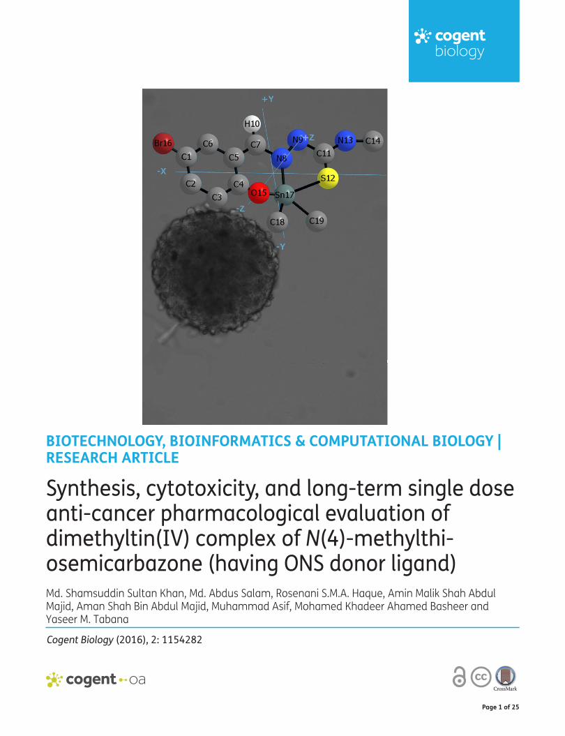
Scheme 1. Synthesis of dimethyltin(IV)-5-bromo-2-hydroxybenzaldehyde-N(4)-methylthiosemicarbazide complex.

Page 5 of 25
Khan et al., Cogent Biology (2016), 2: 1154282http://dx.doi.org/10.1080/23312025.2016.1154282
2. Experimental
2.1. Materials and methodsAll reagents were purchased from Fluka, Aldrich and JT Baker. All solvent were used without further purification. Melting point was measured by the Stuart Scientific SMP1 melting point apparatus. UV–Vis spectra were recorded in DMSO solution with a Perkin Elmer Lambda 25 UV-vis spectrophotom-eter. Infrared (IR) spectra were recorded by the Perkin Elmer System 2000 spectrophotometer using the KBr disk method in the range of 4,000–400 at room temperature. 1H, 13C, and 119Sn NMR spectra were recorded on a Bruker 500 MHz-NMR spectrophotometer relative to SiMe4 and Me4Sn in DMSO solvent. Elemental analysis was conducted by the Perkin Elmer 2400 Series-11 CHN analyzer. Molar conductivity measurements were carried out with a Jenway 4510 conductivity meter using a DMSO solvent.
2.2. Synthesis of 5-bromo-2-hydroxybenzaldehyde-N(4)-methylthiosemicarbazone (H2BHBM)A solution of 5-bromo-2-hydroxybenzaldehyde (0.95 g, 4.75 mmol) in ethanol (20 ml) was added to a solution of 4-methyl-3-thiosemicarbazide (0.5 g, 4.75 mmol) in ethanol (20 ml). The resulting yel-low solution was refluxed with stirring for 2 h and then filtered. A yellow product (formed at room temperature) was then filtered, washed with ethanol, and air dried. M.p: 185–187°C, (1.06 g, 78%). UV-vis (DMSO) λmax/nm: 260, 322, 361. FT-IR (KBr, cm−1)νmax: 3332 (s, OH), 3195 (s, NH), 1620 (m, C=N), 1556 (s, Caro-O), 1368, 856 (w, C–S), 988 (m, N–N). 1H NMR (DMSO-d6, ppm): 11.42 (s, 1H, OH), 10.35 (s, 1H, N-NH), 8.57 (s, 1H, CS-NH), 8.57 (s, 1H, CH = N), 8.15, 7.83, 6.83, (s, d, d, aromatic- H), 3.01 (s, CH3). 13C NMR (DMSO-d6,ppm): 187.48 (C=S), 155.44 (C=N), 137.01–111.04 (aromatic-C), 30.80 (CH3). Anal. Calc. for C9H10BrN3OS: C, 37.47; H, 3.47; N, 14.57. Found: C, 37.60; H, 3.82; N, 14.45%.
2.3. Synthesis of [Me2Sn(BHBM)] complex5-bromo-2-hydroxybenzaldehyde-N(4)-methylthiosemicarbazide (0.228 g, 1.0 mmol) was dissolved in absolute methanol (10 mL) under nitrogen atmosphere in a round bottom reaction flask. Methanol solution of dimethyltin(IV) dichloride (0.219 g, 1.0 mmol) was added dropwise and resulted into a yellow solution. The resulting reaction mixture was refluxed for 4 h (Figure 1(A)) and cooled to room temperature. The yellow microcrystals were obtained from slow evaporation of the resulting solu-tion at room temperature. The microcrystals were filtered, and washed with a small amount of cold methanol, and dried in vacuum over silica gel. Yield: 0.35 g, 78%: Mp.: 240–242°C: Molar conductance (DMSO) Ω−1 cm2 mol−1: 10.36: UV-Visible (DMSO) λmax (nm): 274, 338, 384, 417: FT-IR (KBr, cm−1) νmax: 3,252 (s, NH), 1,597 (m, C=N), 1,537 (s, Caro–O), 1,022 (w, N–N), 1,322, 828 (m, C–S), 652 (w, Sn–C), 565 (w, Sn–O), 455 (w, Sn–N). 1H NMR (DMSO-d6, ppm): 9.81 (s, 1H, CS-NH), 8.43 (s, 1H, s, CH=N), 7.25–7.42 (m, 3H, aromatic-H), 3.05 (m, 3H, CH3), 1.07 (s, 6H, Sn–CH3). 13C NMR (DMSO-d6, ppm): 178.11 (C=S), 147.32 (C=N), 110.54–145.43 (aromatic-C), 31.45 (CH3), 18.46 (Sn-CH3). Anal. Calc. for C11H14N3BrSOSn: C, 30.38; H, 3.24; N, 9.66%. Found: C, 30.34; H, 3.20; N, 9.61%.
2.4. Thermal and chemical stability studiesBHBM powder (50 mg) was transferred into the glass vials and applied 60°C in the oven for 48 h. After that, some portion of the compound was incubated with LiOH and the amount of BHBM was meas-ured. The degraded quantity of the compound was calculated using mass balance. The effect of pH on the compound and its chemical stability was determined by incubation of the compound in an aqueous HCl (pH 2) and Krebs-Heneseleit bicarbonate buffer (pH 7.4) at 37°C for 2 h followed by the analytical HPLC assay for BHBM.
2.5. Stability in human serumBHBM solution (10 mM) with water was added to preheated (37°C) human serum (Sigma) with the resulting concentration 0.5 mM. The 500 μL solution then withdrawn at appropriate intervals and added to 500 μL acetonitrile containing 0.1% trifluoroacetic acid to deproteinize the serum. The so-lution was vortexed and centrifuged for 10 min at 2,000 rpm and filtered by 0.45 μm PTFE filters (Biofil). The filtrate was analyzed by RP-HPLC method.

Page 6 of 25
Khan et al., Cogent Biology (2016), 2: 1154282http://dx.doi.org/10.1080/23312025.2016.1154282
The HPLC analytical method was developed on Agilent 1200 series coupled to a photodiode array detector (Agilent, CA, USA). Chromatographic separation was achieved at room temperature (25οC) with a C4 reversed-phase column (Thermo Scientific™, USA 2504.6 mm, 5-μM particle size) using a gradient solvent system that consisted of mobile phase acetonitrile and 0.05% formic acid, 50:50 (v/v), pH 5, running at an isocratic mode at flow rate 1.0 ml/min. The injected volume of the sample was 20 μl and the total run time was 15 min with BHBM eluting at 3.2 min. The absorbance was set at 200–450 nm and the UV was achieved at 220 nm which was used for quantification. Quantification was carried out using HPLC retention time and UV spectrum of the analyte. The method was specific and sensitive with a lower limit of quantification of 1 ng/mL and ChemStation LC3D software was used to ensure quantification of BHBM. The variation of intraday and interday was minimum < 10%.
2.6. Antioxidant activity
2.6.1. DPPH radical scavenging assayDPPH radical scavenging assay was conducted by method of Khan et al. (2013). In brief, 100 μL of molecule 26 was dissolved in MeOH:H2O (1:1) and 100 μL DPPH (200 μmol L−1) prepared in metha-nol and incubated at room temperature for 30 min. Ascorbic acid was used as reference standards. The amount of remaining DPPH was evaluated at 517 nm. The results were expressed as EC50 and the results (I%) were calculated by following equation:
I% = 1 −Asample
Ablank× 100
Figure 1. (A) Synthesis of dimethyl tin(IV): 5-bromo-2-hydroxy benzaldehyde-N(4)-methyl thiosemicarbazone; (B) IR spectrum; (C) UV-visible spectrum of dimethyltin(IV)-N(4)-methylthiosemicarbazone complex; (D) 1H NMR spectrum; (E) 13C NMR spectrum.

Page 7 of 25
Khan et al., Cogent Biology (2016), 2: 1154282http://dx.doi.org/10.1080/23312025.2016.1154282
where, Ablank is the absorbance of the control reaction (containing all reagents except the test mate-rial). EC50 was calculated from the graph plotted by percentage inhibition of BHBM.
2.6.2. Ferric-reducing antioxidant power (FRAP) assayFRAP was used to determine the antioxidant capacity of BHBM using the method of Benzie and Strain (1996). Single concentration of BHBM was mixed with the 150 μL FRAP working solution (300 mmol L−1 acetate buffer, pH 3.6, 10 mmol L−1 TPTZ in 40 mmol L−1 HCl and 20 mmol L−1 FeCl3 in a ratio of 10:1:1). The mixed solution was incubated for 8 min and read at 600 nm by Tecan microplate reader. The results were calculated from the standard curve of Ferrous sulfate (FeSO4.7H2O) as reference standard and expressed as nmol Fe2+ equivalent μg−1 compound.
2.6.3. Intracellular ROS activityHCT116 cells were seeded in a 48-well plate for 2 days. After 80% confluent of cells, treated the cells by BHBM for 24 h. Cells were washed by PBS and 100 μl ROS reagents were added in each well. Cells were incubate at 37°C for 30 min. Cell lysis buffer was added to the cells after the incubation period and centrifuged at 10,000 rpm for 10 min. Supernatant was collected and diluted with PBS and read in Tecan fluorescence microplate reader at 485–520 nm. Standard curve of the ROS standard was prepared and concentration was determined using the standard curve.
2.7. Ionization constants (pKa) and lipophilicity (logD 7.4) descriptorsThe ionization constants of compounds were determined using UV-metric methods. pKa is the 50% protonated state of the molecule that is measured by analyzing changes in multi-wavelength UV spectra during acid-base titration of the sample. This method is more suitable for colored compound due to pH-sensitive chromophores. Potentiometric titration was conducted by at least four separate titrations for BHBM compound. BHBM solutions (20 ml with 1 mM) were prepared by adjusting the ionic strength to 0.15 M with KCl. The solutions’ pH was stated with 1.8 using 0.5 N HCl. The solutions were titrated with 0.5 N KOH to the pH of 12.2. Titrations were performed in inert gas system (N2). Ionization constants were determined by spectral analysis as described by Carlos, Martínez, and Dardonville (2013). Briefly, the raw OD values of UV-spectra recorded and processed as following using Microsoft Excel program. At first, OD values of the BHBM were subtracted from blank solutions. Second, data were normalized as Abs400 nm = 0. The spectral difference plot provides the wave-lengths of maximum positive and negative absorbance. Absorbance difference Vs pH was plotted from the acid spectra and the spectra at every other pH at the chosen wavelengths. Grahpad Prism program was used to determine the pKa of BHBM using these data worked out by nonlinear regres-sion using Equation (1):
�HA is the extinction coefficients of the acid forms of the compound and the minimum absorbance difference curve,
�A− is the extinction coefficients of the base forms of the compound and maximum absorbance difference curve,St is the total compound concentration
Lipophilicity (LogD7.4) of the BHBM was determined by shake-flask procedure at pH 7.4. Phosphate buffer solution was prepared with ionic strength adjusted to 0.15 M with KCl. N-octanol was added to the buffer and shaking for four hour to be saturated of the two phases. BHBM was added to the saturated solution and shaken for 20 min until the partitioning equilibrium of solutes was reached. The solution was centrifuged at 10,000 rpm, 10 min. After separation, BHBM was quantified in the aqueous phase and octanol phase by UV spectrophotometer (Shimadzu) at 230 nm and the follow-ing equation was used to calculate the Log D7.4.
(1)Absorbance total =
(
�HA−�A−
)
× [10(pH−pKa)]
1 + 10(pH−pKa)× St

Page 8 of 25
Khan et al., Cogent Biology (2016), 2: 1154282http://dx.doi.org/10.1080/23312025.2016.1154282
2.8. Measurement of the generation of NO from BHBM and its apoptotic effect in HCT116The amount of NO generated from BHBM was determined in the cultured cancer cells with DMEM media and DMEM without cells. BHBM was added to the media for 5 or 60 min at 37°C. The genera-tion of NO from BHBM was measured by quantification of nitrite (NO2) using Griess reagent. HCT116 Cell culture supernatant was harvested and mixed with Griess reagent. The mixture was incubated for 10 min at room temperature. The absorbance was recorded at 550 nm using an ELISA reader. Standard curve was constructed from the serial dilutions of sodium nitrite (0.5–100 M) and was used to determine the nitrite concentrations.
Effect of NO donors on HCT116 cancer cell survival was determined by seeding the cells in 96-well plate. Cells were treated with or without BHBM as NO donors. The plate with the BHBM was incubated for 1 h at 37°C. MTT solution (5 mg/ml) was then added to the each well and incubated for another hour at 37°C. Live cells converted into the dark-blue formazan product from the yellow tetrazolium salt of MTT. Formazan was solubilized with DMSO (100 μl per well) and the absorbance at 570 nm was measured in an ELISA reader (Multiscan RC). The absorption of formazan is directly related to the number of viable cells.
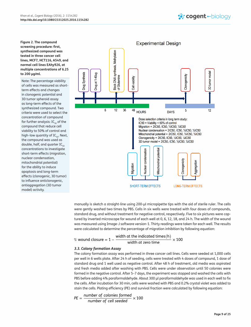
3. Anticancer study design After synthesizing the compound, anticancer activity was screened to determine the short-term and long-term efficacy of BHBM (Figure 2). This strategy determined the dose repetition necessity and effect of single dose for long-term activity.
3.1. Antiproliferative assayHuman cancer cell lines MCF-7 (breast), A549 (lung, non-small cells), HCT-116(colon), and EAhy926 (normal) endothelial cells were collected from ATCC USA. Normal epithelial MDCK cell line was a kind gift from Michael M. Gottesman, Laboratory of Cell Biology, National Cancer Institute, USA. We cul-tured the cells in RPMI 1640 (GIBCO BRL, Life Technologies) and DMEM supplemented with 5% of fetal bovine serum and penicillin (final concentration of 1 mg/mL) and streptomycin (final concen-tration of 200 μg/mL). A confluence of 70–80% cells were used for seeding in 96-well plates (100 μL cells/well) and incubated (0.25–250 μg/mL) for 48 h at 37°C and 5% of CO2.
Before seeding, old medium was aspirated from the cell culture flask and added freshly prepared media after washing by sterile phosphate buffered saline (PBS) (pH 7.4), 2–3 times. Trypsin was added followed by discarding PBS and distributed evenly onto cell surfaces. Cells were incubated at 37°C in 5% CO2 for 2–5 min. To detach the cells properly, the flasks were gently tapped and observed under inverted microscope. About 5 mL of fresh media (10% FBS) was added to neutralize trypsin activity. Cells were counted and seeded 10,000 cells per well. The plate was incubated for 24 h to be ready for the treatment of cells. Six low to high doses were used in the treatment. After 48 h of treat-ment, MTT reagents were added and incubated for 4 h. 20 μl MTT lysis solution was added to each well and read the plate at 570 and 620 nm wavelengths using a high-end TecanM200 Pro multimode microplate reader. Obtained data were analyzed to determine the concentration of compound which inhibited the cell growth by 50% (IC50) from the optical density. Tamoxifen and 5-FU were used as the standard drug. The results are calculated from two independent experiments, each done in triplicate.
3.2. Migration assayWe performed migration assay using three cancer cell lines. Cells were cultured and 20,000 cells were seeded per well in six-well plate. We kept cells to become 95% confluent monolayer to create wound at the middle of the well from corner to corner. The easiest and cheapest way to create wound
LogD = Log
(
CoctCaq
)
Page 9 of 25
Khan et al., Cogent Biology (2016), 2: 1154282http://dx.doi.org/10.1080/23312025.2016.1154282
manually is sketch a straight-line using 200-μl micropipette tips with the aid of sterile ruler. The cells were gently washed two times by PBS. Cells in six wells were treated with four doses of compounds, standard drug, and without treatment for negative control, respectively. Five to six pictures were cap-tured by inverted microscope for wound of each well at 0, 6, 12, 18, and 24 h. The width of the wound was measured using Image-J software version-1. Thirty readings were taken for each well. The results were calculated to determine the percentage of migration inhibition by following equation:
3.3. Colony formation AssayThe colony formation assay was performed in three cancer cell lines. Cells were seeded at 1,000 cells per well in 6 wells plate. After 24 h of seeding, cells were treated with 4 doses of compound, 1 dose of standard drug and 1 well used as negative control. After 48 h of treatment, old media was aspirated and fresh media added after washing with PBS. Cells were under observation until 50 colonies were formed in the negative control. After 5–7 days, the experiment was stopped and washed the cells with PBS before adding 4% paraformaldehyde. About 300 μl paraformaldehyde was used in each well to fix the cells. After incubation for 30 min, cells were washed with PBS and 0.2% crystal violet was added to stain the cells. Plating efficiency (PE) and survival fraction were calculated by following equation:
% wound closure = 1 −width at the indicated times
(
h)
width at zero time× 100
PE =number of colonies formed
number of cell seeded× 100
Figure 2. The compound screening procedure: first, synthesized compound was tested in three cancer cell lines, MCF7, HCT116, A549, and normal cell lines EAhy926, at multiple concentrations of 6.25 to 200 μg/ml.
Note: The percentage viability of cells was measured as short-term effects and changes in clonogenic potential and 3D tumor spheroid assay as long-term effects of the synthesized compound. Two criteria were used to select the concentration of compound for further analysis: IC50 of the compound that reduce cell viability to 50% of control and high–low quantity of IC50. Next, the compound was used as double, half, and quarter IC50 concentrations to investigate short-term effects (migration, nuclear condensation, mitochondrial potential) for the ability to induce apoptosis and long-term effects (clonogenic, 3D tumor) to influence anticlonogenic, antiaggregation (3D tumor model) activity.
Page 10 of 25
Khan et al., Cogent Biology (2016), 2: 1154282http://dx.doi.org/10.1080/23312025.2016.1154282
3.4. Apoptotic assay
3.4.1. Determination of nuclear condensation by Hoechst 33258 stain20,000 cells were seeded per well in 48-well plate. Cells were treated after 48 h of incubation with 70–80% confluent monolayer. Lung cancer cell (A549) was treated with four concentrations of BHBM time dependently at 6 h, 12 h, and 24 h. Cells were fixed by adding 4%, 300 μl paraformaldehyde in each well and incubate for 20 min. After incubation, the cells were washed with PBS and added 100-μl hoechst 33258 stain (1 μg/ml in PBS). After 30 min of incubation, cells were observed under an inverted microscope to capture the pictures to analyze irregular-shaped cells and regular-shaped cells. Cells with bright and shiny presents the condition of condensed nucleus and shrinked cyto-plasm denoted to apoptotic morphology. These apoptotic cells were counted for four fields per well. The percentage of apoptotic cells were determined by following equation:
3.4.2. Determination of mitochondrial potentialLung cancer cell line A549 was used to study the mitochondrial inhibitory potential of BHBM. Cells were treated as described in a hoecht staining assay. During the addition of hoecht stain, rhodamine 123 (5 μg/ml PBS) was added, followed by hoecht at the same quantity. Definition and calculation of mitochondrial potential condition are similar to nuclear condensation as it is bright and shiny with irregular shape.
3.5. Spheroid assayWe optimize the spheroid assay through hanging drop aggregation method. Hanging drops were prepared using lung cancer cell (A549) suspension containing glucose. Per hanging drops containing 20,000 cells were placed on the petridish lid (100 mm) and incubated it as inverted position over the dishes for 3 days. Spheroid was formed inside the hanging drops and transferred them from lid to 48-well plate after coated with 1% agarose. Diameter of spheroid was calculated to determine the inhibitory potential of BHBM by Image-J soft.
3.6. Effect of BHBM in apoptotic targets of Caspase 3/7, 8, 9 The assay was carried out according to manufacturer’s instructions (Promega, USA). The cells were seeded in 96-well plates at 20 x 104 cells/well in 200-μl medium and incubated for 24 h to allow the attachment of cells. The treated cells were incubated for approximately 18 h. Then, 100 μl of the caspase reagent containing cell lysis buffer and specific luminogenic substrate for caspase 3/7, 8, and 9. After incubating the plates for 30 min, the luminescence intensity was measured using micro-plate reader (Tecan, Switzerland). The activity of the BHBM in caspase was represented by fold change of the caspase activity upon treatment with BHBM. The fold changes in the caspase activity were measured by the following formula:
where: Ls = Luminescence reading of wells treated with tested extract
Lb = Luminescence reading of blank
Lc = Luminescence reading of wells treated with vehicle
The results were stated as mean of fold changes in caspase activity in comparison to control ± SD (n = 3).
% Apoptotic index =number of apoptotic cell
total number of live cell× 100
Fold change =Ls − LbLc − Lb
Page 11 of 25
Khan et al., Cogent Biology (2016), 2: 1154282http://dx.doi.org/10.1080/23312025.2016.1154282
3.7. Computational QSARCODESSA was used to generate a set of molecular descriptors used in the QSAR analysis (da Silva et al., 2012; Katritsky, Lobanov, & Karelson, 1996; Pacheco et al., 2013). In the statistical analysis for the QSAR study, the following parameters were employed: maximum number of descriptors per model, 1; the criterion of significance of 15 parameters, 0.01; level of high correlation, 0.99; and level of intercorrelation significance, 0.8.
3.8. Computational molecular dockingThe synthesized compound was designed and the structure was analyzed using Chembiodraw Ultra v.13.0 and CODESSA. The protein structure file is taken from PDB (www.rcsb.org/pdb) was cleaned from polymers, water molecules, and heteroatoms. Automated docking is used to determine the orientation of BHBM binding with NFκB. A genetic algorithm method implemented in the program Auto Dock Vina (Trott & Olson, 2010) was employed. Gasteiger charges were added, merged non-polar hydrogens, and set TORSDOF to six rotatable bonds to BHBM. For docking calculations, Gasteiger Marsili partial charges are assigned to the BHBM and nonpolar hydrogen atoms were merged (Gasteiger & Marsili, 1980). All torsions are allowed to rotate during docking. The grid map is cen-tered at the protein active site and is adjusted such that it can accommodate the binding site of the protein at the torsional degree of freedom with 0.5 units. The genetic algorithm is applied for mini-mization, using default parameters. The number of docking runs, the population in the genetic algo-rithm, the number of energy evaluations, and the maximum number of interactions are 50, 250, 100,000, and 10,000, respectively.
4. Results and discussion
4.1. Chemistry and characterization The reaction of Me2SnCl2 with 5-bromo-2-hydroxybenzaldehyde-N(4)-methylthiosemicarbazone in 1:1 mol ratio takes place according to Figure 1. This newly synthesized complex is yellow solids, air stable and soluble in DMSO, DMF, MeOH, CHCl3, and CH2Cl2 solvents. The composition of this complex was confirmed by their analytical data and structures were suggested by spectroscopic investiga-tions. The electronic spectrum of complex showed bands at 274, 338, and 384, nm corresponding to n-π* transition of the azomethine portion and π-π* transitions of the aromatic ring and thiolate function. The band at 417 nm was suggested ligand → metal charge transfer (LMCT) (Figure 1(C)). In IR spectrum, the sharp, strong, single peak at 3,252, 1,597, 1,537, 1,022, and 1,322, 828 cm−1 for complex gives evidence for the presence of ν(NH), ν(C=N), ν(Caro-O), ν(N-N), and ν(C-S) bands, respec-tively (Figure 1(B)). The appearance of new bands of the complexes at 565 and 455 cm−1 assigned to ν(Sn–O) and ν(Sn–N) supports the bonding of nitrogen and oxygen to tin(IV) (Haque & Salam, 2015), which are not observed in the spectra of the ligand (H2BHBM). The peak at 652 cm−1 in the IR spec-trum of complex supports the presence of ν(Sn–C) band in the structure. The 1H NMR spectra of the ligand also exhibit OH and N-NH proton signals which disappear in the complex, showing the involve-ment of phenolic oxygen and azomethine nitrogen in bonding with the tin(IV) atom. However, in the 1H NMR spectrum of complex, the signals of protons of the ligand from protons of the organic groups in the tin(IV) confirms metal to ligand in the complex (Figure 1(C)). Additionally, 13C NMR spectral data support the proposed structures. Shifts in the positions of carbons adjacent to azomethine ni-trogen (C=N) and phenolic oxygen atom clearly indicate the bonding of the azomethine nitrogen and phenolic oxygen atoms to tin(IV) atom (Figure 1(D)). In 119Sn NMR spectra of complex, δ(119Sn) value is found at −171.23 ppm, indicating that the tin(IV) atom is five-coordinate in this complex (Holeček, Nádvorník, Handlíř, & Lyčka, 1986).
4.2. ADME physicochemical properties of BHBM In this study, we determined the dissociation constants (pKa) lipophilicity of the BHBM. Dissociation constants (pKa) of the BHBM were determined by the potentiometric titration in aqueous solution using a 96-well plate in multiscan microplate reader. The value of the absorbance was collected and measured the pKa. BHBM is a soluble compound and pKa values are high. Shake-flask technique was used to determine the lipophilicity of BHBM using partition pair buffered water (pH 7.4)/noctanol. The

Page 12 of 25
Khan et al., Cogent Biology (2016), 2: 1154282http://dx.doi.org/10.1080/23312025.2016.1154282
distribution coefficients (log D7.4) are shown in Table 2. BHBM showed the lower value for lipophilic-ity. Stability of BHBM in serum and in high temperature also was defined, but no unstable effect was found from HPLC data.
4.3. Antiproliferative mechanism of BHBMAntiproliferative effect of the 5-bromo-2-hydroxy benzaldehyde-N(4)-methylthiosemicarbazide (BHBM) was investigated on breast (MCF7), colon (HCT 116), lung (A549), and endothelial (EAhy926) cell lines. Antiproliferative activity was determined by the MTT method and the activity of BHBM was compared with positive control of Tamoxifen and 5-fluorouracil. The concentration of BHBM that elicited 50% inhibition of proliferation of cancer cells (IC50) (Figure 3) was varied in different cell lines. The analyzed data obtained from BHBM noted that MCF7 and HCT 116 Cell lines were most sensitive to cancer cells as attested by BHBM that positively inhibited the cells growth at IC50 of 6 and 4 μg/ml, respectively (Figure 3). A549 cells were the least sensitive to BHBM. BHBM showed IC50 values in the range of 20–18 μg/mL. Indeed, it also showed low selectivity index (SI) for A549 cancer cells, pre-senting weak antiproliferative activity against nontumorogenic endothelial EAhy926 cells and epi-thelial cells MDCK (Figure 3, Table 1). BHBM was also promising against HCT 116 cells by exhibiting IC50 around 4 μg/mL (Table 1). HCT 116 cells were highly sensitive to BHBM (Table 1) may be due to high affinity of compounds to DNA (Han & Yang, 2002), the structure of the molecule and the coor-dination number of the tin atoms (Collinson & Fenton, 1996; Koch, Baul, & Chatterjee, 2008; Xanthopoulou et al., 2008). BHBM compromised with the MCF7, HCT 116, and A549 cells growth by 50% when used at higher concentrations 12, 8, and 40 μg/mL, respectively, while HCT16 cells were most sensitive to BHBM. Cytostatic activity was observed at the higher concentration of BHBM may be due to stopping the cell growth at synthetic (S) phase, during which time the doubling of DNA occurs. For HCT 116 cells BHBM was the most potent with IC50 values of lower than 8 μg/mL, which may be due to the type of alkyl groups (Br) attached to the Sn moiety. Also, BHBM exhibited the larg-est spectrum of action. BHBM showed considerable antiproliferative activity (IC50 between 5 and
Figure 3. Photomicrographic images of different cancer cell lines and normal endothelial cells treated with BHBM tamoxifen, and 5-fluoruracil (5FU).
Note: Images were obtained from EVOS inverted microscope after 48 h of treatment. MCF7 (A: untreated, B: treated), HCT116 (C: untreated, D: treated), A549 (E: untreated, F: treated) cells were incubated with BHBM for 48 h at different doses. Percent inhibition of cell proliferation (G) was obtained and values were expressed as μg/ml. MCF7 (X), HCT116 (Y), A549 (Z) cells were incubated with tamoxifen and 5-FU for 48 h at different doses. Percent inhibition of cell proliferation (J) was obtained and values were expressed as μg/ml. Results are presented as mean ± SD of three separate experiments (n = 6).

Page 13 of 25
Khan et al., Cogent Biology (2016), 2: 1154282http://dx.doi.org/10.1080/23312025.2016.1154282
20 μg/ml) against all the cancer cell lines (Table 1). These preliminary data may provide the cyto-static effects than cytotoxic effects of the BHBM to the specific candidate target other than healthy cells (IC50, 40 μg/ml in EAhy926 and IC50, 55 μg/ml in MDCK) through the additive or synergistic ef-fects of the substituents of BHBM.
Moreover, BHBM might form a stable form of the chelates with the transition metals found in the cells. This formation inhibits enzymatic reaction in the cells for glucose uptake, DNA and protein synthesis. The coordination further increases the activity of hydrazones by the additional C=N bonds. Metal ion losses the polarity due to sharing of its positive charge within chelating ring with donor atom during coordination. It results the lipophilicity of the central metal atom which provides more efficient permeation through the lipid layers of the cancer cells. The chelation form might block the metabolic pathways of the DNA synthesis of neoplastic cells and inhibit the proliferation.
Antiproliferative activity of BHBM may be achieved by two mechanisms. Chelation of hydrazone to Sn(IV) from the cells may have higher efficiency to inhibit the DNA synthesis of the cancer cell lines (Richardson & Bernhardt, 1999). The chelation may produce cytotoxic oxygen radicals that prevent the uptake of essential nutrients for cancer cells (Richardson & Bernhardt, 1999). BHBM complex is a kind of monomeric structure in which the Sn(IV) center coordinated with the enolic tridentate ligand (L) in the ONMe chelate mode and exhibits five-coordinated trigonal bipyramidal geometry. The synthesized compound revealed the anticancer activity to the human cancer cell line of HCT 116 (colon), A549 (lung), and MCF7 (breast). This result demonstrated that the alkyl group bound with Sn (IV) center and sulfur (S) present in the complex have strong activity on their in vitro anticancer property. Min Hong et al. have studied previously the organotin(IV) complexes with 2-hydroxy-1-naphthaldehyde 5-chloro-2-hydroxybenzoylhydrazone with IC50 values of 0.09 μg/ml to <10 μg/ml in A549 cells and 0.5–4.1 μg/ml in HCT-8 cells (Min et al., 2013). In this study, the authors described the anticancer activity as dose dependent which was increased by high concentration because of long carbon chain butyl ligand and organic ligand R. Also, coordination geometry to tin atom may have possible role to provide the anticancer effect (Min et al., 2013). The present study of the BHBM showed long-term anticancer effect with the single dose without any repetitive dose. While the other organotin complexes with O-Sn-S coordination showed very toxic effects from low to high doses, BHBM with the methyl complex has a significant effect at lower doses only.
The cytotoxic effects of the similar group of compound named 5-bromo-2-hydroxybenzalde-hyde-4, 4-dimethylthiosemicarbazone was observed with the IC50 of 3.23–5.46 μM from the study of Haque and Salam (2015). The detailed long-term cytotoxic/cytostatic effect and long-term dose-dependent and mechanistic activity were not studied yet for this group of compound. In this study, we performed the single-dose long-term effect of the BHBM in in vitro 3D tumor model and other in vitro studies. The novel nonenolic bidentate O-Sn-S-coordination of Schiff base ligand in synthesized methyl complex was documented for the first time.
Table 1. IC50 and selectivity index (SI) of the various melanoma (cancer) cells
Note: SI is the ration of IC50 for normal cell line and cancerous cell line after 48 h of treatment. IC50 was measured after 48 h treatment with compounds using MTT staining and it was expressed as % of vehicle (0.05% DMSO)-treated control. For comparison normal cells endothelial cell line EAhy926 and normal epithelial cell line MDCK and effects of standard chemo drugs Tamoxifen and 5-fluorouracil were used. Results are presented as mean ± SD of three separate experiments (n = 6).
Samples IC50 (μg/ml) on MCF7
IC50 (μg/ml) on
HCT 116
IC50 (μg/ml) on A549
IC50 (μg/ml) on
EAhy926
IC50 (μg/ml) on MDCK
Selectivity Index (SI) NO generation
effect in HCT116
(IC50 μg/ml)
MCF7 HCT 116 A549
BHBM 6.0±0.57 4.0 ± 0.65 20 ± 0.54 40 ± 1.2 55 ± 1.7 7 ± 2.5 10 ± 2.8 2 ± 0.8 4.5 ± 1.7
Tamoxifen 7.0±0.98 – – 20 ± 2.5 15 ± 2.87 3 ± 1.5 – – ND
5 fluoro-uracil
– 2.0 ± 0.75 3.0 ± 0.78 20±2.9 10±2.5 – 10 ± 1.7 6 ± 0.1.9 ND
Page 14 of 25
Khan et al., Cogent Biology (2016), 2: 1154282http://dx.doi.org/10.1080/23312025.2016.1154282
NO generation effect of BHBM in the HCT 116 cell viability was determined and found 25 μM nitrite after 5 min and 188 μM nitrite after 1 h from NO donors of BHBM in cultured RPMI, respectively. At higher concentration of nitrite BHBM showed IC50 of 4.5 μg/ml. This result reveals the extracellular apoptotic activity of BHBM and suggests the interaction of NO and oxygen metabolites to kill the cells.
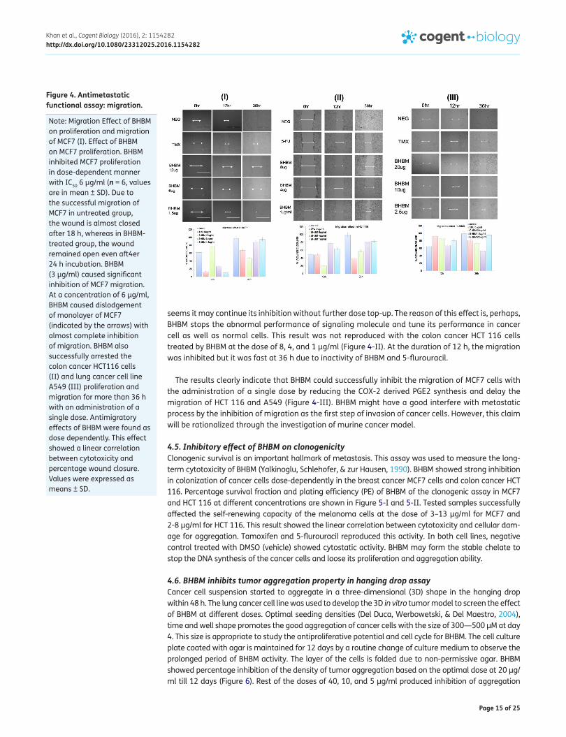
4.4. BHBM showed antimigratory properties on different cancer cells at optimum dosesBHBM were further investigated for antimigratory potential in comparison with Tamoxifen and 5-fluorouracil, using wound-healing assay that is important hallmarks of metastasis. In a previous report, Tamoxifen and 5-fluorouracil showed inhibition of migration of cancer cells (Borley et al., 2008; Hayot et al., 2002) and thus were used as a positive control in this study. BHBM was evaluated for antimigratory activity using the IC50 value of the respective cell lines. Human breast, colon, and lung cancer cells of MCF7, HCT 116, and A549, respectively, were allowed to migrate in the presence of BHBM with different concentrations for 0 to 36 h and antimigratory effects were calculated. The results shown in Figure 4 indicated that BHBM had significant antimigratory properties in compari-son with the negative control group and competed its activity with the inhibitory potential of Tamoxifen and 5-flurouracil. BHBM showed the percentage inhibition of migration of the cancer cells in a time-dependent manner. The lower doses of BHBM exhibited the percentage inhibition dose dependently, but higher doses unable to produce dose-dependent inhibition in lung cancer A549 cells (Figure 4). During the 12 h, BHBM showed its lower activity than at 36 h. BHBM started its inhibi-tory activity after 12 h and its activity was increased in course of cumulative time. In all three cancer cells, same mechanism of action was observed from BHBM that was substantial increase of its activ-ity during the period of 12–36 h. This result reveals that BHBM has less toxic effect that reproduces the safety of BHBM although IC50 between the range of 20–5 μg/ml obtained from MTT assay (Figure 3). Earlier published literature reported that dimethyltin was less toxic than trimethyltin and toxicity depended on the nature of the R group attached with Sn (Blunden, Cusak, & Hill, 1985). Also, If BHBM produces strong cytotoxicity by inhibition of mitochondrial oxidative phosphorylation, it could not reveal its antimigratory activity (Aldridge, 1976). The degree of toxic action may be con-trolled by the interaction of amino acid of cancer cells at certain active sites (Hall & Zuckerman, 1977) of imidazole N-H of histidine residue and an S-H group (Aldridge, 1976). Both of these groups present in the BHBM. Recently, published study synthesized the isonicotinoyl hydrazone derivatives as 2-hydroxy-1-naphthaldehyde isonicotinoyl hydrazone and its iron complexes (Lovejoy & Richardson, 2002; Yuan et al., 2004), and di-2-pyridyl ketone isonicotinoyl hydrazone (Bernhardt et al., 2003; Becker et al., 2003), Aroyl hydrazones of pyridoxal and salicylaldehyde hydrazone (Johnson et al., 1982; van Reyk et al., 2000), 2-Benzoxazolyl and 2-benzimidazolyl hydrazones (Easmon et al., 2001), Cyano acetic acid hydrazones of 3- and 4-acetylpyridine (EL-Hawash et al., 2006), 2,6-dimethylimidazo[2,1-b]-(Cima & Ballarin, 1999; Pellerito et al., 2005; Yin & Chen, 2006) thiazole-5-carbohydrazide and the derivatives of hydrazine pyrimidines (Cocco et al., 2006; Terzioglu & Gürsoy, 2003), Organotin(IV) complexes of 2-hydroxy-1-naphthaldehyde 5-chloro-2-hydroxy benzo-ylhydrazone, and isothiazolehydrazones for anticancer activity. These compounds have lack of S-H group, but this group is presented in the BHBM to produce an optimum effect to kill the cancer cell and its growth with slow and less toxic mechanism. In a comparison with past synthesized organo-tin (IV) complex, most of the organotin complex was highly toxic than BHBM (Table 1 and supple-mentary Table 1). Oppositely, BHBM could kill the cells at the optimum dose and the higher doses are not toxic. Thus, the results are suggesting the linear correlation between cytotoxicity and antimigra-tory activity. In case of lung cancer cells, the migration may be through caveolin-1 and protein ki-nase B (Akt) signaling pathway (Brazil & Hemmings, 2001). BHBM may block the activation of this signaling molecule and produce antimigratory activity. For breast cancer cells, PGE2 plays a vital role in the migration. It is assumed that BHBM may intervene with the migrational capacity of this signal-ing molecule to inhibit the migration at the dose of 12 μg/ml till 36 h. During the first 12 h, BHBM was unable to control the migration, but it stopped the migration after 12 h. The reason may be due to delay of weakening the signaling molecules specifically. The opposite effect was found from Tamoxifen and lower doses of BHBM at 6 μg/ml and 1.5 μg/ml which were highly active at first 12 h, but not active for a longer period of time. BHBM significantly showed its prolonged activity and it
Page 15 of 25
Khan et al., Cogent Biology (2016), 2: 1154282http://dx.doi.org/10.1080/23312025.2016.1154282
seems it may continue its inhibition without further dose top-up. The reason of this effect is, perhaps, BHBM stops the abnormal performance of signaling molecule and tune its performance in cancer cell as well as normal cells. This result was not reproduced with the colon cancer HCT 116 cells treated by BHBM at the dose of 8, 4, and 1 μg/ml (Figure 4-II). At the duration of 12 h, the migration was inhibited but it was fast at 36 h due to inactivity of BHBM and 5-flurouracil.
The results clearly indicate that BHBM could successfully inhibit the migration of MCF7 cells with the administration of a single dose by reducing the COX-2 derived PGE2 synthesis and delay the migration of HCT 116 and A549 (Figure 4-III). BHBM might have a good interfere with metastatic process by the inhibition of migration as the first step of invasion of cancer cells. However, this claim will be rationalized through the investigation of murine cancer model.
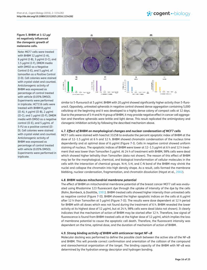
4.5. Inhibitory effect of BHBM on clonogenicityClonogenic survival is an important hallmark of metastasis. This assay was used to measure the long-term cytotoxicity of BHBM (Yalkinoglu, Schlehofer, & zur Hausen, 1990). BHBM showed strong inhibition in colonization of cancer cells dose-dependently in the breast cancer MCF7 cells and colon cancer HCT 116. Percentage survival fraction and plating efficiency (PE) of BHBM of the clonogenic assay in MCF7 and HCT 116 at different concentrations are shown in Figure 5-I and 5-II. Tested samples successfully affected the self-renewing capacity of the melanoma cells at the dose of 3–13 μg/ml for MCF7 and 2-8 μg/ml for HCT 116. This result showed the linear correlation between cytotoxicity and cellular dam-age for aggregation. Tamoxifen and 5-flurouracil reproduced this activity. In both cell lines, negative control treated with DMSO (vehicle) showed cytostatic activity. BHBM may form the stable chelate to stop the DNA synthesis of the cancer cells and loose its proliferation and aggregation ability.
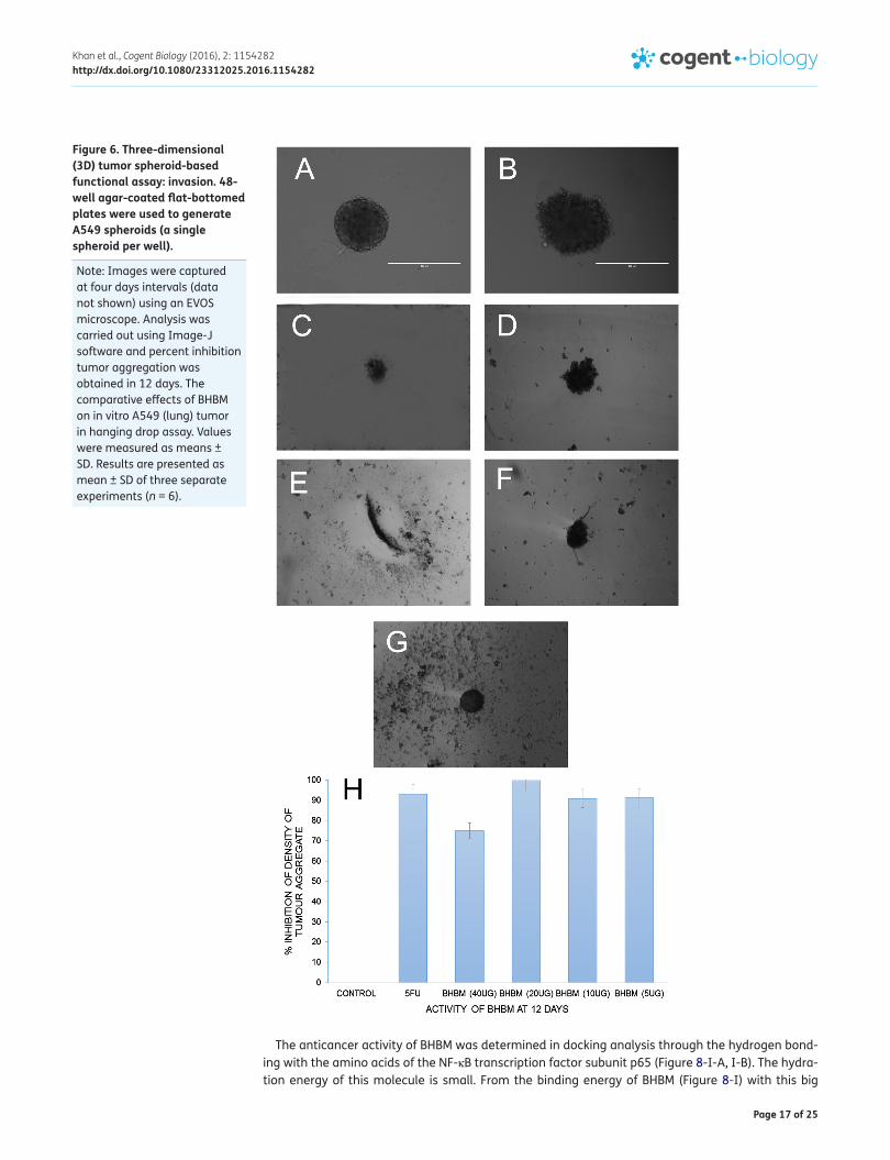
4.6. BHBM inhibits tumor aggregation property in hanging drop assayCancer cell suspension started to aggregate in a three-dimensional (3D) shape in the hanging drop within 48 h. The lung cancer cell line was used to develop the 3D in vitro tumor model to screen the effect of BHBM at different doses. Optimal seeding densities (Del Duca, Werbowetski, & Del Maestro, 2004), time and well shape promotes the good aggregation of cancer cells with the size of 300—500 μM at day 4. This size is appropriate to study the antiproliferative potential and cell cycle for BHBM. The cell culture plate coated with agar is maintained for 12 days by a routine change of culture medium to observe the prolonged period of BHBM activity. The layer of the cells is folded due to non-permissive agar. BHBM showed percentage inhibition of the density of tumor aggregation based on the optimal dose at 20 μg/ml till 12 days (Figure 6). Rest of the doses of 40, 10, and 5 μg/ml produced inhibition of aggregation
Figure 4. Antimetastatic functional assay: migration.
Note: Migration Effect of BHBM on proliferation and migration of MCF7 (I). Effect of BHBM on MCF7 proliferation. BHBM inhibited MCF7 proliferation in dose-dependent manner with IC50 6 μg/ml (n = 6, values are in mean ± SD). Due to the successful migration of MCF7 in untreated group, the wound is almost closed after 18 h, whereas in BHBM-treated group, the wound remained open even aft4er 24 h incubation. BHBM (3 μg/ml) caused significant inhibition of MCF7 migration. At a concentration of 6 μg/ml, BHBM caused dislodgement of monolayer of MCF7 (indicated by the arrows) with almost complete inhibition of migration. BHBM also successfully arrested the colon cancer HCT116 cells (II) and lung cancer cell line A549 (III) proliferation and migration for more than 36 h with an administration of a single dose. Antimigratory effects of BHBM were found as dose dependently. This effect showed a linear correlation between cytotoxicity and percentage wound closure. Values were expressed as means ± SD.
Page 16 of 25
Khan et al., Cogent Biology (2016), 2: 1154282http://dx.doi.org/10.1080/23312025.2016.1154282
similar to 5-flurouracil at 5 μg/ml. BHBM with 20 μg/ml showed significantly higher activity than 5-fluro-uracil. Oppositely, untreated spheroids in negative control showed dense aggregation containing 5,000 cells/drop at the beginning and it was developed to a highly dense colony of compact cells at 12 days. Due to the presence of S-H and N-H group of BHBM, it may provide negative effect in cancer cell aggrega-tion and therefore spheroids were brittle and light dense. This result replicated the antimigratory and clonogenic inhibition activity by following the described mechanism above.
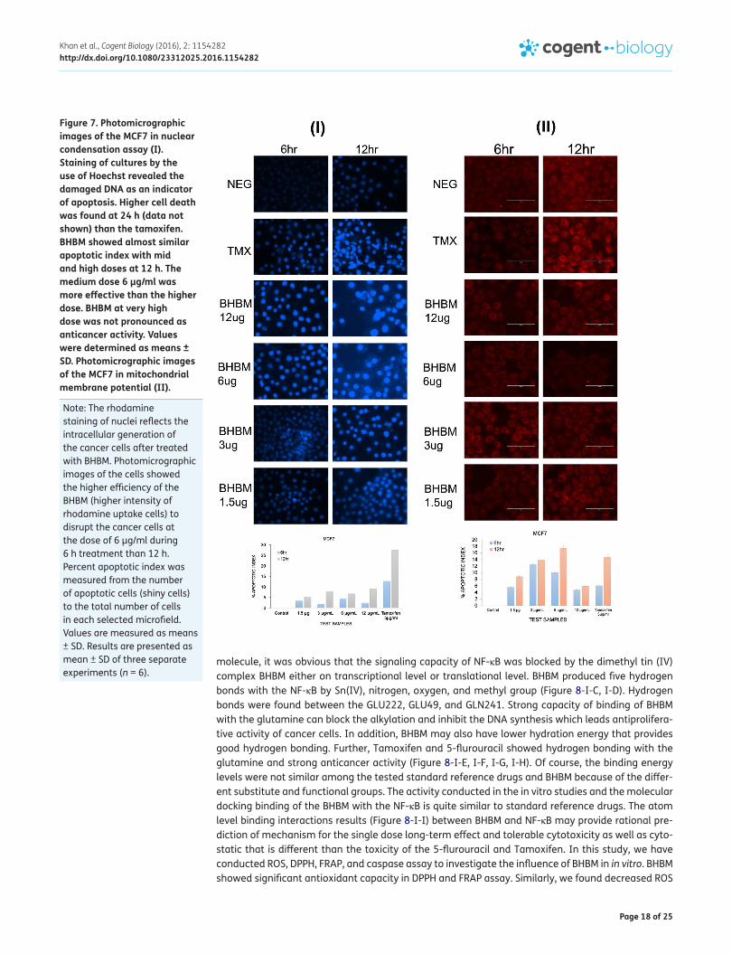
4.7. Effect of BHBM on morphological changes and nuclear condensation of MCF7 cellsMCF7 cells were stained with hoechst 33258 to evaluate the percent apoptotic index of BHBM at the dose of 12–1.5 μg/ml at 6 h and 12 h. BHBM showed chromatin condensation of the nucleus time dependently and at optimal dose of 6 μg/ml (Figure 7-I). Cells in negative control showed uniform staining of nucleus. The apoptotic indices of BHBM were lower at 12–1.5 μg/ml at 6 h and 12 h treat-ment that was lower than Tamoxifen 5 μg/ml. At 24 h of treatment with BHBM, 98% cells were dead which showed higher lethality than Tamoxifen (data not shown). The reason of this effect of BHBM may be for the morphological, chemical, and biological transformation of cellular molecules in the cells with the interaction of chemical groups. N-H, S-H, and C-N bond of the BHBM may shrink the nuclei and collapse the chromatin into high density shape. As a result, cells formed the membrane blebbing, nuclear condensation, fragmentation, and chromatin dissolution (Hayot et al., 2002).
4.8. BHBM reduces mitochondrial membrane potential The effect of BHBM on mitochondrial membrane potential of the breast cancer MCF7 cell was evalu-ated using Rhodamine 123 fluorescent dye through the uptake of intensity of the dye by the cells (Rahn, Bombeck, & Doolittle, 1991). BHBM-treated cells showed higher intensity than untreated cells as negative control (Figure 7-II). BHBM showed the higher apoptotic indices on the cells at 6 μg/ml after 12 h than Tamoxifen at 5 μg/ml (Figure 7-II). The results were dose dependent at 12 h period for BHBM with all doses which was not found during the treatment of 6 h. BHBM revealed the lower activity at its highest dose of 12 μg/ml, but at 24 h, 98% cells were dead (data not shown). It clearly indicates that the mechanism of action of BHBM may be started after 12 h. Therefore, low signal of fluorescence is found from BHBM-treated cells at the higher dose of 12 μg/ml, which implies the loss of membrane potential to cause the apoptotic cell death. Therefore, the fluorescent intensity was dependent on the time, optimal dose, and the duration of mechanism of action of BHBM.
4.9. Strong binding activity of BHBM with anticancer target NF-κBMolecular docking was performed to define the possible clash between the active site of the NF-κB and BHBM. This will provide correct confirmation and orientation of the collision of the compound and stereochemical organization of the target. The binding capacity of the BHBM with NF-κB was determined by the hydration energy descriptor and hydrogen bonding.
Figure 5. BHBM at 1-12 μg/ml negatively influenced the clonogenic growth of melanoma cells.
Note: MCF7 cells were treated with BHBM 12 μg/ml (I-A), 6 μg/ml (I-B), 3 μg/ml (I-C), and 1.5 μg/ml (I-F), DMEM media with DMSO as a Negative Control (I-E), and 5 μg/mL of tamoxifen as a Positive Control (I-D). Cell colonies were stained with crystal violet and counted. Anticlonogenic activity of BHBM was expressed as percentage of control treated with vehicle (0.05% DMSO). Experiments were performed in triplicate. HCT116 cells were treated with BHBM 8 μg/ml (II-A), 4 μg/ml (II-B), 2 μg/ml (II-C), and 1 μg/ml (II-F), DMEM media with DMSO as a negative control (II-E), and 5 μg/mL of 5-FU as a positive control (II-D). Cell colonies were stained with crystal violet and counted. Anticlonogenic activity of BHBM was expressed as percentage of control treated with vehicle (0.05% DMSO). Experiments were performed in triplicate.
Page 17 of 25
Khan et al., Cogent Biology (2016), 2: 1154282http://dx.doi.org/10.1080/23312025.2016.1154282
The anticancer activity of BHBM was determined in docking analysis through the hydrogen bond-ing with the amino acids of the NF-κB transcription factor subunit p65 (Figure 8-I-A, I-B). The hydra-tion energy of this molecule is small. From the binding energy of BHBM (Figure 8-I) with this big
Figure 6. Three-dimensional (3D) tumor spheroid-based functional assay: invasion. 48-well agar-coated flat-bottomed plates were used to generate A549 spheroids (a single spheroid per well).
Note: Images were captured at four days intervals (data not shown) using an EVOS microscope. Analysis was carried out using Image-J software and percent inhibition tumor aggregation was obtained in 12 days. The comparative effects of BHBM on in vitro A549 (lung) tumor in hanging drop assay. Values were measured as means ± SD. Results are presented as mean ± SD of three separate experiments (n = 6).
Page 18 of 25
Khan et al., Cogent Biology (2016), 2: 1154282http://dx.doi.org/10.1080/23312025.2016.1154282
molecule, it was obvious that the signaling capacity of NF-κB was blocked by the dimethyl tin (IV) complex BHBM either on transcriptional level or translational level. BHBM produced five hydrogen bonds with the NF-κB by Sn(IV), nitrogen, oxygen, and methyl group (Figure 8-I-C, I-D). Hydrogen bonds were found between the GLU222, GLU49, and GLN241. Strong capacity of binding of BHBM with the glutamine can block the alkylation and inhibit the DNA synthesis which leads antiprolifera-tive activity of cancer cells. In addition, BHBM may also have lower hydration energy that provides good hydrogen bonding. Further, Tamoxifen and 5-flurouracil showed hydrogen bonding with the glutamine and strong anticancer activity (Figure 8-I-E, I-F, I-G, I-H). Of course, the binding energy levels were not similar among the tested standard reference drugs and BHBM because of the differ-ent substitute and functional groups. The activity conducted in the in vitro studies and the molecular docking binding of the BHBM with the NF-κB is quite similar to standard reference drugs. The atom level binding interactions results (Figure 8-I-I) between BHBM and NF-κB may provide rational pre-diction of mechanism for the single dose long-term effect and tolerable cytotoxicity as well as cyto-static that is different than the toxicity of the 5-flurouracil and Tamoxifen. In this study, we have conducted ROS, DPPH, FRAP, and caspase assay to investigate the influence of BHBM in in vitro. BHBM showed significant antioxidant capacity in DPPH and FRAP assay. Similarly, we found decreased ROS
Figure 7. Photomicrographic images of the MCF7 in nuclear condensation assay (I). Staining of cultures by the use of Hoechst revealed the damaged DNA as an indicator of apoptosis. Higher cell death was found at 24 h (data not shown) than the tamoxifen. BHBM showed almost similar apoptotic index with mid and high doses at 12 h. The medium dose 6 μg/ml was more effective than the higher dose. BHBM at very high dose was not pronounced as anticancer activity. Values were determined as means ± SD. Photomicrographic images of the MCF7 in mitochondrial membrane potential (II).
Note: The rhodamine staining of nuclei reflects the intracellular generation of the cancer cells after treated with BHBM. Photomicrographic images of the cells showed the higher efficiency of the BHBM (higher intensity of rhodamine uptake cells) to disrupt the cancer cells at the dose of 6 μg/ml during 6 h treatment than 12 h. Percent apoptotic index was measured from the number of apoptotic cells (shiny cells) to the total number of cells in each selected microfield. Values are measured as means ± SD. Results are presented as mean ± SD of three separate experiments (n = 6).

Page 19 of 25
Khan et al., Cogent Biology (2016), 2: 1154282http://dx.doi.org/10.1080/23312025.2016.1154282
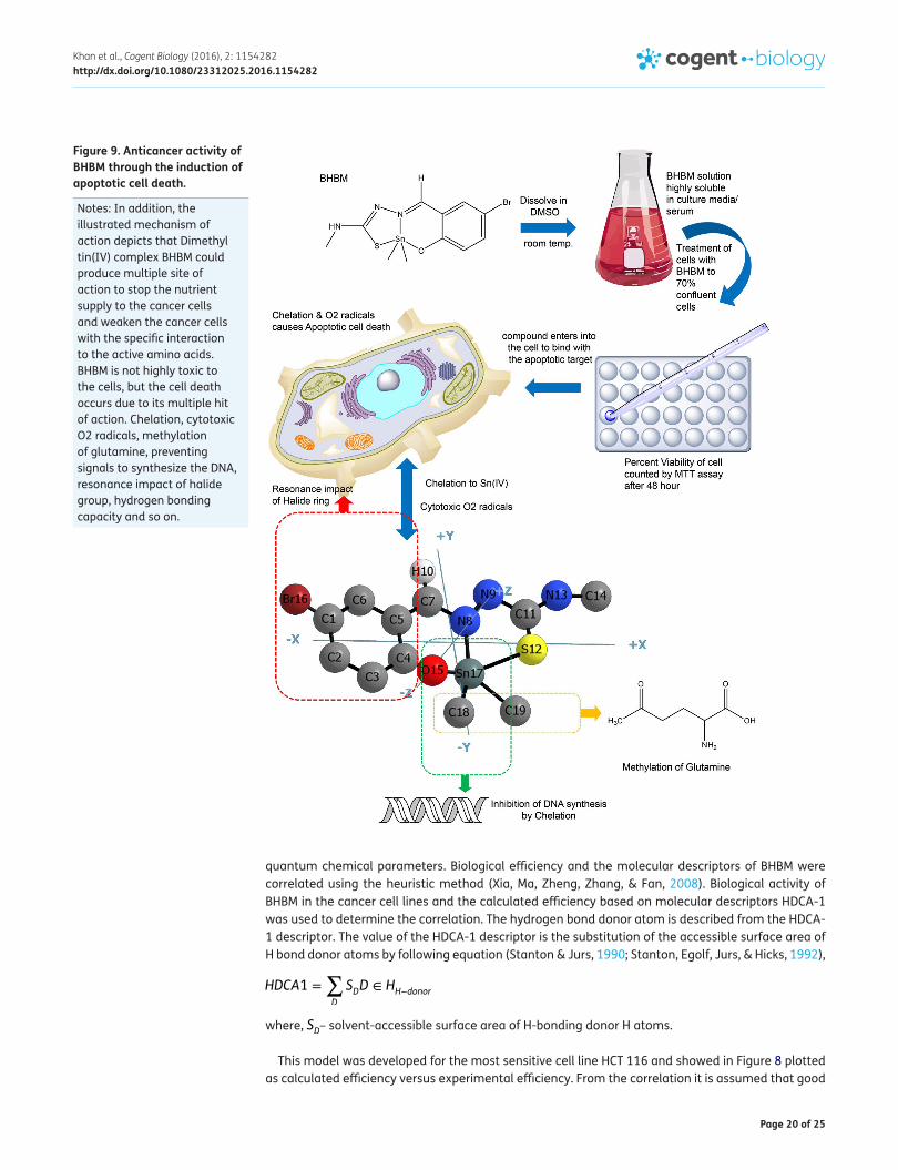
level in the BHBM-treated HCT 116 cells at the dose of 4 and 8 μg/ml. HCT 116 cells become under stress due to this reduced level of ROS (Table 2). The reason of the antioxidant activity of the BHBM may be due to form of chelation of Sn(IV), resonance impact of aromatic rings, hydrogen bonds, and the accumulation of Sn in the cancer cells than normal cells (Figure 9). Concurrently, due to the ROS stress in HCT 116 cells by BHBM, caspases and NF-κB have been modulated to kill the cells. The ef-fects of BHBM on caspases 3/7, 8, and 9 were significantly increased in the levels of activated cas-pases observed in the hoecht and rhodamin staining at the nucleus morphology by caspase 3/7, while initiation of cell death by caspase 8 and 9 through ligation of cell death receptors and disrup-tion of mitochondrial membrane integrity, respectively. The results showed that there was signifi-cant increase in caspase activities due to the influence of BHBM at 4 and 8 μg/ml in compared to control (Table 2).
4.10. QSAR of BHBM for anticancer activityQSAR studies were performed to find the relationship between anticancer activity and molecular structure. This correlation was determined by the multiple linear regression model between molecu-lar descriptors and the efficiency of the BHBM. Anticancer activity was dependent on the nature of the substitution of different parts of the structure. The essential chemical features of the test com-pound for anticancer activity were identified by the QSAR of Hansch approach. The structural analy-sis of BHBM was carried out in CODESSA software. This software was used to determine the set of molecular descriptors because of its ability to generate more than 400 molecular descriptors such as molecular constitution, geometry, and topology and with electrostatic, thermodynamic, and
Figure 8. Visualization of Ligand and protein interaction profile: surface visualization (I-A) and cartoon display (I-B) of NF-κB and active site residue interaction of protein NF-κB (I-D, I-F, I-H).
Note: Molecular docking was performed in Autodock Vina. The binding energy obtained from docking analysis reflected the anticancer activity that correlates QSAR model. Descriptor Solvent accessible surface area (SASA), surface area (SA) of BHBM was generated by CODESSA software (X, Y, Z). SASA is very important to correlate the molecule structure with the anticancer activity. QSAR model was developed in CODESSA using this SASA descriptor by the multiple linear regression (MLR) method. Calculated efficiency versus the experimental efficiency of the training set molecules were plotted by MLR method (H). Results are presented as mean ± SD of three separate docking experiments.
Page 20 of 25
Khan et al., Cogent Biology (2016), 2: 1154282http://dx.doi.org/10.1080/23312025.2016.1154282
quantum chemical parameters. Biological efficiency and the molecular descriptors of BHBM were correlated using the heuristic method (Xia, Ma, Zheng, Zhang, & Fan, 2008). Biological activity of BHBM in the cancer cell lines and the calculated efficiency based on molecular descriptors HDCA-1 was used to determine the correlation. The hydrogen bond donor atom is described from the HDCA-1 descriptor. The value of the HDCA-1 descriptor is the substitution of the accessible surface area of H bond donor atoms by following equation (Stanton & Jurs, 1990; Stanton, Egolf, Jurs, & Hicks, 1992),
where, SD– solvent-accessible surface area of H-bonding donor H atoms.
This model was developed for the most sensitive cell line HCT 116 and showed in Figure 8 plotted as calculated efficiency versus experimental efficiency. From the correlation it is assumed that good
HDCA1 =∑
D
SDD ∈ HH−donor
Figure 9. Anticancer activity of BHBM through the induction of apoptotic cell death.
Notes: In addition, the illustrated mechanism of action depicts that Dimethyl tin(IV) complex BHBM could produce multiple site of action to stop the nutrient supply to the cancer cells and weaken the cancer cells with the specific interaction to the active amino acids. BHBM is not highly toxic to the cells, but the cell death occurs due to its multiple hit of action. Chelation, cytotoxic O2 radicals, methylation of glutamine, preventing signals to synthesize the DNA, resonance impact of halide group, hydrogen bonding capacity and so on.

Page 21 of 25
Khan et al., Cogent Biology (2016), 2: 1154282http://dx.doi.org/10.1080/23312025.2016.1154282
solvent accessible surface area leads to the biological activity. The reason may be due to the pres-ence of hydroxyl groups in the BHBM’s substitute of the meta- and para-positions.
The used compounds in developing the QSAR model consisted with a series of test set molecules, training set molecules, and validation set molecules were labeled in CODESSA during multiple linear regression analysis. The quality of this analysis was justified by the parameters of correlation coef-ficient (R), standard error of the estimate (SEE), variance ratio (F) at specified degrees of freedom (df) (Table 3), (Figure 8-II).
Additionally, anticancer activity of the BHBM was led by the substitution of N-H, S-H, and aromatic ring with the Sn(IV) molecule. It was observed that BHBM inhibited the activity of cancer cells with 100% in comparing with the standard drugs of Tamoxifen and 5-flurouracil at the optimal doses for different cancer cell lines. In most of the anticancer experiments, it was found that BHBM showed its 100% capacity in killing the cancer cells after a certain period of time, i.e. after few hours. This may be due to the presence of electron donating groups in the Sn(IV) ring coordinated with the oxygen, nitrogen, and sulfur atom to form the stable chelate with the active amino acid of the receptor site which has electron deficiency. In this reaction, the resonance of the molecule is important too. It is clearly observed from the molecular docking analysis that positive steric parameter at O15, Sn17, C18, and C19 position explains why the small groups of the BHBM has better fit with the receptor cavity (Figure 8-I). The possible hydrophobic interaction due to presence of the aromatic ring, and aliphatic group plays a vital role for proper entry of BHBM into the hydrophobic pocket of the receptor (Figure 8-I).
Hydration energy is a useful physicochemical parameters to define the discharged energy in con-tact with the water molecules for a molecule. The higher value of the hydration energy of the mol-ecule indicates better solubility in water. Hydration is also depends on the size of the molecule and small molecules performs the high solubility. The solvent accessible surface is of BHBM and the de-veloped model provides an insight of hydration energy. The good correlation with training set mol-ecules indicates the presence of hydration energy is high in BHBM. It provides the mechanism of action of BHBM as anticancer activity through the hydration process and solubility of the molecule. The hydrogen bonding acceptors atoms in cancer cell lines MCF7, HCT 116, and A549 impose a great-er effect in increasing the hydration energy which is highly negative. As a result, the IC50 of the BHBM was increased and showed 100% anticancer activity as well as other standard drugs of Tamoxifen and 5-flurouracil. In a comparison with the BHBM, it had a lower anticancer activity in A549 cells than MCF7 and HCT 116. The reason may be due to low hydration energy from the cells. The
Table 2. pKa and LogD physicochemical properties of BHBM
Note: Effect of BHBM on levels of caspases 3/7, 8, and 9. A significant increase in the apoptotic markers, caspases 3/7, 8 and 9 was observed after 48 h to HCT116 cells. BHBM at 4 and 8 μg/mL significantly increased the levels of caspases 8 and 9. Higher concentration of BHBM had an effect on caspase 8 and 9 induction that was comparable to the control. The results are mean values ± SEM (n=3). Free radical scavenging activity of the BHBM measured by DPPH assay. Results are expressed as mean ± SEM (n=3). Effect of the BHBM on ROS level. The relative ROS fluorescence intensity in cultures of HCT116 cells treated with 4 and 8 μg/mL of BHBM over 48 h was measured using the ROS assay reagent. HCT116 cells incubated with two different concentrations of BHBM showed a dose-dependent decrease ROS levels compared to the control. Results are means ± SEM of three experiments (n = 6).
Compounds pKa LogD Antioxidant CaspaseDPPH EC50
(μg/mL)FRAP (nmol
Fe+2 eq./mg
compound)
ROS Caspase 3 Caspase 8 Caspase 9
BHBM 7.38 ± 0.12 −0.92 ± 1.5 320.50 ± 12.18 54.83 ± 0.48 66.23% decrease
0.735 ± 0.87 0.71 ± 0.58 0.661 ± 0.97
Ascorbic acid – – 7.30 ± 0.01 – – – – –
control – – – – – 0.588 ± 0.43 0.651 ± 1.2 0.551 ± 0.35
Page 22 of 25
Khan et al., Cogent Biology (2016), 2: 1154282http://dx.doi.org/10.1080/23312025.2016.1154282
molecular docking analysis also revealed that the greater impact of hydration energy due to either role of BHBM or NF-κB (subunit P65 and P50) for the hydrogen bond acceptor atoms during interaction.
5. ConclusionWe provide a comprehensive suite of simple, and reproducible method for synthesis of [Me2Sn(BHBM)] that recapitulates in vitro hallmarks of anticancer activity and that at the same time QSAR with mo-lecular docking analysis provides a quantitative mechanism of action with high-throughput pre-clinical studies. We provide evidence that by manipulating the substituents of Sn(IV) complex with S-H, N-H, C-N, Me-Sn-Me, aromatic ring with halogen atoms have the potential to enhance the tar-geted anticancer activity that may be selective to the specific target active site through chelation and alkylation of glutamine and narrow down the nutrient supply to the cancer cells with prolonged duration of mechanism in a harmless manner of chemotherapeutic treatment. The strategy of sin-gle-dose screening by designing a organotin(IV) compound with good water and human fluid solu-bility by adding the polar substituents, and halogen atoms might be useful for candidate anticancer drug prior to in vivo studies.
Abbreviations:
DMEM Dulbecco's Modified Eagle Medium
MTT dimethyl thiazolyl diphenyl tetrazolium
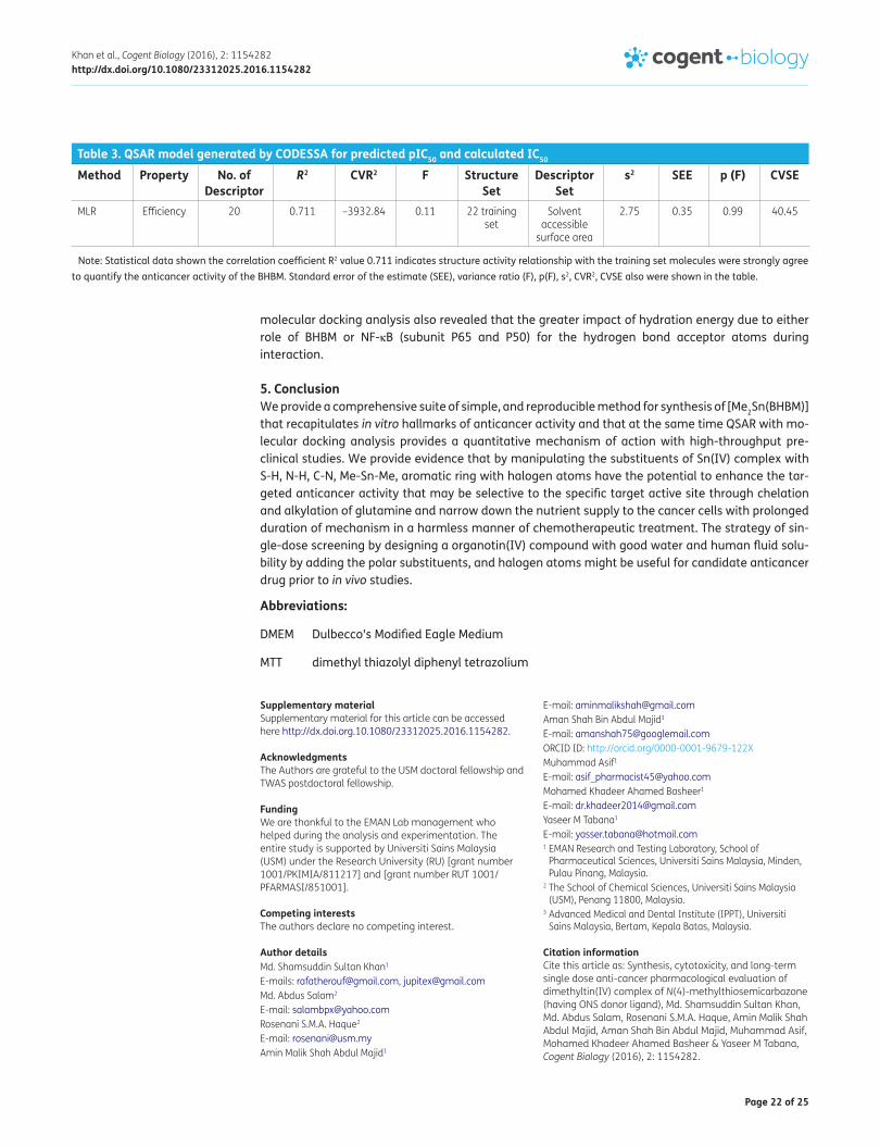
Table 3. QSAR model generated by CODESSA for predicted pIC50 and calculated IC50
Note: Statistical data shown the correlation coefficient R2 value 0.711 indicates structure activity relationship with the training set molecules were strongly agree to quantify the anticancer activity of the BHBM. Standard error of the estimate (SEE), variance ratio (F), p(F), s2, CVR2, CVSE also were shown in the table.
Method Property No. of Descriptor
R2 CVR2 F Structure Set
Descriptor Set
s2 SEE p (F) CVSE
MLR Efficiency 20 0.711 –3932.84 0.11 22 training set
Solvent accessible
surface area
2.75 0.35 0.99 40.45
Supplementary materialSupplementary material for this article can be accessed here http://dx.doi.org.10.1080/23312025.2016.1154282.
AcknowledgmentsThe Authors are grateful to the USM doctoral fellowship and TWAS postdoctoral fellowship.
FundingWe are thankful to the EMAN Lab management who helped during the analysis and experimentation. The entire study is supported by Universiti Sains Malaysia (USM) under the Research University (RU) [grant number 1001/PKIMIA/811217] and [grant number RUT 1001/PFARMASI/851001].
Competing interestsThe authors declare no competing interest.
Author detailsMd. Shamsuddin Sultan Khan1
E-mails: [email protected], [email protected]. Abdus Salam2
E-mail: [email protected] S.M.A. Haque2
E-mail: [email protected] Malik Shah Abdul Majid1
E-mail: [email protected] Shah Bin Abdul Majid3
E-mail: [email protected] ID: http://orcid.org/0000-0001-9679-122XMuhammad Asif1
E-mail: [email protected] Khadeer Ahamed Basheer1
E-mail: [email protected] M Tabana1
E-mail: [email protected] EMAN Research and Testing Laboratory, School of
Pharmaceutical Sciences, Universiti Sains Malaysia, Minden, Pulau Pinang, Malaysia.
2 The School of Chemical Sciences, Universiti Sains Malaysia (USM), Penang 11800, Malaysia.
3 Advanced Medical and Dental Institute (IPPT), Universiti Sains Malaysia, Bertam, Kepala Batas, Malaysia.
Citation informationCite this article as: Synthesis, cytotoxicity, and long-term single dose anti-cancer pharmacological evaluation of dimethyltin(IV) complex of N(4)-methylthiosemicarbazone (having ONS donor ligand), Md. Shamsuddin Sultan Khan, Md. Abdus Salam, Rosenani S.M.A. Haque, Amin Malik Shah Abdul Majid, Aman Shah Bin Abdul Majid, Muhammad Asif, Mohamed Khadeer Ahamed Basheer & Yaseer M Tabana, Cogent Biology (2016), 2: 1154282.

Page 23 of 25
Khan et al., Cogent Biology (2016), 2: 1154282http://dx.doi.org/10.1080/23312025.2016.1154282
Cover imageSource: Authors.
ReferencesAldridge, W. N. (1976). The influence of organotin compounds
on mitochondrial functions. In J. J. Zuckerman (Ed.), Organotin compounds, new chemistry and applications (Vol. 157, Chapter 13, pp. 186–196). New York, NY: American Chemical Society.
Becker, E. M., Lovejoy, D. B., Greer, J. M., Watts, R., & Richardson, D. S. (2003). Identification of the di-pyridyl ketone isonicotinoyl hydrazone (PKIH) analogues as potent iron chelators and anti-tumour agents. British Journal of Pharmacology, 138, 819–830.
Benzie, I. F. F., & Strain, J. J. (1996). The ferric reducing ability of plasma (frap) as a measure of “antioxidant power”: The FRAP assay. Analytical Biochemistry, 239, 70–76. http://dx.doi.org/10.1006/abio.1996.0292
Bernhardt, P. V., Caldwell, L. M., Chaston, T. B., Chin, P., & Richardson, D. R. (2003). Cytotoxic iron chelators: Characterization of the structure, solution chemistry and redox activity of ligands and iron complexes of the di-2-pyridyl ketone isonicotinoyl hydrazone (HPKIH) analogues. Journal of Biological Inorganic Chemistry, 8, 866–880. http://dx.doi.org/10.1007/s00775-003-0486-z
Blunden, S. J., Cusak, P. A., & Hill, R. (1985). The industrial uses of tin chemicals. London: Royal Society of Chemistry.
Borley, A. C., Hiscox, S., Gee, J., Smith, C., Shaw, V., Barrett-Lee, P., & Nicholson, R. I. (2008). Anti-oestrogens but not oestrogen deprivation promote cellular invasion in intercellular adhesion-deficient breast cancer cells. Breast Cancer Research, 10, R103. http://dx.doi.org/10.1186/bcr2206
Brazil D. P., & Hemmings, B. A. (2001). Ten years of protein kinase B signalling: A hard Akt to follow. Trends in Biochemical Sciences., 26, 657–664. http://dx.doi.org/10.1016/S0968-0004(01)01958-2
Carlos, H., Martínez, R., & Dardonville, C. (2013). Rapid Determination of Ionization Constants (pKa) by UV Spectroscopy Using 96-Well Microtiter Plates. ACS Medicinal Chemistry Letters, 4, 142–145.
Cima, F., & Ballarin, L. (1999). TBT-induced apoptosis in tunicate haemocytes. Applied Organometallic Chemistry, 13, 697. http://dx.doi.org/10.1002/(ISSN)1099-0739
Cocco, M. T., Congiu, C., Lilliu, V., & Onnis, V. (2006). Synthesis and in vitro antitumoral activity of new hydrazinopyrimidine- 5-carbonitrile derivatives. Bioorganic & Medicinal Chemistry, 14, 366–372. http://dx.doi.org/10.1016/j.bmc.2005.08.012
Collery, P., & Perchery, C. (1993). Clinical experience with tumor-inhibiting gallium complexes. In B. K. Keppler (Ed.), Metal complexes in cancer chemotherapy (pp. 249–258). Weinheim: VCH.
Collier, W. A. (1929). Zur experimentellen Therapie der Tumoren. Zeitschrift für Hygiene und Infektionskrankheiten, 110, 169–174. http://dx.doi.org/10.1007/BF02175963
Collinson, S. R., & Fenton, D. E. (1996). Coordination Chemistry Reviews, 148, 19–40. http://dx.doi.org/10.1016/0010-8545(95)01156-0
da Silva, D. L., Reis, F. S., Muniz, D. R., Ruiz, A. L. T. G., de Carvalho, J. E., Sabino, A. A., & Modolo, L. V. (2012). Free radical scavenging and antiproliferative properties of Biginelli adducts. Bioorganic & Medicinal Chemistry, 20, 2645–2650. http://dx.doi.org/10.1016/j.bmc.2012.02.036
Del Duca, D., Werbowetski, T., & Del Maestro, R. F. (2004). Spheroid preparation from hanging drops: Characterization of a model of brain tumor invasion. Journal of Neuro-Oncology, 67, 295–303. http://dx.doi.org/10.1023/B:NEON.0000024220.07063.70
Easmon, J. G., Puerstinger, G., Roth, T., Fiebig, H. H., Jenny, M., Jaeger, W., … Hofmann, J. (2001). 2-benzoxazolyl and 2-benzimidazolyl hydrazones derived from 2-acetylpyridine: A novel class of antitumor agents. International Journal of Cancer, 94, 89–96. http://dx.doi.org/10.1002/(ISSN)1097-0215
EL-Hawash, S. A. M., Abdel Wahab, A. E., & El-Demellawy, M. A. (2006). Cyanoacetic acid hydrazones of 3-(and 4-)acetylpyridine and some derived ring systems as potential antitumor and anti-hcv agents. Archiv der Pharmazie, 339, 14–23. http://dx.doi.org/10.1002/(ISSN)1521-4184
Gasteiger, J., & Marsili, M. (1980). Iterative partial equalization of orbital electronegativity - a rapid access to atomic charges. Tetrahedron, 36, 3219–3228. http://dx.doi.org/10.1016/0040-4020(80)80168-2
Gielen, M. (2003). An overview of forty years organotin chemistry developed at the Free Universities of Brussels ULB and VUB. Journal of the Brazilian Chemical Society, 14, 870–877.
Hall, W. T., & Zuckerman, J. J. (1977). Tin(II) and dimethyltin(IV) derivatives of amino acids. Inorganic Chemistry, 16, 1239–1241. http://dx.doi.org/10.1021/ic50171a053
Han, G., & Yang, P. (2002). Synthesis and characterization of water-insoluble and water-soluble dibutyltin(IV) porphinate complexes based on the tris(pyridinyl)porphyrin moiety, their anti-tumor activity in vitro and interaction with DNA. Journal of Inorganic Biochemistry, 91, 230. http://dx.doi.org/10.1016/S0162-0134(02)00369-0
Haque, R. A., & Salam, M. A. (2015). Synthesis, spectroscopic properties and biological activity of new mono organotin(IV) complexes with 5-bromo-2-hydroxybenzaldehyde-4, 4-dimethylthiosemicarbazone. Cogent Chemistry, 1, 1045212. http://dx.doi.org/10.1080/23312009.2015.1045212
Hayot, C., Farinelle, S., De Decker, R., Decaestecker, C., Darro, F., Kiss, R., & Van Damme, M. (2002). In vitro pharmacological characterizations of the anti-angiogenic and anti-tumor cell migration properties mediated by microtubule-affecting drugs, with special emphasis on the organization of the actin cytoskeleton. International Journal of Oncology, 21, 417–425.
Holeček, J., Nádvorník, M., Handlíř, K., & Lyčka, A. (1986). 13C and 119Sn NMR spectra of Di-n-butyltin(IV) compounds. Journal of Organometallic Chemistry, 315, 299. http://dx.doi.org/10.1016/0022-328X(86)80450-8
Johnson, D. K., Murphy, T. B., Rose, N. J., Goodwin, W. H., & Pickart, L. (1982). The experimental therapy for the tumors. Journal of Hygiene and Infectious Diseases, 67, 159–165. http://dx.doi.org/10.1016/S0020-1693(00)85058-6
Katritsky, A. R., Lobanov, V. S., & Karelson, M. (1996). CODESSA: Reference manual, University of Florida (Version 2). Gainesville, FL: Semichem, Terrace.
Khan, M. S. S., Syeed, S. H., Uddin, M. H., Akter, L., Ullah, M. A., Jahan, S., & Rashid, M. H. (2013). Screening and evaluation of antioxidant, antimicrobial, cytotoxic, thrombolytic and membrane stabilizing properties of the methanolic extract and solvent-solvent partitioning effect of Vitex negundo Bark. Asian Pacific Journal of Tropical Disease, 3, 393–400. http://dx.doi.org/10.1016/S2222-1808(13)60090-0
Koch, B., Baul, T. S. B., & Chatterjee, A. (2008). Cell proliferation inhibition and antitumor activity of novel alkyl series of diorganotin(IV) compounds. Journal of Applied Toxicology, 28, 430–438. http://dx.doi.org/10.1002/(ISSN)1099-1263
Lovejoy, D. B., & Richardson, D. R. (2002). Novel “hybrid” iron chelators derived from aroylhydrazones and thiosemicarbazones demonstrate selective antiproliferative activity against tumor cells. Blood, 100,

Page 24 of 25
Khan et al., Cogent Biology (2016), 2: 1154282http://dx.doi.org/10.1080/23312025.2016.1154282
666–676. http://dx.doi.org/10.1182/blood.V100.2.666
Min, H., Yin, H., Zhang, X., Chuan, L., Caihong, Y., & Shuang, C. (2013). Di- and tri-organotin(IV) complexes with 2-hydroxy-1-naphthaldehyde 5-chloro-2-hydroxybenzoylhydrazone: Synthesis, characterization and in vitro antitumor activities. Journal of Organometallic Chemistry, 724, 23–31.
Mouayed, A. H., Muhammad, A. I., Muhammad, A., Rosenani, A. H., Mohammed, B. K. A., Amin, M. S. A. M., & Teoh, S. G. (2015). A synthesis, crystal structures and in vitro anticancer studies of new thiosemicarbazone derivatives. Phosphorus Sulfur and Silicon and the Related Elements, 190, 1498–1508.
Pacheco, S. R., Braga, T. C., da Silva, D. L., Horta, L. P., Reis, F. S., Ruiz, A. L., … de Fatima, Â (2013). Biological activities of eco-friendly synthesized hantzsch adducts. Medicinal Chemistry, 9, 889–896. http://dx.doi.org/10.2174/1573406411309060014
Pellerito, C., D’Agati, P., Fiore, T., Mansueto, C., Mansueto, V., Stocco, G., … Pellerito, L. (2005). Synthesis, structural investigations on organotin(IV) chlorin-e6 complexes, their effect on sea urchin embryonic development and induced apoptosis. Journal of Inorganic Biochemistry, 99, 1294–1305. http://dx.doi.org/10.1016/j.jinorgbio.2005.03.002
Pruchnik, F. P., Bańbuła, M., Ciunik, Z., Chojnacki, H., Latocha, M., Skop, B., … Nasulewicz, A. (2002). Structure, properties and cytostatic activity of triorganotin (aminoaryl)carboxylates. European Journal of Inorganic Chemistry, 3214–3221. http://dx.doi.org/10.1002/1099-0682(200212)2002:12<3214::AID-EJIC3214>3.0.CO;2-K
Rahn, C. A., Bombeck, D. W., & Doolittle, D. J. (1991). Assessment of mitochondrial membrane potential as an indicator of cytotoxicity. Fundamental and Applied Toxicology, 16, 435–448. http://dx.doi.org/10.1016/0272-0590(91)90084-H
Reddy, D. M., Qazi, N. A., Sawant, S. D., Bandey, A. H., Srinivas, J., Shankar, M., … Sampath Kumar, H. M. (2011). Design and synthesis of spiro derivatives of parthenin as novel anti-cancer agents. European Journal of Medical Chemistry, 46, 3210–3217
Richardson, D. R. (1997). Cytotoxic analogs of the iron(III) chelator pyridoxal isonicotinoyl hydrazone: Effects of complexation with copper(II), gallium(III), and iron(III) on their antiproliferative activities. Antimicrobial Agents & Chemotherapy, 41, 2061–2063.
Richardson, D. R., & Bernhardt, P. V. (1999). Crystal and molecular structure of 2-hydroxy-1-naphthaldehyde isonicotinoyl hydrazone (NIH) and its iron(III) complex: An iron chelator with anti-tumour activity. Journal of Biological Inorganic Chemistry, 4, 266–273. http://dx.doi.org/10.1007/s007750050312
Saxena, A. K., & Huber, F. (1989). Organotin compounds and cancer chemotherapy. Coordination Chemistry Reviews, 95, 109–123. http://dx.doi.org/10.1016/0010-8545(89)80003-7
Stanton D. T., & Jurs, P. C. (1990). Development and use of charged partial surface area structural descriptors in computer-assisted quantitative structure-property
relationship studies. Analytical Chemistry, 62, 2323–2329. http://dx.doi.org/10.1021/ac00220a013
Stanton D. T., Egolf, L. M., Jurs, P. C., & Hicks, M. G. (1992). Computer-assisted prediction of normal boiling points of pyrans and pyrroles. Journal of Chemical Information and Modeling, 32, 306–316. http://dx.doi.org/10.1021/ci00008a009
Tabassum, S., & Pettinari, C. (2006). Chemical and biotechnological developments in organotin cancer chemotherapy. Journal of Organometallic Chemistry, 691, 1761–1766. http://dx.doi.org/10.1016/j.jorganchem.2005.12.033
Terzioglu, N., & Gürsoy, A. (2003). Synthesis and anticancer evaluation of some new hydrazone derivatives of 2,6-dimethylimidazo[2,1-b][1,3,4]thiadiazole-5-carbohydrazide. European Journal of Medicinal Chemistry, 38, 781–786. http://dx.doi.org/10.1016/S0223-5234(03)00138-7
Tian, L., Qian, B., Sun, Y., Zheng, X., Yang, M., Li, H., & Xueli, L. (2005). Synthesis, structural characterization and cytotoxic activity of diorganotin(IV) complexes ofN-(5-halosalicylidene)-α-amino acid. Applied Organometallic Chemistry, 19, 980–987. http://dx.doi.org/10.1002/(ISSN)1099-0739
Trott, O., & Olson, A. J. (2010). AutoDock Vina: Improving the speed and accuracy of docking with a new scoring function, efficient optimization and multithreading. Journal of Computational Chemistry, 31, 455–461.
van Reyk, D. V., Sarel, S., & Hunt, N. (2000). Inhibition of in vitro lymphoproliferation by three novel iron chelators of the pyridoxal and salicyl aldehyde hydrazone classes. Biochemical Pharmacology, 60, 581–587. http://dx.doi.org/10.1016/S0006-2952(00)00347-6
Xanthopoulou, M. N., Hadjikakou, S. K., Hadjiliadis, N., Milaeva, E. R., Gracheva, J. A., Tyurin, V. Y., … Charalabopoulos, K. (2008). Biological studies of new organotin(IV) complexes of thioamide ligands. European Journal of Medicinal Chemistry, 43, 327–335. http://dx.doi.org/10.1016/j.ejmech.2007.03.028
Xia, B., Ma, W., Zheng, B., Zhang, X., & Fan, B. (2008). Quantitative structure-activity relationship studies of a series of non-benzodiazepine structural ligands binding to benzodiazepine receptor. European Journal of Medicinal Chemistry, 43, 1489–1498. http://dx.doi.org/10.1016/j.ejmech.2007.09.004
Yalkinoglu, A. O., Schlehofer, J. R., & zur Hausen, H. (1990). Inhibition of N-methyl-N′'-nitro-n-nitrosoguanidine-induced methotrexate and adriamycin resistance in CHO cells by adeno-associated virus type 2. International Journal of Cancer, 45, 1195–1203. http://dx.doi.org/10.1002/(ISSN)1097-0215
Yin, H. D., & Chen, S. W. (2006). Synthesis and characterization of organotin(IV) compounds with Schiff base of o-vanillin-2-thiophenoylhydrazone. Journal of Organometallic Chemistry, 691, 3103–3108. http://dx.doi.org/10.1016/j.jorganchem.2006.03.003
Yuan, J., Lovejoy, D. B., & Richardson, D. R. (2004). Novel di-2-pyridyl-derived iron chelators with marked and selective antitumor activity: In vitro and in vivo assessment. Blood, 104, 1450–1458. http://dx.doi.org/10.1182/blood-2004-03-0868

Page 25 of 25
Khan et al., Cogent Biology (2016), 2: 1154282http://dx.doi.org/10.1080/23312025.2016.1154282
© 2016 The Author(s). This open access article is distributed under a Creative Commons Attribution (CC-BY) 4.0 license.You are free to: Share — copy and redistribute the material in any medium or format Adapt — remix, transform, and build upon the material for any purpose, even commercially.The licensor cannot revoke these freedoms as long as you follow the license terms.
Under the following terms:Attribution — You must give appropriate credit, provide a link to the license, and indicate if changes were made. You may do so in any reasonable manner, but not in any way that suggests the licensor endorses you or your use. No additional restrictions You may not apply legal terms or technological measures that legally restrict others from doing anything the license permits.
Cogent Biology (ISSN: 2331-2025) is published by Cogent OA, part of Taylor & Francis Group. Publishing with Cogent OA ensures:• Immediate, universal access to your article on publication• High visibility and discoverability via the Cogent OA website as well as Taylor & Francis Online• Download and citation statistics for your article• Rapid online publication• Input from, and dialog with, expert editors and editorial boards• Retention of full copyright of your article• Guaranteed legacy preservation of your article• Discounts and waivers for authors in developing regionsSubmit your manuscript to a Cogent OA journal at www.CogentOA.com


![Evaluation of the angiogenesis activity of Crataeva magna ... · Semin Cancer Biol 2003;13(2):159–167. [6] Lay-Jing Seaw, Hooi-Kheng Beh, Amin Malik Shah, Abdul Majid, Vikneswaram](https://img.pdfslide.us/doc/110x75/5fb85fef5fa3175bc342b814/evaluation-of-the-angiogenesis-activity-of-crataeva-magna-semin-cancer-biol.jpg)


![[Dec., 2016] ZULKIFLI ABDUL MAJID · [Zulkifli Abdul Majid] CURRICULUM VITAE 3 4. Committee Member, Seminar Keselamatan Sistem Gas bagi Sektor Pendidikan, Suruhanjaya Tenaga, Malaysia,](https://img.pdfslide.us/doc/110x75/5ffbb1e222b87e0dc7428c32/dec-2016-zulkifli-abdul-majid-zulkifli-abdul-majid-curriculum-vitae-3-4-committee.jpg)