Embed Size (px)
Citation preview

Journal of Organometallic Chemistry 690 (2005) 383–393
www.elsevier.com/locate/jorganchem
Synthesis and structural characterization of N-para-ferrocenylbenzoyl amino acid ethyl esters and the X-ray crystal structures of the
glycyl and (±)-2-aminobutyric acid derivativeFc-C6H4CONHCH(C2H5)CO2Et
David Savage a, Gwen Malone a, John F. Gallagher a,b,*, Yoshiteru Ida c,Peter T.M. Kenny a,b,*
a School of Chemical Sciences, Dublin City University, Dublin 9, Irelandb National Institute of Cellular Biotechnology, Dublin City University, Dublin 9, Ireland
c School of Pharmaceutical Sciences, Showa University, Hatanodai, Shinagawa-ku, Tokyo 142-8555, Japan
Received 1 August 2004; accepted 1 August 2004
Available online 22 October 2004
Abstract
A series of N-para-ferrocenyl benzoyl amino acid ethyl esters 1–8 have been prepared by coupling para-ferrocenyl benzoic acid
with the amino acid esters using the conventional 1,3-dicyclohexylcarbodiimide (DCC), 1-hydroxybenzotriazole (HOBt) protocol.
The amino acids employed in the synthesis were glycine, LL-alanine, LL-leucine, LL-phenylalanine, b-alanine, 4-aminobutyric acid, (±)-
2-aminobutyric acid and 2-aminoisobutyric acid. The compounds were fully characterized by a range of spectroscopic techniques
such as NMR and mass spectrometry. In addition the X-ray crystal structures of the glycyl 1 and (±)-2-aminobutyrate 7 derivatives
have been determined. Analysis of relevant fragments in crystal structures on the Cambridge Structural Database indicates a relative
paucity of common fragments such as the a-aminobutyrate group in comparison to the glycyl moiety.
� 2004 Elsevier B.V. All rights reserved.
Keywords: Ferrocene; Bioorganometallic chemistry; Amino acid esters; X-ray crystal structures; Database analysis; Systematics
1. Introduction
The organometallic compound ferrocene is a promis-ing candidate for incorporation in novel materials due
to its stability, spectroscopic, electrochemical properties
and ease of use [1]. As a direct consequence of these
properties, research in the area of ferrocenyl derivatives
has seen a dramatic increase in attention over the past
decade, primarily for the ultimate goals of achieving no-
vel sensor compounds, peptide mimetic models and
unnatural drugs [2–10]. The synthesis and structural
0022-328X/$ - see front matter � 2004 Elsevier B.V. All rights reserved.
doi:10.1016/j.jorganchem.2004.08.047
* Corresponding authors. Tel.: +353 1 7005689; fax: +353 1
7005503.
E-mail addresses: [email protected] (J.F. Gallagher), peter.
[email protected] (P.T.M. Kenny).
characterization of a variety of N-ferrocenoyl and
N-ferrocenyl amino acid and peptide derivatives has
been reported [11–25]. We now report the synthesis ofa series of para-ferrocenyl benzoyl amino acid ethyl es-
ters 1–8. The ferrocenyl moiety is linked to the amino
acid residues through a para-benzoyl group. The com-
pounds were fully characterized by 1H NMR, 13C
NMR spectroscopy and mass spectrometry. In addition
the X-ray crystal structures of compounds 1 and 7 are
reported and compared with a related structure [22].
The compounds are composed of three key moieties,namely (i) an electroactive core, (ii) a conjugated linker
that can act as a chromophore and (iii) an amino acid
derivative that can interact with other molecules via
hydrogen bonding.
384 D. Savage et al. / Journal of Organometallic Chemistry 690 (2005) 383–393
2. Results and discussion
2.1. Synthesis
para-Ferrocenyl benzoic acid was prepared as previ-
ously reported [22]. This acid was coupled under basicconditions to the free N-terminal amino acid esters of
glycine, LL-alanine, LL-leucine, LL-phenylalanine, b-ala-nine, 4-aminobutyric acid, (±)-2-aminobutyric acid
and 2-aminoisobutyric acid in the presence of dic-
yclohexylcarbodiimide (DCC) and 1-hydroxybenzo-
triazole (HOBt). The resulting N-para-ferrocenyl
benzoyl amino acid esters 1–8 were obtained as yel-
low/orange colored crystals (Scheme 1). The yields ob-tained ranged between 48% and 68% and all gave
analytical and spectroscopic data in accordance with
the proposed structures. The compounds are reasona-
bly stable however they slowly decompose over a per-
iod of time. The N-para-ferrocenyl benzoyl ethyl esters
1–8 were characterized by a combination of 1H NMR,13C NMR, DEPT-135 and 1H–13C COSY (HMQC)
spectroscopy and by either fast atom bombardment(FAB) 1–4, electrospray ionization (ESI) 5, 6 or ma-
trix assisted laser desorption ionization mass spectr-
ometry (MALDI) 7, 8. Crystals of sufficient quality
for X-ray diffraction studies were obtained for com-
pounds 1 and 7.
2.2. 1H and 13C NMR spectroscopic analysis
All the proton and carbon chemical shifts for com-
pounds 1–8 were unambiguously assigned by a combi-
nation of DEPT-135 and 1H–13C-COSY (HMQC).
The 1H and 13C NMR spectra for compounds 1–8
showed peaks in the ferrocene region characteristic of
a mono substituted ferrocene moiety [4,5,22]. The pro-
tons in the ortho position of the substituted Cp ring ap-
pear in the region d 4.65–4.89 whereas the protons in themeta position occur in the range d 4.17–4.41. The unsub-stituted Cp ring appears in the region d 3.77–4.02. The
O
OMe
H2N
O
NH
R
COOEt
Fe
Fe
1-8
Scheme 1. Synthesis of the N-ferrocenylbenzoyl amino acid esters 1–8, (i) NaN
ethyl ester.
protons of the para-disubstituted benzoyl group appear
as two doublets in the region d 7.38–7.8. For example, in
the case of the b-alanine derivative 5, the aromatic pro-
tons are present as two doublets at d 7.61 and d 7.76,
respectively. The unsubstituted C5H5 ring appears as a
singlet in the 1H NMR spectrum at d 4.01 whereas theortho and meta protons on the substituted Cp ring are
present at d 4.87 and d 4.39, respectively. The NH pro-
ton appears as a triplet at d 8.56 (J = 5.2 Hz) and a quar-
tet at d 3.5 (J = 7.2 Hz) corresponds to the
NHCH2CH2CO-protons. The triplet at d 2.59 (J = 7.2
Hz) is due to the second methylene group adjacent to
the carbonyl group.
The 13C NMR spectra of compounds 1–8 show sig-nals in the region d 66.9–83.9 indicative of a monosub-
stituted ferrocene subunit [4,5,22]. The ipso carbon of
the substituted Cp ring appears in a narrow range of d83.5–83.9. This signal is not present in the DEPT 135
spectra. The carbon atoms of the aromatic ring are vis-
ible in the region d 125.7–144.2. The methylene carbon
atoms of the derivatives were identified by DEPT-135.
A complete assignment of the 1H and 13C NMR spectraof N-{para-(ferrocenyl)benzoyl}-b-alanine ethyl ester 5
is presented in Table 1.
2.3. Mass spectrometry
Since the introduction of soft ionization techniques
such as fast atom bombardment mass spectrometry
(FABMS), electrospray ionization mass spectrometry(ESIMS) and matrix assisted laser desorption ioniza-
tion (MADLI), a wide range of thermolabile and
non-volatile compounds can be subjected to mass
spectrometric analysis [26–29]. As the compounds
were not amenable to electron ionization studies, the
soft ionization techniques were employed in the anal-
ysis and confirmed the correct relative molecular mass.
Examination of the mass spectra revealed the presenceof both radical-cation [M]+�, and protonated molecu-
lar ion species, [M + H]+. The abundance of the
O
OMe
O
OH
ii
i
iii
Fe
Fe
O2, HCl, 5 �C, (ii) NaOH/MeOH, (iii) DCC, HOBt, Et3N, Amino acid

Table 2
Selected bond lengths and angles (A, �) for molecules A/B in 1, and 7
A/B in 1 7
Fe1. . .Cg1 1.635(3)/1.643(3) 1.6472(13
Fe2. . .Cg2 1.648(4)/1.645(4) 1.6485(16
Cg1. . .Fe1. . .Cg2 177.7(2)/179.41(15) 179.50(16
C1–O1 1.248(7)/1.243(7) 1.232(3)
C3–O2 1.203(7)/1.193(7) 1.180(3)
C3–O3 1.317(7)/1.345(7) 1.332(3)
C1–N1 1.327(8)/1.324(8) 1.330(3)
C2–N1 1.439(7)/1.440(7) 1.462(3)
C2–C3 1.499(8)/1.499(8) 1.526(4)
C1–C34 1.490(8)/1.498(8) 1.506(3)
Fe1–C11–C31 125.5(4)/126.9(4) 128.54(18
O1–C1–N1 121.3(6)122.1(6) 123.0(3)
O1–C1–C34 121.5(6)/121.6(6) 121.5(2)
C1–N1–C2 124.2(6)/124.7(6) 122.2(2)
N1–C1–C34 117.1(6)/116.4(6) 115.5(2)
N1–C2–C3 112.0(5)/111.5(5) 107.5(2)
N1–C2–C6 –/– 113.3(2)
Fe1–C11–C31–C36 65.0(8)/110.4(6) 118.9(2)
C12–C11–C31–C36 �23.3(10)/19.2(10) 27.6(4)
N1–C1–C34–C33 �157.0(6)/153.1(6) 143.3(3)
C2–N1–C1–C34 176.3(6)/175.5(5) �179.0(2)
C1–N1–C2–C3 �107.5(7)/120.3(7) 126.9(3)
N1–C2–C3–O3 �160.8(6)/156.1(6) 150.9(2)
where Cg1/Cg3 and Cg2/Cg4 are the centroids of the (g5-C5H4)/(g5
C5H5) rings in molecules A and B in 1, respectively.
Table 11H and 13C spectroscopic data for 5
FeNH
O
OO
12
3
4
5
6 7
8
910
11
12 13
1516
17 18
19 20
21
22
14
Site 1H NMR 13C NMR HMQC
1 83.6
2,3 4.87 66.9
4,5 4.39 69.8
6–10 4.01 69.8
11 143
12, 16 7.61 125.7
13, 15 7.76 127.7
14 131.7
17 166.5
18 3.5 34.2
19 2.59 31
20 171.7
21 4.07 60.3
22 1.18 14
D. Savage et al. / Journal of Organometallic Chemistry 690 (2005) 383–393 385
[M + H]+ species was greatest in the analysis of samples 5
and 6 by ESI. However analysis of compounds 1–4 by
FAB and 7 and 8 by MALDI generated radical-cations
[M]+� as the most abundant species. Cation adducts cor-responding to [M + Na]+ and [M + K]+ were also pre-
sent. Addition of a dilute solution of potassium iodide
(KI) to samples 1–4 and the liquid matrix on the probe
tip generated an intense signal 39 Daltons higher due
to an [M + K]+ adduct. The vast majority of analytes
subjected to analysis by FAB and MALDI furnish prot-
onated molecular ion species or cation adducts
[26,28,29]. It has been reported that the molecular radicalcation of ferrocene and not the protonated molecule is
generated during analysis by MALDI [30]. Structurally
significant fragment ions were observed in the FAB mass
spectra for compounds 1–4. A fragment ion is present at
m/z 261 confirming the presence of a ferrocenylphenyl
subunit at the N-terminal. The signal present at m/z
289 is due to cleavage at the benzoyl C@O function. This
fragment ion at m/z 289 was also observed in theMALDI and ESI spectra.
2.4. X-ray crystallographic studies of 1 and 7
The single crystal X-ray structures of 1 and 7 have
been determined, with selected bond lengths and angles
listed in Table 2 with crystallographic details given in
the footnote. The molecular and crystal structures ofboth 1 and 7 are also compared with the previously re-
ported N-{para-(ferrocenyl)benzoyl}-LL-alanine methyl
ester 9 [22]. There is an increasing number of metallo-
cene based amino acid/peptide structures on the Cam-
bridge Structural Database (CSD) [31], however
)
)
)
)
-
research on compounds incorporating the Fc–C6H4-
moiety are still relatively rare in comparison to the
plethora of Fc systems reported, where Fc = (g5-C5H5)Fe(g
5-C5H4).
2.4.1. Molecular and crystal structure study of 1The glycine derivative 1 (Fig. 1) contains two mole-
cules, which differ significantly in conformation in the
asymmetric unit of space group P21/c (No. 14). The ferr-
ocenyl (g5-C5) cyclopentadienyl groups are slightly stag-
gered with C1n. . .Cg1. . .Cg2. . .C2n torsion angles(n = 1–5) in the range 9.5(8)–11.5(7)� and 7.9(6)–
8.9(5)� for molecules A and B, respectively. The
Fe1A. . .Cg1/Cg2 distances are 1.635(3)/1.648(4) A,
while the Cg1. . .Fe1. . .Cg2 angle is 177.7(2)�: corre-
sponding data for Fe1B. . .Cg3/Cg4 in B are 1.643(3)/
1.645(4) A and 179.41(15)� (where Cg1/Cg3 and Cg2/
Cg4 are the centroids of the (g5-C5H4) and (g5-C5H5)
rings in A/B, respectively). The most important geomet-ric parameters are amide C@O 1.248(7)/1.243(7) A, OC–
NH 1.327(8)/1.324(8) A, HN–CH 1.439(7)/1.440(7) A,
ester C@O/CO 1.203(7)/1.317(7) A and 1.193(7)/
1.345(7) A, respectively, which are similar to the dimen-
sions in previously reported systems, such as the LL-ala-
nine derivative 9 [22]. Other important bond length/
angle parameters are listed for comparison in Table 2
and will be discussed for both systems 1 and 2.The most important structural feature in 1 is the ori-
entation of the carboxylate group with respect to the
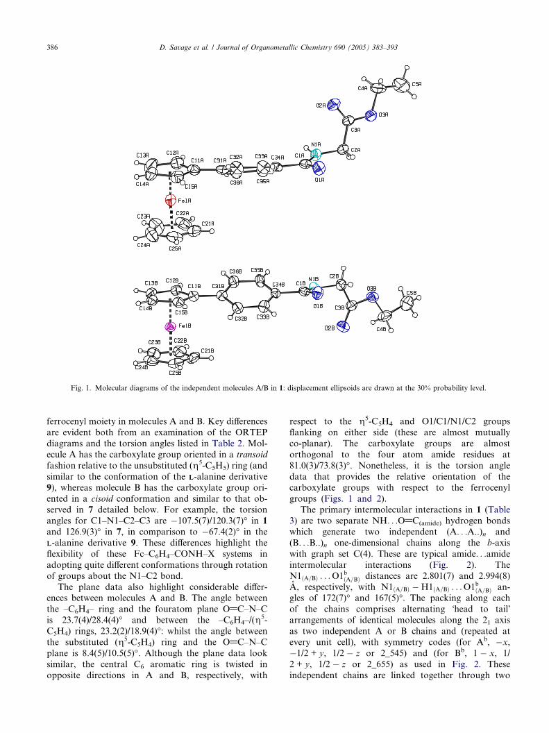
Fig. 1. Molecular diagrams of the independent molecules A/B in 1: displacement ellipsoids are drawn at the 30% probability level.
386 D. Savage et al. / Journal of Organometallic Chemistry 690 (2005) 383–393
ferrocenyl moiety in molecules A and B. Key differences
are evident both from an examination of the ORTEP
diagrams and the torsion angles listed in Table 2. Mol-
ecule A has the carboxylate group oriented in a transoid
fashion relative to the unsubstituted (g5-C5H5) ring (and
similar to the conformation of the LL-alanine derivative
9), whereas molecule B has the carboxylate group ori-
ented in a cisoid conformation and similar to that ob-served in 7 detailed below. For example, the torsion
angles for C1–N1–C2–C3 are �107.5(7)/120.3(7)� in 1
and 126.9(3)� in 7, in comparison to �67.4(2)� in the
LL-alanine derivative 9. These differences highlight the
flexibility of these Fc–C6H4–CONH–X systems in
adopting quite different conformations through rotation
of groups about the N1–C2 bond.
The plane data also highlight considerable differ-ences between molecules A and B. The angle between
the –C6H4– ring and the fouratom plane O@C–N–C
is 23.7(4)/28.4(4)� and between the –C6H4–/(g5-
C5H4) rings, 23.2(2)/18.9(4)�: whilst the angle between
the substituted (g5-C5H4) ring and the O@C–N–C
plane is 8.4(5)/10.5(5)�. Although the plane data look
similar, the central C6 aromatic ring is twisted in
opposite directions in A and B, respectively, with
respect to the g5-C5H4 and O1/C1/N1/C2 groups
flanking on either side (these are almost mutually
co-planar). The carboxylate groups are almost
orthogonal to the four atom amide residues at
81.0(3)/73.8(3)�. Nonetheless, it is the torsion angle
data that provides the relative orientation of the
carboxylate groups with respect to the ferrocenyl
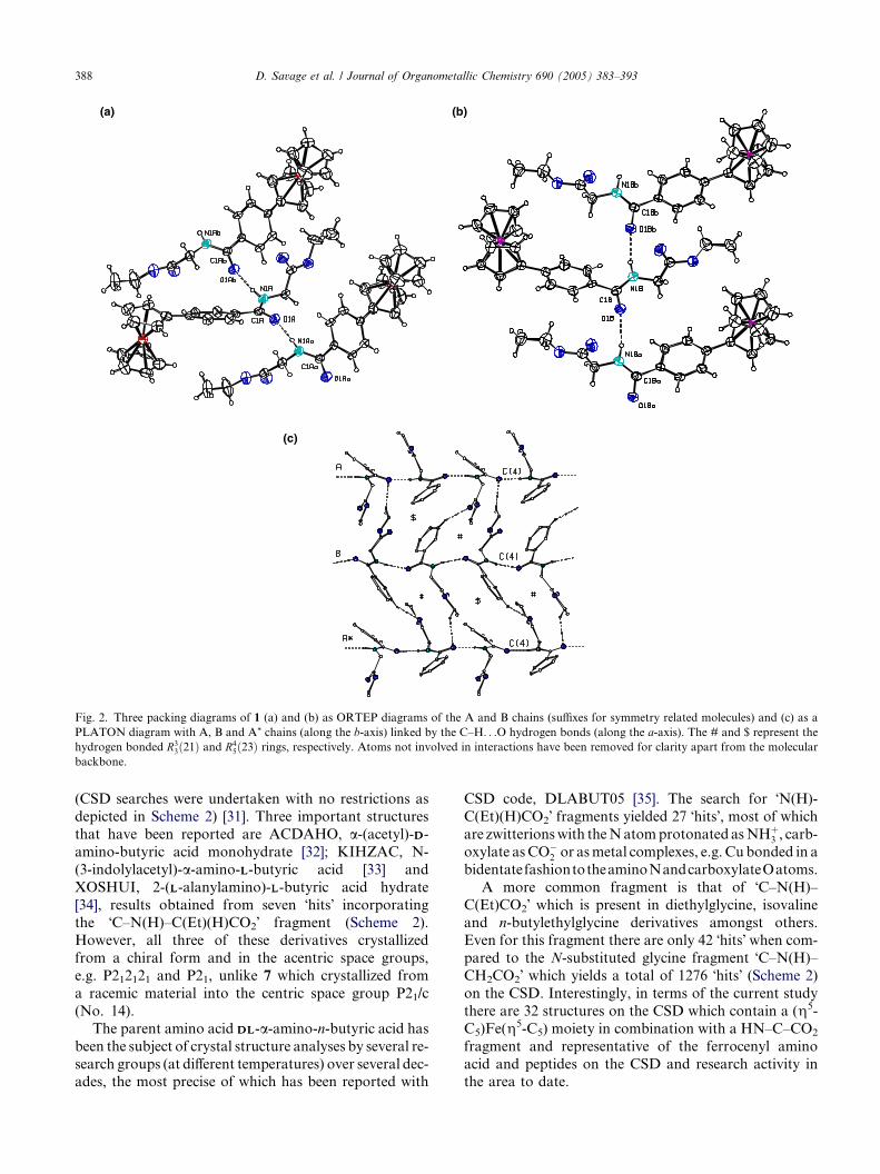
groups (Figs. 1 and 2).The primary intermolecular interactions in 1 (Table
3) are two separate NH. . .O@C(amide) hydrogen bonds
which generate two independent (A. . .A..)n and
(B. . .B..)n one-dimensional chains along the b-axis
with graph set C(4). These are typical amide. . .amide
intermolecular interactions (Fig. 2). The
N1ðA=BÞ . . .O1bðA=BÞ distances are 2.801(7) and 2.994(8)
A, respectively, with N1ðA=BÞ �H1ðA=BÞ . . .O1bðA=BÞ an-gles of 172(7)� and 167(5)�. The packing along each
of the chains comprises alternating �head to tail�arrangements of identical molecules along the 21 axis
as two independent A or B chains and (repeated at
every unit cell), with symmetry codes (for Ab, �x,
�1/2 + y, 1/2 � z or 2_545) and (for Bb, 1 � x, 1/
2 + y, 1/2 � z or 2_655) as used in Fig. 2. These
independent chains are linked together through two

Table 3
Intermolecular Interactions (A, �) for molecules A/B in 1, and 7
1
DH. . .Aa D–H H. . .A D. . .A D–H. . .A Symmetryb
N1A–H1A. . .O1A 0.91(8) 1.89(8) 2.801(7) 172(7) 2_545
N1B–H1B. . .O1B 0.92(6) 2.09(6) 2.994(8) 167(5) 2_655
C4B–H4B2. . .O1A 0.97 2.56 3.310(8) 134 1_655
C36B–H36B. . .O2A 0.93 2.50 3.390(8) 161 2
C2A–H2A2. . .O1A 0.97 2.41 2.786(8) 103 Intra
C2B–H2B1. . .O1B 0.97 2.42 2.808(8) 104 Intra
7
N1–H1. . .O1 0.80(3) 2.18(3) 2.962(3) 165(2) 2_655
C2–H2. . .O1 0.98 2.35 2.787(3) 106 Intra
C4–H4B. . .O1 0.97 2.52 3.353(4) 145 3_666
C36–H36. . .O2 0.93 2.52 3.391(3) 157 4_575
a D–H. . .A signifies Donor–Hydrogen. . .Acceptor.b Symmetry operations as detailed in the text and from SHELXL97. In Figs. 2 and 4, suffixes are used for simplicity to designate the symmetry
related molecules.
D. Savage et al. / Journal of Organometallic Chemistry 690 (2005) 383–393 387
weaker C–H. . .O interactions into 2D sheets through
C4B. . .O1Ai 3.310(8) A and C36B. . .O2Aii 3.390(8)
A interactions (with symmetry operations: (i) 1 + x,
y, z and (ii) �x, �1/2 + y, 1/2 � z). In Fig. 2 the #
and $ represent the R33ð21Þ and R4
5ð23Þ rings, thus gen-
erated by the C–H. . .O interactions. Aggregation of
the two independent chains occurs in solution through
N–H. . .O@C(amide) interactions and upon crystalliza-tion the packing in the crystal structure retains two
distinct transoid and cisoid type conformations linked
through N–H. . .O@C hydrogen bonds (Fig. 2). This
is not unusual and structures with more than one mol-
ecule in the asymmetric unit are a common though
largely unexplained occurrence. There is no significant
C–H. . .p(arene) or p. . .p stacking interactions in 1.
2.4.2. Molecular and crystal structure study of 7In the (±)-butyrate derivative 7, one molecule is pre-
sent in the asymmetric unit in space group P21/c (Fig. 3).
The ferrocenyl (g5-C5) groups are essentially eclipsed
with torsion angles C1n. . .Cg1. . .Cg2. . .C2n (n = 1–5)
in the range 4.0(3)–4.6(2)�. The Fe1. . .Cg1/Cg2 distancesare 1.6472(13)/1.6485(16) A, while the Cg1. . .Fe1. . .Cg2angle is almost linear at 179.50(8)� (where Cg1 and Cg2are the centroids of the (g5-C5H4) and (g5-C5H5) rings,
respectively). Important geometric parameters include
amide C@O 1.232(3) A, OC–NH 1.330(3) A, HN–CH
1.462(3) A and ester C@O/C–O 1.180(3)/1.332(3) A,
respectively, and similar to the dimensions in 1. Major
differences to note are N1–C2–C3, 112.0(5)/111.5(5)� in1, 110.24(16)� in 9, 107.5(2)� in 7 giving a spread of 5�at C2. On substituting H by CH3 and CH2CH3 alongthe three compound structural series, the increasing ster-
ic congestion is reflected in a contraction of this angle at
C2 in 7.
The primary intermolecular interaction in 7 (Table 3)
is N–H. . .O@C(amide) generating a one-dimensional
chain along the b-axis with graph set C(4), N. . .Ob
2.962(3) A, NH. . .Ob 165(2)� (symmetry code, (b)
1 � x, 1/2 + y, 1/2 � z or 2_655). This interaction is ca.
0.15 A longer than the N–H. . .O@C(amide) hydrogen
bond in the (A. . .A..)n chain of 1 though slightly shorter
by 0.03 A than in the (B. . .B..)n chain. There are two
CH. . .O intermolecular interactions present in 7, which
are longer, with C. . .O distances of 3.353(4), 3.391(3)A, the former along the a-axis direction (Fig. 4). There
are no significant C–H. . .p(arene) or p. . .p stacking
interactions.
In 7 the –C6H4– ring and the fouratom O@C–N–C
plane are oriented at 36.94(16)� and for the –C6H4–/
(g5-C5H4) rings, 27.91(18)�: whereas the angle between
(g5-C5H4) and the O@C–N–C plane is 9.1(3)�. As
viewed in Fig. 3, these data show a significant twist be-tween the central C6 aromatic ring and the two moieties
flanking on either side (which are almost mutually co-
planar). These results serve to demonstrate the flexibility
in the orientation of these rings/planes in the molecular
structures of 1, 7 and 9 in the solid state. The carboxy-
late and ethyl groups at C2 in 7 are distinctly different
from 1 though more similar to molecule B in 1. In 7
the angles C1–N1–C2–C3/N1–C2–C3–O2, 126.9(3)�/�33.0(4)� also contrast with �67.4(2)�/�26.5(2)� in 9.
From above, structure 7 retains a cisoid-type conforma-
tion in contrast to the transoid conformation in 9 and in
A (1).
2.4.3. Analysis of fragments in the cambridge structural
database
A search of the Cambridge Structural Database(January 2004 – version v5.25) for structures incorpo-
rating the C–N(H)–C(Et)(H)CO2 moiety (for N-substi-
tuted systems) reveals that it is a system which had
been barely studied when compared to the more com-
mon amino acid derivatives, e.g. glycine or LL-alanine
(a) (b)
(c)
Fig. 2. Three packing diagrams of 1 (a) and (b) as ORTEP diagrams of the A and B chains (suffixes for symmetry related molecules) and (c) as a
PLATON diagram with A, B and A* chains (along the b-axis) linked by the C–H. . .O hydrogen bonds (along the a-axis). The # and $ represent the
hydrogen bonded R33ð21Þ and R4
5ð23Þ rings, respectively. Atoms not involved in interactions have been removed for clarity apart from the molecular
backbone.
388 D. Savage et al. / Journal of Organometallic Chemistry 690 (2005) 383–393
(CSD searches were undertaken with no restrictions as
depicted in Scheme 2) [31]. Three important structures
that have been reported are ACDAHO, a-(acetyl)-DD-amino-butyric acid monohydrate [32]; KIHZAC, N-
(3-indolylacetyl)-a-amino-LL-butyric acid [33] and
XOSHUI, 2-(LL-alanylamino)-LL-butyric acid hydrate
[34], results obtained from seven �hits� incorporating
the �C–N(H)–C(Et)(H)CO2� fragment (Scheme 2).However, all three of these derivatives crystallized
from a chiral form and in the acentric space groups,
e.g. P212121 and P21, unlike 7 which crystallized from
a racemic material into the centric space group P21/c
(No. 14).
The parent amino acid DLDL-a-amino-n-butyric acid has
been the subject of crystal structure analyses by several re-
search groups (at different temperatures) over several dec-ades, the most precise of which has been reported with
CSD code, DLABUT05 [35]. The search for �N(H)-
C(Et)(H)CO2� fragments yielded 27 �hits�, most of which
are zwitterionswith theNatomprotonated asNHþ3 , carb-
oxylate asCO�2 or asmetal complexes, e.g. Cu bonded in a
bidentatefashiontotheaminoNandcarboxylateOatoms.
A more common fragment is that of �C–N(H)–
C(Et)CO2� which is present in diethylglycine, isovaline
and n-butylethylglycine derivatives amongst others.Even for this fragment there are only 42 �hits� when com-
pared to the N-substituted glycine fragment �C–N(H)–
CH2CO2� which yields a total of 1276 �hits� (Scheme 2)
on the CSD. Interestingly, in terms of the current study
there are 32 structures on the CSD which contain a (g5-
C5)Fe(g5-C5) moiety in combination with a HN–C–CO2
fragment and representative of the ferrocenyl amino
acid and peptides on the CSD and research activity inthe area to date.

Fig. 3. Molecular diagram of 7 (top) and a packing diagram (bottom) showing the N–H. . .O@C hydrogen bonded chain using ORTEP (suffixes for
symmetry related molecules): displacement ellipsoids are drawn at the 30% probability level.
D. Savage et al. / Journal of Organometallic Chemistry 690 (2005) 383–393 389
3. Summary
The N-para-ferrocenyl benzoyl amino acid ethyl es-
ters 1–8 have been prepared by coupling para-ferroce-nyl benzoic acid with the amino acid esters using
the conventional 1,3-dicyclohexylcarbodiimide (DCC),
1-hydroxy- benzotriazole (HOBt) protocol The com-
pounds were fully characterized by a range of spectro-
scopic techniques such as NMR and mass
spectrometry.
The X-ray crystal structures of the glycyl 1 and
(±)-2-aminobutyrate 7 derivatives have been deter-mined. Analysis of relevant fragments in crystal struc-
tures on the Cambridge Structural Database indicates
a relative paucity of common fragments such as the
a-aminobutyrate group in comparison to the glycyl
moiety.
4. Experimental
4.1. General procedures
All chemicals were purchased from Sigma/Aldrich and
used as received. Commercial grade reagents were used
without further purification, however, solvents were puri-
fied prior to use. Melting points were determined using aGriffin melting point apparatus and are uncorrected.
Infrared spectra were recorded on a Nicolet 405 FTIR
spectrometer and UV–Vis spectra on a Hewlett–Packard

Fig. 4. A stereoview of the important intermolecular interactions in 7. The ferrocenyl moiety and ethyl H atoms have been removed for clarity. The
hydrogen bonding rings can be clearly discerned and comprise the C(4) chain and hydrogen bonded ring motifs.
CN
C
H
Fragments used and 'hits' found in the CSD searches
7
OO
H
CN
H
27
OO
H
CN
C
H
42
OO
CN
H
1276
OO
H
H
Scheme 2. Cambridge Structural Database fragment search.
390 D. Savage et al. / Journal of Organometallic Chemistry 690 (2005) 383–393
8452A diode array UV–Vis spectrophotometer. NMR
spectra were obtained on a Bruker AC 400 NMR spec-
trometer operating at 400 MHz for 1H NMR and 100
MHz for 13C NMR. The 1H and 13C NMR chemicalshifts (ppm) are relative to TMS and all coupling con-
stants (J) are in Hertz. Positive ion fast atom bombard-
ment mass spectra were obtained on a JEOL SX102
double focussing mass spectrometer employing meta-
nitrobenzyl alcohol as the liquid matrix. Electrospray
ionization mass spectra were obtained on an Applied
Biosystems QSTAR quadrupole time of flight mass spec-
trometer and matrix assisted laser desorption ionizationmass spectra on a Bruker Ultraflex TOF/TOFmass spec-
trometer employing a nitrogen laser at 337 nm.
The single crystal X-ray data for 1 and 7were collected
on a Siemens P4 diffractometer at 294(1) K: x-scans forthe h range 2–26�. Data reductions for both compounds
were standard with the absorption correction DIFABS
used for 1 and w-scans were used for the absorption cor-
rection to 7 (6 reflections with 4� increments). The struc-tures were solved by direct methods and refined using full
matrix least squares methods using SHELXS97 and
SHELXL97, respectively [36]. Hydrogen atoms were
treated as riding atoms except for the amino H atom,
which was refined with isotropic displacement parame-
ters in both structures. The molecular and crystal struc-
ture diagrams drawings were generated using PLATON
[37]. Database searches were undertaken using the Cam-
bridge Structural Database [31].
4.2. General procedure for the synthesis of N-para-ferro-
cenyl benzoyl amino acid esters 1–8
4.2.1. N-{para-(ferrocenyl)benzoyl} glycine ethyl ester 1Glycine ethyl ester hydrochloride (0.3 g, 2.2 mmol)
and triethylamine (0.5 ml) were added to a solution of
para-ferrocenyl benzoic acid (0.5 g, 1.6 mmol), 1-hydroxybenzotriazole (0.3 g, 2.2 mmol) and 1,3-dic-
yclohexylcarbodiimide (0.45 g, 2.2 mmol) in CH2Cl2(50 ml) at 0 �C. After 30 min, the solution was raised
to room temperature and allowed to proceed for 48 h.
The precipitated N,N-dicyclohexylurea was removed
by filtration and the filtrate washed with water, 10%
potassium hydrogen carbonate, 5% citric acid and
dried over MgSO4. Recrystallization from petroleumether (40–60 �C): ethyl acetate furnished the title com-
pound as orange needles. The crystals were of suffi-
cient quality for an X-ray diffraction study (0.351 g,
56%).
m.p. 102–104 �C, E1/2 = 513 mV.
Mass spectrum: found: [M]+� 391,
C21H21N1O3Fe requires: 391.I.R. mmax (KBr): 3308, 1735, 1700, 1685, 1211 cm�1.
UV–Vis kmax CH2Cl2: 358 (e 2290), 454 (e 740) nm.

D. Savage et al. / Journal of Organometallic Chemistry 690 (2005) 383–393 391
1H NMR (400 MHz) d (DMSO): 8.90 (1H, d, J = 7.6
Hz –CONH–), 7.79 (2H, d, J = 8 Hz, ArH), 7.64 (2H, d,
J = 8 Hz, ArH), 4.89 {2H, t, J = 2 Hz, ortho on (g5-
C5H4)}, 4.41 {2H, t, J = 2 Hz, meta on (g5-C5H4)},4.12 (2H, q, J = 7.2 Hz, –OCH2CH3), 4.02 {5H, s, (g5-
C5H5)}, 3.99 (2H, d, J = 5.6 Hz, –NHCH2CO–), 1.21
(3H, t, J = 7.2 Hz, –OCH2CH3).13C NMR (100 MHz) d (DMSO): 170.4, 166.9, 143.4,
131.0, 127.8, 125.8, 83.5, 69.9, 67.0, 60.8 (�ve DEPT),
33.7 (�ve DEPT), 14.4.
4.2.2. N-{para-(ferrocenyl)benzoyl}-LL-alanine ethyl ester2
LL-Alanine ethyl ester hydrochloride (0.3 g, 2.0 mmol)
was used. Recrystallization from petroleum ether (40–60
�C): ethyl acetate furnished the title compound as an or-
ange solid (0.38 g, 59%).
m.p. 104–106 �C, E1/2 = 505 mV,
½a�25D ¼ þ28� (c = 2.1, EtOH).Mass spectrum: found: [M]+� 405,
C22H23N1O3Fe requires: 405.
I.R. mmax (KBr): 2928, 1750, 1648, 1509 cm�1.
UV–Vis kmax CH2Cl2: 358 (e 2670), 454 (e 860) nm.
1H NMR (400 MHz) d (DMSO): 8.47 (1H, d, J = 6.8
Hz, –CONH–), 7.56 (2H, d, J = 8 Hz, ArH), 7.38 (2H, d,
J = 8 Hz, ArH), 4.65 {2H, s, ortho on (g5-C5H4)}, 4.41–4.48 {1H, m, –CH(CH3)}, 4.17 {2H, s, meta on (g5-
C5H4)}, 4.12 (2H, q, J = 7.6 Hz, –OCH2CH3), 3.77
{5H, s, (g5-C5H5)}, 1.48 {3H, d, J = 7.2 Hz,
–CH(CH3)}, 1.21 (3H, t, J = 7.6 Hz, –OCH2CH3).13C NMR (100 MHz) d (DMSO): 173.5, 166.5, 143.3,
131.1, 128.0, 125.7, 83.5, 69.9, 67.0, 60.8 (�ve DEPT),
48.7, 17.1, 14.5.
4.2.3. N-{para-(ferrocenyl)benzoyl}-LL-leucine ethyl ester
3LL-Leucine ethyl ester hydrochloride (0.3 g, 1.5 mmol)
was used. Recrystallization from petroleum ether (40–60
�C): ethyl acetate furnished the title compound as brown
solid (0.46 g, 68%).
m.p. 127–129 �C, E1/2 = 501 mV,½a�20D ¼ þ3� (c = 1.2, EtOH).
Mass spectrum: found: [M]+� 447,
C25H29N1O3Fe requires: 447.
I.R. mmax (KBr): 3331, 2933, 1728, 1614 cm�1.
UV–Vis kmax CH2Cl2: 352 (e 1550), 454 (e 450) nm.
1H NMR (400 MHz) d (DMSO): 8.66 (1H, d, J = 7.6
Hz, –CONH–), 7.80 (2H, d, J = 8 Hz, –ArH), 7.62 (2H,d, J = 8 Hz, –ArH), 4.88 {2H, s, ortho on (g5-C5H4)},
4.50–4.60 [1H, m, –CH{CH2CH(CH3)2}], 4.41 {2H, s,
meta on (g5-C5H4)}, 4.08 (2H, q, J = 6.8 Hz, –
OCH2CH3), 4.02 {5H, s, (g5-C5H5)}, 1.57–1.81
[3H, m, –CH{CH2CH(CH3)2}], 1.19 (3H, t, J = 6.8
Hz, –OCH2CH3), 0.93 [3H, d, J = 6.4 Hz,
–CH{CH2CH(CH3)2}], 0.86 [3H, d, J = 6.4 Hz,
–CH{CH2CH(CH3)2}].13C NMR (100 MHz) d (DMSO): 173.1, 166.9, 143.3,
131.1, 128.0, 125.7, 83.5, 69.9, 67.0, 60.1 (�ve DEPT),
51.4, 39.6 (�ve DEPT), 24.8, 23.2, 21.5, 14.4.
4.2.4. N-{para-(ferrocenyl)benzoyl}-LL-phenylalanine ethylester 4
LL-Phenylalanine ethyl ester hydrochloride (0.3 g, 1.3
mmol) was used. Recrystallization from petroleum ether
(40–60 �C): ethyl acetate furnished the title compound asan orange solid (0.38 g, 61%).
m.p. 134–136 �C, E1/2 = 498 mV,
½a�20D ¼ þ48� (c = 2, EtOH).
Mass spectrum: found: [M]+� 481,
C28H27N1O3Fe requires: 481.
I.R. mmax (KBr): 2929, 1710, 1636, 1106 cm�1.
UV–Vis kmax CH2Cl2: 358 (e 3370), 454 (e 1070) nm.
1H NMR (400 MHz) d (DMSO): 8.80 (1H, d, J = 7.6
Hz, –CONH–), 7.73 (2H, d, J = 8.4 Hz, –ArH), 7.61
(2H, d, J = 8.4 Hz, –ArH), 7.21–7.32 (5H, m, –ArH),
4.88 {2H, s, ortho on (g5-C5H4)}, 4.54–4.68 {1H, m,
–CH (CH2Ph)}, 4.41 {2H, s, meta on (g5-C5H4)}, 4.10
(2H, q, J = 7.2 Hz, –OCH2CH3), 4.02 {5H, s, (g5-
C5H5)}, 3.11–3.17 {2H, m, –CH(CH2Ph)}, 1.16 (3H, t,J = 7.2 Hz, –OCH2CH3).
13C NMR (100 MHz) d (DMSO): 172.3, 166.8, 143.4,
138.0, 131.1, 129.4, 128.6, 127.9, 126.9, 125.7, 83.5, 69.9,
68.1, 67.1, 66.9, 61.0 (�ve DEPT), 54.8, 33.7 (�ve
DEPT), 14.4.
4.2.5. N-{para-(ferrocenyl)benzoyl}-b-alanine ethyl ester5
b-Alanine ethyl ester hydrochloride (0.3 g, 2.0
mmol) was used. Recrystallization from diethyl ether
furnished the title compound as orange needles (0.34
g, 52%).
m.p. 137–138 �C, E 0� = 131 mV.
Mass spectrum: found: [M + H]+ 406.1080,
C22H24N1O3Fe requires: 406.1106.I.R. mmax (KBr): 2930, 1708, 1651, 1629 cm�1.
UV–Vis kmax CH2Cl2: 354 (e 1900), 452 (590) nm.
1H NMR (400 MHz) d (DMSO): 8.56 (1H, t,
J = 5.2 Hz, –CONH–), 7.76 (2H, d, J = 8 Hz, –ArH),
7.61 (2H, d, J = 8 Hz, –ArH), 4.87 {2H}, s, ortho on
(g5-C5H4)}, 4.39 {2H, s, meta on (g5-C5H4)}, 4.07
(2H, q, J = 7.6 Hz, –OCH2CH3), 4.01 {5H, s, (g5-C5H5)}, 3.50 (2H, q, J = 7.2 Hz, –CH2CH2–), 2.59

392 D. Savage et al. / Journal of Organometallic Chemistry 690 (2005) 383–393
(2H, t, J = 7.2 Hz, –CH2CH2–), 1.18 (3H, t, J = 7.6
Hz, –OCH2CH3).13C NMR (100 MHz) d(DMSO): 171.7, 166.5, 143.0,
131.7, 127.7, 125.7, 83.6, 69.8, 66.9, 60.3 (�ve DEPT),
34.2 (�ve DEPT), 31.0 (�ve DEPT), 14.0.
4.2.6. N-{para-(ferrocenyl)benzoyl}-4-aminobutyric acid
ethyl ester 64-Aminobutyric acid ethyl ester hydrochloride (0.3 g,
1.8 mmol) was used. Recrystallization from diethyl ether
furnished the title compound as orange needles (0.37 g,
55%).
m.p. 80–82 �C, E 0� = 132 mV.Mass spectrum found: [M + H]+ 420.1264,
C23H26N1O3Fe requires: 420.1262,
I.R. mmax (KBr): 2930, 1733, 1623, 1547 cm�1.
UV–Vis kmax CH2Cl2: 354 (e 1260), 450 (e 370) nm.
1H NMR (400 MHz) d (DMSO): 8.45 (1H, t, J = 5.2
Hz, –CONH–), 7.77 (2H, d, J = 8 Hz, –ArH), 7.61 (2H,
d, J = 8 Hz, –ArH), 4.88 {2H, s, ortho on (g5-C5H4)},4.40 {2H, s, meta on (g5-C5H4)}, 4.06 (2H, q, J = 7.6
Hz, –OCH2CH3), 4.02 {5H, s, (g5-C5H5)}, 3.39 (2H, q,
J = 7.2 Hz, –NHCH2CH2CH2–), 2.36 (2H, t, J = 7.2
Hz, –NHCH2CH2CH2–), 1.78 (2H, quint, J = 7.2 Hz,
–NHCH2CH2CH2–), 1.17 (3H, t, J = 7.6Hz, –OCH2CH3).13C NMR (100 MHz) d (DMSO): 173.1, 166.4, 142.8,
131.9, 127.7, 125.7, 83.6, 69.8, 66.9, 60.1 (�ve DEPT),
33.7 (�ve DEPT), 31.4 (�ve DEPT), 24.9 (�ve DEPT),14.5.
4.2.7. N-{para-(ferrocenyl) benzoyl} (±)-2-aminobutyric
acid ethyl ester 7(±)-2-Aminobutyric acid ethyl ester hydrochloride
(0.3 g, 1.8 mmol) was used. Recrystallization from
petroleum ether (40–60�) furnished the title compound
as orange needles. The crystals were of sufficient qualityfor an X-ray diffraction study (0.37 g, 55%).
m.p. 105–107 �C, E 0� = 136 mV.
Mass spectrum: found: [M]+� 419.105,
C23H25N1O3Fe requires: 419.118.
I.R. mmax (KBr): 3305, 2981, 2928, 1734, 1639, 1609,
1542, 1522 cm�1.
UV–Vis kmax MeCN: 350 (e 1190), 451 (e 350) nm.
1H NMR (400 MHz) d (CDCl3): 7.66 (2H, d, J = 8.4
Hz, ArH), 7.44 (2H, d, J = 8.4 Hz, ArH), 6.69 (1H, d,
J = 7.6 Hz, –CONH–), 4.72 {1H, q, J = 6.4 Hz, –CH
(CH2CH3)}, 4.63 {2H, s, ortho on (g5-C5H4)}, 4.31
{2H, s, meta on (g5-C5H4)}, 4.18 (2H, q, J = 7.2 Hz,
–OCH2CH3), 3.96 {5H, s, (g5-C5H5)}, 1.91–1.98
{1H, m, –CH(CH2CH3)}, 1.74–1.89 {1H, m,–CH(CH2CH3)}, 1.24 (3H, t, J = 7.2 Hz, –OCH2CH3),
0.91 {3H, t, J = 7.2 Hz, CH(CH2CH3)}.
13C NMR (100 MHz) d (CDCl3): 173.1, 167.3, 144.2,
131.5, 127.6, 126.2, 83.9, 70.2, 70.1, 67.2, 61.9 (�ve
DEPT), 54.0, 26.2 (�ve DEPT), 14.6, 9.9.
4.2.8. N-{para-(ferrocenyl) benzoyl}-isobutyricacid ethyl
ester 8Isobutyric acid ethyl ester hydrochloride (0.3 g, 1.8
mmol) was used. Recrystallization from diethyl ether
furnished the title compound as an orange solid (0.22
g, 55%).
m.p. 130–132 �C, E 0� = 132 mV.
Mass spectrum: found: [M]+� 419.110,
C23H25N1O3Fe requires: 419.118.I.R. mmax (KBr): 3223, 2928, 1741, 1621, 1610, 1567,
1521 cm�1.
UV–Vis kmax MeCN: 367 (e 2900), 453 (e 1060) nm.
1H NMR (400 MHz) d (CDCl3): 7.64 (2H, d, J = 8
Hz, ArH), 7.44 (2H, d, J = 8 Hz, ArH), 6.77 (1H, s,
–CONH–), 4.63 {2H, s, ortho on (g5-C5H4)}, 4.31
{2H, s, meta on (g5-C5H4)}, 4.19 (2H, q, J = 7.2 Hz,–OCH2CH3), 3.97 {5H, s, (g5-C5H5)}, 1.63 {6H, s,
–C(CH3)2}, 1.23 (3H, t, J = 7.2 Hz, –OCH2CH3).13C NMR (100 MHz) d (CDCl3): 175.4, 166.8, 143.9,
132.2, 127.5, 126.2, 83.9, 70.2, 70.0, 67.1, 62.1 (�ve
DEPT), 57.3, 25.1, 14.6.
4.3. Crystallographic footnotes for (1) and (7)
Crystallographic data 1: chemical formula
C21H21NO3Fe, red block, molecular weight 391.24
g mol�1, monoclinic, space group P21/c (No. 14),
a = 15.417(2), b = 9.712(3), c = 25.084(4) A, b =97.740(5)�, V = 3721.4(13) A3, Z = 8, density = 1.397
g cm�3 (calc.), F(0 0 0) = 1632, l = 0.830 mm�1,
absorption correction range 0.746–0.960, 9666 reflec-
tions in the range 2–26�, 7332 unique, 3336 > 2r(I),259 parameters, R factor = 0.070, wR2 = 0.136,
GOF = 1.02, density range in the final difference map
is �0.38 to +0.35 e A�3 (highest peaks in close prox-
imity to the ester ethoxy group and iron atom).Crystallographic data 7: chemical formula
C23H25NO3Fe, red block, molecular weight 419.29
g mol�1, monoclinic, space group P21/c (No. 14),
a = 14.500(2), b = 9.4360(10), c = 15.316(2) A, b =
105.940(10)�, V = 2015.0(4) A3, Z = 4, density = 1.382
g.cm�3 (calc.), F(0 0 0) = 880, l = 0.772 mm�1, absorp-
tion correction range 0.699–0.723, 10122 reflections in
the range 2–28�, 4890 unique, 3785 > 2r(I), 259 param-eters, R factor = 0.047, wR2 = 0.118, GOF = 1.03, den-
sity range in the final difference map is �0.63 to +0.58
e A�3 (highest peaks in close proximity to the iron
atom).

D. Savage et al. / Journal of Organometallic Chemistry 690 (2005) 383–393 393
5. Supplementary material
Crystallographic data for the structural analyses have
been deposited with the Cambridge Crystallographic
Data Centre, CCDC numbers 235738 and 235739 for
1 and 7, respectively. Copies of this information maybe obtained free of charge from The Director, CCDC,
12 Union road, Cambridge, CB2 1EZ, UK (fax: +44-
1223-336033; e-mail: [email protected]).
Acknowledgements
DS thanks the Irish American Partnership and Dub-lin City University for the funding of a studentship
award 1999–2002. We also thank Ms Kimiko Shiohara
for FABMS measurements.
References
[1] (a) G. Jaouen (Ed.), J. Organomet. Chem., 589, 1999, pp. 1–126;
(b) R.D. Adams (Ed.), J. Organomet. Chem., 619, 1999, pp. 1–875.
[2] V. Degani, A. Heller, J. Am. Chem. Soc. 110 (1988) 2615.
[3] M. Kira, T. Matsubara, H. Shinohara, M. Sisido, Chem. Lett.
(1997) 89.
[4] H.-B. Kraatz, J. Lusztyk, G.D. Enright, Inorg. Chem. 36 (1997)
2400.
[5] J.F. Gallagher, P.T.M. Kenny, M.J. Sheehy, Inorg. Chem.
Commun. 2 (1999) 327.
[6] A. Nomoto, T. Moriuchi, S. Yamazaki, A. Ogawa, T. Hirao, J.
Chem. Soc. Chem. Commun. (1998) 1963.
[7] T. Moriuchi, A. Nomoto, K. Yoshida, A. Ogawa, T. Hirao, J.
Am. Chem. Soc. 123 (2001) 68.
[8] T. Moriuchi, A. Nomoto, K. Yoshida, T. Hirao, Organometallics
20 (2001) 1008.
[9] T. Itoh, S. Shirakami, N. Ishida, Y. Yamashita, T. Yoshida,
H.-S. Kim, Y. Wataya, Bioorg. Med. Chem. Lett. 10 (2000) 1657.
[10] J.F. Gallagher, P.T.M. Kenny, M.J. Sheehy, Inorg. Chem.
Commun. 2 (1999) 200.
[11] H.-B. Kraatz, D.M. Leek, A. Houmam, G.D. Enright, J. Lusztyk,
D.D.M. Wayner, J. Organomet. Chem. 589 (1999) 38.
[12] A. Hess, J. Sehnert, T. Weyhermuller, N. Metzler-Nolte, Inorg.
Chem. 39 (2000) 5437.
[13] O. Brosch, T. Weyhermuller, N. Metzler-Nolte, Inorg. Chem. 39
(2000) 323.
[14] T. Moriuchi, K. Yoshida, T. Hirao, Organometallics 20 (2001)
3101.
[15] Y.M. Xu, H.-B. Kraatz, Tet. Lett. 42 (2001) 2601.
[16] T. Moriuchi, K. Yoshida, T. Hirao, J. Organomet. Chem. 637
(2001) 75.
[17] A. Wieckowska, R. Bilewicz, A. Misicka, M. Pietraszkiewicz, K.
Bajdor, L. Piela, Chem. Phys. Lett. 350 (2001) 447.
[18] H.-B. Kraatz, Y.M. Xu, P. Saweczko, J. Organomet. Chem. 637
(2001) 335.
[19] P. Stepnicka, I. Cisarova, New J. Chem. 26 (2002) 1389.
[20] S. Maricic, U. Berg, T. Frejd, Tetrahedron 58 (2002) 3085.
[21] S. Maricic, T. Frejd, J. Org. Chem. 67 (2002) 7600.
[22] D. Savage, J.F. Gallagher, Y. Ida, P.T.M. Kenny, Inorg. Chem.
Commun. 5 (2002) 1034.
[23] D.R. van Staveren, T. Weyhermuller, N. Metzler-Nolte, J. Chem.
Soc. Dalton Trans. (2003) 210.
[24] J.L. Kuo, J.H. Liao, C.T. Chen, C.H. Huang, C.S. Chen, J.M.
Fang, Org. Lett. 5 (2003) 1821.
[25] M.J. Sheehy, J.F. Gallagher, M. Yamashita, Y. Ida, J. White-
Colangelo, J. Johnson, R. Orlando, P.T.M. Kenny, J. Organomet.
Chem. 689 (2004) 1511.
[26] M. Barber, R.S. Bordoli, R.D. Sedgwick, A.N. Tyler, J. Chem.
Soc. Chem. Commun. (1981) 325.
[27] J.B. Fenn, J. Am. Soc. Mass Spectrom. 4 (1993) 524.
[28] M. Karas, D. Bachmann, U. Bahr, F. Hillenkamp, Int. J. Mass
Spectrom. Ion Processes 78 (1987) 53.
[29] K. Tanaka, H. Waki, Y. Ido, S. Akita, Y. Yoshida, T. Yoshida,
Rapid Commun. Mass Spectrom. 2 (1988) 151.
[30] T. Donovan McCarley, R.L. McCarley, P.A. Limbach, Anal.
Chem. 70 (1998) 4376.
[31] F.H. Allen, O. Kennard, Chem. Des. Automat. News 8 (1993)
131.
[32] A. Bavaso, E. Benedetti, B. Diblasio, G. Morelli, C. Pedone,
Acta Crystallogr. B 37 (1981) 1132.
[33] B. Kojic-Prodic, B. Nigovic, D. Horvatic, Z. Ruzic-Toros, V.
Magnus, W.L. Duax, J.J. Stezowski, N. Bresciani-Pahor, Acta
Crystallogr. B 47 (1991) 107.
[34] C.H. Gorbitz, Acta Crystallogr. C 58 (2002) o533.
[35] J. Voogd, J.L. Derissen, Acta Crystallogr. B 36 (1980) 3175.
[36] G.M. Sheldrick, SHELXL97, University of Gottingen, Gottingen,
1997.
[37] A.L. Spek, PLATON, University of Utrecht, Holland, 1998.