Embed Size (px)
Citation preview

Accepted Manuscript
Synthesis and spectral properties of new hydrazone dyes and their Co(III) azocomplexes
Tarek Aysha, Antonín Lyčka, Stanislav Luňák, Oldřich Machalický, Mervat Elsedik,Radim Hrdina
PII: S0143-7208(13)00139-3
DOI: 10.1016/j.dyepig.2013.04.012
Reference: DYPI 3911
To appear in: Dyes and Pigments
Received Date: 26 January 2013
Revised Date: 10 April 2013
Accepted Date: 12 April 2013
Please cite this article as: Aysha T, Lyčka A, Luňák Jr S, Machalický O, Elsedik M, Hrdina R, Synthesisand spectral properties of new hydrazone dyes and their Co(III) azo complexes, Dyes and Pigments(2013), doi: 10.1016/j.dyepig.2013.04.012.
This is a PDF file of an unedited manuscript that has been accepted for publication. As a service toour customers we are providing this early version of the manuscript. The manuscript will undergocopyediting, typesetting, and review of the resulting proof before it is published in its final form. Pleasenote that during the production process errors may be discovered which could affect the content, and alllegal disclaimers that apply to the journal pertain.

MANUSCRIP
T
ACCEPTED
ACCEPTED MANUSCRIPT
1
Synthesis and spectral properties of new hydrazone dyes and their Co(III) azo
complexes
*Tarek Ayshaa, Antonín Lyčkab,c, Stanislav Luňák Jrd, Oldřich Machalickýa, Mervat Elsedika,e
and Radim Hrdinaa, aInstitute of Organic Chemistry and Technology, Faculty of Chemical Technology, University of
Pardubice, Studentská 95, CZ-53210 Pardubice, Czech Republic (*on leave from, National
Research Centre, El Buhouth st, Dokki, Cairo, Egypt, Po. Box: 12311). bResearch Institute for Organic Syntheses (VUOS), Rybitví 296, CZ-533 54 Pardubice, Czech
Republic. cUniversity of Hradec Králové, Faculty of Science, Rokitanského 62, CZ 500 03 Hradec Králové
3, Czech Republic. dFaculty of Chemical Technology, University of Pardubice, Studentská 95, CZ-53210 Pardubice,
Czech Republic. eNational Research Centre, El Buhouth st, Dokii, Cairo, Egypt, P. O. Box: 12311.
*Corresponding author: Tarek Aysha
E-mail address: [email protected]
Dying Printing & Textile Auxiliaries Department, Textile Research Division, National
Research Centre, El Buhouth st, Dokki, Cairo, Egypt, P. O. Box: 12311

MANUSCRIP
T
ACCEPTED
ACCEPTED MANUSCRIPT
2
Abstract
A series of six keto-hydrazone dyes was prepared by azo coupling of diazotised substituted
2-aminophenols with pyrrolinone esters. All keto- hydrazone compounds were found as a
mixtures of E and Z isomers by 1H NMR. Irrespective to the position of nitro substituent on the
phenol ring, all compounds fluoresce strongly only in solvent glass at 77 K except 4-nitro
derivatives which also weakly fluoresce in solution and in solid state at room temperature. Using
these hydrazones as tridentate O–N–O´ ligands, six symmetrical 2:1 octahedral Co(III)
complexes were prepared. Multinuclear NMR combined with 15N labelled hydrazone derivative
proved that the starting mixture of hydrazone isomers was converted exclusively to E-azo
configuration in complexes with coordinated nitrogen atoms coming solely from phenolic
residues. The considerably different effect of 4- and 5-nitrophenol substituents on absorption
spectra of the ligands and complexes was ascribed to prevailing azo character of an electronic
structure of a ligand in the complex, based on TD DFT calculations.
Keywords:
Hydrazone; Pyrrolinone; Metal-azo complex; Absorption spectra; Fluorescence spectra; DFT.
1. Introduction
Azo coupling of diazotized anilines with β–diketones, keto-esters and keto-amides (e.g.
acetoacetanilides or pyrazolones) forms exclusively keto-hydrazones, i.e. the formal hydroxy-azo
/ keto-hydrazone equilibrium is strongly shifted towards the latter tautomer [1]. Arising keto-
hydrazones can exist as E and Z isomers with respect to exocyclic C=N bond, both being
stabilized by intramolecular hydrogen bonds [2]. Configuration change (switching by
isomerization) can be induced by light or heat [3], pH change [2] or coordination-coupled proton
transfer [4]. If 2-aminophenols are used as active components, o,o -́dihydroxy-azo compounds
are formal products of azo coupling. Such compounds (either as azo, or hydrazone tautomers)
can be used as tridentate ligands forming usually octahedral metal-complex azo dyes mainly in
combination with Cr(III) and Co(III) cations in commercial production [1]. Although their
application in textile and leather industry becomes problematic, because of health and ecological

MANUSCRIP
T
ACCEPTED
ACCEPTED MANUSCRIPT
3
risks [1, 5-6], metal-complex azo dyes remain in the centre of scientific interest, because of their
potential applications in technologies like optical data recording [7-8] or catalysis [9].
Hydrazones based on a coupling of p-substituted diazonium salts with pyrrolinone esters
arise as a mixture of E and Z isomers, in which the type of hydrogen bonding drives a
conformation of side carboxy ester group [10-11]. Their absorption spectra show a moderate
bathochromic 'shift, if the aniline contains a p-electron donor substituent, while room
temperature fluorescence is observed only in the opposite case, i.e. with strong electron-acceptor
substituents in the same position. The latter observation was ascribed to the lowering of the
competitive excited state photoisomerization rate induced by substituent [12]. The first aim of
the presented study was a modification of an aniline part by o-hydroxy substituent (Scheme 1)
and to study the effect of (potential) second hydrogen bond on an equilibrium composition of the
isomers. Furthermore, the hydrogen bonding in the excited state was expected to form a more
rigid molecular structure and increase the fluorescence quantum yield.
As these hydrazones contain several oxygen and nitrogen atoms formally able to occupy
the coordination positions in octahedral complexes, the synthesis of the metal-azo complexes,
their characterization and a study of their spectral properties was the second aim of this study.
Among the most frequently used transition metals the Co(III) central cations were chosen, as
they are known to be diamagnetic and enable the estimation of the key structural features of
metal-azo complexes by multinuclear NMR techniques [13-14].
N2+ OH
NHO
EtOOC
Ar
Cl-
+
NH
N NH
O
EtOOC
Ar
OH
R
R
0°C
R Ar
NO2
NO2
5
4
H
H
4
5
NO2
NO2
2a
2b
2c
2d
2e
2f
Scheme 1: Preparation of starting hydrazone dyes

MANUSCRIP
T
ACCEPTED
ACCEPTED MANUSCRIPT
4
NH
N NH
O
EtOOC
Ar
OH
R
100°C
R Ar
NO2
NO2
5
4
H
H
4
5
NO2
NO2
3a
3b
3c
3d
3e
3f
CoSO4
pH = 9-10
R
R
NNO
ON NO
OCo
NH
COOEt
Ar
NH
EtOOC
Ar
Na+
Scheme 2: Preparation of cobalt complexes
2. Experimental and theoretical procedures
2.1 Materials and instruments
Tetrahydrofuran (THF), 1,4-dioxane, dimethylsulfoxide (DMSO), N-methyl-2-
pyrrolidone (NMP) and 2-methyltetrahydrofuran (MTHF) of spectroscopic grade were purchased
from Fluka. Other solvents were purchased from Penta or Lach-Ner s.r.o, Czech Republic. 2-
Amino phenol, 2-amino-4-nitrophenol and 2-amino-5-nitrophenol were all purchased from Aldrich
Chemical Company and used without further purifications.
The UV/vis absorption spectra were recorded using Perkin-Elmer Lambda 35
spectrophotometer and 1 cm quartz cuvette. The dye solutions in THF were prepared in the dark
at concentrations 1×10⁻⁵ mol/L and measured immediately. A Perkin-Elmer LS55 fluorescence
spectrophotometer was used to measure low temperature fluorescence spectra in MTHF. The

MANUSCRIP
T
ACCEPTED
ACCEPTED MANUSCRIPT
5
fluorescence quantum yields in solution (φF) were determined using 4-dicyanomethylene-2-
methyl-6-[p(dimethylamino)styryl]-4H-pyran (DCM) (φF = 0.57) in 1-propanol as the standard
[15].
An EA 1108 FISONS instrument was used for elemental analysis.
Thin-layer chromatography (TLC) was performed using Kieselgel 60 F254 (Merck, Darmstadt,
Germany), for observation of reaction progress and the purity of the prepared intermediates and
dyes using different eluents.
Melting points were measured on a Büchi 510 melting point apparatus.
Positive-ion and negative-ion atmospheric pressure chemical ionization (APCI) mass spectra
were measured on an ion trap analyzer Esquire 3000 (Bruker Daltonics, Bremen, Germany) in
the range m/z 50-1000. The samples were dissolved in acetonitrile and analysed by direct
infusion at the flow rate 100 µL.min–1. The selected precursor ions were further analysed by
MS/MS analyses under the following conditions: the isolation width m/z= 4, the collision
amplitude in the range 0.7-1.0 V depending on the precursor ion stability, the temperature of
drying gas was 330°C, the APCI temperature was 400°C, the tuning parameter compound
stability was 100%, the flow rate and the pressure of nitrogen were 4 L.min–1 and 45 psi,
respectively.
Chromatographic apparatus consisted of a LC 1100 Series (Agilent Technologies, USA)
and the ion trap mass spectrometer MSD TRAP XCT Plus system (Agilent Technologies, USA)
equipped with ESI and APCI probes was used. Negative-ion ESI mass spectra were recorded in
mass range 50-1500 m/z in all experiments. The ion trap analyzer was tuned to obtain an optimal
response in the range of expected m/z values (target mass was set to m/z = 500). Other ESI ion
source parameters were as follows: drying gas flow 8 L.min-1, nebulizer gas pressure 40 psi,
drying gas. Samples were dissolved in methanol in appropriate concentrations for MS detection.
Injection volumes of 20 µL, a flow-rate of 0.2 mL.min–1 and column temperature of 30 °C were
used in all analysis. The separation was performed in following chromatographic system: An
octadecyl silica cartridge column, Zorbax Eclipse XDB C18 (150×2.1 mm i.e. 5 µm particle size)
purchased from Agilent (HPST Prague, Czech Republic) was used for the separation of samples.
Gradient was employed, with 20 mmol L–1 ammonium acetate in water (solvent A, pH 4) and

MANUSCRIP
T
ACCEPTED
ACCEPTED MANUSCRIPT
6
acetonitrile (solvent B): from 5 % B in 0-3 min, then 5-100% in 3-20 min to 100 % B in 20-30
min.
FT-IR spectra were recorded as KBr pellets over the range 4000–200 cm–1 using a Perkin–Elmer
system 2000 FT spectrometer.
The 1H and 13C NMR spectra were recorded on a Bruker Avance II 400 spectrometer at 400.13
MHz and 100.62 MHz, respectively. The samples were dissolved in hexadeuteriodimethyl
sulfoxide. The 1H and 13C NMR chemical shifts were referenced to the central signal of the
solvent (δ= 2.55 and 39.6, respectively).
2.2 Synthesis of hydrazones
The procedure of the synthesis of hydrazones is the same as described in our previous
study [11] based on corresponding amine in diazotization step and coupling with pyrrolinone
ester as a secondary component at 0 °C.
Ethyl-5-oxo-2-phenyl-4-[2-(2-hydroxyphenyl)hydrazono]-4,5-dihydro-1H-pyrrole-3-
carboxylate (2a).
Preparative yield 86 %; purified by recrystallization from ethanol, mp = 210-212°C. 1H-NMR (400 MHz, DMSO-d6, δ, ppm) Z isomer: 1.24 (3H, t, 3J = 7.1 Hz, CH3); 4.19 (2H, q, 3J
= 7.1 Hz, CH2); 10.27 (1H, s, OH); 11.38 (1H, br. s, –CONH–); 13.26 (1H, br. s, –NHN=). 1H-NMR (400 MHz, DMSO-d6, δ, ppm) E isomer: 0.91 (3H, t, 3J = 7.1 Hz, CH3); 4.08 (2H, q, 3J
= 7.1 Hz , CH2); 10.02 (1H, s, OH); 11.23 (1H, br. s, –CONH–); 12.90 (1H, br. s, –NHN=).
6.88-6.99 (m), 7.44-7.53 (m). 7.56-7.61 (m) and 7.62-7.69 (m) aromatic protons of both E and
Z isomers.
IR (KBr), νmax/cm-1: 3439, 3142, 3049, 2994, 1697, 1673, 1569, 1496, 1449, 1420, 1380, 1250,
1206, 1039, 829, 743.
MS analysis Mr= 351 g/mol. Positive-ion MS: m/z 352 [M+H]+, 100%; m/z 306 [M+H-
C2H5OH]+, Negative-ion MS: m/z 350 [M-H]–,100%; m/z 304 [M-H-C2H5OH]–.
Elemental analysis: calculated (C19H17N3O4): C (64.95%), H (4.88%), N (11.96%). Found: C
(64.73 %), H (4.94%), N (11.87%).
Ethyl-5-oxo-2-phenyl-4-[2-(2-hydroxy-5-nitrophenyl)hydrazono]-4,5-dihydro-1H-pyrrole-
3- carboxylate (2b).

MANUSCRIP
T
ACCEPTED
ACCEPTED MANUSCRIPT
7
Preparative yield 61 %; purified by recrystalization from ethanol, mp = 256-258°C. 1H-NMR (400 MHz, DMSO-d6, δ, ppm) Z isomer: 1.33 (3H, t, 3J = 7.1 Hz, CH3); 4.21 (2H, q, 3J
= 7.1 Hz, CH2); 11.30 (1H, s, OH); 11.52 (1H, br. s, –CONH–); 13.14 (1H, br. s, –NHN=). 1H-NMR (400 MHz, DMSO-d6, δ, ppm) E isomer: 0.92 (3H, t, 3J = 7.1 Hz , CH3); 4.07 (2H, q, 3J = 7.1 Hz, CH2); 11.12(1H, s, OH); 11.39 (1H, br.s, –CONH–); 13.05 (1H, br. s, –NHN=). 7.06
(d, 3J = 8.8 Hz), 7.11 (d, 3J = 8.8 Hz), 7.48-7.55 (m), 7.65-7.72 (m), 7.87 (dd, 4J = 2.7 and 3J =
8.8 Hz), 7.91 (dd, 4J = 2.7 and 3J = 8.8 Hz), 8.26 (d, 4J = 2.7 Hz) and 8.40 (d, 4J = 2.7 Hz),
aromatic protons of both E and Z isomers.
IR (KBr), νmax/cm-1: 3386, 3162, 3031, 2984, 1677, 1571, 1529, 1493, 1448, 1421, 1340, 1289,
1250, 1218, 1036, 823, 774, 744.
MS analysis Mr= 396 g/mol. Positive-ion MS: m/z 397 [M+H]+, 100%; m/z 351 [M+H-
C2H5OH]+, Negative-ion MS: m/z 395 [M-H]–, 100%.
Elemental analysis: calculated (C19H16N4O6): C (57.58%), H (4.07%), N (14.14%). Found: C
(57.29%), H (4.29%), N (14.58%).
Ethyl-5-oxo-2-phenyl-4-[2-(2-hydroxy-4-nitrophenyl)hydrazono]-4,5-dihydro-1H-pyrrole-
3-carboxylate (2c).
Preparative yield 76 %; purified by recrystallization from ethanol, mp = 266-269°C. 1H-NMR (400 MHz, DMSO-d6, δ, ppm) Z isomer: 1.20 (3H, t, 3J = 7.1 Hz, CH3); 4.18 (2H, q, 3J
= 7.1 Hz, CH2); 11.25 (1H, s, OH); 11.53 (1H, br. s, –CONH–); 13.21 (1H, br. s, –NHN=). 1H-NMR (400 MHz, DMSO-d6, δ, ppm) E isomer: 0.80 (3H, t, 3J = 7.1 Hz, CH3); 4.06 (2H, q, 3J
= 7.1 ± 0.2 Hz, CH2); 11.01 (1H, s, OH); 11.40 (1H, br. s, –CONH–); 13.18 (1H, br. s, –NHN=).
7.48-7.68 (m), 7.69 (d,4J = 2.5 Hz), 7.74 (d, 4J = 2.5 Hz), 7.85 (dd, 4J = 2.5 and 3J = 8.8 Hz),
7.89 (dd, 4J = 2.5 and 3J = 8.8 Hz), aromatic protons of both E and Z isomers.
IR (KBr), νmax/cm-1: 3404, 3228, 3162, 3057, 2984, 1676, 1581, 1573, 1423, 1384, 1340, 1289,
1254, 1216, 1186, 1033, 821, 763, 747.
MS analysis Mr= 396 g/mol. Positive-ion MS: m/z 397 [M+H]+, 100%; m/z 351 [M+H-
C2H5OH]+, Negative-ion MS: m/z 395 [M-H]–, 100%.
Elemental analysis: calculated (C19H16N4O6): C (57.58%), H (4.07%), N (14.14%). Found: C
(57.43%), H (4.11%), N (14.19%).

MANUSCRIP
T
ACCEPTED
ACCEPTED MANUSCRIPT
8
Ethyl-5-oxo-2-(naphthalen-2-yl)-4-[2-(2-hydroxyphenyl)hydrazono]-4,5-dihydro-1H-
pyrrole-3-carboxylate (2d).
Preparative yield 91 %; purified by re-precipitation from dioxan, cyclohexane, mp=254-256°C. 1H-NMR (400 MHz, DMSO-d6, δ, ppm) Z isomer: 1.22 (3H, t, 3J = 7.1 Hz, CH3); 4.22 (2H, q, 3J
= 7.1 Hz, CH2); 10.29 (1H, s, OH); 11.50 (1H, br. s, –CONH–); 13.30 (1H, br. s, –NHN=). 1H-NMR (400 MHz, DMSO-d6, δ, ppm) E isomer: 0.83 (3H, t, 3J = 7.1 Hz, CH3); 4.07 (2H, q, 3J
= 7.1 Hz, CH2); 10.03 (1H, s, OH); 11.34 (1H, br. s, –CONH–); 12.96 (1H, br. s, –NHN=). 6.89
– 6.99 (m), 7.52 – 7.67 (m), 7.68 (dd, 4J = 2.0 and 3J = 8.6 Hz), 7.72 (dd, 4J = 2.0 and 3J = 8.6
Hz), 8.01 (d, 3J = 8.6 Hz) and 8.03 (d, 3J = 8.6 Hz), 8.20 (d, 4J = 2.0 Hz) and 8.25 (d, 4J = 2.0
Hz), aromatic protons of both E and Z isomers.
IR (KBr), νmax/cm-1: 3431, 3177, 3061, 2982, 1671, 1601, 1565, 1423, 1310, 1261, 1242, 1226,
1210, 1184, 824, 761, 739.
MS analysis Mr= 401 g/mol. Positive-ion MS: m/z 402 [M+H]+, m/z 356 [M+H-C2H5OH]+, 100
%, Negative-ion MS: m/z 400 [M-H]–, 100 %.
Elemental analysis: calculated (C23H19N3O4): C (68.82%), H (4.77%), N (10.47%). Found: C
(68.50%), H (4.45%), N (10.40%).
Ethyl-5-oxo-2-(naphthalen-2-yl)-4-[2-(2-hydroxy-5-nitrophenyl)hydrazono]-4,5-dihydro-
1H-pyrrole-3-carboxylate (2e).
Preparative yield 80 %; purified by re-precipitation from dioxan, cyclohexane, mp =217-219°C. 1H-NMR (400 MHz, DMSO-d6, δ, ppm) Z isomer: 1.31 (3H, t, 3J = 7.1 Hz, CH3); 4.23 (2H, q, 3J
= 7.1 Hz, CH2); 11.31 (1H, s, OH); 11.62 (1H, br. s, –CONH–); 13.19 (1H, br. s, –NHN=). 1H-NMR (400 MHz, DMSO-d6, δ, ppm) E isomer: 0.82 (3H, t, 3J = 7.1 Hz, CH3); 4.08 (2H, q, 3J
= 7.1 Hz, CH2); 11.16 (1H, s, OH); 11.50 (1H, br. s, –CONH–); 13.10 (1H, br. s, –NHN=). 7.07
(d, 3J = 8.5 Hz), 7.12 (d, 3J = 8.5 Hz), 7.61-7.78 (m), 7.89 (dd, 4J = 2.9 and 3J = 8.5 Hz), 7.95
(dd, 4J = 2.9 and 3J = 8.5 Hz), 7.99-8.11 (m), 8.24 (d, 4J = 2.9 Hz) and 8.40 (d, 4J = 2.9 Hz),
aromatic protons of both E and Z isomers.
IR (KBr), νmax/cm-1: 3385, 3168, 3039, 2985, 1679, 1628, 1587, 1438, 1401, 1340, 1268, 1252,
1211, 1186, 821, 774, 742.

MANUSCRIP
T
ACCEPTED
ACCEPTED MANUSCRIPT
9
MS analysis Mr= 446 g/mol. Positive-ion MS: m/z 447 [M+H]+, 100 %; m/z 401 [M+H-
C2H5OH]+, Negative-ion MS: m/z 445 [M-H]–, 100 %.
Elemental analysis: calculated (C23H18N4O6): C (61.88%), H (4.06%), N (12.55%). Found: C
(61.63%), H (4.28%), N (12.50%).
Ethyl-5-oxo-2-(naphthalen-2-yl)-4-[2-(2-hydroxy-4-nitrophenyl)hydrazono]-4,5-dihydro-
1H-pyrrole-3-carboxylate (2f).
Preparative yield 73 %; purified by re-precipitation from dioxan, cyclohexane, mp =274-277°C. 1H-NMR (400 MHz, DMSO-d6, δ, ppm) Z isomer: 1.22 (3H, t, 3J = 7.1 Hz, CH3); 4.21 (2H, q, 3J
= 7.1 Hz, CH2); 11.28 (1H, s, OH); 11.65 (1H, br. s, -CONH-); 13.27 (1H, br. s, -NHN=). 1H-NMR (400 MHz, DMSO-d6, δ, ppm) E isomer: 0.81 (3H, t, 3J = 7.1 Hz, CH3); 4.06 (2H, q, 3J
= 7.1 Hz, CH2); 11.05 (1H, s, OH); 11.52 (1H, br. s, -CONH-); 13.24 (1H, br. s, -NHN=). 7.58 –
7.78 (m), 7.81 – 7.92 (m), 7.97 - 8.09 (m), 8.20-8.26 (m), aromatic protons of both E and
Z isomers.
IR (KBr), νmax/cm-1: 3411, 3230, 3168, 3059, 2988, 1677, 1626, 1583, 1429, 1382, 1340, 1281,
1244, 1214, 1180, 821, 763, 747.
MS analysis Mr= 446 g/mol. Positive-ion MS: m/z 447 [M+H]+, 100 %; m/z 401 [M+H-
C2H5OH]+, Negative-ion MS: m/z 445 [M-H]–, 100 %.
Elemental analysis: calculated (C23H18N4O6): C (61.88%), H (4.06%), N (12.55%). Found: C
(61.44%), H (4.19%), and N (12.51%).
2.3 Synthesis of cobalt complexes
General procedure
Starting hydrazone (5×10⁻³ mol) was dissolved in DMF (20 mL), metal salt (CoSO4.7H2O)
(2.5×10⁻³ mol) was dissolved in (30 mL) of distilled water then added to the hydrazone solution.
The reaction mixture was maintained under alkali conditions, pH= 9-10 by slowly addition of
sodium hydroxide solution 1M. The mixture was heated and vigorously stirred at 100°C, the
reaction was followed by TLC until all ligand disappeared, then the mixture was cooled down to
room temperature, poured into crushed ice (50 g) then filtrated off, washed by distilled water
(50 mL) and dried. The crude product was purified by dissolving in 1,4-dioxan (20 mL) and then

MANUSCRIP
T
ACCEPTED
ACCEPTED MANUSCRIPT
10
re-precipitated by cyclohexane (30 mL). Preparative yields, melting points, NMR data, mass
spectroscopy and elemental analysis are as follows.
Complex 3a
Preparative yield 89 %; mp >300°C. 1H-NMR (400 MHz, DMSO-d6, δ, ppm): 1.42 (6H, t, 3J = 7.1 Hz, 2×CH3); 4.38 (4H, q, 3J = 7.1
Hz, 2×CH2); 6.56 (2x1H, d, 3J = 8.3 Hz); 6.60 (2x1H, m); 6.85 (2x1H, m); 7.29 (2x1H, t, 3J =
7.2 Hz); 7.36 (2x2H, dd, 3J = 8.0 Hz, 3J = 7.2 Hz); 7.50 (2x2H, d, 3J = 8.0 Hz); 8.17 (2x1H, d, 3J
= 7.8 Hz); 11.37 (2x1H, br. s, 2×–CONH–). 13C-NMR (100 MHz, DMSO-d6, δ, ppm): 14.4 (2×CH3), 59.5 (2×CH2), 105.1, 122.9, 131,
131.5, 144.8, 146.8, 168 (2×all C), 113.7, 114.5, 117.3, 126.9, 127.6, 127.4, 127.9 (2× all CH),
164.5 (2×COO).
IR (KBr), νmax/cm-1: 3405, 3060, 2981, 1676, 1586, 1470, 1400, 1383, 1312, 1270, 1217, 1040,
848, 747, 697, 532.
MS analysis Mr= 758+Na. Negative-ion MS: m/z 757 [M-H]–, 100%; m/z 711 [M-H-C2H5OH]–.
Elemental analysis: calculated (C38H30N6O8CoNa): C (58.47%), H (3.87%), N (10.77%). Found:
C (58.90%), H (4.12%), N (10.80%).
Complex 3b
Preparative yield 79 %; mp >300°C. 1H-NMR (400 MHz, DMSO-d6, δ, ppm): 1.52 (6H, t, 3J = 7.1, 2×CH3); 4.40 (4H, q, 3J = 7.1,
2×CH2); 6.71 (2x1H, d, 3J = 9.1 Hz); 7.35-7.44 (2x3H, m); 7.55 (2x2H, m); 7.88 (2x1H, dd, 3J =
9.1 Hz and 4J = 2.8 Hz); 9.06 (2x1H, d, 4J = 2.8 Hz); 11.39 (2H, br. s, 2×–CONH–). 13C-NMR (100 MHz, DMSO-d6, δ, ppm): 14.3 (2×CH3), 59.9 (2×CH2), 104.6, 123.2, 124.1,
130.4, 135.7, 136, 145.7, 146.5 (2×all C), 110, 116.5, 123.2, 128, 128.3, 128.5 (2× all CH),
163.8 (2×COO).
IR (KBr), νmax/cm-1: 3440, 3051, 2979, 1671, 1579, 1477, 1443, 1406, 1386, 1340, 1278, 1216,
1077, 828, 789, 752, 518.
MS analysis Mr= 848+Na. Negation-ion MS: m/z 847 [M-H]–, 100%; m/z 801 [M-H-C2H5OH]–
.Elemental analysis: calculated (C38H28N8O12CoNa): C (52.42%), H (3.24%), N (12.87%).
Found: C (52.66%), H (3.59%), N (12.94%).

MANUSCRIP
T
ACCEPTED
ACCEPTED MANUSCRIPT
11
Complex 3c
Preparative yield 81 %; mp >300°C. 1H-NMR (400 MHz, DMSO-d6, δ, ppm): 1.34 (6H, t, 3J = 7.1, 2×CH3); 4.31 (4H, q, 3J = 7.1,
2×CH2); ); 7.30 (2x1H, d,4J = 2.0 Hz); 7.38-7.44 (2x3H, m); 7.55 (2x2H, dd, 3J = 8.0 Hz); 7.64
(2x1H,dd, 3J = 7.1 Hz, 4J = 2.0 Hz); 8.30 (2x1H, d, 3J = 7.1 Hz); 11.97 (2H, br. s, 2×–CONH–). 13C-NMR (100 MHz, DMSO-d6, δ, ppm): 14.4 (2×CH3), 59.9 (2×CH2), 104.8, 126, 130.3,
136.2, 145.2, 146.6, 152.9, 166 (2×all C), 110.8, 110.9, 113.8, 128.1, 128.2, 128.7 (2× all CH),
163.8 (2×COO).
IR (KBr), νmax/cm-1: 3431, 3053, 2981, 2929, 1683, 1599, 1463, 1444, 1409, 1384, 1330, 1272,
1247, 1071, 818, 775, 742, 518.
MS analysis Mr= 848+Na. Negative-ion MS: m/z 847 [M-H]–, 100 %; m/z 801 [M-H-C2H5OH]–.
Elemental analysis: calculated (C38H28N8O12CoNa): C (52.42%), H (3.24%), N (12.87%). Found:
C (52.73%), H (3.61%), N (13.04%).
Complex 3d
Preparative yield 83 %; mp >300°C. 1H-NMR (400 MHz, DMSO-d6, δ, ppm): 1.43 (6H, t, 3J = 7.1 Hz, 2×CH3); 4.41 (4H, q, 3J = 7.1
Hz, 2×CH2); ); 6.62 (2x1H, m); 6.88 (2x1H, m); 7.51-7.58 (2x2H, m); 7.65-7.74 (2x2H, m);
7.83-7.92 (2x3H, m); 7.99-8.05 (2x2H, m); 8.23 (2x1H, d, 3J = 7.1 Hz); 11.57 (2H, br. s, 2×–
CONH–). 13C-NMR (100 MHz, DMSO-d6, δ, ppm): 14.3 (2×CH3), 59.5 (2×CH2), 105.4, 123, 128.5,
131.3, 132.1, 132.5, 145, 146.8, 168 (2×all C), 114.9, 117.3, 118.7, 125.8, 125.9, 126.1, 126.2,
126.9, 127, 127.3, 127.9 (2× all CH), 164.5 (2×COO), 820, 777, 746, 505.
IR (KBr), νmax/cm-1: 3419, 3055, 2979, 2930, 1662, 1587, 1471, 1400, 1384, 1312, 1268, 1235,
1194, 1102, 861, 777, 746, 505.
MS analysis Mr= 858+Na. Negative-ion MS: m/z 857 [M-H]–, 100%; m/z 811 [M-H-C2H5OH]–.
Elemental analysis: calculated (C46H34N6O8CoNa): C (62.73%), H (3.89%), N (9.54%). Found:
C (63.10%), H (4.20%), N (10.01%).
Complex 3e
Preparative yield 80 %; mp >300°C.

MANUSCRIP
T
ACCEPTED
ACCEPTED MANUSCRIPT
12
1H-NMR (400 MHz, DMSO-d6, δ, ppm): 1.47 (6H, t, 3J = 7.1 Hz, 2×CH3); 4.42 (4H, q, 3J = 7.1
Hz, 2×CH2); 6.75 (2x1H, d, 3J = 7.2 Hz); 7.54-7.58 (2x3H, m); 7.70 (2x1H, d, 3J = 8 Hz); 7.89-
7.96 (2x4H, m); 8.10 (2x1H, m); 9.09 (2x1H, d, 4J = 2.2 Hz); 12.01 (2H, br. s, 2×–CONH–). 13C-NMR (100 MHz, DMSO-d6, δ, ppm): 14.3 (2×CH3), 59.9 (2×CH2), 104.9, 123.3, 127.9,
132.4, 132.6, 135.7, 136, 145.8, 146.5, 173.8 (2×all C), 110, 116.5, 124.2, 126.3, 126.5, 126.7,
127.2, 127.4, 127.5, 128.2 (2× all CH), 163.8 (2×COO).
IR (KBr), νmax/cm-1: 3431, 3055, 2979, 2928, 1672, 1580, 1477, 1407, 1385, 1340, 1282, 1215,
1191, 1078, 821, 787, 751, 517.
MS analysis Mr= 948+Na. Negative-ion MS: m/z 947 [M-H]–, 100 %; m/z 901 [M-H-C2H5OH]–.
Elemental analysis: calculated (C46H38N6O12CoNa): C (56.90%), H (3.32%), N (11.54%). Found:
C (57.07%), H (3.55%), N (11.98%).
Complex 3f
Preparative yield 84%; mp >300°C. 1H-NMR (400 MHz, DMSO-d6, δ, ppm): 1.41 (6H, t, 3J = 7.1 Hz, 2×CH3); 4.42 (4H, q, 3J = 7.1
Hz, 2×CH2); 7.41 (2x1H, d, 4J = 1.9 Hz); 7.55-7.59 (2x2H, m); 7.66-7.71 (2x2H, m); 7.91
(2x1H, m); 7.95 (2x2H, m); 8.10 (2x1H, m); 8.35 (2x1H, d, 3J = 7.1 Hz); 11.96 (2H, br. s, 2×–
CONH–). 13C-NMR (100 MHz, DMSO-d6, δ, ppm): 14.4 (2×CH3), 59.9 (2×CH2), 105, 126.1, 127.9,
132.2, 132.5, 132.7, 145.3, 146.8, 153, 166 (2×all C), 110, 110.9, 113.6, 126.2, 126.7, 126.9,
127.4, 127.6, 127.9, 128.3 (2× all CH), 163.8 (2×COO).
IR (KBr), νmax/cm-1: 3441, 3061, 2981, 2926, 1680, 1600, 1462, 1437, 1410, 1384, 1326, 1278,
1224, 1193, 1071, 819, 780, 744, 515.
MS analysis Mr= 948+Na. Negative-ion MS: m/z 947 [M-H]–, 100 %; m/z 901 [M-H-C2H5OH]–.
Elemental analysis: calculated (C46H38N6O12CoNa): C (56.90%), H (3.32%), N (11.54%). Found:
C (57.17%), H (3.63%), N (11.83%).
2.4 Quantum chemical calculations
The theoretical calculation of hydrazones 2a – 2c based on density functional theory
(DFT) were carried out. The ground state geometry was optimized using B3LYP functional in
combination with 6-311G(d,p) basis set. TD DFT calculations of excitation energies were carried

MANUSCRIP
T
ACCEPTED
ACCEPTED MANUSCRIPT
13
out with the same xc functional and the broader basis set 6-311+G(2d,p). Solvent effect was
taken into account through non-equilibrium polarized continuum model (PCM). Modelling of the
complexes on this theoretical level was out of the computational abilities. All calculation codes
came from Gaussian09W program suite [16].
3. Results and discussion
3.1 Synthesis and NMR assignment
Two sets of hydrazones based on phenyl and 2-naphthyl pyrrolinone esters were prepared
with at least 60% yield. The procedure of the preparation and purification was similar to the
synthesis of only para substituted derivatives [11] (Scheme 1). Six cobalt (III) complexes were
prepared from these hydrazone ligands with the reaction yields over 70 % (Scheme 2). The
reaction mixture had to be vigorously stirred to facilitate an oxidation of Co(II) to Co(III). No
base catalysed hydrolysis and consequent formation of o,o -́carboxyhydroxyazo complexes was
observed.
NMR analysis of 2a-2f, using also (2b) N15 analogue, prepared by the same procedure but
with Na15NO2 (99% 15N) for the diazotization of aniline derivative [10], confirmed, that all
hydrazones exist as a mixture of two isomers and the Z isomer is more abundant than E isomer. 1H chemical shifts of methyl and methylene protons of carboxy ester group indicate the s-trans
conformation in the former and s-cis in the latter isomer [10].
Multinuclear magnetic resonance spectroscopy is undoubtedly a very useful tool for studies
of metal complex azo dyes (see Refs. [17-20] and references cited therein). It is possible to
characterize equivalence or non-equivalence of two ligands in 2:1 complexes, to determine
which of two nitrogen atoms is involved in coordination, as well as to describe the coordination
sphere of the metal atom, i.e. its coordination number. The compounds studied in this work differ
from previously analysed samples in the structure of passive component while the active
components (starting substituted hydroxyanilines) are the same as those previously used. Such a
situation allows assignment of 1H and 13C chemical shifts of the most important parts of these
complexes as shown in Tables 1 and 2 and Figure 1. Having accomplished this it can be
concluded that both ligands in 2:1 complexes are equivalent (at least on the NMR time scale)
since only one set of data were observed both in 1H and 13C NMR spectra. The 1H and 13C

MANUSCRIP
T
ACCEPTED
ACCEPTED MANUSCRIPT
14
chemical shifts of protons and carbons of active components are practically the same within an
experimental error. It means that, in contrast to the starting ligands existing completely in
hydrazone form, cobalt (III) complexes exist in (E)-azo configuration and cobalt atom is six-
coordinated being bound to oxygens, which originate in hydroxyl and CONH groups, and
nitrogens from substituted anilines as starting material for all ligands.
Figure 1
Table 1
Table 2
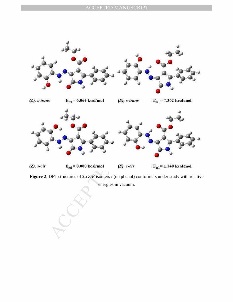
3.2 DFT structure of hydrazones
NMR analysis confirms the presence of both E and Z isomer in a reaction product and
establishes the conformation of the ester group. Both isomers are probably stabilized by
hydrogen bonding from N–H to C=O coming either from pyrrolinone (Z isomer) or ester group
(E isomer) as shown in Figure 2. Such intramolecular H-bonding was found for phenylazo-
pyrazolones in the former case [21] and for phenylazo ß–ketoesters in the latter case [21] by X-
ray diffractometry. On the other hand, there is no experimental evidence, whether the hydrogen
from phenol group can mediate the H-bond to more distant nitrogen. Consequently, the DFT
optimization of the structures of both possible conformers with respect to hydroxyl orientation
(Figure 2 for compound 2a) was carried out for each isomer of compounds 2a-2c. Relative
energies are summarized in Table 3.
Figure 2
Table 3

MANUSCRIP
T
ACCEPTED
ACCEPTED MANUSCRIPT
15
There is generally a clear trend in DFT results: irrespective to the presence and position
of the nitro substituent, E and Z isomers with the same conformation on the phenol unit are of
similar energy, explaining thus the presence of both isomers in reaction product. The absolute
values of energy prefer the Z-isomer in vacuum and the slightly more polar E isomer in a polar
environment. A second H-bond supporting s-cis conformation of phenol ring is strongly
preferred in all cases. An application of the last result as a definitive description of a final
conformation is somewhat speculative as the computations do not take into account the specific
interactions of phenol (H-bonding) with solvent or with another molecule in the solid-state. The
results on o-hydroxy-phenylazo ß-ketoesters show the preference of a s-trans conformer [22] by
X-ray diffractometry, although the model DFT calculation predicted the opposite situation.
Consequently the excitation energies were calculated on both conformations of both isomers.
Azo - enol tautomers were not studied so detailed, as they were not detected in DMSO solution
by NMR. Several orientational calculations has shown, that irrespective to substituent their
energies are about 17-19 kcal/mol higher as compared to the most stable hydrazone tautomer,
explaining thus their absence in studied solutions.
3.3 Absorption and fluorescence of hydrazones
Originally, the absorption spectra of 2a-2f were measured in NMP, in order to be directly
comparable with previously reported derivatives with no o-hydroxy substituent on the phenyl
ring [11]. While the spectra of compounds 2a and 2d without nitro substituent have one
structureless band in visible region, only 3-4 nm hypsochromically shifted as compared to
previously reported isomeric p-hydroxy derivatives [11], all four nitro derivatives has shown also
the second, considerably bathochromically shifted, absorption band (Figure 3, Table 4). This new
band was more intense and less shifted for the 5-nitro derivatives 2b and 2e, while less intense
but remarkably more shifted for the 4-nitro-derivatives 2c and 2f. We consider the new long-
wavelength band as an absorption band on O–-anion isomer, rising due to the presence of water
in hygroscopic solvent (NMP) even if it is in very low concentration. This idea was supported
both experimentally by the effect of added water on the spectra of 2b in acetone (Figure 4), and

MANUSCRIP
T
ACCEPTED
ACCEPTED MANUSCRIPT
16
by PCM TD DFT computations of the anions, which qualitatively match the experiment (545 nm
for 2b and 684 nm for 2c, compared to experimental values 545 nm and 624 nm, respectively).
Higher concentration of an anion in 5-nitro derivatives as compared to 4-nitro derivatives and its
absence in 2a and 2d was tentatively ascribed to an acidity of phenol protons, which probably
behave in analogy with nitro-phenols only (pKa = 9.89, 8.28 and 7.15 for phenol, m-NO2-phenol
and p-NO2-phenol, respectively [23]).
Absorption spectra of compounds 2a-2f were thus also measured at room temperature in
THF, in which no long-wavelength absorption band was detected (Figure 5) and molar
absorptivities could be estimated (Table 4). All hydrazones have broad absorption band with
only one maximum as expected comparing with para substituted derivatives [11] with slight
bathochromic shift about 8 nm when going from phenyl to 2-naphthyl derivative. A small
bathochromic shift of non-dissociated forms is observed when going from THF to more polar
NMP. Effect of nitro substitution is also quite small: 5-nitro substitution causes hypsochromic
(and hypochromic) shift, while 4-nitro a bathochromic (and hyperchromic) shift, both about 10
nm.
Figure 3
Table 4
Figure 4
Figure 5
PCM (THF) TD DFT computed excitation energies of various isomers / conformers of
compounds 2a-2c qualitatively confirm the observed effect of a nitro group on the absorption
spectra (Table 5). Nevertheless, quantitatively the effect of nitro substitution is slightly
overestimated. The theoretical prediction is quite clear for 2a and 2c, i.e. irrespective to isomer /
rotamer arrangement, there is only one strong band in visible area, corresponding to allowed S0-
S1 (HOMO – LUMO) transition, while S0 – S2 transition is weak, with wavelength under 400 nm
and of HOMO-1 – LUMO (2a) or HOMO – LUMO+1 (2c). The situation for 2b is rather
complicated, as original LUMO+1 orbital is considerably stabilized by 5-nitro substituent, so
both HOMO – LUMO and HOMO – LUMO+1 transitions fall into the visible range and the

MANUSCRIP
T
ACCEPTED
ACCEPTED MANUSCRIPT
17
character of the lowest excited state depends on conformation of the phenol ring. Anyway, only
one of these transitions is always strong and thus driving the absorption spectrum.
Table 5
All compounds under study show strong fluorescence in MTHF solvent glass, as shown in
Figure 6 as an example. On the other hand only 4-nitro derivatives 2c (Figure 7) and 2f show
weak fluorescence in solution (φF = 0.005 for 2c) and in solid-state. There is a considerable
effect of nitro substitution on the shape of low temperature fluorescence excitation and emission
spectra. On the other hand, all of the compounds show fluorescence in MTHF solvent glass, as
shown in Figure 6 as an example. There is a considerable effect of nitro substitution on the shape
of fluorescence excitation and emission spectra. While 0-0 vibronic transition forms an absolute
maximum in emission for all nitro derivatives, the excitation maxima correspond to 0-0
transition only in the case of 4-nitro derivatives 2c and 2f, which thus show the lowest change in
geometry when going from the ground to the fluorescent excited state. The excitation and
emission spectra show relatively low Stokes shift between both 0-0 vibronic transition, i.e. there
were observed no specific spectral features, like abnormal Stokes shift, relating to excited state
proton transfer [24]. The Stokes shift is considerably lower for nitro derivatives (e.g. 1730 cm-1
for 2a vs. 860 cm-1 for 2c), that further supports the above mentioned lowest reorganization in S1
state for 4-nitro derivatives. There are no significant differences in the spectra of 5-nitro
derivatives with respect to either unsubstituted or 4-nitro derivatives, so the fluorescence comes
very probably from the same type of S1 state, i.e. the lowest relaxed excited state of 2b and 2e is
of the same HOMO – LUMO type as in other compounds, which means that the close in energy
S2 state of HOMO – LUMO+1 type does not affect the fluorescence ability.
As compared with the previously reported derivatives [11], the spectral maxima of o-
hydroxy derivative 2a in NMP are almost the same as for p-hydroxy derivative. 2-Hydroxy
substitution brings a considerable bathochromic and bathofluoric shift (about 25 nm) for
compound 2c with respect to the 4-nitro derivative [11], i.e. the electron-donating effect of o-
hydroxyl dominates the electron-accepting effect of the nitro group. The same reason (in S1
MANUSCRIP
T
ACCEPTED
ACCEPTED MANUSCRIPT
18
state) leads to a considerable decrease of a room temperature fluorescence quantum yield of 2c
with respect to the 4-nitro derivative (φF = 0.1 in NMP [11]).
Finally, while the derivatives without an o-hydroxy substituent have shown solid-state
fluorescence in the range 554-672 nm easily visually detectable, irrespective of the character of
the p-substituent or of type of 5-aryl [11], the o-hydroxy substituent in the compounds under
study either completely quenches the fluorescence, or considerably decreases its intensity (2c).
The nature of this specific process is not very clear and will not be discussed, as there is a lack of
information on the molecular packing and consequent interactions in the solid-state.
Figure 6
Figure 7
3.4 Absorption spectra of complexes
There was found no fluorescence of the complexes under study in solution, solvent glass
and solid-state. The changes in absorption spectra induced by irradiation of diluted solutions are
under study and will be published later. The absorption spectra of all synthesized complexes 3a-
3f were measured in NMP and THF immediately after preparation at room temperature. On the
contrary to hydrazone series, no qualitative difference in spectral behaviour in both solvents was
observed. The spectra are shown on Figures 8 and 9 and the spectral data are summarized in
Table 6. The spectral curves do not show well-resolved vibronic structure but from the data it
appears that in the case of 3a, 3b, 3d and 3e the absolute maximum is 0-1 band and a long
wavelength shoulder band relates to 0-0 band, while the opposite is observed for 3c and 3f, i.e. 0-
0 band is an absolute maximum and 0-1 band corresponds to a short wavelength shoulder. There
is a moderate bathochromic (4-8 nm) shift when going from the phenyl to 2-naphthyl derivatives,
but the rise of molar absorptivity accompanying conjugation extension is considerable. Azo
complexes of o,o -́dihydroxy class show usually broad absorption bands with molar
absorptivities under 50 000 L.mol–1.cm–1, so the absorption spectrum of e.g. compound 3f with
one relatively narrow band near 600 nm, resolved vibronic structure and ε over 80 000 L.mol–

MANUSCRIP
T
ACCEPTED
ACCEPTED MANUSCRIPT
19
1.cm–1 in NMP makes this type of complexes interesting for further structure / properties
research, as e.g. DVD-R media require the values of ε over 100 000 L.mol–1.cm–1 [7].
The absorption spectra of complexes are generally bathochromically shifted with respect to
starting hydrazone ligands. The effect of the presence and position of the nitro group is
considerably different. While a 5-nitro substituent causes small hypsochromic shift (about –10
nm of 2b vs. 2a), 4-nitro substituted compounds are a bit bathochromically shifted (about +10
nm of 2c vs. 2a) in the hydrazone series (Table 4). On the other hand, the effect of nitro
substitution is much more specific in complex series (Table 6): 5-nitro substituent causes very
small bathochromic shift (about +5 nm of 3b vs. 3a), while 4-nitro substitution shifts the
corresponding vibronic bands more than 50 nm bathochromically (3c vs. 3a). The computed
excitation energies of strong allowed bands falling into the visible region of hypothetical (not
detected) azo tautomers of ligands 2a, 2b and 2c (452 nm, 454 nm and 531 nm in THF,
respectively) show the same trend as the spectra of complexes, indirectly confirming the azo
character of ligands in complexes, as derived also from NMR.
Table 6
Figure 8
Figure 9
4. Conclusion
All new keto-hydrazone dyes were present as a mixture of E and Z isomers, so the
original expectations on improving the selectivity of a reaction towards only one isomer by its
stabilization by additional H-bonding were not fulfilled. All compounds fluoresce strongly only
in solvent glass at 77 K, while fluoresce in solution or in solid state is either very weak or totally
absent. Thus the rigidity of the molecules in excited state by intramolecular H-bonding was not
increased. In summary, there was not found any experimental evidence of the secondary H-
bonding both in the ground and excited state. The effect of o-hydroxy substituent was thus only
electron-donating.

MANUSCRIP
T
ACCEPTED
ACCEPTED MANUSCRIPT
20
Using these hydrazones as tridentate O–N–O´ ligands, six new symmetrical 2:1
octahedral Co(III) complexes were prepared. Multinuclear NMR proved that the starting mixture
of hydrazone isomers was converted solely to E-azo configuration in the complexes. The
considerably different effect of 4- and 5-nitrophenol substituents on the absorption spectra of the
ligands and complexes was ascribed to azo character of an electronic structure of the ligands in
complexes. 4-Nitrophenyl substituted complexes show relatively narrow absorption bands near
600 nm and high molar absorptivities, giving thus some perspective to this type of compounds in
optical data recording. Further research of the structural modifications to improve these
properties is now in progress.
Acknowledgement
T. Aysha and R. Hrdina thank the Ministry of Education, Youth and Sports of the Czech
Republic for financial support (Project CZ.1.07/2.3.00/30.0021 “Enhancement of R&D Pools of
Excellence at the University of Pardubice”).
References:
[1] Zollinger H. Color Chemistry: syntheses, properties, and applications of organic dyes and
pigments. 2nd rev. ed. Weinheim; New York: VCH, 1991.
[2] Landge SM, Tkatchouk E, Benítez D, Lanfranchi DA, Elhabiri M, Goddard, III WA,
Aprahamian I. Isomerization Mechanism in Hydrazone-Based Rotary Switches:Lateral Shift,
Rotation, or Tautomerization? J. Am. Chem. Soc. 2011; 133(25):9812-9823.
[3] Chaur MN, Collado, Lehn J-M. Tautomeric Switching and Metal-Cation Sensing of Ligand-
Equipped 4-Hydroxy-/4-oxo-1,4-dihydroquinolines. Chem. Eur. J. 2012; 18 (23): 7269 – 7277.
[4] Su X, Robbins FR, Aprahamian I. Switching through Coordination-Coupled Proton Transfer.
Angew. Chem. Int. Ed. 2011; 50(8): 1841 –1844.
[5] Szymczyk M, Freeman HS. Design, synthesis, and characterization of new iron-complexed
azo dyes. Dyes Pigments. 2007; 72(1):8-15.
[6] Szymczyk M, El-Shafei A, Freeman HS. Metal-complexed dyes. Rev. Prog. Color. 2004;
34(1):39-57.

MANUSCRIP
T
ACCEPTED
ACCEPTED MANUSCRIPT
21
[7] Mustroph H, Stollenwerk M, Bressau V. Current Developments in Optical Data Storage with
Organic Dyes. Angew. Chem. Int. Ed. 2006; 45(13): 2016 – 2035.
[8] Li X, Wu Y, Gu D, Gan F. Spectral, thermal and optical properties of metal(II)-azo
complexes for optical recording media. Dyes Pigments. 2010; 86(2):182-189.
[9] Karipcin F, Dede B, Percin-Ozkorucuklu S, Kabalcilar E. Mn(II), Co(II) and Ni(II)
complexes of 4-(2-thiazolylazo)resorcinol: Syntheses, characterization, catalase-like activity,
thermal and electrochemical behaviour. Dyes Pigments. 2010; 84:14-18.
[10] Lyčka A, Luňák S. Jr., Aysha, T., Holuša, R., Hrdina, R. A 1H, 13C and 15N NMR
spectroscopic and GIAO DFT study of ethyl 5-oxo-2-phenyl-4-(2-phenylhydrazono)-4,5-
dihydro-1H-pyrrole-3-carboxylate. Tetrahedron Lett. 2010;51:3149-51.
[11] Aysha T, Luňák S Jr., Lyčka A, Hrdina R. Synthesis, absorption and fluorescence of
hydrazone colorants based on pyrrolinone esters. Dyes Pigments. 2011; 91(2):170-6.
[12] Cordes T, Schadendorf T, Priewisch B, Rück-Braun B, Zinth W. The Hammett relationship
and reactions in the excited electronic state: hemithioindigo Z/Ephotoisomerization. J. Phys.
Chem. A 2008; 112:581-8.
[13] Lyčka A, Rys P, Skrabal P. Al-27, N-15, C-13 and H-1 NMR spectra of the 2 : 1
aluminium(III) complexes of some azo dyes. Magn. Reson. Chem. 1998; 36(4):279-84.
[14] Lyčka A, Jirman J, Koloničný A. N-15, C-13, and H-1 NMR spectra of azo and hydrazo
compounds derived from 1,3,3-trimethyl-2-methylidene-2,3-dihydroindole (Fischer base).
Collect. Czech Chem. Comm. 1998; 63(7):1012-20.
[15] Bondarev S, Knyukshto V, Stepuro V, Stupak A and Turban A. Fluorescence and electronic
structure of the laser dye DCM in solutions and in polymethylmethacrylate. Journal of applied
spectroscopy. 2004; 71:194-201.
[16] Gaussian 09, Revision A.1, Frisch MJ, Trucks GW, Schlegel HB, Scuseria GE, Robb MA,
Cheeseman JR, Scalmani G, Barone V, Mennucci B, Petersson GA, Nakatsuji H, Caricato M, Li
X, Hratchian HP, Izmaylov AF, Bloino J, Zheng G, Sonnenberg JL, Hada M, Ehara M, Toyota
K, Fukuda R, Hasegawa J, Ishida M, Nakajima T, Honda Y, Kitao O, Nakai H, Vreven T,
Montgomery, Jr. JA, Peralta JE, Ogliaro F, Bearpark M, Heyd JJ, Brothers E, Kudin KN,

MANUSCRIP
T
ACCEPTED
ACCEPTED MANUSCRIPT
22
Staroverov VN, Kobayashi R, Normand J, Raghavachari K, Rendell A, Burant JC, Iyengar SS,
Tomasi J, Cossi M, Rega N, Millam JM, Klene M, Knox JE, Cross JB, Bakken V, Adamo C,
Jaramillo J, Gomperts R, Stratmann RE, Yazyev O, Austin AJ, Cammi R, Pomelli C, Ochterski
JW, Martin RL, Morokuma K, Zakrzewski VG, Voth GA, Salvador P, Dannenberg JJ, Dapprich
S, Daniels AD, Farkas O, Foresman JB, Ortiz JV, Cioslowski J, Fox DJ. Gaussian, Inc.,
Wallingford CT, 2009
[17] Lyčka A, Jirman J, Cee A. N-15, C-13 and H-1-NMR Spectra of the 2-1 Cobalt(III)
Complexes of Some Azo Dyes. Magn Reson Chem. 1990; 28(5):408-13.
[18] Lyčka A. Multinuclear NMR of Azo Dyestuffs. Annual report, NMR spectra. 1993; 26:247-
51.
[19] Lyčka A, Holeček J. N-15, C-13 and H-1 NMR spectra of three 2 : 1 cobalt(III) complexes
of 1-(2-carboxyphenyl)azo-2-naphthol. Dyes Pigments. 2003; 57(2):115-9.
[20] Lyčka A, Luštinec D, Holeček J, Nádvornik M, Holčapek M. Al-27 N-15 C-13 and H-1
NMR spectra and negative-ion electrospray mass spectra of the 2:1 aluminium(III) complexes of
azo dyes derived from anthranilic acid. Dyes Pigments. 2001; 50(3):203-9.
[21] Skoweranda J, Bukowska-Strzyzewska M, Strzyzewski W. Molecular structure of isopropyl
1-phenyl-4-phenylhydrazono-5-oxo-3-pyrazolecarboxylate. J. Chem. Cryst. 1994; 24: 517-520.
[22] Kopylovich MN, Mahmudov KT, Guedes da Silva MFC, Figiel PJ, Karabach YY,
Kuznetsov ML, Luzyanin KV, Pombeiro AJL. Ortho-Hydroxyphenylhydrazo-ß-Diketones:
Tautomery, Coordination Ability, and catalytic Activity of Their Copper(II) Complexes toward
Oxidation of Cyclohexane and Benzylic Alcohols. Inorg. Chem. 2011; 50: 918-931.
[23] CRC Handbook of Chemistry and Physics, 78th Edition 1997-1998, CRC Press INC., New
York.
[24] Joshi H, Kamounah FS, Gooijer C, van der Zwan G, Antonov L. Excited state
ntramolecular proton transfer in some tautomeric azo dyes and Schiff bases containing an
intramolecular hydrogen bond. J. Photochem. Photobiol .A Chem 2002; 152:183-191.

MANUSCRIP
T
ACCEPTED
ACCEPTED MANUSCRIPT
Table 1: 1H, 13C NMR data for compounds 3a, 3b and 3c
3a 3b 3c H/C No.
δ(1H) δ(13C) δ(1H) δ(13C) δ(1H) δ(13C)
1 (NH) 11.37 - 11.39 - 11.97 -
2 - 131.5 - 135.7 - 136.2
3 - 105.1 - 104.6 - 104.8
4 - 122.9 - 124.1 - 126
5 - 144.8 - 145.7 - 146.6
COO - 164.5 - 163.8 - 163.8
CH2 4.38 59.5 4.40 59.9 4.31 59.9
CH3 1.42 14.4 1.52 14.3 1.34 14.4
1// - 131 - 130.4 - 130.3
2// 7.50 127.6 7.56 128.3 7.55 128.2
3// 7.36 127.9 7.40 128 7.42 128.1
4// 7.29 127.4 7.40 128.5 7.38 128.7
1/ - 146.8 - 146.5 - 152.9
2/ - 168 - 173.8 - 166
3/ 6.56 117.3 6.71 116.5 7.37 110.8
4/ 6.84 126.9 7.87 123.2 - 145.2
5/ 6.60 113.7 - 136 7.65 110.9
6/ 8.17 114.5 9.06 110 8.30 113.8

MANUSCRIP
T
ACCEPTED
ACCEPTED MANUSCRIPT
Table 2: 1H, 13C NMR data for compounds 3d, 3e and 3f
3d 3e 3f H/C No.
δ(1H) δ(13C) δ(1H) δ(13C) δ(1H) δ(13C)
1(NH) 11.57 - 12.01 - 11.96 -
2 - 131.3 - 135.7 - 136.2
3 - 105.4 - 104.9 - 105
4 - 123 - 123.3 - 126.1
5 - 145 - 145.8 - 146.8
COO - 164.5 - 163.8 163.8
CH2 4.41 59.5 4.42 59.9 4.42 59.9
CH3 1.43 14.3 1.47 14.3 1.41 14.4
1/ - 146.8 - 146.5 - 153
2/ - 168 - 173.8 - 166
3/ 6.62 117.3 6.75 116.5 7.41 110.8
4/ 6.88 125.9 7.70 126.3 - 145.3
5/ 6.63 118.7 - 136 7.67 110.9
6/ 8.23 114.9 9.09 110 8.35 113.6
3d 2-naphthyl δ(1H): 7.51-8.03 (overlapped multiplets)
3d δ(13C): 128.5, 132.1, 132.5 (all C), 125.8, 126.1, 126.2, 126.9, 127.0, 127.3, 127.9 (all CH)
3e 2-naphthyl δ(1H): 7.54-8.10 (overlapped multiplets)
3e δ(13C): 127.9, 132.4, 132.6 (all C), 124.2, 126.5, 126.7, 127.2, 127.4, 127.5, 128.2 (all CH)
3f 2-naphthyl δ(1H): 7.57-8.10 (overlapped multiplets)
3f δ(13C): 127.9, 132.5, 132.7 (all C), 126.2, 126.7, 126.9, 127.4, 127.6, 127.9, 128.3 (all CH)

MANUSCRIP
T
ACCEPTED
ACCEPTED MANUSCRIPT
Table 3: Relative energies of various conformers of 2a-2c in vacuum and in THF (according to
Figure 2).
Compound
Configuration
on C=N bond
(Isomer)
Conformation
on C–N bond
(Rotamer)
Relative ground state
energy in vacuum 1
(kcal/mol)
Relative ground
state energy in
THF 2 (kcal/mol)
2a
E
Z
s-cis
s-trans
s-cis
s-trans
1.340
7.362
0.000
6.064
0.000
3.758
0.860
3.932
2b
E
Z
s-cis
s-trans
s-cis
s-trans
0.276
7.910
0.000
5.897
0.000
4.358
1.362
4.805
2c
E
Z
s-cis
s-trans
s-cis
s-trans
0.635
5.615
0.000
4.950
0.000
3.170
1.405
3.874 1 B3LYP/6-311G(d,p), the most stable conformation of 2b is 0.737 kcal/mol more stable than 2c
2 PCM (THF) B3LYP/6-311+G(2d,p), the most stable conformation of 2b is 0.378 kcal/mol
more stable than 2c

MANUSCRIP
T
ACCEPTED
ACCEPTED MANUSCRIPT
Table 4: Absorption and fluorescence maxima of compounds 2a-2f at room (NMP and THF) and
low (MTHF) temperature (all compounds are the mixtures of E and Z isomers).
Excitation
(77 K)
Emission
(77 K) Compound
Absorption
maxima in
NMP [nm]
Absorption
maxima in
THF [nm]
Absorption
coefficient in
THF
[L.mol–1.cm–1] 0-1 0-0 0-0 0-1
2a 460 454 33550 469 493 539 567
2b 468, 546 445 27800 463 484 509 539
2c 477, 624 465 41350 471 505 528 555
2d 466 463 39200 477 499 541 570
2e 470, 555 453 37350 464 493 514 547
2f 484, 639 473 45000 484 512 535 573

MANUSCRIP
T
ACCEPTED
ACCEPTED MANUSCRIPT
Table 5: PCM (THF) TD DFT excitation energies (λ00) and oscillator strengths (fosc) of 2a-2c for
various isomers and conformers as shown on Figure 2 for 2a.
HOMO LUMO
HOMO LUMO+1
resp.
HOMO-1 LUMO Compound
Configuration
on C=N bond
(Isomer)
Conformation
on C–N bond
(Rotamer)
λ00 fosc λ00 fosc
2a
E
Z
s-cis
s-trans
s-cis
s-trans
460
449
471
459
0.759
0.786
0.819
0.849
364
349
378
363
0.066
0.010
0.012
0.028
2b
E
Z
s-cis
s-trans
s-cis
s-trans
440
482
456
484
0.849
0.060
0.731
0.084
470
433
465
447
0.051
0.775
0.160
0.762
2c
E
Z
s-cis
s-trans
s-cis
s-trans
490
485
502
498
0.964
1.004
0.970
0.995
395
388
396
390
0.034
0.059
0.025
0.056

MANUSCRIP
T
ACCEPTED
ACCEPTED MANUSCRIPT
Table 6: Absorption maxima and molar absorption coefficient of complexes 3a-3f in THF
Dye
Absorption
maxima in
NMP [nm]
Absorption
coefficient in
NMP
[L.mol–1.cm–1]
Absorption
maxima in
THF [nm]
Absorption
coefficient in
THF
[L.mol–1.cm–1]
3a 519 41600 514 34000
3b 525 60000 519, 546 (s) 38700
3c 600 63200 566 (s), 591 50300
3d 529 53000 518 39000
3e 532 77000 526, 553 (s) 52400
3f 604 84000 574 (s), 597 62500
Underlined values represent the absolute absorption maxima

MANUSCRIP
T
ACCEPTED
ACCEPTED MANUSCRIPT
R
NO2
NO2
5'
4'
H3a
3b
3c
R
R
NNO
ON NO
OCo
NH
COOEtNH
EtOOCNa+
12
34
5
1'2'
3'
4'5'
6'
1''2''
3''
4''
6'
5'4'
3'
2' 1'
5
43
21
Na+
NNO
ON NO
OCo
NH
COOEtNH
EtOOC
R
R
3f
3e
3d H
4'
5'
NO2
NO2
R
Figure 1: C, H numbering used in NMR assignment of synthesized cobalt complexes.
MANUSCRIP
T
ACCEPTED
ACCEPTED MANUSCRIPT
Figure 2: DFT structures of 2a Z/E isomers / (on phenol) conformers under study with relative
energies in vacuum.

MANUSCRIP
T
ACCEPTED
ACCEPTED MANUSCRIPT
0
0.1
0.2
0.3
0.4
350 450 550 650 750
A
nm
2a 2b 2c 2d 2e 2f
Figure 3: Absorption spectra of 2a-2f in NMP.

MANUSCRIP
T
ACCEPTED
ACCEPTED MANUSCRIPT
0
0.2
0.4
0.6
0.8
1
350 450 550 650
Rel
ativ
e in
teni
sity
nm
Pure aceton 0.4 % water 1 % water
2 % water 3 % water 4 % water
Figure 4: Absorption spectra of 2b in acetone in presence of different concentration of water.

MANUSCRIP
T
ACCEPTED
ACCEPTED MANUSCRIPT
0
0.1
0.2
0.3
0.4
0.5
0.6
300 350 400 450 500 550 600 650 700 750 800
A
nm
2a 2b 2c 2d 2e 2f
Figure 5: Absorption spectra of hydrazones 2a-2f in THF.

MANUSCRIP
T
ACCEPTED
ACCEPTED MANUSCRIPT
Figure 6: Absorption (room temperature) and fluorescence excitation and emission spectra in
MTHF at 77 K for 2a, 2b, 2d, 2e

MANUSCRIP
T
ACCEPTED
ACCEPTED MANUSCRIPT
0
0.2
0.4
0.6
0.8
1
300 400 500 600 700
Rel
ativ
e in
teni
sity
nm
Absorbance at 300 K Excitation at 77 K Fluorescence at 77 K
Fluorescence at 300K Excitation at 300K Fluorescence in solid state
Figure 7: Absorption at room temperature (MTHF), excitation and emission spectra at low
temperature (MTHF) of compound 2c together with its solid-state emission at room temperature.

MANUSCRIP
T
ACCEPTED
ACCEPTED MANUSCRIPT
0
0.2
0.4
0.6
0.8
1
350 450 550 650 750
A
nm
3a 3b 3c 3d 3e 3f
Figure 8: Absorption spectra of complexes 3a-3f in NMP.

MANUSCRIP
T
ACCEPTED
ACCEPTED MANUSCRIPT
0
0.1
0.2
0.3
0.4
0.5
0.6
0.7
300 400 500 600 700 800
A
nm
3a 3b 3c 3d 3e 3f
Figure 9: Absorption spectra of complexes 3a-3f in THF.

MANUSCRIP
T
ACCEPTED
ACCEPTED MANUSCRIPT
• Six new o-hydroxy substituted hydrazones based on pyrrolinone esters were prepared as a mixture of E and Z isomers.
• Their absorption and fluorescence spectra were studied experimentally and theoretically by TD DFT.
• Six new 2:1 symmetrical Co(III) complexes were prepared using these hydrazones as tridentate ligands.
• The starting mixture of hydrazone isomers was converted solely to E-azo configuration in
complexes by NMR spectroscopy. • Different effect of nitro group position in ligands and complexes on the absorption
spectra was discussed.


![APPLICATION OF AZO-CALIX[4]PYRROLE DYES IN DYEING OF ...shodhganga.inflibnet.ac.in/bitstream/10603/4671/11/11_chapter 6.pdfThe dyeing performance of azo-calix[4]pyrrole dyes on fibres](https://img.pdfslide.us/doc/110x75/5e4d1c555de91f77ce488c17/application-of-azo-calix4pyrrole-dyes-in-dyeing-of-6pdf-the-dyeing-performance.jpg)