Embed Size (px)
Citation preview
Chapter – 1 Introduction
1
1. INTRODUCTION
Pharmaceutical chemistry is focused on quality aspects of medicines and aims to
assure their fitness. There is a lot of collaboration between chemists and biologists while
searching for a lead on a new drug or doing research on a preclinical drug candidate.
While looking into the drug safety profile, collaboration with toxicologists and
pharmacologists is required. Drug discovery research is a highly creative and stimulating
work environment where people are driven to succeed by personal and scientific
objectives.
Heterocyclic compounds are organic compounds that contain a ring structure
containing atom in addition to carbon, such as sulphur, oxygen, nitrogen as part of the
ring. Many of the heterocyclic ring systems are of fundamental importance to living
systems. For instance, nucleic acids are derivatives of the purine and pyrimidine ring
systems. Many heterocycles have important pharmaceutical properties and have a wide
range of applications: they are predominant among the types of compounds used as
pharmaceuticals, as agro chemicals and as veterinary products. They are also used as
optical brightening agents, as antioxidants, as copolymers, solvents, photographic
sensitizers, corrosion inhibitors and additives with a variety of other functions. Many
dye stuffs and pigments have heterocyclic structures.
Heterocyclic compounds are of fundamental importance to biological process
and are wide spread of natural products. Heterocyclic compounds are finding an
increasing use as intermediates in organic synthesis. They offer a high degree of
structural diversity and have proven to be broadly and economically useful as

Chapter – 1 Introduction
2
therapeutic agents. Some heterocyclic amines (HCAs) found in cooked meat are known
carcinogens.
1.1. 1,8-NAPHTHYRIDINE
Naphthyridine is the name commonly given to the fused-ring system resulting from the
fusion of two pyridine rings through two adjacent carbon atoms, each ring thus
containing only one nitrogen atom. This name was suggested by Reissert,who, in 1893,
made the first representative of the series, since 1,8-naphthyridine was considered to
be the naphthalene analogue of pyridine. Sixpossible naphthyridines are:
N
N
N
NN
N
N N
N N
N
N
1,5-napthyridine 1,6-napthyridine 1,7-napthyridine
1,8-napthyridine 2,6-napthyridine
2,7-napthyridine
(1) (2) (3)
(4)(5)
(6)
1,8-Naphthyridine derivatives are reported to possess a wide spectrum of
biological activities such as diuretic1,antimalarial2, anti-inflammatory3,
antitumor4,antihypertensive5 and antibacterial activites.6,7 1,8-Naphthyridine
derivatives have attracted considerable attention because the 1,8-naphthyridine
skeleton is present in many compounds that have been isolated from natural
substances, with various biological activities. Nalidixic acid (7), for example, possesses
strong antibacterial activity and used mainly for the treatment of urinary tract infections

Chapter – 1 Introduction
3
with gram negative pathogens. In addition, Gemifloxacin (8) is antimicrobial and
antibacterial. It is known that (E)- and (Z)-O-(diethylamino)ethyl oximes of 1,8-
naphthyridine series (9) are potential drugs for local anaesthesia8, and 1-(2-
fluorobenzyl)-3-(2-tolyl)-1,8-naphthyridin-2(1H)-one (10) is used for the treatment of
memory disorders, in particular, Alzheimer’s disease9. 2-Amino-N-hydroxy-1,8-
naphthyridine-3-carboxamidine(11) possesses herbicidal properties and used for the
selective control of weeds in barley, wheat, maize, sorghum and rice crops10.
(8)
N N
O
COOH
C2H5
CH3
(7)
N NH
NOCH2CH2Et2
R1
R2
(9)
N N
O
COOHF
NH3CON
NH2
(10)
(11)
N N
O
COOH
F
F
N
NH2
N
F
N
NH
O
COOH
N
H5C2
Chapter – 1 Introduction
4
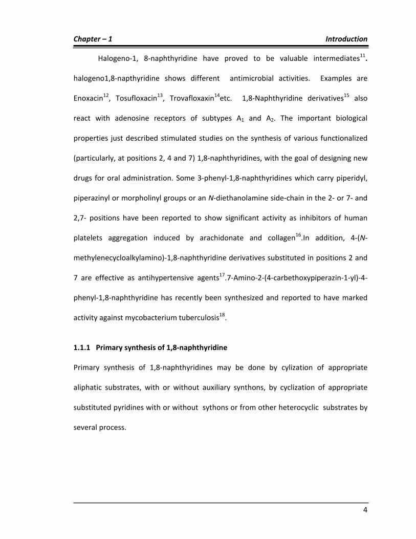
Halogeno-1, 8-naphthyridine have proved to be valuable intermediates11.
halogeno1,8-napthyridine shows different antimicrobial activities. Examples are
Enoxacin12, Tosufloxacin13, Trovafloxaxin14etc. 1,8-Naphthyridine derivatives15 also
react with adenosine receptors of subtypes A1 and A2. The important biological
properties just described stimulated studies on the synthesis of various functionalized
(particularly, at positions 2, 4 and 7) 1,8-naphthyridines, with the goal of designing new
drugs for oral administration. Some 3-phenyl-1,8-naphthyridines which carry piperidyl,
piperazinyl or morpholinyl groups or an N-diethanolamine side-chain in the 2- or 7- and
2,7- positions have been reported to show significant activity as inhibitors of human
platelets aggregation induced by arachidonate and collagen16.In addition, 4-(N-
methylenecycloalkylamino)-1,8-naphthyridine derivatives substituted in positions 2 and
7 are effective as antihypertensive agents17.7-Amino-2-(4-carbethoxypiperazin-1-yl)-4-
phenyl-1,8-naphthyridine has recently been synthesized and reported to have marked
activity against mycobacterium tuberculosis18.
1.1.1 Primary synthesis of 1,8-naphthyridine
Primary synthesis of 1,8-naphthyridines may be done by cylization of appropriate
aliphatic substrates, with or without auxiliary synthons, by cyclization of appropriate
substituted pyridines with or without sythons or from other heterocyclic substrates by
several process.

Chapter – 1 Introduction
5
From a single pyridine substrate
5-Methyl-3-(m-tolylethynyl)-2-pyridinamine (12) on treatment with sodium
ethoxide in the presence of ethanol on cyclisation produced 4-ethoxy-6-methyl-2-m-
tolyl-1,8-naphthyridine(13).19 3-(2-Ethoxycarbonylvinyl)-2-pyridinamine (14) on
treatment with sodium ethoxide underwent cyclisation to form 1,8-naphthyridin-2(1H)-
one (7) in ethanol.(15)20
N NH2
CH3
O
CH3
C2H5ONa
N N
CH3
OC2H5
CH3
(12) (13)
N NH2
CH=CHCO2C2H5
N NH
O
C2H5ONa
(14) (15)
2,5-Bis (3-aminopropyl)pyridine (16) on treatment with NaNH2 in toluene after
cyclisation gave 2-(3-aminopropyl)-1,2,3,4-tetrahydro-1,8-naphthyridine(17)21. 6-(2-
acetyl-1-methylethylidene) amino-2-pyridinamine (18) gives 5,7-dimethyl-1,8-
naphthyridin-2-amine (19) after cyclisation on treatment with NaNH2 in presence of
phosphoric acid at 100oc 22.

Chapter – 1 Introduction
6
N (CH2)3NH2NH
N (CH2)3NH2
H2N(H2C)3
NaNH2
(16) (17)
N N=CHCH2COCH3 N N CH3
NaNH2
H2N H2N
CH3
(18) (19)
From pyridine substrate and synthon(S)
3-(2-Nitrovinyl)-2-pyridinamine (20) underwent condensation with benzaldehyde
(PhCHO) in xylene and gave 3-nitro-2-phenyl-1,8-naphthyridine(21)23.2-
Pyridinamine(22) on treatment with benzaldehyde and subsequently with acetic acid
produced 2-phenyl-1,8-naphthyridine-4-carboxylic acid (23) in ethanol24.
N NH2N N C6H5
NO2CH=CHNO2
C6H5CHO
(20) (21)
N NH2 N N NH2
C6H5CHO
CH3COOH
COOH
(22) (23)

Chapter – 1 Introduction
7
2-Amino-3-pyridinecarbonitrile (24) on treatment with m-chlorobenzyl cyanide,
KOH/H2O on microwave irradiation produced 3-m-chlorophenyl-1,8 naphthyridin-2-
amine (25). The same substrate (23) with ethyl cyano acetate in ethanol and trace
amounts of piper dine gave 2-oxo-1,2-dihydro-1,8-naphthyridine-3-carbonitrile (26)25
NH2
CHO m-chlorobenzyl cyanide
N N NH2
Cl
N NH
O
CNCNCH2COOC2H5
(24) (25)
(26)
From other heterocyclic substrates
Pyrido[2,3-d]pyrimidines as substrates
Pyrido[2,3-d] pyrimidine (27) with malononitrile in methanol, gave 2-amino-1,8-
naphthyridine-3-carbonitrile (28) and with ethyl cyano acetate gave ethyl 2-amino-1,8-
naphthyridine-3-carboxylate.(29)26
N
N
N
N N
CN
NH2
N N NH2
COOEt
(27)
(28)
(29)
CN-CH2-CN
CNCOOC2H5

Chapter – 1 Introduction
8
Pyrrolo[2,3-b]pyridines as substrates
Pyrrolo[2,3-b]pyridines(30) und`erwent pyrolysis in chloroform to give a separable
mixture of 1,8-naphthyridine (R=H) and 3-chloro-1,8-naphthyridine .(R= Cl) (31)
CHCl3
N N
R
N NH
(30) (31)
1.1.2 Reactions of 1,8-napthyridines
C-Amination
1,8-Naphthyridine (32) on treatment with NaNH2 in NH3 and then KMnO4 gave
1,8-naphthyridin-2-amine (34) the intermediate dihydro adduct (33) was clearly
involved27.
N N N NH
N N
NaNH2
NH3
[O]
NH2 NH2
(32) (33) (34)
Halogenation
1,8-Naphthyridine hydro bromide (35) gave a separable mixture of 3-bromo-1,8-
naphthyridine (36) and 3,6-dibrimo-1,8-naphthyridine (37) on treatment with Br2 in
PhNO2 at 175oc yield was 30% each. 28
N N
Br
N N
Br
N N
BrBr
+Br2, PhNO2
(35) (36) (37)

Chapter – 1 Introduction
9
N-oxidation
1,8-napthyridine undergo oxidation in the presence of H202 and benzoic acid to form
its 1-oxide derivative.(38)29
N N
H2O2
N N+
o(38)
1,8-Naphthyridine-1-oxide (39) with o-methoxyphenyllithium gave 2-o-
methoxyphenyl-1,8-naphthyridine (40) in diethylether30.
N N
O
N NH
C6H4OMe
LiC6H4OMe
(39) (40)
1.2 Synthetic routes of 1,4-napthyridine nucleus
Four routes involved in synthesis of 1,8-napthyridine ring
a) Niementowski synthesis b)Friedlander synthesis c)Reductive cyclization d)
Intramolecular nucleophilic cyclization
Niementowski synthesis
In the first route Hitherto31 described was an extension of the Niementowski synthesis
to the preparation of 1,8-napthyridine-2,4-diols. 7-phenyl - 1,8-napthyridine-2,4-diols
(42) by the condensation of ethyl-2-amino-6-phenyl nicotinate (41) with simple esters
in the presence of sodium.

Chapter – 1 Introduction
10
N NH2
COOEt
+ R-CH2COOC2H5
N N OH
OH
R
Ph(41)
(42)
Friedlander synthesis
In the second route Friedlander was described 32For the synthesis of [1,8] naphthyridine
derivatives containing phosphorus. The first 2, 3 alkene substituted [1,8] naphthyridine
(44) bearing a phosphorus moiety have been synthesized by the Fried lander
annulations of 2-aminonicotinaldehyde (43) with diphenyl phosphoryl cyclopentanones.
NH2
CHO
+O
R
PX
Ph
Ph N N CH3P PhPh
X(44)(43)
The Friedlander condensation of 2- amino nicotinaldehyde with active methylene
compounds in the presence of catalyst Lithium chloride under the two non-
conventional methods like microwave Irradiation and by grinding in a mortar afforded
the corresponding 1,8 – Naphthyridines (45)33 Both these methodologies are attractive
as they are relatively nontoxic, economical and highly effective .The results shows that
microwave procedure as slightly superior to the solid-state methods in terms of
reduced time period and better yields.
N N CH3
O
Ph+ H3C
O
CH2
O
PhLiCl
MW
(45)
N NH2
CHO
Chapter – 1 Introduction
11
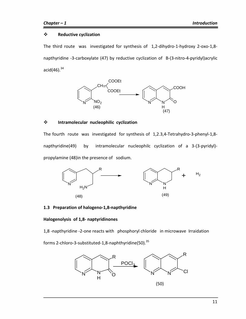
Reductive cyclization
The third route was investigated for synthesis of 1,2-dihydro-1-hydroxy 2-oxo-1,8-
napthyridine -3-carboxylate (47) by reductive cyclization of B-(3-nitro-4-pyridyl)acrylic
acid(46).34
N NO2
CHCOOEt
COOEt
N NH
COOH
O(46)
(47)
Intramolecular nucleophilic cyclization
The fourth route was investigated for synthesis of 1,2.3,4-Tetrahydro-3-phenyl-1,8-
napthyridine(49) by intramolecular nucleophilc cyclization of a 3-(3-pyridyl)-
propylamine (48)in the presence of sodium.
NNH2
R
N NH
R
+ H2
(49)(48)
1.3 Preparation of halogeno-1,8-napthyridine
Halogenolysis of 1,8- naptyridinones
1,8 -napthyridine -2-one reacts with phosphoryl chloride in microwave Irraidation
forms 2-chloro-3-substituted-1,8-naphthyridine(50).35
N
R
NH
O
POCl3
N N Cl
R
(50)

Chapter – 1 Introduction
12
1.3.1 Reactions of halogeno-1,8-napthyridine
Aminolysis
4-Bromo-1,8-naphthyridine (51) gave 1,8-naphthyridin-4-amine (52) on treatment with
NH3 in phenol at 170oc.
N N
Br
N N
NH2
NH3/C6H5OH
(51) (52)
2-chloro-3-nitro-1,8-naphthyridine (53) gave 3-nitro-1,8-naphthyridin-2-amine
(54) on treatment with NH3 in ethanol at 110oc.36,37
N N
No2
Cl
R
N N
R
NH2
No2
NH3/C2H5OH
(53) (54)
Hydrolysis
In alkaline hydrolysis 2-chloro-1,8-napthyridine reacts with NaOH to from 1,8-
napthyridine-2-(1H)-one38(55). In acidic condition 4-bromo-1,8-napthyridine with Hcl to
from 1,8-napthyridine-2(1H)-one.39

Chapter – 1 Introduction
13
N N ClN N
HO
N N
Br
N NH
O
NaoH
HCl
(55)
(55)
1.4 Preparation of amino-1,8-naphthyridine
From Acylamino-1,8-naphthyridine
2-Acetamido-7-ethoxy-4-phenyl-1,8-naphthyridine (56) reflux with sodium ethoxide in
ethanol gave 7-ethoxy-4-phenyl-1,8-naphthyridin-2-amine (57).40
N N
C6H5
NHCOCH3C2H5O N N
C6H5
NH2C2H5O
C2H5ONa/C2H5OH
(56) (57)
Conversion of azido to amino naphthyridine
4-Azido-7-methyl-2-phenyl-1,8-naphthyridine (58) gave 7-methyl-2-phenyl-1,8-
naphthyridin-4-amine (59) with Pd/C in methanol, H2O at 20oC41
N N
N3
C6H5H3C N N
NH2
C6H5H3C
Pd/C, CH3OH
(58) (59)

Chapter – 1 Introduction
14
1. 4.1 Reactions of amino-1,8-naphthyridine
2-p-(Benzylideneamino) phenyl-1,8-naphthyridine(60) underwent oxidation to give 2-p-
benzamidophenyl-1,8-naphthyridine (61) on treatment with m-ClC6H4CO3H.42,43
N N
N=CHC6H5
N N
NHC6H5
m-ClC6H4CO3H
(60) (61)
1,8-Naphthyridine-2,7-diamine (62) with nitrosobenzene gave 2,7-bis phenyl
azo-1,8-naphthyridine (63) on treatment with alcoholic KOH in H2O.44
N N N=NPhPhN=NN N NH2H2N
C6H5NO
C2H5OH,KOH
(62) (63)
5,7-Dimethyl-1,8-naphthyridin-2-amine (64, R= NH2) gave 2-isocyanato-5,7-
dimethyl-1,8-naphthyridine (65, R = NCO) as a minor product (7%) via an imidazo[1,2-
a][1,8]naphthyridine intermediate. The same substrate (64, R = NH2) with butyl
isocyanate afforded 2-N-butylureido-5,7-dimethyl-1,8-naphthyridine (66, R =
NHCONHBu) on reflux with toluene.45
N N
CH3
CH3R N N
CH3
CH3R
R-NH2 (64) (65) = R-NCO
(66)= NHCONHBu

Chapter – 1 Introduction
15
1.5 Preparation of 1,8-naphthyridine carboxylic acid
By hydrolysis of 1,8-naphthyridinecarbonitriles
2-Phenyl-1,8-naphthyridine-3-carbonitrile (67, R= CN) gave the corresponding 3-
carboxylic acid (68, R= CO2H) on reflux with alcoholic KOH in water for 14 h at 700c , the
yield was found to be 76%46.
N N Ph
CN
N N Ph
COOH
KOH/C2H5OH
(67) (68)
By oxidation of 1,8-naphthyridinecarbaldehydes
1,8-Naphthyridine-2,7-dicarbaldehyde (69, R= H) gave 1,8-naphthyridine-2,7-
dicarboxylic acid (70, R =H) on refluxing with 80% HNO3. 47
N N CHO
R
OHC N N CO2H
R
HO2C
HNO3(R=H)orNaClO2(R=OC8H17)
(69) (70)
1.5.1 Reaction of 1,8-naphthyridine carboxylic acid
Decarboxylation
7-Amino-4-oxo-1,4-dihydro-1,8-naphthyridine-2-carboxylic acid (65) gave 2-phenyl-1,8-
naphthyridine (72) on treating with Cu bronze at 260oc.

Chapter – 1 Introduction
16
N N PhNH
N
O
HO
O
NH2
Cu Bronze
(71) (72)
1.6 . Pyrazolones and Isoxazolinone:
1.6.1 . Chemistry of Pyrazolones
The oxo derivatives of pyrazolines, known as pyrazolones, are best classified as follows:
5-pyrazolone, also called 2-pyrazolin-5-one73 (73); 4-pyrazolone, also called 2-pyrazolin-
4-one (74); and 3-pyrazolone, also called 3-pyrazolin-5-one (75). Within each class of
pyrazolones many tautomeric forms are possible; for simplicity only one form is shown.
NH
NO
NH
N
O
NH
NO H
(73) (74) (75)
NN
R
OH
NNH
R
O
NN
R
OH
NN
R
O
Enol Keto Enol Keto
(76) (77)
Substitution at N1 decreases the possible number of tautomers: for 3-
pyrazolones, two tautomeric forms are possible, (76) and (77), which in non polar

Chapter – 1 Introduction
17
solvents are both present in about the same ratio. 5-pyrazolones exhibit similar
behavior.
In 4-pyrazolones, the enol form predominates, although the keto form has also
been observed. The tautomeric character of the pyrazolones is also illustrated by the
mixture of products isolated aftercertain reactions. Thus alkylation normally takes place
at C4, but on occasion it is accompanied by alkylation on O and N. Similar problems can
arise during acylation and carbamoylation reactions, which also favor C4. Pyrazolones
react with aldehydes and ketones at C4 to form a carbon–carbon double bond, eg (78).
Coupling takes place when pyrazolones react with diazonium salts to produce azo
compounds, eg (79).
Compounds of type (79) are widely used in the dye industry. The Mannich
reaction also takes place at C4, as does halogenation and nitration. The important
analgesic aminoantipyrine (80)on photolysis in methanol undergoes ring fission to yield
(81).48
NN
R
O
R
R
NN
R
O
NN
R
(78) (79)

Chapter – 1 Introduction
18
NNO
NH2 CH3
CH3
CH3 OH
hv NH
O
OH
O
NHCH3
CH3
(80) (81)
1.6.2 Synthesis of pyrazolone derivatives
The pyrazolone-3-carboxylic acid (83) has been isolated by reaction of oxazolone
(82) with hydrazonyl chloride.49
O
NAr2
H
OAr1
+ NH N
COOR
Ar3
Cl
(C4H9)4N+Br
-
Na2CO3
NN
Ar1
O
CH3
COORNH
Ar2
O
(82) (83)
NN
CH2
C(CH3)3(H3C)3C
+ NC
CN
Na H NN
C(CH3)3
ONC
NH2 C(CH3)3
(84)
The preferred synthetic method for the title compounds utilizes the reaction of
hydrazines with bifunctional compounds, such as β-diketones and esters, and β-keto
acetylenic compounds. In an alternative procedure, diazo compounds replace

Chapter – 1 Introduction
19
hydrazines and ring formation takes place via 1,3-dipolar cycloaddition. Pyrazoles and
pyrazolones are widely used in the pharmaceutical industry to alleviate inflammation,
fever, pain, and infections. To a lesser extent, they are also used as insecticides and
herbicides. Pyrazolones linked to azo compounds are extensively used in the dye
industry; some pyrazolines display insecticidal activity.50
Pyrazolones with a free NH group are easily nitrosated and give rise to
nitrosamines, which cause tumors in the liver of test animals. The analgesics antipyrine
(85) and aminopyrine (86), if admixed with nitrites, are mutagenic when tested in vitro;
however, when tested in the absence of nitrites, negative results are obtained51
NN CH3O
CH3
.
NN
CH3
ON
CH3
CH3
CH3
(85) (86)
Pyrazolone-type drugs, such as phenylbutazone and sulfinpyrazone, are
metabolized in the liver by micro-somal enzymes, forming glucuronide metabolites that
are easily excreted because of enhanced water solubility.
The pyrazolone derivatives, which include dipyrone (87), antipyrine (85),
aminopyrine (86) and propyphenazone, are widely used analgesics. Dipyrone, the most
widely used pyrazolone, has been the most studied. Dipyrone is an inhibitor of cyclo-
oxygenase but, unlike aspirin, its effect is rapidly reversible. The inhibition of
prostaglandin biosynthesis contributes to the analgesic activity of the pyrazolone

Chapter – 1 Introduction
20
derivatives. Unlike the Non-steroidal anti-inflammatory agents (NSAIDs) generally, the
pyrazolone derivatives antipyrine, aminopyrine and propyphenazone are minimally
bound to plasma proteins. The pyrazolones undergo extensive biotransformation,
aminopyrine and dipyrone being converted to active metabolites. The most frequently
reported side effects of the pyrazolone derivatives are skin rashes. Gastrointestinal side
effects are rare.
NN
CH3
ON
CH3
CH3
NaO3S
OH2
(87)
1.6.3 Important pyrazolone and isoxazoline derivative in pharmaceuticals:
Some of the pharmaceuticals that incorporate the pyrazole nucleus are given below.
Their main uses are as antipyretic, anti-inflammatory, and analgesic agents. To a lesser
extent, they have shown efficacy as antibacterial/antimicrobial, antipsychotic, anti-
emetic, and diuretic agents. The analgesic aminopyrine, the antipyretic dipyrone, and
the anti-inflammatory phenylbutazone (88), though once widely prescribed, are rarely
used in the 1990s on account of their tendency to cause agranulocytosis. Pyrazolone
derivatives as like benzimidazole derivatives have been found to possess some
interesting pharmacological activities. eg. antipyrine, ampyrone, edaravone, etc.

Chapter – 1 Introduction
21
(88)
NNO
O(CH2)3CH3
S
S
NH2O
O
NH2
OO NNO
O(H2C)3
CH3
O
O
Cl
(89)butaglycon (90)feclobuzo,
NNO
OCH3
O
NNO
OS
O
(91) kebuzone (92) sulfinpyrazone
NNO
O(CH2)3CH3

Chapter – 1 Introduction
22
NNNH2
O
CH3
Cl
Cl
NNO CH3
CH3
(93) muzolimin (94) phenazobz
NN
CH3
O NN
CH3
O
NH2
CH3
Edaravone (95) Amprone (96)
Pyrazolones react with diazonium salts, an important process in the dye industry.
The majority of dyes are having pyrazolone nucleus with an azo linkage attached at C4,
eg, (97) and (98).
NNO
R2
R1
CH3
R''
NN
NaO3S
NNO
R1NN
NN
NN
O
R1
Cl
Cl
(98) (97)

Chapter – 1 Introduction
23
The survey of the pertinent literature reveals that isoxazolines have been found
to possess a wide range of biological activity such as anti bacterial,52 anti HIV53, anti-
inflammatory54,anticancer55, etc. Some Isoxazole derivatives (99) have been reported as
anti-tubercular, anti bacterial and antifungal agets.56
O
N O
R
(99)
Azopyrazoles (100) and azoisoxazoles (101) are possessing good antifungal
activity57. Similarly, 2-alkyl isoxazolidine derivatives have been as antifungal agents.58
NN
N
NH
R1
NN
N
O
R1
(100) (101)
1.7 PYRAZOLE
1.7.1 CHEMISTRY OF PYRAZOLE RING SYSTEM:
Pyrazole is a hetero aromatic compound, consists of doubly unsaturated five membered
ring containing two adjacent nitrogen atoms (102). Pyrazole is a 1,2-diazole and as its
name implies, it may be considered as an azapyrrole. Many drugs and medicinal (e.g.
antipyrine) contain a pyrazole ring and many dye stuffs are derived from it.

Chapter – 1 Introduction
24
(102)
NNH
Knorr introduced the name pyrazole for these compounds to denote that the
nucleus was derived from pyrrole by replacement of a carbon by nitrogen and he
synthesized many members of the class and systemically investigated their properties.
Chemical properties:
1. Pyrazoles are aromatic compounds and the ring system is more stable than
pyrrole and less reactive.
2. Electrophilic substitution reactions occur readily, the attack being at the 4th
position.
3. Bromination can be effected in organic solvents using hypobromite to yield 4-
bromo derivative.
4. Direct iodination occur using iodine and sodium acetate.
5. Nitration and sulphonation reactions are less facile.
The pyrazoles are class of heterocyclic compounds and the pyrazole skeleton
constitutes an important central template for a wide variety of biologically active
compounds. The pyrazole nucleus has been reported to possess a wide spectrum of
biological activities such as anti-inflammatory59 antibacterial60, analgesic62, antifungal61,
anti viral62, Hypoglycemic63, anticancer 64 and antitubercular.65

Chapter – 1 Introduction
25
Pyrazole related marketed drugs:
H3C
NN CF3
SO O
H2N
Celecoxib
H3C
O
SO O
H3C
O
Rofecoxib
NO
SO O
H2NCH3
Valdecoxib
NH3C
SO O
H3C
N
Cl
Etoricoxib
O
NN CF2H
SO O
H2N
Deracoxib
F
N
OH
NH
O
S
N
S
MeloxicamO O
1.8 Isoxazole
Isoxazole (103) is a five membered heterocyclic compound having two hetero atoms:
oxygen at position 1 and nitrogen at position 2. Claisen first reported an isoxazole (103)
for a product from the reaction of 1,3 diketone with hydroxylamine hydrochloride66
Isoxazole nucleus has been reported to possess a wide spectrum of biological activities
such as anti-inflammatory67, analgesic68, antituberculosis69,Hypoglycemic70,antcancer.71

Chapter – 1 Introduction
26
NO
(103) 1.8.1 Chemistry of Isoxazole
Isoxazoles can be prepared by various methods; some of them are described as under.
Crawley L. S. and Fan Shawe W. J.72
R CH CH
O
R1 + NH2OH.HCl N
O
R
R1
have prepared substituted isoxazole (104) from α,β-
unsaturated carbonyl compounds, hydroxyl amine hydrochloride and KOH in methanol.
(104)
Kalirajan et al.73
have synthesized and check antimicrobial screening against
various gram positive and gram negative bacteria and anti fungal activity against various
fungal stains compared with standard drug (Ampicillin and Ketoconazole) using solvent
control.
NO
H3CO
74
(105)

Chapter – 1 Introduction
27
Isoxazole related marketed drugs:
ONNHS
O
ONH2
Sulfisoxazole
NO
CH2 S
O
O
NH2
Zonisamide
1.9 Pyrimidine-2-one
Pyridones, which belongs to an important group of heterocyclic compounds, havebeen
extensively explored for their applications in the field of medicinal chemistry.
Pyridones, with a carbonyl group at position 2 (106) have been subject of
extensive studyin recent past.
NH
O
(106)
Pyridone derivatives have been found to possess variety of therapeutic
activitiesLike antiviral75, antimicrobial76, anticancer77, antiHIV78 Pesticidal79Herbicidal.80
Pyridones are derivatives of pyrimidines with carbonyl group at 2-position (I).Some 2-
pyridones are physiologically as well as pharmacologically important which areas under,
eg. Ciclopirox (107), and Amrinone (108).

Chapter – 1 Introduction
28
NH
O
NH2
N
N O
CH3
OH
(107) (108) 1.9.1 Chemistry of pyrimidine-2-one
K. Folkers and S. A. Harris81 have synthesized 3-cyano-2-pyridone (109) bythe
condensation of cyano acetamide with 1,3-diketone or 3-ketoester.
CH3COCH2COCH2OC2H5 + NH2COCH2CN
NH
O
CN
CH3
CH2OC2H5
(109)
Pednekar82 have synthesized fused 2-pyridone derivatives (110), (111) and (112) as
usefulheterocyclic moieties as they possess broad spectrum of biological activities such
asantiviral, CNS depressant, bactericidal and ulcer inhibitor.
NH
NNH
O
CH3NC
N
NOO
CH3
CH3
NC
NH
O
CH3
NC
Ph
PhO
(110) (111) (112)

Chapter – 1 Introduction
29
Upadhyay et al.83 have documented cyanopyridone derivatives, which showed
antifungal and antileishmanial activities. E. Amer84 prepared 3-cyano-2-
pyridonederivatives (113) displaying high antimicrobial activity. Abou El-Fotooh andco-
workers.85 have demonstrated pyridones (114) as anticancer agent.
NNH
O
CN
Ar
NH
Ar
O
NC
N
NH
O
CN
CH3
O
(113) (114)
1.10. Introduction of analgesic activity
1.10.1 pain
The struggle to relieve pain began with the origin of humanity. Ancient writings , both
serious and fanciful ,dealt with secret remedies, religious rituals and other methods of
pain relief, slowly paving way to the present modern era of synthetic analgesics.
Tainter has divided the history of analgsic drugs into 4 major eras, namely:
a) The period of discovery and use of naturally occurring plant drugs.
b) The isolation of pure plant principles from the natural sources and their
identification with analgesic action.
c) The development of organic chemistry and the first synthetic analgesics.

Chapter – 1 Introduction
30
d) The development of modern pharmacologic techniques, making it possible to
undertake a systematic testing of new analgesics.
Pain is sensation transmitted from sensory nerves through the spinal cord to the
sensory area of the cerebrum where the sensations are perceived.
It is defined by the International Association for the Study of Pain (IASP) as “an
unpleasant sensory and emotional experience associated with actual or potential tissue
damage, or described in terms of such damage”. Pain is referred to as the sensation of
an injured tissue:
Whenever there is a damage to a tissue
During onset of a disease
Due to inflammation
Pain may be external (due to any injury; e.g. cut) or may be internal (due to
onset of a disease; e.g. headache, muscular pain, etc.).
1.10.2 Types of pain
Pain can be classified as acute or chronic. The distinction between acute and chronic
pain is not based on its duration of sensation, but rather the nature of the pain itself. In
general, physicians are more comfortable treating acute pain, which has as its source
soft tissue damage, infection and/or inflammation. It can be modulated and removed by
treating its cause and through combined strategies using analgesics to treat the pain
and antibiotics to treat the infection. In general, while it is uncomfortable to experience,

Chapter – 1 Introduction
31
it is easy to treat; is distinguished by having a specific cause and purpose, and generally
produces no persistent psychological reaction.86
Acute pain is also called as fast pain, perception is very rapid, usually within 0.1
second after a stimulus is applied, because the nerve impulses conduct along
medium-diameter, myelinated axons called A-delta fibres. This type of pain also
known as fast, sharp, or pricking pain. The pain felt from a needle puncture or
knife cut to the skin. Acute pain is not felt in deeper tissues of the body87
Chronic pain is also called as slow pain, by contrast, begins a second or more
after stimuli is applied and then gradually increases in intensity over a period of
several seconds or minutes. Impulses for slow pain conduct along small-
diameter, unmyelinated C fibers. This type of pain, which may be excruciating, is
also referred to as burning, aching, or throbbing pain. Slow pain can occur both
in the and in deeper tissue or internal organs.87
Cutaneous pain is caused by injury to the skin or superficial tissues. Cutaneous
nociceptors terminate just below the skin, and due to the high concentration of
nerve endings, produce a well-defined, localized pain of short duration.
Examples of injuries that produce cutaneous pain include paper cuts, minor cuts,
minor (first degree) burns and lacerations. 87
Somatic Pain is due tostimulation of nociceptors in the integument and
supporting structures, namely, striated muscles, joints, periosteum, bones, and
nerve trunks by direct extension through fascial planes and lymphatic spread77

Chapter – 1 Introduction
32
Visceral pain, Thecause for visceral pain could be spasm of the smooth muscles
of hollow viscus, distention of the capsule of solid organs, inflammation,
chemical irritation, traction or twisting of mesentery, ischemia and necrosis, or
tumour encroachment of the pelvis and presacral regions .88
Referred pain the pain is felt in or just deep to the skin that overlies the
stimulated organ. This pain may also felt in surface area far from the stimulated
organ. In general, the visceral organ involved and the area to which the pain is
referred are served by the same segment of the spinal cord.87
Phantom limb pain is the sensation of pain from a limb that has been lost or
from which a person no longer receives physical signals like itching, pressure,
tingling. Cerebral cortex interprets impulses arising in the proximal portions of
sensory neurons that previously carried impulses from the limb as coming from
the nonexistent limb.87
Neuropathic pain, or "neuralgia", can occur as a result of injury or disease to the
nerve tissue itself. This can disrupt the ability of the sensory nerves to transmit
correct information to the thalamus, and hence the brain interprets painful
stimuli even though there is no obvious or known physiologic cause for the pain.
Neuropathic pain is, as stated above, the disease of pain. It is not the sole
definition for chronic pain, but does meet its criteria.89

Chapter – 1 Introduction
33
1.10.3 Physiology of nociception (Commonly physiology of pain)
This section, except in the paragraph on pain in consciousness, for historical
reasons uses pain to refer to nociception. Where both a historical pain term and
a modern nociception term are common, a bracketed pain term is included. E.g.
Nociceptors (Pain receptors).
"Nociception is the term introduced almost 100 years ago by the great
physiologist Sherrington in 1906 to make clear the distinction between detection
of a noxious event or a potentially harmful event and the psychological and
other responses to it90.
Nociception is the system which carries information about noxious stimulus,
usually associated with tissue damage to the spinal cord and brain.
1.10.4 Transmission of nociception (pain) signals
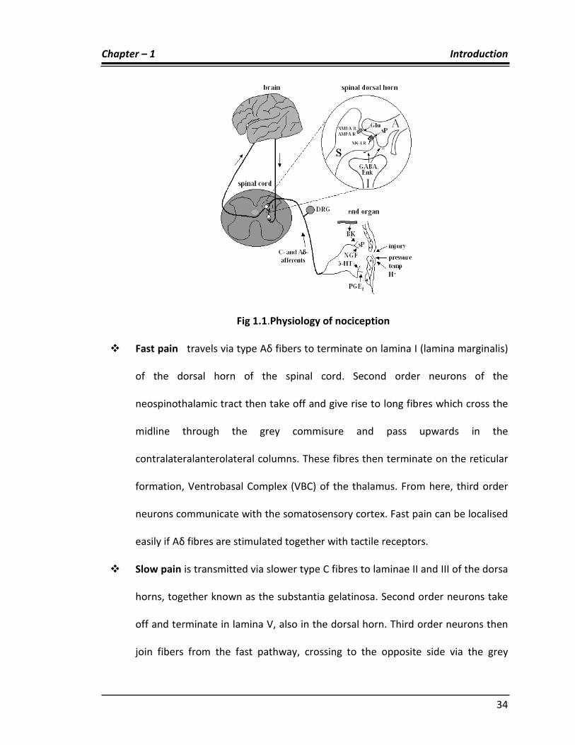
There are 2 pathways for transmission of nociception in the central nervous system.
These are the neospinothalamic tract (for fast pain) and the paleospinothalamic tract
(for slow pain) as shown in Fig 1.1
Chapter – 1 Introduction
34
Fig 1.1.Physiology of nociception
Fast pain travels via type Aδ fibers to terminate on lamina I (lamina marginalis)
of the dorsal horn of the spinal cord. Second order neurons of the
neospinothalamic tract then take off and give rise to long fibres which cross the
midline through the grey commisure and pass upwards in the
contralateralanterolateral columns. These fibres then terminate on the reticular
formation, Ventrobasal Complex (VBC) of the thalamus. From here, third order
neurons communicate with the somatosensory cortex. Fast pain can be localised
easily if Aδ fibres are stimulated together with tactile receptors.
Slow pain is transmitted via slower type C fibres to laminae II and III of the dorsa
horns, together known as the substantia gelatinosa. Second order neurons take
off and terminate in lamina V, also in the dorsal horn. Third order neurons then
join fibers from the fast pathway, crossing to the opposite side via the grey

Chapter – 1 Introduction
35
commisure, and traveling upwards through the anterolateral pathway. These
neurons terminate widely in the brain stem, with one tenth of fibres stopping in
the thalamus, and the rest stopping in the medulla, pons and tectum of midbrain
mesencephalon, periaqueductal grey. Slow pain is poorly localized.91
1.11 Introduction of anti-inflammatory activity
1.11.1 Inflammation
Inflammation92 is defined as the local response of living mammalian tissues to the injury
due to an agent.
Agents causing inflammation are
Physical agents like heat, cold, radiation, mechanical trauma.
Chemical agents like organic and inorganic poisons.
Infective agents like bacteria, viruses and their toxins.
Immunological agents like cell-mediated and antigen-antibody reactionsearliest
response to tissue inury. These alterations include: haemodynamic changes and
changes in vascular permeability.
Cellular events:
The cellular phase of inflammation consists of 2 processes:
Exudation of leukocytes:
The escape of leukocytes from the lumen of microvasculature to the intestinal tissue is
the most important featurevof inflammatory response. In acute inflammation,

Chapter – 1 Introduction
36
polymorphonuclear neutrophils (PMNs) comprise the first line of body defence ,
followed later by monocytes and macrophages.
1.11.2 Signs of inflammation
The Roman writer Celsus in 1st century A.D. named the famius 4 cardinal signs of
inflammations as:
• Rubor (Redness)
• Tumour (Swelling)
• Calor (Heat)
• Dolor (Pain)
To these, fifth sign functio laesa (loss of function) was later added by Virchow.
As evident from the above discussion Inflammation is one of the causes of Pain,
while Pain is one of the signs of Inflammation. Thus it can be inferred that Pain and
Inflammation often occurs simultaneously.
1.11.3 Types of inflammation92
Depending upon the defense capacity of the host and duration of response,
inflammation can be classified as acute and chronic.
Acute Inflammation is of short duration and representsthe early body reaction
and is usually followed by repair.
Chronic Inflammation is of longer duration and occurs either after the causative
agent of acute inflammation persists for a long time, or the stimulus is such that
it induces chronic inflammation from the beginning

Chapter – 1 Introduction
37
Chronic inflammation is subdivided into 2 types
• Non-Specific, which the irritant substance produces a non-specific chronic
inflammatory reaction with formation of granulation tissue and healing by
fibrosis e.g. osteomyelitis, chronic ulcer.
• Specific, when the injurious agent causes a characteristic histologic tissue
response e.g. tuberculosis, leprosy, syphilis.
1.11.4 Characteristics
Inflammation has two main components: cellular and exudative.
The cellular component involves the movement of white blood cells from blood
vessels into the inflamed tissue. The white blood cells, or leukocytes, take on an
important role in inflammation; they extravasate (filter out) from the capillaries into
tissue, and act as phagocytes, picking up bacteria and cellular debris. They may also aid
by walling off an infection and preventing its spread.
The exudative component involves the movement of fluid, usually containing
many important proteins such as fibrin and immunoglobulins (antibodies). Blood vessels
are dilated upstream of an infection (causing redness and heat) and constricted
downstream while capillary permeability to the affected tissue is increased, resulting in
a net loss of blood plasma into the tissue, giving rise to edema or swelling. The swelling
distends the tissues, compresses nerve endings, and thus causes pain.93
If inflammation of the affected site persists, released cytokines IL-1 and TNF will
activate endothelial cells to up regulate receptors VCAM-1, ICAM-1, E-selectin, and L-
selectin for various immune cells. Receptor up regulation increases extravasation of

Chapter – 1 Introduction
38
Neutrophils, monocytes, activated T-helper and T-cytotoxic, and memory T and B cells
to the infected site.93
Neutrophils are characteristic of inflammation in the early stages. They are the
first cells to appear in an infected area, and any section of recently inflamed (within a
couple of days or so) tissue viewed under a microscope will appear packed with them.
They are easily identified by their multilobed nuclei and granular cytoplasm and perform
many important functions, including phagocytosis and the release of extracellular
chemical messengers. Neutrophils only live for a couple days in these interstitial
areas, so if the inflammation persists for a longer duration then they are gradually
replaced by longer lived monocytes.94
1.11.5 Leukocytes and Cytokines
Various leukocytes are involved in the initiation and maintenance of inflammation.
Generally speaking, acute inflammation is mediated by granulocytes or
polymorphonuclear leukocytes, while chronic inflammation is mediated by mononuclear
cells such as monocytes and macrophages.
These cells can be further stimulated to maintain inflammation through the
action of an adaptive cascade involving lymphocytes: T cells, B cells, and antibodies as
shown in Fig 1.2. These inflammatory cells are
• Mast cells, which release histamine and prostaglandin in response to activation
of stretch receptors. This is especially important in cases of trauma.

Chapter – 1 Introduction
39
• Macrophages which release TNF-α, IL-1 in response to activation of toll-like
receptors.
Figure- 1.2 Mechanism action of cox-2 receptor
1.11.6 Remedies of pain and inflammation
An Analgesic may be defined as a drug bringing about insensibility to pain without loss
of consciousness. In other words, agents that decrease pain or which relieves pain are
called as Analgesics or Analgetics meaning Pain-killers. Although ‘Analgetic’ is
grammatically correct, common use has made the term ‘Analgesic’ to ‘Analgetic’ for the
description of pain killing drugs. Pain relieving agents are also called as antinociceptives.
The effect of pain-killing is known as Analgesia. The effect is brought about by increasing
the threshold of pain which is felt when an internal or external stimulus is given.
Threshold of Pain = Lowest perceptible degree Intensity of Pain
From the above equation it is evident that as the Threshold of Pain is increased,
the Intensity of Pain is decreased.
Analgesic drugs can be classified into two groups:

Chapter – 1 Introduction
40
1.11.7 Opioids: Narcotic analgesics
Opioids analgesics are also known as narcotic analgesics. They are used to relieve severe
pain and are often prescribed to patients recovering from operations and serious
injuries. Thet are basically centrally-acting analgesics i.e they exert their action by acting
on the Central Nervous System. Opioid analgesics are classified into two categories viz.
Morphine drivatives and Phenanthrene derivatives. Example of opioid analgesics include
morphine, papaverine.
Figure: 1.3 – Inflammatory Pathways95
1.11.8 Non-opioids or NSAIDs:
Non-opioids, also called non-narcotic drugs or Non-steroidal anti-inflammatory drugs,
usually abbreviated to NSAIDs, are drugs with analgesic, antipyretic and anti-
inflammatory effects - they reduce pain, fever and inflammation. The term "non-
steroidal" is used to distinguish these drugs from steroids, which (among a broad range

Chapter – 1 Introduction
41
of other effects) have a similar eicosanoid-depressing, anti-inflammatory action. They
are basically peripherally-acting analgesics and exert their action by interfering with the
formation of the eicosanoids from arachidonic acid. Many non-opioid analgesics can be
bought over-the-counter at chemists and supermarkets. Clinical use of NSAIDs is
associated with significant toxicity particularly in the gastrointestinal tract and kidney.
Various approaches such as formulation & co-administration (of agents to protect the
stomach), chemical manipulation and synthesis of new safer anti-inflammatory drugs
are reported to overcome the toxicity of NSAIDs.. The most prominent members of this
group of drugs are aspirin, ibuprofen, and naproxen partly because they are available
over-the-counter in many areas. Paracetamol (acetaminophen) has negligible anti-
inflammatory activity, and is strictly speaking not an NSAID. NSAIDs within a group will
tend to have similar characteristics and tolerability. There is little difference in clinical
efficacy between the NSAIDs when used at equivalent doses. Rather, differences
between compounds tended to be with regards to dosing regimens (related to the
compound's elimination half-life), route of administration, and tolerability profile.
NSAID drugs sometimes referred to as non-narcotic analgesic or aspirin-like drugs.96 The
NSAIDs are classified as follows:
Salicylates e.g.Aspirin
Arylalkanoic acids e.g.Diclofenac
2-Arylpropionic acids (profens) e.g.Ibuprofen
N-Arylanthranilic acids (fenamic acids) e.g.Mefenamic acid
Pyrazolidine derivatives e.g.Phenylbutazone
Chapter – 1 Introduction
42
Oxicams e.g.Piroxicam
COX-2 Inhibitors e.g.Celecoxib (FDA alert [1])
Sulphonanilides e.g.Nimesulide
Others e.g.Licofelone
1.11.9 Pharmacological actions of NSAIDs
All the NSAIDs have actions very similar to those of aspirin. The three main therapeutic
effects are An anti-inflammatory effect: modification of the inflammatory action, an
analgesic effect: reduction of pain and an antipyretic effect: loweing of body
temperature when this is raised in disease. (i.e.fever)
In addition, all the NSAIDs share, to a gretae or lesser degree, the same type of
mechanisam-based side effects. these include:
• gastric irriation,which may range from simple discomfort to ulcer formation
• An effect on renal blood flow in the compromised kidney
• A tendency to prolong bleeding through inhibition of platelet function.
Controversially, it is argued that they may also all-but especially COX-2 selective drugs-
increase like likelihood of thromotic events such as myocardial infarction by inhibiting
prostaglandin (PG I2) synthesis.
A number of aryl and heteroaryl substituted compounds such as Diclofenac97,
lumiracoxib98 Etodolac 97 have been characterized asnon-steroidal anti-inflammatory
drugs (NSAIDS).

Chapter – 1 Introduction
43
Important Marketed Non-Steroidal Anti-Inflammatory Drugs:
CH2COOH
NHClCl
CH3
H3CO CH2COOH
SOCH3
H3CO
NCH3
CH2COOH
O
Cl
Diclofenac Indomethacin sulindac
PROFEN DERIVATIVES
CH3
COOH
F
CH3COOH
CH3
CH3
Ibuprofen Flubiprofen
O
CH3
COOH
H3CO
CH3
COOH
Ketoprofen Naproxen
OXICAMS
NS
OO
CH3
OH
O
NH NS
NS
OO
CH3
OH
O
NH N

Chapter – 1 Introduction
44
Selective COX-2-inhibitors
NN
CF3
H2NO2S
CH3
O O
H3CO2S
Celecoxib Rofecoxib
1.12 Introduction of antibacterial activity
A drug which kills or inhibits the growth of microbes is known as antimicrobial
agent.Invitro tests are used as screening procedure for new agents to test the
susceptibility if individual isolates from infections to determine which of the available
drugs might be useful therapeutically. Due to development of sulfonamides and
penicillin’s invitro measurement of susceptibility of microbes to chemotherapeutic
agents have been used.A drug is considered to be bacteriostatic or fungistatic when
they inhibit the growth of bacteria or fungi respectively and bactericidal or fungicidal
due to its ability to kill bacteria or fungi.
Important factors for antimicrobial activity are size of the inoculums, metabolic
state of microbes,PH,temperature and duration of interaction, concentration of
inhibitor and presence of Interference substances. The development of resistance
among various pathogenic microbes towards antibiotics has increased the impetus for
investigating new antimicrobial agents.when compound was found to have positive

Chapter – 1 Introduction
45
index, a new series of related compounds are synthesized in the hope that one of them
would be more effective than the existing one.
1.12.1 Anti bacterial activity:
The emergence of resistance to the major classes of antibacterial agent is recognized as
a significant medical crisis and serious health concern. Particularly, the emergence of
multi drug-resistance strains of Gram-positive bacterial pathogens is a problem of ever
increasing significance. As the limited number of antimicrobial classes and the common
occurrence of resistance within and between classes, the search for antibacterial agents
with novel mechanism of actions is always remains an important and challenging task.
The control of microorganism is critical for the prevention and treatment of
disease. Microorganisms also grow on and within other organism, and microbial
colonization can lead to disease, disability, and death. Thus the control or destruction of
microorganisms residing within the bodies of humans and other animals is great
importance.
Modern medicine is dependant on chemotherapeutic agents, chemical agents
that are used to treat disease. Chemotherapeutic agents destroy pathogenic
microorganisms or inhibit their growth at concentrations low enough to avoid
undesirable damage to the host. Most of these agents are antibiotics, microbial
products or their derivatives that can kill susceptible microorganisms or inhibit their
growth. Drugs such as the sulfonamides are sometimes called antibiotics although they
are synthetic chemotherapeutic agents, not microbially synthesized.

Chapter – 1 Introduction
46
Antibiotics are chemical substances excreted by some microorganism which
inhibit the growth and development of other microbes. Some of these drugs that were
obtained naturally were put to chemical modifications in attempts to enhance beneficial
effects while minimizing the toxic effects. The resultant modified product is termed as
semi synthetic antibiotics. Most antibiotic currently used are semi synthetic. The
chemist has synthesized many drugs that have got the antibacterial property and less
toxicity. These drugs are called synthetic antibiotic drugs. Naturally occurring antibiotic,
their semisynthetic derivatives and synthetic antibiotics have got the same target. i.e.,
antimicrobial action. Hence all these drugs were put together to be called antimicrobial
agents.
1.12.2. Drug resistance:
The emergence of drug resistance bacteria is posing a major problem in antimicrobial
therapy. The frequency varies with the organism and the antibiotic used. At first, there
is an emergence of a small number of drug resistant bacteria which sooner multiplies
selectively in the presence of the drug at the cost of sensitive bacteria.
1.12.3. Types of drug resistance:
Drug resistance is of two types, primary and acquired.
1. Primary resistance: some bacteria possess an innate property of resistance to
certain drug, e.g. resistance of E.coli to penicillin.
2. Acquired resistance: it results either from mutation or gene transfer.

Chapter – 1 Introduction
47
1.12.4. Recent targets for finding antibacterial agents
Beta-Ketoacyl-acyl carrier protein (KAS) synthase III encoded by the fabH gene is
thought to catalyze the first elongation reaction of type II fatty acid synthesis in bacteria
and plant plastids. Beta-ketoacyl-acyl carrier protein synthase (KAS) I is important
enzyme system for the construction of the unsaturated fatty acid carbon skeletons
characterizing E. coli membrane lipids. Recent research reported that Type II fatty acid
synthesis (FAS II) pathway is an attractive targete for their efficacy against infections
caused by multi-resistant Gram-positive bacteria and Gram-negative bacteria99. Among
the related FAS II enzymes, beta ketoacyl-acyl carrier protein synthase (KAS) is an
essential target for novel antibacterial drug design100-101.
The enzyme bacterial peptide deformylase (PDF) is another novel target for
novel antibacterial agents. The metalloproteases enzyme, Bacterial peptide deformylase
(PDF) deformylates the N-formyl methionine of newly synthesized polypeptides through
Fe2+-mediated catalytic reaction. PDF is essential in prokaryotes and this enzyme is
absent in mammalian cells and provides a unique target for antimicrobial chemotherapy
102-105. Thus, it may be another target for new chemotherapeutic agents.
Lipopolysaccharides constitute the outer leaflet of the outer membrane of Gram-
negative bacteria and are therefore essential for cell growth and viability. The
glycosyltransferase (GT) enzyme, heptosyltransferase WaaC involved in the synthesis of
the inner core region of lipopolysaccharides. It catalyzes the addition of the first l-
glycero-d-manno-heptose molecule to one molecule of 3-deoxy-d-manno-oct-2-ulosonic
acid (Kdo) residue of the Kdo2-lipid A molecule. These heptose is an essential

Chapter – 1 Introduction
48
component of the Lipopolysaccharides core domain; its absence results in a truncated
lipopolysaccharide associated with the deep-rough phenotype causing a greater
susceptibility to antibiotic. Thus, WaaC represents a promising target in antibacterial
drug design.106
1.12.5 Anti fungal activity:
The object of antifungal drug discovery has become a subject of greater challenge due
to increasing incidences of fungal drug resistance. This appears due largely to the
extensive use of antifungal agents to treat fungal infections. In the past decade, number
of patients diagnosed with fungal infections have increased drastically, whereas,
relatively very few clinically useful drugs were discovered. The azole derivatives such as
such as clotrimazole, fluconazole, itraconazole, ketoconazole, etc. have been widely
used to treat a verity of fungal infections. These azole derivatives inhibit the fungal
enzyme 14-alpha demethylase which is essential for the ergosterol synthesis pathway
leads to the depletion of this steroidal compound in the cell membrane and
accumulation of toxic intermediate sterols, leads increased membrane permeability and
inhibition of fungal growth107-109. But broad usage of these drugs led to development of
acquired resistance especially among Candida albicans. Thus, searching not only
improved version of existing drug but also for new drug targets has become an urgent
need110.recent reports showed that 2-glutamine, D-fructose-6-phosphate
aminotransferase known as a new target for antifungals, it catalyzes a complex reaction
involving ammonia transfer from L-glutamine to fructose-6-phosphate, followed by
isomerisation of the formed fructosamine-6-phosphate to glucosamine-6-phosphate.111