Embed Size (px)
Citation preview
Optical Materials 34 (2012) 1535–1542
Contents lists available at SciVerse ScienceDirect
Optical Materials
journal homepage: www.elsevier .com/locate /optmat
Synthesis and luminescent properties of blue sextuple-hydrogen-bondself-assembly molecular duplexes bearing 4-phenoxy-1,8-naphthalimide moieties
Jingjing Liu a, Yanhu Li b, Yi Wang a, Huiqin Sun c, Zhiyun Lu a,⇑, Hongbin Wu b, Junbiao Peng b,⇑, Yan Huang a,⇑a College of Chemistry, Sichuan University, Chengdu 610064, PR Chinab Institute of Polymer Optoelectronic Materials and Devices, South China University of Technology, Guangzhou 510640, PR Chinac Analytical and Testing Center, Sichuan University, Chengdu 610064, PR China
a r t i c l e i n f o
Article history:Received 27 December 2011Accepted 18 March 2012Available online 29 April 2012
Keywords:NaphthalimideH-bond self-assemblyElectroluminescence
0925-3467/$ - see front matter � 2012 Elsevier B.V. Ahttp://dx.doi.org/10.1016/j.optmat.2012.03.022
⇑ Corresponding authors. Fax: +86 28 85410059 (Z87110606 (J. Peng).
E-mail addresses: [email protected] (Z. Lu), [email protected] (Y. Huang).
a b s t r a c t
Two novel blue light-emitting sextuple hydrogen-bonding self-assembly molecular duplexes bearing 4-phenoxy-1,8-naphthalimide fluorophores, namely PhNIHB and 2TPhNIHB, have been synthesized andcharacterized. Compared with their small molecular counterparts PhNI and 2TPhNI, the objective com-pounds exhibit 13–22 nm blue-shifted fluorescent emission, and much higher photoluminescence quan-tum yields (0.34 vs 0.18 for PhNIHB; 0.42 vs 0.27 for 2TPhNIHB) in solid state; and their thermal andmorphological stability have been improved as well. Employing 2TPhNIHB or 2TPhNI as emitter, non-doped solution-processed light-emitting diodes with structure of ITO/PEDOT: PSS (40 nm)/PVK(40 nm)/blue emitter (70–80 nm)/CsF (1.5 nm)/Al (120 nm) have been fabricated. The 2TPhNI-baseddevice gives yellow emission [CIE (0.38, 0.49)] with poor maximum luminous efficiency (LEmax) of0.13 cd/A and external quantum efficiency (EQEmax) of 0.06%. The 2TPhNIHB-based device, however,gives blue-green emission [CIE (0.25, 0.34)], with much higher efficiency relative to 2TPhNI-based one(LEmax of 0.37 cd/A and EQEmax of 0.35%). The effective isolation of the naphthalimide fluorescent coresas well as the suppressed formation of exciplex at the PVK/emitter interface by these oligoamide motifsare suggested to be responsible for the improved EL performance.
� 2012 Elsevier B.V. All rights reserved.
1. Introduction
Owing to their potential applications in flat panel displays aswell as light sources, organic light-emitting diodes (OLEDs) haveattracted enormous attention all over the world in the past twodecades [1–4]. Among devices emitting RGB primary colors, theblue ones compromising both high efficiency and good chromatic-ity are regarded to be more essential, because deeper blue devicewould result in lower power consumption in the case of OLEDs[5]. However, compared with well-developed green OLEDs (lumi-nous efficiency > 26 lm/W at 20 mA/cm2) [6], the performance ofdeep blue ones is much inferior (maximum luminous effi-ciency < 6 lm/W) [7], consequently, the development of highly effi-cient pure blue materials should be of great significance [8].Moreover, to suppress the unfavorable concentration quenchingof light-emitting fluorophores, a doping technique is generallyadopted for fabrication of OLEDs [7,9–11]. This procedure, how-ever, may result in relatively poor device durability due to thephase separation between guest and host under prolonged electric
ll rights reserved.
. Lu, Y. Huang), fax: +86 20
[email protected] (J. Peng),
stress as well as high working temperatures [12,13]. Therefore, in-tense research efforts have been focused on the exploitation of no-vel host-emitting blue electroluminescent (EL) materials [14–21].This is really a challenging task since the candidates should possessboth intrinsic wide bandgap to realize pure blue emission [21], andweak intermolecular interaction between the fluorophores to guar-antee good color purity as well as high photoluminescence (PL)quantum yield (QY) in condensed state [22]. In addition, theyshould be able to form high quality amorphous films with goodmorphological stability [23], and should show excellent thermo-stability [8].
To develop non-doped blue emitters, bulky substituents[14–16,21], strongly twisted multichromophoric molecular struc-tures [2,17,18] or highly branched dendrons [19,20] are generallyincorporated with the deep-blue fluorescent cores. Recently, wedemonstrated that the introduction of multiple H-bonds self-assembly motif into the fluorophore is an effective and successfulway for constructing novel OLEDs emitters with high efficiency,and the objective self-assembly dimer bearing two sextuple oligoa-mide H-bonding strands exhibits dramatically improved EL perfor-mance compared with its small-molecular counterpart [24].Furthermore, in our recent attempts to explore novel deep bluefluorophores, we find that 4-phenoxy-substituted 1,8-naphthali-mide derivatives could give efficient pure blue PL emission in
1536 J. Liu et al. / Optical Materials 34 (2012) 1535–1542
solution [25]. Nevertheless, in solid state, most of them suffer fromsevere molecular aggregation arising probably from the stackingbetween planar naphthalimide moieties, hence render red-shiftedPL emission with dropped PL QY. Additionally, these moleculesshow high propensity for crystallization, thereby the relative poorfilm morphology would restrict their application as non-dopedOLED emitters. To solve these problems, herein, we report theincorporation of multiple H-bonds self-assembly motif with thedeep blue 4-phenoxy-naphthalimide fluorophore. Both the twoobjective molecular duplexes exhibit blue PL emission with im-proved chromaticity, PL QY, thermal stability and film morphologyin condensed state relative to their small molecular counterparts.Moreover, they could form uniform and oxygen-stable pinhole-freeamorphous thin films by facile solution-process procedure.
2. Experimental section
2.1. General information and materials
All the chemicals commercially available were used directlywithout further purification unless otherwise stated. All the sol-vents were of analytical grade and freshly distilled prior to use.Anhydrous N,N-dimethylformamide and dichloromethane were re-fluxed with calcium hydride and diphosphorous pentoxide respec-tively, followed by fresh distillation before use. 1H NMR, two-dimensional NMR (NOESY) and 13C NMR spectra were recordedon a Bruker AVANCE-400 or AVANCE-600 spectrometer in CDCl3
using TMS as internal standard. High resolution MS spectra weremeasured with a Q-TOF Premier ESI mass spectrometer (Micro-mass, Manchester, UK). Thermogravimetric analysis (TGA) wasperformed on a TGA Q500 instrument under nitrogen atmosphereat a heating rate of 10 �C/min. UV–Vis absorption spectra of the10�5 mol/L CHCl3 solution and solid thin film samples were mea-sured on a SHIMADZU UV-2100 spectrophotometer. PL spectrawere recorded on a HITACHI F-7000 fluorescence spectrophotom-eter at 298 K, while thin film samples were spin-coated from cor-responding chloroform solution with concentration of 25 mg/mLat a speed of 2000 rpm on quartz substrates. PL efficiencies ofthe objective compounds were determined in 10�5 mol/L CHCl3
solution using 10�5 mol/L quinine sulfate in 0.05 mol/L H2SO4
(u = 0.55) [26] as standard under excitation of 365 nm, while abso-lute PL quantum yields and Commission International de l’Eclai-rage (CIE) coordinates of solid powder and neat films weredetermined on a Horiba Jobin Yvon-Edison fluoromax-4 fluores-cence spectrometer equipped with an integrating sphere and digi-tal photometer. Cyclic voltammetry (CV) measurement was carriedout on a PARSTAT 2273 electrochemical workstation at room tem-perature in anhydrous acetonitrile solution with tetrabutylammo-nium perchlorate (0.1 mol/L) as the supporting electrolyte at ascanning rate of 100 mV/s. The CV system was constructed usinga platinum plate, a Ag/AgNO3 (0.1 mol/L in acetonitrile) electrodeand a platinum wire as the working electrode, quasi-referenceelectrode and counter electrode, respectively, and each measure-ment was calibrated with a ferrocene/ferrocenium (Fc/Fc+) redoxcouple as internal standard. Scanning electron microscope (SEM)images of thin films were obtained on a JEOL JSM-5900 LV instru-ment with an accelerating voltage of 20 kV. X-ray diffraction (XRD)data of thin films were generated by using the Philips DX-100sealed-tube X-ray generator (Cu target; I = 0.2 nm) with power of40 kV and 35 mA. The OLED structure employed in this study is:ITO/PEDOT: PSS (40 nm)/PVK (40 nm)/emitter (70–80 nm)/CsF(1.5 nm)/Al (120 nm), where poly(3,4-ethylenedioxythiophene):poly(styrenesulfonic acid) (PEDOT: PSS) (Batron-P 4083) acts asanode buffer material, PVK [poly(N-vinylcarbazole)] acts as holetransporting material, and the active area of OLED is 0.19 cm2. Prior
to use, the indium-tin oxide (ITO) (with a sheet resistance of 20 X/h and a thickness of 120 nm) precoated glass substrate wascleaned in ultrasonic baths of detergent solution, deionized water,acetone, and 2-propanol in sequence. After degreasing, the ITOsubstrate was oxidized and cleaned in a UV-ozone chamber underoxygen plasma (Instrument: Plasma-Preen II-l862, by introducingoxygen into the chamber with a rate of 4–5 mL/s under 2–3 � 10�3 Pa) for 15 min. Thin films of PVK and emissive layers wereprepared from solutions with concentration of 20 mg/mL in chloro-benzene or p-xylene respectively by spin-coating at 1500 rpm in aglove box (VAC Co.) with N2 circulation (with <1 ppm oxygen andwater). The thickness of polymer layer was determined by measur-ing the thickness of the reference film prepared under very similarconditions with profilometry (Tenco Alfa-Step 500). The depositionof electron-injecting CsF (1.5 nm) layer followed with an Al cap-ping layer (120 nm) were realized through a shadow mask in a vac-uum chamber of �3 � 10�4 Pa, and the layer thickness wasmonitored by a crystal thickness monitor (Sycon). The current den-sity–voltage–luminance characteristics of the OLEDs were re-corded on a calibrated silicon photodiode driven by Keithley 236source. External quantum efficiencies were measured in an inte-grating sphere.
2.2. Synthesis
The detailed synthetic routes to the target compounds (abbrevi-ated as PhNIHB and 2TPhNIHB) as well as the reference com-pounds (abbreviated as PhNI and 2TPhNI) are outlined inScheme 1. Intermediates 1a-1b [24], 1c-1d [25], II-V [24] and smallmolecular counterparts PhNI and 2TPhNI [25] were preparedaccording to procedures we reported previously.
2.2.1. Synthesis of N-[2-(3-hexanamido-5-nitrobenzoyloxy) ethyl]-4-phenoxy-1,8-naphthalimide (1e-1)
A solution of 1b (2.8 g, 10 mmol) and 4-dimethylaminopyridine(DMAP) (2.44 g, 20 mmol) in 200 mL dry CH2Cl2 was cooled to 0–5 �C, then 1-ethyl-3-[3-(dimethylamino)propyl]carbodiimidehydrochloride (EDCI) (3.83 g, 20 mmol) and 1d-1 (2.67 g, 8 mmol)were added in turn, and the mixture was stirred overnight at roomtemperature. The reactant was quenched, washed with saturatedaqueous NaHCO3 solution, water and brine in sequence, and thendried over anhydrous sodium sulfate. Upon removal of the solvent,the crude product was purified by column chromatography (eluent:CHCl3/AcOEt = 10/1), yield: 70%, m.p.: 122–123 �C, 1H NMR(400 MHz, CDC13, TMS) d (ppm): 8.89(s, 1H, NH), 8.73(d, 1H,J = 8.4 Hz, ArH), 8.67(d, 1H, J = 7.2 Hz, ArH), 8.46(d, 1H, J = 8.4 Hz,ArH), 8.44(s, 1H, ArH), 8.18(s, 1H, ArH), 7.79(t, 1H, J = 8 Hz, ArH),7.66(s, 1H, ArH), 7.49(t, 2H, J = 8 Hz, ArH), 7.32(t, 1H, J = 7.6 Hz,ArH), 7.19(d, 2H, J = 8.4 Hz, ArH), 6.90(d, 1H, J = 8.4 Hz, ArH), 4.66–4.70(m, 4H, ANCH2CH2OA), 2.40(t, 2H, J = 7.6 Hz, ACH2CO), 1.72–1.75(m, 2H, ACH2), 1.34–1.36(m, 4H, ACH2), 0.90(t, 3H, J = 6.8 Hz,ACH3). TOF-MS: m/z 596.21 (M + H+); calcd. for Mw + H+: 596.20.
2.2.2. Synthesis of N-[2-(3-hexanamido-5-nitrobenzoyloxy)ethyl]-4-(2-tert-butylphenoxy)-1,8-naphthalimide (1e-2)
Compound 1e-2 was synthesized with the similar procedure as1e-1, using 1b and 1d-2 as starting materials, yield: 76%, m.p.:210–211 �C, 1H NMR (400 MHz, CDC13, TMS) d (ppm): 8.91(s, 1H,NH), 8.76(d, 1H, J = 8.4 Hz, ArH), 8.70(d, 1H, J = 7.2 Hz, ArH),8.48(d, 1H, J = 8 Hz, ArH), 8.46(s, 1H, ArH), 8.19(s, 1H, ArH), 7.82(t,1H, J = 8 Hz, ArH), 7.65(s, 1H, ArH), 7.52–7.54(dd, 1H, J = 7.6 Hz,2 Hz, ArH), 7.22–7.29(m, 2H, ArH), 6.99(d, 1H, J = 7.6 Hz, ArH),6.91(d, 1H, J = 8.4 Hz, ArH), 4.66–4.71(m, 4H, ANCH2CH2OA),2.41(t, 2H, J = 7.6 Hz, ACH2CO), 1.72–1.74(m, 2H, ACH2), 1.40(s,9H, t-BuH), 1.34–1.38(m, 4H, ACH2), 0.91(t, 3H, J = 6.8 Hz, ACH3).TOF-MS: m/z 652.27 (M + H+); calcd. for Mw + H+: 652.26.
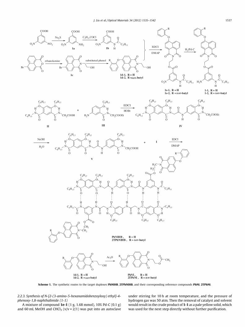
Scheme 1. The synthetic routes to the target duplexes PhNIHB, 2TPhNIHB, and their corresponding reference compounds PhNI, 2TPhNI.
J. Liu et al. / Optical Materials 34 (2012) 1535–1542 1537
2.2.3. Synthesis of N-[2-(3-amino-5-hexanamidobenzoyloxy) ethyl]-4-phenoxy-1,8-naphthalimide (1-1)
A mixture of compound 1e-1 (1 g, 1.68 mmol), 10% Pd-C (0.1 g)and 60 mL MeOH and CHCl3 (v/v = 2/1) was put into an autoclave
under stirring for 10 h at room temperature, and the pressure ofhydrogen gas was 50 atm. Then the removal of catalyst and solventwould result in the crude product of 1-1 as a pale yellow solid, whichwas used for the next step directly without further purification.
1538 J. Liu et al. / Optical Materials 34 (2012) 1535–1542
2.2.4. Synthesis of N-[2-(3-amino-5-hexanamidobenzoyloxy) ethyl]-4-(2-tert-butylphenoxy)-1,8-naphthalimide (1-2)
Compound 1-2 was synthesized with the same procedure as 1-1, using 1e-2 as starting materials, and was used for the next stepwithout further purification.
2.2.5. Synthesis of the duplex PhNIHBTo the 0–5 �C solution of V (1.46 g, 1.68 mmol) and DMAP
(0.31 g, 2.52 mmol) in 60 mL dry CH2Cl2 was added EDCI (0.48 g,2.52 mmol). Then a solution of 1-1 (0.95 g, 1.68 mmol) in 50 mLCH2Cl2 was added dropwise under argon atmosphere. The mixturewas stirred for 48 h at room temperature, then was quenched andwashed with saturated aqueous NaHCO3 solution, water and brinesubsequently, then dried over anhydrous sodium sulfate. Removalof the solvent under vacuum would result in the crude duplexPhNIHB, which was further purified by precipitation from ethanolfor three times, followed by recrystallization from CHCl3/EtOH formore than three times. White solid, yield: 50%, m.p.: 193.9 �C. 1HNMR (400 MHz, CDC13, TMS) d (ppm): 10.13(s, 1H, NH), 10.12(s,1H, NH), 9.66(s, 1H, NH), 9.50(s, 1H, NH), 9.13(s, 1H, NH), 9.06(s,1H, ArH), 8.82(s, 1H, ArH), 8.68(t, 2H, J = 7.6 Hz, ArH), 8.62(s, 1H,ArH), 8.55(dd, 1H, J = 9.2 Hz, 2.4 Hz, ArH), 8.48(d, 1H, J = 8 Hz,ArH), 8.03(d, 1H, J = 2.8 Hz, ArH), 7.89(t, 1H, J = 5.2 Hz, NH),7.75(t, 1H, J = 7.6 Hz, ArH), 7.45(t, 2H, J = 8 Hz, ArH), 7.27(d, 1H,J = 16 Hz, ArH), 7.17(m, 3H, ArH), 6.93(d, 1H, J = 9.2 Hz, ArH),6.90(d, 1H, J = 8.4 Hz, ArH), 6.51(s, 1H, ArH), 4.65(m, 4H, H2CACH2),4.43(s, 2H, CH2), 4.35(s, 2H, CH2), 4.04–4.18(m, 6H, OCH2), 3.40(m,2H, CH2), 2.41(t, 2H, J = 7.6 Hz, CH2), 1.83–2.02(m, 6H, CH2), 1.16–1.63(m, 44H, CH2), 0.75–0.93(m, 15H, CH3). 13C NMR (100 MHz,CDCl3) d (ppm): 171.05, 165.53, 165.36, 165.04, 163.46, 163.16,162.49, 162.44, 160.46, 159.51, 158.85, 153.81, 152.57, 138.48,134.82, 132.00, 131.76, 131.09, 130.00, 129.32, 128.81, 127.57,125.39, 124.48, 123.31, 122.94, 121.44, 119.91, 119.66, 118.99,116.65, 115.63, 115.43, 112.89, 112.74, 112.57, 112.35, 111.95,111.91, 111.82, 111.73, 109.56, 95.62, 69.58, 68.94, 68.75, 60.74,44.62, 39.58, 37.83, 35.71, 31.15, 30.83, 30.80, 30.72, 28.65,28.56, 28.52, 28.35, 28.31, 28.24, 27.93, 27.77, 26.19, 25.45,25.36, 25.29, 24.02, 21.84, 21.72, 21.69, 21.65, 13.24, 13.06,13.02. TOF-MS: m/z 2828.3462 (M + H+), 1414.6213 (M/2 + H+);calcd. for Mw + H+: 2828.5752, Mw/2 + H+: 1414.7876.
0.0
0.3
0.6
0.9
300 350 400 450 5000.0
0.3
0.6
0.9 in thin film
Nor
mal
ized
Abs
orpt
ion
Inte
nsit
y (a
.u.)
PhNI 2TPhNI PhNIHB 2TPhNIHB
in solution
Wavelength (nm)
PhNI 2TPhNI PhNIHB 2TPhNIHB
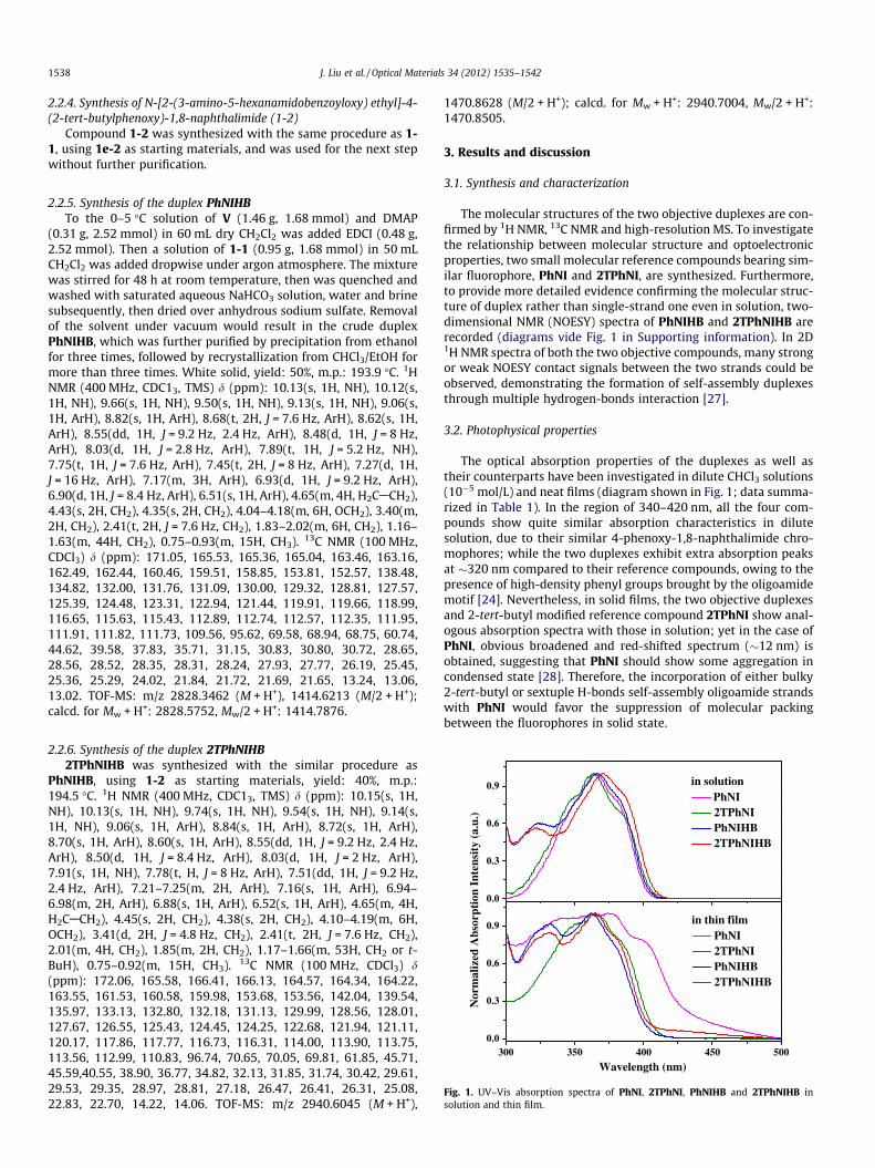
Fig. 1. UV–Vis absorption spectra of PhNI, 2TPhNI, PhNIHB and 2TPhNIHB insolution and thin film.
2.2.6. Synthesis of the duplex 2TPhNIHB2TPhNIHB was synthesized with the similar procedure as
PhNIHB, using 1-2 as starting materials, yield: 40%, m.p.:194.5 �C. 1H NMR (400 MHz, CDC13, TMS) d (ppm): 10.15(s, 1H,NH), 10.13(s, 1H, NH), 9.74(s, 1H, NH), 9.54(s, 1H, NH), 9.14(s,1H, NH), 9.06(s, 1H, ArH), 8.84(s, 1H, ArH), 8.72(s, 1H, ArH),8.70(s, 1H, ArH), 8.60(s, 1H, ArH), 8.55(dd, 1H, J = 9.2 Hz, 2.4 Hz,ArH), 8.50(d, 1H, J = 8.4 Hz, ArH), 8.03(d, 1H, J = 2 Hz, ArH),7.91(s, 1H, NH), 7.78(t, H, J = 8 Hz, ArH), 7.51(dd, 1H, J = 9.2 Hz,2.4 Hz, ArH), 7.21–7.25(m, 2H, ArH), 7.16(s, 1H, ArH), 6.94–6.98(m, 2H, ArH), 6.88(s, 1H, ArH), 6.52(s, 1H, ArH), 4.65(m, 4H,H2CACH2), 4.45(s, 2H, CH2), 4.38(s, 2H, CH2), 4.10–4.19(m, 6H,OCH2), 3.41(d, 2H, J = 4.8 Hz, CH2), 2.41(t, 2H, J = 7.6 Hz, CH2),2.01(m, 4H, CH2), 1.85(m, 2H, CH2), 1.17–1.66(m, 53H, CH2 or t-BuH), 0.75–0.92(m, 15H, CH3). 13C NMR (100 MHz, CDCl3) d(ppm): 172.06, 165.58, 166.41, 166.13, 164.57, 164.34, 164.22,163.55, 161.53, 160.58, 159.98, 153.68, 153.56, 142.04, 139.54,135.97, 133.13, 132.80, 132.18, 131.13, 129.99, 128.56, 128.01,127.67, 126.55, 125.43, 124.45, 124.25, 122.68, 121.94, 121.11,120.17, 117.86, 117.77, 116.73, 116.31, 114.00, 113.90, 113.75,113.56, 112.99, 110.83, 96.74, 70.65, 70.05, 69.81, 61.85, 45.71,45.59,40.55, 38.90, 36.77, 34.82, 32.13, 31.85, 31.74, 30.42, 29.61,29.53, 29.35, 28.97, 28.81, 27.18, 26.47, 26.41, 26.31, 25.08,22.83, 22.70, 14.22, 14.06. TOF-MS: m/z 2940.6045 (M + H+),
1470.8628 (M/2 + H+); calcd. for Mw + H+: 2940.7004, Mw/2 + H+:1470.8505.
3. Results and discussion
3.1. Synthesis and characterization
The molecular structures of the two objective duplexes are con-firmed by 1H NMR, 13C NMR and high-resolution MS. To investigatethe relationship between molecular structure and optoelectronicproperties, two small molecular reference compounds bearing sim-ilar fluorophore, PhNI and 2TPhNI, are synthesized. Furthermore,to provide more detailed evidence confirming the molecular struc-ture of duplex rather than single-strand one even in solution, two-dimensional NMR (NOESY) spectra of PhNIHB and 2TPhNIHB arerecorded (diagrams vide Fig. 1 in Supporting information). In 2D1H NMR spectra of both the two objective compounds, many strongor weak NOESY contact signals between the two strands could beobserved, demonstrating the formation of self-assembly duplexesthrough multiple hydrogen-bonds interaction [27].
3.2. Photophysical properties
The optical absorption properties of the duplexes as well astheir counterparts have been investigated in dilute CHCl3 solutions(10�5 mol/L) and neat films (diagram shown in Fig. 1; data summa-rized in Table 1). In the region of 340–420 nm, all the four com-pounds show quite similar absorption characteristics in dilutesolution, due to their similar 4-phenoxy-1,8-naphthalimide chro-mophores; while the two duplexes exhibit extra absorption peaksat �320 nm compared to their reference compounds, owing to thepresence of high-density phenyl groups brought by the oligoamidemotif [24]. Nevertheless, in solid films, the two objective duplexesand 2-tert-butyl modified reference compound 2TPhNI show anal-ogous absorption spectra with those in solution; yet in the case ofPhNI, obvious broadened and red-shifted spectrum (�12 nm) isobtained, suggesting that PhNI should show some aggregation incondensed state [28]. Therefore, the incorporation of either bulky2-tert-butyl or sextuple H-bonds self-assembly oligoamide strandswith PhNI would favor the suppression of molecular packingbetween the fluorophores in solid state.
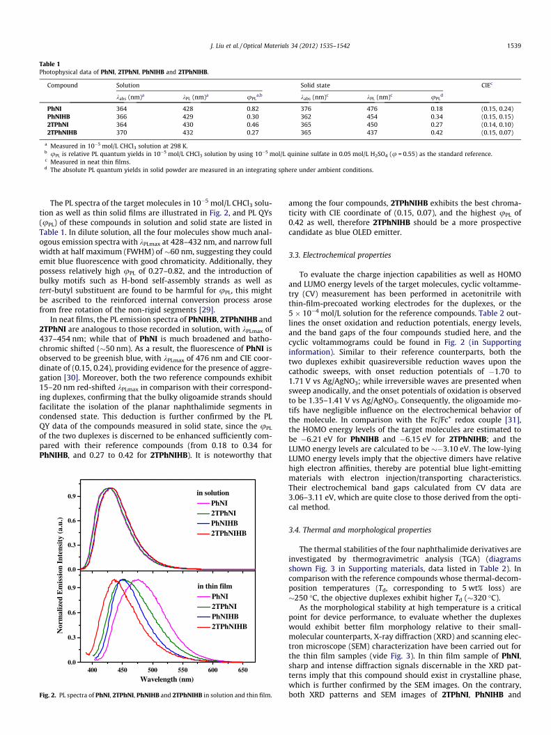
Table 1Photophysical data of PhNI, 2TPhNI, PhNIHB and 2TPhNIHB.
Compound Solution Solid state CIEc
kabs (nm)a kPL (nm)a uPLa,b kabs (nm)c kPL (nm)c uPL
d
PhNI 364 428 0.82 376 476 0.18 (0.15, 0.24)PhNIHB 366 429 0.30 362 454 0.34 (0.15, 0.15)2TPhNI 364 430 0.46 365 450 0.27 (0.14, 0.10)2TPhNIHB 370 432 0.27 365 437 0.42 (0.15, 0.07)
a Measured in 10�5 mol/L CHCl3 solution at 298 K.b uPL is relative PL quantum yields in 10�5 mol/L CHCl3 solution by using 10�5 mol/L quinine sulfate in 0.05 mol/L H2SO4 (u = 0.55) as the standard reference.c Measured in neat thin films.d The absolute PL quantum yields in solid powder are measured in an integrating sphere under ambient conditions.
J. Liu et al. / Optical Materials 34 (2012) 1535–1542 1539
The PL spectra of the target molecules in 10�5 mol/L CHCl3 solu-tion as well as thin solid films are illustrated in Fig. 2, and PL QYs(uPL) of these compounds in solution and solid state are listed inTable 1. In dilute solution, all the four molecules show much anal-ogous emission spectra with kPLmax at 428–432 nm, and narrow fullwidth at half maximum (FWHM) of �60 nm, suggesting they couldemit blue fluorescence with good chromaticity. Additionally, theypossess relatively high uPL of 0.27–0.82, and the introduction ofbulky motifs such as H-bond self-assembly strands as well astert-butyl substituent are found to be harmful for uPL, this mightbe ascribed to the reinforced internal conversion process arosefrom free rotation of the non-rigid segments [29].
In neat films, the PL emission spectra of PhNIHB, 2TPhNIHB and2TPhNI are analogous to those recorded in solution, with kPLmax of437–454 nm; while that of PhNI is much broadened and batho-chromic shifted (�50 nm). As a result, the fluorescence of PhNI isobserved to be greenish blue, with kPLmax of 476 nm and CIE coor-dinate of (0.15, 0.24), providing evidence for the presence of aggre-gation [30]. Moreover, both the two reference compounds exhibit15–20 nm red-shifted kPLmax in comparison with their correspond-ing duplexes, confirming that the bulky oligoamide strands shouldfacilitate the isolation of the planar naphthalimide segments incondensed state. This deduction is further confirmed by the PLQY data of the compounds measured in solid state, since the uPL
of the two duplexes is discerned to be enhanced sufficiently com-pared with their reference compounds (from 0.18 to 0.34 forPhNIHB, and 0.27 to 0.42 for 2TPhNIHB). It is noteworthy that
0.0
0.3
0.6
0.9
400 450 500 550 600 6500.0
0.3
0.6
0.9
PhNI 2TPhNI PhNIHB 2TPhNIHB
in solution
Nor
mal
ized
Em
issi
on I
nten
sity
(a.
u.)
Wavelength (nm)
PhNI 2TPhNI PhNIHB 2TPhNIHB
in thin film
Fig. 2. PL spectra of PhNI, 2TPhNI, PhNIHB and 2TPhNIHB in solution and thin film.
among the four compounds, 2TPhNIHB exhibits the best chroma-ticity with CIE coordinate of (0.15, 0.07), and the highest uPL of0.42 as well, therefore 2TPhNIHB should be a more prospectivecandidate as blue OLED emitter.
3.3. Electrochemical properties
To evaluate the charge injection capabilities as well as HOMOand LUMO energy levels of the target molecules, cyclic voltamme-try (CV) measurement has been performed in acetonitrile withthin-film-precoated working electrodes for the duplexes, or the5 � 10�4 mol/L solution for the reference compounds. Table 2 out-lines the onset oxidation and reduction potentials, energy levels,and the band gaps of the four compounds studied here, and thecyclic voltammograms could be found in Fig. 2 (in Supportinginformation). Similar to their reference counterparts, both thetwo duplexes exhibit quasireversible reduction waves upon thecathodic sweeps, with onset reduction potentials of �1.70 to1.71 V vs Ag/AgNO3; while irreversible waves are presented whensweep anodically, and the onset potentials of oxidation is observedto be 1.35–1.41 V vs Ag/AgNO3. Consequently, the oligoamide mo-tifs have negligible influence on the electrochemical behavior ofthe molecule. In comparison with the Fc/Fc+ redox couple [31],the HOMO energy levels of the target molecules are estimated tobe �6.21 eV for PhNIHB and �6.15 eV for 2TPhNIHB; and theLUMO energy levels are calculated to be ��3.10 eV. The low-lyingLUMO energy levels imply that the objective dimers have relativehigh electron affinities, thereby are potential blue light-emittingmaterials with electron injection/transporting characteristics.Their electrochemical band gaps calculated from CV data are3.06–3.11 eV, which are quite close to those derived from the opti-cal method.
3.4. Thermal and morphological properties
The thermal stabilities of the four naphthalimide derivatives areinvestigated by thermogravimetric analysis (TGA) (diagramsshown Fig. 3 in Supporting materials, data listed in Table 2). Incomparison with the reference compounds whose thermal-decom-position temperatures (Td, corresponding to 5 wt% loss) are�250 �C, the objective duplexes exhibit higher Td (�320 �C).
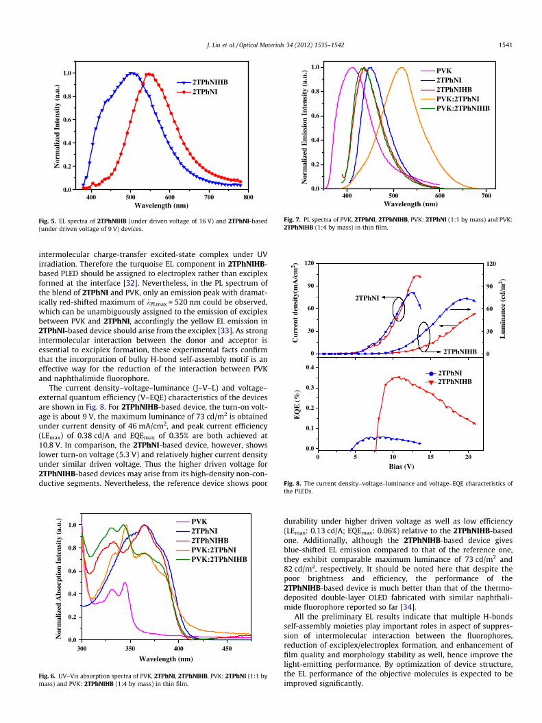
As the morphological stability at high temperature is a criticalpoint for device performance, to evaluate whether the duplexeswould exhibit better film morphology relative to their small-molecular counterparts, X-ray diffraction (XRD) and scanning elec-tron microscope (SEM) characterization have been carried out forthe thin film samples (vide Fig. 3). In thin film sample of PhNI,sharp and intense diffraction signals discernable in the XRD pat-terns imply that this compound should exist in crystalline phase,which is further confirmed by the SEM images. On the contrary,both XRD patterns and SEM images of 2TPhNI, PhNIHB and
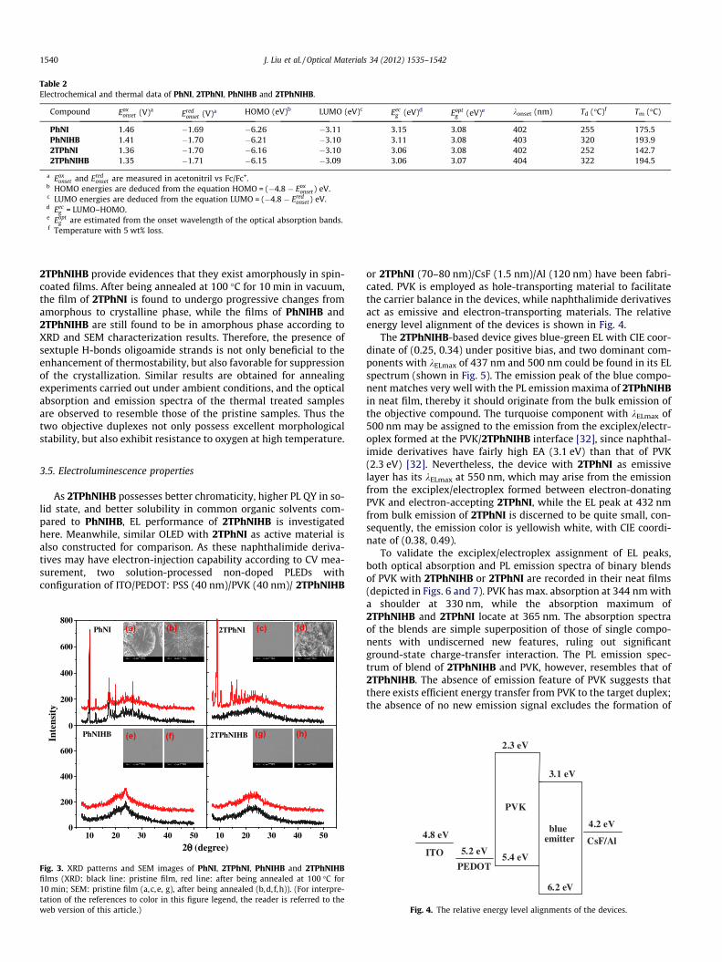
Table 2Electrochemical and thermal data of PhNI, 2TPhNI, PhNIHB and 2TPhNIHB.
Compound Eoxonset (V)a
Eredonset (V)a HOMO (eV)b LUMO (eV)c Eec
g (eV)d Eoptg (eV)e konset (nm) Td (�C)f Tm (�C)
PhNI 1.46 �1.69 �6.26 �3.11 3.15 3.08 402 255 175.5PhNIHB 1.41 �1.70 �6.21 �3.10 3.11 3.08 403 320 193.92TPhNI 1.36 �1.70 �6.16 �3.10 3.06 3.08 402 252 142.72TPhNIHB 1.35 �1.71 �6.15 �3.09 3.06 3.07 404 322 194.5
a Eoxonset and Ered
onset are measured in acetonitril vs Fc/Fc+.b HOMO energies are deduced from the equation HOMO = (�4.8 � Eox
onset) eV.c LUMO energies are deduced from the equation LUMO = (�4.8 � Ered
onset) eV.d Eec
g = LUMO–HOMO.e Eopt
g are estimated from the onset wavelength of the optical absorption bands.f Temperature with 5 wt% loss.
1540 J. Liu et al. / Optical Materials 34 (2012) 1535–1542
2TPhNIHB provide evidences that they exist amorphously in spin-coated films. After being annealed at 100 �C for 10 min in vacuum,the film of 2TPhNI is found to undergo progressive changes fromamorphous to crystalline phase, while the films of PhNIHB and2TPhNIHB are still found to be in amorphous phase according toXRD and SEM characterization results. Therefore, the presence ofsextuple H-bonds oligoamide strands is not only beneficial to theenhancement of thermostability, but also favorable for suppressionof the crystallization. Similar results are obtained for annealingexperiments carried out under ambient conditions, and the opticalabsorption and emission spectra of the thermal treated samplesare observed to resemble those of the pristine samples. Thus thetwo objective duplexes not only possess excellent morphologicalstability, but also exhibit resistance to oxygen at high temperature.
3.5. Electroluminescence properties
As 2TPhNIHB possesses better chromaticity, higher PL QY in so-lid state, and better solubility in common organic solvents com-pared to PhNIHB, EL performance of 2TPhNIHB is investigatedhere. Meanwhile, similar OLED with 2TPhNI as active material isalso constructed for comparison. As these naphthalimide deriva-tives may have electron-injection capability according to CV mea-surement, two solution-processed non-doped PLEDs withconfiguration of ITO/PEDOT: PSS (40 nm)/PVK (40 nm)/ 2TPhNIHB
0
200
400
600
800
10 20 30 40 500
200
400
600
10 20 30 40 50
2TPhNIHB
2TPhNIPhNI
PhNIHBInte
nsit
y
2 (degree)
(a) (b) (c) (d)
(e) (f) (g) (h)
Fig. 3. XRD patterns and SEM images of PhNI, 2TPhNI, PhNIHB and 2TPhNIHBfilms (XRD: black line: pristine film, red line: after being annealed at 100 �C for10 min; SEM: pristine film (a,c,e, g), after being annealed (b,d, f,h)). (For interpre-tation of the references to color in this figure legend, the reader is referred to theweb version of this article.)
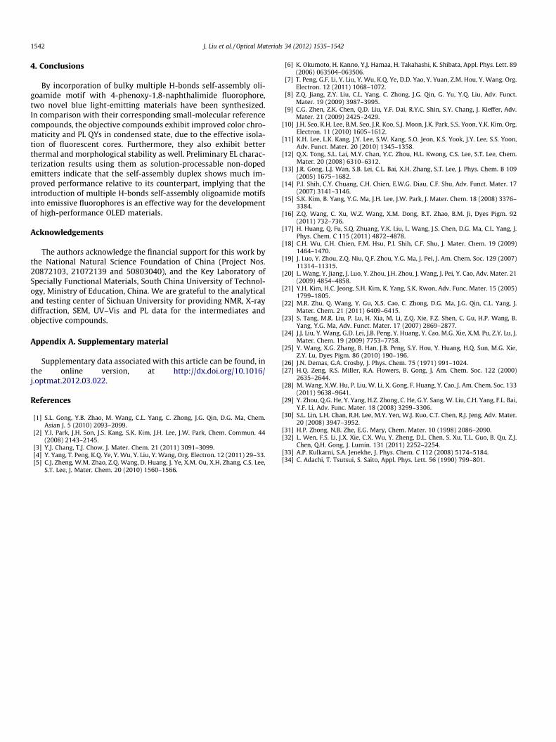
or 2TPhNI (70–80 nm)/CsF (1.5 nm)/Al (120 nm) have been fabri-cated. PVK is employed as hole-transporting material to facilitatethe carrier balance in the devices, while naphthalimide derivativesact as emissive and electron-transporting materials. The relativeenergy level alignment of the devices is shown in Fig. 4.
The 2TPhNIHB-based device gives blue-green EL with CIE coor-dinate of (0.25, 0.34) under positive bias, and two dominant com-ponents with kELmax of 437 nm and 500 nm could be found in its ELspectrum (shown in Fig. 5). The emission peak of the blue compo-nent matches very well with the PL emission maxima of 2TPhNIHBin neat film, thereby it should originate from the bulk emission ofthe objective compound. The turquoise component with kELmax of500 nm may be assigned to the emission from the exciplex/electr-oplex formed at the PVK/2TPhNIHB interface [32], since naphthal-imide derivatives have fairly high EA (3.1 eV) than that of PVK(2.3 eV) [32]. Nevertheless, the device with 2TPhNI as emissivelayer has its kELmax at 550 nm, which may arise from the emissionfrom the exciplex/electroplex formed between electron-donatingPVK and electron-accepting 2TPhNI, while the EL peak at 432 nmfrom bulk emission of 2TPhNI is discerned to be quite small, con-sequently, the emission color is yellowish white, with CIE coordi-nate of (0.38, 0.49).
To validate the exciplex/electroplex assignment of EL peaks,both optical absorption and PL emission spectra of binary blendsof PVK with 2TPhNIHB or 2TPhNI are recorded in their neat films(depicted in Figs. 6 and 7). PVK has max. absorption at 344 nm witha shoulder at 330 nm, while the absorption maximum of2TPhNIHB and 2TPhNI locate at 365 nm. The absorption spectraof the blends are simple superposition of those of single compo-nents with undiscerned new features, ruling out significantground-state charge-transfer interaction. The PL emission spec-trum of blend of 2TPhNIHB and PVK, however, resembles that of2TPhNIHB. The absence of emission feature of PVK suggests thatthere exists efficient energy transfer from PVK to the target duplex;the absence of no new emission signal excludes the formation of
ITO
4.8 eV
PEDOT
5.2 eV5.4 eV
2.3 eV
PVK
blueemitter
3.1 eV
6.2 eV
4.2 eV
CsF/Al
Fig. 4. The relative energy level alignments of the devices.
400 500 600 700 8000.0
0.2
0.4
0.6
0.8
1.0
Nor
mal
ized
Int
ensi
ty (
a.u.
)
Wavelength (nm)
2TPhNIHB 2TPhNI
Fig. 5. EL spectra of 2TPhNIHB (under driven voltage of 16 V) and 2TPhNI-based(under driven voltage of 9 V) devices.
400 500 600 7000.0
0.2
0.4
0.6
0.8
1.0
Nor
mal
ized
Em
issi
on I
nten
sity
(a.
u.)
Wavelength (nm)
PVK 2TPhNI 2TPhNIHB PVK:2TPhNI PVK:2TPhNIHB
Fig. 7. PL spectra of PVK, 2TPhNI, 2TPhNIHB, PVK: 2TPhNI (1:1 by mass) and PVK:2TPhNIHB (1:4 by mass) in thin film.
0
30
60
90
120
Lum
inan
ce (
cd/m
2 )
Cur
rent
den
sity
(mA
/cm
2 )
0
30
60
90
120
2TPhNI
2TPhNIHB
0 5 10 15 200.0
0.1
0.2
0.3
0.4
EQ
E (
%)
Bias (V)
2TPhNI2TPhNIHB
Fig. 8. The current density–voltage–luminance and voltage–EQE characteristics of
J. Liu et al. / Optical Materials 34 (2012) 1535–1542 1541
intermolecular charge-transfer excited-state complex under UVirradiation. Therefore the turquoise EL component in 2TPhNIHB-based PLED should be assigned to electroplex rather than exciplexformed at the interface [32]. Nevertheless, in the PL spectrum ofthe blend of 2TPhNI and PVK, only an emission peak with dramat-ically red-shifted maximum of kPLmax = 520 nm could be observed,which can be unambiguously assigned to the emission of exciplexbetween PVK and 2TPhNI, accordingly the yellow EL emission in2TPhNI-based device should arise from the exciplex [33]. As strongintermolecular interaction between the donor and acceptor isessential to exciplex formation, these experimental facts confirmthat the incorporation of bulky H-bond self-assembly motif is aneffective way for the reduction of the interaction between PVKand naphthalimide fluorophore.
The current density–voltage–luminance (J–V–L) and voltage–external quantum efficiency (V–EQE) characteristics of the devicesare shown in Fig. 8. For 2TPhNIHB-based device, the turn-on volt-age is about 9 V, the maximum luminance of 73 cd/m2 is obtainedunder current density of 46 mA/cm2, and peak current efficiency(LEmax) of 0.38 cd/A and EQEmax of 0.35% are both achieved at10.8 V. In comparison, the 2TPhNI-based device, however, showslower turn-on voltage (5.3 V) and relatively higher current densityunder similar driven voltage. Thus the higher driven voltage for2TPhNIHB-based devices may arise from its high-density non-con-ductive segments. Nevertheless, the reference device shows poor
300 350 400 4500.0
0.2
0.4
0.6
0.8
1.0 PVK 2TPhNI 2TPhNIHB PVK:2TPhNI PVK:2TPhNIHB
Nor
mal
ized
Abs
orpt
ion
Inte
nsit
y (a
.u.)
Wavelength (nm)
Fig. 6. UV–Vis absorption spectra of PVK, 2TPhNI, 2TPhNIHB, PVK: 2TPhNI (1:1 bymass) and PVK: 2TPhNIHB (1:4 by mass) in thin film.
the PLEDs.
durability under higher driven voltage as well as low efficiency(LEmax: 0.13 cd/A; EQEmax: 0.06%) relative to the 2TPhNIHB-basedone. Additionally, although the 2TPhNIHB-based device givesblue-shifted EL emission compared to that of the reference one,they exhibit comparable maximum luminance of 73 cd/m2 and82 cd/m2, respectively. It should be noted here that despite thepoor brightness and efficiency, the performance of the2TPhNIHB-based device is much better than that of the thermo-deposited double-layer OLED fabricated with similar naphthali-mide fluorophore reported so far [34].
All the preliminary EL results indicate that multiple H-bondsself-assembly moieties play important roles in aspect of suppres-sion of intermolecular interaction between the fluorophores,reduction of exciplex/electroplex formation, and enhancement offilm quality and morphology stability as well, hence improve thelight-emitting performance. By optimization of device structure,the EL performance of the objective molecules is expected to beimproved significantly.
1542 J. Liu et al. / Optical Materials 34 (2012) 1535–1542
4. Conclusions
By incorporation of bulky multiple H-bonds self-assembly oli-goamide motif with 4-phenoxy-1,8-naphthalimide fluorophore,two novel blue light-emitting materials have been synthesized.In comparison with their corresponding small-molecular referencecompounds, the objective compounds exhibit improved color chro-maticity and PL QYs in condensed state, due to the effective isola-tion of fluorescent cores. Furthermore, they also exhibit betterthermal and morphological stability as well. Preliminary EL charac-terization results using them as solution-processable non-dopedemitters indicate that the self-assembly duplex shows much im-proved performance relative to its counterpart, implying that theintroduction of multiple H-bonds self-assembly oligoamide motifsinto emissive fluorophores is an effective way for the developmentof high-performance OLED materials.
Acknowledgements
The authors acknowledge the financial support for this work bythe National Natural Science Foundation of China (Project Nos.20872103, 21072139 and 50803040), and the Key Laboratory ofSpecially Functional Materials, South China University of Technol-ogy, Ministry of Education, China. We are grateful to the analyticaland testing center of Sichuan University for providing NMR, X-raydiffraction, SEM, UV–Vis and PL data for the intermediates andobjective compounds.
Appendix A. Supplementary material
Supplementary data associated with this article can be found, inthe online version, at http://dx.doi.org/10.1016/j.optmat.2012.03.022.
References
[1] S.L. Gong, Y.B. Zhao, M. Wang, C.L. Yang, C. Zhong, J.G. Qin, D.G. Ma, Chem.Asian J. 5 (2010) 2093–2099.
[2] Y.I. Park, J.H. Son, J.S. Kang, S.K. Kim, J.H. Lee, J.W. Park, Chem. Commun. 44(2008) 2143–2145.
[3] Y.J. Chang, T.J. Chow, J. Mater. Chem. 21 (2011) 3091–3099.[4] Y. Yang, T. Peng, K.Q. Ye, Y. Wu, Y. Liu, Y. Wang, Org. Electron. 12 (2011) 29–33.[5] C.J. Zheng, W.M. Zhao, Z.Q. Wang, D. Huang, J. Ye, X.M. Ou, X.H. Zhang, C.S. Lee,
S.T. Lee, J. Mater. Chem. 20 (2010) 1560–1566.
[6] K. Okumoto, H. Kanno, Y.J. Hamaa, H. Takahashi, K. Shibata, Appl. Phys. Lett. 89(2006) 063504–063506.
[7] T. Peng, G.F. Li, Y. Liu, Y. Wu, K.Q. Ye, D.D. Yao, Y. Yuan, Z.M. Hou, Y. Wang, Org.Electron. 12 (2011) 1068–1072.
[8] Z.Q. Jiang, Z.Y. Liu, C.L. Yang, C. Zhong, J.G. Qin, G. Yu, Y.Q. Liu, Adv. Funct.Mater. 19 (2009) 3987–3995.
[9] C.G. Zhen, Z.K. Chen, Q.D. Liu, Y.F. Dai, R.Y.C. Shin, S.Y. Chang, J. Kieffer, Adv.Mater. 21 (2009) 2425–2429.
[10] J.H. Seo, K.H. Lee, B.M. Seo, J.R. Koo, S.J. Moon, J.K. Park, S.S. Yoon, Y.K. Kim, Org.Electron. 11 (2010) 1605–1612.
[11] K.H. Lee, L.K. Kang, J.Y. Lee, S.W. Kang, S.O. Jeon, K.S. Yook, J.Y. Lee, S.S. Yoon,Adv. Funct. Mater. 20 (2010) 1345–1358.
[12] Q.X. Tong, S.L. Lai, M.Y. Chan, Y.C. Zhou, H.L. Kwong, C.S. Lee, S.T. Lee, Chem.Mater. 20 (2008) 6310–6312.
[13] J.R. Gong, L.J. Wan, S.B. Lei, C.L. Bai, X.H. Zhang, S.T. Lee, J. Phys. Chem. B 109(2005) 1675–1682.
[14] P.I. Shih, C.Y. Chuang, C.H. Chien, E.W.G. Diau, C.F. Shu, Adv. Funct. Mater. 17(2007) 3141–3146.
[15] S.K. Kim, B. Yang, Y.G. Ma, J.H. Lee, J.W. Park, J. Mater. Chem. 18 (2008) 3376–3384.
[16] Z.Q. Wang, C. Xu, W.Z. Wang, X.M. Dong, B.T. Zhao, B.M. Ji, Dyes Pigm. 92(2011) 732–736.
[17] H. Huang, Q. Fu, S.Q. Zhuang, Y.K. Liu, L. Wang, J.S. Chen, D.G. Ma, C.L. Yang, J.Phys. Chem. C 115 (2011) 4872–4878.
[18] C.H. Wu, C.H. Chien, F.M. Hsu, P.I. Shih, C.F. Shu, J. Mater. Chem. 19 (2009)1464–1470.
[19] J. Luo, Y. Zhou, Z.Q. Niu, Q.F. Zhou, Y.G. Ma, J. Pei, J. Am. Chem. Soc. 129 (2007)11314–11315.
[20] L. Wang, Y. Jiang, J. Luo, Y. Zhou, J.H. Zhou, J. Wang, J. Pei, Y. Cao, Adv. Mater. 21(2009) 4854–4858.
[21] Y.H. Kim, H.C. Jeong, S.H. Kim, K. Yang, S.K. Kwon, Adv. Func. Mater. 15 (2005)1799–1805.
[22] M.R. Zhu, Q. Wang, Y. Gu, X.S. Cao, C. Zhong, D.G. Ma, J.G. Qin, C.L. Yang, J.Mater. Chem. 21 (2011) 6409–6415.
[23] S. Tang, M.R. Liu, P. Lu, H. Xia, M. Li, Z.Q. Xie, F.Z. Shen, C. Gu, H.P. Wang, B.Yang, Y.G. Ma, Adv. Funct. Mater. 17 (2007) 2869–2877.
[24] J.J. Liu, Y. Wang, G.D. Lei, J.B. Peng, Y. Huang, Y. Cao, M.G. Xie, X.M. Pu, Z.Y. Lu, J.Mater. Chem. 19 (2009) 7753–7758.
[25] Y. Wang, X.G. Zhang, B. Han, J.B. Peng, S.Y. Hou, Y. Huang, H.Q. Sun, M.G. Xie,Z.Y. Lu, Dyes Pigm. 86 (2010) 190–196.
[26] J.N. Demas, G.A. Crosby, J. Phys. Chem. 75 (1971) 991–1024.[27] H.Q. Zeng, R.S. Miller, R.A. Flowers, B. Gong, J. Am. Chem. Soc. 122 (2000)
2635–2644.[28] M. Wang, X.W. Hu, P. Liu, W. Li, X. Gong, F. Huang, Y. Cao, J. Am. Chem. Soc. 133
(2011) 9638–9641.[29] Y. Zhou, Q.G. He, Y. Yang, H.Z. Zhong, C. He, G.Y. Sang, W. Liu, C.H. Yang, F.L. Bai,
Y.F. Li, Adv. Func. Mater. 18 (2008) 3299–3306.[30] S.L. Lin, L.H. Chan, R.H. Lee, M.Y. Yen, W.J. Kuo, C.T. Chen, R.J. Jeng, Adv. Mater.
20 (2008) 3947–3952.[31] H.P. Zhong, N.B. Zhe, E.G. Mary, Chem. Mater. 10 (1998) 2086–2090.[32] L. Wen, F.S. Li, J.X. Xie, C.X. Wu, Y. Zheng, D.L. Chen, S. Xu, T.L. Guo, B. Qu, Z.J.
Chen, Q.H. Gong, J. Lumin. 131 (2011) 2252–2254.[33] A.P. Kulkarni, S.A. Jenekhe, J. Phys. Chem. C 112 (2008) 5174–5184.[34] C. Adachi, T. Tsutsui, S. Saito, Appl. Phys. Lett. 56 (1990) 799–801.


![Hexastore: Sextuple Indexing for Semantic Web Data …2.2.2 Multiple-indexing Approaches Harth and Decker [27] proposed storing RDF data based on multiple indices, while taking into](https://img.pdfslide.us/doc/110x75/5ed3ae49c4540779ca664dde/hexastore-sextuple-indexing-for-semantic-web-data-222-multiple-indexing-approaches.jpg)