Embed Size (px)
Citation preview

SYNTHESIS AND ION SENSING PROPERTIES OF NOVEL BORADIAZAINDACENE DYES
A THESIS SUBMITTED TO THE GRADUATE SCHOOL OF NATURAL AND APPLIED SCIENCES
OF THE MIDDLE EAST TECHNICAL UNIVERSITY
BY
NALAN ZALİM
IN PARTIAL FULFILLMENT OF THE REQUIREMENTS FOR THE DEGREE OF
MASTER OF SCIENCE
IN
THE DEPARTMENT OF CHEMISTRY
SEPTEMBER 2003

Approval of the Graduate School of Natural and Applied Sciences Prof. Dr. Canan Özgen Director
I certify that this thesis satisfies all requirements as a thesis for the degree of Master of Science. Prof. Dr. Teoman Tinçer Chairman of the Department This is to certify that we have read this thesis and in our opinion, it is fully adequate, in scope and quality, as a thesis for the degree of Master of Science.
Prof. Dr. Engin U. Akkaya Supervisor Examining Committee Members Prof. Dr. İdris M. Akhmedov Prof. Dr. İnci Gökmen Prof. Dr. Engin U. Akkaya Assoc. Prof. Dr. Deniz Üner Assoc. Prof. Dr. Özdemir Doğan
ABSTRACT
SYNTHESIS AND ION SENSING PROPERTIES OF NOVEL
BORADIAZADACENE DYES
Zalim, Nalan
M. S., Department of Chemistry
Supervisor: Prof. Dr. Engin U. AKKAYA
September 2003, 74 pages
The derivatives of boradiazaindacene (BODIPY) are highly fluorescent dyes which
have quantum yields near 1.0. These dyes that have exceptional spectral and
photophysical stability as compared to other fluorescent groups are used for several
different applications.
The fluorescent sensor molecules for the detection of cations with PET
(photoinduced-electron transfer) mechanism in general have been obtained by the
differentiation of the BODIPY core. The extension of conjugation over the pyrrole
ring shifts the absorption and emission at longer wavelength. Moreover, it is seen
that red emission occurs from an ICT (intramolecular-charge transfer) state.
iii

In this study, we designed and synthesized an unsymetrically substituted
BODIPY dye series, carrying a cation-sensitive phenylazacrown ether group
conjugated to the core, and investigated the ion sensing properties of these
compounds. Both aza-crown derivatives displayed selectivity towards Ca(II).
Keywords: Chemosensor, BODIPY, azacrown ether, photoinduced-charge transfer
iv
ÖZ
BORADİAZAİNDASEN BOYARMADDELERİNİN SENTEZİ VE İYON
ALGILAYICI ÖZELLİKLERİ
Zalim, Nalan
Yüksek Lisans, Kimya Bölümü
Tez Yöneticisi: Prof. Dr. Engin U. AKKAYA
Eylül 2003, 74 sayfa
Boradiazaindasen (BODIPY) türevleri kuantum verimleri 1.0’e yaklaşan çok parlak
floresan boyarmaddelerdir. Diğer floresan gruplarla kıyaslandığında fevkalade
spektral ve fotofiziksel kararlılığı olan bu boyarmaddelerin çok farklı uygulama
alanları vardır.
Boradiazaindasen (BODIPY) yapısı türevlendirilerek çoğunlukla PET
(photoinduced-electron transfer) mekanizmasıyla katyon sinyalleyen molecular
algılayıcılar elde edilmiştir. Pirol halkası üzerinden konjügasyonun uzatılması, daha
uzun dalga boyunda absorpsiyon ve emisyonu mümkün kılmıştır. Ayrıca kırmızı
emisyonun bir ICT (intramolecular-charge transfer) düzeyinden olduğu
anlaşılmaktadır.
v

Bu çalışmada, konjüge olarak ana gruba bağlı katyon algılayıcı fenil-aza-taç
eter grup taşıyan, simetrik olmayan, bir boradiazaindasen boyarmadde serisi
tasarlayıp sentezlemiş ve iyon algılayıcı özellikleri incelenmiştir. Sonuçlar her iki
aza-taç eter türevinin de Ca(II) katyonu için seçicilik gösterdiğini ortaya
çıkarmıştır.
Anahtar kelimeler: Moleküler algılayıcı, BODIPY, azataç eter, photoinduced-yük
aktarımı.
vi

To my family
vii

ACKNOWLEDGMENTS
I would like to express my sincere thanks to my supervisor Prof. Dr. Engin
U. Akkaya for his guidance, support and patience during the course of this research
as well as his unlimited knowledge and experience that I have benefited from
greatly.
I want to thank to my family for their support, understanding and
encouragement.
My gratitude to the NMR technician Fatoş is endless.
I would like to thank to our group members. I can never forget the beautiful
friendship in our Lab, B-09 (underground organization).
viii

TABLE OF CONTENTS
ABSTRACT………………………………………………………………………..iii
ÖZ…………………………………………………………………………………...v
DEDICATION……………………………………………………………………..vii
ACKNOWLEDGEMENTS……………………………………………………….viii
TABLE OF CONTENTS…………………………………………………………...ix
LIST OF TABLES………………………………………………………………....xii
LIST OF FIGURES……………………………………………………………….xiii
CHAPTER
1. INTRODUCTION
1.1 Supramolecular Chemistry…………………………………………………...1
1.2 A Brief Introduction to Fluorescence………………………………………...4
1.2.1 Fluorescence as a Signal Transduction Mechanism……………………5
1.3 Molecular Recognition……………………………………………………....7
1.4 Chemosensors……………………………………………………………......8
1.4.1 Fluorescent Chemosensors
1.5 Principles of Fluorescent Chemosensor Design for Cation Recognition…...11
1.5.1 Fluorescent Photoinduced Electron Transfer (PET) Cation Sensors....14
1.5.2 Fluorescent Photoinduced Charge Transfer (PCT) Cation Sensors…..19
ix
1.6 4,4-Difluoro-4-bora-3a,4a-diaza-s-indacene (BODIPY) Dyes.......................23
1.7 Aim of the Study…………………………………………………………….33
2. EXPERIMENTAL
2.0 Instrumentation……………………………………………………………...34
2.1 Synthesis of 16-Phenyl-1,4,7,10,13-pentaoxa-16-aza-cyclooctadecane
Substituted BODIPY Dye……………………………..................................35
2.1.1 Synthesis of 16-Phenyl-1,4,7,10,13-pentaoxa-16-aza-
Cyclooctadecane……………………………………………………..35
2.1.2 Synthesis of 4-(1,4,7,10,13-Pentaoxa-16-aza-cyclooctadec-16-
yl) benzaldehyde……………………………………………………..36
2.1.3 Synthesis of 4,4-Difluoro-4-bora-3a,4a-diaza-s-indacene
(BODIPY)……………………………………………………………37
2.1.4 Synthesis of 4,4-Difluoro-4-bora-3a,4a-diaza-s-indacene
(BODIPY) Dye………………………………………………………38
2.2 Synthesis of 13-Phenyl-1,4,7,10-tetraoxa-13-aza-cyclopentadecane
Substituted BODIPY Dye…………………………………………………39
2.2.1 Synthesis of 13-Phenyl-1,4,7,10-tetraoxa-13-aza-
cyclopentadecane…………………………………………………...39
2.2.2 Synthesis of 4-(1,4,7,10-Tetraoxa-13-aza-cyclopentadec-13-yl)-
benzaldehyde……...…………………………………………………41
2.2.3 Synthesis of 4,4-Difluoro-4-bora-3a,4a-diaza-s-indacene
(BODIPY) Dye…………………………………………………….....42
3. RESULTS AND DISCUSSION………………………………………………....44
3.1 Synthesis and Photophysical Properties of BODIPY Dye (8)……………...46
3.2 Synthesis and Photophysical Properties of BODIPY Dye (12)…………….52
x

4. CONCLUSION…………………………………………………………………..57
REFERENCES……………………………………………………………………...58
APPENDIX…………………………………………………………………………64
xi

LIST OF TABLES
TABLE
Table 1. Spectroscopic data of 8 and 8-H+ in different solvets at 298 K................32
xii
LIST OF FIGURES
FIGURES
1.1 From molecular components to supramoler systems…………………………....2
1.2 Jablonski Diagram……………………………………………………………….4
1.3 Main aspects of fluorescent molecular sensors for cation recognition………....13
1.4 Principle of cation recognition based on cation control of photoinduced
electron transfer in nonconjugated donor-acceptor systems................................16
1.5 Some examples of fluorescent PET sensors for various cations..........................18
1.6 Spectral displacements of PCT sensors resulting from interaction of a bound
cation with an electron-donating or electron-withdrawing group……………...20
1.7 Some examples for fluorescent PCT sensors for various cations.......................22
1.8 Structure of 4,4-Difluoro-4-bora-3a,4a-diaza-s-indacene (BODIPY,BDP).......23
1.9 Chemical structures of 1a-e, 2a-b, 3a-b and 4a-c..............................................24
1.10 Structure of compound 5...................................................................................26
1.11 Plots for quenching of 5 with S-PEA and R-PEA in acetonitrile......................26
xiii
1.12 The effect of phosphate binding in fluorescence..............................................27
1.13 Emission spectrum of the Zn(II) complex in response to increasing
phosphate concentrations.................................................................................28
1.14 Structure of compound 6...................................................................................29
1.15 Absorption spectrum of 6 and emission spectra of the complexes with
cations...............................................................................................................29
1.16 Changes in fluorescence intensity of 6 upon addition of metal ions................29
1.17 Structure of compound 7...................................................................................30
1.18 Structure of compound 8...................................................................................31
1.19 Steady-state spectra of 8 and 8-H+ in acetonitrile.............................................32
2.1 Synthesis of 16-Phenyl-1,4,7,10,13-pentaoxa-16-aza-cyclooctadecane (3).......36
2.2 Synthesis of 4-(1,4,7,10,13-Pentaoxa-16-aza-cyclooctadec-16-yl)-
benzaldehyde (4).................................................................................................37
2.3 Synthesis of 4,4-Difluoro-4-bora-3a,4a-diaza-s-indacene (BODIPY) (7)..........38
2.4 Synthesis of 4,4-difluoro-4-bora-3a,4a-diaza-s-indacene (BODIPY) dye (8)....39
2.5 Synthesis of 13-Phenyl-1,4,7,10-tetraoxa-13-aza-cyclopentadecane (10)..........40
2.6 Synthesis of 4-(1,4,7,10-Tetraoxa-13-aza-cyclopentadec-13-yl)-
benzaldehyde (11)................................................................................................42
xiv
2.7 Synthesis of 4,4-Difluoro-4-bora-3a,4a-diaza-s-indacene (BODIPY)
Dye (12)..............................................................................................................43
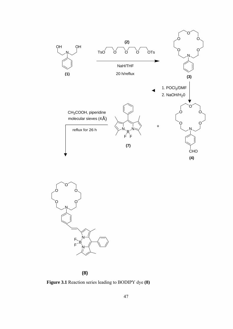
3.1 Reaction series leading to BODIPY dye (8)......................................................47
3.2 Absorption spectra of BODIPY dye 8, 8-(metal cations) and 8-TFA
(Trifluoroacetic acid) in acetonitrile...................................................................48
3.3 Emission spectrum of BODIPY dye 8, as a function of increasing TFA
(Trifluoroacetic acid) concentration in acetonitrile............................................49
3.4 Emission spectra of free ligand 8 and 8-(metal cations) in acetonitrile.............50
3.5 Emission spectrum of BODIPY dye 8 in response to increasing calcium ion
concentration......................................................................................................51
3.6 Reaction series leading to BODIPY dye (12)....................................................53
3.7 Absorption spectra of BODIPY dye 12, 12-(metal cations) and 12-TFA
(Trifluoroacetic acid) in acetonitrile..................................................................54
3.8 Emission spectrum of BODIPY dye 12, as a function of increasing TFA
(trifluoroacetic acid) concentration in acetonitrile............................................54
3.9 Emission spectra of free ligand 12 and 12-(metal cations) in acetonitrile........55
3.10 Emission spectrum of BODIPY dye 12 in response to increasing calcium
ion concentration in acetonitrile......................................................................56
A.1 1H-NMR spectrum of (3)..................................................................................64
A.2 13C-NMR spectrum of (3).................................................................................65
xv

A.3 1H-NMR spectrum of (4)...................................................................................66
A.4 13C-NMR spectrum of (4)..................................................................................67
A.5 1H-NMR spectrum of (7)...................................................................................68
A.6 1H-NMR spectrum of (8)...................................................................................69
A.7 1H-NMR spectrum of (10).................................................................................70
A.8 13C-NMR spectrum of (10)................................................................................71
A.9 1H-NMR spectrum of (11).................................................................................72
A.10 13C-NMR spectrum of (11)..............................................................................73
A.11 1H-NMR spectrum of (12)...............................................................................74
xvi

CHAPTER 1
INTRODUCTION
1.1 Supramolecular Chemistry
Supramolecular chemistry is, in Jean-Marie Lehn’s words, “Chemistry beyond the
molecule”, and its goal is to control over the intermolecular noncovalent bond [1-2].
It is a highly interdisciplinary field of science covering the chemical,
physical, and biological features of the chemical species of greater complexity than
molecules themselves that are held together and organized by means of
intermolecular (non-covalent) binding interactions [3].
Supramolecular chemistry is one of the most vigorous and fast-growing
fields of chemical endeavor. Its interdisciplinary nature has brought about wide
ranging collaborations between physicists, theorists and computational modelers,
crystallographers, inorganic and solid state chemists, synthetic organic chemists,
biochemists and biologists. In contrast to molecular chemistry which studies the
features of the entities constructed from atoms linked by covalent bonds,
supramolecular deals with the complex entities formed by the association of two or
more chemical species held together by intermolecular non-covalent forces. The
objects of supramolecular chemistry are “supramolecular entities, supermolecules
1
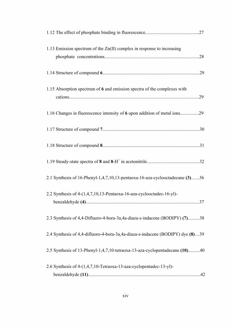
possessing features as well defined as those of molecules themselves. One may say
that supermolecules are to molecules and the intermolecular bond what molecule are
to atom and covalent bond” [1, 2, 4]. So, the supermolecule represents the next level
of the molecule. Figure 1.1 illustrates this relationship between molecular and
supramolecular chemisty in terms of both structures and functions.
Figure 1.1 From molecular components to supramolecular systems [5].
2
Nature of Supramolecular Interactions
The non-covalent interactions have a constitutive role in supramolecular structures.
The non-covalent interactions involved in supramolecular entities may be a
combination of several attractive and repulsive interactions which present different
degrees of strength, directionality, dependence of distance and angles, e.g. ion-
pairing, hydrogen bonding, cation-π, π-π interactions, electrostatic forces, metal ion
coordination, donor-acceptor interactions etc. Their strengths range from weak or
moderate as in hydrogen bonds, to strong or very strong for metal ion coordination.
The former responsible for the overall shape of many proteins, recognition of
substrates by numerous enzymes, and for the double helix structure of DNA,
whereas the latter makes available, by means of a single metal ion binding strengths
that lie in the domain of antigen-antibody complexes (or higher), where many
individual interactions are involved. All of these interactions are vital for
supramolecular systems and effect relating both to the host and guest as well as their
surroundings (e.g. solvation, crystal lattice, gas phase etc.).
In contrast to the covalent interactions that dominate in classical molecules,
non-covalent interactions are however in general weak interactions that bind
together different kinds of building blocks into supramolecular entities. Covalent
bonds are generally shorter than 2 Å, while non-covalent interactions function
within range of several angstroms. The formation of a covalent bond requires
overlapping of partially occupied orbitals of interacting atoms, which share a pair of
electrons. In non-covalent interactions, in turn, no overlapping is necessary because
the attraction comes from the electrical properties of the building blocks. Because of
these weak non-covalent interactions supramolecular species are thermodynamically
3
less stable, kinetically more labile and dynamically more flexible than molecules.
1.2 A Brief Introduction to Fluorescence
The phenomenon of fluorescence was known by the middle of the nineteenth
century. British scientist Sir George G. Stokes first made the observation that the
mineral fluorspar exhibits fluorescence when illuminated with ultraviolet light, and
he coined the word "fluorescence". Stokes observed that the fluorescing light has
longer wavelengths than the excitation light, a phenomenon that has become to be
known as the Stokes shift. Fluorescence microscopy is an excellent method of
studying material that can be made to fluoresce, either in its natural form or when
treated with chemicals capable of fluorescing.
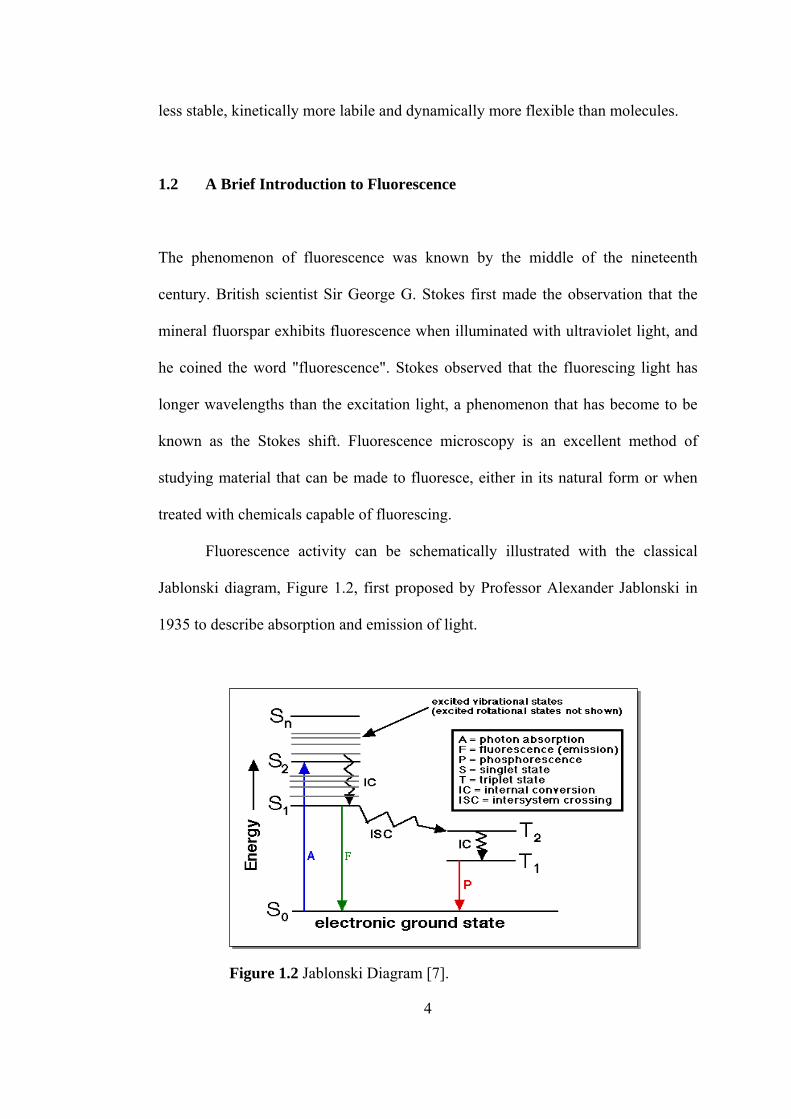
Fluorescence activity can be schematically illustrated with the classical
Jablonski diagram, Figure 1.2, first proposed by Professor Alexander Jablonski in
1935 to describe absorption and emission of light.
Figure 1.2 Jablonski Diagram [7].
4
Once a molecule has absorbed energy in the form of electromagnetic radiation,
there are a number of routes by which it can return to ground state, one of which is
fluorescence. The fluorescence phenomenon may be explained as follows [6];
The absorption of a quantum of light by a molecule results in the elevation
of an electron from the molecule’s ground electronic state (S0) to one of several
vibrational levels in the electronic excited state. In solution, the excited state
molecule rapidly relaxes to the lowest vibrational level of the lowest electronic state
(S1). The energy thus stored in the excited state may be released in several ways.
The electron may return to the electronic ground state with only the release of heat
(radiationless relaxation). After relaxing thermally to the lowest vibrational level of
the S1 state, the electron may return to the S0 state with light emission
(fluorescence) or, if the molecule is sufficiently long-lived in the S1 state, it may
cross into a lower energy triplet state (T1) which is named as intersystem crossing
(ISC). Relaxation from the T1 state to the S0 state can also occur with light emission
in solids (phosphorescence), by the release of energy (radiationless transition), or by
chemical reaction.
1.2.1 Fluorescence as a Signal Transduction Mechanism
Signal transduction is the mechanism by which an interaction of sensor with analyte
yields a measurable form of energy, which can be characterized by various
spectroscopies (e.g., UV, visible, NMR) or may yield electrochemical responses [6].
Fluorescence is identified as the optimal signal transduction mechanism in
potential sensing applications. There are many reasons for which fluorescence is an
5
enormously sensitive technique, because the observing wavelength is always longer
than that of the exciting wavelength, so that a signal may be read versus zero or near
zero background and thus the sensitivity is very high. Owing to the sensitivity of the
fluorescence, even low fluorophore levels can be detected. This optical method
typically works in the millisecond domain, much faster than the time resolution of
other methods. The fluorescence of most biological samples generates background
signal up to about 650 nm, in contrast to some applications in which background
fluorescence is not a problem. Long wavelength fluorophores (i.e., near-IR)
generally avoid these interferences, although sometimes at the cost of severely
narrowing the signal transduction mechanisms that function in such compounds. In
addition to, fluorescence signaling gives the monitoring of both excitation and
emission wavelengths. Fluorescence usually is not destructive so that, fluorescent
chemosensors can be used at all levels of organization from whole organs to isolated
tissue and can be incorporated into the intact functioning cells without destruction.
Fluorescence has the potential for real-time sensing without the need for many steps
associated with other analytical methods. Many fluorescent probes have been
identified that charge intensity in response to analytes of interest, and more has been
synthesized for high specificity and/or sensitivity. Fluorescence signaling can be
used in the creation of remote sensing applications with fiber optic techniques. Fiber
optic sensors (called optodes) permit wireless communication between the detection
element and the analyte, making it particularly attractive for in situ remote sensing
applications [8-9].
6
1.3 Molecular Recognition
Molecular recognition is the key component of the emerging science of
supramolecular chemistry.
Molecular recognition is defined as the energy and the information engaged
in the binding and selection of substrate(s) by a given receptor molecule; it may
involve a specific function [10].
Molecular recognition implies the (molecular) storage and (supramolecular)
read out of molecular information. The source of information processing at the
supramolecular level is represented by molecular recognition events. They may
cause changes in electronic, ionic, optical, and conformational properties and so
translate themselves into the generation of a signal. Molecular recognition processes
may play a role in several key steps: (1) the building up the device from its
components; (2) its integration into supramolecular arrangements; (3) the selective
operation on given species; (4) the response to external physical or chemical stimuli
(light, electrons, ions, molecules, etc.), that may regulate the operation of the device
and switch it on or off; (5) the nature of the signals generated and of the signal
conversion effected (photon-photon, photon-electron, etc.) [3].
Molecular recognition is involved in many fields such as chemistry, biology,
medicine, environment, etc., so that it may be one of the cornerstones of
supramolecular chemistry. In particular, selective detection of metal cations
involved in biological processes (e.g., sodium, potassium, calcium, magnesium), in
clinical diagnosis (e.g., lithium, potassium, aluminum) or in pollution (e.g., lead,
mercury, cadmium) has been given substantial attention [11].
7

There must be large difference between the binding free energies of a given
substrate and of the other substrates on account of high recognition by a receptor
molecule. With the purpose of achievement large differences in affinity several
requirements must be considered [3]:
• Steric (shape and size) complementarity between receptor and substrate;
• Interactional complementarity, i.e. occurrence of complementary binding
sites such as electrostatic, hydrogen bond donor/acceptor, in the correct
disposition on substrate and receptor in order to achieve matching electronic
and nuclear distribution (electrostatic, H-bonding and van der Waals) maps;
• Large contact areas between receptor and substrate;
• Multiple interaction sites, in view of the fact that non-covalent interactions
are weak than covalent bonds;
• Strong overall binding; so as to efficient recognition both high stability and
high selectivity, strong binding of receptor and substrate is required;
• Medium effects play also an important role through the interaction of solvent
molecules with receptor and substrate as well as with each other; hence two
partners should present geometrically matched as solvophobic or solvophilic
domains.
1.4 Chemosensors
A sensor is a device that yields measurable signal in response when interacts with
matter or energy. Chemosensors are the molecules of abiotic origin that signal the
presence of matter of energy [6].
8
A molecular sensor is composed of signaling unit, receptor and substrate. A
receptor portion of the sensor must attract the substrate (guest, analyte). This is the
basis of the molecular recognition event, and receptor can be any of the systems,
such as crown ethers, cryptands, cavitands and so on. The receptor also be in
communication with a signaling unit that is responsive to the guest binding. The
signaling unit can yield signal in the different form, e.g., an emission of
electromagnetic radiation (photochemical sensing), a current (electrochemical
sensing) or an otherwise externally measurable change (e.g. in color or pH).
1.4.1 Fluorescent Chemosensors
A fluorescent chemosensor is a molecule of abiotic origin that gives fluorescence
signal in response when complexes to an analyte reversibly [6]. Fluorescent sensors
undergo photophysical changes (extinction coefficient, fluorescence intensity, shifts
in absorption and emission spectra) due to analyte binding.
There are three matters, fundamental to the design of fluorescent
chemosensors, which have to be known:
• The way of binding of a molecular entity with selectivity.
• How can one generate signals from such binding processes those are easy to
measure?
• The intersect mechanisms for binding and fluorescence signal transduction.
There are some requirements to be satisfied by the fluorescent chemosensors in
order to make use of the advantages of them [12-13]:
9
• The chemosensor must bind the desired analyte selectively as compared to
others present in the medium.
• The chemosensor must show sufficient discrimination between the species
desired and potential candidates.
• The fluorescence should be as intense as possible; also photostability in the
presence of dissolved oxygen is wanted.
• Excitation wavelengths should outnumber 340 nm, due to requirements of
expensive quartz rather than glass microscope optics caused from shorter
wavelengths; in addition to shorter wavelengths are strongly absorbed by
nucleic acids and aromatic amino acids.
• Emission wavelengths should exceed 500 nm to reduce overlap with
autofluorescence of some biological samples.
• Toxicity should be minimized.
• There should a large wavelength shift in the excitation or emission spectrum
or both that is caused from binding of analyte, so that ratioing of signals at
two excitation or two emission wavelengths can be performed [14-15]. Such
rationing is extremely precious for getting rid of the irrelevancies such as cell
thickness, dye concentration, and wavelength-independent variations in
illumination intensity and detection efficiency.
• High extinction coefficients (10-3-10-4 M-1 cm-1) are necessary for a good
signal of the small concentrations of the chemosensor and for ignoring any
buffering of ion fluctuations.
• The solubility of the probes in desired medium in which recognition takes
place is important. Not only are the water soluble probes but also probes in
10
organic phase can be desirable, because parameters such as nature of solvent
(polarity, hydrogen bonding ability, protic or aprotic character), pH, ionic
strength, etc. play a role due to ability of them affecting not only the
efficiency and selectivity of binding, but also the photophysical
characteristic of the fluorophore.
• Binding to cellular constituents and membranes should be minimized or
carefully controlled for specificity. To impermeant through membranes the
ability of highly water solubility should be given by adding enough charged
polar groups such as carboxylates to the chemosensors, so that once
introduced into cells it does not rapidly leak out again. Also these polar
groups should be prevented by nonpolar protecting groups removable by
cytoplasmic enzymes, so that large populations of cells can be loaded with
the indicator by incubating them with the nonpolar membrane-permeant
derivative.
1.5 Principles of Fluorescent Chemosensor Design for Cation Recognition
Ion recognition, especially cation recognition, is a subject of reasonable
interest because of its implications in many fields: chemistry, biology, medicine,
environment, etc. Sodium, potassium, magnesium, calcium are very important
cations for some biological processes such as transmission of nerve impulses,
regulation of cell activity, constitution in metalloenzymes. Detections of lithium and
potassium are vital for the treatment of manic depression and high blood pressure,
respectively. Detection of mercury, lead and cadmium is also significant because of
11
their toxicity for organisms. Fluorescent sensors undergo physical changes,
(extinction coefficient, fluorescence intensity, shifts in absorption and emission
spectra) as marked as possible upon ion binding [16]. In the design of such sensors,
recognition of ions requires special care because attention should be paid to both
recognition and signaling moieties. Such fluorescent sensors are named as the
fluoroionophore that are composed of a fluorophore and ionophore (receptor) linked
to each other, Figure 1.3. The signaling moiety acts as signal transducer, i.e. it
converts the formation (recognition event) into an optical signal expressed as the
changes in the photophysical characteristics of the fluorophore. These fluorescent
sensors are two types with respect to the kind of the linkage of flourophore and
receptor. These are conjugate chemosensor and intrinsic chemosensor. In the
conjugate chemosensor the receptor is linked to the fluorophore via spacer, however
in the intrinsic chemosensor some atoms and groups participating in the
complexation belong to the fluorophore. In the structure more than one ionophore
and/or more than one fluorophore can present. The connection between the receptor
and fluorophore is a very important aspect of fluorescent chemosensor design since
the strongest perturbation of the photophysical properties of the fluorophore by the
ion is desired.
For designing of fluorescence chemosensors for cations, there are an enormous
number of factors that should be paid attention, some of which are listed below:
• Size match between cation and host (receptor) cavity;
• Electrostatic charge;
• Solvent (polarity, hydrogen bonding and coordinating ability);
• Size match between cation and host (receptor) cavity;
12
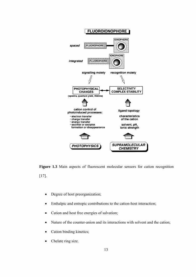
Figure 1.3 Main aspects of fluorescent molecular sensors for cation recognition
[17].
• Degree of host preorganization;
• Enthalpic and entropic contributions to the cation-host interaction;
• Cation and host free energies of salvation;
• Nature of the counter-anion and its interactions with solvent and the cation;
• Cation binding kinetics;
• Chelate ring size.
13
The photophysical changes of a fluorescent probe on recognition should be as
marked as possible. Probes undergoing shifts of emission and/or excitation spectra
(or appearance or disappearance of bands) are preferable to those that undergo only
changes in fluorescence intensity: indeed, after calibration the ratio of the
fluorescence intensities at two appropriate emission and excitation wavelengths
provides a measure of the ion or molecule concentration which is independent of
the probe concentrations (provided that the ion is in excess) and is insensitive to
intensity of incident light, scattering, inner-filter effects, and photobleaching [18].
The photophysical changes of a fluorescent chemosensor during interaction
with analyte can involve various photoinduced processes, such as energy transfer,
excimer or exciplex formation or disappearance, twisted intramolecular charge
transfer, etc., but the most important two of them are:
• Photoinduced Electron Transfer (PET)
• Photoinduced Charge Transfer (PCT)
1.5.1 Fluorescent Photoinduced Electron Transfer (PET) Cation
Sensors
Fluorescent PET sensors can be formalized as ‘fluorophore-spacer-receptor’
systems of modular structure [19]. The fluorescent PET sensors also named as
intrinsic chemosensors in which the ligand is an integral part of fluorophore π-
system. It is well-established that fluorescence signal to be obtained using such
probes is stronger and/or more information by creating a different absorbing species
on metal ion binding, enabling rationing of the emission signals thereby cancelling
14
concentration dependent artifacts [20]. To make arrangements for various
excitation/emission wavelengths and for a variety of a given guest, the photon- and
guest-interaction sites can be chosen. This choice has several constraints; however
for the design logic of fluorescent PET sensors these constraints are very important.
The energy stored in the fluorophore excited state upon photon absorption must be
sufficient to oxidize the guest-free receptor and to simultaneously reduce the
fluorophore [21-22]. This is the thermodynamic criterion for a PET sensor, but if
the cationic guest is considered it is easy to inhibit the PET process since the
oxidation potential of the guest occupied receptor is considerably higher than that of
the guest-free receptor. The excited state energy of the optically pumped
fluorophore thus remains unused and return as a photon. In the PET sensors the
spacer maintains the modularity of the system, which results in the additivity of
component parameters, and is only violated by relatively long range forces [23-24].
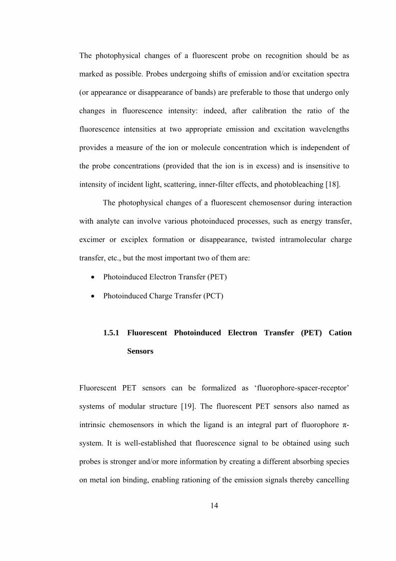
Figure 1.4 illustrates how a cation can control the photoinduced electron
transfer (PET) in a fluoroionophore in which the cation receptor is an electron
donor (e.g. amino group) and the fluorophore plays the role of an acceptor. Upon
excitation of the fluorophore, an electron of the highest occupied molecular orbital
(HOMO) is promoted to the lowest unoccupied molecular orbital (LUMO), which
enables PET from the HOMO of the donor (belonging to the free cation receptor) to
that of the fluorophore, causing fluorescence quenching of the latter. Upon cation
binding, the redox potential of the donor is raised so that the relevant HOMO
becomes lower in energy than that of the fluorophore; consequently, PET is not
possible any more and fluorescence quenching is suppressed; that means,
fluorescence intensity is enhanced upon cation binding [18].
15
Example:
O O
O
O
N
O
CH2 K+
O O
O
O
N
O
CH2
ΦF = 0.003 (in methanol) ΦF = 0.14
Figure 1.4 Principle of cation recognition based on cation control of photoinduced
electron transfer in nonconjugated donor-acceptor systems [17].
(Example from [25]).
The fluorescence of aromatic hydrocarbons is quenched by aliphatic or
aromatic amines due to photoinduced electron transfer from the latter to the former.
In spite of the fact that most PET fluorescent sensors are based on this
process, other PET mechanisms can happen with transition metal ions [26-27].
There are various examples of PET sensors some of which are shown in
Figure1.5.
16

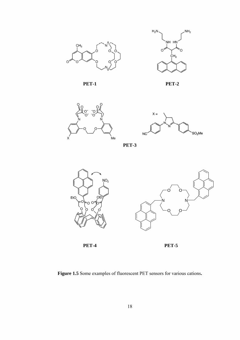
PET-1 is the cryptant-based PET sensor. It has beeen successfully used for
monitoring levels of potassium in blood and across biological membranes [28].
PET-2 is the podand-based PET sensor [29-30]. It can bind Cu(II) and Ni(II) and
favor oxidation of these cations to the trivalent state. Fluorescence quenching of the
anthracene upon binding should be ascribed in this case to an electron transfer from
the reducing divalent metal center. PET-3 is an example of chelating PET sensor. It
efficiently binds divalent hard cation of Ca2+ [31]. PET-4 is the calixarene-based
PET sensor that is designed for selective recognition of Na+. It contains four
carbonyl functions that bind Na+. Complexation with Na+ prevents close approach
of pyrene and nitrobenzene and thus reduces the probability of PET. The
fluorescence quantum yield increases from 0.0025 to 0.016 [32]. PET-5 is the PET
sensor involving excimer formation. There is a large change in the
monomer/excimer ratio because of cation binding. There is a concomitant increase
in the overall fluorescence emission as a result of the reduction of PET from the
nitrogen atom to the phenyl groups. Among the investigated metal ions, the larger
stability constants of the complexes were obtained for K+ and Ba2+, in accordance
with the size of these cations with respect to the crown diameter [33].
17
PET-1 PET-2 PET-3
N
O O
N
O O
PET-4 PET-5
Figure 1.5 Some examples of fluorescent PET sensors for various cations.
18
1.5.2 Fluorescent Photoinduced Charge Transfer (PCT) Cation
Sensors
A fluorescent PCT sensor contains an electron-donating group (often an amino
group) conjugated to an electron-withdrawing group. Upon excitation by light, the
PCT sensor undergoes intramolecular charge transfer from the donor to the acceptor;
so that a change in dipole moment can occur. This change results in a Stokes shift
that depends on the micro environment of the fluorophore. If cations interact closely
with the donor or the acceptor moiety of the fluorophore, photophysical properties of
the fluorophore will change, because the complexed cation affects the efficieny of
the intramolecular charge transfer [16-18].
Interaction of an electron donor group (like an amino group) within
fluorophore with a cation causes the reduction of the electron-donating character of
this group. A blue shift of the absorption spectrum and a decrease of the extinction
coefficient are expected because of the resulting reduction of conjugation. On the
other hand, an interaction of a cation with the acceptor group enhances the electron-
withdrawing character of this group; as a result that the absorption spectrum is red-
shifted and molar absorption coefficient is increased. The fluorescence spectra are in
principle shifted in the same direction as those of the absorption spectra. Besides of
these shifts, changes in quantum yields and lifetimes are often observed. All these
photophysical effects are noticeably dependent on the charge and the size of the
cation and selectivity of these effects are expected.
The photophysical changes upon cation binding can also be described in
terms of charge dipole interaction [34]. Let us consider only the case where the
19
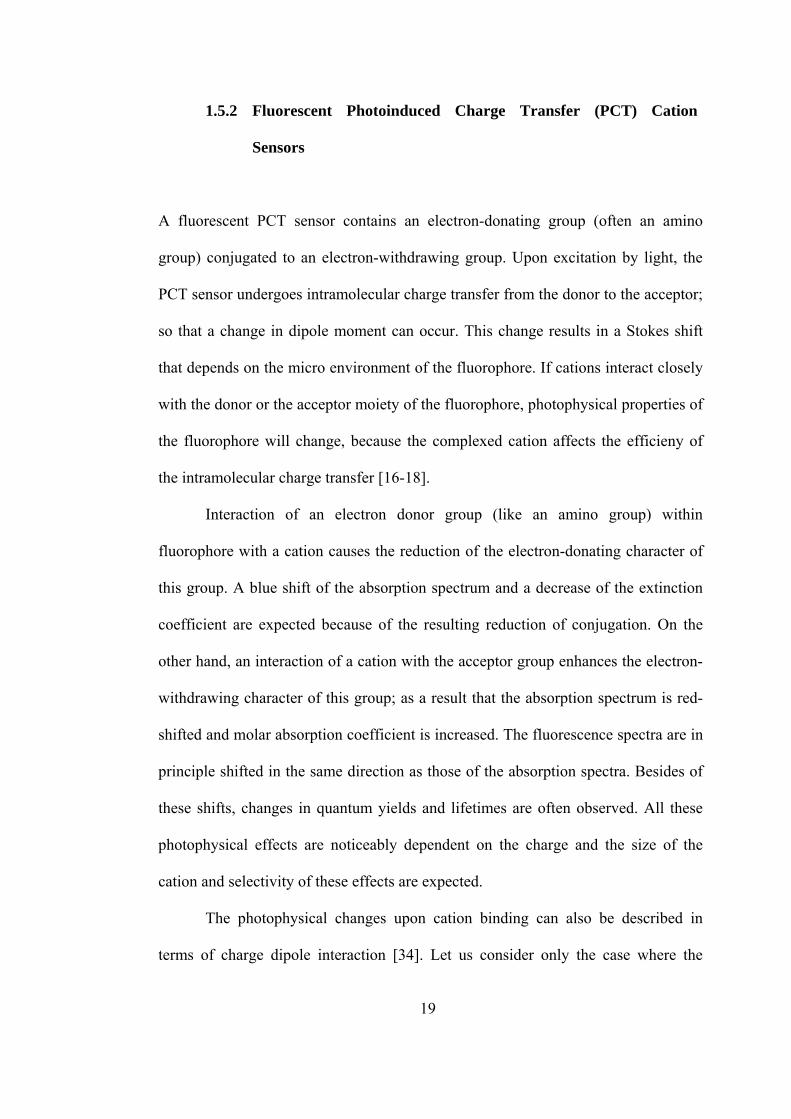
dipole moment in the excited state is larger than that in the ground state. Then, when
the cation interacts with the donor group, the excited state is more strongly
destabilized by the cation than the ground state, and a blue shift of the absorption
and emission spectra is expected. Conversely, when the cation interacts with the
acceptor group, the excited state is more stabilized by the cation than the ground
state, and this leads to a red shift of the absorption and the emission spectra, Figure
1.6.
Figure 1.6 Spectral displacements of PCT sensors resulting from interaction of a
bound cation with an electron-donating or electron-withdrawing group [17].
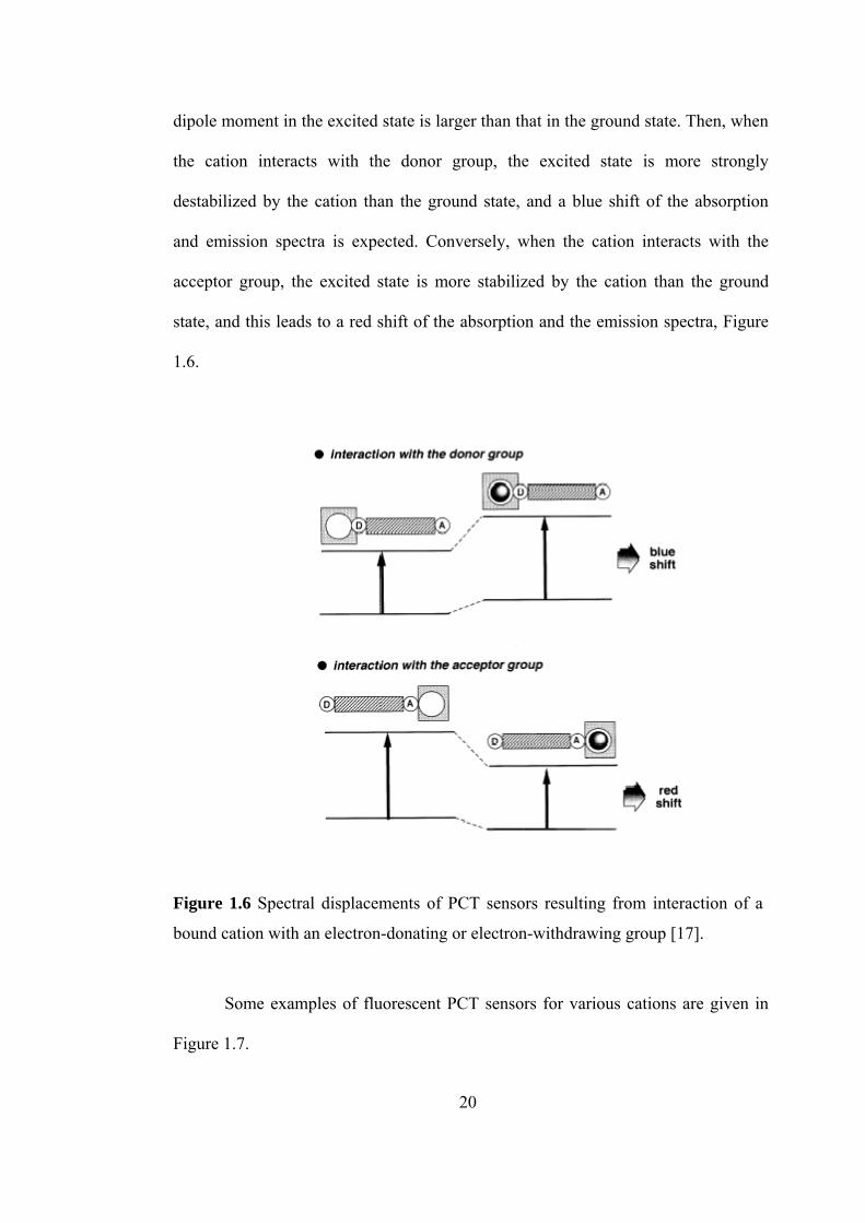
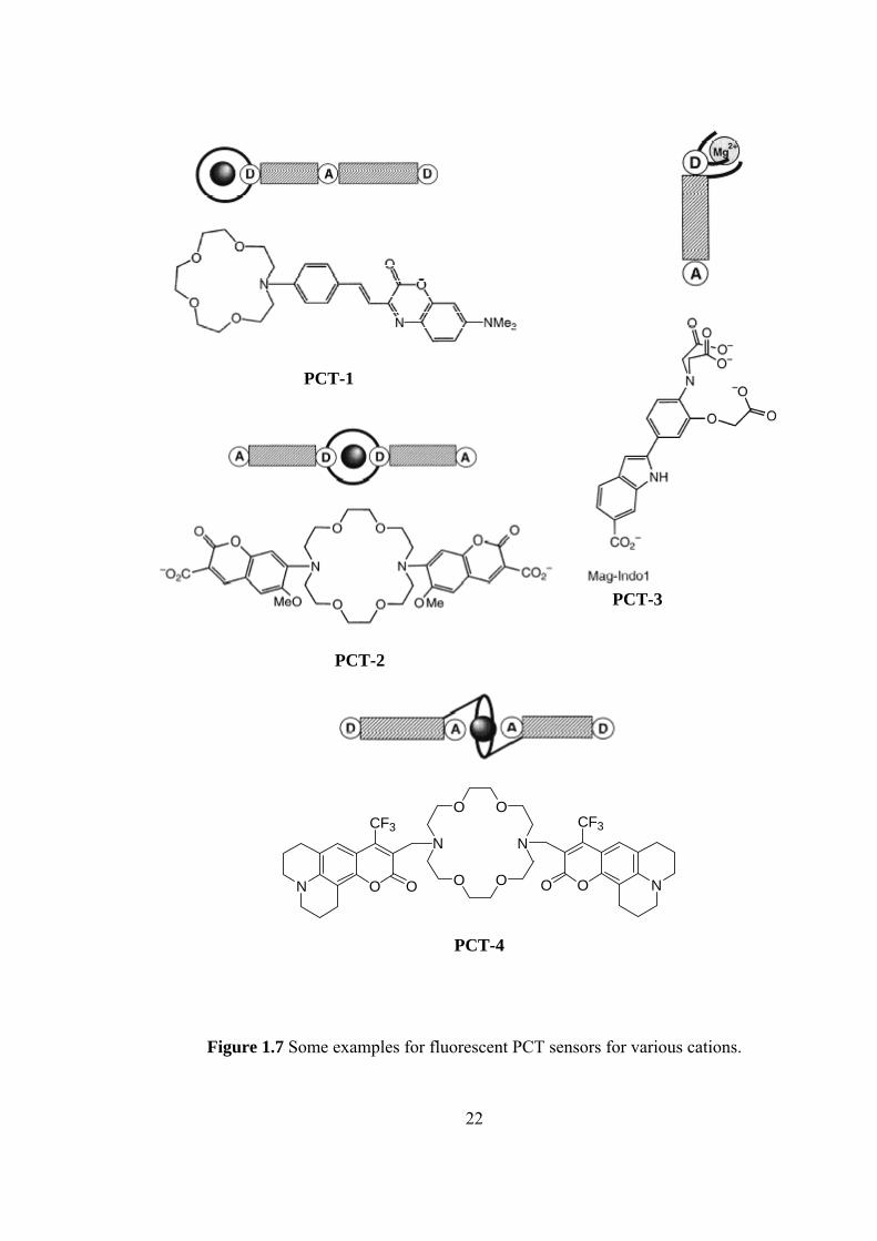
Some examples of fluorescent PCT sensors for various cations are given in
Figure 1.7.
20

PCT-1 is one of the first crown-containing fluorescent PCT sensors that have
been designed by Valuer. The fluorescence maximum shifts from 642 nm for the
free ligand to 574 nm for the Ca2+ complex in acetonitrile [35-37]. It is not only
responsive to alkaline-earth cations but also to divalent heavy metal ions Hg2+ and
Pb2+ [38]. PCT-2 shows selectivity for K+. The oxygen atom of the methoxy
substituent of the fluorophore can interact with a cation; binding efficiency and
selectivity for potassium are thus better than those of the crown alone [39]. PCT-3
(Mag-Indo1) is a chelating PCT sensor that is selective for Mg2+ [40]. PCT-4 is a
PCT sensor in which the bound cation interacts with an electron-withdrawing group.
In the sensor, the carbonyl groups of the two coumarin moieties participate in the
complexes. Direct interaction between these groups and the cation explains the high
stability of constants and the photophysical changes. In addition to the shifts of the
absorption and emission spectra, an interesting specific increase in the fluorescence
quantum yield upon binding of K+ and Ba2+ ions has been observed [41].
21
PCT-1
PCT-4
Figure 1.7 Some examples for fluorescent PCT sensors for various cations.
PCT-3
PCT-2
N
O O
N
O ON O O
CF3
O NO
CF3
22
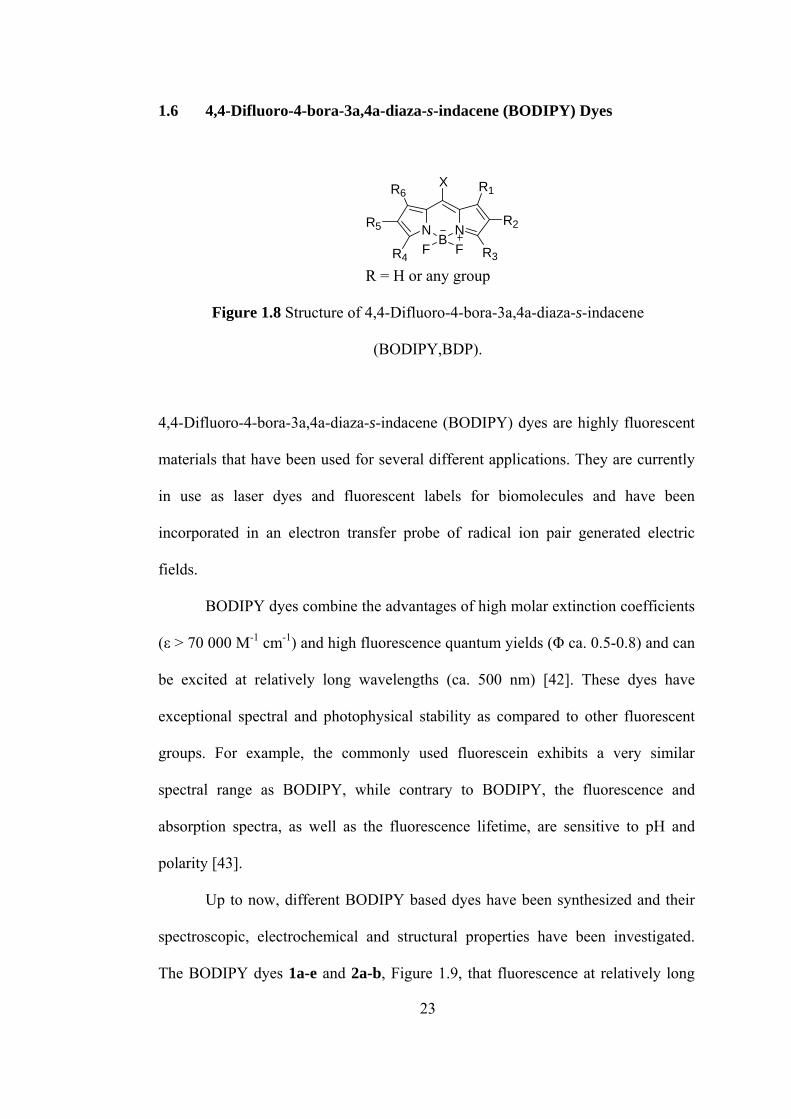
1.6 4,4-Difluoro-4-bora-3a,4a-diaza-
Figure 1.8 Structure of 3a,4a-diaza-s-indacene
4,4-Difluoro-4-bora-3a,4a-diaza-s-indacene (BODIPY) dyes are highly fluorescent
BODIPY dyes combine the advantages of high molar extinction coefficients
(ε > 70
ow, different BODIPY based dyes have been synthesized and their
spectro
NB
N
s-indacene (BODIPY) Dyes
F F
R6
R4 R3
R1X
R2R5
R = H or any group
4,4-Difluoro-4-bora-
(BODIPY,BDP).
materials that have been used for several different applications. They are currently
in use as laser dyes and fluorescent labels for biomolecules and have been
incorporated in an electron transfer probe of radical ion pair generated electric
fields.
000 M-1 cm-1) and high fluorescence quantum yields (Φ ca. 0.5-0.8) and can
be excited at relatively long wavelengths (ca. 500 nm) [42]. These dyes have
exceptional spectral and photophysical stability as compared to other fluorescent
groups. For example, the commonly used fluorescein exhibits a very similar
spectral range as BODIPY, while contrary to BODIPY, the fluorescence and
absorption spectra, as well as the fluorescence lifetime, are sensitive to pH and
polarity [43].
Up to n
scopic, electrochemical and structural properties have been investigated.
The BODIPY dyes 1a-e and 2a-b, Figure 1.9, that fluorescence at relatively long
23
wavelengths were synthesized by Burgess [44]. These dyes have several potential
applications. They could aid the design of fluorescent chemosensors, act as receptor
molecules for dynamics, structure and function of biomolecules, provide light-
harvesting systems for artificial photosynthesis, and for multipigment arrays in
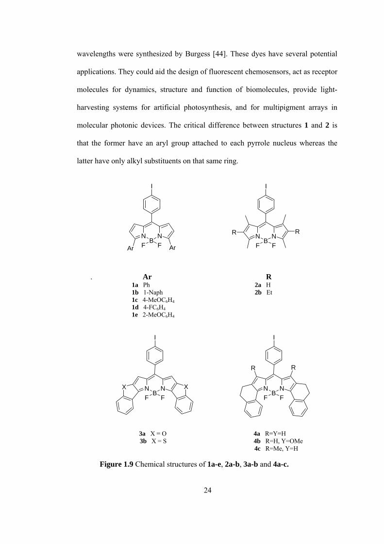
molecular photonic devices. The critical difference between structures 1 and 2 is
that the former have an aryl group attached to each pyrrole nucleus whereas the
latter have only alkyl substituents on that same ring.
. Ar R
1a Ph 2a H
3b X = S 4b R=H, Y=OMe =H
NB
N
F FAr Ar
I
NB
NF F
RR
I
1b 1-Naph 2b Et 1c 4-MeOC6H4 1d 4-FC6H4
1e 2-MeOC6H4
I
NB
NF F
X X NB
NF F
I
RR
3a X = O 4a R=Y=H 4c R=Me, Y
Figure 1.9 Chemical structures of 1a-e, 2a-b, 3a-b and 4a-c.
24
UV ab compounds 1
lso have been prepared
sorption and fluorescence emission data were compared for
and 2. Absorption and fluorescence emission maxima for compounds 1 occur at
higher wavelengths than for compounds 2 because of the added conjugation in
compounds 1, and the Stokes shifts for the aryl-substituted compounds 1 are larger
than for the alkyl-substituted compounds 2. However, the fluorescence quantum
yields for the aryl-substituted compounds tend to be lower than the corresponding
alkyl-substituted ones which are caused from nonradiative energy loss due to
spinning motions about the C-aryl single bonds. Also electrochemical properties of
these compounds have been investigated by Burgess [44]. These studies
demonstrate that electron-withdrawing and electron-releasing aryl substituents have
less impact on the oxidation and reduction potentials than expected, consistent with
twisting of the aryl substituents out of the BODIPY plane.
The constrained, aryl-substituted BODIPY dyes a
[45-47]. The planar aromatic compounds 3a-b and 4a-c, Figure 1.9, [48] were
synthesized and investigated to see if they have more favorable fluorescence
characteristics than the unconstrained systems 2, because molecular constraints are
well-known to enhance fluorescence. Dye types 3 and 4 have relatively rigid
conformations caused by heteroatom or ethylene bridge linkers that preclude free
rotation of the substituted-benzene molecular fragments. In the event, the new types
3 and 4 have longer λmax abs (620-660 nm) and λmax fluor (630-680 nm) values than
compounds 2. They also exhibit higher extinction coefficients (> 100 000 M-1 cm-1,
except for 3b). Their fluorescent quantum yields are high (up to 0.72 for 4c), with
the exception of compound 3b.
25
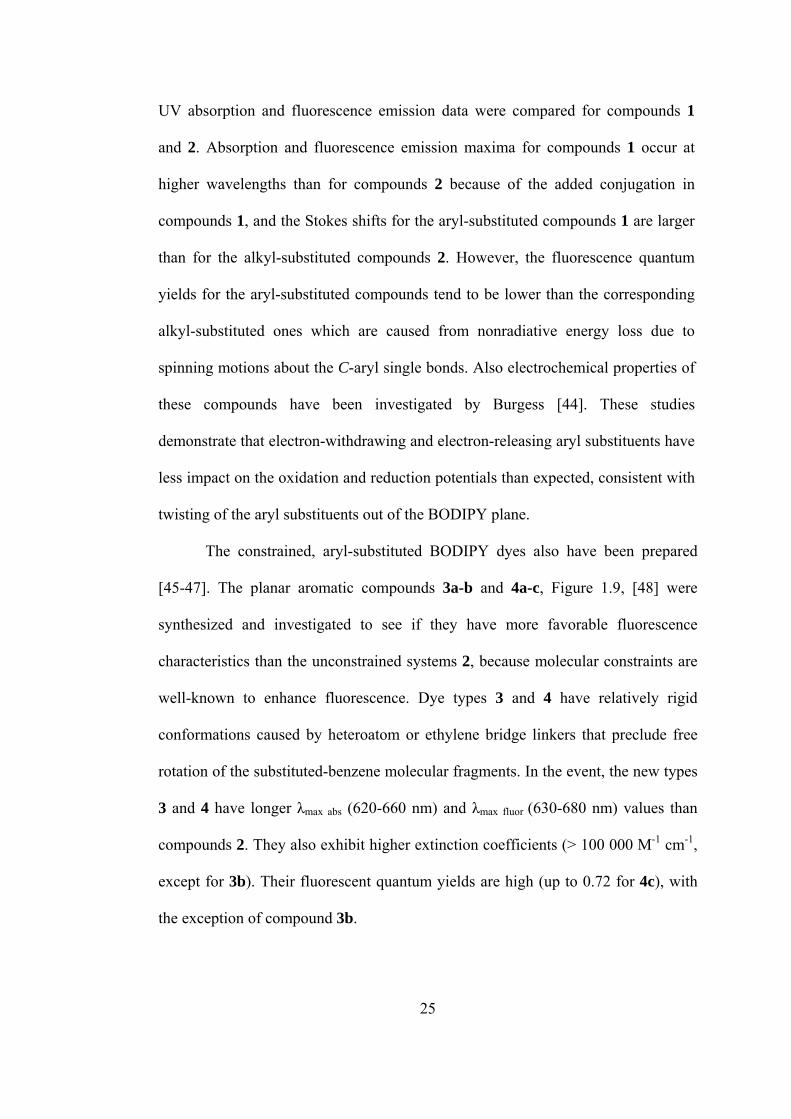
The BODIPY dyes that show chiral discrimination were synthesized.
Compound 5 in Figure 1.10 [49] is the optically active binaphthalene boron-
dipyrromethane (BODIPY) which shows chiral discrimination towards the
enantiomers of 1-phenylethylamine (PEA) by distinguishable quenching rates of the
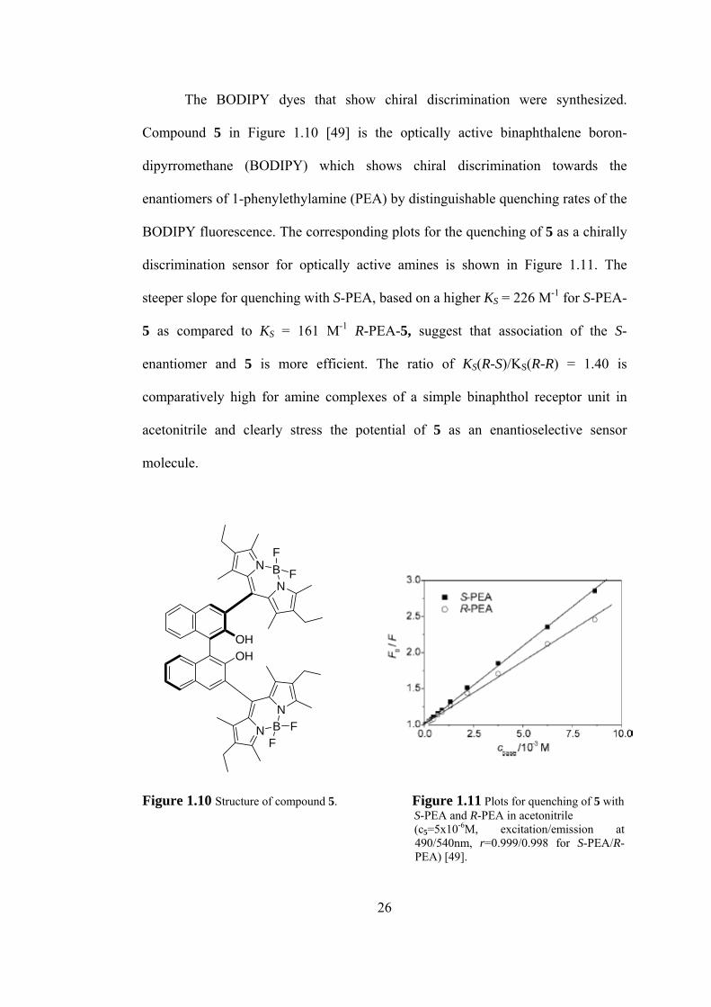
BODIPY fluorescence. The corresponding plots for the quenching of 5 as a chirally
discrimination sensor for optically active amines is shown in Figure 1.11. The
steeper slope for quenching with S-PEA, based on a higher KS = 226 M-1 for S-PEA-
5 as compared to KS = 161 M-1 R-PEA-5, suggest that association of the S-
enantiomer and 5 is more efficient. The ratio of KS(R-S)/KS(R-R) = 1.40 is
comparatively high for amine complexes of a simple binaphthol receptor unit in
acetonitrile and clearly stress the potential of 5 as an enantioselective sensor
molecule.
BN
N
BN
N
OHOH
F
F
FF
Figure 1.10 Structure of compound 5. Figure 1.11 Plots for quenching of 5 with S-PEA and R-PEA in acetonitrile (c5=5x10⁻⁶M, excitation/emission at
490/540nm, r=0.999/0.998 for S-PEA/R-PEA) [49].
26
The combination of the chiral recognition features of a substituted 1,1’-
binaphthalene unit with the favorable spectroscopic properties of the BODIPY
chromophore presents a promising approach towards “ON-OFF” signaling of chiral
analytes, advantageous both in spectral region and time domain.
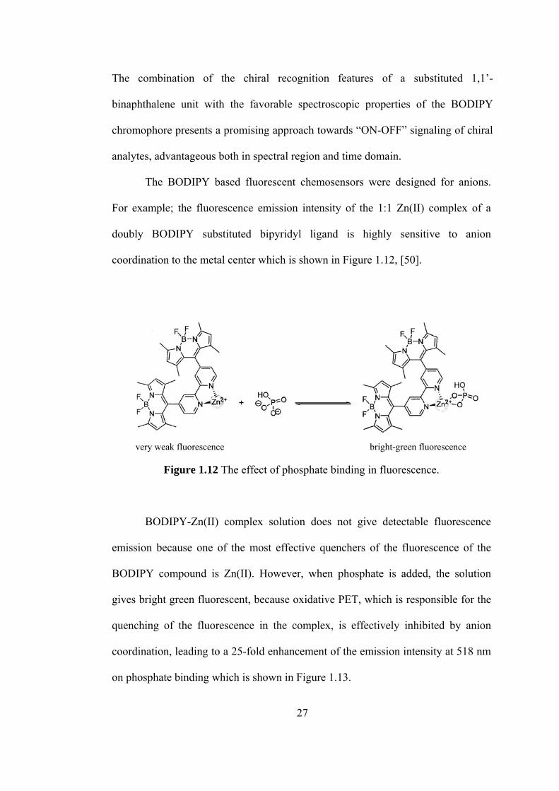
The BODIPY based fluorescent chemosensors were designed for anions.
For example; the fluorescence emission intensity of the 1:1 Zn(II) complex of a
doubly BODIPY substituted bipyridyl ligand is highly sensitive to anion
coordination to the metal center which is shown in Figure 1.12, [50].
very weak fluorescence bright-green fluorescence
Figure 1.12 The effect of phosphate binding in fluorescence.
BODIPY-Zn(II) complex solution does not give detectable fluorescence
emission because one of the most effective quenchers of the fluorescence of the
BODIPY compound is Zn(II). However, when phosphate is added, the solution
gives bright green fluorescent, because oxidative PET, which is responsible for the
quenching of the fluorescence in the complex, is effectively inhibited by anion
coordination, leading to a 25-fold enhancement of the emission intensity at 518 nm
on phosphate binding which is shown in Figure 1.13.
27
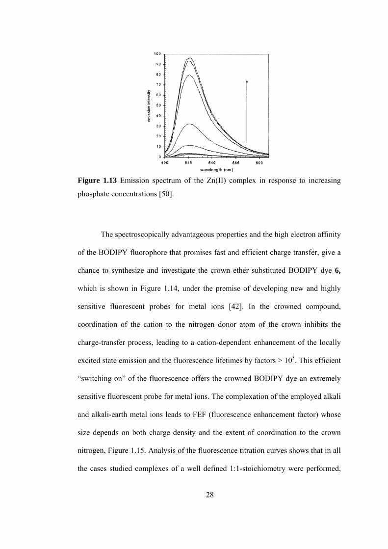
Figure 1.13 Emission spectrum of the Zn(II) complex in response to increasing
phosphate concentrations [50].
The spectroscopically advantageous properties and the high electron affinity
of the BODIPY fluorophore that promises fast and efficient charge transfer, give a
chance to synthesize and investigate the crown ether substituted BODIPY dye 6,
which is shown in Figure 1.14, under the premise of developing new and highly
sensitive fluorescent probes for metal ions [42]. In the crowned compound,
coordination of the cation to the nitrogen donor atom of the crown inhibits the
charge-transfer process, leading to a cation-dependent enhancement of the locally
excited state emission and the fluorescence lifetimes by factors > 103. This efficient
“switching on” of the fluorescence offers the crowned BODIPY dye an extremely
sensitive fluorescent probe for metal ions. The complexation of the employed alkali
and alkali-earth metal ions leads to FEF (fluorescence enhancement factor) whose
size depends on both charge density and the extent of coordination to the crown
nitrogen, Figure 1.15. Analysis of the fluorescence titration curves shows that in all
the cases studied complexes of a well defined 1:1-stoichiometry were performed,
28
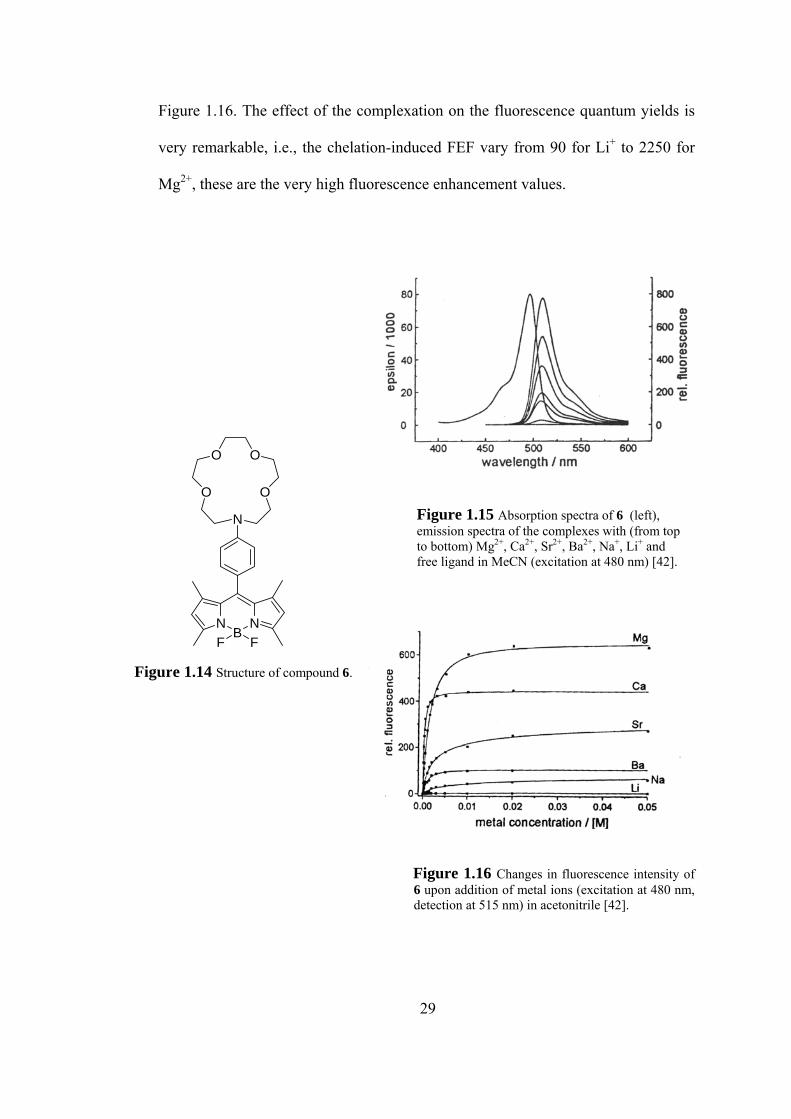
Figure 1.16. The effect of the complexation on the fluorescence quantum yields is
very remarkable, i.e., the chelation-induced FEF vary from 90 for Li+ to 2250 for
Mg2+, these are the very high fluorescence enhancement values.
OO
O O
N
NB
NF F
Figure 1.15 Absorption spectra of 6 (left), emission spectra of the complexes with (from top to bottom) Mg2+, Ca2+, Sr2+, Ba2+, Na+, Li+ and free ligand in MeCN (excitation at 480 nm) [42].
Figure 1.14 Structure of compound 6.
Figure 1.16 Changes in fluorescence intensity of 6 upon addition of metal ions (excitation at 480 nm, detection at 515 nm) in acetonitrile [42].
29
In addition, thia aza crown substituted BODIPY dye that shows a strong
fluorescence enhancement selectively with HgII, AgI, and CuII is synthesized [51].
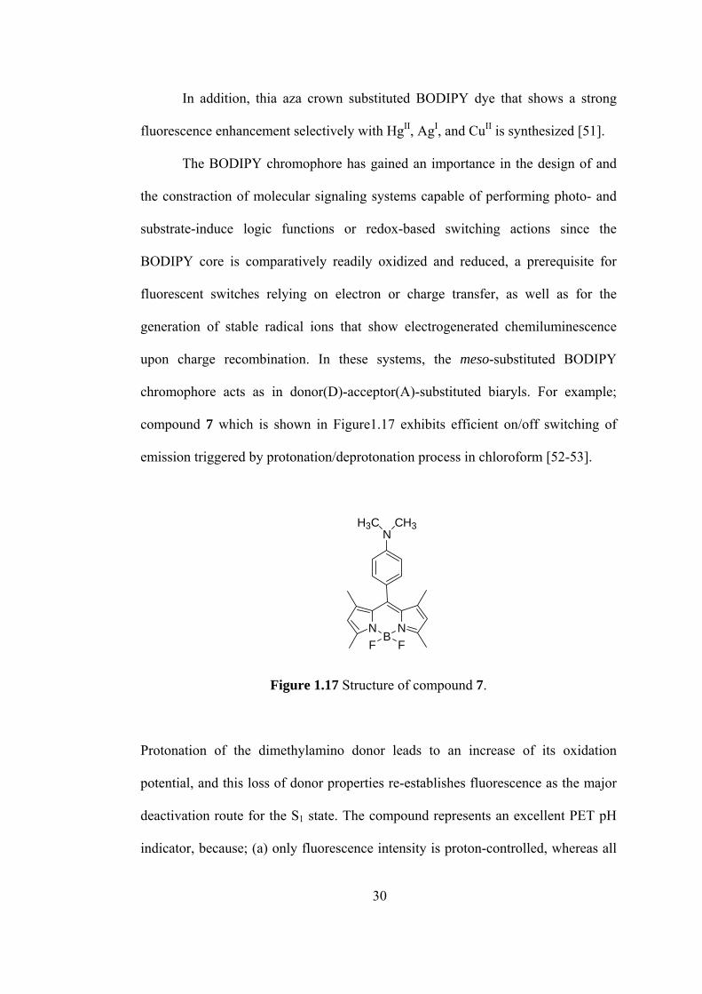
The BODIPY chromophore has gained an importance in the design of and
the constraction of molecular signaling systems capable of performing photo- and
substrate-induce logic functions or redox-based switching actions since the
BODIPY core is comparatively readily oxidized and reduced, a prerequisite for
fluorescent switches relying on electron or charge transfer, as well as for the
generation of stable radical ions that show electrogenerated chemiluminescence
upon charge recombination. In these systems, the meso-substituted BODIPY
chromophore acts as in donor(D)-acceptor(A)-substituted biaryls. For example;
compound 7 which is shown in Figure1.17 exhibits efficient on/off switching of
emission triggered by protonation/deprotonation process in chloroform [52-53].
NB
NF F
NH3C CH3
Figure 1.17 Structure of compound 7.
Protonation of the dimethylamino donor leads to an increase of its oxidation
potential, and this loss of donor properties re-establishes fluorescence as the major
deactivation route for the S1 state. The compound represents an excellent PET pH
indicator, because; (a) only fluorescence intensity is proton-controlled, whereas all
30
other spectral parameters are pH invariant, (b) it has very high photostability, (c)
high fluorescence quantum yield, and (d) long excitation wavelength which make
the sensor excitable by inexpensive blue-green light-emitting diodes.
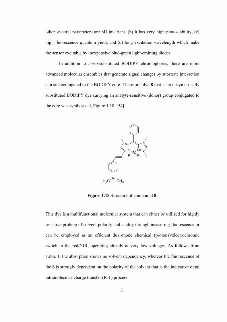
In addition to meso-substituted BODIPY chromophores, there are more
advanced molecular ensembles that generate signal changes by substrate interaction
at a site conjugated to the BODIPY core. Therefore, dye 8 that is an unsymetrically
substituted BODIPY dye carrying an analyte-sensitive (donor) group conjugated to
the core was synthesized, Figure 1.18, [54].
NB
NF F
NCH3H3C
Figure 1.18 Structure of compound 8.
This dye is a multifunctional molecular system that can either be utilized for highly
sensitive probing of solvent polarity and acidity through measuring fluorescence or
can be employed as an efficient dual-mode chemical (protons)/electrochromic
switch in the red/NIR, operating already at very low voltages. As follows from
Table 1, the absorption shows no solvent dependency, whereas the fluorescence of
the 8 is strongly dependent on the polarity of the solvent that is the indicative of an
intramolecular-charge transfer (ICT) process.
31
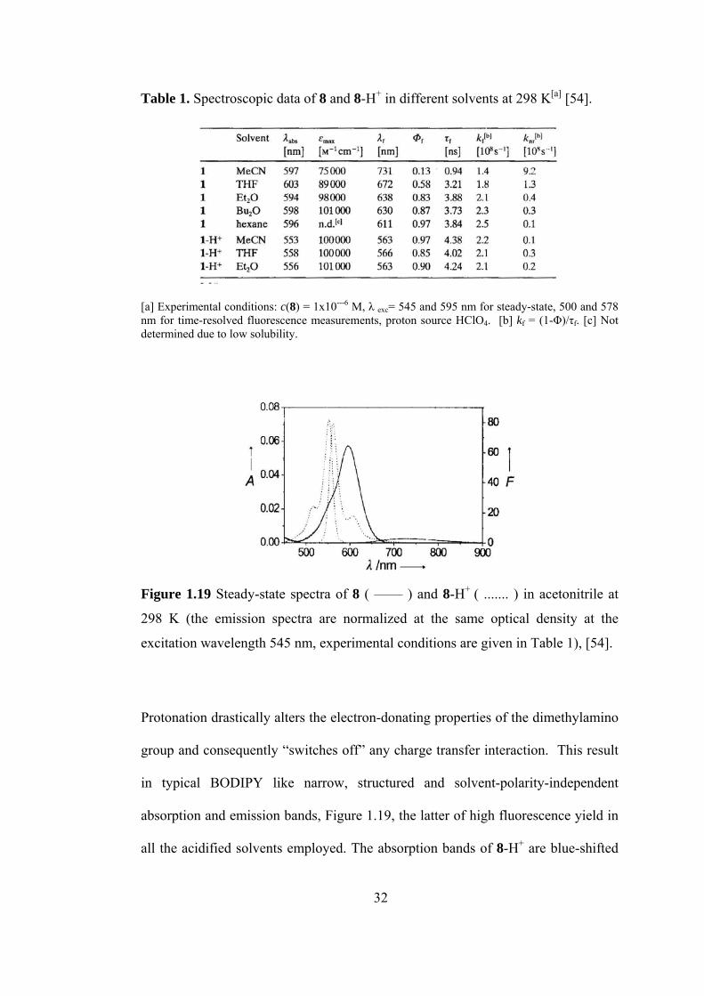
Table 1. Spectroscopic data of 8 and 8-H+ in different solvents at 298 K[a] [54].
[a] Experimental conditions: c(8) = 1x10---6 M, λ exc= 545 and 595 nm for steady-state, 500 and 578 nm for time-resolved fluorescence measurements, proton source HClO4. [b] kf = (1-Φ)/τf. [c] Not determined due to low solubility.
Figure 1.19 Steady-state spectra of 8 ( —— ) and 8-H+ ( ....... ) in acetonitrile at
298 K (the emission spectra are normalized at the same optical density at the
excitation wavelength 545 nm, experimental conditions are given in Table 1), [54].
Protonation drastically alters the electron-donating properties of the dimethylamino
group and consequently “switches off” any charge transfer interaction. This result
in typical BODIPY like narrow, structured and solvent-polarity-independent
absorption and emission bands, Figure 1.19, the latter of high fluorescence yield in
all the acidified solvents employed. The absorption bands of 8-H+ are blue-shifted
32
by about 40 nm compared to those of 8, and the width of the band is further
reduced, stressing the influence of the conjugated (unprotonated) dimethylamino
substituent on the spectroscopic properties of 8 in solvents any polarity.
1.7 Aim of the Study
Our aim in this thesis study is to synthesize and investigate the ion sensing
properties of novel boradiazaindacene dyes.
We have synthesized a series of BODIPY based dyes carrying a cation-
sensitive phenylazacrown ether group conjugated to the polymethinic part of the
chromophore. These dyes are the examples for efficient CT (charge transfer)
systems in which the extension of the conjugation over the pyrrole ring shifts the
absorption and emission to longer wavelength. These chemosensors are expected to
generate signal changes by metal ion complexation and protonation. With this
study, we present a new design concept for sensitive probes for metal ions showing
absorption and fluorescence changes in the red/NIR. We expect this project to open
a new path for the synthesis and development of novel sensors of cations in
different media, which require red to near-IR excitation and emission. Such an
cation chemosensor would be very valuable in studying various biological
phenomena.
33
CHAPTER 2
EXPERIMENTAL
2.0 Instrumentation
In this study, the compounds are characterized by Nuclear Magnetic Resonance
technique. 1H and 13C-Nuclear Magnetic Resonance spectra were recorded on a
Bruker Instruments Avance Series-Spectropin DPX-400 Ultra shield (400 MHz)
High Performance digital FT-NMR Spectrometer (METU NMR Laboratory by
using CDCl3 as solvent and chemical shifts δ in ppm from tetramethylsilane as an
internal reference). Spin multiplicities are indicated by the following symbols: s
(singlet), d (doublet), t (triplet), m (multiplet).
Electronic absorption spectra were recorded on a Shimadzu UV-1601
spectrophotometer. A Perkin-Elmer LS 50 B luminescence spectrometer was used
for recording the fluorescence-emission spectra. The emission was detected with a
Hamamatsu R928 PMT. All instrumental parameters were controlled by
Fluorescence Data Manager Software (FLDM). Measurements were conducted at
25ºC using a 1x0.5 cm rectangular quartz cuvette.
Column chromatography was performed using Merck Silica Gel 60 (particle
size:0.040-0.0963, 230-400 mesh ASTM). All reactions were monitored by thin
34
layer chromatography using Merck Silica Gel 60 F254 TLC Aluminum Sheets
20x20cm. All chemicals and solvents purchased from Aldrich Chemical Company
unless otherwise stated.
2.1 Synthesis of 16-(4-Propenyl-phenyl)-1,4,7,10,13-pentaoxa-16-aza-
cylooctadecane Substituted BODIPY Dye (8)
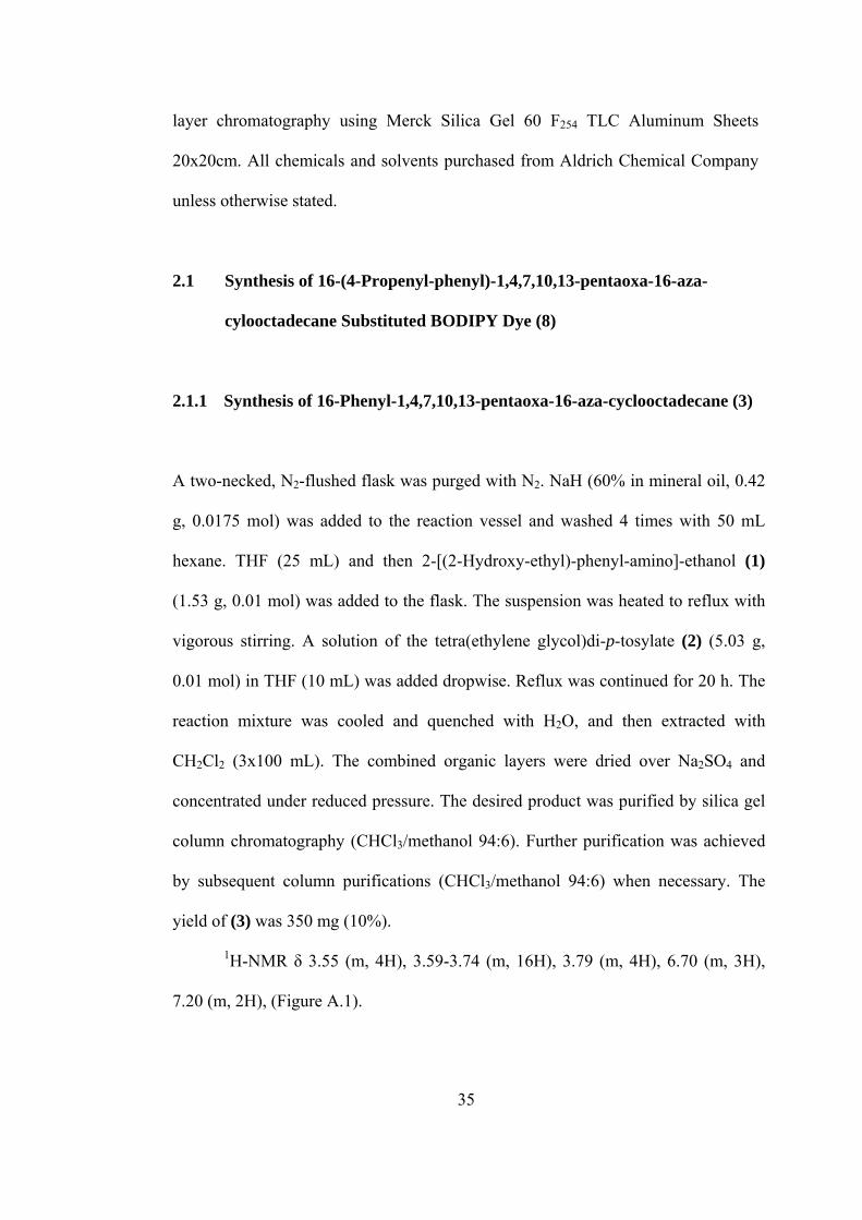
2.1.1 Synthesis of 16-Phenyl-1,4,7,10,13-pentaoxa-16-aza-cyclooctadecane (3)
A two-necked, N2-flushed flask was purged with N2. NaH (60% in mineral oil, 0.42
g, 0.0175 mol) was added to the reaction vessel and washed 4 times with 50 mL
hexane. THF (25 mL) and then 2-[(2-Hydroxy-ethyl)-phenyl-amino]-ethanol (1)
(1.53 g, 0.01 mol) was added to the flask. The suspension was heated to reflux with
vigorous stirring. A solution of the tetra(ethylene glycol)di-p-tosylate (2) (5.03 g,
0.01 mol) in THF (10 mL) was added dropwise. Reflux was continued for 20 h. The
reaction mixture was cooled and quenched with H2O, and then extracted with
CH2Cl2 (3x100 mL). The combined organic layers were dried over Na2SO4 and
concentrated under reduced pressure. The desired product was purified by silica gel
column chromatography (CHCl3/methanol 94:6). Further purification was achieved
by subsequent column purifications (CHCl3/methanol 94:6) when necessary. The
yield of (3) was 350 mg (10%).
1H-NMR δ 3.55 (m, 4H), 3.59-3.74 (m, 16H), 3.79 (m, 4H), 6.70 (m, 3H),
7.20 (m, 2H), (Figure A.1).
35
13C-NMR δ 52.40, 56.31, 61.42, 69.43, 70.71, 70.86, 112.80, 117.00, 129.63,
148.35, (Figure A.2).
OO
O
OON
O O O O OTsTsN
NaH/THF
OHOH
20 h/reflux(1)
(2)
(3)
Figure 2.1 Synthesis of 16-Phenyl-1,4,7,10,13-pentaoxa-16-aza-cyclooctadecane (3)
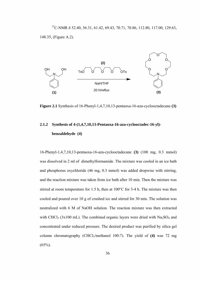
2.1.2 Synthesis of 4-(1,4,7,10,13-Pentaoxa-16-aza-cyclooctadec-16-yl)-
benzaldehyde (4)
16-Phenyl-1,4,7,10,13-pentaoxa-16-aza-cyclooctadecane (3) (100 mg, 0.3 mmol)
was dissolved in 2 ml of dimethylformamide. The mixture was cooled in an ice bath
and phosphorus oxychloride (46 mg, 0.3 mmol) was added dropwise with stirring,
and the reaction mixture was taken from ice bath after 10 min. Then the mixture was
stirred at room temperature for 1.5 h, then at 100°C for 3-4 h. The mixture was then
cooled and poured over 10 g of crushed ice and stirred for 30 min. The solution was
neutralized with 6 M of NaOH solution. The reaction mixture was then extracted
with CHCl3 (3x100 mL). The combined organic layers were dried with Na2SO4 and
concentrated under reduced pressure. The desired product was purified by silica gel
column chromatography (CHCl3/methanol 100:7). The yield of (4) was 72 mg
(65%).
36
1H-NMR δ 3.43-3.48 (m, 16H), 3.55 (m, 8H), 6.56 (d, 2H, J=9.0), 7.53 (d,
2H, J=9.0), 9.54 (s, H), (Figure A.3).
13C-NMR δ 51.82, 68.75, 71.15, 71.23, 111.41, 125.64, 132.53, 153.10,
190.49, (Figure A.4).
OO
O
OON
OO
O
OON
CHO
1. POCl3/DMF
2. NaOH/H20
(3) (4)
Figure 2.2 Synthesis of 4-(1,4,7,10,13-Pentaoxa-16-aza-cyclooctadec-16-yl)-
benzaldehyde (4)
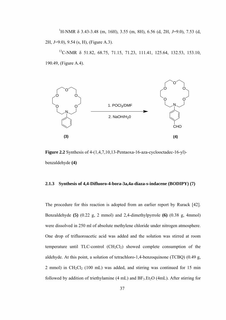
2.1.3 Synthesis of 4,4-Difluoro-4-bora-3a,4a-diaza-s-indacene (BODIPY) (7)
The procedure for this reaction is adopted from an earlier report by Rurack [42].
Benzaldehyde (5) (0.22 g, 2 mmol) and 2,4-dimethylpyrrole (6) (0.38 g, 4mmol)
were dissolved in 250 ml of absolute methylene chloride under nitrogen atmosphere.
One drop of trifluoroacetic acid was added and the solution was stirred at room
temperature until TLC-control (CH2Cl2) showed complete consumption of the
aldehyde. At this point, a solution of tetrachloro-1,4-benzoquinone (TCBQ) (0.49 g,
2 mmol) in CH2Cl2 (100 mL) was added, and stirring was continued for 15 min
followed by addition of triethylamine (4 mL) and BF3.Et2O (4mL). After stirring for
37
another 30 min the reaction mixture was washed with water and dried over Na2SO4,
and solvent was evaporated in vacuo. The residue was chromatographed on a silica
column (CH2Cl2). The yield of (7) was 280 mg (0.86 mmol, 43%).
1H NMR δ 1.37 (s, 6H), 2.56 (s, 6H), 5.98 (s, 2H), 7.26-7.30 (m, 2H), 7.47-
7.50 (m, 3H), (Figure A.5).
HO
HN NB
N
F F
+ 21. CF3COOH, RT, 3 h
2. TCBQ, RT, 15 min3. NH3, BF3.OEt2, RT, 30 min
(5) (6) (7)
Figure 2.3 Synthesis of 4,4-Difluoro-4-bora-3a,4a-diaza-s-indacene (BODIPY) (7)
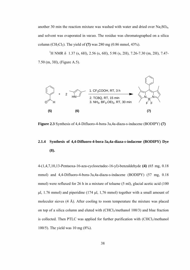
2.1.4 Synthesis of 4,4-Difluoro-4-bora-3a,4a-diaza-s-indacene (BODIPY) Dye
(8).
4-(1,4,7,10,13-Pentaoxa-16-aza-cyclooctadec-16-yl)-benzaldehyde (4) (65 mg, 0.18
mmol) and 4,4-Difluoro-4-bora-3a,4a-diaza-s-indacene (BODIPY) (57 mg, 0.18
mmol) were refluxed for 26 h in a mixture of toluene (5 ml), glacial acetic acid (100
µl, 1.76 mmol) and piperidine (174 µl, 1,76 mmol) together with a small amount of
moleculer sieves (4 Å). After cooling to room temperature the mixture was placed
on top of a silica column and eluted with (CHCl3/methanol 100/3) and blue fraction
is collected. Then PTLC was applied for further purification with (CHCl3/methanol
100/5). The yield was 10 mg (8%).
38
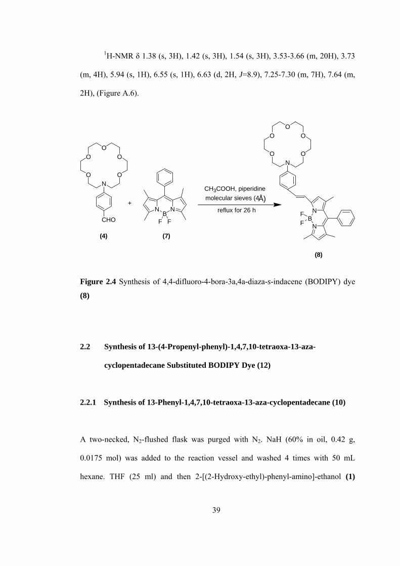
1H-NMR δ 1.38 (s, 3H), 1.42 (s, 3H), 1.54 (s, 3H), 3.53-3.66 (m, 20H), 3.73
(m, 4H), 5.94 (s, 1H), 6.55 (s, 1H), 6.63 (d, 2H, J=8.9), 7.25-7.30 (m, 7H), 7.64 (m,
2H), (Figure A.6).
OO
O
OON
CHON
BN
FF
OO
O
OON
CH3COOH, piperidineÅ)molecular sieves (4
reflux for 26 hNB
N
F F
+
(4) (7)
(8)
Figure 2.4 Synthesis of 4,4-difluoro-4-bora-3a,4a-diaza-s-indacene (BODIPY) dye
(8)
2.2 Synthesis of 13-(4-Propenyl-phenyl)-1,4,7,10-tetraoxa-13-aza-
cyclopentadecane Substituted BODIPY Dye (12)
2.2.1 Synthesis of 13-Phenyl-1,4,7,10-tetraoxa-13-aza-cyclopentadecane (10)
A two-necked, N2-flushed flask was purged with N2. NaH (60% in oil, 0.42 g,
0.0175 mol) was added to the reaction vessel and washed 4 times with 50 mL
hexane. THF (25 ml) and then 2-[(2-Hydroxy-ethyl)-phenyl-amino]-ethanol (1)
39
(1.53 g, 0.01 mol) was added to the flask. The suspension was heated to reflux with
vigorous stirring. A solution of the tri(ethylene glycol)di-p-tosylate (9) (4.58 g, 0.01
mol) in THF (10 ml) was added dropwise. Reflux was continued for 20 h. The
reaction mixture was cooled and quenched with H2O, and then extracted with
CH2Cl2 (3x100 ml). The combined organic layers were dried over Na2SO4 and
concentrated under reduced pressure. The desired product was purified by silica gel
column chromatography (CHCl3/methanol 100:7). Further purifications were
achieved by subsequent columns (CHCl3/methanol 100:7) when necessary. The
yield of (10) was 230 mg (8%).
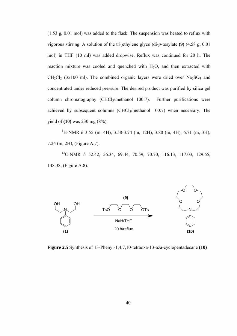
1H-NMR δ 3.55 (m, 4H), 3.58-3.74 (m, 12H), 3.80 (m, 4H), 6.71 (m, 3H),
7.24 (m, 2H), (Figure A.7).
13C-NMR δ 52.42, 56.34, 69.44, 70.59, 70.70, 116.13, 117.03, 129.65,
148.38, (Figure A.8).
NOHOH
O O O OTsTs
OO
O O
N
NaH/THF
20 h/reflux
(9)
(10)(1)
Figure 2.5 Synthesis of 13-Phenyl-1,4,7,10-tetraoxa-13-aza-cyclopentadecane (10)
40
2.2.2 Synthesis of 4-(1,4,7,10-Tetraoxa-13-aza-cyclopentadec-13-yl)-
benzaldehyde (11)
13-Phenyl-1,4,7,10-tetraoxa-13-aza-cyclopentadecane (10) (100 mg, 0.34 mmol)
was dissolved in 3 ml of dimethylformamide. The mixture was cooled in an ice bath
and phosphorus oxychloride (52 mg, 0.340 mmol) was added dropwise with stirring,
and the reaction mixture was taken from ice bath after 10 min. Then the mixture was
stirred at room temperature for 1.5 h, then at 100°C for 3-4 h. The mixture was then
cooled and poured over 10 g of crushed ice and stirred for 30 min. The solution was
neutralized with 6 M of NaOH solution. The reaction mixture was then extracted
with CHCl3 (3x100 ml). The combined organic layers were dried with Na2SO4 and
concentrated under reduced pressure. The desired product was purified by silica gel
column chromatography (CHCl3/methanol 100:7). The yield was 78 mg (71%).
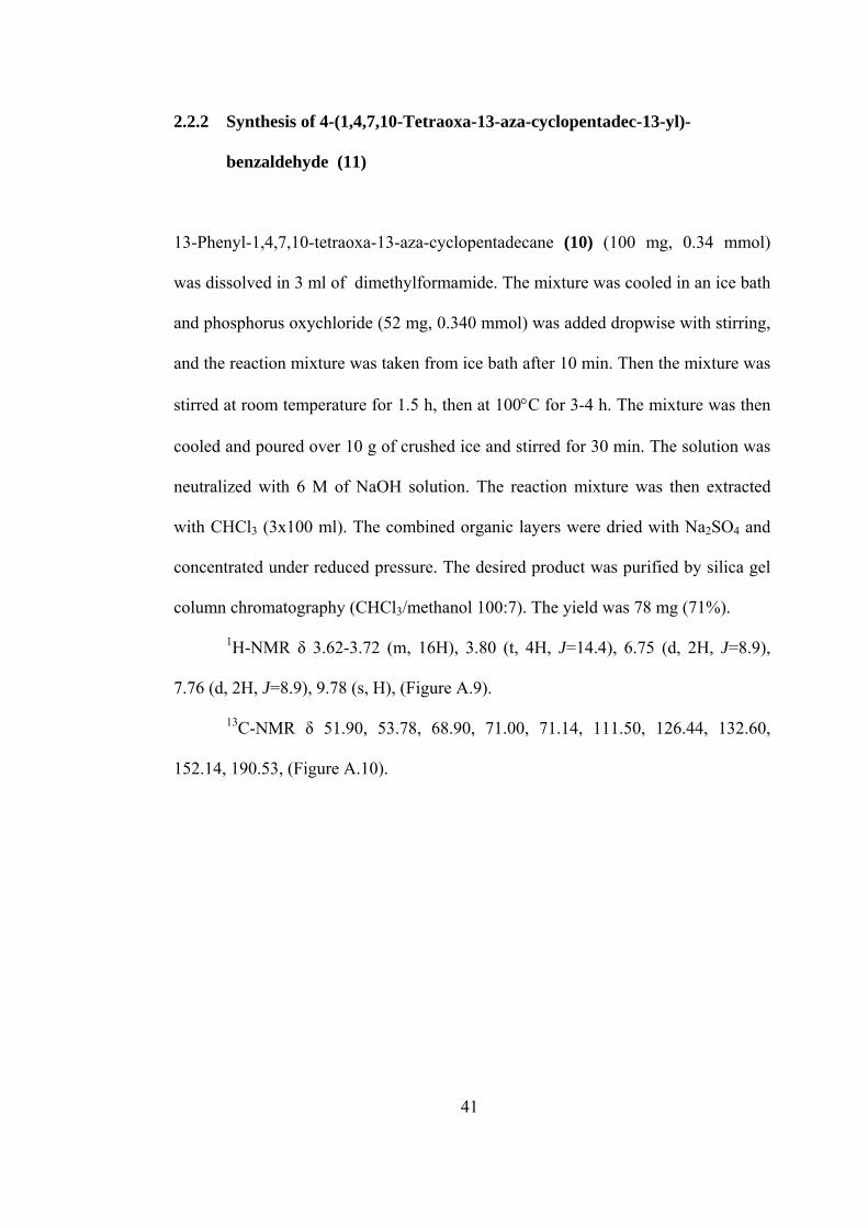
1H-NMR δ 3.62-3.72 (m, 16H), 3.80 (t, 4H, J=14.4), 6.75 (d, 2H, J=8.9),
7.76 (d, 2H, J=8.9), 9.78 (s, H), (Figure A.9).
13C-NMR δ 51.90, 53.78, 68.90, 71.00, 71.14, 111.50, 126.44, 132.60,
152.14, 190.53, (Figure A.10).
41
OO
O O
N
OO
O O
N
CHO
1. POCl3/DMF
2. NaOH/H20
(10) (11)
Figure 2.6 Synthesis of 4-(1,4,7,10-Tetraoxa-13-aza-cyclopentadec-13-yl)-
benzaldehyde (11)
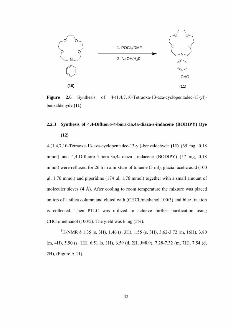
2.2.3 Synthesis of 4,4-Difluoro-4-bora-3a,4a-diaza-s-indacene (BODIPY) Dye
(12)
4-(1,4,7,10-Tetraoxa-13-aza-cyclopentadec-13-yl)-benzaldehyde (11) (65 mg, 0.18
mmol) and 4,4-Difluoro-4-bora-3a,4a-diaza-s-indacene (BODIPY) (57 mg, 0.18
mmol) were refluxed for 26 h in a mixture of toluene (5 ml), glacial acetic acid (100
µl, 1.76 mmol) and piperidine (174 µl, 1,76 mmol) together with a small amount of
moleculer sieves (4 Å). After cooling to room temperature the mixture was placed
on top of a silica column and eluted with (CHCl3/methanol 100/3) and blue fraction
is collected. Then PTLC was utilized to achieve further purification using
CHCl3/methanol (100/5). The yield was 6 mg (5%).
1H-NMR δ 1.35 (s, 3H), 1.46 (s, 3H), 1.55 (s, 3H), 3.62-3.72 (m, 16H), 3.80
(m, 4H), 5.90 (s, 1H), 6.51 (s, 1H), 6.59 (d, 2H, J=8.9), 7.28-7.32 (m, 7H), 7.54 (d,
2H), (Figure A.11).
42
OO
O O
N
CHON
BN
FF
OO
O O
N
NB
N
F F
CH3COOH, piperidineÅ)molecular sieves (4
reflux for 26 h
(11)
(12)
+
(7)
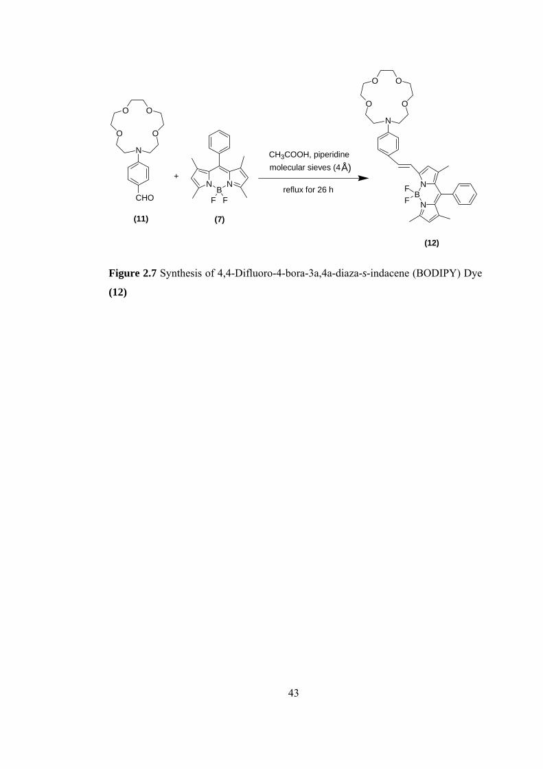
Figure 2.7 Synthesis of 4,4-Difluoro-4-bora-3a,4a-diaza-s-indacene (BODIPY) Dye
(12)
43
CHAPTER 3
RESULTS AND DISCUSSION
In this study, we designed and synthesized an unsymetrically substituted BODIPY
(4,4-Difluoro-4-bora-3a,4a-diaza-s-indacene) dye series. In these systems, the
substituents that are phenylazacrown ethers carrying cation-sensitive group, either
monoaza-15-crown-5 or monoaza-18-crown-6, are conjugated to the BODIPY core.
We also investigated ion-sensing properties of these BODIPY based dye systems.
BODIPY dyes are highly fluorescent materials that have been used for several
different applications. These BODIPY dyes combine the advantages of high molar
extinction coefficients (ε > 70 000 M-1 cm-1) and high fluorescence quantum yields
(Φ ca. 0.5-0.8) and can be excited at relatively long wavelengths (ca. 500 nm).
These dyes have exceptional spectral and photophysical stability as compared to
other fluorescent groups.
BODIPY dyes that are sensitive toward both external physical and chemical
triggers provide a versatile basis for the constructions of sophisticated switches that
communicate, for instance, through changes in electrochromic and/or fluorescence
properties. The design of such BODIPY based systems is important since the
44
BODIPY core is comparatively readily oxidized and reduced, a prerequisite for
fluorescent switches relying on electron or charge transfer (CT), as well as for the
generation of stable radical ions that show electrogenerated chemiluminescence
upon charge recombination. In these systems, the meso-substituted BODIPY
chromophore acts as in donor(D)-acceptor(A)-substituted biaryls so that the
switching process hardly influences the absorption and emission wavelengths.
In this study, we designed and synthesized more advanced molecular
ensembles that generate signal changes by substrate interaction at a site conjugated
to the BODIPY core. The molecular sensors that we synthesized are ones of the
first examples of an unsymmetrically substituted BODIPY dyes with an analyte-
responsive receptor in the polymethinic part of the chromophore. In our systems,
the analyte-sensitive group is either monoaza-12-crown-4 or monoaza-15-crown-5
that plays also the role of an electron donor groups because of N atom they have.
Having the strong and analytically valuable proton-induced effects on the
absorption and emission behaviour of the amino-substituted BODIPY derivatives,
we examined the photophysical changes of the dyes we synthesized upon
complexation to the some metal cations in acetonitrile in order to test their possible
application as fluorescent probes. The crowned BODIPY dyes presented here are
examples for very efficient CT (charge transefer) systems. In these systems
coordination to the nitrogen donor atom of the receptor not only leads to changes in
fluorescence intensity and lifetime but also provides additional information via
spectral shifts in both absorption and emission.
45
3.1 Synthesis and Photophysical Properties of BODIPY Dye (8)
We followed three steps for the synthesis of BODIPY dye (8) which is shown in
Figure 3.1. In the first step, 18-membered lariat azacrown ether, that is phenyl
substituted, was synthesized by cyclization of 2-[(2-Hydroxy-ethyl)-phenyl-amino]-
ethanol (1) with tetra(ethylene glycol)di-p-tosylate (2). The yield was much lower
than that expected because of difficulties we had in purification. Then 16-Phenyl-
1,4,7,10,13-pentaoxa-16-aza-cyclooctadecane (3) that was obtained in first step was
formylated with Vilsmeier and Haack reaction. The appearance of the aldehyde
proton at δ 9.68 ppm and the disappearance of the peak of proton at the para-
position of the phenyl at δ 7.28 in the 1H-NMR spectrum show a successful reaction.
Finally, the formyl substituted compound (4) was reacted with 4,4-Difluoro-4-bora-
3a,4a-diaza-s-indacene (BODIPY) and the desired final product that is
functionalized boron-dipyrromethane dye was obtained.
After synthesis of the BODIPY dye (8), we investigated the photophysical
behaviour of upon complexation with some metal cations and proton. In
spectrometric measurements acetonitrile was used as solvent, because the BODIPY
dyes we synthesized are the intrinsically donor-acceptor-substituted
fluoroionophores (ICT probes) that show strong solvent-dependent behavior. In a
nonpolar solvent, such as hexane, emission occurs only from a LE (locally excited)
state, whereas in a more polar solvent, such as acetonitrile, an ultrafast excited-state
charge-transfer reaction from the amino donor to the basic fluorophore takes place.
This results in strong quenching of the LE emission and appearance of a
bathochromically shifted emission from a lower lying CT state, both fluorescence
quantum yields being low.
46
O
OO
OON
(3)
igure 3.1 Reaction series leading to BODIPY dye (8)
O O O O OTsTs
NaH/THF
20 h/reflux
(2)
NOHOH
(1)
OO
O
OON
CHO
(4)
1. POCl3/DMF
2. NaOH/H20
NB
N
F F
(7)
+
CH3COOH, piperidineÅ)molecular sieves (4
reflux for 26 h
NB
N
FF
OON
OO
O
(8)
F
47
The efficient nonradiative deactivation process can be utilized to construct a very
efficient molecular switch. Protonation as well as complexation blocks the CT
process and switches on the LE emission again; i.e., a strong increase in LE
fluorescence quantum yield and lifetime with very large fluorescent enhancement
factors.
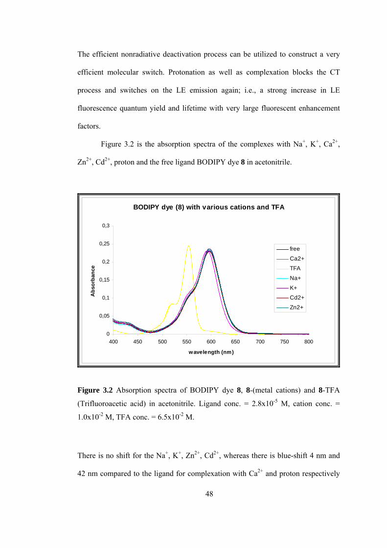
Figure 3.2 is the absorption spectra of the complexes with Na+, K+, Ca2+,
Zn2+, Cd2+, proton and the free ligand BODIPY dye 8 in acetonitrile.
BODIPY dye (8) with various cations and TFA
0
0,05
0,1
0,15
0,2
0,25
0,3
400 450 500 550 600 650 700 750 800
wavelength (nm)
Abs
orba
nce
free
Ca2+TFA
Na+
K+Cd2+
Zn2+
Figure 3.2 Absorption spectra of BODIPY dye 8, 8-(metal cations) and 8-TFA
(Trifluoroacetic acid) in acetonitrile. Ligand conc. = 2.8x10⁻⁵ M, cation conc. =
1.0x10⁻² M, TFA conc. = 6.5x10⁻² M.
There is no shift for the Na+, K+, Zn2+, Cd2+, whereas there is blue-shift 4 nm and
42 nm compared to the ligand for complexation with Ca2+ and proton respectively
48
as expected, because when the nitrogen atom in crown ether playing the role of
electron donor interacts with a cation or proton, the latter reduces the electron-
donating character of this atom; owing to the resulting reduction of conjugation, a
blue shift of the absorption spectrum is expected.
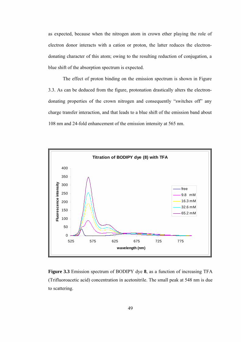
The effect of proton binding on the emission spectrum is shown in Figure
3.3. As
hes off” any
harge transfer interaction, and that leads to a blue shift of the emission band about
108 nm and 24-fold enhancement of the emission intensity at 565 nm.
can be deduced from the figure, protonation drastically alters the electron-
donating properties of the crown nitrogen and consequently “switc
c
Titration of BODIPY dye (8) with TFA
0
50
100
150
200
250
300
350
400
525 575 625 675 725 775
wavelength (nm)
Fluo
resc
ence
inte
nsity
free9.8 mM
16.3 mM
32.6 mM65.2 mM
Figure 3.3 Emission spectrum of BODIPY dye 8, as a function of increasing TFA
(Trifluoroacetic acid) concentration in acetonitrile. The small peak at 548 nm is due
to scattering.
49
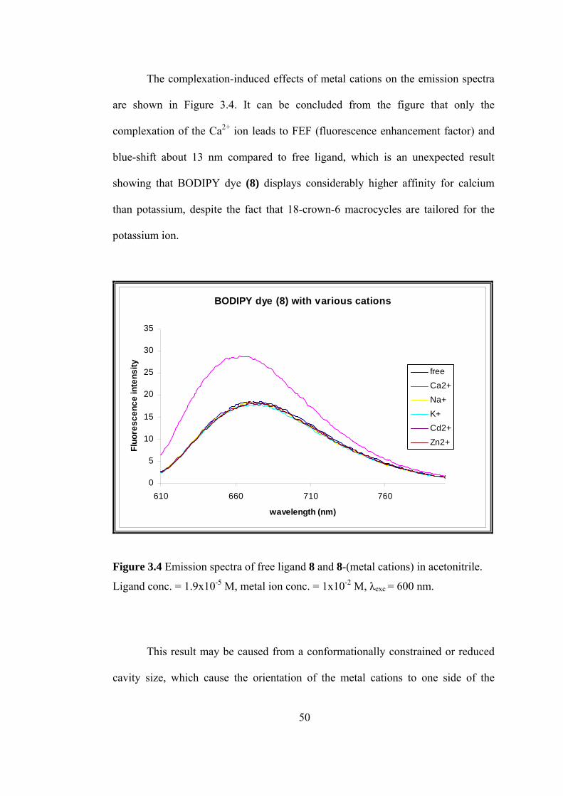
The complexation-induced effects of metal cations on the emission spectra
are shown in Figure 3.4. It can be concluded from the figure that only the
complexation of the Ca2+ ion leads to FEF (fluorescence enhancement factor) and
blue-shift about 13 nm compared to free ligand, which is an unexpected result
showing that BODIPY dye (8) displays considerably higher affinity for calcium
than potassium, despite the fact that 18-crown-6 macrocycles are tailored for the
potassium ion.
BODIPY dye (8) with various cations
0
5
10
15
20
25
30
35
610 660 710 760
wavelength (nm)
Fluo
resc
ence
inte
nsity freeCa2+
Na+
K+Cd2+
Zn2+
Figure 3.4 Emission spectra of free ligand 8 and 8-(metal cations) in acetonitrile.
Ligand conc. = 1.9x10⁻⁵ M, metal ion conc. = 1x10⁻² M, λexc = 600 nm.
hich cause the orientation of the metal cations to one side of the
This result may be caused from a conformationally constrained or reduced
cavity size, w
50
macroc
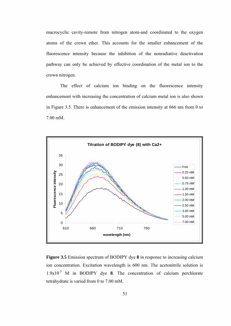
th increasing the concentration of calcium metal ion is also shown
Figure 3.5. There is enhancement of the emission intensity at 666 nm from 0 to
7.00 mM.
yclic cavity-remote from nitrogen atom-and coordinated to the oxygen
atoms of the crown ether. This accounts for the smaller enhancement of the
fluorescence intensity because the inhibition of the nonradiative deactivation
pathway can only be achieved by effective coordination of the metal ion to the
crown nitrogen.
The effect of calcium ion binding on the fluorescence intensity
enhancement wi
in
Titration of BODIPY dye (8) with Ca2+
0
5
10
15
20
25
30
35
610 660 710 760
wavelength (nm)
Fluo
resc
ence
inte
nsity
free
0.25 mM
0.50 mM
0.75 mM
1.00 mM
1.50 mM
2.00 mM
2.50 mM
3.00 mM
5.00 mM
7.00 mM
Figure 3.5 Emission spectrum of BODIPY dye 8 in response to increasing calcium
ion con
1.9x10⁻⁵ M in BODIPY dye 8. The concentration of calcium perchlorate
centration. Excitation wavelength is 600 nm. The acetonitrile solution is
tetrahydrate is varied from 0 to 7.00 mM.
51
3.2 Synthesis and Photophysical Properties of BODIPY Dye (12)
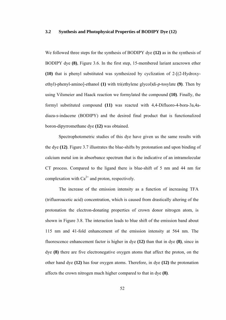
We followed three steps for the synthesis of BODIPY dye (12) as in the synthesis of
BODIPY dye (8), Figure 3.6. In the first step, 15-membered lariant azacrown ether
(10) that is phenyl substituted was synthesized by cyclization of 2-[(2-Hydroxy-
ethyl)-p
cene (BODIPY) and the desired final product that is functionalized
oron-dipyrromethane dye (12) was obtained.
Spectrophotometric studies of this dye have given us the same results with
the dye (12). Figure 3.7 illustrates the blue-shifts by protonation and upon binding of
calcium metal ion in absorbance spectrum that is the indicative of an intramolecular
CT process. Compared to the ligand there is blue-shift of 5 nm and 44 nm for
complexation with Ca2+ and proton, respectively.
The increase of the emission intensity as a function of increasing TFA
(trifluoroacetic acid) concentration, which is caused from drastically altering of the
protonation the electron-donating properties of crown donor nitrogen atom, is
shown in Figure 3.8. The interaction leads to blue shift of the emission band about
115 nm and 41-fold enhancement of the emission intensity at 564 nm. The
uorescence enhancement factor is higher in dye (12) than that in dye (8), since in
mpared to that in dye (8).
henyl-amino]-ethanol (1) with tri(ethylene glycol)di-p-tosylate (9). Then by
using Vilsmeier and Haack reaction we formylated the compound (10). Finally, the
formyl substituted compound (11) was reacted with 4,4-Difluoro-4-bora-3a,4a-
diaza-s-inda
b
fl
dye (8) there are five electronegative oxygen atoms that affect the proton, on the
other hand dye (12) has four oxygen atoms. Therefore, in dye (12) the protonation
affects the crown nitrogen much higher co
52
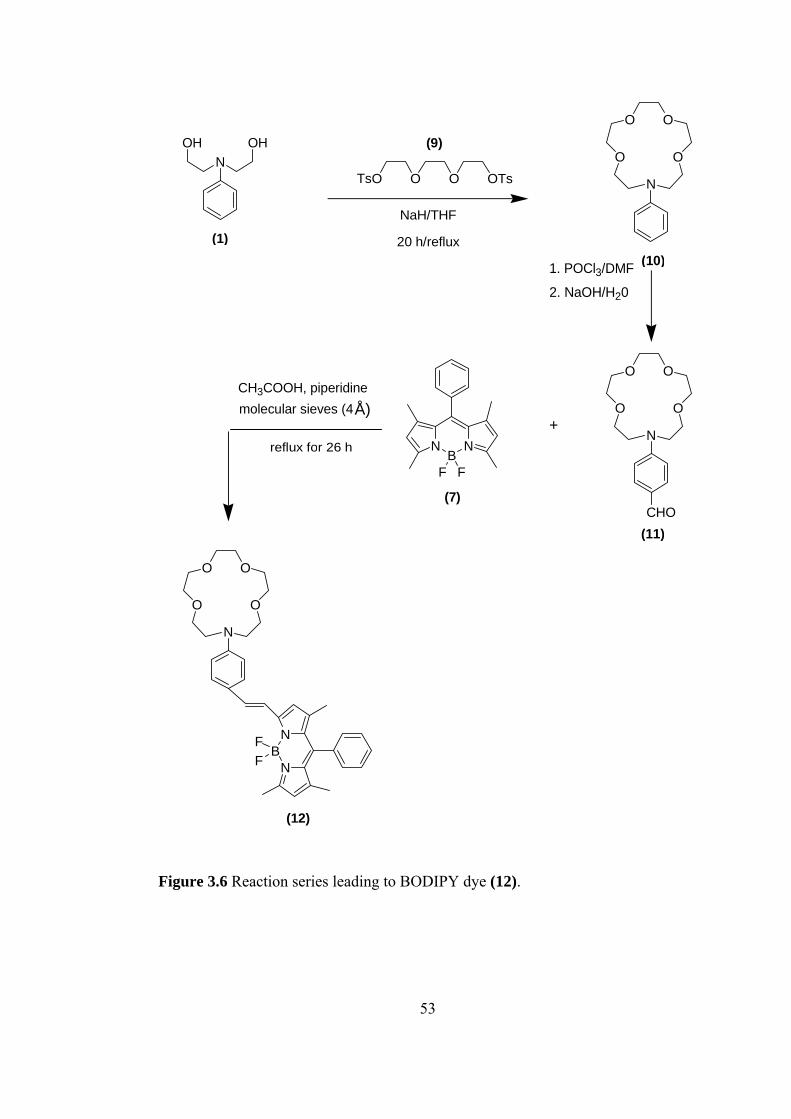
53
NOHOH
(1)
O O O OTsTs
NaH/THF
N
(10)
OO
O O
2. NaOH/H20
1. POCl /DMF
N
3COOH, piperidineÅ)molecular sieves (4
reflux for 26 h
OO
O O
20 h/reflux
(9)
N
CHO
F F
N
(11)
3
+
BN
(7)
CH
N
N
FF
(12)
Figure 3.6 Reaction series leading to BODIPY dye (12).
B
OO
O O
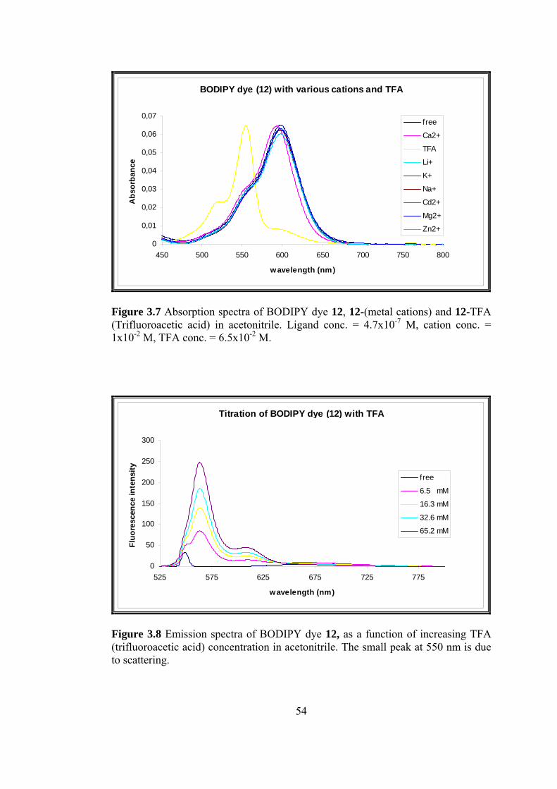
BODIPY dye (12) with various cations and TFA
0
0,01
0,02
0,03
0,04
0,05
0,06
0,07
450 500 550 600 650 700 750 800
wavelength (nm)
Abs
orba
nce
free
Ca2+
TFA
Li+
K+
Na+
Cd2+
Mg2+
Zn2+
igure 3.7 Absorption spectra of BODIPY dye 12, 12-(metal cations) and 12-TFA rifluoroacetic acid) in acetonitrile. Ligand conc. = 4.7x10⁻⁷ M, cation conc. =
x10-2 M, TFA conc. = 6.5x10⁻² M.
F(T1
Titration of BODIPY dye (12) with TFA
0
50
100
150
200
250
300
525 575 625 675 725 775
wavelength (nm)
Fluo
resc
ence
inte
nsity
free
6.5 mM
16.3 mM
32.6 mM
65.2 mM
igure 3.8 Emission spectra of BODIPY dye 12, as a function of increasing TFA (trifluoroacetic acid) concentration in acetonitrile. The small peak at 550 nm is due to scattering.
F
54
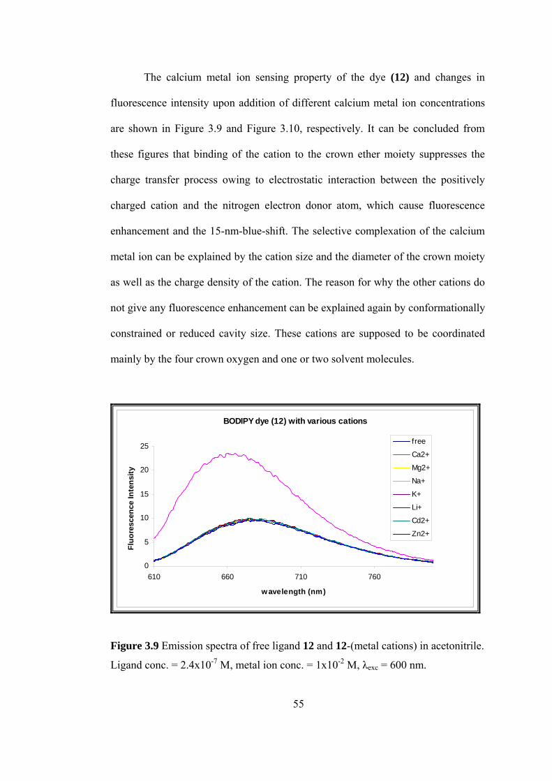
The calcium metal ion sensing property of the dye (12) and changes in
fluorescence intensity upon addition of different calcium metal ion concentrations
are shown in Figure 3.9 and Figure 3.10, respectively. It can be concluded from
these figures that binding of the cation to the crown ether moiety suppresses the
charge transfer process owing to electrostatic interaction between the positively
charged cation and the nitrogen electron donor atom, which cause fluorescence
enhancement and the 15-nm-blue-shift. The selective complexation of the calcium
metal ion can be explained by the cation size and the diameter of the crown moiety
as well as the charge density of the cation. The reason for why the other cations do
These cations are supposed to be coordinated
ainly by the four crown oxygen and one or two solvent molecules.
not give any fluorescence enhancement can be explained again by conformationally
constrained or reduced cavity size.
m
BODIPY dye (12) with various cations
0
5
10
15
20
25
610 660 710 760
wavelength (nm)
Fluo
resc
ence
Inte
nsity
free
Ca2+
Mg2+
Na+
K+
Li+
Cd2+
Zn2+
igand conc. = 2.4x10⁻⁷ M, metal ion conc. = 1x10⁻² M, λexc = 600 nm.
Figure 3.9 Emission spectra of free ligand 12 and 12-(metal cations) in acetonitrile.
L
55
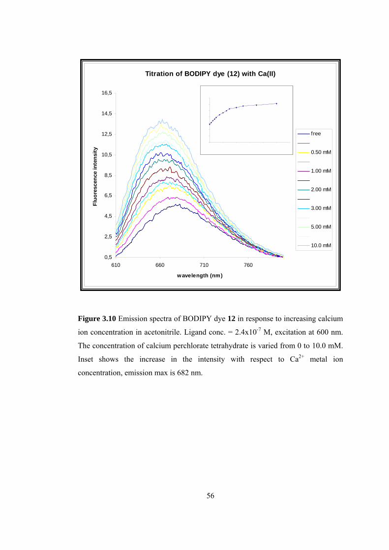
Titration of BODIPY dye (12) with Ca(II)
0,5
2,5
4,5
6,5
8,5
14,5
16,5Fl
uore
scen
nten
ty
0.25 mM
0.75 mM
1.50 mM
2.50 mM
4.00 mM
7.00 mM
Calcium titration of BODIPY dye (12) emission max; 682 nm
0
4
6
10
12
0 2 4 6 8 10 12
concentration (mM)
inte
n
2
8
14
sity
Figure 3.10 Emission spectra of BODIPY dye 12 in response to increasing calcium
ion concentration in acetonitrile. Ligand conc. = 2.4x10⁻⁷ M, excitation at 600 nm.
The concentration of calcium perchlorate tetrahydrate is varied from 0 to 10.0 mM.
Inset shows the increase in the intensity with respect to Ca2+ metal ion
concentration, emission max is 682 nm.
10,5
12,5
610 660 710 760
wavelength (nm)
ce i
si
free
0.50 mM
1.00 mM
2.00 mM
3.00 mM
5.00 mM
10.0 mM
56
CHAPTER 4
CONCLUSION
This study covers the synthesis and ion sensing properties of a series of novel
boradiazaindacene dyes.
The molecular sensors that we synthesized in this study are ones of the first
examples of an unsymetrically substituted boradiazaindacene dyes with an analyte-
responsive receptor in the polymethinic part of the chromophore. The extension of
the absorption and emission to longer
avelength in our systems. The boradiazaindacene dyes presented here are
Thus, with these dyes, we present a new design concept for sensitive probes
r calcium metal ion showing absorption and fluorescence changes in the red/NIR.
specially considering the selective signaling of calcium ions, which is very
portant biological analyte, one can foresee practical applications in many fields
y appropriate derivatization of the compounds developed by us. Further research in
is area will continue in our lab.
the conjugation over the pyrrole ring shifts
w
examples for efficient CT (charge transfer) systems that can be transferred to LE
(locally excited) systems by stable calcium metal ion complexation or protonation
in polar acetonitrile.
fo
E
im
b
th
57
R
[1] Lehn, J.-M., Angew. Chem. 1988, 0, 91.
] Lehn, J.-M., Angew. Chem. Int. Ed. Engl. 1988, 27, 89.
[3] Lehn, J.-M., Supramolecular Chemistry – Concepts and Perspectives, VCH,
Czarnic, A.W. (Ed), ACS Symposium Series 538, American Chemical
u.edu/~chemistry/chemiluminescence/JABLONSKI.html
EFERENCES
10
[2
Weinheim, 1995.
[4] Lehn, J.-M, Science, 1985. 227, 849.
[5] Vögtle, F., Supramolekulare Chemie, Teubner, Stuttgart, 1989, p 6.
[6] Czarnik, A. W., “Supramolecular Chemistry, Fluorescence, and Sensing”,
Society, Washingtion, D.C.,1992.
[7] http://www.shs
(11.08.2003).
Chemical Society, Washingtion, D.C.,1992.
Czarnic, A.W. (Ed), ACS Symposium Series 538,
American Chemical Society, Washingtion, D.C.,1992.
[8] Czarnik, A. W., “Fluorescent Signal Transduction in Molecular Sensors and
Dosimeters”, Czarnic, A.W. (Ed), ACS Symposium Series 538, American
[9] Szmacinski, H., Lakowicz, J. R., “Lifetime-Based Sensing Using Phase-
Modulation Fluorimetry”,
58
[10] Lehn, J.-M., Struct. Bonding, 1973, 16, 1.
[11] Valeur, B., Molecular Luminescence Spectroscopy-Methods and Applications,
Part 3, Ed. Schulman, S. G., John Wiley&sons, New York, 1993 pp 25-84.
Tsein, R. Y., “Fluorescent and Photochemical Probes of Dynamic
Biochemical Signals insida Living Cells”, Czarnic, A.W. (Ed), ACS
hemical Society, Washingtion,
D.C.,1992.
3] Minta, A., Tsein, R. Y., J. Biol. Chem., 1989, 264, 19449.
, M., Trends Biochem. Sci., 1986, 11, 450.
ska, J., Taylor, D. L., Fluorescence
Microscopy of Living Cells in Culture Part B, Methods in Cell Biology, Vol.
ress, San
Diego, 1989.
[16]
gy Transfer”, Czarnic, A.W. (Ed), ACS
Symposium Series 538, American Chemical Society, Washingtion,
7] Valeur, B., Leray, I., Coordination Chemistry Reviews, 2000, 205, 3.
[18]
nd Chemical Sensing, Lakowicz,
J. R. (Ed.), Plenum Press, New York, 1994, 4, pp 21-48.
[12]
Symposium Series 538, American C
[1
[14] Tsien, R. Y., Poenie
[15] Bright, G. R., Fisher, G. W., Rogow
30 (Taylor D. L. and Wang, Y. –L., Eds.), pp 157-192. Academic P
Valuer, B., Bourson, Jean, Pouget, J., “Ion Recognition Detected by Changes
in Photoinduced Charge or Ener
D.C.,1992.
[1
Valuer, B., “Fluorescent Probe Design for Ion Recognition” in Topics in
Fluorescence Spectroscopy, Probe Design a
59
[19] Bissell, R. A., de Silva, A. P., Gunaratne, H. Q. N., Lynch, P. L. M.,
Maguire, G. E. M., Sandanayake, K. R. A. S., Chem. Soc. Rev., 1992, 21,
0] Akkaya, E.U., Oğuz, U., Tetrahedron Letters, 1997, 38, 4509.
[21]
he, R. A. D. D.,
Biosensors, 1989, 4, 169.
3] de Silva, A. P., de Silva, A. S., Dissanayake, A S., Sandanayake, K. R. A. S.,
[24]
[26] , Lichelli, M., Pallavicini, P., Sacchi, D., Taglietti, A., Analyst,
1996, 121, 1763.
7] Bergonzi, R., Fabrizzi, L., Lichelli, M., Mangano, C., Coord. Chem. Rev.,
[28] ani, D., Lucas, M.
E., Kurtz, I., Am. J. Physiol., 1990, 258, F438.
187.
[2
Bissell, R. A., de Silva, A. P., Gunaratne, H. Q. N., Lynch, P. L. M., McCoy,
C. P., Maguire, G. E. M., Sandanayake, K. R. A. S., Top. Curr. Chem, 1993,
168, 223.
[22] Bryan, A.J., de Silva, A. P., de Silva, A. S., Rupasing
[2
J. Chem. Soc. Chem. Commun., 1989, 1054.
de Silva, A. P., Sandanayake, K. R. A. S., J. Chem. Soc., Chem. Commun.,
1989, 1183.
[25] Prasanna de Silva, A., de SilvA S. A., J. Chem. Soc., Chem. Commun.,
1986,1709.
Fabrizzi, L.
[2
1998, 170, 31-46.
Golchini, K., Mackovic-Basic, M., Gharib, S. A., Masilam
60
[29] Fabrizzi, L., Lichelli, M., Pallavacini P., Perotti, A., Sacchi, D., Angew.
Chem. Int. Ed. Ing., 1994, 33, 1975.
0] Fabrizzi, L., Lichelli, M., Pallavacini P., Perotti, Taglietti, A., Sacchi, A.,
[32] akaki, T., Shinkai, S., J. Chem. Soc. Chem. Commun., 1992, 730.
4] Löhr, H.-G., Vögtle, F., Acc. Chem. Res., 1985, 18, 65.
[35] . P., Valeur, B., J. Chem. Soc.
Chem. Commun., 1988, 5, 384.
[36] , S., Le Bris, M.-T., Guette’, J. P., Valeur, B., J. Phys. Chem.,
1988, 92, 6233.
[37] s, S., Bourson, J., Dallery, L., Valeur, B., New J. Chem, 1990,
14, 617.
[38] , Bennett, J., Tweedy, S. H., Elshani, S., Wai, C. M.,
Talanta, 1998, 46, 573.
[39] lamanli, Z., Gosper, J., Sammes, P. G., J. Chem. Soc.
Perkin Trans. 2, 1994, 513.
[40] bes and Research Chemicals,
Molecular Probes, Inc, Eugene, OR, USA.
[3
Chem. Eur. J. 2, 1996, 167.
[31] de Silva, A. P., Gunaratne, H. Q. N., J. Chem. Soc. Chem. Commun., 1990, 186.
Aoki, I., S
[33] Kubo, K., Kato, N., Sakurai, T., Bull. Chem. Soc. Jpn., 1997, 70, 3041.
[3
Fery-Forgues, S., Le Bris, M.-T., Guette’, J
Fery-Forgues
Fery-Forgue
Addleman, R. S.
Crossley, R., Goo
Haugland, R. P., Handbook of Fluorescent Pro
61
[41] Bourson, J., Pouget, J., Valuer, B., J. Phys. Chem., 1993, 97, 4552.
2] Kollmannsberg, M., Rurack, K., Resch-Genger, U., Daub, J., J. Phys. Chem.
3] Bergstrőm, F., Mikhalyov, I., Hägglőf, P., Wortmann R., Ny, T., Johansson,
Burgess, K., J. Ogr. Chem, 1999, 64, 7813.
Chen, J., Reibenspies, J., Derecskei-Kovacs, A., Burgess, K., Chem.
ahedron Letters, 2000, 41, 2303.
0] Coskun, A., Baytekin, B., Akkaya, E. U., Tetrahedron Letters, 2003, 44,
1] Kollmannsberg, M., Rurack, K., Resch-Genger, U., Daub, J., J. Am. Chem.
[4
A, 1998, 102, 10211-10220.
[4
L.B.-Å., Journal of American Chemical Society, 2001.
[44] Burghart, A., Kim, H., Welch, M. B., Thoresen, L. H., Reibenspies, J.,
[45]
Commun., 1999, 2501.
[46] Kim, H., Burghart, A., Welch, M.B., Reibenspies, J., Burgess, K., Chem.
Commun., 1999,1889.
[47] Chen, J., Burghart, A., Wan, C.-W., Thai, L., Ortiz, C., Reibenspies, J.,
Burgess, K., Tetr
[48] Chen, J., Burghart, A., Derecskei-Kovacs, A., Burgess, K., J. Org. Chem.,
2000, 65, 2900.
[49] Beer, G., Rurack, K., Daub, J., Chem. Commun., 2001, 1138.
[5
5649.
[5
Soc., 2000, 122, 968.
62

[52] Werner, T., Huber, C., Heinl, S., Kollmannsberg, M., Daub, J., Wolfbeis, O.
S., Fresenius J. Anal. Chem., 1997, 539, 150.
[53] T., Heinl, S., Breu, J., Daub, J., Angew. Chem.
Int. Ed. Ingl., 1997, 36, 1333.
[54] m. Int. Ed., 2001, 40,
385.
Kollmannsberg, M., Gareis,
Rurack, K., Kollmannsberg, M., Daub, J., Angew. Che
63

APPENDIX
64
O
OO O
ON
Figu
re A
.1 1 H
-NM
R sp
ectru
m o
f com
poun
d (3
).
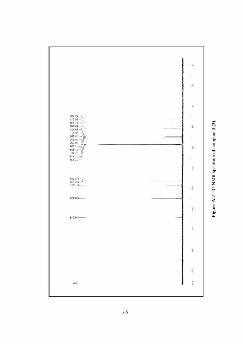
poun
d (3
).Fi
gure
A.2
13C
-NM
R sp
ectru
m o
f com
65
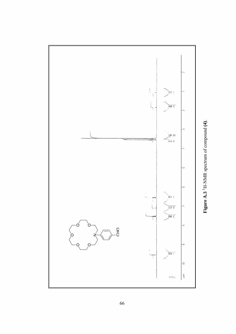
OO
O OO
N CH
O
Figu
re A
.3 1 H
-NM
R sp
ectru
m o
f com
poun
d (4
).
66
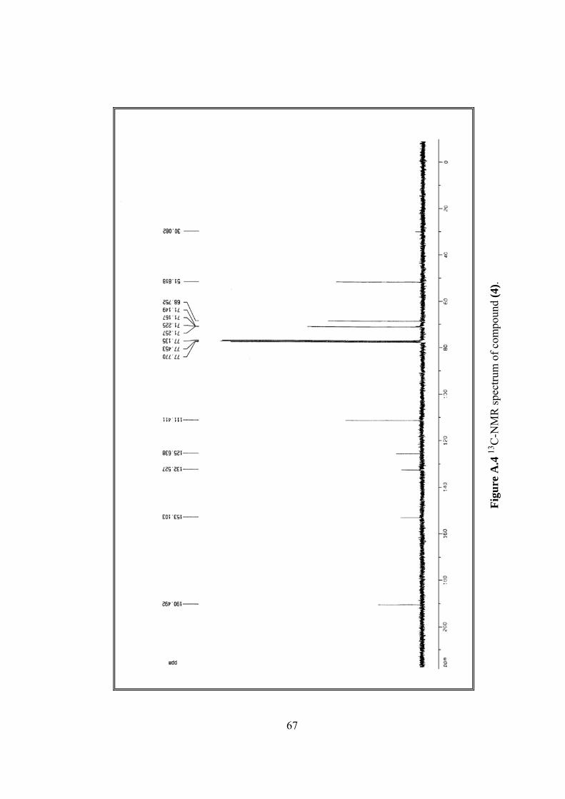
.Fi
gure
A.4
13C
-NM
R sp
ectru
m o
f com
poun
d (4
)
67
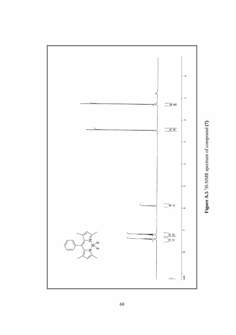
68
NB
N
FF
Figu
re A
.5 1 H
-NM
R sp
ectru
m o
f com
poun
d (7
).
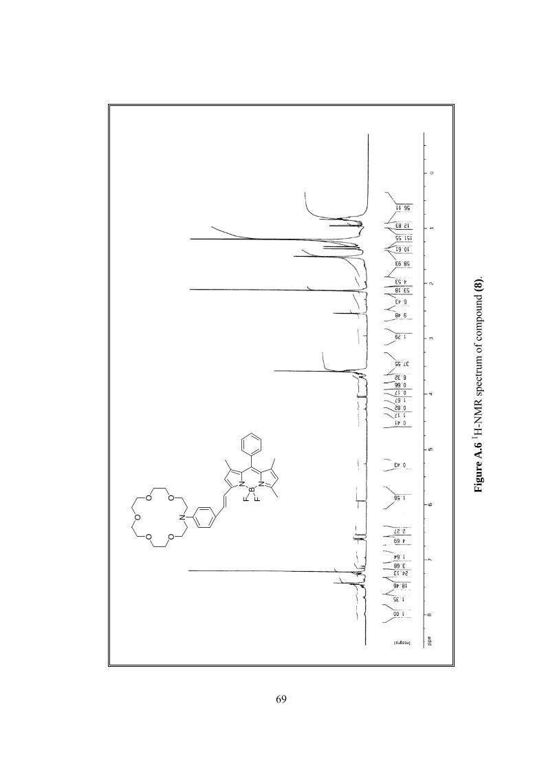
NB
N
F F
OO
O OO
N
NB
N
F F
OO
O OO
N
1 H-N
MR
spec
trum
of c
ompo
und
(8).
Figu
re A
.6
69
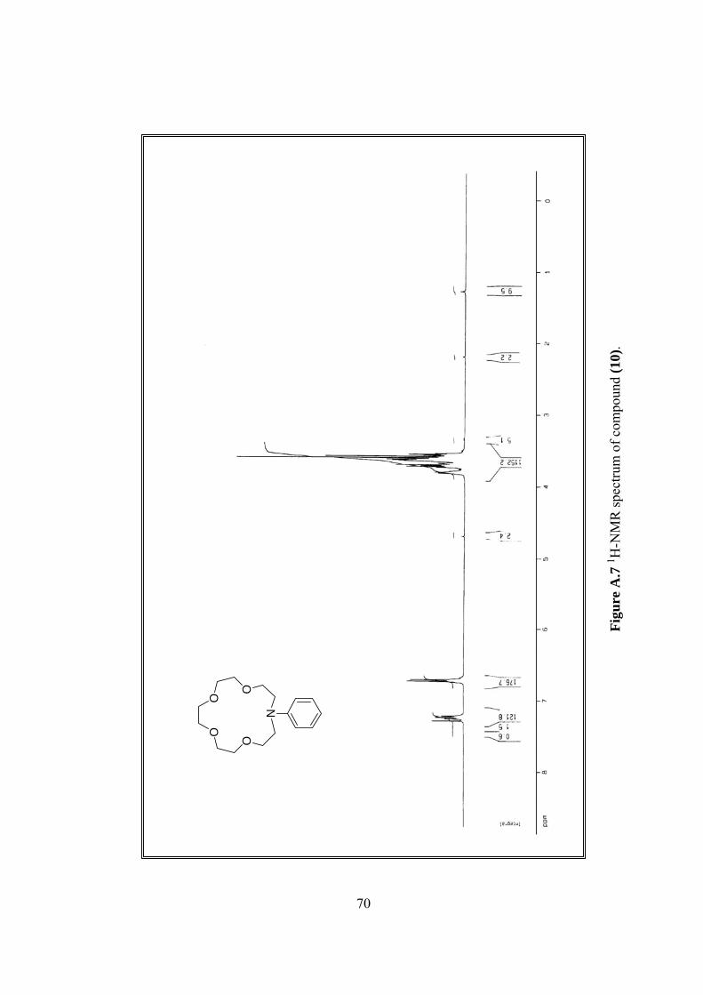
OO
OO
N
.Fi
gure
A.7
1 H-N
MR
spec
trum
of c
ompo
und
(10)
70
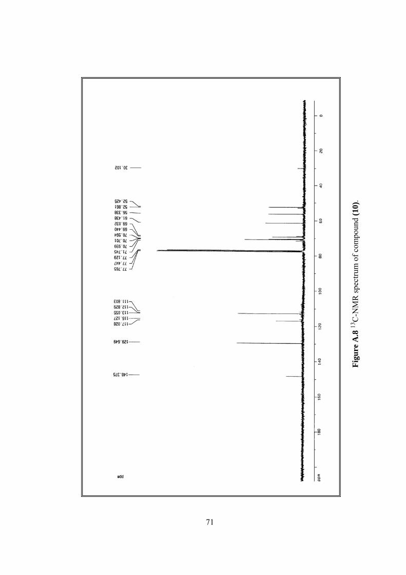
Figu
re A
.8 13
C-N
MR
spec
trum
of c
ompo
und
(10)
.
71
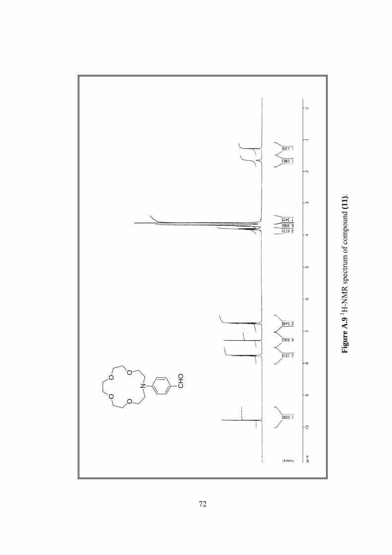
OO
OO
N CH
O
Figu
re A
.9 1 H
-NM
R sp
ectru
m o
f com
poun
d (1
1).
72
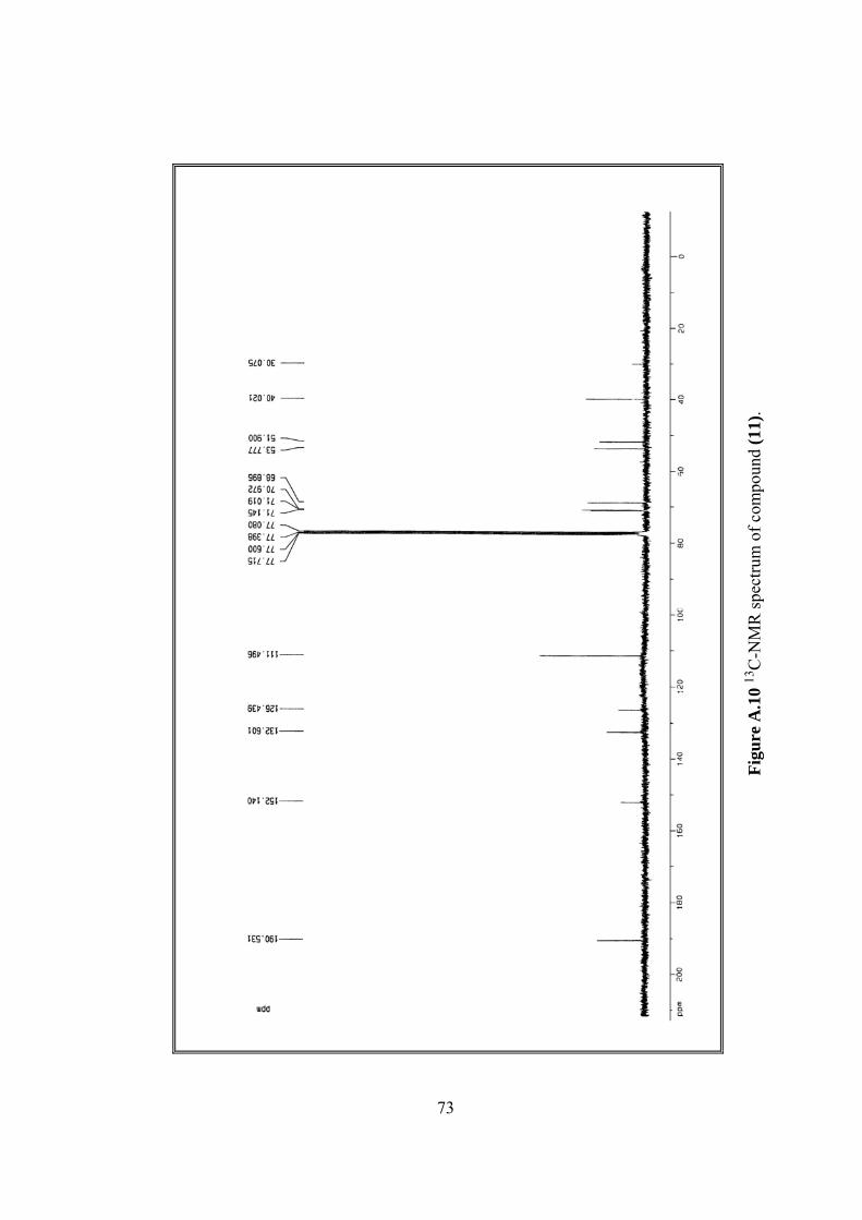
Figu
re A
.10
1 3C
-NM
R sp
ectru
m o
f com
poun
d (1
1).
73
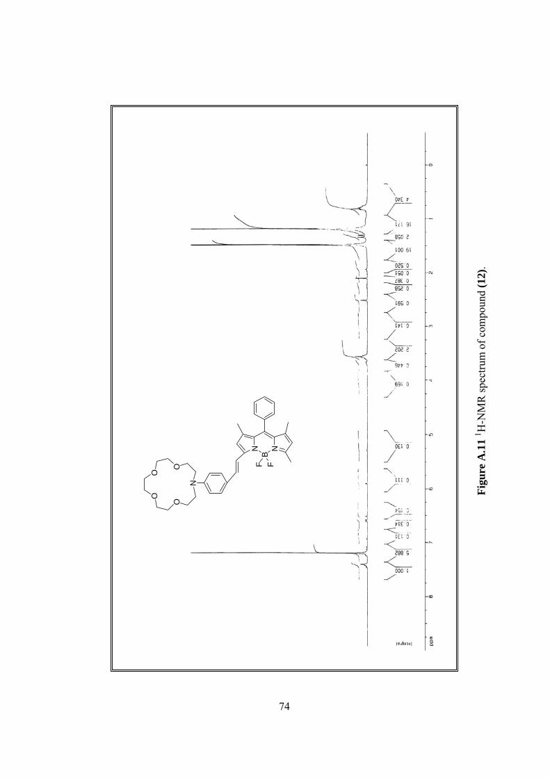
NB
N
F F
OO
OO
N
Figu
re A
.11
1 H-N
MR
spec
trum
of c
ompo
und
(12)
.
74