Embed Size (px)
Citation preview
Louisiana State UniversityLSU Digital Commons
LSU Doctoral Dissertations Graduate School
2007
Synthesis and functionalizations of tetrapyrrolederivativesLijuan JiaoLouisiana State University and Agricultural and Mechanical College, [email protected]
Follow this and additional works at: https://digitalcommons.lsu.edu/gradschool_dissertations
Part of the Chemistry Commons
This Dissertation is brought to you for free and open access by the Graduate School at LSU Digital Commons. It has been accepted for inclusion inLSU Doctoral Dissertations by an authorized graduate school editor of LSU Digital Commons. For more information, please [email protected].
Recommended CitationJiao, Lijuan, "Synthesis and functionalizations of tetrapyrrole derivatives" (2007). LSU Doctoral Dissertations. 1309.https://digitalcommons.lsu.edu/gradschool_dissertations/1309
SYNTHESIS AND FUNCTIONALIZATIONS OF TETRAPYRROLE
DERIVATIVES
A Dissertation
Submitted to the Graduate Faculty of the
Louisiana State University and
Agricultural and Mechanical College
in partial fulfillment of the
requirements for the degree of
Doctor of Philosophy
In
The Department of Chemistry
by
Lijuan Jiao
B.S., Shandong University, China 2000
M. S., University of Science and Technology of China, 2003
December, 2007

ii
DEDICATION
This Dissertation is dedicated to my parents in China for their unconditional love, their
continuous support, and their understanding of every decision that I made throughout of my life.
谨以此文献给我远在中国的亲爱的父母,感谢他们这麽多年以来所给予我的无微不
至的关爱,默默无闻的支持与理解。

iii
ACKNOWLEDGEMENTS
First of all, I would like to give my sincere thanks to my advisor - Professor Kevin M.
Smith. He is a great advisor that every student would ask for. He also has great personality. He is
kind, generous, optimistic and knowledgeable. He provided me abundant guidance, freedom and
support, and continuous encouragement throughout my Ph.D. studies. He led me into porphyrin
chemistry and provided me with invaluable opportunities to know porphyrins and to enjoy
porphyrin research. He has always been there for me when I need suggestions and help with my
research, and also to help with my career plan and job interviews. Without his unconditional
support, my Ph.D. studies would have been more lengthy. I will always be proud of once being
his student and having worked under his guidance.
My special thanks go to Professor Graça Vicente. She is a wonderful woman,
independent, brilliant, but also kind and always willing to provide help to all of her students. She
helped me with everything ever since I first met with her at LSU. She gave me suggestions in my
studies, research, job interview and even my everyday life. It is hard to study in a science major,
especially for a woman. Some time, I felt so frustrated that I even thought about quitting. But she
was there, set a role model for me, and let me regain confidence to continue my studies and my
research. She let me know how to balance career and everyday life.
My special thanks also go to Professor William Crowe, Professor Brian Hales and
Professor Henry James. Professor William Crowe is a great teacher, and he is also a kind,
generous and knowledgeable person. I really enjoy his great lectures in the Organic Chemistry
class. He cared about his students and tries his best to help us to learn essential organic chemistry.
I also enjoyed my research discussions with him, which were very helpful for smoothing my
research process. Professor Brian Hales gave me great lectures in the ―Bioinorganic Chemistry‖
iv
classes, which helped me to learn interesting things outside of organic chemistry. I thank
Professor Henry James for being on my committee.
My special thanks also go to Ms. Sherry Wilkes. She has provided her generous help to
us ever since my husband and I first arrived at LSU. Her generous support continued throughout
our doctoral studies. My thanks also go to my colleges: Dr. Frank Fronczek in the LSU X-ray
facility for those beautiful X-rays of my crystals; Dr. Tracy McCarley in the LSU-MS facility for
teaching me how to operate the MALDI-MS; Mr. Guangyu Li and Dr. Dale Treleaven for
training me to do NMR; Dr. Thomas Weldeghiorghis for the 2D-NMRs in the chlorin-e6 project;
Mr. Tim Jensen for helping me to learn cell-culture techniques and fluorescence microscopy; Dr.
Irena Nesterova from Dr. Soper’s research group for teaching me how to do fluoroesence
quantum yield experiments and her helpful chemistry discussions.
My thanks also go to everyone from both the Smith and Vicente research groups,
especially Wei Liu, Celinah Mwakwari, Jianming Lu, Brahma Ghosh and Michael Easson for
once being my lab mates and making working in the lab comfortable. My thanks also go to the
other group members: Owendi Ongayi, Vijay Gottumukula, Jodie Hargus, Ravi Kumar,
Raymond Luguya, Caleb Clark, Boyd Laurent, Hairong Li, Hillary Tanui, Kiran Allam,
Giuseppe Pomarico, Anatol Litoshka, Martha Sibrian-Vasquez and Federica Mandoj.
My deepest thanks go to my parents, Mr. Congjian Jiao and Ms. Yuying Li. I want to
thank them for their love and their continuous support. They provided me with the strength and
courage to take opportunities and face challenges throughout my life. I also want to thank my
brothers and sisters, Mr. Tao Deng, Mr. Feng Deng, Ms. Mei Deng and Ms. Ping Li. Thanks for
all their care, love and interactions in my life. I also want thank all my friends, especially Dr.
Fang Chen, for being my best friend and always being there for me.

v
My last, but not least, appreciation goes to my loved husband, Mr. Erhong Hao. He
always stands next to me and supports me. He provides me with the deepest love, the continuous
support and encouragement. He offers me his broad shoulder to let my mind rest when I was
exhausted from research. It is him who makes chemistry research romantic and attractive. It is
him who always believed in me and kept on pushing me to do my best. It is also him who always
reminds me to cherish and follow my dreams, so that I will never lose myself. Without him, I
would never be able to finish my doctoral research in such a short period. Also, without him, my
life will not be so bright, colorful and lovely.
vi
TABLE OF CONTENTS
Dedication…...………………………………………….……………………………..…….…....ii
Acknowledgements…..……………………...…………………………………..........................iii
Abstract………………..……………………………………………………………..…..….....viii
Chapter 1. Introduction……………………………………………………………………...….1
1.1History of Tetrapyrroles……………………………………………………………….…..…...1
1.2Overview of Porphyrins……………………………………….…………………..….….….…5
1.3Benzoporphyrins…………………………….…………………..…………………………....20
1.4Overview of Chlorin-e6 as a PDT Photosensitizer………………………….………...............24
1.5Overview of Porphycenes………….……………………...………………………………….28
1.6References……………………………………...………………………………………..……30
Chapter 2. β, β’-Fused Methylenepropanoporphyrins………………………...……………..37
2.1Introduction…………………………………………………………………………………...37
2.2Results and Discussion………………...……………………………………………………..39
2.3Experiment………………………………………………………………...………………….68
2.4Conclusions and Future Work……………………………………………...……………..….74
2.5References…………………………………………………………………...………………..75
Chapter 3. Benzoporphyrins from the Ring-Closing-Metathesis……….………………...…79
3.1Introduction…………………………………………………………………………………...79
3.2Results and Discussion…………………………………………...…………………………..81
3.3Conclusions……………………………………………………………………...…………....98
3.4Experiment…………………………………………...……………………………………….98
3.5References……………………………………………………………………...…….….…..105
Chapter 4. “Hangman Porphyrin” Analogs………………………………………………....107
4.1Introduction………………………………………………………………………………….107
4.2Results and Discussion………………………………...………………………………...….110
4.3Conclusions…..…………………………...……………………………………………...…121
4.4Experiment…………………………………………………………………………………..121
4.5References…………………………………………………………………………...………130
Chapter 5. Total Synthesis of Porphycenes and Improved Synthesis of 2,2’-Bipyrroles...133
5.1Introduction………………………………………………………………………………….133
5.2Results and Discussion………………………………………………………………...…....138
5.3Conclusions and Future Work…………………………………………..………………......156
5.4Experiment…………………………………………………………………………………..156
5.5References…………………………………………………………………………………...175
Chapter 6. The Unique Regiochemistry of Mono-(L)-aspartylchlorin-e6……………..…..178
6.1Introduction………………………………………………………………………………….178

vii
6.2Results and Discussion………………………………………………………..…………....180
6.3Conclusion……………………………………………………………………………….....192
6.4Experiment………………………………………………………………………………….193
6.5References…………………………………………………………………………………..196
Appendix: Letters of Permission………………...…...………………………………………198
Vita…...…………………………………………………………………………………...……202
viii
ABSTRACT
Chapter 1 of this Dissertation presents a brief overview of the history of tetrapyrrole
derivatives and of their fundamental properties. Overviews of porphyrins, benzoporphyrins,
chlorins and porphycenes are presented.
Work presented in Chapter 2 through Chapter 4 mainly focuses on the syntheses and
functionalization of chromophore-extended porphyrin derivatives. Several new synthetic routes
for the syntheses and functionalizations of extended porphyrins either at the β-position or at the
meso-position of porphyrin are developed. From these improved synthetic routes, the regio-
selective syntheses of porphyrin derivatives are described. Chapter 2 mainly focuses on the
syntheses of β,β’-fused methylenepropanoporphyrins and related porphyrin dimers. Chapter 3
mainly describes a new synthetic route for selective synthesis of benzoporphyrin regioisomers
and Chapter 4 mainly discusses new work on the efficient synthesis of the so-called ―Hangman
Porphyrin‖ analogs.
Chapter 5 consists two parts. The first part is devoted results of work on the total
synthesis of an important porphycene derivative, 9-capronyl-oxytetrakis(methyoxyethyl)-
porphycene, which has already been shown to have attractive potential applications in
photodynamic therapy of tumors. The second part of Chapter 5 concerns the improved syntheses
of 2,2’-bipyrrole, which is an important part of our effort to improve the synthesis of porphycene
and related tetrapyrrole derivatives. The potential utility of these 2,2’-bipyrroles as bio-probes
and ion-binding reagents are also tested.
Chapter 6 reports mechanistic studies on the unique regio-selective formation of mono-
(L)-aspartylchlorin-e6. This important photodynamic therapy (PDT) photosensitizer has recently
undergone a structural revision, and the work reported in this Chapter provides a rationale for the

ix
formation of the unexpected regioisomeric structure now known to belong to mono-(L)-
aspartylchlorin-e6.
1
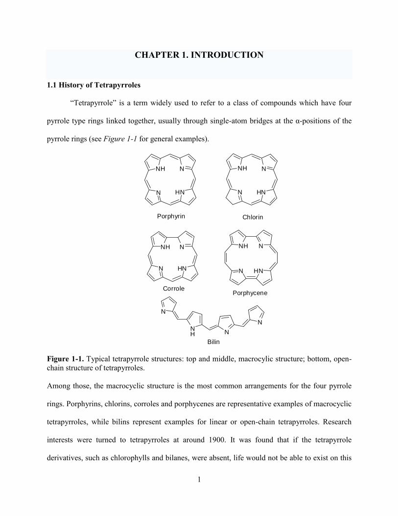
CHAPTER 1. INTRODUCTION
1.1 History of Tetrapyrroles
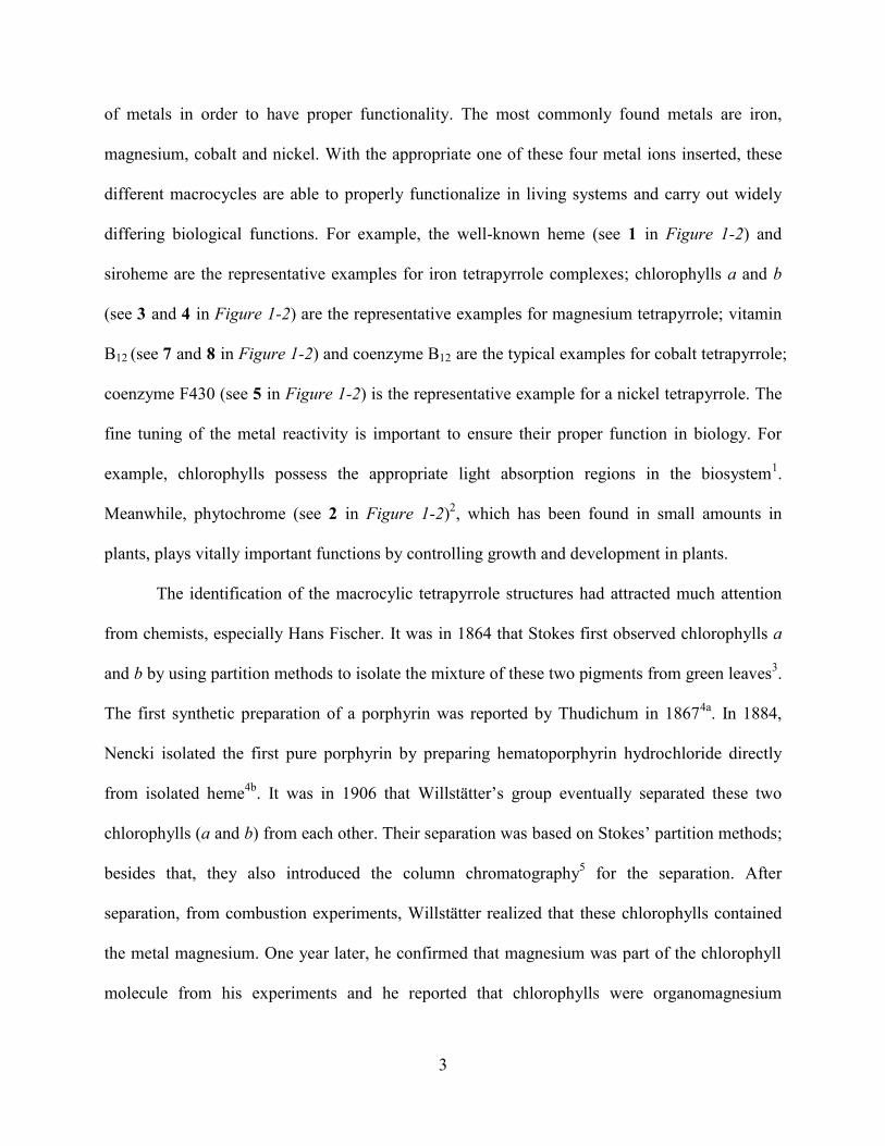
―Tetrapyrrole‖ is a term widely used to refer to a class of compounds which have four
pyrrole type rings linked together, usually through single-atom bridges at the α-positions of the
pyrrole rings (see Figure 1-1 for general examples).
N
NH N
N
N
NH N
HN N
NH N
HN
Porphyrin Chlorin
Bilin
NH N
HNNN
NH N
HN
CorrolePorphycene
Figure 1-1. Typical tetrapyrrole structures: top and middle, macrocylic structure; bottom, open-
chain structure of tetrapyrroles.
Among those, the macrocyclic structure is the most common arrangements for the four pyrrole
rings. Porphyrins, chlorins, corroles and porphycenes are representative examples of macrocyclic
tetrapyrroles, while bilins represent examples for linear or open-chain tetrapyrroles. Research
interests were turned to tetrapyrroles at around 1900. It was found that if the tetrapyrrole
derivatives, such as chlorophylls and bilanes, were absent, life would not be able to exist on this
2
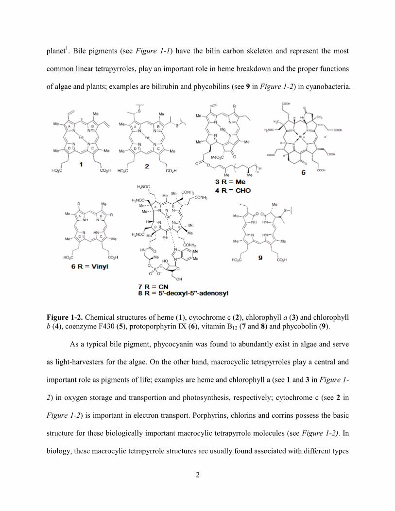
planet1. Bile pigments (see Figure 1-1) have the bilin carbon skeleton and represent the most
common linear tetrapyrroles, play an important role in heme breakdown and the proper functions
of algae and plants; examples are bilirubin and phycobilins (see 9 in Figure 1-2) in cyanobacteria.
Figure 1-2. Chemical structures of heme (1), cytochrome c (2), chlorophyll a (3) and chlorophyll
b (4), coenzyme F430 (5), protoporphyrin IX (6), vitamin B12 (7 and 8) and phycobolin (9).
As a typical bile pigment, phycocyanin was found to abundantly exist in algae and serve
as light-harvesters for the algae. On the other hand, macrocyclic tetrapyrroles play a central and
important role as pigments of life; examples are heme and chlorophyll a (see 1 and 3 in Figure 1-
2) in oxygen storage and transportion and photosynthesis, respectively; cytochrome c (see 2 in
Figure 1-2) is important in electron transport. Porphyrins, chlorins and corrins possess the basic
structure for these biologically important macrocylic tetrapyrrole molecules (see Figure 1-2). In
biology, these macrocylic tetrapyrrole structures are usually found associated with different types
3
of metals in order to have proper functionality. The most commonly found metals are iron,
magnesium, cobalt and nickel. With the appropriate one of these four metal ions inserted, these
different macrocycles are able to properly functionalize in living systems and carry out widely
differing biological functions. For example, the well-known heme (see 1 in Figure 1-2) and
siroheme are the representative examples for iron tetrapyrrole complexes; chlorophylls a and b
(see 3 and 4 in Figure 1-2) are the representative examples for magnesium tetrapyrrole; vitamin
B12 (see 7 and 8 in Figure 1-2) and coenzyme B12 are the typical examples for cobalt tetrapyrrole;
coenzyme F430 (see 5 in Figure 1-2) is the representative example for a nickel tetrapyrrole. The
fine tuning of the metal reactivity is important to ensure their proper function in biology. For
example, chlorophylls possess the appropriate light absorption regions in the biosystem1.
Meanwhile, phytochrome (see 2 in Figure 1-2)2, which has been found in small amounts in
plants, plays vitally important functions by controlling growth and development in plants.
The identification of the macrocylic tetrapyrrole structures had attracted much attention
from chemists, especially Hans Fischer. It was in 1864 that Stokes first observed chlorophylls a
and b by using partition methods to isolate the mixture of these two pigments from green leaves3.
The first synthetic preparation of a porphyrin was reported by Thudichum in 18674a
. In 1884,
Nencki isolated the first pure porphyrin by preparing hematoporphyrin hydrochloride directly
from isolated heme4b
. It was in 1906 that Willstätter’s group eventually separated these two
chlorophylls (a and b) from each other. Their separation was based on Stokes’ partition methods;
besides that, they also introduced the column chromatography5 for the separation. After
separation, from combustion experiments, Willstätter realized that these chlorophylls contained
the metal magnesium. One year later, he confirmed that magnesium was part of the chlorophyll
molecule from his experiments and he reported that chlorophylls were organomagnesium

4
complexes. After that, experiments were performed to remove the magnesium and to generate a
metal-free product, named ―pheophytin‖. After chemical manipulation, such as burning,
oxidization, reduction, and pyrolysis, many of the degradation products were found to be
pyrroles or contain pyrrolic residues. Using less drastic degradation, much of the original
molecules were retained and found to be deeply colored stable substances. Then, in 1912,
Küster6 suggested that pheophytin and chlorophylls shared a similar macrocyclic structure, in
which four pyrrole derived rings were joined to each other by methane bridges. This structure is
now known as the porphyrin macrocycle. Initially, when Küster proposed his suggested structure,
Fischer (who was the major porphyrin researcher of the day) doubted his results. It was because
Fisher thought there would be a stability problem issue with the large ring-size present in
Küster’s proposal. Then, in 1925, Keilin7
discovered that heme (see 1 in Figure 1-2) was an
organic complex of iron. His discovery was soon confirmed by Fischer and Kämmerer. Also,
iron was removed from heme, and protoporphyrin IX was generated. The subsequent chemical
manipulation provided similar results as those of pheophytin. One year later, in 1926, Fisher
synthesized etioporphyrin-I. From his own synthesis, Fisher also realized the presence of similar
aromatic structure as suggested by Küster and accepted Küster’s idea. By the late 1930’s,
Willstätter and Fischer had already worked out the complex structures of both chlorophyll a (see
3 in Figure 1-2) and heme (see 1 in Figure 1-2) from hemoglobin8. It was a surprise to discover
that although chlorophyll and heme were such difference in their appearances and functionalities,
they actually shared a similar basic macrocylic structure and gave similar visible spectra. Later
on, the structures of heme (see 1 in Figure 1-2) and protoporphyrin IX (6) opened the way to
understanding how the prosthetic group of the cytochromes c isolated from many living things
could play a crucially important role in biological electron transport9.

5
In the 1940’s chromatography became widely used as an important tool in product
separation and purification; this provided great opportunities to study the complex structures of
tetrapyrroles. It was in 1948 that vitamin B12 was first isolated as deep red colored crystals10
. Its
structure was studied in both the USA and Britain, separately by two teams led by Karl Folkers
and Sir Alexander Todd. Based on their studies and the efforts of many other research groups, in
1953, cobalt was found in vitamin B12. At that time, even the macrocyclic ligand that held cobalt
was identified11
. It was found that there was a direct link between rings A and D to form a
smaller macrocycle in vitamin B12, which is different from heme (1) and chlorophyll a (3). The
parent ring system of vitamin B12 was named corrin. Then, in 1960, Woodward accomplished
the first total synthesis of chlorophyll a12
.
1.2 Overview of Porphyrins
1.2.1 Introduction
Figure 1-3. Fischer (left) and IUPAC (middle and right) nomenclature systems for porphyrins.
Porphyrin research has been well-established for over a century. It was Thudichum4a
who
first isolated a porphyrin from hemoglobin in 1867. Ever since then, attracted to their interesting
physical, chemical, and spectroscopic properties and the essential biological functionalities of
porphyrins, scientists from many different areas have devoted themselves to porphyrin research.
So far, there has been at least nine Nobel Prizes in Chemistry have been awarded for outstanding

6
achievements in porphyrin chemistry. Most recently there have also been published the twenty
volumes of ―The Porphyrin Handbook‖. Here, only a brief overview of related porphyrins
aspects will be discussed, which is intended to help the reader better understand the research
projects that will be discussed in detail in the following chapters of this Dissertation.
Porphyrins naturally occur as colored pigments and have been described as ―the Pigments
of Life‖12
. Porphyrins and their derivatives occur widely in nature and play important roles in
biological processes. Representative examples of porphyrin derivatives are hemes, (found in
myoglobins, hemoglobins, cytochromes, catalases and peroxidases), chlorophylls and
bacteriochlorophylls. Nature uses them in the most important processes of photosynthesis
(chlorophylls and bacteriochlorophylls), and in oxygen-transportation, in electron-transfer and
also in catalytic oxidations (hemes). For example, heme, the iron(II) complex of protoporphyrin
IX, is the prosthetic group in hemoglobins and myoglobins. These heme proteins play the
essential roles of transporting and storing molecular oxygen, which is needed for all cellular
respiration. Heme can catalyze the oxidation of substrates using hydrogen peroxide in
peroxidases, and catalyze the breakdown of hydrogen peroxide to water and oxygen in catalases.
Besides these naturally existing porphyrin derivatives, synthetic porphyrins have found
important applications in the medical research area. Due to their intriguing physical, chemical
and biological properties, porphyrins and their metalated complexes have also attracted lots of
interests from various interdisciplinary research areas.
It was in 1912 that Küster first proposed the existence of the intricate porphyrin ring
system, which was late confirmed by Fischer. Now it has been well-accepted that the porphyrin
macrocycle is an aromatic system, consisting of four pyrrole units which are linked by four sp2
hybridized meso-carbons and this model has been confirmed by many hundreds of X-ray
7
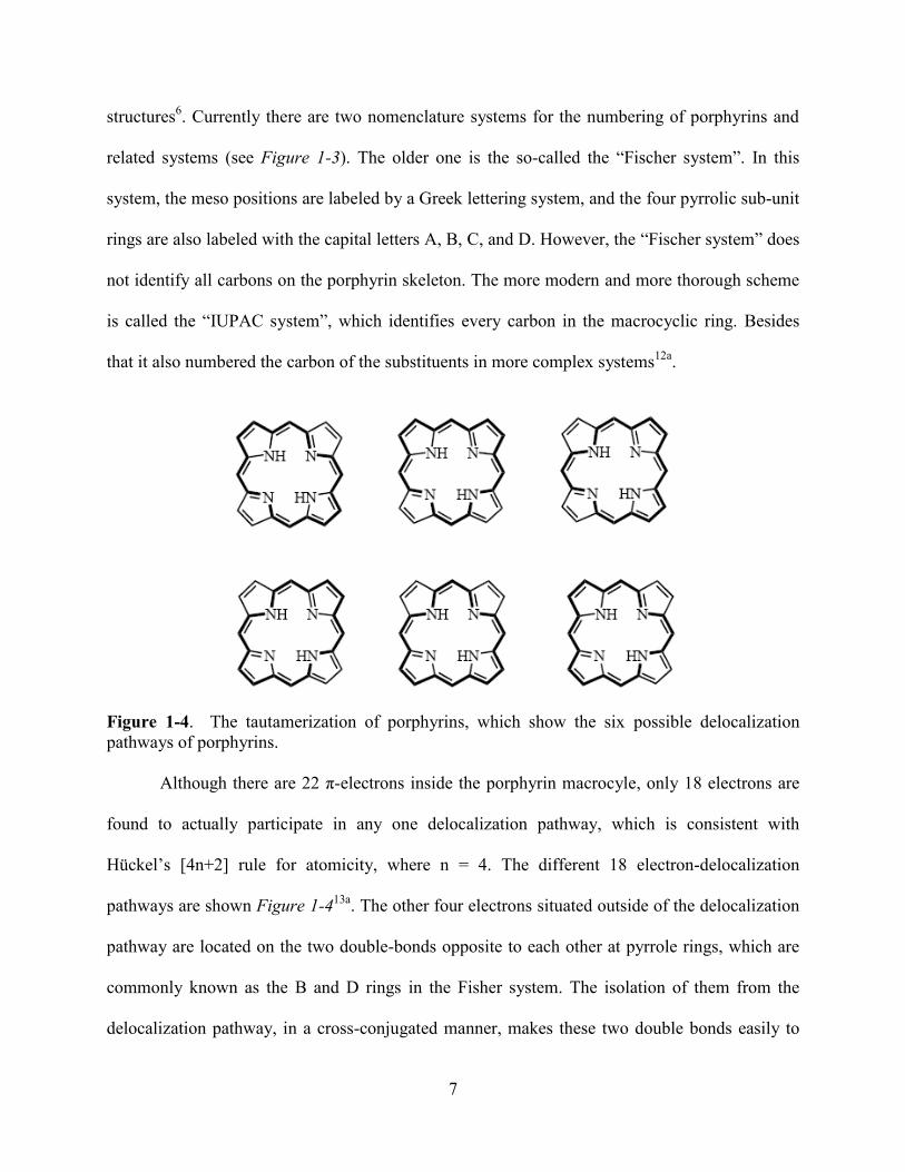
structures6. Currently there are two nomenclature systems for the numbering of porphyrins and
related systems (see Figure 1-3). The older one is the so-called the ―Fischer system‖. In this
system, the meso positions are labeled by a Greek lettering system, and the four pyrrolic sub-unit
rings are also labeled with the capital letters A, B, C, and D. However, the ―Fischer system‖ does
not identify all carbons on the porphyrin skeleton. The more modern and more thorough scheme
is called the ―IUPAC system‖, which identifies every carbon in the macrocyclic ring. Besides
that it also numbered the carbon of the substituents in more complex systems12a
.
Figure 1-4. The tautamerization of porphyrins, which show the six possible delocalization
pathways of porphyrins.
Although there are 22 π-electrons inside the porphyrin macrocyle, only 18 electrons are
found to actually participate in any one delocalization pathway, which is consistent with
Hückel’s [4n+2] rule for atomicity, where n = 4. The different 18 electron-delocalization
pathways are shown Figure 1-413a
. The other four electrons situated outside of the delocalization
pathway are located on the two double-bonds opposite to each other at pyrrole rings, which are
commonly known as the B and D rings in the Fisher system. The isolation of them from the
delocalization pathway, in a cross-conjugated manner, makes these two double bonds easily to
8
be reduced or oxidized using numerous reactions. Typical examples are: catalytic hydrogenation,
reduction with diimide, and hydroboration. The reduction products for porphyrins are usually
chlorins or bacteriochlorins. As a typical aromatic system, porphyrin is also able to undergo a
number of electrophilic aromatic substitution reactions (EAS), such as nitration, halogenation
and formylation on any unsubstituted meso- and/or β-pyrrolic positions13b-c
. However, the
quaternary α-pyrrolic carbons rarely participate in any kind of reactions. It was found that the
reducing of the aromatic character from the delocalization pathways can induce dramatic
changes in the spectroscopic properties14
.
There are two pyrrolenine nitrogen atoms (pKa~6) and two inner-core NH groups
(pKa~16) in porphyrins. The former can act as a base to accept protons and the latter can act as
an acid to provide protons. The pyrrolenine nitrogen atoms can be protonated by strong acids,
such as sulfuric acid and trifluoroacetic acid (TFA). The inner-core nitrogen protons can be
removed by bases and metals. Also the presence of these nitrogen atoms makes most of the
porphyrin derivatives amphoteric and shows both acidic and basic behavior13b-c
. Also they can
serve as an inner chelating pocket and provided various opportunities for chemical modifications.
In most cases, the insertion of metals into the porphyrin macrocycles is easy and the removal of
them can be achieved with Brønsted-Lowry acids without affecting the macrocyclic conjugation.
Some represent metalloporphyrins are Cu, Ni, Zn, Fe and Co centered porphyrins.
The NMR spectra of the aromatic tetrapyrrole show anisotropic effects15
. When there is a
magnetic field applied, a ring current is generated and a local magnetic field similar to that in
benzene is induced. In the proton NMR spectrum, the interior nitrogen protons normally appear
between δ -4 and -2 due to the high shielding by the ring current. The deshielded meso-protons
usually appear at very low field (δ ~ 10 ppm) and the pyrrolic protons are also deshielded and

9
tend to resonate at δ 8 to 9, which shows big shift compared to that of pyrrole at δ ~ 6 ppm.
However, when there is aggregate formation of these porphyrins, their NMR spectra tends to be
hard to assign.
Visible absorption spectroscopy is also a powerful tool to probe the structure of the
tetrapyrrole chromophore of porphyrins. The macrocyclic conjugation gives several
characteristic weak absorption bands, which are called Q bands and are located between 450-700
nm; there is one major absorption band known as the Soret band, which is an intense absorption
band (ε > 100,000) located between 400 and 450 nm16
. The Soret band is characteristic of the
macrocyclic conjugation, and it disappears when the aromatic delocalization pathway is
disrupted. Porphyrin derivatives show deep colors and have strong absorptions in the visible
region near 400 nm, with their molar extinction coefficients to be about 105 Mol/L. The color
difference among porphyrins is attributed to the different absorption spectra associated with
different unique tetrapyrrolic structure. For example, natural porphyrins have dark red colors, but
their reduced form, such as chlorins, show dark green or blue green colors. Thus the modification
of the peripheral double bond of the porphyrins can cause changes of the absorption spectra, both
the intensity and the wavelength. However, as long as the 18 π-electron cyclic pathway remains,
the intense Soret band would also remains. The Soret band is absent only when porphyrin
macrocyclic conjugation is disrupted. On the other hand, although the chelation, pH and different
peripheral substituent arrays change the absorbance energy intensities and even change the color
of the compounds, it usually only involves the Q band absorptions, and leaves the Soret band
intact.
10
1.2.2 Synthetic Methodologies:
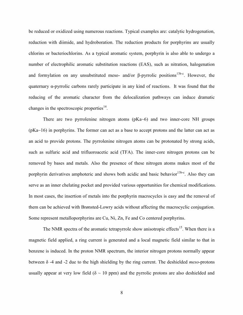
Figure 1-5. Historically and biologically important porphyrins.
Figure 1-6. Chemical structure of heme, chlorophyll a and bacteriochlorophyll.
Fisher, the ―father of porphyrin chemistry‖, reported the first total synthesis of the
porphyrins etioporphyrin-III and octamethylporphyrin, in 1926 (see Figure 1-5). In 1929 Fischer
synthesized and named protoporphyrin-IX (see Figure 1-5), which is the free base porphyrin of
hemin17
. Since then, a large number of synthetic routes have been developed for the preparation
of both symmetrical and unsymmetrical porphyrin derivatives for structural, mechanistic,
synthetic and biological studies. Here, some of the most commonly used synthetic methodologies
are described. Porphyrin syntheses often started from the syntheses of a large class of pyrroles.
Hans Fischer had already perfected early pyrrole synthetic work and also the syntheses of a
11
variety of porphyrins18
. His early research work had inspired many research groups throughout
the world to participate and devote their efforts to improve porphyrin synthetic methodology.
Now, there are several routes to generate porphyrins. One of them is to modify natural products.
For example, the modification of chlorophylls a or b, bacteriochlorophylls and hemin (see
Figure 1-6) can be used to generate very desirable porphyrins.
Although porphyrins can be generated from total synthesis starting from monopyrrolic
subunits, the types of porphyrins that can be generated are very limited. Recently, many
improved synthetic routes have been reported, which now provide easy access to useful
porphyrins. Currently the syntheses of symmetric porphyrins including both the octa-ß
substituted or tetra-meso substituted porphyrins, involve the tetramerization of a suitable
monopyrrolic subunit19
. However, little progress has been made for the unsymmetric porphyrin
synthesis.
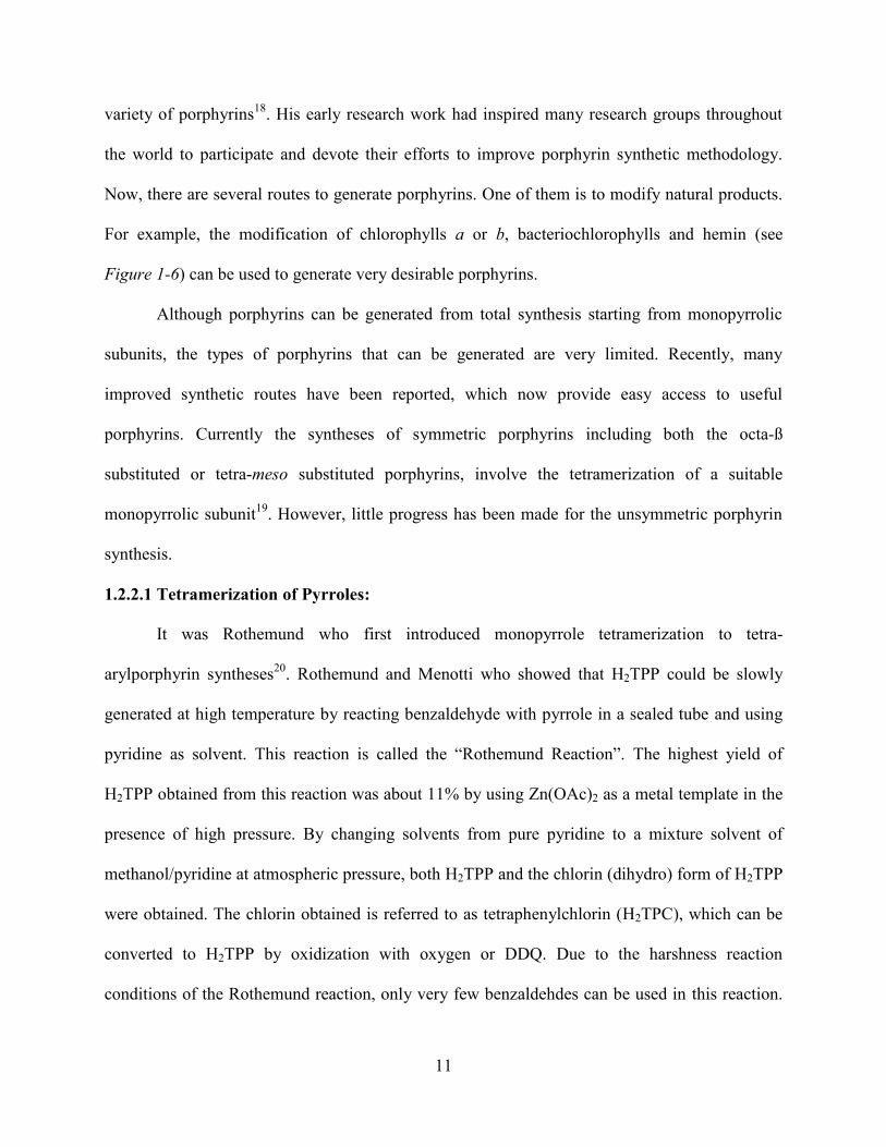
1.2.2.1 Tetramerization of Pyrroles:
It was Rothemund who first introduced monopyrrole tetramerization to tetra-
arylporphyrin syntheses20
. Rothemund and Menotti who showed that H2TPP could be slowly
generated at high temperature by reacting benzaldehyde with pyrrole in a sealed tube and using
pyridine as solvent. This reaction is called the ―Rothemund Reaction‖. The highest yield of
H2TPP obtained from this reaction was about 11% by using Zn(OAc)2 as a metal template in the
presence of high pressure. By changing solvents from pure pyridine to a mixture solvent of
methanol/pyridine at atmospheric pressure, both H2TPP and the chlorin (dihydro) form of H2TPP
were obtained. The chlorin obtained is referred to as tetraphenylchlorin (H2TPC), which can be
converted to H2TPP by oxidization with oxygen or DDQ. Due to the harshness reaction
conditions of the Rothemund reaction, only very few benzaldehdes can be used in this reaction.
12
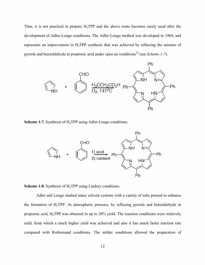
Thus, it is not practical to prepare H2TPP and the above route becomes rarely used after the
development of Adler-Longo conditions. The Adler-Longo method was developed in 1964, and
represents an improvement in H2TPP synthesis that was achieved by refluxing the mixture of
pyrrole and benzaldehyde in propionic acid under open air conditions21
(see Scheme 1-7).
Scheme 1-7. Synthesis of H2TPP using Adler-Longo conditions.
Scheme 1-8. Synthesis of H2TPP using Lindsey conditions.
Adler and Longo studied many solvent systems with a variety of salts present to enhance
the formation of H2TPP. At atmospheric pressure, by refluxing pyrrole and benzaldehyde in
propionic acid, H2TPP was obtained in up to 20% yield. The reaction conditions were relatively
mild, from which a much higher yield was achieved and also it has much faster reaction rate
compared with Rothemund conditions. The milder conditions allowed the preparation of

13
porphyrins with a wide variety of functionalities attached. Although it gave a vast improvement
over the Rothemund method, this reaction condition still had its limitations. It was still relative
harsh and more sensitive functionalities failed to survive. Also the purifications became more
difficult due to the formation of tar. Despite all these drawbacks, it was still the most efficient
method for the syntheses of meso-tetra-alkylporphyrins at that time.
In 1986, Lindsey optimized the method and developed an improved synthesis of
porphyrins, under so called ―Lindsey conditions‖ (see Scheme 1-8). This is by far the most
effective route for synthesizing symmetrical porphyrins. For example, the preparation of
porphyrins with the same substituents on all four meso-positions, or all eight of β-pyrrolic
positions, or a combination of them. Under Lindsey conditions, the synthesis of porphyrin is
done in two steps through the formation of porphyrinogen from monopyrrole tetramerization and
a subsequent separate oxidation22
.
It was successfully demonstrated by Lindsey that the formation of H2TPP could be
achieved under equilibrium conditions, and under this situation many functional groups could
survive. A colorless porphyrinogen was first formed, followed by a subsequent oxidation step
with p-chloranil or DDQ. H2TPP was formed by dissolving benzaldehyde and pyrrole in
dichloromethane in a 10-2
M solution. The acid catalyst (BF3•Et2O or TFA) was typically added
at a dilution of 10-3
M. The yields of porphyrins generated under these conditions were improved
to around 30-40%. It was found that the use of p-chloranil for the oxidation typically gave higher
yields than the case when DDQ was used as oxidation reagent. Lindsey and coworkers
discovered that not only the oxidation reagent, but the reaction time, the concentration of starting
materials and the acid catalyst could also affect the reaction. It was found by altering the
concentration of acid, the yield was only slightly affected. However, the yields of H2TPP were
14
decreased a lot by varying the reagent concentrations to ten-fold higher and ten-fold lower than
10-2
M. The oxidation rate depended on the oxidizing agent. For example, the oxidation can
finish within minutes by using DDQ, while it requires an hour to complete the oxidation when p-
chloranil was used. The efficiency of Lindsey conditions have been proved for both tetra-
arylporphyrins and meso-tetra-alkylporphyrins, giving unprecedented yields.
1.2.3 Applications of Synthetic Porphyrins
Porphyrins play vital roles in biological systems, not only in nature but also in
applications in material science and medicine areas.
1.2.3.1 Applications in Material Science
Porphyrins-based molecular wires are appealing because polyporphyrin systems
containing redox active and/or photoactive units, which allows the long-distance delocalization
of electron density, thus makes them ideal systems for electron- or energy- transfer23
. Earlier
research using porphyrins and related compounds to study electron transfer was focused on
modeling the photosynthetic reaction center and on a better understanding of the complex
mechanism of photosynthesis23a
. Covalently-linked bisporphyrins and quinine-substituted
porphyrin dimers and trimers were built and used to mimic the electron transfer process in
biological systems. Recently, more study has been focused on using supramolecular porphyrin
arrays as potential photonic molecular wires.
Certain heme-containing enzymes such as cytochrome P450 have been found to perform
hydroxylation of alkanes and epoxidation of unfunctionalized alkenes24
. Noncovalent hydrogen-
bonding interactions have been found to be the most important factor in dynamically regulating
the active site for PCET (Proton-Coupled Electron Transfer) reactivity inside some of these
enzyme systems. As a result, a lot of research has been developed to design porphyrin molecules
15
for biomimetic models for heme-containing enzymes. Recently, porphyrins functionalized with
hydrogen-bond synthons have been widely studied because they could be used as efficient
building blocks for construction of supramolecules with appealing structural and electronic
properties. Among these, the so-called ―Hangman Porphyrins‖ have attracted much recent
interest, because of the prospective application they have for unraveling the hydrogen bonding
effect on energy and electron-transfer reaction. Due to the rigidity of the spacer used in these
systems, a side-to-side arrangement of the porphyrin macrocycle and hydrogen-bond
functionality has been shown. These hangman porphyrins have simplified the construction of
biomoleculaes with engineered distal sites as platforms able to control both proton and electron
transfer, which provides the opportunity to precisely control the functional nature of a hydrogen-
bonding group25
. Despite these advantages, the elucidation of structure/reactivity relationships of
PCET catalysis is difficult in porphyrin systems. Significant challenges are posed by the lengthy
total synthesis and tedious purification of porphyrin platforms and their intractability to modular
modifications26
. In the meanwhile, heteroatom-bridged calixarenes, such as the oxygen-bridged
calix[4]arenes, are easy to access and modify and also display unique chemical and physical
properties27
. Inspired by the easy availability and the unique discrete 1,3-alternate conformations
of oxacalix[4]arenas, the design and synthesis of hangman porphyrin analogs with
oxacalix[4]arene as spacer to hang hydrogen synthons over porphyrin macrocycle28
become very
attractive, and this will be discussed in Chapter 4.
1.2.3.2 Biological Applications of Porphyrins - Photodynamic Therapy (PDT)
PDT is a binary therapy which combines light and a photosensitizer in the presence of
oxygen to destroy tumors or unwanted tissues29
(see Figure 1-9). It has been found that many
16
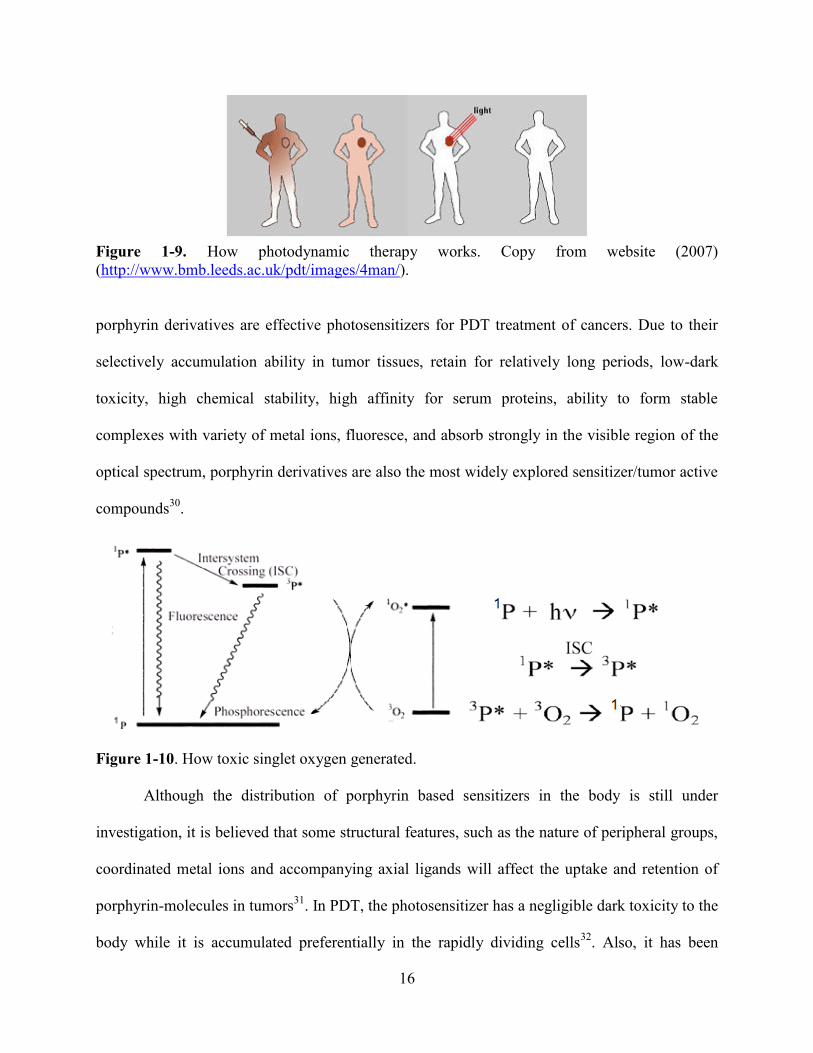
Figure 1-9. How photodynamic therapy works. Copy from website (2007)
(http://www.bmb.leeds.ac.uk/pdt/images/4man/).
porphyrin derivatives are effective photosensitizers for PDT treatment of cancers. Due to their
selectively accumulation ability in tumor tissues, retain for relatively long periods, low-dark
toxicity, high chemical stability, high affinity for serum proteins, ability to form stable
complexes with variety of metal ions, fluoresce, and absorb strongly in the visible region of the
optical spectrum, porphyrin derivatives are also the most widely explored sensitizer/tumor active
compounds30
.
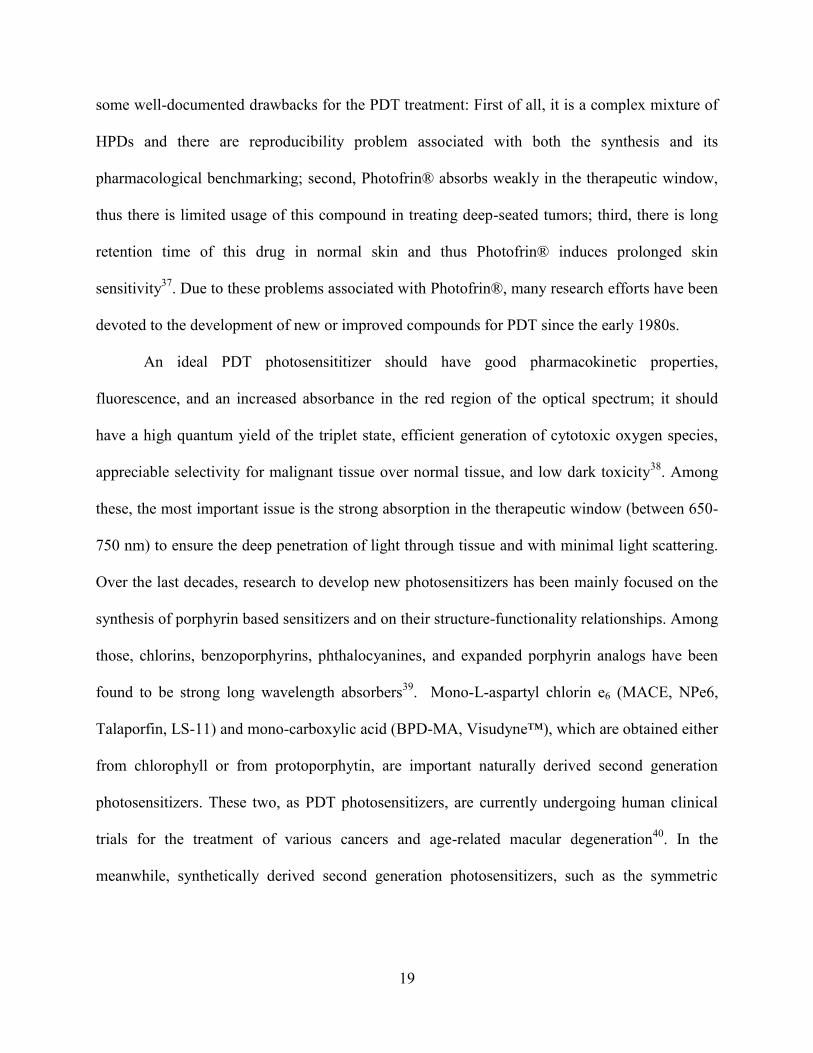
Figure 1-10. How toxic singlet oxygen generated.
Although the distribution of porphyrin based sensitizers in the body is still under
investigation, it is believed that some structural features, such as the nature of peripheral groups,
coordinated metal ions and accompanying axial ligands will affect the uptake and retention of
porphyrin-molecules in tumors31
. In PDT, the photosensitizer has a negligible dark toxicity to the
body while it is accumulated preferentially in the rapidly dividing cells32
. Also, it has been

17
hypothesized that porphyrins have the tendency of association with plasma proteins, particularly
low density lipoproteins (LDL), while the increasing level of LDL receptors has been found in
cancer cells33
. Usually, photosensitizers are injected into the bloodstream and it is generally
believed that amphiphilic molecules that bear both hydrophobic sites and hydrophilic sites
should improve tumor-specificity (see Figure 1-9). At the absence of light, the photosensitizer is
harmless and has no effect to either healthy or abnormal tissue. When exposed to a carefully
regulated specific light dose, it becomes activated and can rapidly destroy the tissue irradiated
with the light. Compared to the other currently available cancer therapeutic methods, such as
chemotherapy, radiotherapy and surgery (or a combination of these methods), PDT has the
advantage of preferential accumulation of the photosensitizer in the target tissue and precise
selectivity of the treatment by controlling the illumination34
. Figure 1-10 shows a modified
Jablonski diagram of the mechanism for PDT. After the light penetrates the tissue, the
photosensitizer is excited and reacts with other substrates, mainly the molecular oxygen to
generate highly cytotoxic species, including singlet oxygen, superoxide anion and hydroxyl
radicals, which can cause irreversible damage to the tumor cells34
. Ground state (1S) is the stable
electronic configuration of photosensitizers35
. With an appropriate wavelength of light irradiation,
the photosensitizer is excited to its singlet excited state (1S*). When it decays to the ground state,
the fluorescence radiation enables the identification of tumor tissue. Meanwhile, it can also
undergo a nonradiative process of inter-system crossing (ISC) to convert the photosensitizer
from a singlet state to a triplet excited state (1T*) or through internal conversion to release the
energy as heat. The relaxation of photosensitizer from the triplet excited state to the ground state
usually through two pathways: release of phosphorence radiation, or non-radiatively transferring
its energy to another molecule with a triplet ground state. ISC involves a change in the electron

18
spin, thus it is a spin-forbidden pathway and imposes a relatively long time on the triplet state,
allowing interaction with adjacent molecules of the photosensitizer. Molecular oxygen has a
triplet ground state and is abundant in tissue. It can interact with the triplet state photosensitizer
to generate highly cytotoxic species. A good photosensitizer can go through this pathway with
high efficiency.
Figure 1-11. Chemical structure of hematoporphyrin.
Hematoporphyrin (see Figure 1-11) was the first evaluated porphyrin for PDT and it is
readily available from blood. Back to the early 1960s, Lipson et al. had already prepared a
derivative of hematoporphyrin (HPD) which displayed an enhanced selectivity for PDT. HPD is
a mixture of hematoporphyrin, protoporphyrin-IX, hydroxyethylvinylporphyrin and other
complex compounds containing dimeric and oligomeric derivatives of hematoporphyrin36
. In
1981, Dougherty et al. prepared a more purified form of HPD, known today as Photofrin®, from
gel exclusion chromatography. Photofrin® was approved in the USA by the Food and Drug
Administration (FDA) and it also is now approved in eleven European countries. Over the last
two decades, Photofrin® has been used successfully in the treatment of both early and advanced
stages of the lung cancer. Although it has shown curative for a range of cancers, Photofrin® has
19
some well-documented drawbacks for the PDT treatment: First of all, it is a complex mixture of
HPDs and there are reproducibility problem associated with both the synthesis and its
pharmacological benchmarking; second, Photofrin® absorbs weakly in the therapeutic window,
thus there is limited usage of this compound in treating deep-seated tumors; third, there is long
retention time of this drug in normal skin and thus Photofrin® induces prolonged skin
sensitivity37
. Due to these problems associated with Photofrin®, many research efforts have been
devoted to the development of new or improved compounds for PDT since the early 1980s.
An ideal PDT photosensititizer should have good pharmacokinetic properties,
fluorescence, and an increased absorbance in the red region of the optical spectrum; it should
have a high quantum yield of the triplet state, efficient generation of cytotoxic oxygen species,
appreciable selectivity for malignant tissue over normal tissue, and low dark toxicity38
. Among
these, the most important issue is the strong absorption in the therapeutic window (between 650-
750 nm) to ensure the deep penetration of light through tissue and with minimal light scattering.
Over the last decades, research to develop new photosensitizers has been mainly focused on the
synthesis of porphyrin based sensitizers and on their structure-functionality relationships. Among
those, chlorins, benzoporphyrins, phthalocyanines, and expanded porphyrin analogs have been
found to be strong long wavelength absorbers39
. Mono-L-aspartyl chlorin e6 (MACE, NPe6,
Talaporfin, LS-11) and mono-carboxylic acid (BPD-MA, Visudyne™), which are obtained either
from chlorophyll or from protoporphytin, are important naturally derived second generation
photosensitizers. These two, as PDT photosensitizers, are currently undergoing human clinical
trials for the treatment of various cancers and age-related macular degeneration40
. In the
meanwhile, synthetically derived second generation photosensitizers, such as the symmetric
20
meso-tetra(m-hydroxyphenyl)chlorin (m-THPC, Foscan®) and Sn(IV)-etiopurpin (SnET2,
PuryltinTM), are in their early clinical trials for the treatment of various cancers41
.
1.3 Benzoporphyrins
1.3.1 Overview of Benzoporphyrins
Benzoporphyrin refers to the type of porphyrin that has aromatic subunits fused at the β-
pyrrolic positions (see Figure 1-12). When there are four aromatic subunits fused at the β-
pyrrolic positions of porphyrin, they are called tetrabenzoporphyrins (TBPs) (see 1-10 in Scheme
1-9).42
TBPs are chemically stable compounds, and have unique chemical, physical and
spectroscopic properties. Among those, their absorption spectra are significantly shifted to the
infrared region due to the extended π-conjugation. Different from porphyrin derivatives, which
can absorb light between 380-400 and 500-560 nm, TBPs have absorbance in the near IR region,
which allows deep light penetration into tissue and they are therefore suitable photosensitizers in
photodynamic therapy (PDT) of cancer43
. They can act as models for naturally occurring
tetrapyrrole derivatives. They have similar physical properties compared with phthalocyanine,
which is one of the most widely studied organic pigments, having many applications in industry
as dyes, inks, catalysts, electrical conductors and other optical materials. Similarly, TBPs have
found applications as opto-electronic materials, nonlinear optical materials, and luminescent
markers for oxygen, near-IR labeling dyes, and pH sensors in biomedical imaging44
.
Benzoporphyrins were first discovered in trace amounts in various oil shales and petroleum45
.
Despite the increasing interests in their syntheses and characterization, researches on
tetrabenzoporphyrins (TBPs) have progressed slowly due to the difficult synthetic access to these
compounds. Above all, unsymmetrical (non-tetra) benzoporphyrins such as the

21
monobenzoporphyrins and dibenzoporphyrin (see Figure 1-12), have proven to be very difficult
to synthesize.
Figure 1-12. Chemical structures of unsymmetrical benzoporphyrins.
1.3.2 Syntheses of Benzoporphyrins
Scheme 1-9. Previous symmetrical benzoporphyrin synthetic routes.
22
Helberger and coworkers first described the syntheses of TBPs from ocyanoacetophenone
and various phthalimidines46
. Back then, there were two main approaches for the synthetic
development of benzoporphyrins: The first approach was the high temperature condensation
using phthalimidine derivatives at the presence of a metal template; the second approach was the
condensation of pyrroles with mesocarbon donors47
. Later on, many improved syntheses of
benzoporphyrin were reported (see Scheme 1-9).
The initial improvement of TBP synthesis was made by Linstead’s group, in which TBPs
were prepared by high temperature condensation (350-400 °C) of 1-6 or 1-7 (see Scheme 1-9) in
the presence of metal salts, such as iron, zinc, magnesium, cadmium, or metallic acetates48
. The
metalated symmetrical benzoporphyrins thus obtained were usually very impure and required
extensive purification processes. Later on, Vogler and Kunkely used the template reaction of 2-
acetylbenzoic acid (see 1-8 in Scheme 1-9) with zinc(II) acetate in the presence of aqueous
ammonia and molecular sieves at 400 °C to improve the reaction efficiency and simplify the
purification process49
. Also, they found that the condensation of phthalimide or its potassium salt
with sodium acetate or malonic acid (see 1-9 in Scheme 1-9) in the presence of zinc(II) acetate at
360 °C could afford TBPs. Vogler and Kunkley’s synthesis affords the corresponding 1-10 in a
more pure form in 17% yield50
.
Metalated symmetrical benzoporphyrins can also be synthesized using Rothemund
conditions, in low yields, from unstable isoindoles through inert atmosphere pyrolysis in the
presence of metals or metal salts, or from refluxing in a high-boiling solvent (1,2,4-
trichlorobenzene or 1-chloronapthalene). Remy improved the synthesis of metalated symmetrical
benzoporphyrins by high temperature condensations of isoindole (see 1-15 in Scheme 1-10) and
formaldehyde in the presence of a metal or metal salt51
. It was found that metal salts play an
23
important role in Remy’s method. In the presence of metal salts the yield achieved for the target
compound was 53%. In the absence of the metal salt, only very low yields were achieved. Under
similar condition, when changing from formaldehyde to benzaldehyde, the metalated
tetraphenyltetrabenzoporphyrin was obtained as the major product together with a mixture of
partially meso-substituted metallo-TBPs52
.
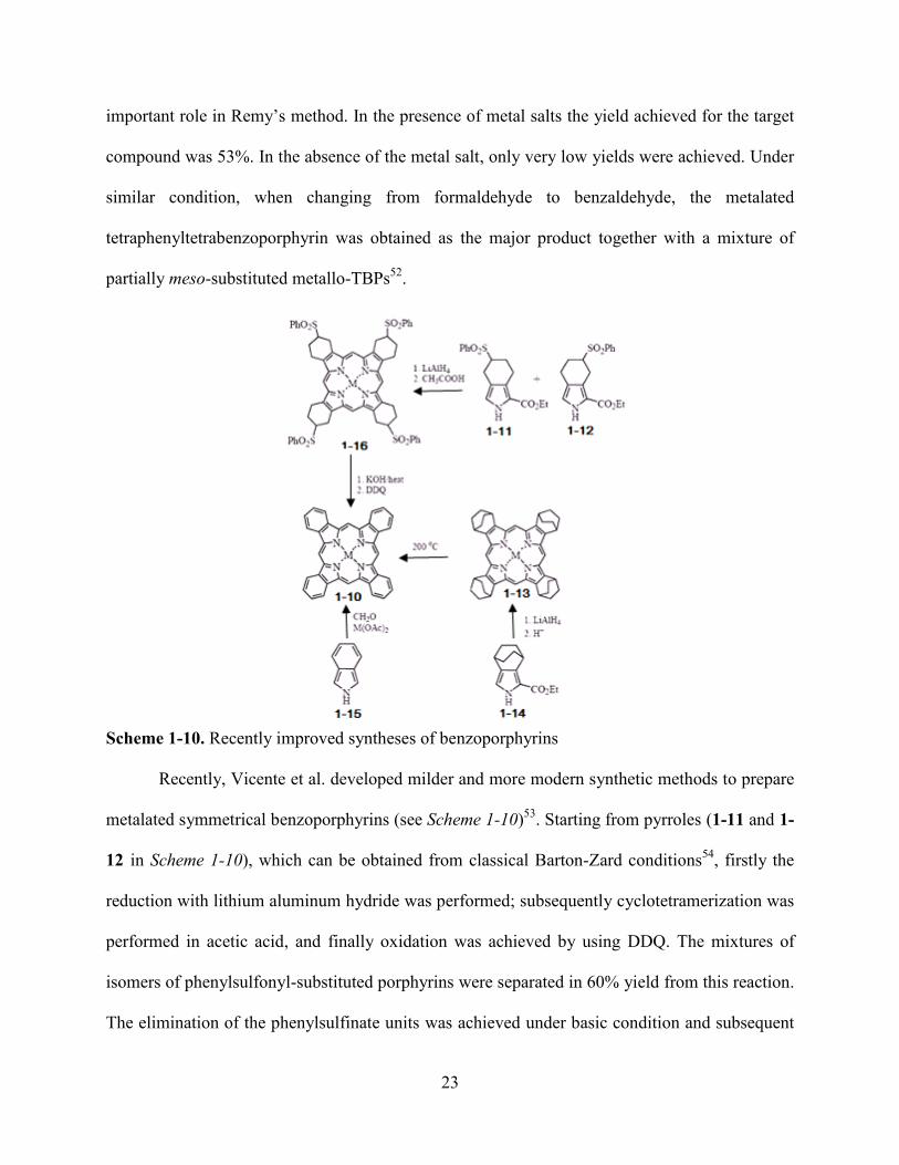
Scheme 1-10. Recently improved syntheses of benzoporphyrins
Recently, Vicente et al. developed milder and more modern synthetic methods to prepare
metalated symmetrical benzoporphyrins (see Scheme 1-10)53
. Starting from pyrroles (1-11 and 1-
12 in Scheme 1-10), which can be obtained from classical Barton-Zard conditions54
, firstly the
reduction with lithium aluminum hydride was performed; subsequently cyclotetramerization was
performed in acetic acid, and finally oxidation was achieved by using DDQ. The mixtures of
isomers of phenylsulfonyl-substituted porphyrins were separated in 60% yield from this reaction.
The elimination of the phenylsulfinate units was achieved under basic condition and subsequent

24
oxidation was performed by using DDQ. After isolation, the target metalated symmetrical
benzoporphyrins 1-10 were obtained in 53% overall yield. Meanwhile, Ono et al. avoided the use
of instable isoindole in the preparation of 1-10 by generating a masked isoindole 1-14 and
subsequently using it for the reaction55
. Ono’s improvement was performed by reducing 1-14
with lithium aluminum hydride, then acid catalyzed cyclotetramerization, and eventually
oxidation with DDQ. After isolation, 1-13 was obtained in 30% yield. The eventual generation of
tetrabenzoporphyrin 1-10 was achieved in very pure form in quantitative yield through a retro-
Diels-Alder reaction by just simply heating 1-13 at 200 °C.
The condensation of benzodipyrromethene hydrobromides and Diels-Alder type reaction
involving β-vinyl porphyrins and activated dienophiles can also be used to generate
benzoporphyrins56
. Compared with porphyrins, unsubstituted TBP has very poor solubility due
to its extended, planar, π-conjugated system and high π-π stacking (aggregation) tendency. Thus,
the physicochemical property evaluation of these compounds has been slow. However, the
tetraaryl substituted tetrabenzoporphyrins have enhanced solubility due to their significantly non-
planar structure due to the steric hindrance effect generated from the four meso-aryl substituents.
1. 4 Overview of Chlorin-e6 as a PDT Sensitizer
The development of the so-called ―2nd
-generation‖ of photosensitizers is aimed to
increase the efficiency of photosensitizers in PDT, expand their applications in PDT, and
maintain the advantages of currently approved photosensitizers. Current research has focused on
the improvement of their photophysical and pharmacokinetic properties. Chlorins have strong
absorptions at the ideal part of the therapeutic region. The electron delocalization pathway of
chlorins is shown in Figure 1-13. Unlike the porphyrins, there are no chlorin-based
photosensitizers that have been approved in the United States by the FDA. However, mono-(L)-
25
aspartylchlorin-e6 is currently in advanced-stages of clinical trials as a 2nd
-generation of
photosensitizer.
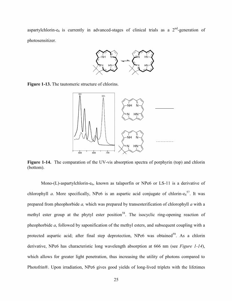
Figure 1-13. The tautomeric structure of chlorins.
HN
N
N
NH
HN
N
N
NH
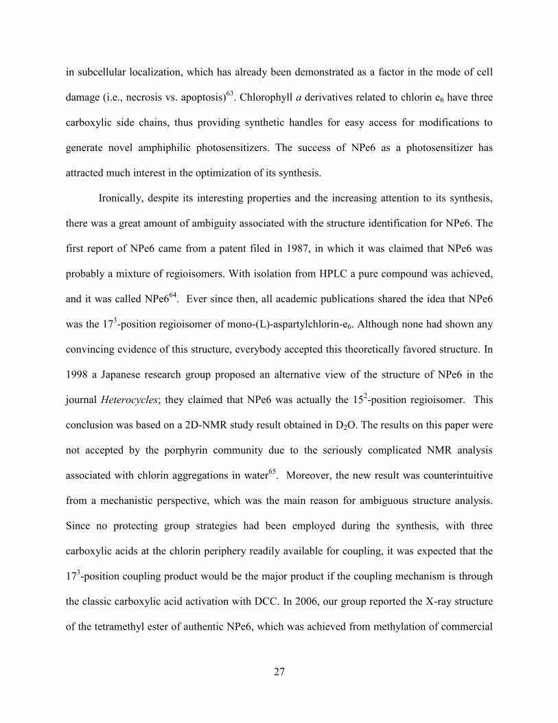
Figure 1-14. The comparation of the UV-vis absorption spectra of porphyrin (top) and chlorin
(bottom).
Mono-(L)-aspartylchlorin-e6, known as talaporfin or NPe6 or LS-11 is a derivative of
chlorophyll a. More specifically, NPe6 is an aspartic acid conjugate of chlorin-e657
. It was
prepared from pheophorbide a, which was prepared by transesterification of chlorophyll a with a
methyl ester group at the phytyl ester position58
. The isocyclic ring-opening reaction of
pheophorbide a, followed by saponification of the methyl esters, and subsequent coupling with a
protected aspartic acid; after final step deprotection, NPe6 was obtained59
. As a chlorin
derivative, NPe6 has characteristic long wavelength absorption at 666 nm (see Figure 1-14),
which allows for greater light penetration, thus increasing the utility of photons compared to
Photofrin®. Upon irradiation, NPe6 gives good yields of long-lived triplets with the lifetimes

26
ranges from 500 to 800 μs, thus giving high yields of cytotoxic singlet oxygen. Furthermore, it
can rapidly clear from normal tissue, having negligible residual photosensitivity in tissues.
Compared with Photofrin® in PDT of cholangiocarcinoma, NPe6 (see Figure 1-15) shows many
advantages: it can reduce tumor volume, inhibit tumor re-growth, and increase depth of tissue
injury up to 67%. Also, the undesired side effect of residual skin photosensitization has been
found to be decreased60
.
Figure 1-15. Chemical structures of temoporfin (left), the main component (hematoporphyrin) of
Photofrin® (middle) and NPe6 (right).
In the binary treatment modalities, stability is especially significant. Degradation
products will shift the light absorbtion wavelength outside of the laser therapy window and
makes the treatment ineffectual. Chlorophyll a derivatives have increased stability due to their
unusual structural characteristics, which is hard to access from current synthetic methods61
. As a
chlorophyll-a derivative, NPe6 has increased stability compared to the other chlorin
photosensitizers, such as temoporfin (Figure 1-15, left), which can readily oxidize back to
porphyrin. In order to improve the efficiency of photosensitizers in PDT, amphiphilicity is
required62
. Recently, it was found that small differences in photosensitizer structure, even
involving regioisomerism of substituents, can bring the huge functional differences, for example
27
in subcellular localization, which has already been demonstrated as a factor in the mode of cell
damage (i.e., necrosis vs. apoptosis)63
. Chlorophyll a derivatives related to chlorin e6 have three
carboxylic side chains, thus providing synthetic handles for easy access for modifications to
generate novel amphiphilic photosensitizers. The success of NPe6 as a photosensitizer has
attracted much interest in the optimization of its synthesis.
Ironically, despite its interesting properties and the increasing attention to its synthesis,
there was a great amount of ambiguity associated with the structure identification for NPe6. The
first report of NPe6 came from a patent filed in 1987, in which it was claimed that NPe6 was
probably a mixture of regioisomers. With isolation from HPLC a pure compound was achieved,
and it was called NPe664
. Ever since then, all academic publications shared the idea that NPe6
was the 173-position regioisomer of mono-(L)-aspartylchlorin-e6. Although none had shown any
convincing evidence of this structure, everybody accepted this theoretically favored structure. In
1998 a Japanese research group proposed an alternative view of the structure of NPe6 in the
journal Heterocycles; they claimed that NPe6 was actually the 152-position regioisomer. This
conclusion was based on a 2D-NMR study result obtained in D2O. The results on this paper were
not accepted by the porphyrin community due to the seriously complicated NMR analysis
associated with chlorin aggregations in water65
. Moreover, the new result was counterintuitive
from a mechanistic perspective, which was the main reason for ambiguous structure analysis.
Since no protecting group strategies had been employed during the synthesis, with three
carboxylic acids at the chlorin periphery readily available for coupling, it was expected that the
173-position coupling product would be the major product if the coupling mechanism is through
the classic carboxylic acid activation with DCC. In 2006, our group reported the X-ray structure
of the tetramethyl ester of authentic NPe6, which was achieved from methylation of commercial

28
available NPe6 with diazomethane66
. This clealy showed that authentic NPe6 is the 152-aspartyl
regioisomer. The unambiguous syntheses of all three NPe6 regioisomers and the difunctional
NPe6 ―DACE‖ were also reported by our group67
. Since the formation of the new NPe6 (the 152-
regioisomer) structure was hard to explain thus it became necessary to discover the underlying
mechanism for the unique formation of this structure. Chapter 6 will present the related
mechanism study results.
1.5 Overview of Porphycene
Figure 1-16. Chemical structure of porphycene (right) and porphyrin (left).
The name of porphycene combines both porphyrin and acene together, in order to
describe its unique structural constitution (see Figure 1-16)68
. The increasing interests in
porphyrin researches led to the discovery of porphyrin isomers such as porphycene in 1986 by
Vogel68
. Two years later, in 1988, he also reported the first heteroporphycene69a
. In 1993, Merz
and coworkers reported the synthesis of tetrathiaporphycenes69b
. One year later, Cava and
coworkers also reported their independent synthesis of this type of porphycene69c
. Following
these initial reports, the synthesis of corrphycene70a
and hemiporphycene70b
were report in 1994,
and the synthesis of isoporphycene71
was reported in 1996. The preparation of porphycenes
usually starts from a 5,5’-diformyl-2,2’-bipyrrole. A low-valent titanium coupling reaction was
used for the final step cyclization reaction, often the McMurry coupling72
. Shortly after the
synthesis of porphycene, Vogel and his coworkers realized that porphycenes possess a strong
intense absorption at the phototherapeutic window for PDT (between 600 and 800 nm), and that
29
this was red-shift compared to porphyrin systems. Thus they proposed that porphycenes might
serve as an important photosensitizer in the photodynamic therapy of cancers.72,73
Ever since
then, many porphycene derivatives have been synthesized. It was found that the peripheral
substitutents of porphycenes can affect the photoexcited triplet states of free-base porphycenes.
Richert et al. prepared the tetramethoxy- and dimethoxy-porphycenes and subsequently
converted them into their 9-acetoxy-substituted derivatives. Among these, 9-acetoxy-2,7,12,17-
tetrakis (β-methoxyyethyl)porphycene has been used for PDT of psoriatic lesions. Also,
extensive preclinical and phase I /II clinical trials with this dye have been performed74
.
Considering the dominant role that metalloporphyrins play in porphyrin chemistry, the
metalation of porphycene also attracted lots of interests. Due to the strong intra-core NH-N
hydrogen bonding and the rectangular shape of the four central nitrogen atoms, the metalation of
porphycene is relatively difficult compared to porphyrins, and the type of metalloporphycenes
that have been reported71a,72, 75
are limited. There are several other ways to modify porphycenes,
which mainly involve: 1) modification at the pyrrolic nitrogens76
; 2) catalytic hydrogenation; 3)
post-synthetic skeletal modification77
.
The characteristic spectrum properties of porphyrinoid aromatic system can be found in
the absorption spectra of porphycenes. There is one split Soret-like band around 370 nm and
three enhanced Q-band absorptions in their UV-vis spectra. With the higher intensity of Q-band
absorption compared to those of porphyrins, porphycenes are more attractive compared with
porphyrin derivatives for application in PDT of cancers72,73
. Compared to porphyrins, the NMR
spectra of porphycene shows great high field shift of the internal NH protons, from around -2~-
4ppm to around +3ppm78
.
30
1.6 References
1 Battersby, A. R. Nat. Prod. Rep., 2000, 17, 507.
2 Fischer, H.; Hess, R.; Hoppe-Seyler’s Z. Physiol. Chem., 1931, 194, 195; b) Siedel, W.;
Fischer, H.; Hoppe-Seyler’s Z. Physiol. Chem., 1933, 214, 145.
3 Stokes, G. G. J. Chem. Soc., 1864, 17, 304.
4 Thudichum; J. L. W. Report Med. Off. Privy. Council, 1867, Appendix 710, 152; b) Nencki,
M., Arch. Exptl. Path. Parmakol. 1888, 24, 430.
5 Robinson, R., Obituary Notices of Fellows of the Royal Society, 1953, 8, 609.
6 Küster, W., Hoppe-Seyler’s Z. Physiol. Chem., 1912, 82, 463.
7 Keilin, D., Proc. R. Soc. London, B, 1925, 98, 312.
8 Fischer, H.; Stern, A., Die Chemie des Pyrrols, Vol. 2, Part 2, Akad Verlag, Leipzig, 1940; b)
Willstätter, R.; Stoll, A., Investigations on Chlorophyll, Science Press, Lancaster, Ohio, 1928.
9 Lemberg, R.; Barrett, J. Cytochromes, Academic Press, London and New York, 1973.
10 Todd, A. R. in Vitamin B12, Zagalak B.; Friedrich, W. Eds., de Gruyter, Berlin, 1979, Page 1;
b) Folkers, K. in Vitamin B12, Zagalak, B.; Friedrich, W. Eds., de Gruyter, Berlin, 1979, Page
7.
11 Hodgkin, D. C., in Vitamin B12, Zagalak, B.; Friedrich, W. Eds., de Gruyter, Berlin, 1979,
Page 19.
12 Vicente, M. G. H.; Smith, K. M. Curr. Org. Chem. 2000, 4, 139; b) Milgrom, L. R. in The
Colors of Life, Oxford University Press, Oxford, New York and Tokyo, 1997.
13 Abraham, R. J.; Medforth, C. J.; Mansfield, K. E.; Simpson, D. J.; Smith, K. M. J. Chem. Soc.
Perkin Trans. 2, 1988, 1365; b) Smith, K. M. in The Porphyrins, Elsvier Press, 1975, pp 9; c)
Boucher, L. J.; Katz, J. J. J. Am. Chem. Soc. 1967, 89,4703.
14 Abraham, R. J.; Medforth, C. J.; Mansfield, K. E.; Simpson, D. J.; Smith, K. M. J. Chem. Soc.
Perkin Trans. 2, 1988, 1365.
15 Smith, K. M.; Goff, D. A.; Abraham, R. J. J. Org. Magn. Reson. 1984, 22, 779; b) Kenner, G.
W.; McCombie, S. W.; Smith, K. M., J. Chem. Soc. Perkin Trans 1, 1973, 21, 2517; c) Senge,
M. O.; Smith, K. M. Photchem. Photobiol. 1991, 54, 841; d) Smith, K. M.; Unsworth, J. F.
Tetrahedron, 1975, 31, 367.
16 Soret, J. L. Compt. Rend. 1883, 97, 1267; b) Weiss, C. J. J. Mol. Spectrosc. 1972, 44, 37.
17 Fischer, H.; Klarer, J. Liebigs Ann. Chem. 1929, 98, 468.
31
18 Kim, J. B.; Adler, A. D.; Longo, F. R. in ―The Porphyrins‖, Dolphin, D. Ed. Academic Press,
New York, 1978, Vol. I, Part A, p. 85.
19 Smith, K. M. in ―Porphyrins and Metalloporphyrins‖, Smith, K. M. Ed., Elsevier,
Amsterdam (1975), p. 32; b) Adler, A. D.; Longo, F. R.; Finarelli, J. D.; Goldmacher, J.;
Assour, J.; Korsakoff, L. J. Org. Chem. 1967, 32, 476.
20 Rothemund, P.; Menotti, A. R. J. Am. Chem. Soc. 1939, 61, 2912.
21 Adler, A.; Longo, F. R.; Finarelli, J. O.; Goldmacher, J.; Assour, J.; Korsakoff, L. J. Org.
Chem. 1967, 32, 476.
22 Lindsey, J. S.; Hsu, H. C.; Schreiman, I. C. Tetrahedron Lett. 1986 , 27 , 4969; b) Rao, P. D.;
Dhanalekshmi, S.; Littler, B.; Lindsey, J. S., J. Org. Chem. 2000, 65,7323; c) Geier, G. R.;
Lindsey, J. S. Tetrahedron 2004, 60, 435; d) Rao, P. D.; Littler, B. J.; Geier, G. R.; Lindsey,
J. S. J. Org. Chem. 2000, 65, 1084.
23 Paolesse, R.; Jaquinod, L.; Sala, F. D.; Nurco, D. J.; Prodi, L.; Montali, M.; Natale, C. D.;
D’Amico, A.; Carlo, A. D.; Lugli, P.; Smith, K. M., J. Am. Chem. Soc. 2000, 122, 11295; b)
Wagner, R. W.; Johnson, T. E.; Lindsey, J. S., J. Am. Chem. Soc. 1996, 118, 11166; c)
Anderson, H. L., Inorg. Chem. 1994, 33, 972; d) Lin, V. S. –Y.; Dimagno, S. G.; Therien, M.
J., Science, 1994, 264, 1105.
24 Dawson, J. H. Science 1988, 240, 433; b) Dismukes, G. C. Science 2001, 292, 447; c)
Tommos, C.; Babcock, G. T. Acc. Chem. Res. 1998, 31, 18; d) Yocum, C. F.; Pecoraro, V. L.
Curr. Opin. Chem. Biol. 1999, 3, 182; e) Sono, M.; Roach, M. P.; Coulter, E. D.; Dawson, J.
H. Chem. Rev. 1996, 96, 2841; f) Ozaki, S.-I.; Roach, M. P.; Matsui, T.; Watanbe, Y. Acc.
Chem. Res. 2001, 34, 818.
25 Chang, C. J.; Chng, L. L.; Nocera, D. G. J. Am. Chem. Soc. 2003, 125, 1866; b) Momenteau,
M.; Reed, C. A. Chem. Rev. 1994, 94, 659; b) Chang, C. J.; Brown, J. D. K.; Chang, M. C. Y.;
Baker, E. A.; Nocera, D. G. Electron Transfer in Chemistry; Balzani, V., Ed.; Wiley-VCH:
Weinheim, Germany, 2001; Vol. 3.2.4, pp 409; c) Cukier, R. I.; Nocera, D. G. Annu. Rev.
Phys. Chem. 1998, 49, 337.
26 Liu, S. Y.; Nocera, D. G. J. Am. Chem. Soc. 2005, 127, 5278.
27 Hao, E.; Fronczek, F. R.; Vicente, M. G. H. J. Org. Chem. 2006, 71, 1233; b) Sommer, N.;
Staab, H. A. Tetrahedron Lett. 1966, 25, 2837; c) Katz, J. L.; Feldman, M. B.; Conry, R. R.
Org. Lett. 2005, 7, 91; d) Katz, J. L.; Geller, B. J.; Conry, R. R. Org. Lett. 2006, 8, 2755; e)
Katz, J. L.; Selby, K. J.; Conry, R. R. Org. Lett. 2005, 7, 3505; f) Maes, W.; Rossom, W. V.;
Hecke, K. V.; Meervelt, L. V.; Dehaen, W. Org. Lett. 2006, 8, 4161; g) Konishi, H.; Tanaka,
K.; Teshima, Y.; Mita, T.; Morikawa, O.; Kobayashi, K. Tetrahedron Lett. 2006, 47, 4041.
28 Jiao, L.; Hao, E.; Fronczek, F. R.; Smith, K. M.; Vicente M. G. H. Tetrahedron, 2007, 63,
4011.

32
29 Vicente, M. G. H. Curr. Med Chem.-Anti-Cancer Agents, 2001, 1, 175; b) Henderson, B. W.;
Dougherty, T. J. Photochem Photobiol. 1992, 55, 145; c) Dougherty, T. J; Comer, C. J;
Henderson, B. W. J Nat. Cancer Inst. 1998, 90, 889; d) Tappeiner, H. V., Jesionek, A.
Munch. Med. Wochenschr 1903, 50, 3042; e) Kaye, A. H.; Morstyn, G.; Apuzzo, M. L. J. J.
Neurosurg. 1988, 69, 1; f) Hausman, W. Biochem. Z. 1909, 14, 275; g) Lipson, R. L.; Baldes
E. J.; Gray, M. J. Cancer 1967, 20, 2255; Dougherty, T. J.; Grindley, G. E.; Fiel, R., et al. J
Natl Cancer Inst. 1975, 55, 115.
30 Woodburn, K. W.; Vardaxis, N. J.; Hill, J. S.; Kaye, A. H.; Phillips, D. R; Photochem.
Photobiol. 1992, 54, 725; b) Woodburn, K. W.; Vardaxis, N. J.; Hill, J. S.; Kaye, A. H.;
Phillips, D. R. Photochem. Photobiol. 1992, 55, 697; c) Boyle, R. W.; Dolphin, D.
Photochem. Photobiol. 1996, 64, 469; d) Jori, G.; Reddi, E. In Photodynamic Therapy of
Neoplastic Disease, Vol. 2, Kessel, D. Eds., 1990; pp117; e) Jori, G. J. Photochem.
Photobiol. B 1996, 36, 87; f) Soncin, M.; Busetti, A.; Reddi, E.; Jori, G.; Rither, B.
D.;Kenney, M. E.; Rodgers, M. A. J. J. Photochem. Photobiol. B 1997, 40, 163; g) Peng, Q.;
Moan, J.; Nesland, J. M. Ultrastruct. Pathol. 1996, 20, 109.
31 Berg, K.; Western, A.; Bommer, J. C.; Moan, J. Photochem. Photobiol. 1990, 52, 481; b)
Ambroz, M.; MacRobert, M. J.; Morgan, J.; Rumbles, G.; Foley, M. S. C.; Phillips, D. J.
Photochem. Photobiol. B 1994, 22, 105; c) Peng, Q.; Moan, J.; Farrants, G.; Danielson, H. E.;
Rimington, C. Cancer Lett. Shannon, Irel. 1991, 58, 17; d) Bottriroli, G.; Croce, A. C.;
Ramponi, R.; Vachi, P. Photochem. Photobiol. 1992, 55, 575; e) Merchat, M.; Spikes, J. D.;
Bertoloni, G.; Jori, G. J. Photochem. Photobiol. B 1996, 35, 149; g) Margaron, P.; Gregoire,
M-J.; Scasnar, V.; Ali, H.; van Lier, J. E. Photochem. Photobiol. 1996, 63, 217.
32 Bonnett, R. Rev. Contemp. Pharmacother. 1999, 10, 1.
33 Kongshaug, M. Int. J. Biochem. 1992, 24, 1239; b) Maziere, J. C.; Morliere, P.; Santus, R. J.
Photochem. Photobiol. B. Biol. 1991, 8, 351; c) Obochi, M. O. K.; Boyle, R. W.; vanLier, J.
E. Photochem. Photobiol. 1993, 57, 634; d) Korbelik, M.; Krosol, G.; Olive, P. L.; Chaplin,
D. J. Br. J. Cancer, 1991, 64, 508-512; e) Krosl, G.; Korbelik, M.; Dougherty, T. J. Br. J.
Cancer, 1995, 71, 549; f)Hamblin, M. R.; Newman, E. L. J. Photochem. Photobiol. B: Biol.
1994, 23, 3; g) Brasseur, N.; Langlois, R.; Lamadeleine, C.; Ouellet, R.; vanLier, J. E.
Photochem. Photobiol. 1999, 69, 345; h) Thomas, J. P.; Girotti, A. W. Photochem. Photobiol.
1989, 49, 241; e) Brault, D. J. Photochem. Photobiol. B: Biol. 1990, 6, 79; f) Peng, Q.; Moan,
J.; Cheng, L. S. Cancer, Lett. 1991, 58, 29; g) Jori, G.; Reddi, E. Int. J. Biochem. 1993, 25,
1369.
34 Chaber, B, A. in Present status and future prospects for treatment of metastatic cancer.;
Honn, K. V.; Powers, W. E.; Sloane, B. F.; Martinus, N.; Eds, 1986, pg 15; b) Liotta, L. A.
Cancer Res. 1986, 46, 1.
35 Vicente, M. G. H. Rev. Port. Quím. 1996, 3, 47; b) Stenberg, E. D.; Dolphin, D.; Brückner, C.
Tetrahedron, 1998, 54, 4152; c) Pandey, R. K.; Zheng, G. Porphyrins as Photosensitizers in
Photodynamic therapy, in The Porphyrin Handbook, Kadish, K. M.; Smith, K. M. Guilard,
R. Eds. Academic press, 2000; d) Dougherty, T. J.; Gomer, C. J.; Henderson, B. W.; Jori, G.;
Kessel, D.; Korberlik, M.; Moan, J.; Peng, Q. J. Natl. Cancer Inst. 1998, 90, 889; e)
33
Schnitmaker, J. J.; Bass, P.; van Leengoed, M. L. L. M.; van der Meulen, F. W.; Star, W. M.;
van Zaudwijk, N. J. Photochem. Photobiol. B. Biol. 1996, 34, 3; f) Hahn, S. M.; Glatstein, E.
Rev. Contemp. Pharmacother. 1999, 10, 69; g) Hsi, R. A.; Rosenthal, D. I.; Glastein, E.
Drugs, 1999, 57, 725; h) Webber, J.; Herman, M.; Kessel, D.; Fromm, D. Annals Surg. 1999,
230, 12; i) Webber, J.; Herman, M.; Kessel, D.; Fromm, D. Langenbeck’s Arch. Surg. 2000,
385, 299; j) Mason, M. D. Rev. Contemp. Pharmother. 1999, 10, 25.
36 Lipson, R. L.; Baldes, E. J.; Olsen, A. M. J. Natl. Cancer Inst. 1961, 26, 1-12
37 Dougherty, T. J. Photochem. Photobiol. 1983, 38, 377; b) Pandey, R. K.; Majchzycki, D. F.;
Smith, K. M. Proc. SPIE 1989, 1065, 164; c) Bonett, R.; Berenbaum, M. C. Adv. Exp. Biol.
Med. 1983, 160, 241; d) Pandey, R. K. Dougherty, T. J. Photochem. Photobiol. 1988, 47, 769;
e) Kessel, D. Photochem. Photobiol. 1986, 44, 193.
38 Bonnett, R. Chem. Soc. Rev. 1995, 24, 19; b) Fisher, A. M. R.; Murphree, A. L.; Gomer, C. J.
Lasers Surg. Med. 1995, 17, 2; c) Sternberg, E. D.; Dolphin, D.; Bruckner, C. Tetrahedron,
1998, 54, 4151; d) Pandey, R. K. J. Porphyrins Phthalocyanines, 2000, 4, 368; e) Ali, H.;
van Lier, J. E. Chem. Rev. 1999, 99, 2379.
39 Pandey, R. K.; Zheng, G. In The Porphyrin Handbook, Vol. 6, Kadish, K. M.; Smith, K. M.
Guillard R. Eds. Academic Press: San Diego, 2000; pp 157.
40 Kessel, D. J. Photochem. Photobiol. B: Biol. 1997, 39, 81; b) Spikes, J. D.; Bommer, J. C. J.
Photochem. Photobiol. B: Biol. 1993, 17, 135; c) Peyman, G. A.; Kazi, A. A.; Moshfeghi, D.;
Unal, M.; Khoobehi, B.; Yoneya, S.; Mori, K.; Rivera, I. Ophthal. Surg. Lasers, 2000, 31,
323.
41 Glanzmann, T.; Forrer, M.; Blant, S. A.; Woodtli, A.; Grosjean, P.; Braichotte, D.; van den
Bergh, H.; Monnier, P.; Wagnieres, G. J. Photochem. Photobiol. B: Biol. 2000, 57, 22; b)
Garbo, G. M. J. Photochem. Photobiol. B: Biol. 1996, 34, 109.
42 Martinsen, J.; Pace, J. L.; Phillips, B.M.; Ibers, A. J. J. Am. Chem. Soc. 1982, 104, 83.
43 Lash, T. D. J. Porph. Phthal, 2001, 5, 267; b) Wolford, S. T.; Novicki, D. L.; Kelly, B.
Fundam. Appl. Toxicol. 1995, 24, 52; c) Sessler, J. L.; Seidel, D. Angew. Chem. Int. Ed. 2003,
42, 5134; d) Brown, S. B.; Truscott, T. G. Chem. Br. 1993, 29, 995; e) Bonnett, R. Chem.
Rev. 1994, 24, 19.
44 Leznoff, C. C., Phthalocyanines: Properties and Applications; VCH Publishers: New York,
1990-1996; Vol. 1-4; b) Volger, A.; Rethwisch, B.; Hutterman, J.; Bensenhard, J. O. Angew.
Chem. Int. Ed. Engl. 1978, 17, 951; c) Vogler, A.; Kunkley, H. Inorg. Chim. Acta. 1980, 44,
L 211; d) Nguyen, K. A.; Pachter, R. J. Chem. Phys. 2001, 114, 10757; e) Hanack, M.;
Zipplies, T. J. Am. Chem. Soc. 1985, 107, 6127; f) Remier, K. J.; Remier, M. M.; Still, M. J.
Can. J. Chem, 1981, 59, 1388; g) Carlson, J. B.; Vouros, P. J. Mass. Spectrosc. 1996, 31,
1403.
45 Baker, E. W., J. Am. Chem. Soc. 1966, 88, 2311; b) Baker, E. W.; Yen, T. F.; Dikie, J. P.;
Rhodes, R. E.; Clarke, L. F. J. Am. Chem. Soc. 1967, 89, 363; c) Kaur, S.; Chicarelli, M. I.;
34
Maxwell, J. R. J. Am. Chem. Soc. 1986, 108, 1347; d) Kaur, S.; Gill, J. P.; Evershed, R. P.;
Eglinton, G.; Maxwell, J. R. J. Chromatogr. 1989, 473, 135; e) Quirke, J. M. E.; Dale, T.;
Britton, E. D.; Yost, R. A.; Trichet, J.; Belayouni, H. Org. Geochem. 1990, 15, 1969.
46 Helberger, J. H.; Ann. 1937, 529, 205; b) Helberger, J. H.; von Rebay, A. ibid. 1937, 531,
279; c) Helberger, J. H.; von Rebay, A.; Hever, D. B. ibid. 1938, 533, 197; c) Helberger, J.
H.; Hever, D. B. ibid. 1938, 536, 173.
47 Kopranenkov, V. N.; Takhanova, E. A.; Lukyanets, E. A. Zh. Org. Khim. 1979, 15, 642; b)
Kopranekov, V. N.; Makaranova, E. A.; Lukyanets, E. A. Zh. Obshch. Khim. 1981, 51, 2727;
c) Bender, C. O.; Bonnett, R.; Smith, R. G. J. Chem. Soc. 1969, 345; d) Bender, C. O.;
Bonnett, R.; Smith, R. G. J. Chem. Soc. 1970, 1251; c) Bender, C. O.; Bonnett, R.; Smith, R.
G. J. Chem. Soc. 1972, 771; d) Bornstein, J.; Remy, D. E.; Sheilds, J. E. J. Chem. Soc. 1972,
1149.
48 Barrett, P. A.; Linstead, R. P.; Rundall, F. G.; Tuey, G. A. P. J. Chem. Soc. 1940, 1079; b)
Linstead, R. P.; Weiss, F. T. J. Chem. Soc. 1950, 2975.
49 Vogler, A.; Kunkely, H. Angew. Chem. Int. Ed. Engl. 1978, 17, 760; b) Kopranenkov, V. N.;
Vorotnikov, A. M.; Ivanova, T. M.; Luk’yanets, E. A. Chem. Heterocyclic Comp. 1988, 24,
1120; c) Kopranenkov, V. N.; Makarova, E. A.; Dashkevich, S. N.; Luk’yanets, E. A. Chem.
Heterocyclic Comp. 1988, 24, 630.
50 Vogler, A.; Kunkley, H.; Rethwisch, B. Inorg. Chim. Acta, 1980, 46, 101.
51 Remy, D. Tetrahedron Lett. 1983, 24, 1451.
52 Cheng, R.-J.; Chen, Y.-R.; Chuang, C.-E. Heterocycles, 1992, 34, 1; b) Cheng, R.-J.; Chen,
Y.-R.; Chuang, C.-E. Polyhedron, 1993, 12, 1353; c) Yasuike, M.; Yamaoka, T.; Ohno, O.;
Ichimura, K.; Morii, H.; Sakuragi, M. Inorg. Chim. Acta. 1991, 182, 83.
53 Vicente, M. G. H.; Tome, A. C.; Walter, A.; Cavaleiro, J. A. S. Tetrahedron Lett. 1997, 38,
3639.
54 Arnold, D. P.; Burgess-Dean, L.; Hubbard, J.; Rahman, M. A. Aust. J. Chem. 1994, 47, 969;
b) Haake, G.; Struve, D.; Montforts, F.-P. Tetrahedron Lett. 1994, 35, 9703.
55 Ito, S.; Murashima, T.; Ono, N. Chem. Commun. 1998, 1661.
56 Bonnett, R.; McManus, K. A. J. Chem. Soc. Chem. Commun. 1994, 1129; b) Morgan, A. R.;
Pangka, V. S.; Dolphin, D. J. Chem. Soc. Chem. Commun. 1984, 1047; c) Yon-Hin, P.;
Wijesekera, T. P.; Dolphin, D. Tetrahedron Lett. 1989, 30, 6135.
57 Spikes, J. D.; Bommer, J. C. In Chlorophylls; Scheer, H. Eds., CRC Press: Boston, 1996, p
1188.

35
58 Smith, K. M.; Goff, D. A.; Simpson, D. J. J. Am. Chem. Soc. 1985, 107, 4946; b) Weller, A.;
Livingston, R. J. Am. Chem. Soc. 1954, 76, 1575; c) Wasielewski, M. R.; Svec, W. A. J. Org.
Chem. 1980, 45, 1969.
59 Cox, M. T.; Jackson, A. H.; Kenner, G. W.; McCombie, S. W.; Smith, K. M. J. Chem. Soc.,
Perkin Trans. 1, 1974, 516; b) Fischer, H.; Orth, H. Die Chemie Pyrrols Akademische
Verlag: Liepzig, vol II, part 2, 1940, p 206.
60 Song, L. M. W.; Wang, K.K.; Zinmeister, Alan R. Cancer 1998, 82 (2), 421.
61 Woodward, R. B. Pure Appl. Chem. 1961, 2, 383; b) Smith, K. M., J Chem Soc. Perk Trans.
1, 1988, 3119.
62 Vicente, M.G. H. Curr. Med Chem.-Anti-Cancer Agents, 2001, 1, 175.
63 Kessel, D. H; Luguya, R.; Vicente, M. G. H. Photochem. Photobiol. 2003, 78, 431-435; b)
Krieg, R. C.; Messmann, H.; Schlottman, K.; Enlicher E.; Seeger, S.; Scholmerich, J.;
Knuechel, R. Photochem. Photobiol. 2003, 78, 393.
64 Bommer, J., Ogden, B. F., Tetrapyrrole Therapeutic Agents, U. S. Patent 4,693,885, Sept 15,
1987.
65 Gomi, S.; Nishizuka, T.; Ushiroda, O.; Takahashi, H.; Sumi, S. Heterocycles 1998, 48, 2231;
b) Abraham, R. J.; Rowan, A. E. In Chlorophylls; Scheer, H. Ed.; CRC Press: Boston, 1996;
pp 797.
66 Hargus, J. A., in Master Dissertation, Louisiana State University, Baton Rouge, 2005.
67 W. G. Roberts, F. -Y. Shiau, J. S. Nelson, K. M. Smith and M. W. Berns. J. Natl. Cancer Inst.
1988, 80, 330; b) W. G. Roberts, K. M. Smith, J. L. McCullough and M. W. Berns.
Photochem. Photobiol. 1989, 49, 431.
68 Vogel, E.; Köcher, M.; Schmickler, H.; Lex, J. Angew. Chem. Int. ed. Engl. 1986, 25, 257.
69 Vogel, E.; Sicken, M.; Röhrig, P.; Schmickler, H.; Lex, J.; Ermer, O. Angew. Chem. Int. ed.
Engl. 1988, 27, 411.
70 Aukauloo, M. A.; Guilard, R. New. J. Chem. 1994, 18, 1205; b) Callot, H. J.; Rohrer, A.;
Tschamber, T. New. J. Chem. 1995, 19, 155; c) Vogel, E.; Bröring, M.; Scholz, P.; Deponte,
R.; Lex, J.; Schmickler, H.; Schaffner, K.; Braslavsky, S. E.; Müller, M.; Pörting, S.;
Weghorn, S. J.; Fowler, C. J.; Sessler, J. L. Angew. Chem. Int. ed. Engl. 1997, 36, 1651.
71 Vogel, E. Heterocycl. Chem. 1996, 33, 1461; b) Vogel, E.; Bröring, M.; Erben, C.; Demuth,
R.; Lex, J.; Nendel, M.; Houk, K. N. Angew. Chem. Int. ed. Engl. 1997, 36, 353.
72 Vogel, E.; Balci, M.; Pramod, K.; Koch, P.; Lex, J.; Ermer, O. Angew. Chem. Int. ed. Engl.
1987, 26, 928.

36
73 Wehrle, B.; Limbach, H.-H.; Köcher, M.; Ermer, O.; Vogel, E. Angew. Chem. Int. ed. Engl.
1987, 26, 934.
74 Jux, N.; Koch, P.; Schmickler, H.; Lex, J.; Vogel, E. Angew. Chem. Int. ed. Engl. 1990, 29,
1385.
75 Richert, C.; Wessels, J. M.; Mueller, M.; Kisters, M.; Benninghaus, T.; Goetz, A. E. J. Med.
Chem., 1994, 37, 2797.
76 Chang, C. K.; Morrison, I.; Wu, W.; Chern, S.-S.; Peng, S.-M. J. Chem. Soc., Chem.
Commun. 1995, 1173; b) Vogel, E.; Köcher, M.; Balci, M.; Teichler, I.; Lex, J.; Schmickler,
H.; Ermer, O. Angew. Chem. Int. ed. Engl. 1987, 25, 931.
77 Vogel, E. Pure Appl. Chem. 1990, 62, 557; b) Vogel, E. Pure Appl. Chem. 1993, 65, 143; c)
Vogel, E. Pure Appl. Chem. 1996, 68, 1335; d) Sessler, J. L. Angew. Chem. Int. ed. Engl.
1994, 33, 1348; e) Toporowicz, M.; Ofir, H.; Levanon, H.; Vogel, E.; Köcher, M.; Pramod,
K.; Fessenden, R. W. Photochem. Photobio. 1989, 50, 37.
37
CHAPTER 2. β,β’-FUSED METHYLENEPROPANOPORPHYRINS
2.1 Introduction
Porphyrins can be used as chemical sensors1-3
, catalysts4,5
, and as molecular devices6,7
.
Among those, porphyrin systems functionalized with redox groups, such as ferrocene, have been
reported in the last decades and have found potential applications in solar energy conversion1-3
. It
was found that the properties of these systems depended on the efficient π-overlap between the
central porphyrin unit and the peripheral substitutents. Lately metallocene-modified porphyrins,
as charge-transfer materials, have also attracted much attention due to their wide applications as
molecular conductors and molecular magnets. Metallocene-modified porphyrin complexes have
been studied over the last decades8-9
. In most cases, the metallocene fragments were connected to
the porphyrin through various spacers at meso- or at β- position of porphyrins. For example, the
direct connection of the metallocene to the meso-positions of porphyrins through a single C-C
bond, an aromatic system, or via conjugated double or triple bonds1,10,11
have been reported.
However, the spectroscopic and electrochemical results indicate negligible strong
communications between the two fragments. This is attributed to the poor overlap (if any)
between the two aromatic π-systems.
In the meanwhile, the design and synthesis of covalently linked multiporphyrin arrays
became a frontier research area in porpyrin chemistry. These multiporphyrin arrays12
have
unique photo-electronic properties13-16
, and have attracted much recent interest because they play
important roles in many areas such as in light harvesting,17
energy and electron transfer,18
and
multielectron redox catalysis19
. The construction of these multiporphyrin arrays also required an
extended π-electron network. Among the fused porphyrin dimers and oligoporphyrins being
reported, significant novel spectroscopic properties20-21
were observed; in particular, the
38
formation of bisporphyrin sandwich complexes has been shown to dramatically change the redox
properties of porphyrins. It was found that in bis(porphyrin) sandwich complexes, the porphyrin
complexes acted as both the electron-donor and electron-acceptor species22
. The initial study in
our group had shown that the fusion of two porphyrins at β-positions could enhance the overlap
of the two π-systems, and enhance the interactions between the redox moieties. The formation of
sandwich complexes can enhance the overlap of porphyrin HOMOs to assist the energetic
situation of both the porphyrin-porphyrin bonding and anti-bonding orbitals. It is reported that
the large metal ion in these bis(porphyrin) sandwich complexes are generally much easier to
oxidize than the corresponding monoporphyrin23-24
, since the large metal ion is able to hold two
porphyrin macrocycles close enough to raise the HOMO energy level. For the metallocene-
containing bisporphyrin and oligoporphyrins, there is long distance communication from
frameworks that should find attractive applications in nanoelectronics.
On the other hand, the covalently linked porphyrin arrays, especially cofacial
porphyrins25,26
have been widely studied in the past two decades because of their unique photo-
electronic properties. Particularly, these cofacial porphyrins have found potential applications as
electron-energy transfer moieties in molecular wires21,27
. So far, several fused bisporphyrins and
oligoporphyrins have been synthesized28
. Among these, the most successful cofacial porphyrins
are the so-called ―Pacman Porphyrin‖ systems, which consist of two octa-alkylporphyrins held
almost cofacial through a single rigid bridge. 25
However, the synthesis of cofacial porphyrins
has been, and will still remain, a challenge.29
The development of new methods for carbon-carbon bond formation is at the heart of
organic synthesis. The most desirable methods are those that are easily accomplished in large
scale, operate near ambient temperature, and do not require drastic reaction conditions. Usually,

39
the construction of carbocyclic rings requires the preparation of highly functionalized
intermediates. The reactions of atomic oxygen with unsaturated organic compounds have
demonstrated that the primary reactive intermediates can undergo extensive rearrangement
before forming isolable oxygenated products30,31
. The reactions of triplet oxygen atoms with
olefins32
, methylenecycloalkanes31
, and phenylethylenes33
have been reported.
The construction of these bisporphyrins and oligoporphyrins required efficient syntheses
of mono-methylenepropanoporphyrins and bismethylenepropanoporpyrins. In this Chapter, the
efficient synthesis of these porphyrin monomers will be discussed. Based on these monomers,
the construction of novel bisporphyrins will also be discussed.
2.2 Results and Discussion
2.2.1 Syntheses of β,β’-Fused Methylenepropanoporphyrins
The tandem carbon-carbon bond formation catalyzed by Pd(0) through [3+2]
cycloaddition has been reported34-37
. In most cases, a strongly electron-withdrawing group, such
as ester, nitrile, nitro, ketone or sulfone, is required to activate the double bond towards the
cycloaddition. In the meanwhile, there are few reports about the reactivity of the activated
double bonds toward this [3+2] cycloaddition38
. Also it is required that the Pd(0) be generated in
situ for the 1,3-dipolar cycloaddition. Solvents also found an important role in this type reaction.
For example, the substitution of toluene with THF could greatly shorten the reaction time and
enhance the cycloaddition yield39
. When considering the readily available selective nitration of
porphyrin, the Pd(0) catalyzed [3+2] cycloaddition reaction of 2-[(trimethylsilyl)methyl]-2-
propen-1-yl acetate with electron-deficient double bonds are very attractive in building
methylenepropanoporphyrins38
. A Michael-addition, followed by the ring-closure reaction, is
believed to be the mechanism40-42
for this reaction.
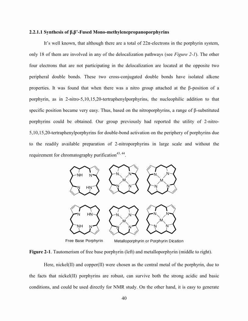
40
2.2.1.1 Synthesis of β,β’-Fused Mono-methylenepropanoporphyrins
It’s well known, that although there are a total of 22π-electrons in the porphyrin system,
only 18 of them are involved in any of the delocalization pathways (see Figure 2-1). The other
four electrons that are not participating in the delocalization are located at the opposite two
peripheral double bonds. These two cross-conjugated double bonds have isolated alkene
properties. It was found that when there was a nitro group attached at the β-position of a
porphyrin, as in 2-nitro-5,10,15,20-tertraphenylporphyrins, the nucleophilic addition to that
specific position became very easy. Thus, based on the nitroporphyrins, a range of β-substituted
porphyrins could be obtained. Our group previously had reported the utility of 2-nitro-
5,10,15,20-tertraphenylporphyrins for double-bond activation on the periphery of porphyrins due
to the readily available preparation of 2-nitroporphyrins in large scale and without the
requirement for chromatography purification43, 44
.
N
NH N
HN N
N N
N
N HN
NNH
M
N
N N
N
M
N
N N
N
M
N N
NN
M
Free Base Porphyrin Metalloporphyrin or Porphyrin Dication
Figure 2-1. Tautomerism of free base porphyrin (left) and metalloporphyrin (middle to right).
Here, nickel(II) and copper(II) were chosen as the central metal of the porphyrin, due to
the facts that nickel(II) porphyrins are robust, can survive both the strong acidic and basic
conditions, and could be used directly for NMR study. On the other hand, it is easy to generate
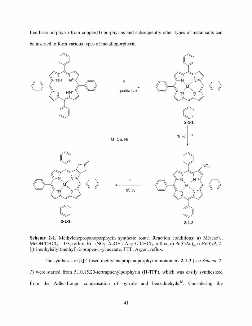
41
free base porphyrin from copper(II) porphyrins and subsequently other types of metal salts can
be inserted to form various types of metalloporphyrin.
N
NH N
HN
N
N N
N
M
a
b
N
N N
N
M
NO2
N
N N
N
M
c
M=Cu, Ni
qualitative
78 %
85 %
2-1-1
2-1-22-1-3
Scheme 2-1. Methylenepropanoporphyrin synthetic route. Reaction conditions: a) M(acac)2,
MeOH/CHCl3 = 1/3, reflux; b) LiNO3, AcOH / Ac2O / CHCl3, reflux; c) Pd(OAc)2, (i-PrO)3P, 2-
[(trimethylsilyl)methyl]-2-propen-1-yl acetate, THF, Argon, reflux.
The syntheses of β,β’-fused methylenepropanoporphyrin monomers 2-1-3 (see Scheme 2-
1) were started from 5,10,15,20-tertraphenylporphyrin (H2TPP), which was easily synthesized
from the Adler-Longo condensation of pyrrole and benzaldehyde45
. Considering the
42
regioselectivity differences between free-base porphyrin and the metalloporphyrin for the
nitration reaction, the insertion of metal before nitration was mandatory46, 47
.
Quantitative metalation of porphyrin to generate 2-1-1 was performed at refluxing
temperature by dissolving H2TPP in methanol/chloroform (v/v = 1/3) in the presence of 10
equivalents of metal acetylacetonate [M(acac)2]. After filtration to remove the excess amount of
M(acac)2, the desired product 2-1-1 was obtained. Further recrystallization was performed from
MeOH/DCM. The subsequent mono-nitration to generate 2-1-2 was performed in
chloroform/acetic acid (AcOH); acetic anhydride (Ac2O) and lithium nitrate (LiNO3) was used as
the nitration-reagent. The resulting 2-1-2-Cu was isolated in 78% yield and a similar yield was
obtained for 2-1-2-Ni. The preparation of mono-methylenepropanoporphyrin 2-1-3 was achieved
by Pd(0) catalyzed [3+2] cycloaddition reaction as described in the literature38
. The catalyst,
Pd(0), was prepared in situ by reacting of Pd(OAc)2 with triisopropylphosphite [(i-PrO)3P] in
THF under strict air-free conditions at room temperature. It generally took 30 minutes for the
generation of Pd(0). It was found that even a minute amount of air could ruin the generation of
the Pd(0) catalyst. The solution changed color from yellow to colorless with the generation of
Pd(0). To this colorless THF solution of Pd(0), was added solid 2-1-2, and followed by 2-
[(trimethylsilyl)methyl]-2-propen-1-yl acetate with exclusion of air. Immediately, this reaction
mixture was placed in a preheated (90 oC) oil bath and refluxed for a period of 2 days. TLC was
used to follow the reaction. Upon the complete consumption of the starting material, the reaction
temperature was raised to 100 oC, and the mixture was refluxed for an additional period of 2 days.
After the reaction mixture was cooled to room temperature, the desired 2-1-3 was purified using
a silica gel column eluted with DCM/hexane (v/v = 1/10). After removing the solvent under
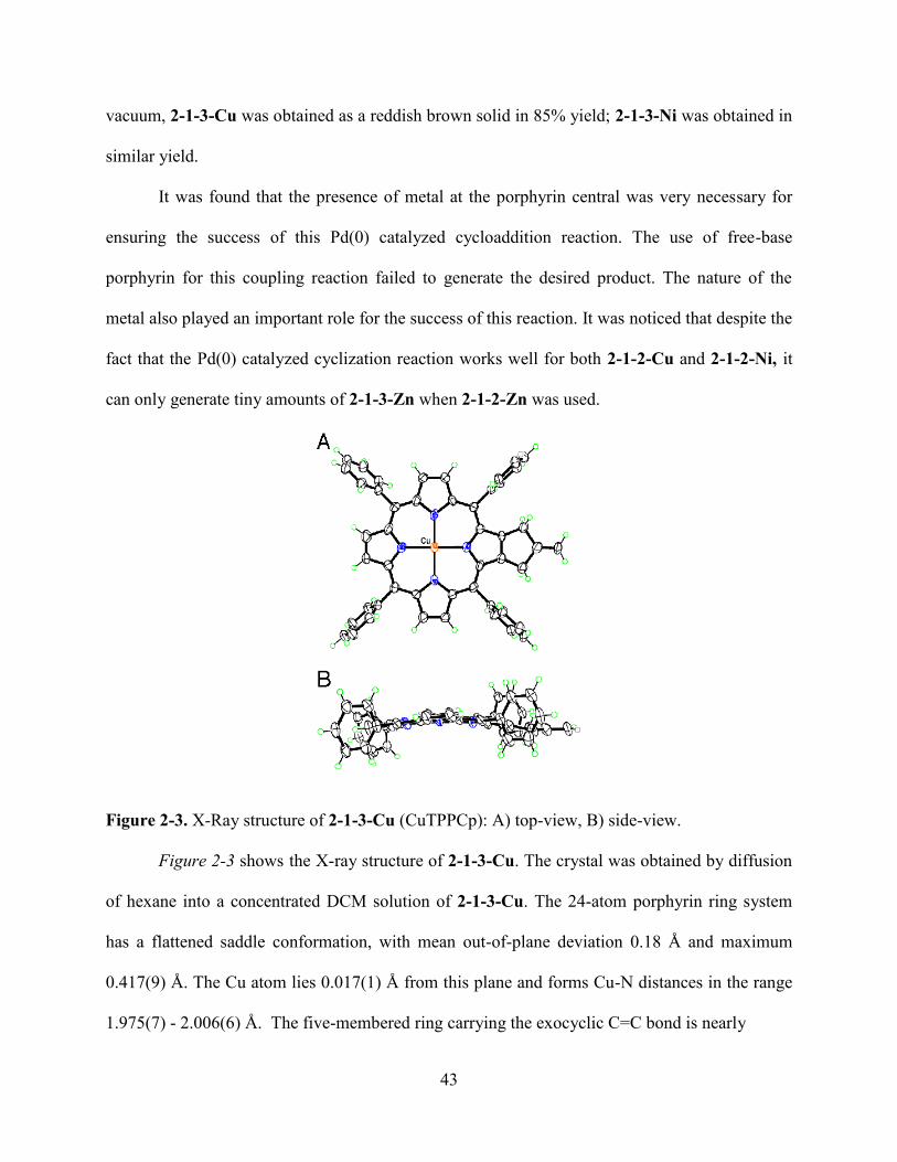
43
vacuum, 2-1-3-Cu was obtained as a reddish brown solid in 85% yield; 2-1-3-Ni was obtained in
similar yield.
It was found that the presence of metal at the porphyrin central was very necessary for
ensuring the success of this Pd(0) catalyzed cycloaddition reaction. The use of free-base
porphyrin for this coupling reaction failed to generate the desired product. The nature of the
metal also played an important role for the success of this reaction. It was noticed that despite the
fact that the Pd(0) catalyzed cyclization reaction works well for both 2-1-2-Cu and 2-1-2-Ni, it
can only generate tiny amounts of 2-1-3-Zn when 2-1-2-Zn was used.
Figure 2-3. X-Ray structure of 2-1-3-Cu (CuTPPCp): A) top-view, B) side-view.
Figure 2-3 shows the X-ray structure of 2-1-3-Cu. The crystal was obtained by diffusion
of hexane into a concentrated DCM solution of 2-1-3-Cu. The 24-atom porphyrin ring system
has a flattened saddle conformation, with mean out-of-plane deviation 0.18 Å and maximum
0.417(9) Å. The Cu atom lies 0.017(1) Å from this plane and forms Cu-N distances in the range
1.975(7) - 2.006(6) Å. The five-membered ring carrying the exocyclic C=C bond is nearly
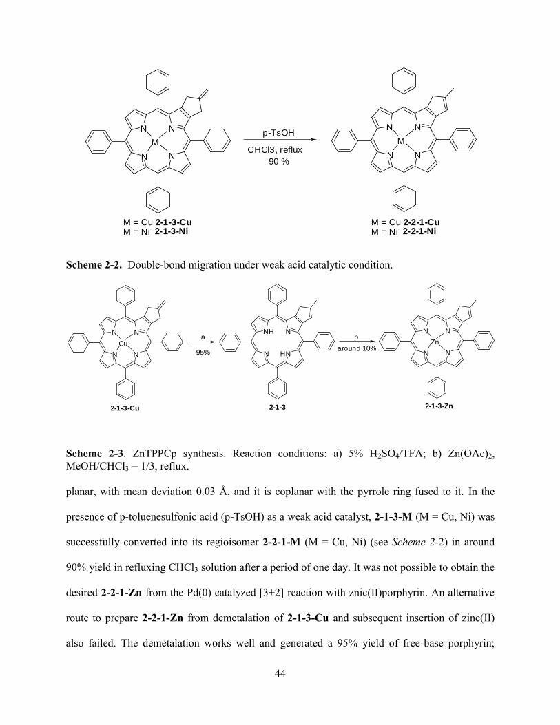
44
N
N N
N
M
N
N N
N
Mp-TsOH
CHCl3, reflux
90 %
M = Cu 2-1-3-CuM = Ni 2-1-3-Ni
M = Cu 2-2-1-CuM = Ni 2-2-1-Ni
Scheme 2-2. Double-bond migration under weak acid catalytic condition.
N
N N
N
Cu
N
NH N
HN N
N N
N
Znaround 10%
a b
95%
2-1-3-Cu 2-1-3 2-1-3-Zn
Scheme 2-3. ZnTPPCp synthesis. Reaction conditions: a) 5% H2SO4/TFA; b) Zn(OAc)2,
MeOH/CHCl3 = 1/3, reflux.
planar, with mean deviation 0.03 Å, and it is coplanar with the pyrrole ring fused to it. In the
presence of p-toluenesulfonic acid (p-TsOH) as a weak acid catalyst, 2-1-3-M (M = Cu, Ni) was
successfully converted into its regioisomer 2-2-1-M (M = Cu, Ni) (see Scheme 2-2) in around
90% yield in refluxing CHCl3 solution after a period of one day. It was not possible to obtain the
desired 2-2-1-Zn from the Pd(0) catalyzed [3+2] reaction with znic(II)porphyrin. An alternative
route to prepare 2-2-1-Zn from demetalation of 2-1-3-Cu and subsequent insertion of zinc(II)
also failed. The demetalation works well and generated a 95% yield of free-base porphyrin;
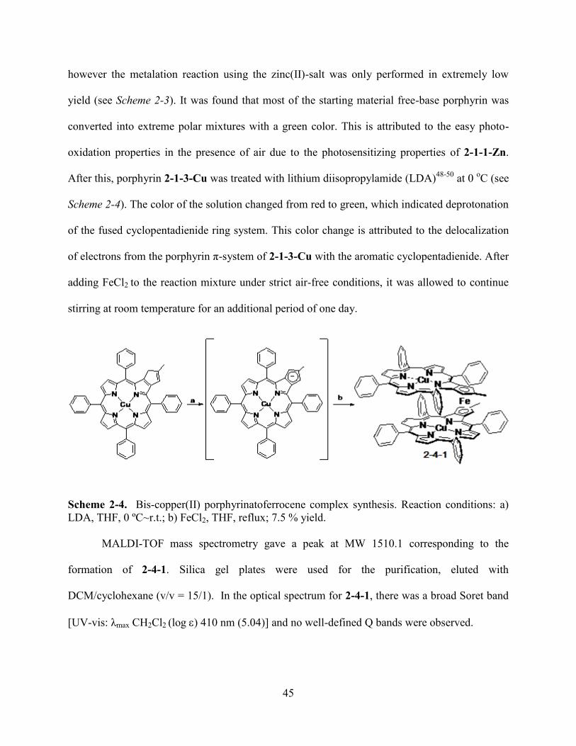
45
however the metalation reaction using the zinc(II)-salt was only performed in extremely low
yield (see Scheme 2-3). It was found that most of the starting material free-base porphyrin was
converted into extreme polar mixtures with a green color. This is attributed to the easy photo-
oxidation properties in the presence of air due to the photosensitizing properties of 2-1-1-Zn.
After this, porphyrin 2-1-3-Cu was treated with lithium diisopropylamide (LDA)48-50
at 0 oC (see
Scheme 2-4). The color of the solution changed from red to green, which indicated deprotonation
of the fused cyclopentadienide ring system. This color change is attributed to the delocalization
of electrons from the porphyrin π-system of 2-1-3-Cu with the aromatic cyclopentadienide. After
adding FeCl2 to the reaction mixture under strict air-free conditions, it was allowed to continue
stirring at room temperature for an additional period of one day.
Scheme 2-4. Bis-copper(II) porphyrinatoferrocene complex synthesis. Reaction conditions: a)
LDA, THF, 0 ºC~r.t.; b) FeCl2, THF, reflux; 7.5 % yield.
MALDI-TOF mass spectrometry gave a peak at MW 1510.1 corresponding to the
formation of 2-4-1. Silica gel plates were used for the purification, eluted with
DCM/cyclohexane (v/v = 15/1). In the optical spectrum for 2-4-1, there was a broad Soret band
[UV-vis: λmax CH2Cl2 (log ) 410 nm (5.04)] and no well-defined Q bands were observed.
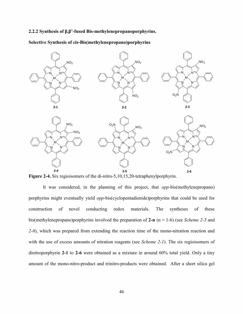
46
2.2.2 Synthesis of β,β’-fused Bis-methylenepropanoporphyrins.
Selective Synthesis of cis-Bis(methylenepropano)porphyrins
N
N N
N
M
NO2
NO2
N
N N
N
M
NO2
N
N N
N
M
NO2
N
N N
N
M
NO2
N
N N
N
M
NO2
N
N N
N
M
NO2
NO2O2N
O2N
O2N
NO2
2-1 2-2 2-3
2-62-52-4
Figure 2-4. Six regioisomers of the di-nitro-5,10,15,20-tetraphenylporphyrin.
It was considered, in the planning of this project, that opp-bis(methylenepropano)
porphyrins might eventually yield opp-bis(cyclopentadienide)porphyrins that could be used for
construction of novel conducting redox materials. The syntheses of these
bis(methylenepropano)porphyrins involved the preparation of 2-n (n = 1-6) (see Scheme 2-5 and
2-6), which was prepared from extending the reaction time of the mono-nitration reaction and
with the use of excess amounts of nitration reagents (see Scheme 2-1). The six regioisomers of
dinitroporphyrin 2-1 to 2-6 were obtained as a mixture in around 60% total yield. Only a tiny
amount of the mono-nitro-product and trinitro-products were obtained. After a short silica gel
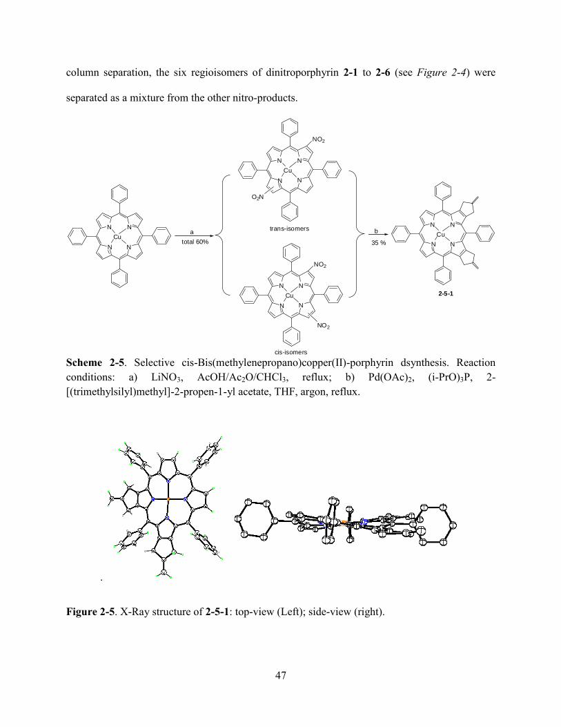
47
column separation, the six regioisomers of dinitroporphyrin 2-1 to 2-6 (see Figure 2-4) were
separated as a mixture from the other nitro-products.
N
N N
N
Cu
NO2
O2N
N
N N
N
Cu
NO2
NO2
N
N N
N
Cua b
total 60% N
N N
N
Cu
35 %
trans-isomers
cis-isomers
2-5-1
Scheme 2-5. Selective cis-Bis(methylenepropano)copper(II)-porphyrin dsynthesis. Reaction
conditions: a) LiNO3, AcOH/Ac2O/CHCl3, reflux; b) Pd(OAc)2, (i-PrO)3P, 2-
[(trimethylsilyl)methyl]-2-propen-1-yl acetate, THF, argon, reflux.
.
Figure 2-5. X-Ray structure of 2-5-1: top-view (Left); side-view (right).
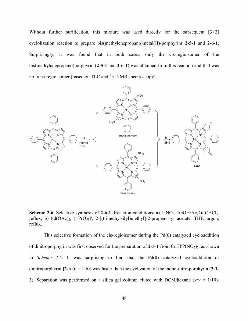
48
Without further purification, this mixture was used directly for the subsequent [3+2]
cyclolization reaction to prepare bis(methylenepropano)metal(II)-porphyrins 2-5-1 and 2-6-1.
Surprisingly, it was found that in both cases, only the cis-regioisomer of the
bis(methylenepropano)porphyrin (2-5-1 and 2-6-1) was obtained from this reaction and that was
no trans-regioisomer (based on TLC and 1H-NMR spectroscopy).
N
N N
N
Ni
NO2
O2N
N
N N
N
Ni
NO2
NO2
N
N N
N
Ni
N
N N
N
Nia b
overall65%
38%
trans-isomers
cis-isomers
2-6-1
Scheme 2-6. Selective synthesis of 2-6-1. Reaction conditions: a) LiNO3, AcOH/Ac2O/ CHCl3,
reflux; b) Pd(OAc)2, (i-PrO)3P, 2-[(trimethylsilyl)methyl]-2-propen-1-yl acetate, THF, argon,
reflux.
This selective formation of the cis-regioisomer during the Pd(0) catalyzed cycloaddition
of dinitroporphyrin was first observed for the preparation of 2-5-1 from CuTPP(NO2)2, as shown
in Scheme 2-5. It was surprising to find that the Pd(0) catalyzed cycloaddition of
dinitroporphyrin [2-n (n = 1-6)] was faster than the cyclization of the mono-nitro-porphyrin (2-1-
2). Separation was performed on a silica gel column eluted with DCM/hexane (v/v = 1/10).
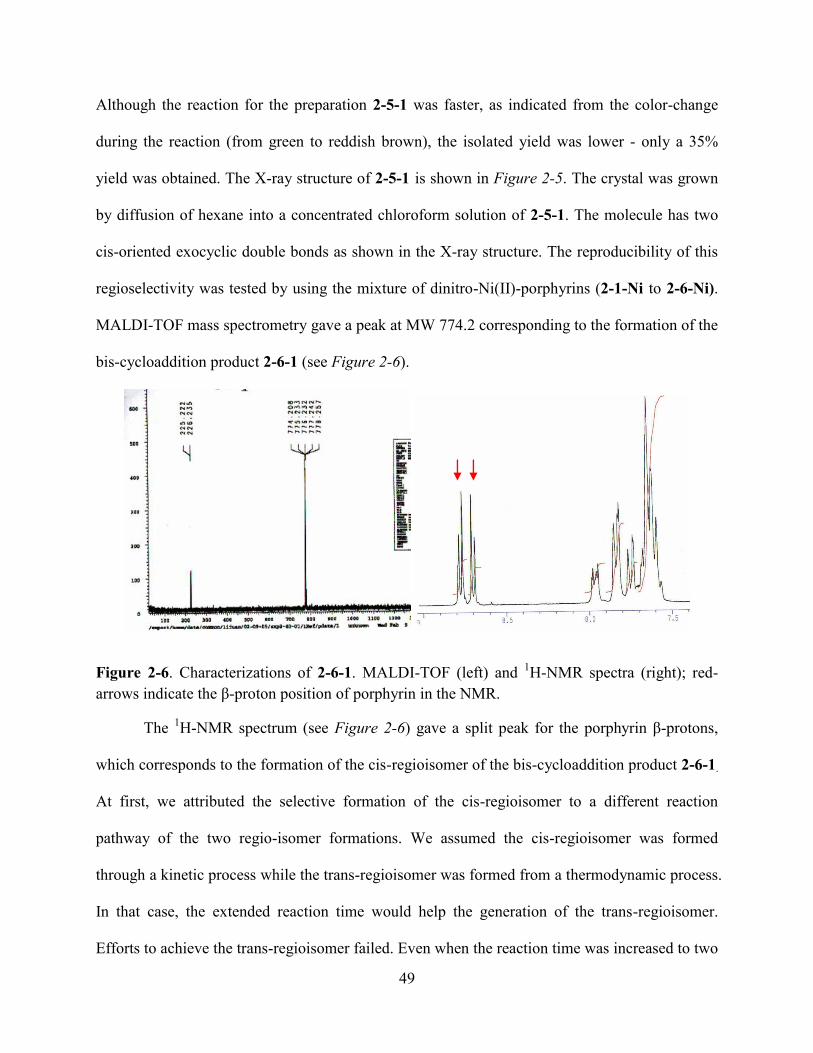
49
Although the reaction for the preparation 2-5-1 was faster, as indicated from the color-change
during the reaction (from green to reddish brown), the isolated yield was lower - only a 35%
yield was obtained. The X-ray structure of 2-5-1 is shown in Figure 2-5. The crystal was grown
by diffusion of hexane into a concentrated chloroform solution of 2-5-1. The molecule has two
cis-oriented exocyclic double bonds as shown in the X-ray structure. The reproducibility of this
regioselectivity was tested by using the mixture of dinitro-Ni(II)-porphyrins (2-1-Ni to 2-6-Ni).
MALDI-TOF mass spectrometry gave a peak at MW 774.2 corresponding to the formation of the
bis-cycloaddition product 2-6-1 (see Figure 2-6).
Figure 2-6. Characterizations of 2-6-1. MALDI-TOF (left) and 1H-NMR spectra (right); red-
arrows indicate the β-proton position of porphyrin in the NMR.
The 1H-NMR spectrum (see Figure 2-6) gave a split peak for the porphyrin β-protons,
which corresponds to the formation of the cis-regioisomer of the bis-cycloaddition product 2-6-1.
At first, we attributed the selective formation of the cis-regioisomer to a different reaction
pathway of the two regio-isomer formations. We assumed the cis-regioisomer was formed
through a kinetic process while the trans-regioisomer was formed from a thermodynamic process.
In that case, the extended reaction time would help the generation of the trans-regioisomer.
Efforts to achieve the trans-regioisomer failed. Even when the reaction time was increased to two
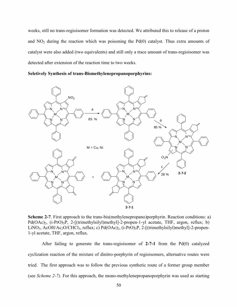
50
weeks, still no trans-regioisomer formation was detected. We attributed this to release of a proton
and NO2 during the reaction which was poisoning the Pd(0) catalyst. Thus extra amounts of
catalyst were also added (two equivalents) and still only a trace amount of trans-regioisomer was
detected after extension of the reaction time to two weeks.
Seletively Synthesis of trans-Bismethylenepropanoporphyrins:
N
N N
N
M
NO2
a
N
N N
N
M
N
N N
N
M
N
N N
N
M
O2N
b
N
N N
N
M +
c
M = Cu, Ni
85 %
85 %
28 %
2-7-1
2-7-2
Scheme 2-7. First approach to the trans-bis(methylenepropano)porphyrin. Reaction conditions: a)
Pd(OAc)2, (i-PrO)3P, 2-[(trimethylsilyl)methyl]-2-propen-1-yl acetate, THF, argon, reflux; b)
LiNO3, AcOH/Ac2O/CHCl3, reflux; c) Pd(OAc)2, (i-PrO)3P, 2-[(trimethylsilyl)methyl]-2-propen-
1-yl acetate, THF, argon, reflux.
After failing to generate the trans-regioisomer of 2-7-1 from the Pd(0) catalyzed
cyclization reaction of the mixture of dinitro-porphyrin of regioisomers, alternative routes were
tried. The first approach was to follow the previous synthetic route of a former group member
(see Scheme 2-7). For this approach, the mono-methylenepropanoporphyrin was used as starting
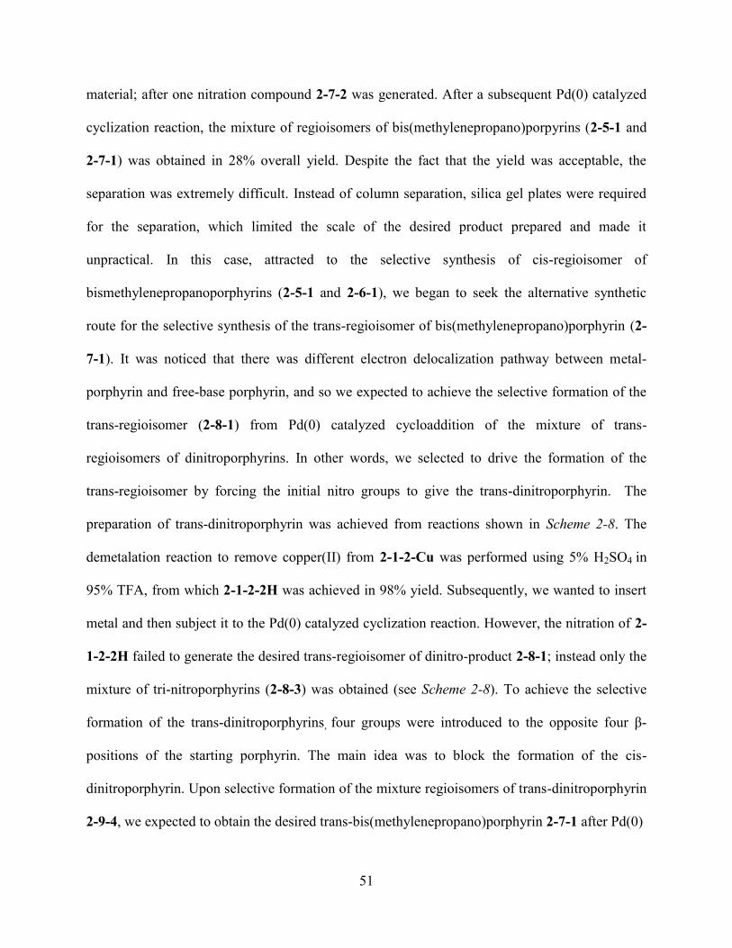
51
material; after one nitration compound 2-7-2 was generated. After a subsequent Pd(0) catalyzed
cyclization reaction, the mixture of regioisomers of bis(methylenepropano)porpyrins (2-5-1 and
2-7-1) was obtained in 28% overall yield. Despite the fact that the yield was acceptable, the
separation was extremely difficult. Instead of column separation, silica gel plates were required
for the separation, which limited the scale of the desired product prepared and made it
unpractical. In this case, attracted to the selective synthesis of cis-regioisomer of
bismethylenepropanoporphyrins (2-5-1 and 2-6-1), we began to seek the alternative synthetic
route for the selective synthesis of the trans-regioisomer of bis(methylenepropano)porphyrin (2-
7-1). It was noticed that there was different electron delocalization pathway between metal-
porphyrin and free-base porphyrin, and so we expected to achieve the selective formation of the
trans-regioisomer (2-8-1) from Pd(0) catalyzed cycloaddition of the mixture of trans-
regioisomers of dinitroporphyrins. In other words, we selected to drive the formation of the
trans-regioisomer by forcing the initial nitro groups to give the trans-dinitroporphyrin. The
preparation of trans-dinitroporphyrin was achieved from reactions shown in Scheme 2-8. The
demetalation reaction to remove copper(II) from 2-1-2-Cu was performed using 5% H2SO4 in
95% TFA, from which 2-1-2-2H was achieved in 98% yield. Subsequently, we wanted to insert
metal and then subject it to the Pd(0) catalyzed cyclization reaction. However, the nitration of 2-
1-2-2H failed to generate the desired trans-regioisomer of dinitro-product 2-8-1; instead only the
mixture of tri-nitroporphyrins (2-8-3) was obtained (see Scheme 2-8). To achieve the selective
formation of the trans-dinitroporphyrins, four groups were introduced to the opposite four β-
positions of the starting porphyrin. The main idea was to block the formation of the cis-
dinitroporphyrin. Upon selective formation of the mixture regioisomers of trans-dinitroporphyrin
2-9-4, we expected to obtain the desired trans-bis(methylenepropano)porphyrin 2-7-1 after Pd(0)
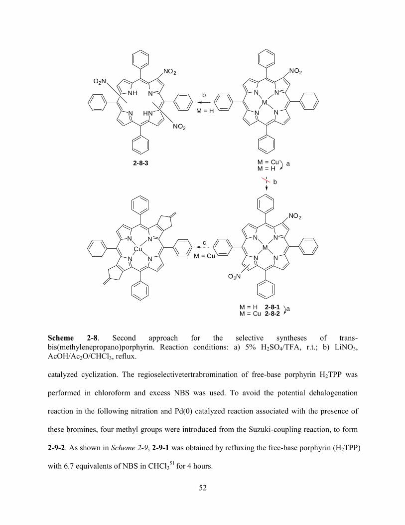
52
N
N N
N
M
NO2
N
N N
N
Cu
N
N N
N
NO2
O2N
M
b
b
N
NH N
HN
NO2
NO2
O2N
M = CuM = H
M = H 2-8-1M = Cu 2-8-2
a
a
c
M = H
M = Cu
2-8-3
Scheme 2-8. Second approach for the selective syntheses of trans-
bis(methylenepropano)porphyrin. Reaction conditions: a) 5% H2SO4/TFA, r.t.; b) LiNO3,
AcOH/Ac2O/CHCl3, reflux.
catalyzed cyclization. The regioselectivetertrabromination of free-base porphyrin H2TPP was
performed in chloroform and excess NBS was used. To avoid the potential dehalogenation
reaction in the following nitration and Pd(0) catalyzed reaction associated with the presence of
these bromines, four methyl groups were introduced from the Suzuki-coupling reaction, to form
2-9-2. As shown in Scheme 2-9, 2-9-1 was obtained by refluxing the free-base porphyrin (H2TPP)
with 6.7 equivalents of NBS in CHCl351
for 4 hours.

53
N
NH N
HN
a
N
NH N
HN
Br
Br
Br
Br
b
N
NH N
HN
CH3
CH3
H3C
H3C
c
N
N N
N
CH3
CH3
H3C
H3C
Nid
N
N N
N
CH3
CH3
H3C
H3C
Ni
O2N
NO2
e
N
N N
N
CH3
CH3
H3C
H3C
Ni
65 % 87%
98%
35%
2-9-1 2-9-2
2-9-32-9-42-9-5
Scheme 2-9. Third approach for the selective formation of trans-
bis(methylenepropano)porphyrin 2-9-5. Reaction conditions: a) NBS, CHCl3, reflux 4 hrs; b)
Pd(PPh3)4, CH3B(OH)2, THF/toluene, argon, reflux; c) Ni(acac)2, MeOH/CHCl3 = 1/3, reflux; d)
LiNO3, AcOH /Ac2O/CHCl3, refluxing; e) Pd(OAc)2, (i-PrO)3P, 2-[(trimethylsilyl)methyl]-2-
propen-1-yl acetate, THF, argon, refluxing for 5 days.
TLC and UV-vis were used to follow this reaction. The reaction was stopped when TLC
indicated no starting material left and the Soret band shifted to 430 nm in the UV-vis spectra.
After stopping this reaction, it was cooled to room temperature and cleaned up using a short
silica gel plug to remove excess NBS, washing with DCM to collect the desired product. After
removing the solvent under vacuum, the solid was washed with methanol three times, and 2-9-1
was obtained in 65% yield as a purple solid. No further purification was performed. The Suzuki-
coupling reaction between 2-9-1 and CH3B(OH)2 was performed by refluxing them in
THF/toluene (v/v = 3/2) under argon. It took a period of two days under strict air-free
condition52
to finish this reaction. Pd(PPh3)4 was used as catalyst, and 2-9-2 was obtained in 87%
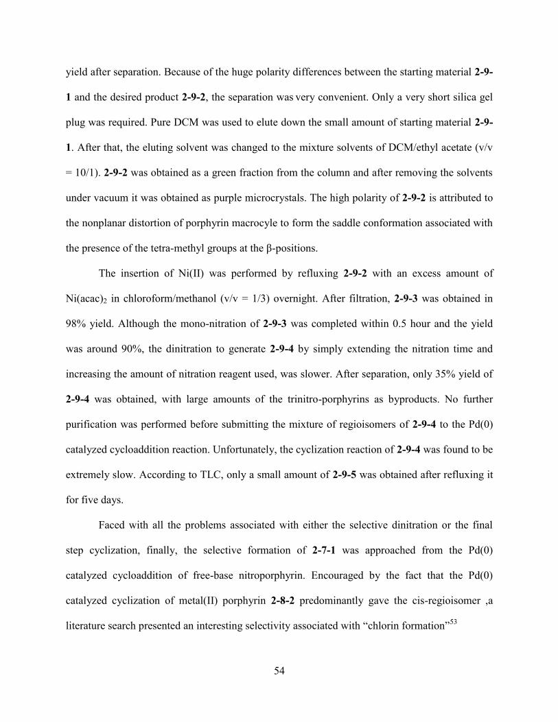
54
yield after separation. Because of the huge polarity differences between the starting material 2-9-
1 and the desired product 2-9-2, the separation was very convenient. Only a very short silica gel
plug was required. Pure DCM was used to elute down the small amount of starting material 2-9-
1. After that, the eluting solvent was changed to the mixture solvents of DCM/ethyl acetate (v/v
= 10/1). 2-9-2 was obtained as a green fraction from the column and after removing the solvents
under vacuum it was obtained as purple microcrystals. The high polarity of 2-9-2 is attributed to
the nonplanar distortion of porphyrin macrocyle to form the saddle conformation associated with
the presence of the tetra-methyl groups at the β-positions.
The insertion of Ni(II) was performed by refluxing 2-9-2 with an excess amount of
Ni(acac)2 in chloroform/methanol (v/v = 1/3) overnight. After filtration, 2-9-3 was obtained in
98% yield. Although the mono-nitration of 2-9-3 was completed within 0.5 hour and the yield
was around 90%, the dinitration to generate 2-9-4 by simply extending the nitration time and
increasing the amount of nitration reagent used, was slower. After separation, only 35% yield of
2-9-4 was obtained, with large amounts of the trinitro-porphyrins as byproducts. No further
purification was performed before submitting the mixture of regioisomers of 2-9-4 to the Pd(0)
catalyzed cycloaddition reaction. Unfortunately, the cyclization reaction of 2-9-4 was found to be
extremely slow. According to TLC, only a small amount of 2-9-5 was obtained after refluxing it
for five days.
Faced with all the problems associated with either the selective dinitration or the final
step cyclization, finally, the selective formation of 2-7-1 was approached from the Pd(0)
catalyzed cycloaddition of free-base nitroporphyrin. Encouraged by the fact that the Pd(0)
catalyzed cyclization of metal(II) porphyrin 2-8-2 predominantly gave the cis-regioisomer ,a
literature search presented an interesting selectivity associated with ―chlorin formation‖53
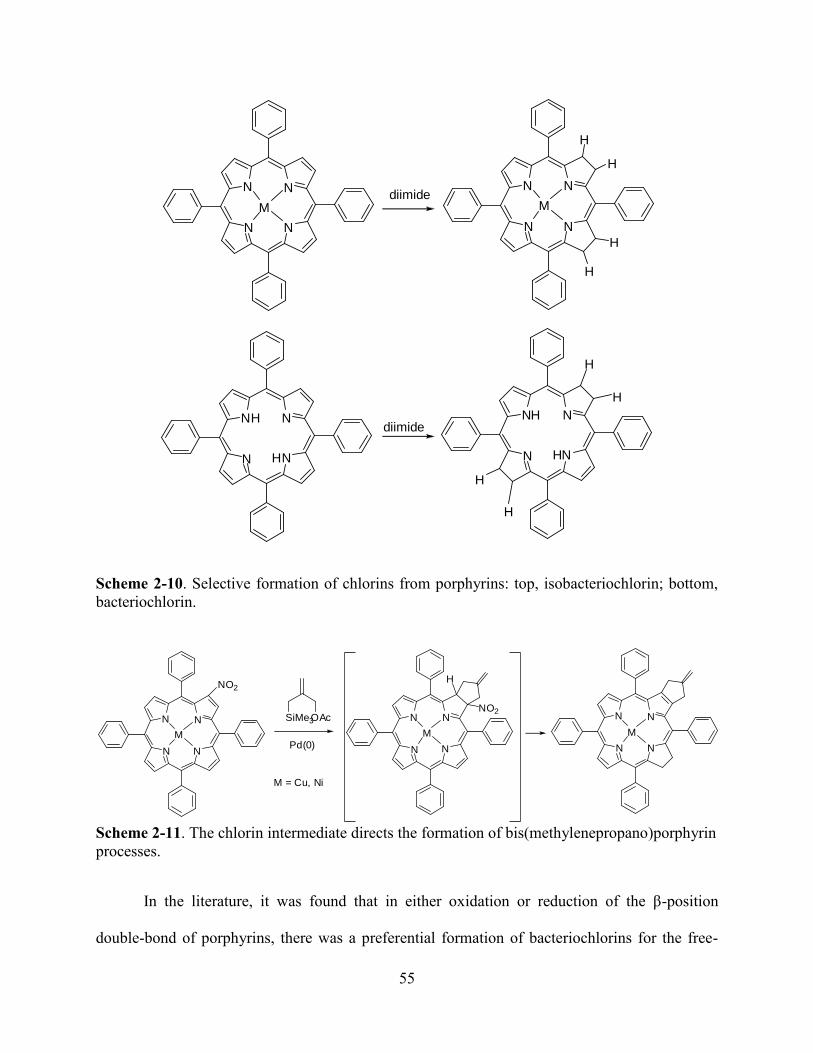
55
N
N N
N
Mdiimide
N
N N
N
M
H
H
H
H
N
NH N
HN N
NH N
HN
H
H
H
H
diimide
Scheme 2-10. Selective formation of chlorins from porphyrins: top, isobacteriochlorin; bottom,
bacteriochlorin.
N
N N
N
M
NO2
M = Cu, Ni
N
N N
N
M
N
N N
N
M
NO2
H
SiMe3OAc
Pd(0)
Scheme 2-11. The chlorin intermediate directs the formation of bis(methylenepropano)porphyrin
processes.
In the literature, it was found that in either oxidation or reduction of the β-position
double-bond of porphyrins, there was a preferential formation of bacteriochlorins for the free-

56
base porphyrin, and isobacteriochlorins for the metal-porphyrin (see Scheme 2-10). We attributed
the selective formation of the cis-regioisomers for the metal-porphyrin to the formation of
chlorin-intermediate (see Scheme 2-11). Since the cyclization of the dinitro-metal(II) porphyrin
had been found result in the selective formation of cis regioisomer, it would be very reasonable
for the achievement of the trans-regioisomer when dinitrated free-base porphyrin was used for
the cyclization.
N
N N
N
M
NO2
M = Cu, Ni
b
N
N N
N
M
NO2
H
NO2
H
N
N N
N
M
NO2
H
NO2NO2
N
N N
N
M
NO2
b
N
N N
N
M
NO2
H
N
N N
N
M
NO2
H
O2N O2N
H
O2N
N
N N
N
M
N
N N
N
M
favored
unfavored
Scheme 2-12. Chlorin induces the regioselectivity for the formation of
bis(methylenepropano)porphyrins.
The chlorin intermediate was observed in the formation of the 2-1-3 and could even be
isolated when performing the cyclization reaction at low temperatures. It was converted into
porphyrin by increasing the temperature from 90 oC to 100
oC over 24 hours or even a longer
period. Two modified routes were planned to improve the synthesis rate and yield. The first
modification was based on reaction shown in Scheme 2-9. It was started from 2-9-4, after
performing the mononitration and by the Pd(0) catalyzed cyclization reactions; the second
nitration was performed and followed by the second Pd(0) catalyzed cyclization reaction.

57
Another modification route was designed to introduce copper(II) instead of nickel(II) into the
porphyrin before dinitration, and then removal of the copper(II) before the final Pd(0) catalyzed
cyclization reaction. By doing this, we envisioned the Pd(0) catalyzed cyclization of the free-
base porphyrin might be able to generate the desired trans-isomers of
bis(methylenepropano)_porphyrin. In the second improved approach, copper(II) was first
removed under acidic condition to obtain the free-base dinitrated porphyrin; then under strictly
air-free condition the Pd(0) catalyzed reaction was performed. Unfortunately, the trans-
bis(methylenepropano)porphyrin was obtained, but in extremely low yield. MALDI-TOF mass
spectrometry gave a peak at 718 corresponding to the formation of 2-13-1, the 1H-NMR
spectrum indicated the formation of this trans-isomer.
N
N N
N
NO2
Cua
N
NH N
HN
NO2
b
NO2
NO2
N
NH N
HN
2-13-1
Scheme 2-13. Forth approach in the selective formation of the trans-
bis(methylenepropano)porphyrin. Reaction conditions: a) 5% H2SO4/TFA, DCM, r.t.; b)
Pd(OAc)2, (i-PrO)3P, 2-[(trimethylsilyl)methyl]-2-propen-1-yl acetate, THF, argon, reflux.
2.2.3. Construction of Cofacial Bisporphyrins
Our group has previously synthesized several fused metallocenoporphyrin derivatives
and bisporphyrin-metallocenes.24
An unexpectedly efficient synthesis of a cofacial metallo-
bisporphyrin was found from a simple DBU catalyzed carbon-carbon bond formation followed
by a self-sensitized oxygenation reaction at room temperature. The β,β’-fused
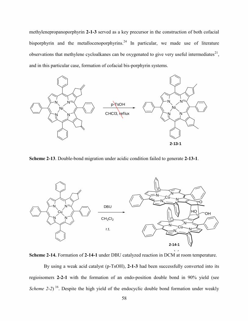
58
methylenepropanoporphyrin 2-1-3 served as a key precursor in the construction of both cofacial
bisporphyrin and the metallocenoporphyrins.24
In particular, we made use of literature
observations that methylene cycloalkanes can be oxygenated to give very useful intermediates21
,
and in this particular case, formation of cofacial bis-porphyrin systems.
p-TsOH
CHCl3, ref luxN
N N
N
Ni
N
N N
N
Ni
2-13-1
Scheme 2-13. Double-bond migration under acidic condition failed to generate 2-13-1.
N
NN
NCu
N
NN
NCu
O
OHHO
(7)
N
N N
N
Cu
DBU
CH2Cl2
r.t.
Scheme 2-14. Formation of 2-14-1 under DBU catalyzed reaction in DCM at room temperature.
By using a weak acid catalyst (p-TsOH), 2-1-3 had been successfully converted into its
regioisomers 2-2-1 with the formation of an endo-position double bond in 90% yield (see
Scheme 2-2) 16
. Despite the high yield of the endocyclic double bond formation under weakly
2-14-1
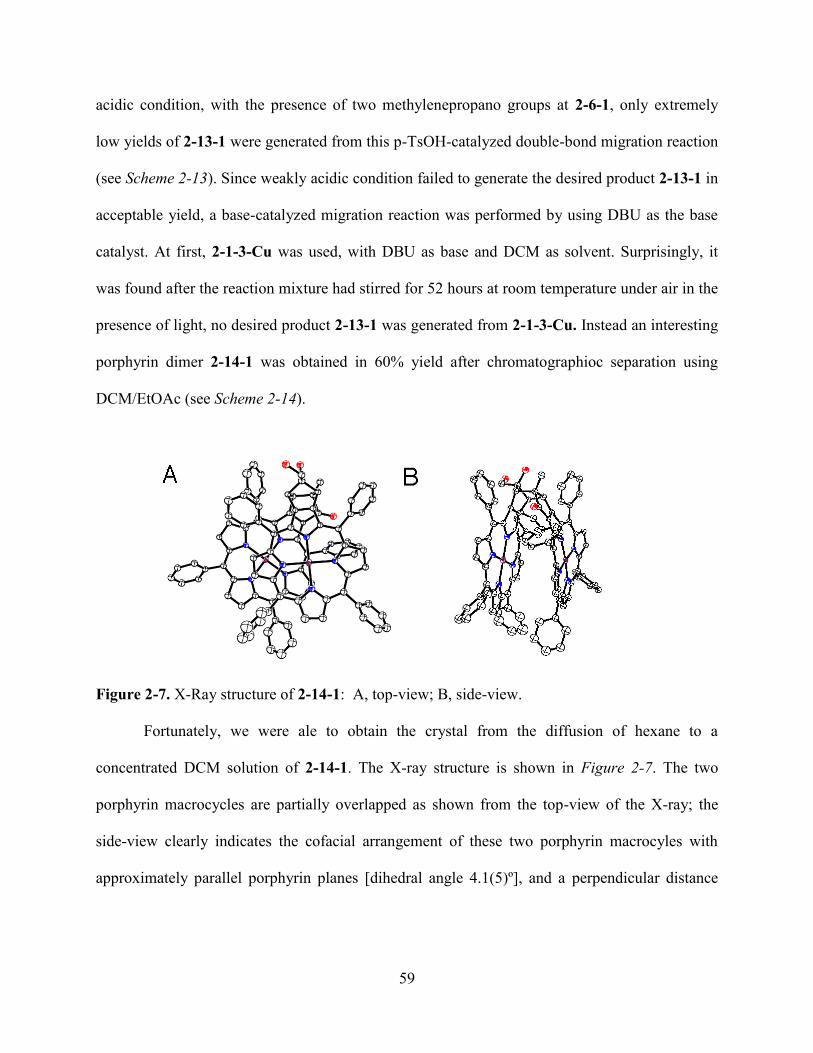
59
acidic condition, with the presence of two methylenepropano groups at 2-6-1, only extremely
low yields of 2-13-1 were generated from this p-TsOH-catalyzed double-bond migration reaction
(see Scheme 2-13). Since weakly acidic condition failed to generate the desired product 2-13-1 in
acceptable yield, a base-catalyzed migration reaction was performed by using DBU as the base
catalyst. At first, 2-1-3-Cu was used, with DBU as base and DCM as solvent. Surprisingly, it
was found after the reaction mixture had stirred for 52 hours at room temperature under air in the
presence of light, no desired product 2-13-1 was generated from 2-1-3-Cu. Instead an interesting
porphyrin dimer 2-14-1 was obtained in 60% yield after chromatographioc separation using
DCM/EtOAc (see Scheme 2-14).
Figure 2-7. X-Ray structure of 2-14-1: A, top-view; B, side-view.
Fortunately, we were ale to obtain the crystal from the diffusion of hexane to a
concentrated DCM solution of 2-14-1. The X-ray structure is shown in Figure 2-7. The two
porphyrin macrocycles are partially overlapped as shown from the top-view of the X-ray; the
side-view clearly indicates the cofacial arrangement of these two porphyrin macrocyles with
approximately parallel porphyrin planes [dihedral angle 4.1(5)º], and a perpendicular distance
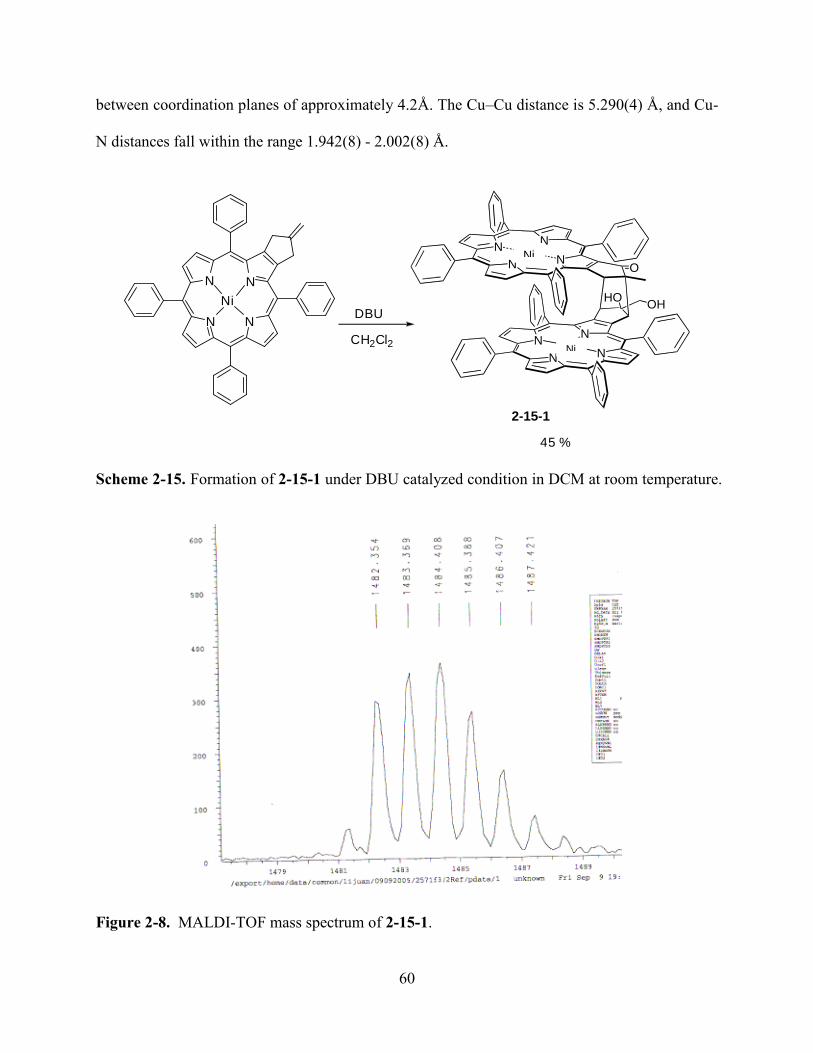
60
between coordination planes of approximately 4.2Å. The Cu–Cu distance is 5.290(4) Å, and Cu-
N distances fall within the range 1.942(8) - 2.002(8) Å.
N
N N
N
NiDBU
CH2Cl2
45 %
N
NN
NCu
N
NN
NCu
O
OHHO
(7)
Scheme 2-15. Formation of 2-15-1 under DBU catalyzed condition in DCM at room temperature.
Figure 2-8. MALDI-TOF mass spectrum of 2-15-1.
Ni
Ni
2-15-1
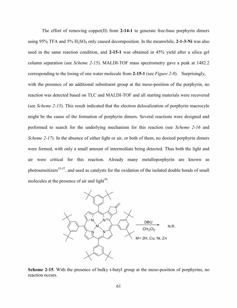
61
The effort of removing copper(II) from 2-14-1 to generate free-base porphyrin dimers
using 95% TFA and 5% H2SO4 only caused decomposition. In the meanwhile, 2-1-3-Ni was also
used in the same reaction condition, and 2-15-1 was obtained in 45% yield after a silica gel
column separation (see Scheme 2-15). MALDI-TOF mass spectrometry gave a peak at 1482.2
corresponding to the losing of one water molecule from 2-15-1 (see Figure 2-8). Surprisingly,
with the presence of an additional substituent group at the meso-position of the porphyrin, no
reaction was detected based on TLC and MALDI-TOF and all starting materials were recovered
(see Scheme 2-15). This result indicated that the electron delocalization of porphyrin macrocyle
might be the cause of the formation of porphyrin dimers. Several reactions were designed and
performed to search for the underlying mechanism for this reaction (see Scheme 2-16 and
Scheme 2-17). In the absence of either light or air, or both of them, no desired porphyrin dimers
were formed, with only a small amount of intermediate being detected. Thus both the light and
air were critical for this reaction. Already many metalloporphyrin are known as
photosensitizers55-57
, and used as catalysts for the oxidation of the isolated double bonds of small
molecules at the presence of air and light58
.
N
N N
N
MDBU
CH2Cl2
M= 2H, Cu, Ni, Zn
N.R.
Scheme 2-15. With the presence of bulky t-butyl group at the meso-position of porphyrins, no
reaction occurs.
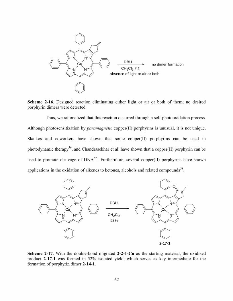
62
N
N N
N
CuDBU
CH2Cl2 r.t.no dimer formation
absence of light or air or both
Scheme 2-16. Designed reaction eliminating either light or air or both of them; no desired
porphyrin dimers were detected.
Thus, we rationalized that this reaction occurred through a self-photooxidation process.
Although photosensitization by paramagnetic copper(II) porphyrins is unusual, it is not unique.
Skalkos and coworkers have shown that some copper(II) porphyrins can be used in
photodynamic therapy56
, and Chandrasekhar et al. have shown that a copper(II) porphyrin can be
used to promote cleavage of DNA57
.
Furthermore, several copper(II) porphyrins have shown
applications in the oxidation of alkenes to ketones, alcohols and related compounds58
.
N
N N
N
Cu
DBU
CH2Cl2 N
N N
N
Cu
O
52%
2-17-1
Scheme 2-17. With the double-bond migrated 2-2-1-Cu as the starting material, the oxidized
product 2-17-1 was formed in 52% isolated yield, which serves as key intermediate for the
formation of porphyrin dimer 2-14-1.
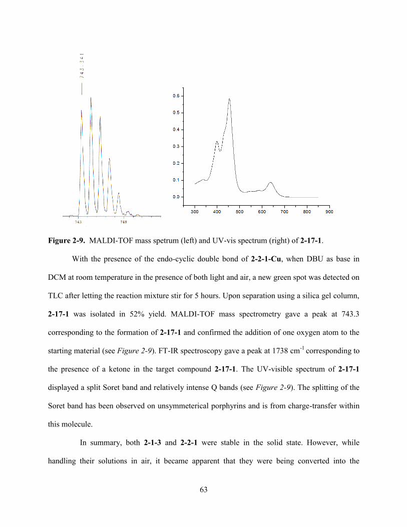
63
Figure 2-9. MALDI-TOF mass spetrum (left) and UV-vis spectrum (right) of 2-17-1.
With the presence of the endo-cyclic double bond of 2-2-1-Cu, when DBU as base in
DCM at room temperature in the presence of both light and air, a new green spot was detected on
TLC after letting the reaction mixture stir for 5 hours. Upon separation using a silica gel column,
2-17-1 was isolated in 52% yield. MALDI-TOF mass spectrometry gave a peak at 743.3
corresponding to the formation of 2-17-1 and confirmed the addition of one oxygen atom to the
starting material (see Figure 2-9). FT-IR spectroscopy gave a peak at 1738 cm-1
corresponding to
the presence of a ketone in the target compound 2-17-1. The UV-visible spectrum of 2-17-1
displayed a split Soret band and relatively intense Q bands (see Figure 2-9). The splitting of the
Soret band has been observed on unsymmeterical porphyrins and is from charge-transfer within
this molecule.
In summary, both 2-1-3 and 2-2-1 were stable in the solid state. However, while
handling their solutions in air, it became apparent that they were being converted into the
64
oxidized byproducts from TLC and MALDI-TOF. In the dark, in solution under argon, both 2-1-
3 and 2-2-1 were perfectly stable. Also, with the separate presence of either air or light, both 2-1-
3 and 2-2-1 were stable. However, once exposed to both light and air, they were rapidly
converted into the oxidized byproducts. The presence of conjugated endocyclic double bond
made 2-2-1 more unstable when exposed to light and air. The major product was an α,β-
unsaturated ketone 2-17-1, which was isolated in 52% yield. Based on these experimental results,
we propose a self-photooxidation mechanism for the formation of these porphyrin dimers (see
Figure 2-10). A literature search revealed numerous examples of photooxygenation of organic
molecules bearing exo-cyclic alkenes. Of particular relevance was the work of Havel21
who
showed that methylenecycloalkanes react with triplet oxygen. Methylenecycloalkanes were
shown to yield epoxides, and alkenones among other oxygenated products. In other work,
alkenyl-linked [60]fullerene derivatives have been shown to self-photooxygenate to give allylic
alcohols,58
and Saracoglu et al.55
have shown, for example, that cycloheptatriene derivatives can
be photooxygenated in the presence of H2TPP (2) as a singlet oxygen sensitizer, to give the
norcaradiene endoperoxides and bis-epoxide derivatives. The relevance of this literature became
apparent when, upon treatment with DBU, porphyrin 2-1-3 afforded a 60% yield of the
oxygenated bis-porphyrin 2-14-1 (see Figure 2-11). As shown in Figure 2-11, we propose
compound 2-1-3 undergoes photooxidation with triplet oxygen to generate epoxide 2-8, from
which the diol 2-9 and allylic alcohol 2-10 is subsequently formed. Meanwhile, the endocyclic
alkene compound 2-2-1 is photo-oxidized to give the epoxide 2-11 and then converted into the
diol 2-12, from which the allylic alcohol 2-13 is generated. Then the Michael addition generates
intermediates 2-17-1 and 2-14. The subsequent Michael addition reaction between anion 2-14
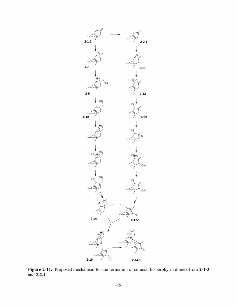
65
Figure 2-11. Proposed mechanism for the formation of cofacial bisporphyrin dimers from 2-1-3
and 2-2-1.
O
H O O H
H O H O
H O H O O
O
H O H O H O H O
H O
O H
H O H O H O
O H
H O O
O
H O
O
H O
O
O H O
~ ~ ~ ~ ~ ~
~ ~ ~ ~ ~ ~
~ ~ ~ ~ ~ ~
~ ~ ~ ~ ~ ~
~ ~ ~ ~ ~ ~
~ ~ ~ ~ ~ ~
~ ~ ~ ~ ~ ~
~ ~ ~ ~ ~ ~
~ ~ ~ ~ ~ ~
~ ~ ~ ~ ~ ~
~ ~ ~ ~ ~ ~
~ ~ ~ ~ ~ ~
~ ~ ~ ~ ~ ~
~ ~ ~ ~ ~ ~
~ ~ ~ ~ ~ ~
~ ~ ~ ~ ~ ~
~ ~ ~ ~ ~ ~
~ ~ ~ ~ ~ ~
~ ~ ~ ~ ~ ~
O
H O H O
~ ~ ~ ~ ~ ~
2-1-3 2-2-1
2-8 2-11
2-9 2-12
2-10 2-13
2-14 2-17-1
2-15 2-14-1
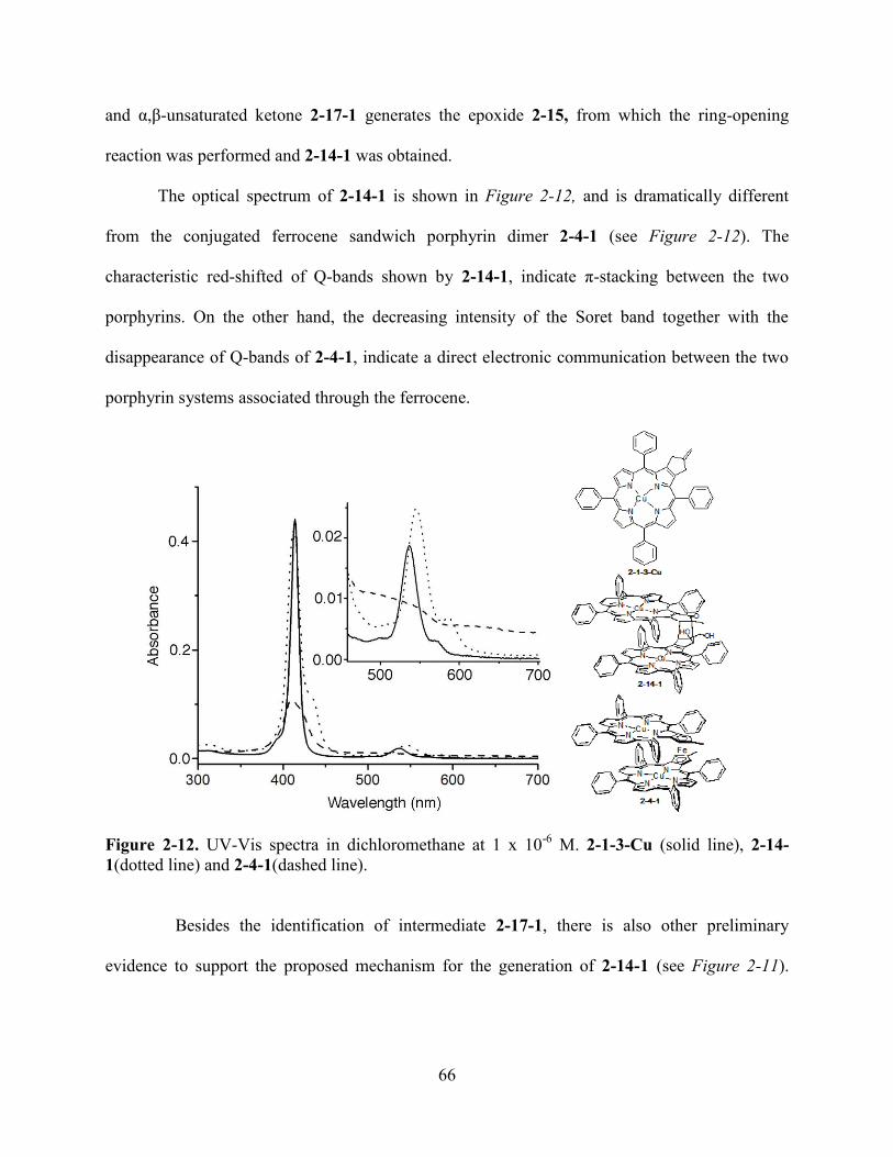
66
and α,β-unsaturated ketone 2-17-1 generates the epoxide 2-15, from which the ring-opening
reaction was performed and 2-14-1 was obtained.
The optical spectrum of 2-14-1 is shown in Figure 2-12, and is dramatically different
from the conjugated ferrocene sandwich porphyrin dimer 2-4-1 (see Figure 2-12). The
characteristic red-shifted of Q-bands shown by 2-14-1, indicate π-stacking between the two
porphyrins. On the other hand, the decreasing intensity of the Soret band together with the
disappearance of Q-bands of 2-4-1, indicate a direct electronic communication between the two
porphyrin systems associated through the ferrocene.
Figure 2-12. UV-Vis spectra in dichloromethane at 1 x 10⁻⁶
M. 2-1-3-Cu (solid line), 2-14-
1(dotted line) and 2-4-1(dashed line).
Besides the identification of intermediate 2-17-1, there is also other preliminary
evidence to support the proposed mechanism for the generation of 2-14-1 (see Figure 2-11).
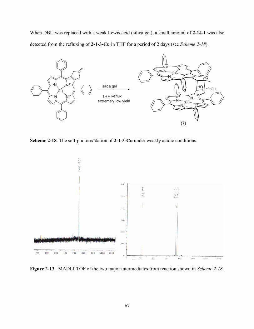
67
When DBU was replaced with a weak Lewis acid (silica gel), a small amount of 2-14-1 was also
detected from the refluxing of 2-1-3-Cu in THF for a period of 2 days (see Scheme 2-18).
N
N N
N
Cu
silica gel
THF
extremely low yield
Reflux
N
NN
NCu
N
NN
NCu
O
OHHO
(7)
Scheme 2-18. The self-photooxidation of 2-1-3-Cu under weakly acidic conditions.
Figure 2-13. MADLI-TOF of the two major intermediates from reaction shown in Scheme 2-18.
68
The yield of 2-14-1 was admittedly low compared to that under basic conditions; however, two
additional porphyrins were isolated as major products. MALDI-TOF mass spectrometry gave a
peak at 743.341 for the less polar one, and another peak at 758.101 for the more polar porphyrin
(see Figure 2-13).
The MALDI-TOF results indicated one or two oxygen atoms have been added to the
starting material 2-1-3-Cu. For the more polar product, besides the 758.101 peak, there was also
another peak at 742.2 shown in the MALDI spectrum, indicating that a facile loss of one oxygen
atom had taken place. The molecular weights of these two products were well in agreement with
the two important intermediates (2-17-1 and 2-14) as proposed in Figure 2-11. We believe that
these two reactions (DBU and/or silica gel) might share similar reaction mechanisms. The slight
difference happened at the nucleophilic addition step: under basic conditions, it was the hydroxyl
group that acted as the nucleophilic reagent while under Lewis acid catalyst conditions, water
was the nucleophilic reagent. The different nucleophilic ability of these two reagents made high
temperature necessary for the acidic condition reaction and explained why a very low yield of 2-
14-1 was obtained under these conditions. The whole mechanism (see Figure 2-11) involves
several mono oxidations of double bonds to form epoxide intermediates. These critically
important major intermediates, epoxides and spiro-epoxides, had been previously reported in the
literature associated with the reactions of methylenecycloalkanes with triplet oxygen11
. In our
case, the triplet oxygen was generated in situ from dioxygen activated by 2-1-3-Cu.
2.3 Experiment
2.3.1 General Information
All reactions were monitored by TLC using 0.25 mm silica gel plates with or without UV
indicator (60F-254). Silica gel (Sorbent Technologies 32-63 m) was used for flash column
69
chromatography. 1H- and
13C-NMR spectra were obtained on either a DPX-250 or an ARX-300
Bruker spectrometer. Chemical shifts (δ) are given in ppm relative to residual CHCl3 (7.26 ppm,
1H), or DCM (5.32 ppm, 1H) unless otherwise indicated. Electronic absorption spectra were
measured on a Perkin Elmer Lambda 35 UV-Vis spectrophotometer. MALDI-TOF mass spectra
were obtained on an Applied Biosystems QSTAR XL, using positive method with dithranol as
matrix. Materials obtained from commercial suppliers were used without further purification.
2. 3.2 Procedure for the Synthesis of 2-14-1
A mixture of β,β’-fused copper(II) methylenepropanoporphyrin 2-1-3-Cu (100 mg, 0.14
mmol) and DBU (100 mL, 0.67 mmol) was dissolved in CH2Cl2 (20 mL) and stirred at room
temperature in air for 52 hours. Upon the completion of the reaction, the separation was
performed on a silica gel column using CH2Cl2 as eluting solvent. The title porphyrin 2-14-1 was
obtained in 60% yield (63 mg, 0.042 mmol). UV/Vis: max CH2Cl2 (log ) 413 nm (5.62), 545
(4.40), 585 (3.83); MS (HR-MALDI-TOF) C96H62Cu2N8O3 (M+): Calcd m/z for 1502.3553;
Found 1502.1324. A crystal of 2-14-1 (See Figure 2-7) was grown by slow diffusion of hexane
into the concentrated dichloromethane solution of 2-14-1.
2.3.3 Procedure for synthesis of porphyrin 2-17-1
A mixture of endocyclic alkene porphyrin 2-2-1 (100 mg, 0.14 mmol) and DBU (100 mL,
0.67 mmol) was dissolved in CH2Cl2 (20 mL) and was stirred at room temperature under air for 5
hours. The separation was performed on a silica gel column using CH2Cl2 /hexane (v/v =1/2) as
eluting solvent. The tile green porphyrin 2-17-1 was obtained in 52% yield (54 mg, 0.072 mmol).
UV/Vis: max CH2Cl2 (log ) 338 nm (4.72), 399 (5.22), 454 (5.47), 549 (4.25), 584 (4.30), 637
(4.65); MALDI-TOF C48H30CuN4O (M+H): Calcd m/z for 742.2; Found 742.5.
70
2.3.4 Procedures for synthesis of 2-1-3
The preparation of 2-1-3-Ni was performed under strictly air-free conditions. The Pd(0)
catalyst was prepared in suit by reacting Pd(OAc)2 (113 mg, 0.5 mmol) with
triisopropylphosphate (3 mmol) in 100 ml dry THF. The reaction mixture was stirred at room
temperature for a period of 30 minutes. Porphyrin 2-1-2-Ni (1.42 g, 2 mmol) was weighed and
added directly into a Schlenck reaction flask. Subsequently 2-[(trimethylsilyl)methyl]-2-propen-
1-yl acetate (0.5 mmol) was added. Then the reaction temperature was raised to 90 oC and the
mixture was refluxed at this temperature for 2 days. TLC and MALDI-TOF were used to follow
this reaction. When most of the starting material was consumed, the reaction temperature was
raised to 100 oC and the mixture was refluxed at this temperature for an additional period of 2
days. Solvent was removed under vacuum and the residue was applied to a silica gel column for
separation; hexane/DCM was used as eluting solvent. After removing the solvent under vacuum,
the desired product 2-1-3-Ni was obtained as a reddish brown solid in 82% yield (1.19 g). MP >
300oC;
1H-NMR (250 MHz, CDCl3) ppm: 8.75-8.72 (m, 6H), 8.01-7.97 (m, 4H), 7.87-7.83 (m,
4H), 7.70-7.63 (m, 12H), 4.99-4.98 (m, 2H), 3.48-3.46 (m, 4H).
2-1-3-Cu was prepared in a similar procedure as described above for 2-1-3-Ni. MALDI-TOF
Calcd. for C48H32N4Cu, 728.3. Found, 728.1. Anal. Calcd for C48H32CuN4.C6H14: C, 79.62; H,
5.69; N, 6.88. Found: C, 79.22; H, 5.72; N, 7.05. UV/vis: λmax DCM (log ε) 414 nm (5.64), 536
(4.27), 568 (3.49).X-Ray data: C48H32CuN4HCl3, triclinic space group P-1, a = 12.940(4), b =
13.185(5), c = 13.694(7) A˚, a = 108.12(2), b = 117.71(2), c = 90.22(2), V = 1935.0(14) A˚ 3, T =
110 K, Z = 2, R = 0.094 (F2 > 2r), Rw = 0.271 (all F2) for 5879 unique data and 514 refined
parameters. CCDC 280898.
71
2.3.5 Procedures for synthesis of 2-2-1
The preparation of 2-2-1-Cu was performed by mixing of porpyrin 2-1-3-Cu (432 mg, 0.6 mmol)
with p-TsOH (17 mg, 0.1 mmol) in 15 mL of chloroform and then refluxed for a period of 24
hours. After cooling down to room temperature, excess of p-TsOH was filtered off. The
remained solution was washed with saturated aqueous Na2CO3 and water. Solvent was removed
under vacuum and the residue was submitted to silica gel column separation. The eluting solvent
was hexane/DCM. The desired product was obtained in 90% yield (390 mg) after
recrystallization from MeOH/DCM. MALDI-TOF: calcd m/z for C48H32CuN4 728.3; found
727.9; UV/vis λmax DCM (log ε) 411 nm (5.51), 540 (4.36).
2-2-1-Ni was prepared similar to 2-2-1-Cu as described above from 2-1-2-Ni. 1H-NMR (250
MHz, CDCl3) ppm 8.78-8.73 (m, 6H), 8.03-7.97 (m, 8H), 7.71-7.66 (m, 12H), 5.57 (s, 1H), 3.10
(s, 2H), 2.10 (s, 3H). MALDI-TOF Calcd. for C52H36N4Ni, 775.6. Found, 775.6.
2-1-2-Ni: 1H-NMR (CDCl3, 250 MHz) ppm 8.99 (s, 1H), 8.71-8.65 (m, 6H), 8.00-7.96 (m, 8H),
7.72-7.62 (m, 12H). MALDI-TOF Calcd. for C44H27N5NiO2, 716.4. Found 716.4.
2.3.6 Procedures for synthesis of 2-6-1 and related Compounds
The preparation of 2-6-1 was similar to that of 2-1-3-Ni. After the in situ preparation of the Pd(0)
catalyst, the mixture of isomers of dinitro-Ni(II)TPP (383 mg, 0.5 mmol) was added into the
reaction mixture and allowed to stir at 90 oC for a period of 2 days. Then the temperature was
raised to 100 oC for an additional 2 days. TLC and MALDI-TOF indicated the selective
formation of cis-isomer 2-6-1-Ni. After separation using silica gel TLC, the desired 2-6-1-Ni was
obtained in 35% yield (134 mg). 1H-NMR (CDCl3, 250 MHz) ppm 8.79 (d, 2H, J = 5.0 Hz), 8.72
(d, 2H, J = 5.0 Hz), 7.93-7.98 (m, 2H), 7.86 (d, 4H, J = 7.5 Hz), 7.77 (d, 2H, J = 7.5 Hz), 7.69-
72
7.60 (m, 12 H), 4.97 (s, 4H), 3.48 (d, 8H, J = 10.0 Hz). MALDI-TOF Calcd. for C52H36N4Ni,
775.6. Found, 775.1. m.p. > 300 oC.
2-5-1 was prepared in a similar procedure as described for 2-6-1-Ni, with a similar yield 33%.
MALDI-TOF Calcd. for C52H36N4Cu, 780.4. Found, 780.0.
2-7-2-Cu: MALDI-TOF Calcd. for C96H62N8Ni2O3 [M-H2O], 1475.0. Found, 1474.7.
2.3.7 Procedure for preparation of 2-9-1
Recrystallization of NBS was performed in hot water followed by drying at 80 °C under vacuum
for a period of 6 h. H2TPP (600 mg, 1.0 mmol) was dissolved in CHCl3 (120 mL) and freshly
recrystallized NBS (1.0 g, 6.0 mmol) was added into the solution which was refluxed for a
period of 4 h. After cooling to room temperature, CHCl3 was removed under vacuum. The
residue was then washed with methanol (2×40 mL) to remove the succinimide impurities. Then
the residue as a purple solid was dissolved in CHCl3 and purified on a silica gel column with
CHCl3 as the eluting solvent. The first fraction from the column was collected and the solvent
was removed under vacuum. The solid was further recrystallized from CHCl3 /CH3OH (1:3).
After filtration and removal of the solvent under vacuum, 3-2 was obtained in 66% yield (600
mg). 1H NMR (CDCl3, 250 MHz): ppm 8.70 (s, 4H), 8.20-8.17 (m, 8H), 7.80-7.78 (m, 12H),
−2.90 (s, 2H). UV–vis (DCM): λmax (nm) (log ε): 437 (5.50), 535 (4.34), 613 (3.60), 687 (4.12).
MALDI-TOF Calcd for C44H26Br4N4: 930.3. Found 930.4.
2.3.8 Procedures for Preparation of 2-9-3
The preparation of 2-9-2 was achieved from Suzuki-coupling reaction52
. 2-9-1 (465 mg, 0.5
mmol), Pd(PPh3)4 (225 mg, 0.02 mmol) anhydrous K2CO3 (1.2 g, 8 mmol),and CH3B(OH)2 (120
mg, 2 mmol) in THF/toluene (v/v = 3/2) 100 ml. The reaction mixture was degassed and then let
it stirred at 90 °C under argon atmosphere for 3 days. After completion of reaction, the solvent
73
was removed under vacuum. The crude product was dissolved in CHCl3 and washed with
saturated aqueous NaHCO3 solution followed by saturated aqueous NaCl solution, and the
organic layer was dried over anhydrous Na2SO4. After reducing the solvent under vacuum, the
reaction mixture was loaded on a silica gel column. Only a very short silica gel plug was
required. Pure DCM was used to elute down the small amount of starting material 2-9-1. After
that, the eluting solvent was changed to the mixture solvents of DCM/ethyl acetate (v/v = 10/1).
2-9-2 was obtained as a green fraction from the column and after removing the solvents under
vacuum it was obtained as purple microcrystals in 87% yield (292 mg). 1H NMR (CDCl3, 250
MHz) ppm: 7.94 (m, 8H), 7.58 (m, 4H), 7.47 (m, 8H), 6.76 (m, 20H), 1.84 (s, 12H), -1.68 (s, 2H).
MALDI-TOF Calcd for C48H36N4 668.8. Found 668.7. The insertion of Ni(II) was performed by
refluxing 2-9-2 (268 mg, 0.4 mmol) with an excess amount of Ni(acac)2 (4mmol) in
chloroform/methanol (v/v = 1/3) overnight. After filtration, 2-9-3 was obtained in 98% yield
(285 mg). 1H-NMR (CDCl3, 250 MHz) ppm 7.47 (m, 8H), 7.12 (m, 4H), 7.02 (m, 8H), 6.68 (m,
20H), 1.58 (s,12H). MALDI-TOF Calcd. for C48H36N4Ni, 727.5. Found, 727.8.
2.3.8 Procedure for Preparation of 2-17-1
A mixture of endocyclic alkene porphyrin 2-2-1-Cu (100 mg, 0.14 mmol) and DBU (100 µL,
0.67 mmol) in DCM (20 mL) was stirred at room temperature under air for 5 h. The mixture was
purified by column chromatography on silica gel using DCM/hexane (v/v = 1/2) as eluent, giving
the green porphyrin 2-17-1 in 52% yield (54 mg, 0.072 mmol). UV/vis: λmax DCM (log ε) 338
nm (4.72), 399 (5.22), 454 (5.47), 549 (4.25), 584 (4.30), 637 (4.65); MALDI-TOF
C48H30CuN4O [M+H]: Calcd m/z for 742.2. Found 742.5.
74
2.3.9 Procedures for Preparation of 2-14-1, 2-15-1 and Related Compounds
A mixture of β,β’-fused copper(II) methylenepropanoporphyrin 2-3-1 (100 mg, 0.14 mmol) and
DBU (100 µL, 0.67 mmol) in DCM (20 mL) was stirred at room temperature in air for 52 h. The
mixture was purified by column chromatography on silica gel using DCM as eluent, giving the
title porphyrin 2-14-1 in 60% yield (63 mg). UV/vis: λmax DCM (log ε) 413 nm (5.62), 545
(4.40), 585 (3.83). MS (HRMALDI-TOF) C96H62Cu2N8O3 (M+): Calcd m/z for 1502.3553.
Found 1502.1324. The crystal of 7 was grown by slow diffusion of hexane into dichloromethane
solution. X-ray data for Compound 2-14-1, C96H60Cu2N8O3, triclinic space group P-1, a =
14.174(6), b = 16.881(8), c = 17.876(10) A ˚, a = 99.97(3),b = 101.23(3), c = 99.171(17), V =
4048(3) A˚ 3
, T = 110 K, Z = 2, R = 0.101 (F2 > 2r), Rw = 0.278 (all F2) for 100,38 unique data
and 448 refined parameters. CCDC 280897.
2-16-1 was obtained in a similar procedure as described above. MALDI-TOF Calcd. for
C96H62N8Ni2O3 [M-H2O]+, 1475.0. Found, 1474.7.
2-4-1: MALDI-TOF: calcd m/z for C96H62Cu2FeN8 1510.5. Found 1510.1. UV/vis: λmax
DCM (log ε) 410 nm (5.04).
2.4 Conclusions and Future Work
Using Pd(0) catalyzed [3+2] cyclization reactions, under strictly air-free conditions, a
series of mono-methylenepropanoporphyrins and bis(methylenepropano)porphyrins had been
synthesized. The selective formation of both cis-bis(methylenepropano)porphyrins and trans-
bis(methylenepropano)porphyrins has been accomplished from the corresponding
nitroporphyrins. Starting from mono-methylenepropanoporphyrins, a new type of self-sensitized
photooxidation reaction was accidently discovered, from which β,β’-cofacial porphyrin dimers
were built. The mechanism for this reaction was also studied in some detail.
75
Future research will be focused on the use of the bis(methylenepropano)porphyrins to
build either ferrocenoporphyrin monomers with two ferrocenes in each structure, and the
building of sandwich ferrocenoporphyrin trimers.
2.5 References
1 Plenoio, H.; Diodone, R. J. Organomet. Chem. 1995, 492, 73.
2 Burrell, A. K.; Campbell, W. M.; Officer, D. L.; Scott, S. M.; Gordon, K. C.; MaDonald, M.
R. J. Chem. Soc. Dalton Trans. 1999, 3349.
3 Beer, P. D. Chem. Soc. Rev. 1989, 18, 409.
4 Beer, P. D.; Kurek, S. S. J. Organomet. Chem. 1989, 366, C6-C8.
5 Ryabov, A. D.; Goral, V. N.; Gorton, L.; Csoregi, E. Chem. Eur. J. 1999, 5, 961.
6 Thornton, N. B.; Wojtowicz, H.; Netzel, T.; Zixton, D. W. J. Phys. Chem. B. 1998, 102, 2101.
7 Boyd, P. D. W.; Burrel, A. K.; Campbell, W. M.; Cocks, P. A.; Gordon, K. C.; Jameson, G.
B.; Officer, D. L.; Zhao, Z. Chem. Commun. 1999, 637.
8 Nemykin, V. N.; Kobayashi, N. Chem. Commun. 2001, 165.
9 Wagner, R. W.; Brown, P. A.; Johnson, T. E.; Lindsey, J. S. J. Chem. Soc., Chem. Commun.
1991, 1463.
10 Rhee, S. W.; Park, B. B.; Do, Y.; Kim, J. Polyhedron 2000, 19, 1961.
11 Wollmann, R. G.; Hendrickson, D. N. Inorg. Chem. 1977, 16, 3079.
12 Burrell, A. K.; Officer, D. L.; Plieger, P. G.; Reid, D. C. W. Chem. Rev. 2001, 101, 2751; (b)
Harvey, P. D. In The Porphyrin Handbook, Kadish, K. M.; Smith, K. M.; Guilard, R. Eds.;
Academic Press: San Diego, CA; 2003; vol. 18, pp 63; (c) Vicente, M. G. H.; Jaquinod, L.;
Smith, K. M. Chem. Commun. 1999, 1771.
13 Wagner, R. W.; Johnson, T. E.; Lindsey, J. S. J. Am. Chem. Soc. 1996, 118, 11166.
14 Anderson, H. L. Inorg. Chem. 1994, 33, 972.
15 Lin, V. S.-Y.; DiMagno, S. G.; Therien, M. J. Science 1994, 264, 1105.
16 Lin, V. S.-Y.; Therien, M. J. Chem. Eur. J. 1995, 1, 645.
17 Recent example: Kuramochi, Y.; Satake, A.; Kobuke, Y. J. Am. Chem. Soc. 2004, 126, 8668.
76
18 Recent examples: Faure, S.; Stern, C.; Guilard, R.; Harvey, P. D. J. Am. Chem. Soc. 2004,
126, 1253.
19 Recent examples: Chang, C. J.; Loh, Z.-H.; Shi, C.; Anson, F. C.; Nocera, D. G. J. Am. Chem.
Soc. 2004, 126, 10013.
20 Collman, J. P.; Kendall, J. L.; Chen, J. L. Inorg. Chem. 2000, 39, 1661.
21 Anderson, H. L.; Martin, S. J.; Bradley, D. D. C. Angew. Chem. Int. Ed. Engl. 1994, 33, 655.
22 Wilson, G. S.; Anderson, H. L. Chem. Commun. 1999, 1539.
23 Buchler, J. W.; De Cian, A.; Fischer, J. ; Hammerschmitt, P. ; Loffler, J.; Scharbert, B.;
Weiss, R. Chem. Ber. 1989, 122, 2219.
24 Bilsel, O.; Rodriguez, J.; Milam, S. N.; Gorlin, P. A.; Girolami, G. S.; Suslick, K. S.; Holten,
D. J. Am. Chem. Soc. 1992, 114, 6528.
25 Bilsel, O.; Rodriguez, J.; Milam, S. N.; Gorlin, P. A.; Girolami, G. S.; Suslick, K. S.; Holten,
D. J. Am. Chem. Soc. 1992, 114, 6528.
26 Collman, J. P.; Wagenknecht, P. S.; Hutchison, J. E. Angew. Chem. Int. Ed. Engl. 1994, 33,
1537.
27 Clement, T. E.; Nurco, D. J.; Smith, K. M. Inorg. Chem. 1998, 37, 1150.
28 Wagner, R. W.; Johnson, T. E.; Lindsey, J. S. J. Am. Chem. Soc. 1996, 118, 11166; (b)
Anderson, H. L. Inorg. Chem. 1994, 33, 972; (c) Lin, V. S.–Y; Therien, M. J. Chem. Eur. J.
1995, 1, 645.
29 Anderson, H. L.; Martin, S. J.; Vradley, D. D. C. Angew. Chem. Int. Ed. Engl. 1994, 33, 655.
(b) Wilson, G. S.; Anderson, H. L. Chem. Commun. 1999, 1539.
30 Sanders, J. K. M. In The Porphyrin Handbook; Kadish, K. M.; Smith, K. M.; Guilard, R.
Eds.; Academic Press: San Diego, CA; 2000; vol. 3, pp 347.
31 Cvetanovic, R. J.; Singleton, D. L. Rev. Chem. Intermed. 1984, 5, 183.
32 Havel, J. J. J. Org. Chem. 1978, 43, 762.
33 Tanner, D. D.; Kandanarachchi, P.; Das, N. C.; Brausen, M.; Vo, C. T.; Camaioni, D. M.;
Franz, J. A. J. Org. Chem. 1998, 63, 4587.
34 Zadok, E.; Rubinraut, S.; and Mazur, Y. J. Org. Chem. 1987, 52, 385.
35 Holzapfel, C. W.; Van der Merwe, T. L. Tetrahedron Lett. 1996, 37, 2303.
36 Schimizu, I.; Ohashi, Y.; Tsuji, J. Tetrahedron Lett. 1984, 25, 5183.

77
37 Ejiri, S.; Yamago, S.; Nakamura, E. J. Am. Chem. Soc. 1992, 114, 8707.
38 Trost, B. M.; Matelich, M. C. J. Am. Chem. Soc. 1991, 113, 9007.
39 Holzapfel, C. W.; Van der Merwe, T. L. Tetrahedron Lett. 1996, 37, 2307.
40 Schimizu, I.; Ohashi, Y.; Tsuji, J. Tetrahedron Lett. 1984, 25, 5183.
41 Trost, B. M.; Chan, D. M. T. J. Am. Chem. Soc. 1979, 101, 6429.
42 Trost, B. M.; Chan, D. M. T. J. Am. Chem. Soc. 1981, 103, 5972.
43 Trost, B. M.; Miller, M. L. J. Am. Chem. Soc. 1988, 110, 3687.
44 Shea, K. M.; Jaquinod, L.; Khoury, R. G.; Smith, K. M. Chem. Commun. 1998, 759.
45 Krattinger, B.; Nurco, D. J.; Smith, K. M. Chem. Commun. 1998, 757.
46 Adler, A. D. et. al. J. Org. Chem. 1967, 32, 476.
47 Shine, H. J.; Padilla, A. G.; Wu, S.-M. J. Org. Chem. 1979, 44, 4069.
48 Wickramasinghe, A.; Jaquinod, L.; Nurco, D. J.; Smith, K. M. Tetrahedron 2001, 57, 4261.
49 Dailey, K. K.; Yap, G. P. A.; Rheingold, A. L.; Rauchfuss, T. B. Angew. Chem. Int. Ed. Engl.
1996, 35, 1833.
50 Lichtenberger, D. L.; Elkadi, Y.; Gruhn, N. E. Organometallics 1997, 16, 5209.
51 Beck, C. U.; Field, L. D.; Hambley, T. W.; Humphrey, P. A.; Masters, A. F.; Turner, P. J.
Organomet. Chem. 1998, 565, 283.
52 Kumar, P. K.; Bhyrappa, P.; Varghese, B. Tetrahedron Lett. 2003, 44, 4849.
53 Zhou, X.; Zhou, Z. Y.; Mak, T. C. W.; Chan, K. S. J. Chem. Soc. Perkin. Trans. 1, 1994,
2519.
54 Whitlock, H. W.; Hanauer, Jr. R.; Oester, M. Y.; Bower, B. K. J. Am. Chem. Soc. 1969, 91,
7485.
55 Wang, H. J. H.; Jaquinod, L.; Nurco, D. J.; Vicente, M. G. H.; Smith, K. M. Chem. Commun.
2001, 2646.
56 Saracoglu, N.; Talaz, O.; Azizoglu, A.; Watson, W. H.; Balci, M. J. Org. Chem. 2005, 70,
5403.
57 Selman, S. H.; Hampton, J. A.; Morgan, A. R.; Keck, R. W.; Balkany, A. D.; Skalkos, D.
Photochem. Photobiol. 1993, 57, 681. (b) Hampton, J. A.; Skalkos, D.; Taylor, P. M.;
Selman, S. H. Photochem. Photobiol. 1993, 58, 100.

78
58 Chandrasekhar, V.; Nagendran, S.; Azhakar, R.; Kumar, M. R.; Srinivasan, A.; Ray, K.;
Chandrashekar, T. K.; Madhavaiah, C.; Verma, S.; Priyakumar, U. D.; Sastry, G. N. J. Am.
Chem. Soc. 2005, 127, 2410.
59 Chronakis, N.; Vougioukalakis, G. C.; Orfanopoulos, M. Org. Lett., 2002, 4, 945.
60 Suslick, K. S.; Acholla, F. V.; Cook, B. R. J. Am. Chem. Soc., 1987, 109(9), 2818.
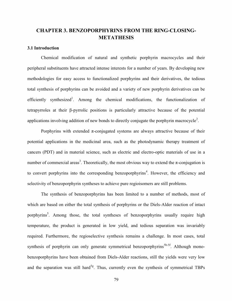
79
CHAPTER 3. BENZOPORPHYRINS FROM THE RING-CLOSING-
METATHESIS
3.1 Introduction
Chemical modification of natural and synthetic porphyrin macrocycles and their
peripheral substituents have attracted intense interests for a number of years. By developing new
methodologies for easy access to functionalized porphyrins and their derivatives, the tedious
total synthesis of porphyrins can be avoided and a variety of new porphyrin derivatives can be
efficiently synthesized1. Among the chemical modifications, the functionalization of
tetrapyrroles at their β-pyrrolic positions is particularly attractive because of the potential
applications involving addition of new bonds to directly conjugate the porphyrin macrocycle2.
Porphyrins with extended π-conjugated systems are always attractive because of their
potential applications in the medicinal area, such as the photodynamic therapy treatment of
cancers (PDT) and in material science, such as electric and electro-optic materials of use in a
number of commercial areas3. Theoretically, the most obvious way to extend the π-conjugation is
to convert porphyrins into the corresponding benzoporphyrins4. However, the efficiency and
selectivity of benzoporphyrin syntheses to achieve pure regioisomers are still problems.
The synthesis of benzoporphyrins has been limited to a number of methods, most of
which are based on either the total synthesis of porphyrins or the Diels-Alder reaction of intact
porphyrins5. Among those, the total syntheses of benzoporphyrins usually require high
temperature, the product is generated in low yield, and tedious separation was invariably
required. Furthermore, the regioselective synthesis remains a challenge. In most cases, total
synthesis of porphyrin can only generate symmetrical benzoporphyrins5b-5f
. Although mono-
benzoporphyrins have been obtained from Diels-Alder reactions, still the yields were very low
and the separation was still hard5g
. Thus, currently even the synthesis of symmetrical TBPs
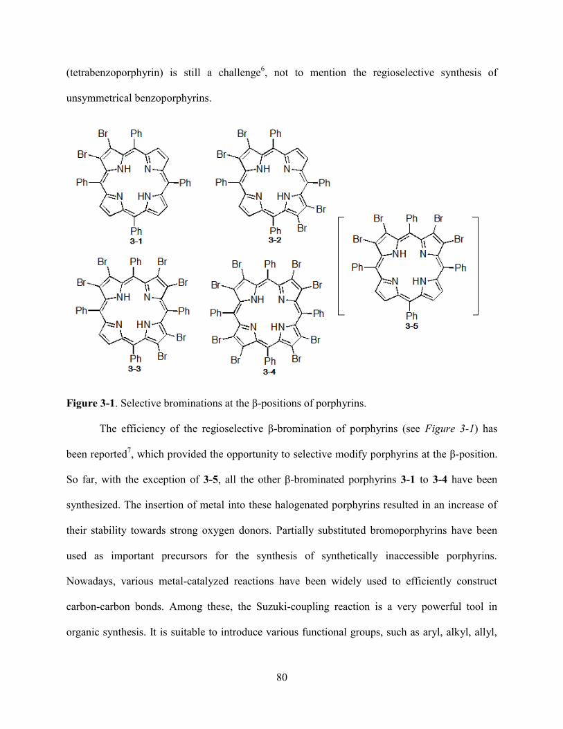
80
(tetrabenzoporphyrin) is still a challenge6, not to mention the regioselective synthesis of
unsymmetrical benzoporphyrins.
Figure 3-1. Selective brominations at the β-positions of porphyrins.
The efficiency of the regioselective β-bromination of porphyrins (see Figure 3-1) has
been reported7, which provided the opportunity to selective modify porphyrins at the β-position.
So far, with the exception of 3-5, all the other β-brominated porphyrins 3-1 to 3-4 have been
synthesized. The insertion of metal into these halogenated porphyrins resulted in an increase of
their stability towards strong oxygen donors. Partially substituted bromoporphyrins have been
used as important precursors for the synthesis of synthetically inaccessible porphyrins.
Nowadays, various metal-catalyzed reactions have been widely used to efficiently construct
carbon-carbon bonds. Among these, the Suzuki-coupling reaction is a very powerful tool in
organic synthesis. It is suitable to introduce various functional groups, such as aryl, alkyl, allyl,
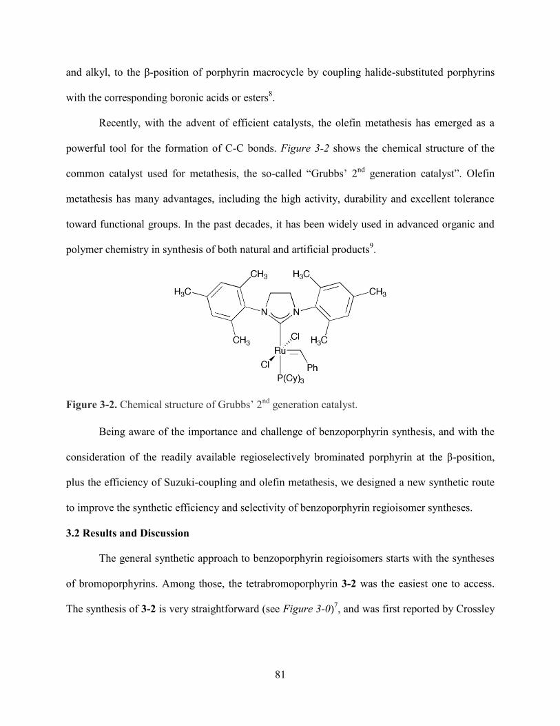
81
and alkyl, to the β-position of porphyrin macrocycle by coupling halide-substituted porphyrins
with the corresponding boronic acids or esters8.
Recently, with the advent of efficient catalysts, the olefin metathesis has emerged as a
powerful tool for the formation of C-C bonds. Figure 3-2 shows the chemical structure of the
common catalyst used for metathesis, the so-called ―Grubbs’ 2nd
generation catalyst‖. Olefin
metathesis has many advantages, including the high activity, durability and excellent tolerance
toward functional groups. In the past decades, it has been widely used in advanced organic and
polymer chemistry in synthesis of both natural and artificial products9.
Figure 3-2. Chemical structure of Grubbs’ 2nd
generation catalyst.
Being aware of the importance and challenge of benzoporphyrin synthesis, and with the
consideration of the readily available regioselectively brominated porphyrin at the β-position,
plus the efficiency of Suzuki-coupling and olefin metathesis, we designed a new synthetic route
to improve the synthetic efficiency and selectivity of benzoporphyrin regioisomer syntheses.
3.2 Results and Discussion
The general synthetic approach to benzoporphyrin regioisomers starts with the syntheses
of bromoporphyrins. Among those, the tetrabromoporphyrin 3-2 was the easiest one to access.
The synthesis of 3-2 is very straightforward (see Figure 3-0)7, and was first reported by Crossley
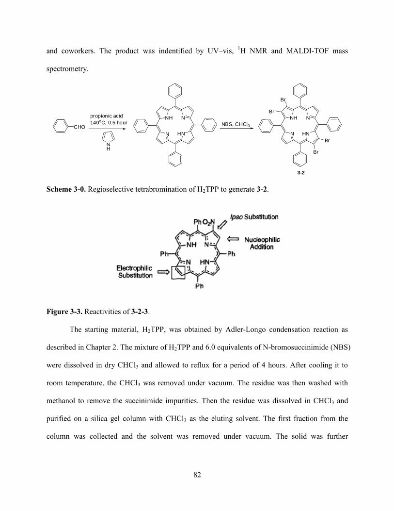
82
and coworkers. The product was indentified by UV–vis, 1H NMR and MALDI-TOF mass
spectrometry.
NH
CHO
propionic acid
140oC, 0.5 hour
N
NH N
HN N
NH N
HN
Br
Br
Br
Br
NBS, CHCl3
3-2
Scheme 3-0. Regioselective tetrabromination of H2TPP to generate 3-2.
Figure 3-3. Reactivities of 3-2-3.
The starting material, H2TPP, was obtained by Adler-Longo condensation reaction as
described in Chapter 2. The mixture of H2TPP and 6.0 equivalents of N-bromosuccinimide (NBS)
were dissolved in dry CHCl3 and allowed to reflux for a period of 4 hours. After cooling it to
room temperature, the CHCl3 was removed under vacuum. The residue was then washed with
methanol to remove the succinimide impurities. Then the residue was dissolved in CHCl3 and
purified on a silica gel column with CHCl3 as the eluting solvent. The first fraction from the
column was collected and the solvent was removed under vacuum. The solid was further
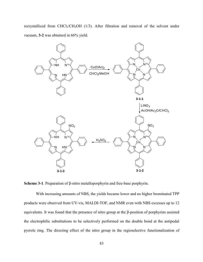
83
recrystallized from CHCl3/CH3OH (1/3). After filtration and removal of the solvent under
vacuum, 3-2 was obtained in 66% yield.
N
N N
N
Cu
NO2
N
N N
N
Cu
N
NH N
HN
NO2
N
NH N
HN
Cu(OAc)2
CHCl3/MeOH
AcOH/Ac2O/CHCl3
LiNO3
H2SO4
3-1-1
3-1-23-1-3
Scheme 3-1. Preparation of β-nitro metalloporphyrin and free-base porphyrin.
With increasing amounts of NBS, the yields became lower and no higher brominated TPP
products were observed from UV-vis, MALDI-TOF, and NMR even with NBS excesses up to 12
equivalents. It was found that the presence of nitro group at the β-position of porphyrins assisted
the electrophilic substitutions to be selectively performed on the double bond at the antipodal
pyrrole ring. The directing effect of the nitro group in the regioselective functionalization of
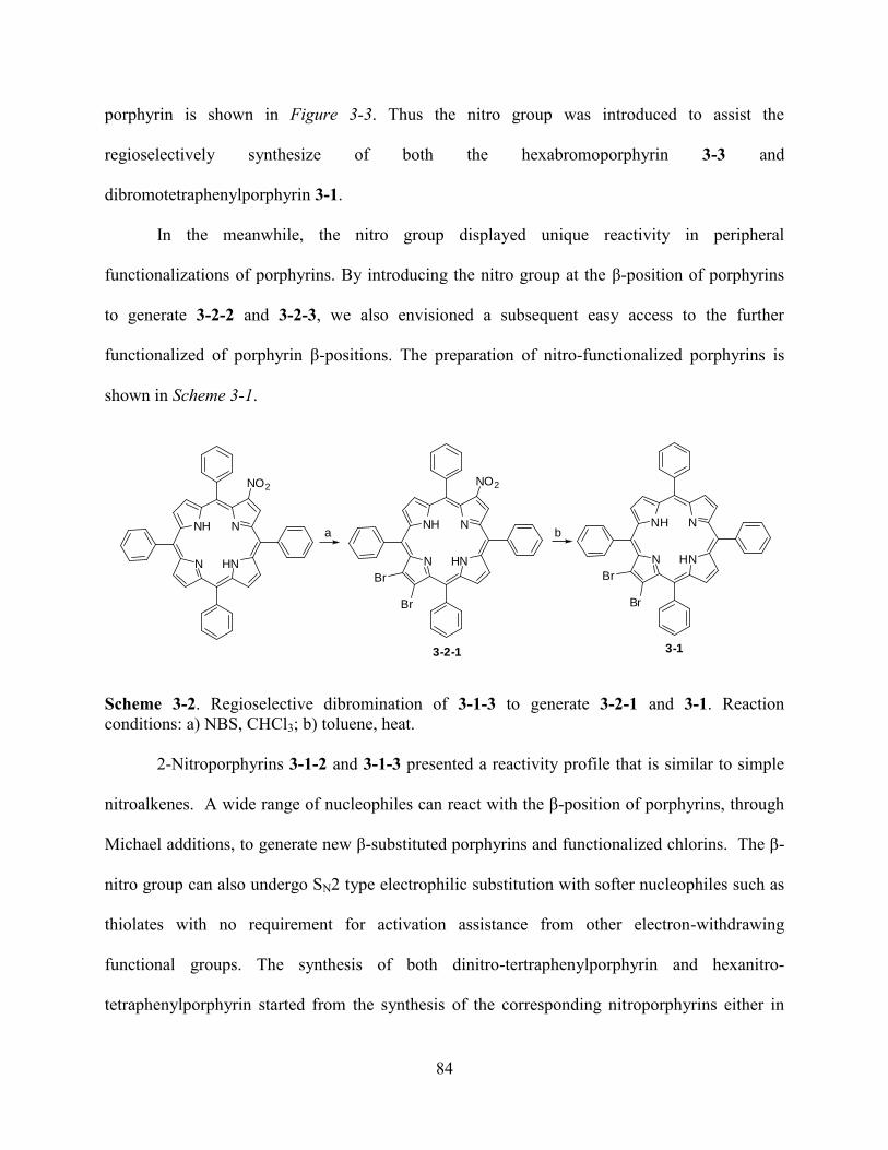
84
porphyrin is shown in Figure 3-3. Thus the nitro group was introduced to assist the
regioselectively synthesize of both the hexabromoporphyrin 3-3 and
dibromotetraphenylporphyrin 3-1.
In the meanwhile, the nitro group displayed unique reactivity in peripheral
functionalizations of porphyrins. By introducing the nitro group at the β-position of porphyrins
to generate 3-2-2 and 3-2-3, we also envisioned a subsequent easy access to the further
functionalized of porphyrin β-positions. The preparation of nitro-functionalized porphyrins is
shown in Scheme 3-1.
N
NH N
HN
NO2
N
NH N
HN
NO2
Br
Br
N
NH N
HN
Br
Br
a b
3-13-2-1
Scheme 3-2. Regioselective dibromination of 3-1-3 to generate 3-2-1 and 3-1. Reaction
conditions: a) NBS, CHCl3; b) toluene, heat.
2-Nitroporphyrins 3-1-2 and 3-1-3 presented a reactivity profile that is similar to simple
nitroalkenes. A wide range of nucleophiles can react with the β-position of porphyrins, through
Michael additions, to generate new β-substituted porphyrins and functionalized chlorins. The β-
nitro group can also undergo SN2 type electrophilic substitution with softer nucleophiles such as
thiolates with no requirement for activation assistance from other electron-withdrawing
functional groups. The synthesis of both dinitro-tertraphenylporphyrin and hexanitro-
tetraphenylporphyrin started from the synthesis of the corresponding nitroporphyrins either in
85
metalated form 3-1-2 or metal-free form 3-1-3 (see Scheme 3-1). As described in Chapter 2,
these nitroporphyrins were obtained in high yields with simple purification due to the huge
polarity difference generated before and after the introduction of the nitro group to the porphyrin.
The demetalation of 3-1-2 to generate 3-1-3 was achieved by using concentrated H2SO4.
Porphyrin 3-1-2 was dissolved in a minimum amount of concentrated sulfuric acid with
alternating stirring and sonication for a period of 1 hour. After this it was poured into a mixture
of ice/water and the 3-1-3 was extracted with CHCl3. The organic phase was subsequently
washed with water and saturated aqueous NaHCO3. After drying over Na2SO4, the solvent was
removed under vacuum. Recrystallization from CHCl3 /MeOH gave 3-1-3 as a dark purple
powder in 74% yield.
The dibromoporphyrin 3-2-1 was obtained from the bromination of metal-free
nitroporphyrin 3-1-3 with 2.4 equivalents of NBS in ethanol-free chloroform (see Scheme 3-2).
The reaction mixture was left to reflux for a period of 12 hours. After cooling it to room
temperature, the reaction mixture was poured through an alumina plug (Grade III) and eluted
with CHCl3. After removal of solvent under vacuum, the resulting solid was further
recrystallized from CHCl3/MeOH. The desired 3-2-1 was eventually obtained as a brown
powder in 80% yield. The nitro group was compatible with the demetalation process. The
removal of the nitro group was performed with NaBH4 as a reducing reagent to generate 3-1
through the corresponding nitrochlorin (2,3-dihydro-2-nitroporphyrin) intermediate, which was
readily converted into tetraphenylporphyrins by refluxing in chloroform with the assistance of
silica gel or in toluene at high temperature (around 100 oC). Porphyrin 3-2-1 was dissolved in
cold dry THF placed in an ice-salt bath and 1.8 equivalents of NaBH4 were added into the
solution under argon. After the reaction mixture was left stir for a period of 1 hour, the ice bath
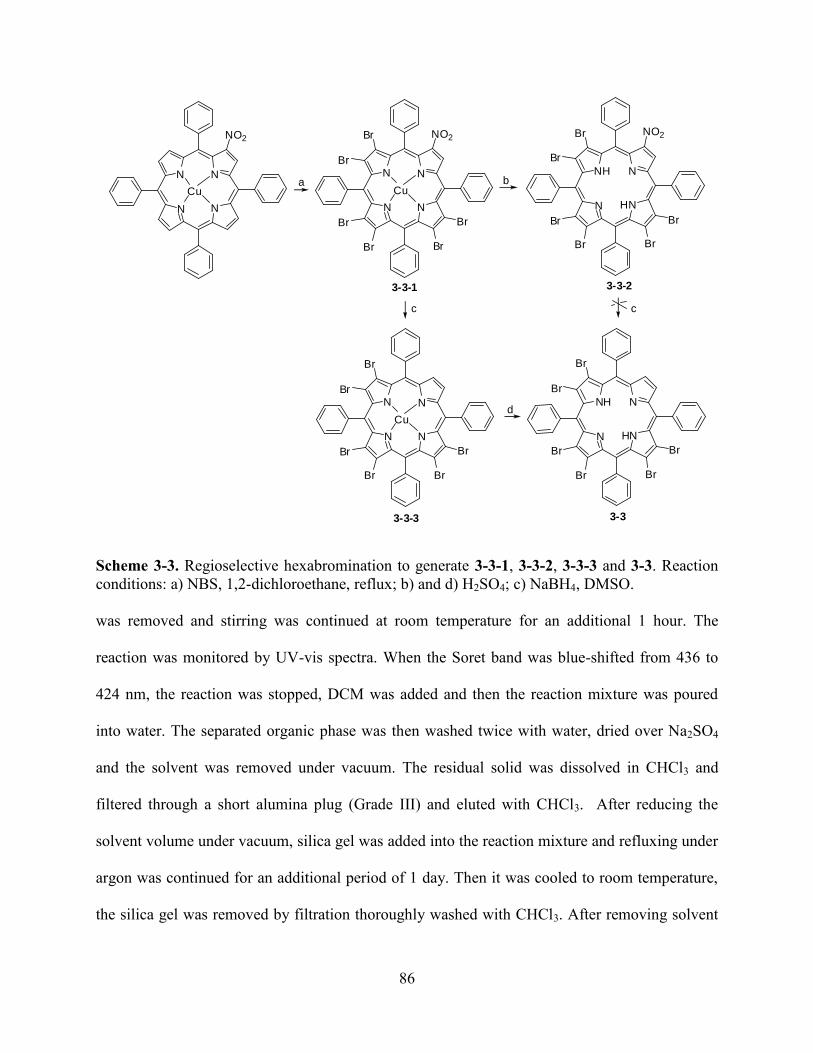
86
N
N N
N
Cu
NO2
N
N N
N
Cu
NO2Br
Br
Br
Br Br
Br
N
NH N
HN
NO2Br
Br
Br
Br Br
Br
N
N N
N
Cu
Br
Br
Br
Br Br
Br
N
NH N
HN
Br
Br
Br
Br Br
Br
a b
c
d
c
3-3-1 3-3-2
3-33-3-3
Scheme 3-3. Regioselective hexabromination to generate 3-3-1, 3-3-2, 3-3-3 and 3-3. Reaction
conditions: a) NBS, 1,2-dichloroethane, reflux; b) and d) H2SO4; c) NaBH4, DMSO.
was removed and stirring was continued at room temperature for an additional 1 hour. The
reaction was monitored by UV-vis spectra. When the Soret band was blue-shifted from 436 to
424 nm, the reaction was stopped, DCM was added and then the reaction mixture was poured
into water. The separated organic phase was then washed twice with water, dried over Na2SO4
and the solvent was removed under vacuum. The residual solid was dissolved in CHCl3 and
filtered through a short alumina plug (Grade III) and eluted with CHCl3. After reducing the
solvent volume under vacuum, silica gel was added into the reaction mixture and refluxing under
argon was continued for an additional period of 1 day. Then it was cooled to room temperature,
the silica gel was removed by filtration thoroughly washed with CHCl3. After removing solvent
87
under vacuum and subsequent recrystallization from CHCl3/MeOH, 3-1 was obtained as a purple
powder in 83% yield. Hexabromoporphyrin 3-3-1 was obtained from the hexabromination of 3-
1-2 with excess amounts of NBS in boiling 1,2-dichloroethane (see Scheme 3-3). To the 1,2-
dichloroethane solution of 3-1-2 was added 10 equivalents NBS and it was refluxed for a period
of 16 hours under argon. After cooling to room temperature, the reaction mixture was poured
into a silica gel plug and eluted with CHCl3. After removal of the solvent under vacuum and
subsequent recrystallization from CHCl3/MeOH, 3-3-1 was obtained as a dark green powder in
68% yield. This reaction was careful monitored by UV-vis spectrophotometry. Extended
refluxing times should be avoided because of the slow degradation of the desired product and a
concomitant decrease in the yield. The demetalation of 3-3-1 to generate 3-3-2 was achieved
with concentrated H2SO4. A minimal amount of DCM was added to dissolve 3-3-1 into a suitable
round bottom flask. After slow removal of the solvent by rotation under vacuum, an oily film
was formed on the inner surface of the flask. Then a minimal amount of concentrated sulfuric
acid was added to the flask to dissolve the film, followed by alternating stirring and sonication
for a period of 1.5 hours. After pouring the mixture into ice/water, 3-3-2 was extracted with
CHCl3. The organic phase was subsequently washed with water and saturated aqueous NaHCO3.
After drying over Na2SO4 the solvent was removed under vacuum. Subsequent recrystallization
from CHCl3/MeOH and filtration gave 3-3-2 as a green powder in 95% yield. The removal of the
nitro group from 3-3-1 to generate 3-3-3 and 3-3 was achieved with NaBH4 as the reducing
reagent. To a dry DMSO solution of 3-3-1 under argon was added 1.8 equivalents of NaBH4 and
the reaction mixture was stirred at room temperature. The solution was initially green and soon
turned brown in color. Stirring was continued for around 2 hours and then the reaction was
stopped (when the Soret band gave a 22 nm blue-shift). After adding DCM, the reaction mixture
88
was poured into water. The organic phase was then washed several times with water. After
reducing the solvent volume under vacuum, the solution was placed in the refrigerator for around
3~6 hours. After filtration, pure 3-3-3 was obtained as a dark brown precipitate, in 55% yield.
Compared to the metalated porphyrin 3-3-1, the green solution of 3-3-2 easily underwent slow
decomposation under even slightly basic conditions. Thus the reduction of 3-3-2 with NaBH4
failed to generate 3-3. Instead, 3-3 was obtained from the demetalation of 3-3-3 with
concentrated H2SO4 in a method similar to the generation of 3-3-2. 3-3-3 was dissolved in
concentrated H2SO4 for a period of 1 hour with alternating stirring and sonication. After it was
poured into the mixture of ice/water and 3-3 was extracted with CHCl3. The organic phase was
subsequently washed with water and saturated aqueous NaHCO3. After drying over Na2SO4, the
solvent was removed under vacuum. Recrystallization from CHCl3/MeOH gave 3-3 as a dark
purple powder in 74% yield.
The Suzuki-coupling reactions were performed in toluene at 100 ºC (see Scheme 3-4).
The mixture of bromoporphyrin (3-1 to 3-4), Pd(PPh3)4 (5 mmol % catalyst /Br) and K2CO3 were
dissolved in freshly distilled toluene, to which 2-allyl-4,4,5,5-tetramethyl-1,3,2-dioxaborolane
was added through a syringe. This coupling reaction required strictly air free conditions. TLC
and MALDI-TOF mass spectrometry were used to follow the reaction. When most of the
bromoporphyrin had been consumed, the reaction was stopped. After diluting with DCM, the
solution was poured into water. The organic layer was separated and dried over anhydrous
Na2SO4. After reducing the solvents under vacuum, the solution was applied to a silica gel
column and separated with DCM/hexane as eluent. After removing solvent under vacuum, 3-4-n
(n = 1 ~ 5) were achieved in 49% ~ 84% yields. The solution was found to have a dramatic color
and polarity change associated with this coupling reaction; the color of the starting materials was

89
reddish brown while it was green for 3-4-n (n = 2,3). The starting materials showed low polarity,
while 3-4-n (n = 2,3) showed very high polarity.
N
NH
N
HN
Br
Br
N
NH
N
HNBr
aBr
Br Br
R R
R = NO2 or H R = NO2 3-4-4R = H 3-4-5
N
NH
N
HN
Br
Br
N
NH
N
HNBr
aBr
N
NH
N
HN
Br
Br
N
NH
N
HNR1 R1
a
R1 = NO2 or H R1 = NO2 3-4-1R1 = H 3-4-2
3-4-3
Scheme 3-4. Suzuki-coupling to generate metathesis precursors. Reaction conditions: a) 2-allyl-
4,4,5,5-tetramethyl-1,3,2-dioxaborolane, anhydrous potassium carbonate, 100 ºC.
Thus, the separation was easily performed. It is surprising to see that the use of 2-allyl-
4,4,5,5-tetramethyl-1,3,2-dioxaborolane in the Suzuki-coupling reaction had indeed provided
high yields. Boron-esters, such as 2-allyl-4,4,5,5-tetramethyl-1,3,2-dioxaborolane, have been
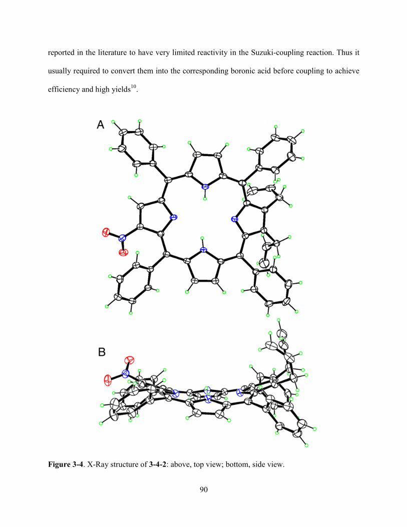
90
reported in the literature to have very limited reactivity in the Suzuki-coupling reaction. Thus it
usually required to convert them into the corresponding boronic acid before coupling to achieve
efficiency and high yields10
.
Figure 3-4. X-Ray structure of 3-4-2: above, top view; bottom, side view.
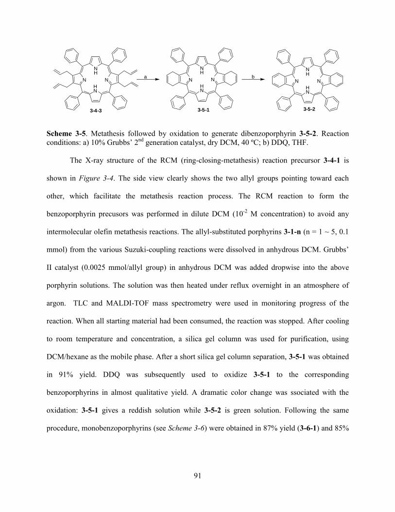
91
N
NH
N
HN
N
NH
N
HN
N
NH
N
HN
a b
3-4-3 3-5-1 3-5-2
Scheme 3-5. Metathesis followed by oxidation to generate dibenzoporphyrin 3-5-2. Reaction
conditions: a) 10% Grubbs’ 2nd
generation catalyst, dry DCM, 40 ºC; b) DDQ, THF.
The X-ray structure of the RCM (ring-closing-metathesis) reaction precursor 3-4-1 is
shown in Figure 3-4. The side view clearly shows the two allyl groups pointing toward each
other, which facilitate the metathesis reaction process. The RCM reaction to form the
benzoporphyrin precusors was performed in dilute DCM (10-2
M concentration) to avoid any
intermolecular olefin metathesis reactions. The allyl-substituted porphyrins 3-1-n (n = 1 ~ 5, 0.1
mmol) from the various Suzuki-coupling reactions were dissolved in anhydrous DCM. Grubbs’
II catalyst (0.0025 mmol/allyl group) in anhydrous DCM was added dropwise into the above
porphyrin solutions. The solution was then heated under reflux overnight in an atmosphere of
argon. TLC and MALDI-TOF mass spectrometry were used in monitoring progress of the
reaction. When all starting material had been consumed, the reaction was stopped. After cooling
to room temperature and concentration, a silica gel column was used for purification, using
DCM/hexane as the mobile phase. After a short silica gel column separation, 3-5-1 was obtained
in 91% yield. DDQ was subsequently used to oxidize 3-5-1 to the corresponding
benzoporphyrins in almost qualitative yield. A dramatic color change was ssociated with the
oxidation: 3-5-1 gives a reddish solution while 3-5-2 is green solution. Following the same
procedure, monobenzoporphyrins (see Scheme 3-6) were obtained in 87% yield (3-6-1) and 85%

92
yield (3-6-2). Also, tribenzoporphyrins (see Scheme 3-7) were obtained in 83% (3-7-1) and 82%
yield (3-7-2).
N
NH
N
HN
R1
N
NH
N
HN
R1
N
NH
N
HN
R1
a b
R1 = NO2 or HR1 = NO2 3-6-3R1 = H 3-6-4
R1 = NO2 3-6-1R1 = H 3-6-2
Scheme 3-6. Metathesis followed by oxidation to generate monobenzoporphyrin 3-6-1 and 3-6-2.
Reaction conditions: a) Grubbs’ 2nd
generation catalyst, dry DCM, 40 ºC; b) DDQ, THF.
N
NH
N
HN
N
NH
N
HN
N
NH
N
HN
a b
R R R
R = NO2 and H R = NO2 3-7-3R = H 3-7-4
R = NO2 3-7-1R = H 3-7-2
Scheme 3-7. Metathesis followed by oxidation to generate tribenzoporphyris 3-7-1 and 3-7-2.
Reaction conditions: a) Grubb’s 2nd
generation catalyst, dry DCM, 40 ºC; b) DDQ, THF.
The X-ray structure of 3-5-2 was shown in Figure 3-5. The porphyrin macrocyle was
adopted a saddle conformation, which was attrictuted to the presence of four phenyl group at the
meso-position of porphyrin. The UV-vis spectra of monobenzoporphyrin (3-6-2),
dibenzoporphyrin (3-5-2) and tribenzoporphyrin (3-7-2) are plotted in Figure 3-6. With the
increase in of the number of benzo-groups attached to the porphyrin macrocyle, there was a clear
red shift of both the Soret band and the Q-bands. This confirmed that the presence of benzo-
groups provides extended conjugation to the porphyrin macrocyle.
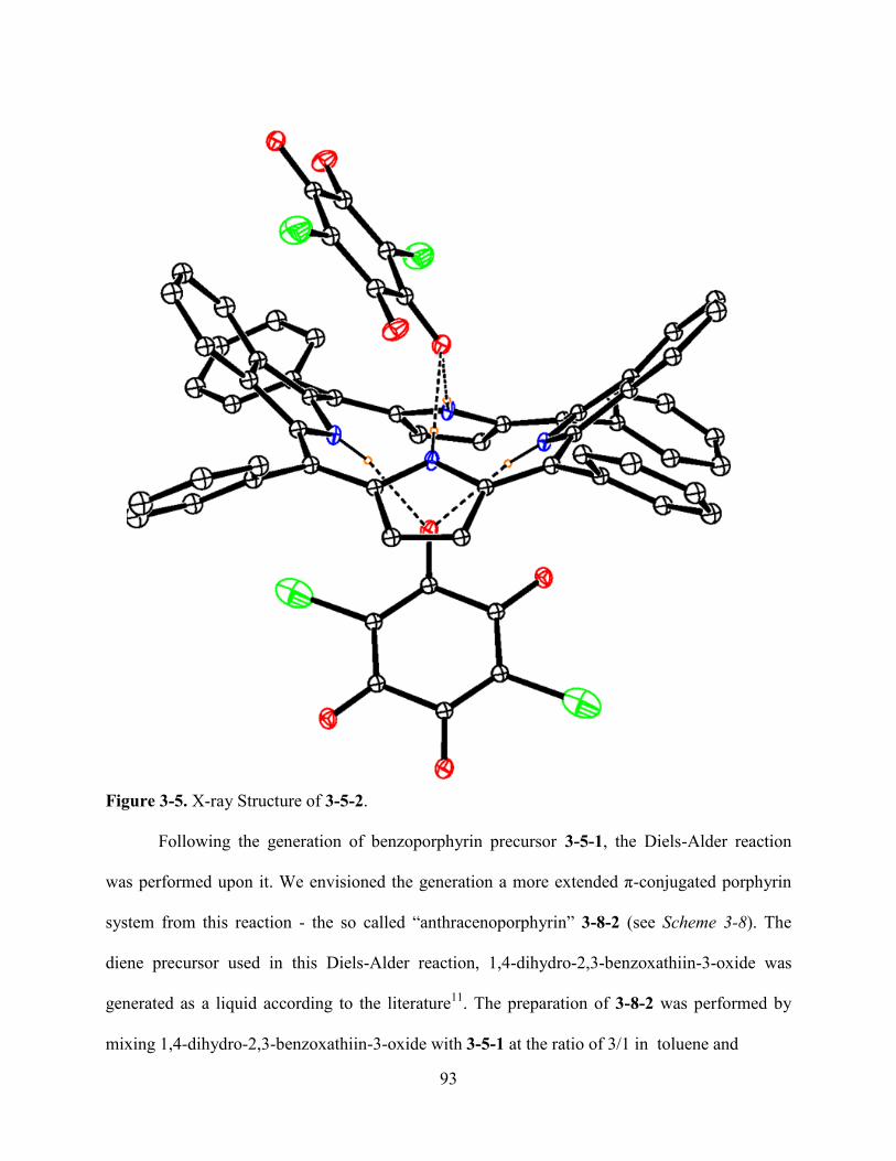
93
Figure 3-5. X-ray Structure of 3-5-2.
Following the generation of benzoporphyrin precursor 3-5-1, the Diels-Alder reaction
was performed upon it. We envisioned the generation a more extended π-conjugated porphyrin
system from this reaction - the so called ―anthracenoporphyrin‖ 3-8-2 (see Scheme 3-8). The
diene precursor used in this Diels-Alder reaction, 1,4-dihydro-2,3-benzoxathiin-3-oxide was
generated as a liquid according to the literature11
. The preparation of 3-8-2 was performed by
mixing 1,4-dihydro-2,3-benzoxathiin-3-oxide with 3-5-1 at the ratio of 3/1 in toluene and

94
Figure 3-6. UV-vis spectra of benzoporphyrins in DCM: 3-5-2 (dash), 3-6-2 (solid) and 3-7-2
(dot).
subsequently refluxing the reaction mixture at 120 ºC under argon. TLC was used to follow the
progress of the reaction. The reaction was stopped when most of the starting material had been
consumed. After separation on a silica gel column, 3-8-1 was obtained in 58% yield. However,
the oxidation of 3-8-1 to generate 3-8-2 was problematic. Firstly, the direct use of 3-8-1 in the
DDQ oxidation in THF resulted in no generation of 3-8-2 both after a long period of room
temperature reaction or at refluxing conditions over 24 hours. Both MALDI-TOF and 1H-NMR
spectra indicated partial oxidation of 3-8-1, as shown in Figure 3-7. The UV-vis spectra gave
absorption peaks (λmax) at 426, 459, 545, 640 and 696 nm. Secondly, the metal zinc was
inserted into the porphyrin central cavity using Zn(OAC)2 in hot MeOH/CHCl3 overnight.
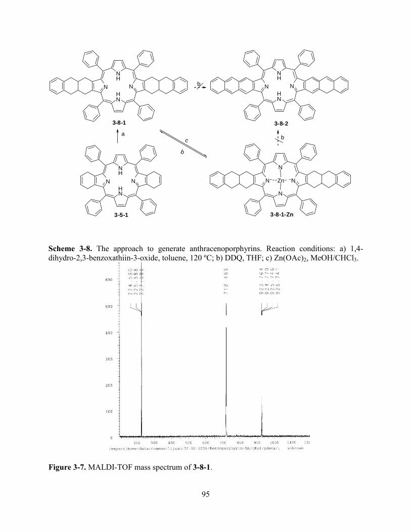
95
N
NH
N
HN
b N
NH
N
HN
3-8-1 3-8-2
N
NH
N
HN
3-5-1
a
N
N
N
N
b
Zn
c
b
3-8-1-Zn
Scheme 3-8. The approach to generate anthracenoporphyrins. Reaction conditions: a) 1,4-
dihydro-2,3-benzoxathiin-3-oxide, toluene, 120 ºC; b) DDQ, THF; c) Zn(OAc)2, MeOH/CHCl3.
Figure 3-7. MALDI-TOF mass spectrum of 3-8-1.
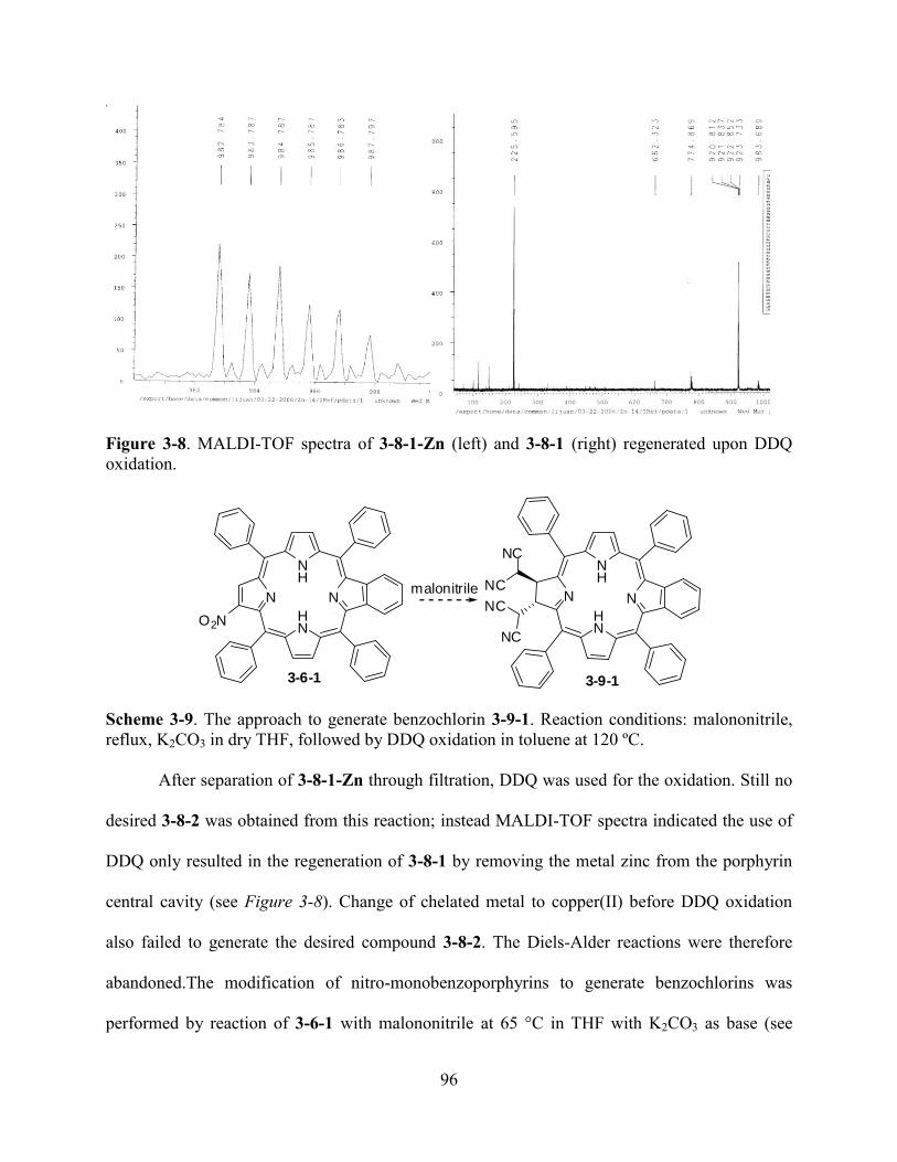
96
Figure 3-8. MALDI-TOF spectra of 3-8-1-Zn (left) and 3-8-1 (right) regenerated upon DDQ
oxidation.
N
NH
N
HN
O2N
3-6-1
N
NH
N
HN
3-9-1
NC
NC
NC
NC
malonitrile
Scheme 3-9. The approach to generate benzochlorin 3-9-1. Reaction conditions: malononitrile,
reflux, K2CO3 in dry THF, followed by DDQ oxidation in toluene at 120 ºC.
After separation of 3-8-1-Zn through filtration, DDQ was used for the oxidation. Still no
desired 3-8-2 was obtained from this reaction; instead MALDI-TOF spectra indicated the use of
DDQ only resulted in the regeneration of 3-8-1 by removing the metal zinc from the porphyrin
central cavity (see Figure 3-8). Change of chelated metal to copper(II) before DDQ oxidation
also failed to generate the desired compound 3-8-2. The Diels-Alder reactions were therefore
abandoned.The modification of nitro-monobenzoporphyrins to generate benzochlorins was
performed by reaction of 3-6-1 with malononitrile at 65 °C in THF with K2CO3 as base (see
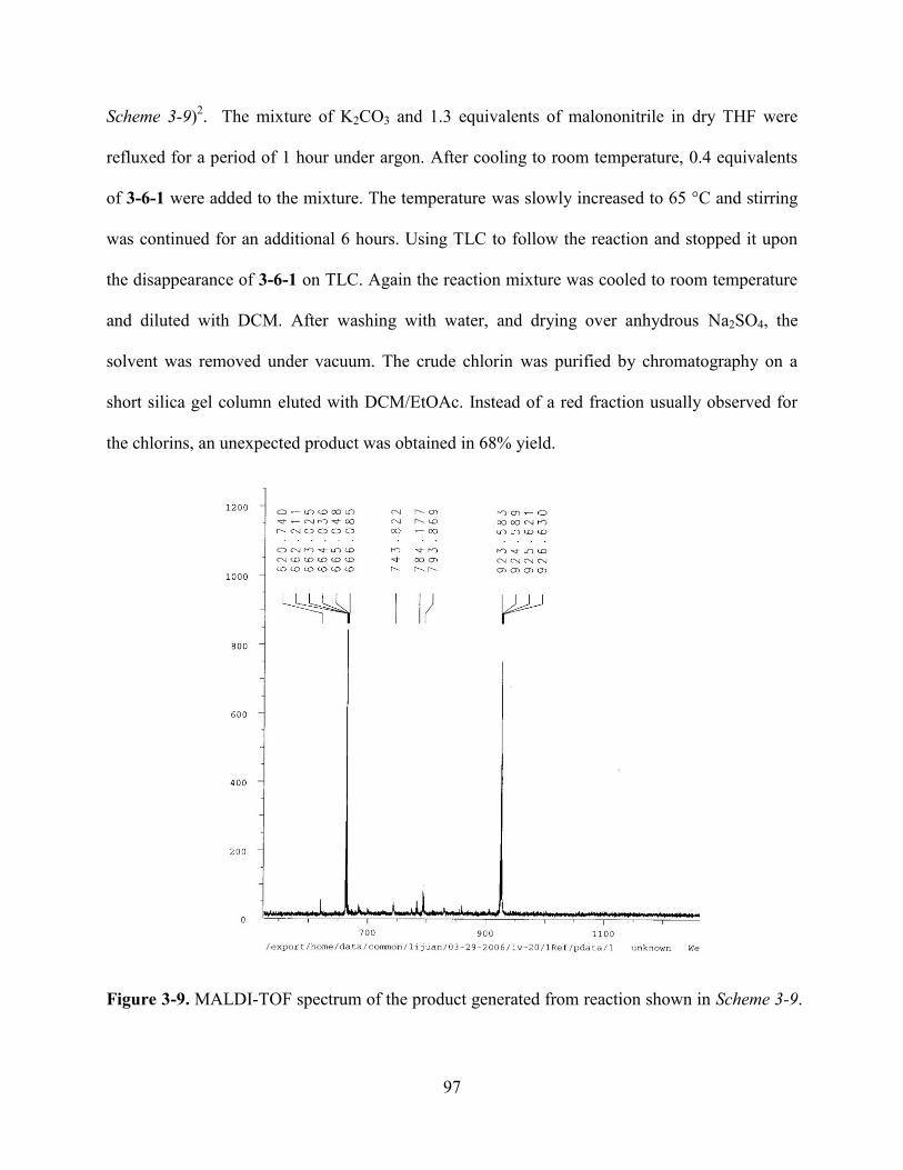
97
Scheme 3-9)2. The mixture of K2CO3 and 1.3 equivalents of malononitrile in dry THF were
refluxed for a period of 1 hour under argon. After cooling to room temperature, 0.4 equivalents
of 3-6-1 were added to the mixture. The temperature was slowly increased to 65 °C and stirring
was continued for an additional 6 hours. Using TLC to follow the reaction and stopped it upon
the disappearance of 3-6-1 on TLC. Again the reaction mixture was cooled to room temperature
and diluted with DCM. After washing with water, and drying over anhydrous Na2SO4, the
solvent was removed under vacuum. The crude chlorin was purified by chromatography on a
short silica gel column eluted with DCM/EtOAc. Instead of a red fraction usually observed for
the chlorins, an unexpected product was obtained in 68% yield.
Figure 3-9. MALDI-TOF spectrum of the product generated from reaction shown in Scheme 3-9.
98
Instead of adding two malonoitrile groups to porphyrin, MALDI-TOF showed a peak at
MW 923.6, corresponding to the addition of four malononitrile groups to the porphyrin
macrocycle (see Figure 3-9). In the meanwhile, it gave a characteristic chlorin band on UV-vis
spectra, which indicated the formation of a chlorin. However, the color of the solution was deep
green instead of red often observed for most of the chlorin system.
3.3 Conclusions
An efficient transition catalyzed synthetic route was developed for the selective syntheses
of the benzoporphyrin regio-isomers. This synthetic route avoided the total synthesis route for
making benzoporphyrin and achieved the high regio-selectivity in generation of benzoporphyrin
regio-isomers. The further usage of the synthetic precursors for the generation of the other type
of extended porphyrin system will also be studied in our group in the future.
3.4 Experiment
3.4.1 General information
All reactions were monitored by TLC using 0.25 mm silica gel plates with or without UV
indicator (60F-254). Silica gel (Sorbent Technologies 32-63 m) was used for flash column
chromatography. 1H- and
13C-NMR spectra were obtained on either a DPX-250 or an ARX-300
Bruker spectrometer. Chemical shifts (δ) are given in ppm relative to residual CHCl3 (7.26 ppm,
1H), or DCM (5.32 ppm, 1H) unless otherwise indicated. Electronic absorption spectra were
measured on a Perkin Elmer Lambda 35 UV-Vis spectrophotometer. MALDI-TOF mass spectra
were obtained on an Applied Biosystems QSTAR XL, using positive method with dithranol as
matrix. Materials obtained from commercial suppliers were used without further purification.
99
3.4.2 Syntheses and Characterization
General procedure for metathesis and subsequent oxidation
The allyl-substituted porphyrins 3-1-n (n = 1 ~ 5, 0.1 mmol) from the various Suzuki-coupling
reactions were dissolved into anhydrous DCM. Grubbs II catalyst (0.0025 mmol/allyl group) in
anhydrous DCM was added dropwise into the above porphyrin solutions. The solution was then
heated under reflux overnight in an atmosphere of argon. TLC and MALDI-TOF spectra were
used in monitoring the reaction. When all starting material had been consumed, the reaction was
stopped. After cooling to room temperature and concentration, a silica gel column was used for
purification, using a mixture of DCM/hexane as the mobile phase. After the short silica gel
column separation, 3-5-1 was obtained in 91% yield. DDQ (182 mg, 0.8 mmol) was
subsequently used to oxidize 3-5-1 to the corresponding benzoporphyrin (64 mg, 0.09 mmol) in
almost qualitative yield. The solution was again found to undergo a dramatic color change
associated with the oxidation: 3-5-1 was reddish in solution while 3-5-2 gave a green solution.
3-6-1: 87% yield. MALDI-TOF Calcd for C48H33N5O2 [M+H]+ 712.271, found 712.149. UV-vis
(DCM) λmax (nm): 383 (log ε 5.10), 439 (5.72), 536 (4.64), 614 (4.11), 680 (4.32). 1H-NMR
(300 MHz, CDCl3) 8.97 (1H, s), 8.85-8.88 (2H, m), 8.63-8.67 (2H, m), 8.21-8.25 (4H, m) 8.09-
8.12 (4H, m), 7.68-7.81 (12H, m), 5.73 (2H, s), 3.27 (4H, s), -2.62 (2H, s). Mp >300 ºC.
3-5-1: 91 % yield. MALDI-TOF Calcd for C52H38N4 [M]+ 718.9, found 718.9.
1H–NMR (250
MHz, CDCl3): ppm 8.50 (s, 4H), 8.15-8.0 (d, 8H, J = 7.5), 7.77-7.66 (m, 12 H), 5.85 (s, 4H),
3.50 (s, 8H), -2.83 (s, 2H). Mp >300 ºC.
3-6-2: 85% yield. MALDI-TOF Calcd for C48H33N4 [M+H]+ 665.271, found 665.201. UV-vis
(DCM), λmax (nm): 407 (log ε 4.95), 425 (5.70), 518 (4.38), 596 (3.88) and 650 (3.01). δH (300
100
MHz, CDCl3) 8.87 (2H, d, J = 5.9 Hz), 8.77 (2H, d, J = 5.9 Hz), 8.71 (2H, s), 8.19-8.24 (8H, m),
7.74-7.93 (12H, m), 7.36-7.39 (2H, m), 7.12-7.16 (2H, m), -2.61 (2H, s). Mp > 300 ºC.
3-5-2: MALDI-TOF Calcd for C52H35N4 [M+H]+ 715.286, found 715.798. UV-vis (DCM) λmax
(nm): 413 (log ε 4.76), 435 (5.67), 525 (4.37), 618 (3.75) and 675 (3.85). δH (300 MHz, CD2Cl2)
8.76 (4H, d, J = 7.38 Hz), 8.53 (4H, d, J = 6.99 Hz), 8.28 (4H, s), 8.12-8.17 (4H, m), 8.00-8.05
(4H, m), 7.84-7.89 (4H, m), 7.56 (8H, s). Mp > 300 ºC.
3-7-1: 83% yield. MALDI-TOF Calcd for C56H35N5O2 [M+H]+
809.279, found 809.422. UV-vis
(DCM) λmax (nm): 394 (log ε 5.02), 448 (5.73), 543 (4.62), 615 (4.26), 682 (4.09). δH (300
MHz, CD2Cl2) 8.93 (1H, s), 8.88-8.92 (2H, m), 8.71-8.73 (2H, d, J = 6.03 Hz), 8.16-8.27 (8H,
m), 7.73-7.91 (12H, m), 7.30-7.34 (2H, d, J = 11.0 Hz), 7.08 (2H, s), -2.35 (2H, s). Mp > 300 ºC.
3-7-2: 82% yield. MALDI-TOF Calcd for C56H37N4 [M+H]+ 765.302, found 765.670. UV-vis
(DCM) λmax (nm): 428 (log ε 4.97), 450 (5.70), 553 (4.31), 590 (4.37), 628 (4.04), 685 (3.86).
δH (250 MHz, CD2Cl2) 8.44 (2H, s), 8.21-8.30 (10H, m), 7.82-7.97 (12H, m), 7.42-7.45 (2H, m),
7.30-7.34 (2H, m), 7.14-7.20 (4H, m), 7.02-7.05 (1H, m), 6.93 (1H, d, J = 7.24 Hz), -1.2 (2H,
brs). Mp >300 ºC.
3-6-3: UV-vis (λmax nm/DCM): 394, 448, 543, 615, 682. 1H-NMR (250 MHz, CDCl3) 8.99 (s,
1H), 8.87-8.82 (m, 2H,), 8.64-8.61 (m, 2H), 8.28 (d, 2H, J = 5.0 Hz), 8.22 (d, 2H, J = 5.0 Hz),
8.13-8.10 (m, 4H), 7.83-7.70 (m, 12H), 5.30 (s, 4H), 3.26 (s, 8H), -2.62 (2H, s). Mp >300 ºC.
3-2-3: 1H-NMR (250 MHz, CDCl3): ppm 9.06 (s, 1H), 9.02 (s, 1H), 8.95 (s, 1H), 8.91 (s, 2H),
8.73 (s, 2H), 8.21 (br, 8H), 7.80 (s, 12H), -2.61 (s, 2H). MALDI-TOF Calcd for C44H29N5O2,
659.7. Found 659.8. Mp >300 ºC.
101
General Procedure for Diels-Alder Reaction
The Diels-Alder reaction was typically performed on 3-5-1. The diene precursor used in this
Diels-Alder reaction, 1,4-dihydro-2,3-benzoxathiin-3-oxide was generated as a liquid according
to the literature11
. The preparation of 3-8-2 was performed by mixing 1,4-dihydro-2,3-
benzoxathiin-3-oride with 3-5-1 in the ratio of 3/1 in toluene and subsequently refluxing the
reaction mixture at 120 ºC under argon. TLC was used to follow the reaction. 3-8-1: UV-vis
(λmax DCM /nm) 427, 456, 545, 584, 642, 698. MALDI Calcd. for C68H46N4, 919.1. Found,
918.6. 3-8-1-Zn: UV-vis (λmax DCM /nm) 440, 470, 579, 641, 732.
The preparation of Benzochlorin 3-9-1.
A mixture of K2CO3 (34 mg, 0.25mmol) and malononitrile (20 µL, 0.3 mmol) in dry THF (1 mL)
was stirred for 1 h at reflux under argon. The reaction mixture was cooled to room temperature,
and 3-6-1 (21 mg, 0.03 mmol) was added to the mixture. The temperature was slowly increased
to 65 °C and the mixture was allowed to stir for 6 h until all starting material and intermediate
cyclopropylchlorin had disappeared (monitored by TLC). The reaction mixture was cooled,
diluted with DCM (20 mL), washed with H2O, dried over anhydrous Na2SO4, filtered, and
evaporated to dryness. The crude chlorin was purified by chromatography on a short silica gel
column eluted with DCM/cyclohexane (2/1), and the red band was collected. UV-vis (λmax
DCM /nm) 424, 481, 515, 547, 629, 686.
Genral procedure for Suzuki-Coupling Reaction
The mixture of bromoporphyrin (3-1 to 3-4) (0.1 mmol) with Pd(PPh3)4 (57.8 mg/Br, 0.05 mmol
/Br) and K2CO3 were dissolved in freshly distilled toluene, to which 2-allyl-4,4,5,5-tetramethyl-
1,3,2-dioxaborolane (33.6 mg/Br, 0.2 mmol/Br) was added through a syringe. Strictly air free
102
conditions was required. TLC and MALDI-TOF were used to follow the reaction. When most of
the bromoporphyrin had been consumed, the reaction was stopped.
3-4-3:1H NMR (250 MHz, CDCl3): ppm 8.37 (s, 4H), 8.15-8.09 (m, 8H), 7.75-7.63 (m, 12 H),
5.69 -5.64 (s, 4H), 4.76 (d, 4H, J = 10.0 Hz), 4.47 (d, 4H, J = 7.5 Hz), 3.51 (s, 8H), -2.50 (s, 2H).
MALDI-TOF Calcd for C56H46N4:775.0. Found 775.8.
3-4-4: 1H NMR (250 MHz, CDCl3): ppm 8.33-8.22 (m, 9H), 7.76-7.71 (m, 12H), 5.35-5.12 (m,
6H), 4.65-4.42 (m, 8H), 4.11-3.97 (m, 4H), 3.40-3.18 (m, 8H), 2.82 (s, 4H), -1.94 (s, 2H).
MALDI-TOF Calcd for C62H53N5O2: 900.1. Found 901.7.
3-4-1: MALDI-TOF Calcd for C50H37N5O2: 740.0. Found 740.0.
General procedures for Bromination
3-2-1: A mixture of 3-2-3 (2.0 g, 3.0 mmol) and N-bromosuccinimide (1.4 g, 2.4 equiv) in dry
chloroform (ethanol free, 250 mL) was refluxing for a period of 12 h. After being cooled to room
temperature, the reaction mixture was poured into an alumina plug (Grade III) and eluted with
CHCl3. After removing of solvent under vacuum, the resulting solid was further recrystallized
from CHCl3/MeOH. The desired 3-2-1 was eventually obtained as a brown powder in 80% yield
(1.97 g). UV-Vis (λmax nm): 442, 543, 695; MALDI-TOF Calcd for C44H27Br2N5O2, 817.5.
Found 817.8.
3-1: NaBH4 (80 mg, 2.2 mmol) was added to a cold solution (ice/NaCl) of 3-2-1 (1.0 g, 1.2
mmol) in dry THF (50 mL) under argon. After allowing the reaction mixture to stir for a period
of 1 h, the ice bath was removed and the mixture was stirred for an additional 1 h. This reaction
was monitored by UV-vis spectrophotometry. When the Soret band was blue shifted from 436 to
424 nm, the reaction was stopped, DCM (200 mL) was added and then the reaction mixture was
poured into water (around 200 mL). The separated organic phase was washed twice with water
103
(200 mL each), and after drying over anhydrous Na2SO4, the solvent was removed under vacuum.
The residual solid was dissolved in DCM and filtered through a short alumina plug (Grade V)
and eluted with CHCl3. After reducing the volume of solvent under vacuum to around 400 mL,
the silica gel (80 g) was added into the reaction mixture and it was refluxed under argon for a
period of 1 d. It was cooled to room temperature and the silica gel was removed by filtration,
followed by a thorough washing with CHCl3. After removing the solvent under vacuum and
subsequent recrystallization from CHCl3/MeOH, 3-1 was obtained as a purple powder in 83%
yield (782 mg). UV-Vis (λmax nm /DCM): 431, 534, 601, 652; MALDI-TOF Calcd for
C44H28Br2N4, 772.5. Found m/z 772.5.
3-3-1: The mixture of 3-1-2 (3.0 g, 4.25 mmol) and NBS (7.5 g, 10 equiv) was dissolved in 1,2-
dichloroethane (300 mL) and was refluxed for a period of 16 h under argon. After cooling to
room temperature, the reaction mixture was poured into a silica gel plug and eluted with CHCl3.
After removing the solvent under vacuum and subsequent recrystallization from CHCl3/MeOH,
the desired product 3-3-1 was obtained as a dark green powder in 68% yield (3.4 g). This
reaction was careful monitored by UV-vis spectrophotometry. Extension of the reflux times
should be avoided in order to prevent the slow degradation of the desired product and a
concomitant decrease in the yield. UV-Vis (λmax nm/DCM): 465 nm, 591, 635; MALDI-TOF
Calcd. for C44H21Br6CuN5O2, 1194.6. Found 1194.9.
3-3-2: Porphyrin 3-3-1 (1.2 g, 1.0 mmol) was placed in a 200 mL round bottom flask and a
minimal amount of DCM was added to dissolve it. After slow removal of the solvent by rotation
under vacuum, an oily film was formed on the surface of the flask. Then concentrated sulfuric
acid (40 mL) was added and the mixture was alternately stirred and sonicated for a period of 1.5
h. It was poured into a mixture of ice/water, and the demetalated product was extracted with

104
CHCl3. The organic phase was subsequently washed with water (40 mL), and saturated aqueous
NaHCO3 (40 mL). After drying over Na2SO4 and reducing the solvent volume to around 50 mL
under vacuum, 50 mL of MeOH was added. After subsequent further concentration, and
filtration, 3-3-2 was obtained as a green powder in 95% yield (1.1 g). UV-Vis (λmax nm /DCM),
472, 579, 635, 751; MALDI-TOF Calcd for C44H23Br6N5O2, 1133.1. Found 1133.0.
3-3-3: Porphyrin 3-3-1 (1.2 mg, 1.0 mmol) was dissolved in dry DMSO (30 mL) under argon,
and NaBH4 (66 mg, 1.8 mmol) was added into the solution with stirring. The solution was
initially green and soon turned into brown. Stirring was continued for around 2 h. When the
Soret absorption band showed a 22 nm blue-shift, the reaction was stopped. After adding 500 mL
of DCM, the reaction mixture was poured into 500 mL of water. The organic phase was washed
several times with water (500 mL each). After reducing the solvent volume to around 30 mL
under vacuum, the solution was placed in the refrigerator for around 3~6 hours. After filtration,
pure 3-3-3 was obtained as a dark brown precipitate in 55% yield (650 mg). MALDI-TOF Calcd
for C44H22Br6N4Cu: 1149.6. Found 1149.6.
3-3. Compared to the metalated starting material, the green solution of 3-3-2 was relatively
unstable even under slightly basic conditions. Thus the reduction of 3-3-2 was not tried. The
desired free-base hexabromotetraphenylporphyrin was obtained from the demetalation of the
corresponding metallo-hexabromotetraphenylporphyrin 3-3-3 using concentrated H2SO4. 3-3-3
(350 mg, 0.3 mmol) was dissolved in 50 mL concentrated sulfuric acid for a period of 1 h with
alternating stirring and sonication. After this it was poured into ice/water, and the demetalated
product was extracted with 50 mL of CHCl3. The organic phase was subsequently washed with
water (50 mL), and saturated aqueous NaHCO3 (50 mL). After drying over anhydrous Na2SO4,
the solvent was removed under vacuum. Recrystallization from CHCl3/MeOH gave 3-3 as a dark

105
purple powder in 74% yield (230 mg). UV-Vis (λmax nm /DCM) 473, 548, 634, 728; MALDI-
TOF Calcd for C44H24Br6N4: 1088.1. Found 1088.2.
3-2: Recrystallization of NBS was performed in hot water followed by drying at 80 °C under
vacuum for a period of 6 h. H2TPP (600 mg, 1.0 mmol) was dissolved in CHCl3 (120 mL) and
freshly recrystallized NBS (1.0 g, 6.0 mmol) was added into the solution which was refluxed for
a period of 4 h. After cooling to room temperature, CHCl3 was removed under vacuum. The
residue was then washed with methanol (2×40 mL) to remove the succinimide impurities. Then
the residue as a purple solid was dissolved in CHCl3 and purified on a silica gel column with
CHCl3 as the eluting solvent. The first fraction from the column was collected and the solvent
was removed under vacuum. The solid was further recrystallized from CHCl3 /CH3OH (1:3).
After filtration and removal of the solvent under vacuum, 3-2 was obtained in 66% yield (0.6 g).
1H NMR (CDCl3, 250 MHz): ppm 8.70 (s, 4H), 8.20-8.17 (m, 8H), 7.80-7.78 (m, 12H), −2.90 (s,
2H). MALDI-TOF Calcd for C44H26Br4N4: 930.3. Found 930.4.
3.5 References
1 Smith, K. M. In Rodd’s Chemistry of the Carbon Compounds; Sainsbury, M., Ed.; Elsevier:
Amsterdam; 1997; Chapter 12, Suppl. to Vol. IVB.; 1997; pp 277.
2 Shea, K. M.; Jaquinod, L.; Smith, K. M.; J. Org. Chem., 1998, 63, 7013.
3 Pandey, R. K.; Zheng, G. In The Porphyrin handbook; Kadish, K. M.; Smith, K. M.;
Guildard, R.; Eds.; Academic Press: San Diego, 2000; Vol. 6, pp. 157. b) Hoffman, B. M.;
Ibers, J. A.; Accounts Chem. Res. 1983, 16, 15. c) Nevin, W. A.; Chamberlain, G. A.; J. Appl.
Phys. 1991, 69, 4324. d) Rao, D. V. G. L. N.; Aranda, F. J.; Roach, J. F.; Remy, D. E. Appl.
Phys. Letters 1991, 58, 1241.
4 Whitlock, H. W.; Hanauer, R.; Oester, M. Y.; Bower, B. K. J. Am. Chem. Soc. 1969, 91,
7485.
5 Senge, M. O. In The Porphyrin handbook; Kadish, K. M.; Smith, K. M.; Guildard, R.; Eds.;
Academic Press: San Diego, 2000; Vol. 1, pp. 284. b) Kopranenkov, V. N.; Dashkevich, S.
N.; Kukyanets, E. A. J. Gen. Chem. USSR. 1981, 51, 2513. c) Remy, D. E. Tetrahedron Lett.
1983, 24, 1451. d) Ichimura, K.; Sakuragi, M.; Morii, H.; Yasuike, M.; Toba, Y. ; Fukui, M. ;
Ohno, O. Inorg. Chim. Acta 1991, 186, 95. e) Ichimura, K.; Sakuragi, M.; Morii, H.; Yasuike,

106
M.; Fukui, M. ; Ohno, O. Inorg. Chim. Acta 1991, 182, 83. f) Vinogradov, S. A.; Wilson, D.
F. J. Chem. Soc., Perkin Trans. 2 1995, 103. g) Ito, S.; Murashima, T.; Uno, N. Chem.
Commun. 1998, 1661. h) Tomé, A. C.; Lacerda, P. S. S.; Neves, M. G. P. M. S.; Cavaleiro, J.
A. S. Chem. Commun., 1997, 1199. i) Silva, A. M. G.; Tomé, A. C.; Neves, M. G. P. M. S.;
Cavaleiro, J. A. S. ; Kappe, C. O. Tetrahedron Lett. 2005, 46, 4723.
6 Bonnett, R.; McManus, K. A. J. Chem. Soc. Perkin Trans. 1, 1996, 2461. b) Vicente, M. G.
H.; Tome, A. C.; Walter, A. ; Cavalerio, J. A. S. Tetrahedron Lett. 1997, 38, 3639. c)
Martinsen, J.; Pace, J. L.; Phillips, B. M.; Ibers, A. J. J. Am. Chem. Soc. 1982, 104, 83.
7 Jaquinod, L. In The Porphyrin handbook; Kadish, K. M.; Smith, K. M.; Guildard, R.; Eds.;
Academic Press: San Diego, 2000; Vol. 5, pp. 202. b) Crossley, M. J.; Burn, P. L.; Chew, S.
S.; Cuttance, C. F.; Newsom, I. A. J. Chem. Soc., Chem. Commun. 1991, 1564. c) Kumar, P.
K.; Bhyrappa, P.; Varghese, B. Tetrahedron Lett. 2003, 44, 4849. d) Mandon, D.;
Ochsenbein, P. ; Fischer, J.; Weiss, R.; Jayaraj, K.; Austin, R. N.; Gold, A. White, P. S.;
Brigaud, O.; Battioni, P.; Mansuy, D. Inorg. Chem., 1992, 31, 2044. e) Jaquinod, L.; Khoury,
R. G.; Shea, K. M.; Smith, K. M. Tetrahedron, 1999, 55, 13151.
8 Suzuki, A. Pure Appl. Chem., 1994, 68, 213. b) Chan, K. S.; Zhou, X.; Lou, B.-S.; Mak, T. C.
W. J. Chem. Soc., Chem. Commun.1994, 271. c) Zhou, X.; Zhou, Z.-Y.; Mak, T. C. W.; Chan,
K. S. J. Chem. Soc., Perkin Trans.1, 1994, 2519.
9 Fürstner, A. Angew. Chem. Int. Ed., 2000, 39, 3012. b) Ivin, K. J. J. Mol. Catal. A-Chem.
1998, 133, 1. c) Grubbs, R. H.; Chang, S. Tetrahedron 1998, 54, 4413. d) Randall, M. L.;
Snapper, M. L. J. Mol. Catal. A-Chem. 1998, 133, 29.
10 Occhiato, E. G.; Trabocchi, A.; Guarna, A. J. Org. Chem. 2001, 66, 2459.
11 Hoey, M. D.; Dittmer, D. C.; J. Org. Chem. 1991, 56, 1947.

107
CHAPTER 4. “HANGMAN PORPHYRIN” ANALOGS
4.1 Introduction
The mechanisms of proton-coupled electron transfer (PCET) processes occur in many
natural systems1–3
. It has been the subject of several investigations using model porphyrin-based
compounds4, 5
. Such studies indicate that either a face-to-face or side-to-side arrangement of the
acid–base and redox sites are crucial for efficient proton and electron transfers6. On the other
hand, the hydrogen-bond framework in heme model systems has served as a determinant in the
heme structure and function. Thus the targeted synthesis of model systems containing one or
more hydrogen-bond functionalities attracts much of research interest7–10
. Porphyrins have
interesting structural and electronic properties, especially the so-called ―hangman porphyrins‖
(see Figure 4-1).
Figure 4-1. Chemical structures of Hangman Porphyrins (left) and related cofacial bisporphyrins
(middle to right).
Hangman porphyrins serve as attractive PCET model systems and allow the control of
both the proton and electron transfers. In the meanwhile, they also provide methods to the
specific modification of PCET model system with specific proton-donating group and specific
arranging it to the special region of metalloporphyrin redox site6. As a potential model system for
investigations of hydrogen-bond frameworks and the related energy transfer in natural
systems6,11–19
, Hangman porphyrins bearing hydrogen synthons with different pKa values have
been reported6, 13– 19
. The acidity of these systems was found to influence the PCET rate and the
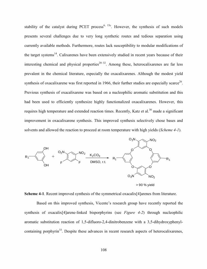
108
stability of the catalyst during PCET process6, 15c
. However, the synthesis of such models
presents several challenges due to very long synthetic routes and tedious separation using
currently available methods. Furthermore, routes lack susceptibility to modular modifications of
the target systems14
. Calixarenes have been extensively studied in recent years because of their
interesting chemical and physical properties20–32
. Among these, heterocalixarenes are far less
prevalent in the chemical literature, especially the oxacalixarenes. Although the modest yield
synthesis of oxacalixarene was first reported in 1966, their further studies are especially scarce26
.
Previous synthesis of oxacalixarene was based on a nucleophilic aromatic substitution and this
had been used to efficiently synthesize highly functionalized oxacalixarenes. However, this
requires high temperature and extended reaction times. Recently, Katz et al.30
made a significant
improvement in oxacalixarene synthesis. This improved synthesis selectively chose bases and
solvents and allowed the reaction to proceed at room temperature with high yields (Scheme 4-1).
O
O O
NO2O2N
O2N NO2
OOH
OHF
O2N NO2
F
R1 R1 R1
K2CO3
DMSO, r.t.
> 90 % yield
Scheme 4-1. Recent improved synthesis of the symmetrical oxacalix[4]arenes from literature.
Based on this improved synthesis, Vicente’s research group have recently reported the
synthesis of oxacalix[4]arene-linked bisporphyrins (see Figure 4-2) through nucleophilic
aromatic substitution reaction of 1,5-difluoro-2,4-dinitrobenzene with a 3,5-dihydroxyphenyl-
containing porphyrin33
. Despite these advances in recent research aspects of heterocalixarenes,
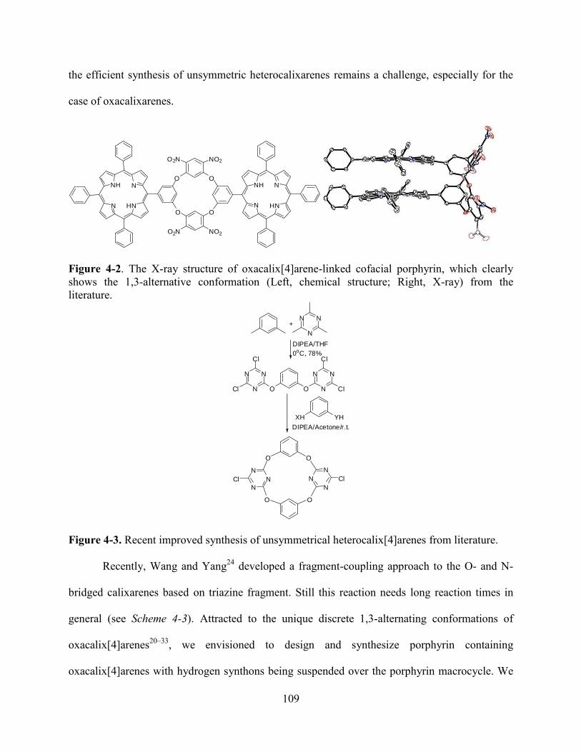
109
the efficient synthesis of unsymmetric heterocalixarenes remains a challenge, especially for the
case of oxacalixarenes.
N
NH N
HN
O
O2N NO2
O
NO2O2N
OO
N
HNN
NH
Figure 4-2. The X-ray structure of oxacalix[4]arene-linked cofacial porphyrin, which clearly
shows the 1,3-alternative conformation (Left, chemical structure; Right, X-ray) from the
literature.
N
N
N
N
N
N N
N
N
O O
Cl
Cl Cl
Cl
N
N
N
N
N
N
OO
O O
ClCl
+
DIPEA/THF
0oC, 78%
XH YH
DIPEA/Acetone/r.t.
Figure 4-3. Recent improved synthesis of unsymmetrical heterocalix[4]arenes from literature.
Recently, Wang and Yang24
developed a fragment-coupling approach to the O- and N-
bridged calixarenes based on triazine fragment. Still this reaction needs long reaction times in
general (see Scheme 4-3). Attracted to the unique discrete 1,3-alternating conformations of
oxacalix[4]arenes20–33
, we envisioned to design and synthesize porphyrin containing
oxacalix[4]arenes with hydrogen synthons being suspended over the porphyrin macrocycle. We
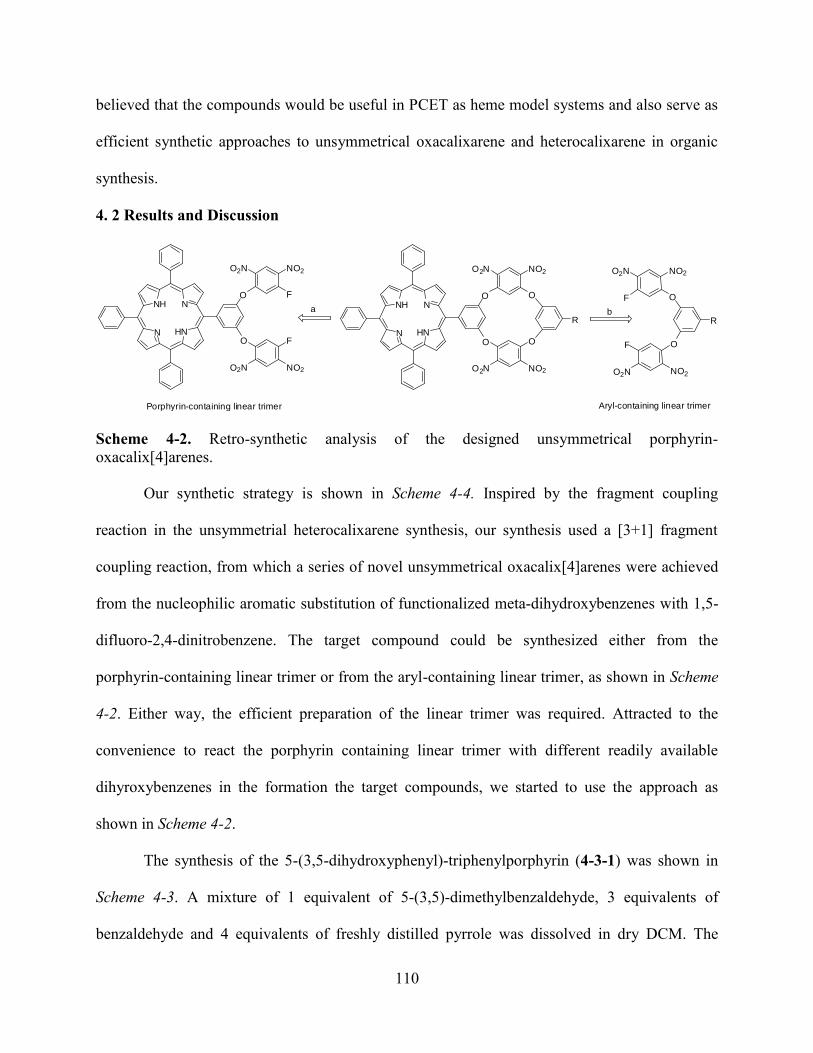
110
believed that the compounds would be useful in PCET as heme model systems and also serve as
efficient synthetic approaches to unsymmetrical oxacalixarene and heterocalixarene in organic
synthesis.
4. 2 Results and Discussion
N
NH N
HN
O
O2N NO2
NO2O2N
O
F
FN
NH N
HN
O
O2N NO2
NO2O2N
O
O
O
R R
O
NO2O2N
O2N NO2
O
F
F
a b
Porphyrin-containing linear trimer Aryl-containing linear trimer
Scheme 4-2. Retro-synthetic analysis of the designed unsymmetrical porphyrin-
oxacalix[4]arenes.
Our synthetic strategy is shown in Scheme 4-4. Inspired by the fragment coupling
reaction in the unsymmetrial heterocalixarene synthesis, our synthesis used a [3+1] fragment
coupling reaction, from which a series of novel unsymmetrical oxacalix[4]arenes were achieved
from the nucleophilic aromatic substitution of functionalized meta-dihydroxybenzenes with 1,5-
difluoro-2,4-dinitrobenzene. The target compound could be synthesized either from the
porphyrin-containing linear trimer or from the aryl-containing linear trimer, as shown in Scheme
4-2. Either way, the efficient preparation of the linear trimer was required. Attracted to the
convenience to react the porphyrin containing linear trimer with different readily available
dihyroxybenzenes in the formation the target compounds, we started to use the approach as
shown in Scheme 4-2.
The synthesis of the 5-(3,5-dihydroxyphenyl)-triphenylporphyrin (4-3-1) was shown in
Scheme 4-3. A mixture of 1 equivalent of 5-(3,5)-dimethylbenzaldehyde, 3 equivalents of
benzaldehyde and 4 equivalents of freshly distilled pyrrole was dissolved in dry DCM. The

111
solution was stirred for 10 minutes under argon. Subsequently, a catalytic amount of a DCM
solution of 2.5M BF3.OEt was added to the reaction mixture. The reaction mixture was stirred
under argon with avoidance of light. TLC was used to follow the reaction. After adding DDQ to
the reaction mixture, stirring was continued for an additional 45 minutes. Then the volume of
solvent was reduced and the residue was applied to a silica gel column using hexane and DCM
as eluting solvents. After removal of solvent under vacuum, 4-3-2 was obtained as purple powder
in 8.5% yield.
N
NH N
HN
HO OH
N
NH N
HN
MeO OMe
MeO
MeO
CHO
NH
BenaldehydePropionic acidRefluxing, 2h
pyrridium hydrochloride
200--220oC 77%
4-3-14-3-2
Scheme 4-3. Synthesis of 3, 5-dihydroxy-tetraphenylporphyrin 4-3-1.
The hydrolysis of 4-3-2 to obtain 5-(3,5-dimethoxyphenyl)-10,15,20-triphenylporphyrin
(4-3-1) was performed at 220 °C using an oil bath. A mixture of 1 equivalent of 4-3-2 and 140
equivalents of pyridine hydrochloride was added to a round-bottom flask and heated to 220 °C in
an oil bath. Upon the melting of the pyridium hydrochloride at 170 °C, the solution turned green
in color. TLC was used to follow the reaction and the reaction was stopped when most of the
starting materials had been consumed. After cooling to room temperature, the reaction mixture
was poured into cold water and extracted with ethyl acetate. The combined organic layers were
washed with aqueous 0.1 M HCl and saturated aqueous NaHCO3. After drying over anhydrous
Na2SO4, the solvent was removed under vacuum. The residue was separated using a silica gel
column eluted with chloroform/ethyl acetate (1:1). The major fraction was collected and was

112
further purified by recrystallization from DCM/hexane. After filtration and removal of the
solvent under vacuum, the target porphyrin 4-3-1 was obtained as a purple powder in 84% yield.
N
NH N
HN
O
O2N NO2
NO2O2N
O
F
FN
NH N
HN
OH
OH
4-3-1 4-4-1
O2N NO2
FF
K2CO3, Acetoner.t.
Scheme 4-4. Synthesis of porphyrin containing linear trimer 4-4-1.
N
NH N
HN
O
O2N NO2
NO2O2N
O
F
F HO
HO
R
4-4-1
R = CHO 4-a
R = COOEt 4-b
R = COOH 4-d
R = OH 4-e
N
NH N
HN
O
O2N NO2
NO2O2N
O
O
O
R
R = CHO 4-5-a
R =COOEt 4-5-b
N
NH N
HN
O
O2N NO2
NO2O2N
O
O
O
N
HNN
NH+
4-5-1
+
K2CO3DMSOr. t.
R =COOH 4-5-d
R = OH 4-5-e
Scheme 4-5. The synthesis of unsymmetrical porphyrin-oxacalix[4]arenes from the porphyrin-
containing linear trimer 4-4-1.
The synthesis of the porphyrin containing the linear trimer 4-4-1 is shown in Scheme 4-6.
A mixture of 1 equivalent of 4-3-1, 4 equivalents of 1,5-difluoro-2,4-dinitrobenzene and eight
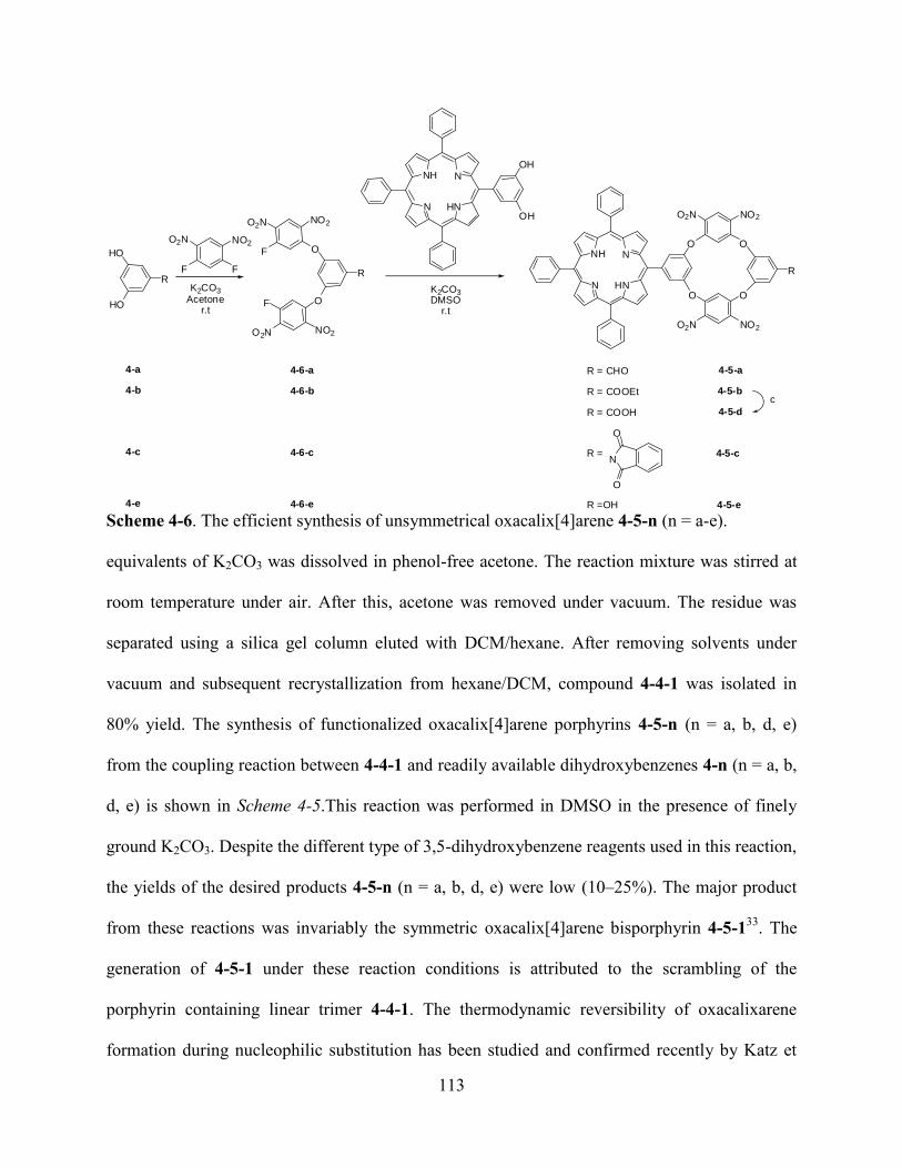
113
R
O
NO2O2N
O2N NO2
O
F
F
R
HO
HO
N
NH N
HN
O
O2N NO2
NO2O2N
O
O
O
R
R = CHO 4-5-a
R = COOEt 4-5-b
R = COOH 4-5-d
R = 4-5-c
R =OH 4-5-e
N
O
O
4-a
4-b
4-c
4-e
4-6-a
4-6-b
4-6-c
4-6-e
c
N
NH N
HN
OH
OH
O2N NO2
FF
K2CO3
Acetoner.t
K2CO3DMSO
r.t
Scheme 4-6. The efficient synthesis of unsymmetrical oxacalix[4]arene 4-5-n (n = a-e).
equivalents of K2CO3 was dissolved in phenol-free acetone. The reaction mixture was stirred at
room temperature under air. After this, acetone was removed under vacuum. The residue was
separated using a silica gel column eluted with DCM/hexane. After removing solvents under
vacuum and subsequent recrystallization from hexane/DCM, compound 4-4-1 was isolated in
80% yield. The synthesis of functionalized oxacalix[4]arene porphyrins 4-5-n (n = a, b, d, e)
from the coupling reaction between 4-4-1 and readily available dihydroxybenzenes 4-n (n = a, b,
d, e) is shown in Scheme 4-5.This reaction was performed in DMSO in the presence of finely
ground K2CO3. Despite the different type of 3,5-dihydroxybenzene reagents used in this reaction,
the yields of the desired products 4-5-n (n = a, b, d, e) were low (10–25%). The major product
from these reactions was invariably the symmetric oxacalix[4]arene bisporphyrin 4-5-133
. The
generation of 4-5-1 under these reaction conditions is attributed to the scrambling of the
porphyrin containing linear trimer 4-4-1. The thermodynamic reversibility of oxacalixarene
formation during nucleophilic substitution has been studied and confirmed recently by Katz et
114
al.30
, which is also in agreement with studies reported for thiocalixarenes34
and with the
invariable formation of symmetrical 4-5-1 and the absence of 4-3-1 in our case. To solve the
scrambling problem of the porphyrin containing linear trimer, the aryl-containing linear trimer
was used for the [3+1] fragment coupling reaction (see Scheme 4-6).
A mixture of 1 equivalent of 4-n (n = a–c, e), 3 equivalents of 1,5-difluoro-2,4-
dinitrobenzene and 4 equivalents of finely ground K2CO3 (<80 mm) was dissolved in acetone at
room temperature. The reaction mixture was stirred for around 1–2 hours at room temperature
under air. TLC was used to follow the reaction. When the reaction was complete, acetone was
removed under vacuum. The residue was separated using a silica gel column eluted with DCM.
After removal of the solvent and washing with hexane (2×10 mL), linear trimer 4-6-n (n = a-c, e)
was obtained as a white solid in 75–85% yields on multi-gram scale. By simply choosing acetone
as solvent and 3 equivalents (rather than two) of 1,5-difluoro-2,4-dinitrobenzene, the formation
of symmetrical oxacalix[4]arene 4-5-1 as a scrambling byproduct was reduced to a minimal
amount. The linear trimers 4-6-n (n = a–c, e) were then used to generate the target functionalized
oxacalix[4]arene porphyrins 4-5-n (n = a-e) in 80–86% yields (see Scheme 4-6). A mixture of 1
equivalent 4-3-1, 1.2 equivalents of 4-6-n (n = a-c, e) and 4 equivalents of finely ground K2CO3
was dissolved in DMSO. The reaction mixture was stirred at room temperature for a period of 30
minutes or up to 3 hours. TLC was used to follow this reaction, and the reaction was stopped
upon complete disappearance of the starting materials. These fragment-coupling reactions are
very efficient and no higher analogs were detected, compared with other fragment-coupling
reactions reported in the literature20–25
. Thus the yields were higher and the separations were
easier. Using DMSO as the solvent in place of acetone in the ring-closure fragment coupling

115
reaction improved the yields of the reaction. However, the use of DMSO instead of acetone
resulted in lower yields of linear trimer formation.
CO2Et
O NO2
NO2O2N
O2N O
FF
Figure 4-4. Chemical structure (left) and X-ray structure (right) of 4-6-b.
The generation of 4-5-d was achieved by hydrolysis of the ester functionality in 4-5-b.
The hydrolysis under basic conditions by using NaOH aqueous solution failed to generate the
desired product; instead the starting material decomposed under these conditions, in agreement
with the scrambling that occurred in the generation of 4-5-1. The hydrolysis under dilute acidic
condition also failed, leaving the starting material intact. The final successful hydrolysis of 4-5-b
was accomplished by refluxing it in a mixture of THF/4 M aqueous HCl (v/v = 1/2) for a period
of 3 days. After separation, 4-5-d was obtained as a purple powder in 95% yield. The
functionalized oxacalix[4]arenoporphyrins 4-5-n (n = a–e, 1) were characterized by HRMS, UV–
vis, fluorescence, and 1H NMR spectroscopy. The structure of trimer 4-6-b was also confirmed
by X-ray crystallography at T=150K (see Figure 4-4). The crystals of 4-6-b were destroyed by
cooling to temperatures lower than 150K, apparently as a result of a phase change. The C–F
distances are 1.331(2) and 1.334(2) A°.
116
The linear trimers 4-6-n (n = a–c, e) shared characteristic 1H NMR spectra, with two
downfield singlets (around 8.9 ppm) for the protons adjacent to the carbons bearing the NO2
groups, and two upfield singlets (around 6.9 ppm) for the protons adjacent to the carbons bearing
the F groups. Based on the characteristic upfield chemical shifts observed in the 1H NMR spectra
of oxacalix[4]arenes for the interior protons on the electrophilic (NO2-bearing) aromatic rings28
,
it is believed that these compounds adopt 1,3-alternating structures in solution20–33
. Recent X-ray
structures26–33
have confirmed this conformation of oxacalix[4]arenes in the solid state.
Table 4-1. 1H NMR shifts for the interior protons on the NO2-bearing benzene rings of
porphyrins 4-5-n (n = a–e, 1).
Porphyrins 4-5-a 4-5-b 4-5-d 4-9-1 4-5-e 4-5-c
(ppm) 6.77 6.43 6.70 6.87 6.70 6.49
Table 4-1 shows the 1H NMR chemical shifts observed for these protons on porphyrins 4-
5-n (n = a–e) and 4-9-1 (see Scheme 4-9). These results suggest that our functionalized
oxacalix[4]arene porphyrins also adopt 1,3-alternating conformations in solution, in agreement
with results reported for symmetrical oxacalix[ 4]arenes26–33
. Furthermore, computer calculations
using the AM1 theoretical model incorporated into the Spartan program were performed to
determine the minimum energy conformations for oxacalix[4]arene porphyrins 4-5-n (n = a-e),
4-9-1. Such calculations have been found reliable for the determination of geometrical
parameters in porphyrin arrays37–40
. Similar optimized geometries were found for all
oxacalix[4]arene porphyrins (see Figure 4-5) and the calculated structure obtained for
bisporphyrin 4-5-1 was in agreement with the crystal data33
. These results suggest, as seen in
Figure 4-5, favorable 1,3-alternating conformations for all oxacalix[4]arene porphyrins, with the
hydrogen-bond synthons hanging over an adjacent pyrrole ring. Such a conformation would

117
Figure 4-5. AM1 calculated conformation of 4-5-d.
N
NH N
HN
OH
OH
HO
HON
NH N
HN
OMe
OMe
MeO
MeOMeO OMe
CHO
CHO NH
R R
R R
R R R R
RR
+
BBr3
R = H 4-7-3R = t-butyl 4-7-4
R = H 4-7-1R = t-butyl 4-7-2
Scheme 4-7. Synthesis of tetrahydroxyporphyrins 4-7-1 and 4-7-2.
provide a face-to-face structural arrangement for the porphyrin macrocycles and hydrogen-bond
synthons, therefore making these compounds suitable as model systems for PCET and hydrogen-
bond investigations. The preparation of 4-7-4 was achieved from a mixed aldehyde condensation
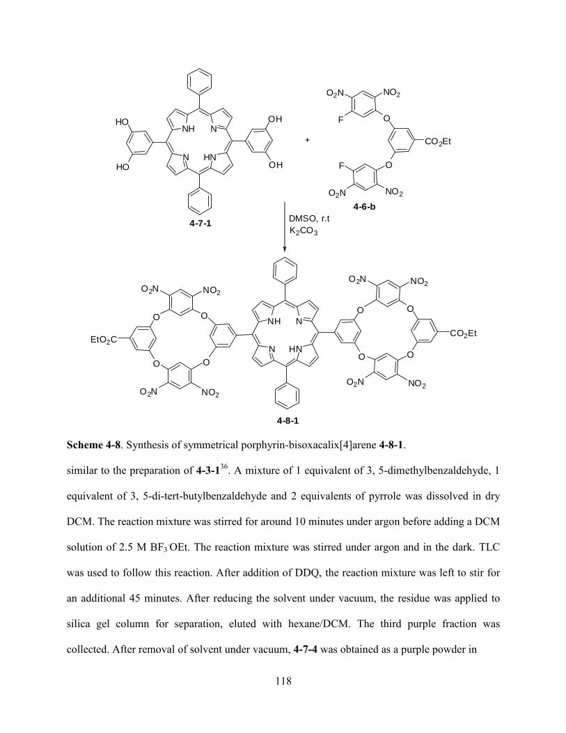
118
CO2Et
O
NO2O2N
O2N NO2
O
F
F
N
NH N
HN
OH
OH
N
NH N
HN
O
O
CO2Et
O
NO2O2N
O2N NO2
O
DMSO, r.t
K2CO3
HO
HO
O
OO
O2N NO2
NO2O2N
O
EtO2C
+
4-8-1
4-7-1
4-6-b
Scheme 4-8. Synthesis of symmetrical porphyrin-bisoxacalix[4]arene 4-8-1.
similar to the preparation of 4-3-136
. A mixture of 1 equivalent of 3, 5-dimethylbenzaldehyde, 1
equivalent of 3, 5-di-tert-butylbenzaldehyde and 2 equivalents of pyrrole was dissolved in dry
DCM. The reaction mixture was stirred for around 10 minutes under argon before adding a DCM
solution of 2.5 M BF3.OEt. The reaction mixture was stirred under argon and in the dark. TLC
was used to follow this reaction. After addition of DDQ, the reaction mixture was left to stir for
an additional 45 minutes. After reducing the solvent under vacuum, the residue was applied to
silica gel column for separation, eluted with hexane/DCM. The third purple fraction was
collected. After removal of solvent under vacuum, 4-7-4 was obtained as a purple powder in
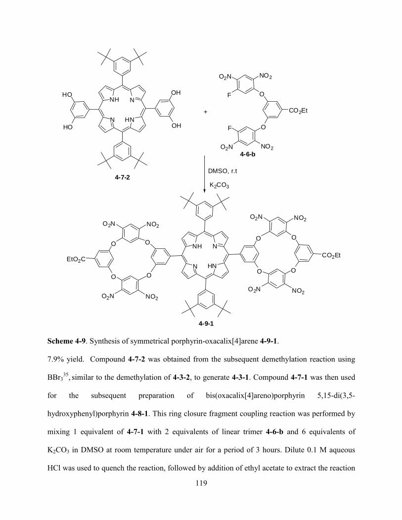
119
CO2Et
O
NO2O2N
O2N NO2
O
F
F
N
NH N
HN
OH
OH
N
NH N
HN
O
O
CO2Et
O
NO2O2N
O2N NO2
O
DMSO, r.t
K2CO3
HO
HO
O
OO
O2N NO2
NO2O2N
O
EtO2C
+
4-7-2
4-6-b
4-9-1
Scheme 4-9. Synthesis of symmetrical porphyrin-oxacalix[4]arene 4-9-1.
7.9% yield. Compound 4-7-2 was obtained from the subsequent demethylation reaction using
BBr335
, similar to the demethylation of 4-3-2, to generate 4-3-1. Compound 4-7-1 was then used
for the subsequent preparation of bis(oxacalix[4]areno)porphyrin 5,15-di(3,5-
hydroxyphenyl)porphyrin 4-8-1. This ring closure fragment coupling reaction was performed by
mixing 1 equivalent of 4-7-1 with 2 equivalents of linear trimer 4-6-b and 6 equivalents of
K2CO3 in DMSO at room temperature under air for a period of 3 hours. Dilute 0.1 M aqueous
HCl was used to quench the reaction, followed by addition of ethyl acetate to extract the reaction
120
mixture from the water. The resulting organic phase was dried over anhydrous Na2SO4 and
purified by alumina column chromatography using DCM/ethyl acetate (v/v = 100/1) as eluting
solvents. After removal of the solvent under vacuum, the major product was obtained in 54%
yield. Although MALDI-TOF mass spectroscopy gave a peak corresponding to the desired
compounds 4-8-1, and TLC always gave one spot for the desired compound, efforts to obtain a
decent NMR spectrum for 4-8-1 failed. We attribute this to the poor solubility of this compound,
which easily formed aggregates in most organic solvents. Meanwhile, we thought that it might
also be attributed to rapid conformational changes of this compound in solution from which
broad NMR signals were observed.
To solve this problem, we introduced bulky groups into the meso-position of porphyrins,
to increase the solubility and help reduce aggregation. The generation of 4-9-1 was from a
similar fragment coupling reaction as in the preparation of 4-8-1, with the substitution of
porphyrin 4-7-1 to 4-7-2. The desired 4-9-1 was obtained as a purple powder in 84% yield. The
presence of the four tert-butyl groups at the meso-position of porphyrin 4-7-2 induced high
solubility of this compound in organic solvents, and its structure, was confirmed by HRMS (a
molecular ion peak was observed at 1923.5460), UV–vis, fluorescence, and 1H NMR
spectroscopy. Based on the characteristic upfield chemical shifts observed in the 1H NMR
spectra of oxacalix[4]arenes for the interior protons on the electrophilic (NO2-bearing) aromatic
rings28
, it is believed that these compounds adopt 1,3-alternating structures in solution20–33
. The
photophysical properties of porphyrins 4-5-n (n= a-e) and 4-9-1 are summarized in Table 4-2.
The long wavelength absorption and fluorescence emission bands for all porphyrins except 4-5-d
were observed between 646–652 and 652–658 nm. The carboxyl group in porphyrin 4-5-d is
probably involved in intermolecular hydrogen-bonding, resulting in its distinct absorbance and
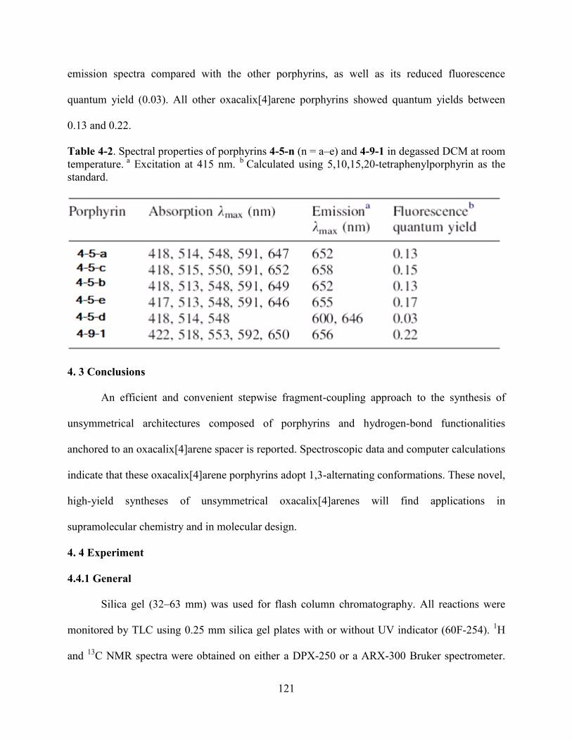
121
emission spectra compared with the other porphyrins, as well as its reduced fluorescence
quantum yield (0.03). All other oxacalix[4]arene porphyrins showed quantum yields between
0.13 and 0.22.
Table 4-2. Spectral properties of porphyrins 4-5-n (n = a–e) and 4-9-1 in degassed DCM at room
temperature. a
Excitation at 415 nm. b
Calculated using 5,10,15,20-tetraphenylporphyrin as the
standard.
4. 3 Conclusions
An efficient and convenient stepwise fragment-coupling approach to the synthesis of
unsymmetrical architectures composed of porphyrins and hydrogen-bond functionalities
anchored to an oxacalix[4]arene spacer is reported. Spectroscopic data and computer calculations
indicate that these oxacalix[4]arene porphyrins adopt 1,3-alternating conformations. These novel,
high-yield syntheses of unsymmetrical oxacalix[4]arenes will find applications in
supramolecular chemistry and in molecular design.
4. 4 Experiment
4.4.1 General
Silica gel (32–63 mm) was used for flash column chromatography. All reactions were
monitored by TLC using 0.25 mm silica gel plates with or without UV indicator (60F-254). 1H
and 13
C NMR spectra were obtained on either a DPX-250 or a ARX-300 Bruker spectrometer.
122
Chemical shifts (δ) are given in ppm relative to CDCl3 (7.27), acetone-d6 (2.05), DMSO-d6 (2.50)
or THF-d8 (1.73) as indicated. Electronic absorption spectra were measured on a Perkin Elmer
Lambda 35 UV–vis spectrophotometer in the 300–800 nm wavelength region with 0.1 nm
accuracy. Fluorescence spectra were measured on a Perkin Elmer LS55 spectrometer in the 500–
800 nm wavelength region with 1 nm accuracy. The fluorescence quantum yields were measured
using the standard method and 5,10,15,20-tetraphenylporphyrin as the standard (quantum yield is
0.11), according to the literature41
. Mass spectra were obtained on Applied Biosystems QSTAR
XL spectrometer. High-resolution mass spectra were obtained on a Q-TOF2 eletrospray at the
mass spectrometry facility of Ohio State University. All solvents were obtained from Fisher
Scientific (HPLC grade, Houston, TX) and used without further purification unless indicated.
Acetone (reagent plus, phenol free, > 99.5%) and DMSO (Biotech grade solvent, 99.8%) were
purchased from Sigma–Aldrich and used without further purification. K2CO3 was ground and
dried at 140 oC. Compounds 4-d
42 and 4-3-1
35 were synthesized according to literature
procedures. Solvents were dried according to literature procedures43
. The computational
simulations used the AM1 semiempirical Hamiltonian method44
incorporated into the quantum
mechanical Spartan program45
. The coordinates used to build the porphyrins in this study were
based on the X-ray crystal structure of 4-5-133
.
4.4.2 Syntheses
4.4.2.1 Aryl-containing Linear Trimer 4-6-a.
3,5-Dihydroxybenzaldehyde (4-a) (276.7 mg, 2.0 mmol) was mixed with 1, 5-difluoro-2, 4-
dinitrobenzene (816.5 mg, 4.0 mmol) and K2CO3 (561.4 mg, 4 mmol) in 10 mL of acetone at
room temperature under air. When the reaction was complete, acetone was removed under
vacuum. The resulting residue was purified by silica gel column chromatography using

123
DCM/ethyl acetate (v/v = 20/1) for elution. After removal of the solvent under vacuum and
washing with hexane (2×10 mL), pure linear trimer 4-6-a was obtained as a white solid in 85%
yield (860.0 mg). 1H NMR (300 MHz, CDCl3) δ 10.03 (s, 1H), 8.98 (s, 1H), 8.95 (s, 1H), 7.58 (d,
2H, J = 2.25 Hz), 7.30 (s, 1H), 7.06 (s, 1H), 7.02 (s, 1H). ESI-MS Calcd. for C19H8F2N4O11 m/z
506.3, found: 506.5.
4.4.2.2 Aryl Containing Linear Trimer 4-6-b.
Ethyl 3,5-dihydroxybenzoate (4-b) (182.0 mg, 1 mmol) was mixed with 1,5-difluoro-2,4-
dinitrobenzene (408.0 mg, 2 mmol) and K2CO3 (560.0 mg, 4 mmol) in 10 mL of acetone at room
temperature under air. When the reaction was complete, acetone was removed under vacuum.
The resulting residue was purified by silica gel column chromatography using DCM for elution.
After removal of the solvent and washing with hexane (2×10 mL), linear trimer 4-6-b was
obtained as a white solid in 82% yield (451.0 mg). 1H NMR (300 MHz, CDCl3) δ 8.94 (s, 1H),
8.91 (s, 1H), 7.74 (d, 2H, J = 2.34 Hz), 7.19 (s, 1H), 6.96 (s, 1H), 6.92 (s, 1H), 4.39 (q, 2H), 1.38
(t, 3H). Anal. Calcd for C21H12F2N4O12: C, 45.83; H, 2.20; N, 10.18. Found: C, 45.79; H, 2.26; N,
9.98. ESI-MS Calcd for C21H12F2N4O12 m/z 550.3, found: 549.8.
4.4.2.3 Aryl Containing Linear Trimer 4-6-c.
Compound 4-c (510.4 mg, 2 mmol) was mixed with 1,5-difluoro-2,4-dinitrobenzene (1.63 g, 8.0
mmol) and K2CO3 (2.21 g, 16 mmol) in 20 mL of acetone at room temperature under air. When
the reaction was complete, acetone was removed under vacuum. The residue was purified by
silica gel column chromatography using DCM to the mixture solvents of DCM/ethyl acetate (v/v
= 20/1) for elution. After removal of the solvent under vacuum and washing with hexane (2×10
mL), linear trimer 4-6-c was obtained as a white solid in 75% yield (935.0 mg). 1H NMR (300

124
MHz, acetone-d6) δ 8.93 (d, 2H, J = 7.67 Hz), 7.93 (m, 4H), 7.69 (s, 1H), 7.65 (s, 1H), 7.60 (d,
2H, J = 2.21 Hz), 7.48 (s, 1H). ESI-MS calcd for C26H11F2N5O12 m/z 623.4, found: 623.8.
4.4.2.4 Porphyrin Conatining Linear Trimer 4-4-1.
5-(3,5-Dihydroxyphenyl)triphenylporphyrin (4-3-1) (32.8 mg, 0.05 mmol) was mixed with 1,5-
difluoro-2, 4-dinitrobenzene (81.6 mg, 0.2 mmol) and K2CO3 (56.0 mg, 0.4 mmol) in 20 mL of
acetone at room temperature under air. After the reaction was complete, acetone was removed
under vacuum. The resulting residue was purified by silica gel column chromatography using the
mixture solvents of DCM/hexane (v/v = 10/1) for elution. Pure linear trimer 4-4-1 was isolated in
80% yield (40.6 mg) after recrystallization from hexane and DCM. 1H NMR (250 MHz, CDCl3)
δ 8.84 (m, 6H), 8.68 (d, 2H, J¼2.37 Hz), 8.19 (m, 8H), 7.73 (m, 11H), 7.05 (m, 1H), 6.80 (m,
2H), -3.03 (s, 2H). HRMS (MALDI-TOF) Calcd for [M+H]+
C56H33F2N8O10 m/z 1015.2288,
found: 1015.2265. UV–vis (DCM) λmax (log ε) 417 (5.84), 513 (4.47), 548 (4.03), 590 (3.85),
646 (3.58) nm.
4.4.2.5 Porphyrin 4-5-a.
Linear trimer 4-6-a (50.1 mg, 0.1 mmol) was mixed with 4-3-1 (64.1 mg, 0.1 mmol) and K2CO3
(60.3 mg, 0.44 mmol) in 20 mL of DMSO at room temperature under air for 1 h (until TLC
showed the complete disappearance of starting material). HCl (0.1 M, 40 mL) was used to
quench the reaction. The water layer was extracted with 100 mL of ethyl acetate, and the organic
layer was washed once with water and dried over anhydrous Na2SO4. The residue was purified
by silica gel column chromatography using DCM for elution. Pure porphyrin 4-5-a was obtained
as a purple solid in 83% yield (92.1 mg) after recrystallization from DCM /hexane. 1H NMR
(250 MHz, THF-d8) δ 10.07 (s, 1H), 8.98 (s, 2H), 8.84 (m, 6H), 8.66 (s, 2H), 8.20–8.21 (m, 6H),
8.13 (d, 2H, J = 2.10 Hz), 8.00 (d, 2H, J = 2.16 Hz), 7.81–7.82 (m, 9H), 7.64 (s, 1H), 7.52 (s, 1H),
125
6.77 (s, 2H), -2.76 (s, 2H). MALDI-TOF Calcd for [M+H]+
C63H37N8O13 m/z 1114.0, found:
1114.0. HRMS (ESI) Calcd for [M+H]+ C63H37N8O13 m/z 1113.2480, found:1113.2501. UV–vis
(DCM) λmax (log ε) 418 (5.88), 514 (4.50), 548 (4.11), 591 (3.93), 647 (3.72) nm.
4.4.2.6 Porphyrin 4-5-b.
Linear trimer 4-6-b (55.0 mg, 0.1 mmol) was mixed with 4-3-1 (64.6 mg, 0.1 mmol) and K2CO3
(56.0 mg, 0.44 mmol) in 20 mL of DMSO at room temperature under air (the reaction was
considered complete after TLC showed the complete disappearance of the starting material). HCl
(0.1 M, 40 mL) was used to quench the reaction. The water layer was extracted with 100 mL of
ethyl acetate, and the organic layer was washed once with water and dried over anhydrous
Na2SO4. The resulting residue was purified by silica gel column chromatography using DCM for
elution. Pure porphyrin 4-5-b was obtained as a purple solid in 86% yield (100.1 mg) after
recrystallization from DCM / hexane. 1H NMR (250 MHz, CDCl3) d 8.93 (s, 2H), 8.82 (m, 6H),
8.41 (s, 4H), 8.16 (m, 6H), 8.04 (d, 2H, J = 2.22 Hz), 7.99 (d, 2H, J = 2.22 Hz), 7.71 (m, 9H),
6.43 (s, 2H), 4.01 (q, 2H), 1.11 (m, 3H), -2.88 (s, 2H). MALDI-TOF Calcd for C65H40N8O14 m/z
1157.058, found: 1159.198. HRMS (MALDI-TOF) Calcd for [M+H]+ C65H41N8O14 m/z
1157.2742, found: 1157.2787. HRMS (ESI) Calcd for [M+H]+ C65H41N8O14 m/z 1157.2742,
found: 1157.2799. UV–vis (DCM) λmax (log ε) 418 (5.89), 513 (4.51), 548 (4.09), 591 (3.88),
649 (3.76) nm.
4.4.2.7 Porphyrin 4-5-c.
Linear trimer 4-6-c (63.3 mg, 0.1 mmol) was mixed with 4-3-1 (65.7 mg, 0.1 mmol) and K2CO3
(60.0 mg, 0.43 mmol) in 10 mL of DMSO at room temperature under air for 3 h (until TLC
showed the complete disappearance of starting material). HCl (0.1 M, 40 mL) was used to
quench the reaction. The water layer was extracted with 100 mL of ethyl acetate, and the organic

126
layer was washed once with water and dried over anhydrous Na2SO4. The residue was purified
by silica gel column chromatography using DCM for elution. Pure porphyrin 4-5-c was obtained
as a purple solid in 83% yield (102.0 mg) after recrystallization from DCM/hexane. 1H NMR
(300 MHz, THF-d8) δ 8.98 (s, 2H), 8.90 (br s, 2H), 8.82 (br s, 4H), 8.20 (br s, 4H), 8.17 (s, 3H),
8.14 (s, 3H), 7.79–7.81 (m, 9H), 7.63 (s, 2H), 7.56 (s, 1H), 7.35 (s, 1H), 6.95 (d, 4H, J = 2.55
Hz), 6.70 (s, 2H), -2.84 (s, 2H). MALDI-TOF Calcd for [M+H]+ C70H40N9O14 m/z 1230.3, found:
1230.8. HRMS (ESI) Calcd for [M+H]+ C70H40N9O14 m/z 1230.2694, found: 1230.2700. UV–vis
(DCM) λmax (log ε) 418 (5.57), 515 (4.20), 550 (3.79), 591 (3.49), 652 (3.43) nm.
4.4.2.8 Bisporphyrin 4-5-1.
This compound was synthesized and characterized as previously reported33
.
4.4.2.9 Porphyrin 4-5-e.
Linear trimer 4-6-e (20.3 mg, 0.02 mmol) was mixed with 4-3-1 (2.5 mg, 0.02 mmol) and
K2CO3 (11.0 mg, 0.08 mmol) in 5 mL of DMSO at room temperature under air for 40 min. HCl
(0.1 M, 20 mL) was used to quench the reaction. The water layer was extracted with 50 mL of
ethyl acetate, and the organic layer was washed once with water and dried over anhydrous
Na2SO4. The residue was purified by silica gel column chromatography using THF/hexane for
elution. Porphyrin 4-5-e was obtained in 20% yield (4.5 mg). 1H NMR (300 MHz, THF-d8)
9.95 (br s,1H), 9.06 (d, 2H, J = 3.90 Hz), 8.93 (s, 2H), 8.84 (s, 4H), 8.71 (d, 2H, J = 2.0 Hz),
8.20–8.28 (m, 6H), 8.13 (d, 2H, J = 4.58 Hz), 7.79–7.81 (m, 9H), 7.61 (s, 1H), 6.86 (s, 2H), 6.75
(s, 1H), 6.70 (d, 2H, J = 6.93 Hz), -2.75 (s, 2H). MALDI-TOF Calcd for [M+H]+ C62H36N8O13
m/z 1102.0, found: 1103.3. HRMS (ESI) Calcd for [M+H]+ C62H37N8O13 m/z 1101.2480, found:
1101.2545. UV–vis (DCM) λmax (logε) 417 (5.75), 513 (4.37), 548 (3.92), 591 (3.71), 646 (3.43)
nm.
127
4.4.2.10 Porphyrin 4-5-d.
Hydrolysis of 4-5-d was obtained by dissolving 4-5-b (24.6 mg, 0.02 mmol) in 10 mL of THF,
followed by addition of 4 M HCl (20 mL). The reaction mixture was refluxed at 60 oC in an oil
bath for 3 days. After completion of the reaction, the mixture was extracted with ethyl acetate
and the organic layer washed with brine. The resulting residue was purified by silica gel column
chromatography using DCM/ethyl acetate (v/v = 20/1) for elution. Pure porphyrin 4-5-d was
obtained in 95% yield (21.5 mg) after recrystallization from DCM/hexane. 1H NMR (300 MHz,
THF-d8) δ 8.97 (s, 1H), 8.93 (d, 2H, J = 4.40 Hz), 8.83 (s, 6H), 8.77 (s, 2H), 8.21 (m, 6H), 8.14
(d, 2H, J = 1.98 Hz), 8.03 (d, 2H, J = 2.06 Hz), 7.79 (d, 9H), 7.53 (s, 1H), 7.49 (s, 1H), 6.87 (s,
2H), -2.74 (s, 2H). MALDI-TOF Calcd for [M+H]+
C63H37N8O14 m/z 1129.2, found: 1130.2.
HRMS (ESI) Calcd for [M+H]+C63H37N8O14 m/z 1129.2429, found: 1129.2405. UV–vis (DCM)
λmax (log 3) 418 (5.43), 514 (3.90), 548 (3.32) nm.
4.4.2.11 5-(3, 5-Dihoxylphenyl)-10,15,20-triphenylporphyrin 4-7-1.
5-(3,5-Dimethoxyphenyl)-10,15,20-triphenylporphyrin 4-7-3 (400 mg, 0.6 mmol) and pyridine
hydrochloride (10.0 g, 86 mmol) were added to a 100 mL round-bottom flask and heated to
220 °C in an oil bath. Upon the melting of pyridium hydrochloride at 170 °C, the solution turned
green in color. TLC indicated the formation of the target porphyrin after 2 h reaction. Stirring
was continued for an additional 4 h, whereupon TLC indicated that most of the starting materials
had been consumed. The reaction was stopped and the mixture was cooled to room temperature.
Then the reaction mixture was poured into 400 mL cold water and extracted with 400 mL ethyl
acetate. The aqueous layer was extracted with ethyl acetate until colorless. The combined
organic layers were washed twice with 0.1 M HCl aqueous solution and once with aqueous
saturated NaHCO3. After drying over anhydrous Na2SO4, the solvent was removed under
128
vacuum. The residue was separated on a silica gel column using chloroform/ethyl acetate 1:1 as
eluting solvent. The major fraction was collected and was further purified by recrystallization
from DCM hexane. After filtration and removal of the solvent under vacuum, porphyrin 4-7-1
was obtained in 84 % yield (160 mg). HRMS (MALDI-TOF) m/z 645.2272, calculated for
C44H28N4O2 645.2290. 1H-NMR (DMSO-d6) δ 9.92 (s, 1H), 9.77 (s, 1H), 9.22 (d, 1H, J = 4.3
Hz), 8.54 (d, 1H, J = 4.9 Hz), 8.28 (d, 1H, J = 3.6 Hz), 8.24 (d, 1H, J = 3.6 Hz), 8.16 (d, 1H, J =
4.4 Hz), 8.12 (d, 1H, J = 4.5 Hz), 8.03 (m, 6H), 7.78 (m, 9H), 7.22 (s, 1H), 7.08 (s, 1H), 5.95 (s,
1H), -0.16 (s, 2H). UV-Vis (DCM) λmax (log ε) 422 (4.9), 455 (4.8), 486 (4.7), 583 (3.7), 640
(3.7), 683 (3.6) nm.
4.4.2.12 5,15-Di(3,5-dihydroxyphenyl)-10,20-di(3,5-ditert-butylphenyl)porphyrin 4-7-2.
3,5-Dimethylbenzaldehyde (0.83 g, 5.0 mmol), 3,5-di-tert-butylbenzaldehyde (1.09 g, 5.0
mmol), and pyrrole (0.70 mL, 10 mmol) were mixed in a 2 L flask. Dry DCM (1000 mL) was
added and the solution was stirred for 10 min under argon before 0.4 mL of 2.5 M BF3.OEt in
DCM was added. The reaction mixture was stirred under argon and in the dark for 2 h. DDQ
(1.64 g) was added and the mixture was stirred for 45 min. The reaction mixture was
concentrated to give a residue that was purified by silica gel column chromatography using a
mixture of hexane and DCM for elution. The third eluted purple fraction contained 5,15-di(3,5-
dimethoxyphenyl)-10,20-di(3,5-di-tert-butylphenyl)porphyrin. The solvent was removed under
vacuum to give 192 mg (7.9% yield) of this porphyrin 4-7-4 as a purple powder. MALDI-TOF
Calcd for [M+H]+ C64H71N4O4 m/z 960.25, found: 960.17.
1H NMR (CDCl3) d 9.07 (d, 4H, J =
4.70 Hz), 9.01 (d, 4H, J = 4.70 Hz), 8.20 (d, 4H, J = 1.68 Hz), 7.91–7.90 (m, 2H), 7.53 (d, 4H, J
= 2.24 Hz), 6.97–6.99 (m, 2H), 1.63 (s, 36H), -2.63 (s, 2H). To a solution of 5, 15-di (3,5-
dimethoxyphenyl)-10,20-di(3,5-di-tert-butylphenyl)porphyrin 4-7-4 (0.096 g, 0.1 mmol) in dry

129
DCM (20 mL) at -20 oC was added dropwise a solution of BBr3 (0.3 mL, 3.1 mmol) in DCM (1
mL) with vigorous stirring under argon over a period of 30 min. The reaction mixture was stirred
at room temperature for a period of 24 h and then poured into water and extracted with ethyl
acetate (3×50 mL). The combined organic layers were washed successively with brine and
aqueous NaHCO3 solutions. The organic solution was dried over Na2SO4 and evaporated to
dryness, giving the title porphyrin 4-7-2 as purple crystals in 91.6% yield (82.7 mg, 0. 092
mmol). MALDI-TOF Calcd for [M+H]+ C60H63N4O4 m/z 904.14, found: 904.21.
1H NMR
(CD2Cl2) δ 9.04 (d, 4H, J = 4.80 Hz), 8.90 (d, 4H, J = 4.75 Hz), 8.70 (s, 4H), 8.15 (d, 4H, J =
1.81 Hz), 7.96–7.94 (m, 2H), 7.26 (d, 4H, J = 2.18 Hz), 6.84–6.82 (m, 2H), 1.56 (s, 36H), -2.75
(s, 2H).
4.4.2.13 Porphyrin 4-9-1.
5,15-Di(3,5-dihydroxyphenyl)-10,20-di(3,5-di-tert-butylphenyl)porphyrin 4-7-2 (36.1 mg, 0.04
mmol) was mixed with linear trimer 4-6-b (44.0 mg, 0.08 mmol) and K2CO3 (33.6 mg, 0.24
mmol) in 10 mL of DMSO at room temperature under air for 3 h. Dilute HCl (0.1 M × 40 mL)
was used to quench the reaction and ethyl acetate (2×25 mL) was used to extract the water layer.
The resulting organic phase was dried over anhydrous Na2SO4 and purified by alumina column
chromatography using DCM/ethyl acetate (v/v = 100/1) for elution. Porphyrin 4-9-1 was
obtained in 84% yield (64.7 mg). 1H NMR (250 MHz, CDCl3) δ 8.95 (br s, 8H), 8.46 (br s, 4H),
8.09 (s, 4H), 8.05 (d, J = 1.86 Hz, 4H), 8.02 (d, J = 1.82 Hz, 4H), 7.92 (s, 2H), 7.25 (br s, 4H),
6.49 (s, 4H), 3.98–4.06 (m, 4H), 1.56 (s, 36H), 1.27 (t, 6H). MALDI-TOF Calcd for [M+H]+
C102H83N12O28 m/z 1923.5, found: 1923.1. HRMS (ESI) calcd for [M+H]+ C102H83N12O28 m/z
1923.5440, found: 1923.5460. UV–vis (DCM) λmax (log ε) 422 (5.79), 518 (4.40), 553 (4.08),
592 (3.87), 650 (3.85) nm.

130
4.4.2.13 Molecular Structures.
The crystal structure of linear trimer 4-6-b was determined using data collected at T =
150 K to q =31.5 with Mo Ka radiation on a Nonius KappaCCD diffractometer. The X-ray
crystallographic data for 3c can be found in supplementary publication CCDC-626294 available
from the Cambridge Crystallographic Data Centre.
4.5 References
1 Dismukes, G. C. Science 2001, 292, 447.
2 Tommos, C.; Babcock, G. T. Acc. Chem. Res. 1998, 31, 18.
3 Yocum, C. F.; Pecoraro, V. L. Curr. Opin. Chem. Biol. 1999, 3, 182.
4 (a) Dawson, J. H. Science 1988, 240, 433; (b) Sono, M.; Roach, M. P.; Coulter, E. D.;
Dawson, J. H. Chem. Rev. 1996, 96, 2841.
5 Ozaki, S.-I.; Roach, M. P.; Matsui, T.; Watanbe, Y. Acc. Chem. Res. 2001, 34, 818.
6 Chang, C. J.; Chng, L. L.; Nocera, D. G. J. Am. Chem. Soc. 2003, 125, 1866.
7 Mims, M. P.; Porras, A. G.; Olson, J. S.; Noble, R.W.; Peterson, J. A. J. Biol. Chem. 1983,
258, 14219.
8 Rohlfs, R. J.; Mathaws, A. J.; Carver, T. E.; Olson, J. S.; Springer, B. A.; Egeberg, K. D.;
Sligar, S. G. J. Biol. Chem. 1990, 265, 3168.
9 Momenteau, M.; Reed, C. A. Chem. Rev. 1994, 94, 659.
10 Chang, C. K.; Kondylis, M. P. J. Chem. Soc., Chem. Commun. 1986, 316.
11 Chang, C. J.; Brown, J. D. K.; Chang, M. C. Y.; Baker, E. A.; Nocera, D. G. Electron
Transfer in Chemistry; Balzani, V., Ed.; Wiley-VCH: Weinheim, Germany, 2001; Vol. 3.2.4,
pp 409.
12 (a) Deng, Y.; Roberts, J. A.; Peng, S.-M.; Chang, C. K.; Nocera, D. G. Angew. Chem., Int. Ed.
1997, 36, 2124; (b) Cukier, R. I.; Nocera, D. G. Annu. Rev. Phys. Chem. 1998, 49, 337.
13 Chang, C. K.; Liang, Y.; Aviles, G. J. Am. Chem. Soc. 1995, 117, 4191.
14 Liu, S. Y.; Nocera, D. G. J. Am. Chem. Soc. 2005, 127, 5278.
131
15 (a) Yeh, C. Y.; Chang, C. J.; Nocera, D. G. J. Am. Chem. Soc. 2001, 123, 1513; (b) Chang, C.
J.; Yeh, C. Y.; Nocera, D. G. J. Org. Chem. 2002, 67, 1403; (c) Chng, L. L.; Chang, C. J.;
Nocera, D. G. Org. Lett. 2003, 5, 2421.
16 Zhang, X. X.; Lippard, S. J. J. Org. Chem. 2000, 65, 5298.
17 Liang, Y.; Chang, C. K. Tetrahedron Lett. 1995, 36, 3817.
18 Chang, C. K.; Bag, N.; Guo, B. M.; Peng, S. M. Inorg. Chim. Acta 2003, 351, 261.
19 Arimura, T.; Brown, C. T.; Springs, S. L.; Sessler, J. L. Chem. Commun. 1996, 2293.
20 König, B.; Fonseca, M. H. Eur. J. Inorg. Chem. 2000, 2303.
21 Lhoták, P. Eur. J. Org. Chem. 2004, 1675.
22 Chambers, R. D.; Hoskin, P. R.; Kenwright, A. R.; Khalil, A.; Richmond, P.; Sandford, G.;
Yufit, D. S.; Howard, J. A. K. Org. Biomol. Chem. 2003, 1, 2137.
23 Li, X.; Upton, T. G.; Gibb, C. L. D.; Gibb, B. C. J. Am. Chem. Soc. 2003, 125, 650.
24 (a) Wang, M.-X.; Yang, H.-B. J. Am. Chem. Soc. 2004, 126, 15412; (b) Wang, M.-X.; Zhang,
X.-H.; Zheng, Q.-Y. Angew. Chem., Int. Ed. 2004, 43, 838.
25 Yang, F.; Yan, L.; Ma, K.; Yang, L.; Li, J.; Chen, L.; You, J. Eur. J. Org. Chem. 2006, 1109.
26 Sommer, N.; Staab, H. A. Tetrahedron Lett. 1966, 25, 2837.
27 Lehmann, F. P. A. Tetrahedron 1974, 30, 727.
28 Gilbert, E. E. J. Heterocycl. Chem. 1974, 11, 899.
29 Bottino, F.; Foti, S.; Pappalardo, S. Tetrahedron 1976, 32, 2567.
30 (a) Katz, J. L.; Feldman, M. B.; Conry, R. R. Org. Lett. 2005, 7, 91; (b) Katz, J. L.; Geller, B.
J.; Conry, R. R. Org. Lett. 2006, 8, 2755; (c) Katz, J. L.; Selby, K. J.; Conry, R. R. Org. Lett.
2005, 7, 3505.
31 Maes, W.; Rossom, W. V.; Hecke, K. V.; Meervelt, L. V.; Dehaen, W. Org. Lett. 2006, 8,
4161.
32 Konishi, H.; Tanaka, K.; Teshima, Y.; Mita, T.; Morikawa, O.; Kobayashi, K. Tetrahedron
Lett. 2006, 47, 4041.
33 Hao, E.; Fronczek, F. R.; Vicente, M. G. H. J. Org. Chem. 2006, 71, 1233.
34 For reversibility in thiocalixarene formation by nucleophilic substitution, see: Freund, T.;
Kübel, C.; Baumgarten, M.; Enkelmann, V.; Gherghel, L.; Reuter, R.; Müllen, K. Eur. J. Org.
Chem. 1998, 63, 555.

132
35 Tanaka, T.; Endo, K.; Aoyama, Y. Bull. Chem. Soc. Jpn. 2001, 74, 907.
36 Lindsey, J. S.; Schreiman, I. C.; Hsu, H. C.; Kearney, P. C.; Marguerettaz, A. M. J. Org.
Chem. 1987, 52, 827.
37 Jollife, K. A.; Langford, S. J.; Oliver, A. M.; Shephard, M. J.; Paddon-Row, M. N. Chem.-
Eur. J. 1999, 5, 2518.
38 Parusel, A. J. Mol. Model. 1998, 4, 366.
39 Hermann, D. T.; Schindler, A. C.; Polborn, K.; Gompper, G.; Stark, S.; Parusel, A.; Grabner,
G.; Kohler, G. Chem.-Eur. J. 1999, 5, 3208.
40 Odobel, F.; Suresh, S.; Blart, E.; Nichlas, Y.; Quintard, J.-P.; Janvier, P.; Questel, J.-Y.; Illien,
B.; Rondeau, D.; Richomme, P.; Haupl, T.; Wallin, S.; Hammarstrom, L. Chem.-Eur. J. 2002,
8, 3027.
41 Fery-Forgues, S.; Lavabre, D. J. Chem. Educ. 1999, 76, 1260.
42 Thorn, M. A.; Denny, G. H.; Babson, R. D. J. Org. Chem. 1975, 40, 1556.
43 Perrin, D. D.; Armarego, W. L. F. Purification of Laboratory Chemicals, 3rd ed.; Pergamon:
Oxford, 1988.
44 Dewar, M. J. S.; Zoebisch, E. G.; Healy, E. F.; Stewart, J. J. P. J. Am. Chem. Soc. 1985, 107,
3902.
45 Spartan, version 2.0; Wavefunction: Irvine, CA, 2001.
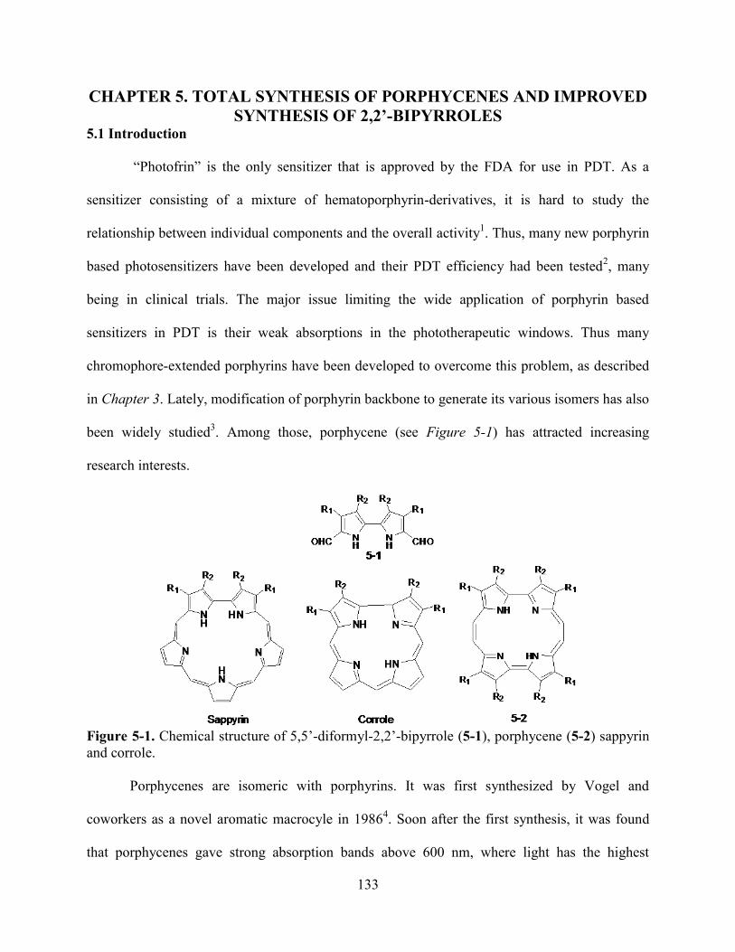
133
CHAPTER 5. TOTAL SYNTHESIS OF PORPHYCENES AND IMPROVED
SYNTHESIS OF 2,2’-BIPYRROLES 5.1 Introduction
―Photofrin‖ is the only sensitizer that is approved by the FDA for use in PDT. As a
sensitizer consisting of a mixture of hematoporphyrin-derivatives, it is hard to study the
relationship between individual components and the overall activity1. Thus, many new porphyrin
based photosensitizers have been developed and their PDT efficiency had been tested2, many
being in clinical trials. The major issue limiting the wide application of porphyrin based
sensitizers in PDT is their weak absorptions in the phototherapeutic windows. Thus many
chromophore-extended porphyrins have been developed to overcome this problem, as described
in Chapter 3. Lately, modification of porphyrin backbone to generate its various isomers has also
been widely studied3. Among those, porphycene (see Figure 5-1) has attracted increasing
research interests.
Figure 5-1. Chemical structure of 5,5’-diformyl-2,2’-bipyrrole (5-1), porphycene (5-2) sappyrin
and corrole.
Porphycenes are isomeric with porphyrins. It was first synthesized by Vogel and
coworkers as a novel aromatic macrocyle in 19864. Soon after the first synthesis, it was found
that porphycenes gave strong absorption bands above 600 nm, where light has the highest
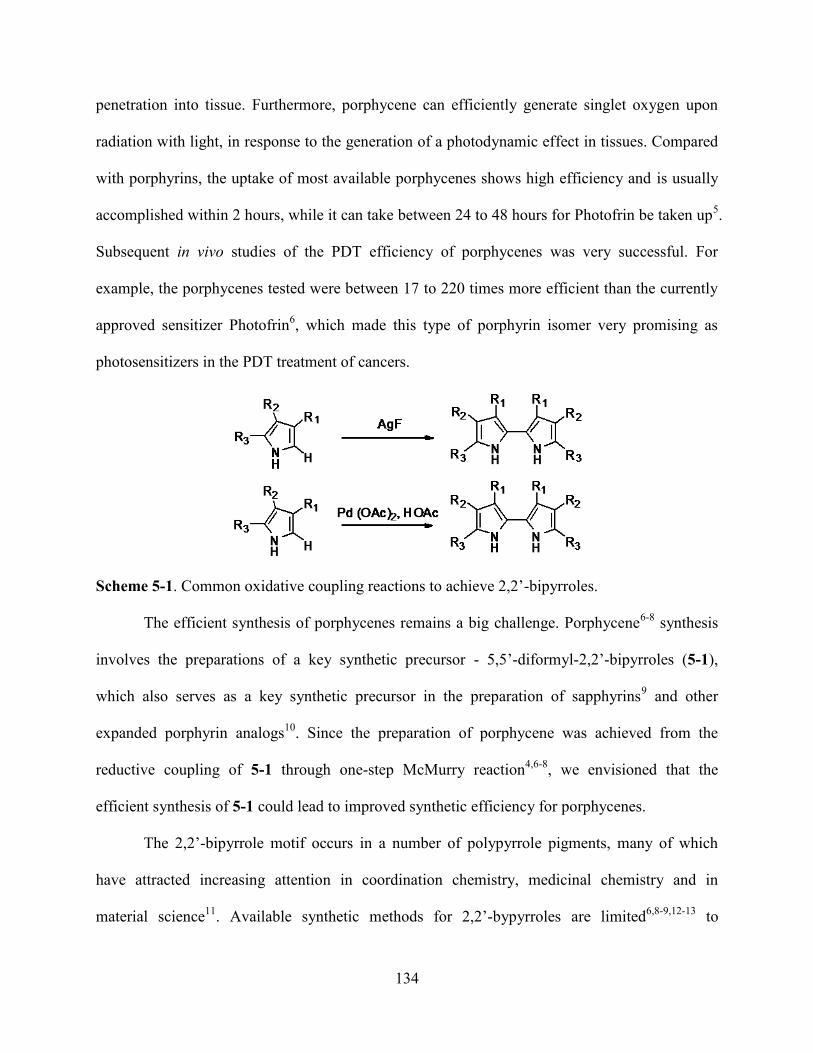
134
penetration into tissue. Furthermore, porphycene can efficiently generate singlet oxygen upon
radiation with light, in response to the generation of a photodynamic effect in tissues. Compared
with porphyrins, the uptake of most available porphycenes shows high efficiency and is usually
accomplished within 2 hours, while it can take between 24 to 48 hours for Photofrin be taken up5.
Subsequent in vivo studies of the PDT efficiency of porphycenes was very successful. For
example, the porphycenes tested were between 17 to 220 times more efficient than the currently
approved sensitizer Photofrin6, which made this type of porphyrin isomer very promising as
photosensitizers in the PDT treatment of cancers.
Scheme 5-1. Common oxidative coupling reactions to achieve 2,2’-bipyrroles.
The efficient synthesis of porphycenes remains a big challenge. Porphycene6-8
synthesis
involves the preparations of a key synthetic precursor - 5,5’-diformyl-2,2’-bipyrroles (5-1),
which also serves as a key synthetic precursor in the preparation of sapphyrins9 and other
expanded porphyrin analogs10
. Since the preparation of porphycene was achieved from the
reductive coupling of 5-1 through one-step McMurry reaction4,6-8
, we envisioned that the
efficient synthesis of 5-1 could lead to improved synthetic efficiency for porphycenes.
The 2,2’-bipyrrole motif occurs in a number of polypyrrole pigments, many of which
have attracted increasing attention in coordination chemistry, medicinal chemistry and in
material science11
. Available synthetic methods for 2,2’-bypyrroles are limited6,8-9,12-13
to
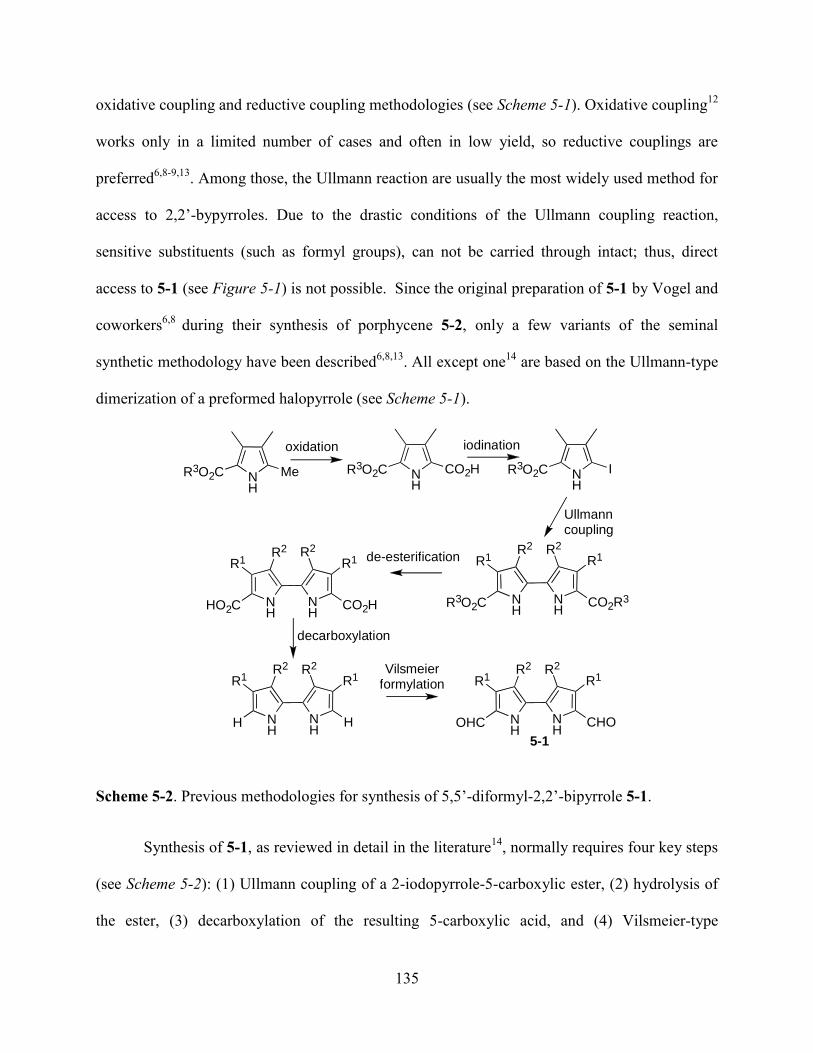
135
oxidative coupling and reductive coupling methodologies (see Scheme 5-1). Oxidative coupling12
works only in a limited number of cases and often in low yield, so reductive couplings are
preferred6,8-9,13
. Among those, the Ullmann reaction are usually the most widely used method for
access to 2,2’-bypyrroles. Due to the drastic conditions of the Ullmann coupling reaction,
sensitive substituents (such as formyl groups), can not be carried through intact; thus, direct
access to 5-1 (see Figure 5-1) is not possible. Since the original preparation of 5-1 by Vogel and
coworkers6,8
during their synthesis of porphycene 5-2, only a few variants of the seminal
synthetic methodology have been described6,8,13
. All except one14
are based on the Ullmann-type
dimerization of a preformed halopyrrole (see Scheme 5-1).
Scheme 5-2. Previous methodologies for synthesis of 5,5’-diformyl-2,2’-bipyrrole 5-1.
Synthesis of 5-1, as reviewed in detail in the literature14
, normally requires four key steps
(see Scheme 5-2): (1) Ullmann coupling of a 2-iodopyrrole-5-carboxylic ester, (2) hydrolysis of
the ester, (3) decarboxylation of the resulting 5-carboxylic acid, and (4) Vilsmeier-type
5-1
N H
R 3 O 2 C M e N H
R 3 O 2 C C O 2 H N H
R 3 O 2 C I
N H
N H
C O 2 R 3
R 1 R 2 R 2 R 1
R 3 O 2 C N H
N H
C O 2 H
R 1 R 2 R 2 R 1
H O 2 C
N H
N H
H
R 1 R 2 R 2 R 1
H N H
N H
C H O
R 1 R 2 R 2 R 1
O H C
o x i d a t i o n i o d i n a t i o n
U l l m a n n c o u p l i n g
d e - e s t e r i f i c a t i o n
d e c a r b o x y l a t i o n
V i l s m e i e r f o r m y l a t i o n

136
diformylation. The Ullmann synthesis of biaryls by the copper-induced reductive coupling of
aromatic halides is of broad synthetic use15a
. Although some substrates will undergo Ullmann
reductive coupling under mild conditions, the typical Ullmann coupling is conducted at high
temperature15b
. Generally, Ullmann-type reductive coupling of pyrroles works well only when
there is an electron-withdrawing group present; high temperature is usually required15b
.
Scheme 5-3. Example of ambient temperature Ullmann-type coupling reaction.
Recently, Liebeskind and coworkers reported an ambient temperature Ullmann-type
coupling reaction15c
, but the requirement of specific type of substrates or specific positions of the
substitutent limits its synthetic application (see Scheme 5-3). They stated that ―The most
noticeable limitation of this process is the lack of reaction of aromatic halide substrates not
possessing a coordinating ortho-substituent‖ 15c
.
Scheme 5-4. Examples of akyl-substituted 2,2’-bipyrroles from a N-protected monopyrrole.
Sessler et al.13a
developed an efficient procedure for the preparation of alkyl-substituted
2,2’-bipyrroles by protection of the pyrrole nitrogen atom before an Ullmann-type coupling
reaction; this was followed by deprotection of the resulting N-substituted 2,2’-bipyrroles.
Vogel13b
et al. later improved the Sessler method by changing the solvent from DMF to toluene
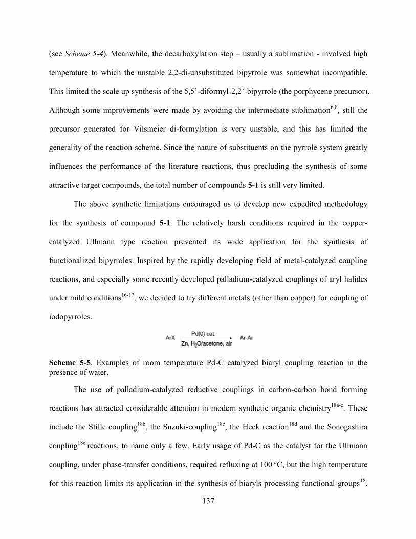
137
(see Scheme 5-4). Meanwhile, the decarboxylation step – usually a sublimation - involved high
temperature to which the unstable 2,2-di-unsubstituted bipyrrole was somewhat incompatible.
This limited the scale up synthesis of the 5,5’-diformyl-2,2’-bipyrrole (the porphycene precursor).
Although some improvements were made by avoiding the intermediate sublimation6,8
, still the
precursor generated for Vilsmeier di-formylation is very unstable, and this has limited the
generality of the reaction scheme. Since the nature of substituents on the pyrrole system greatly
influences the performance of the literature reactions, thus precluding the synthesis of some
attractive target compounds, the total number of compounds 5-1 is still very limited.
The above synthetic limitations encouraged us to develop new expedited methodology
for the synthesis of compound 5-1. The relatively harsh conditions required in the copper-
catalyzed Ullmann type reaction prevented its wide application for the synthesis of
functionalized bipyrroles. Inspired by the rapidly developing field of metal-catalyzed coupling
reactions, and especially some recently developed palladium-catalyzed couplings of aryl halides
under mild conditions16-17
, we decided to try different metals (other than copper) for coupling of
iodopyrroles.
Scheme 5-5. Examples of room temperature Pd-C catalyzed biaryl coupling reaction in the
presence of water.
The use of palladium-catalyzed reductive couplings in carbon-carbon bond forming
reactions has attracted considerable attention in modern synthetic organic chemistry18a-e
. These
include the Stille coupling18b
, the Suzuki-coupling18c
, the Heck reaction18d
and the Sonogashira
coupling18e
reactions, to name only a few. Early usage of Pd-C as the catalyst for the Ullmann
coupling, under phase-transfer conditions, required refluxing at 100 °C, but the high temperature
for this reaction limits its application in the synthesis of biaryls processing functional groups18
.

138
Recently, Pd-C co-catalyzed biaryl synthesis was reported to take place at room temperature (see
Scheme 5-5) by simply adding water into the reaction system17
. Meanwhile 2-formyl-5-
iodopyrroles have been used in Sonogashira coupling reactions (see Scheme 5-6) with TMS-
acetylene to build acetylenic and diacetylenic diformyldipyrroles19
.
Scheme 5-6. Examples of 2-formyl-5-iodopyrroles directly used in the coupling reaction.
5.2 Results and Discussion
5.2.1 Total Synthesis of Porphycene (CpoTMPn)
NNH
N HN
O
O O
O
O
O
Figure 5-2. Chemical structure of CpoTMPn.
Porphycenes containing two or four methoxyethyl side chains, such as CpoTMPn (see
Figure 5-2), are able to enhance and accelerate cellular uptake6. The membrane solubility of
photosensitizers is assumed to be critical in their photodynamic efficiency. The presence of
nonionic polar side chains in CpoTMPn was found to be able to strongly enhance cellular uptake
and antitumor activity of porpycenes3,6, 20
.
The total synthesis followed the procedures reported by Vogel et al.21
and Richert, et al.6,
22 but with small modifications (see Scheme 5-7 through Scheme 5-10). The synthesis of
diketone 5-7-2 involved the preparation of activated metal zinc. The metal zinc activation was
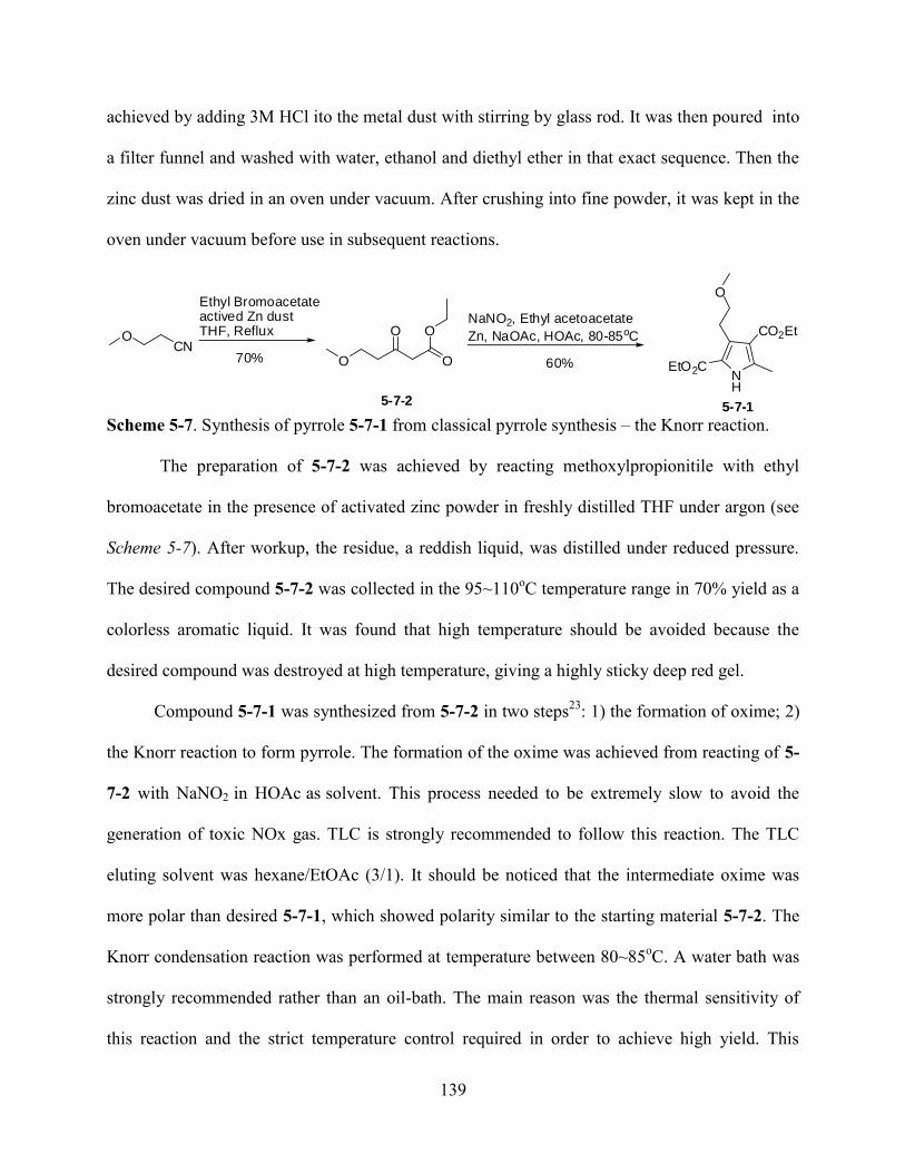
139
achieved by adding 3M HCl ito the metal dust with stirring by glass rod. It was then poured into
a filter funnel and washed with water, ethanol and diethyl ether in that exact sequence. Then the
zinc dust was dried in an oven under vacuum. After crushing into fine powder, it was kept in the
oven under vacuum before use in subsequent reactions.
NH
OCN
Ethyl Bromoacetateactived Zn dustTHF, Reflux
O
OO
O
NaNO2, Ethyl acetoacetate
Zn, NaOAc, HOAc, 80-85oC CO2Et
EtO2C
O
70% 60%
5-7-15-7-2
Scheme 5-7. Synthesis of pyrrole 5-7-1 from classical pyrrole synthesis – the Knorr reaction.
The preparation of 5-7-2 was achieved by reacting methoxylpropionitile with ethyl
bromoacetate in the presence of activated zinc powder in freshly distilled THF under argon (see
Scheme 5-7). After workup, the residue, a reddish liquid, was distilled under reduced pressure.
The desired compound 5-7-2 was collected in the 95~110oC temperature range in 70% yield as a
colorless aromatic liquid. It was found that high temperature should be avoided because the
desired compound was destroyed at high temperature, giving a highly sticky deep red gel.
Compound 5-7-1 was synthesized from 5-7-2 in two steps23
: 1) the formation of oxime; 2)
the Knorr reaction to form pyrrole. The formation of the oxime was achieved from reacting of 5-
7-2 with NaNO2 in HOAc as solvent. This process needed to be extremely slow to avoid the
generation of toxic NOx gas. TLC is strongly recommended to follow this reaction. The TLC
eluting solvent was hexane/EtOAc (3/1). It should be noticed that the intermediate oxime was
more polar than desired 5-7-1, which showed polarity similar to the starting material 5-7-2. The
Knorr condensation reaction was performed at temperature between 80~85oC. A water bath was
strongly recommended rather than an oil-bath. The main reason was the thermal sensitivity of
this reaction and the strict temperature control required in order to achieve high yield. This
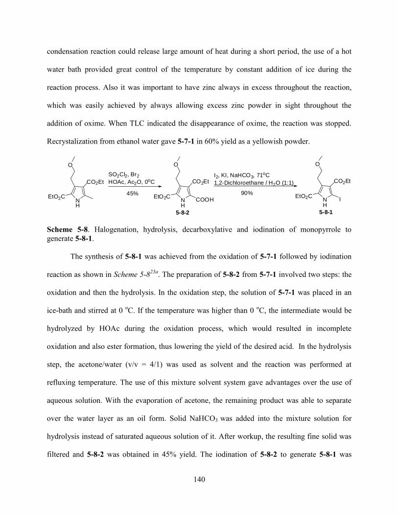
140
condensation reaction could release large amount of heat during a short period, the use of a hot
water bath provided great control of the temperature by constant addition of ice during the
reaction process. Also it was important to have zinc always in excess throughout the reaction,
which was easily achieved by always allowing excess zinc powder in sight throughout the
addition of oxime. When TLC indicated the disappearance of oxime, the reaction was stopped.
Recrystalization from ethanol water gave 5-7-1 in 60% yield as a yellowish powder.
NH
CO2Et
EtO2C
O
NH
CO2Et
IEtO2C
O
SO2Cl2, Br2
HOAc, Ac2O, 0oC
NH
CO2Et
COOHEtO2C
O
I2, KI, NaHCO3, 71oC
1,2-Dichloroethane / H2O (1:1)
45% 90%
5-8-2 5-8-1
Scheme 5-8. Halogenation, hydrolysis, decarboxylative and iodination of monopyrrole to
generate 5-8-1.
The synthesis of 5-8-1 was achieved from the oxidation of 5-7-1 followed by iodination
reaction as shown in Scheme 5-823a
. The preparation of 5-8-2 from 5-7-1 involved two steps: the
oxidation and then the hydrolysis. In the oxidation step, the solution of 5-7-1 was placed in an
ice-bath and stirred at 0 oC. If the temperature was higher than 0
oC, the intermediate would be
hydrolyzed by HOAc during the oxidation process, which would resulted in incomplete
oxidation and also ester formation, thus lowering the yield of the desired acid. In the hydrolysis
step, the acetone/water (v/v = 4/1) was used as solvent and the reaction was performed at
refluxing temperature. The use of this mixture solvent system gave advantages over the use of
aqueous solution. With the evaporation of acetone, the remaining product was able to separate
over the water layer as an oil form. Solid NaHCO3 was added into the mixture solution for
hydrolysis instead of saturated aqueous solution of it. After workup, the resulting fine solid was
filtered and 5-8-2 was obtained in 45% yield. The iodination of 5-8-2 to generate 5-8-1 was
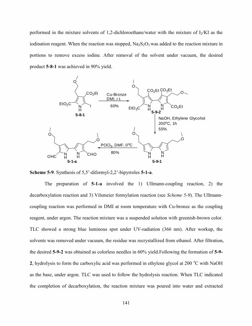
141
performed in the mixture solvents of 1,2-dichloroethane/water with the mixture of I2/KI as the
iodination reagent. When the reaction was stopped, Na2S2O3 was added to the reaction mixture in
portions to remove excess iodine. After removal of the solvent under vacuum, the desired
product 5-8-1 was achieved in 90% yield.
NH
CO2Et
IEtO2C
O
NH
CO2Et
EtO2C
O
NH
CO2Et
OCO2Et
Cu-BronzeDMI, r.t.
NaOH, Ethylene Glycohol
200oC, 1h
POCl3, DMF, 0oC
NHOHC
O
NH
CHO
O
NH
O
NH
O
60%
55%
80%
5-1-a
5-8-15-9-2
5-9-1
Scheme 5-9. Synthesis of 5,5’-diformyl-2,2’-bipyrroles 5-1-a.
The preparation of 5-1-a involved the 1) Ullmann-coupling reaction, 2) the
decarboxylation reaction and 3) Vilsmeier formylation reaction (see Scheme 5-9). The Ullmann-
coupling reaction was performed in DMI at room temperature with Cu-bronze as the coupling
reagent, under argon. The reaction mixture was a suspended solution with greenish-brown color.
TLC showed a strong blue luminous spot under UV-radiation (366 nm). After workup, the
solvents was removed under vacuum, the residue was recrystallized from ethanol. After filtration,
the desired 5-9-2 was obtained as colorless needles in 60% yield.Following the formation of 5-9-
2, hydrolysis to form the carboxylic acid was performed in ethylene glycol at 200 oC with NaOH
as the base, under argon. TLC was used to follow the hydrolysis reaction. When TLC indicated
the completion of decarboxylation, the reaction mixture was poured into water and extracted
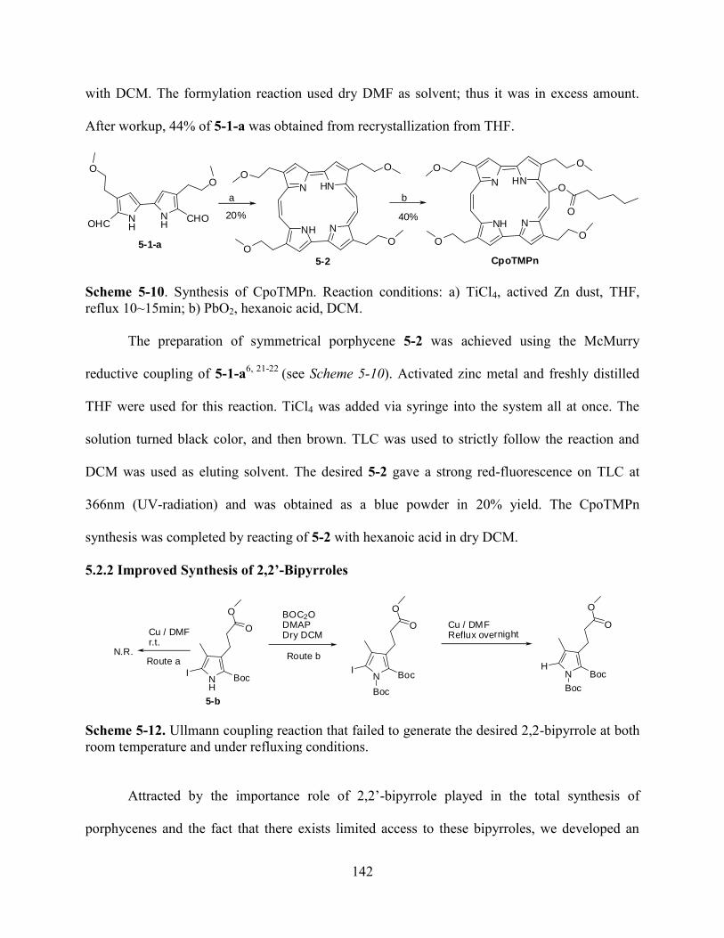
142
with DCM. The formylation reaction used dry DMF as solvent; thus it was in excess amount.
After workup, 44% of 5-1-a was obtained from recrystallization from THF.
NNH
N HN
NHOHC
O
NH
CHO
O
O
OO
O
NNH
N HN
O
O O
O
O
O20% 40%
a b
5-1-a
5-2 CpoTMPn
Scheme 5-10. Synthesis of CpoTMPn. Reaction conditions: a) TiCl4, actived Zn dust, THF,
reflux 10~15min; b) PbO2, hexanoic acid, DCM.
The preparation of symmetrical porphycene 5-2 was achieved using the McMurry
reductive coupling of 5-1-a6, 21-22
(see Scheme 5-10). Activated zinc metal and freshly distilled
THF were used for this reaction. TiCl4 was added via syringe into the system all at once. The
solution turned black color, and then brown. TLC was used to strictly follow the reaction and
DCM was used as eluting solvent. The desired 5-2 gave a strong red-fluorescence on TLC at
366nm (UV-radiation) and was obtained as a blue powder in 20% yield. The CpoTMPn
synthesis was completed by reacting of 5-2 with hexanoic acid in dry DCM.
5.2.2 Improved Synthesis of 2,2’-Bipyrroles
NH
BocI
O
Cu / DMFr.t.
O
N.R.
BOC2ODMAPDry DCM
N BocI
O
O
Boc
Cu / DMFReflux overnight
N BocH
O
O
Route aRoute b
Boc
5-b
Scheme 5-12. Ullmann coupling reaction that failed to generate the desired 2,2-bipyrrole at both
room temperature and under refluxing conditions.
Attracted by the importance role of 2,2’-bipyrrole played in the total synthesis of
porphycenes and the fact that there exists limited access to these bipyrroles, we developed an
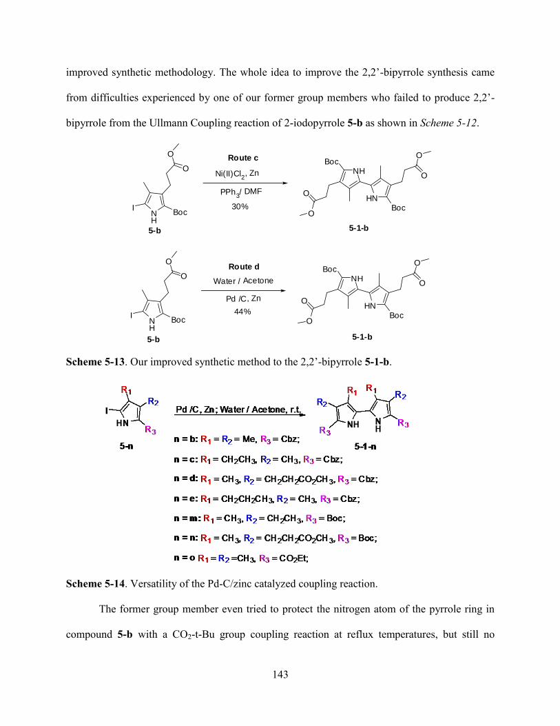
143
improved synthetic methodology. The whole idea to improve the 2,2’-bipyrrole synthesis came
from difficulties experienced by one of our former group members who failed to produce 2,2’-
bipyrrole from the Ullmann Coupling reaction of 2-iodopyrrole 5-b as shown in Scheme 5-12.
NH
BocI
O
HNBoc
O
PPh3/ DMF
Ni(II)Cl2, Zn
OO
NHBoc
O
O
NH
BocI
O
HNBoc
O
Pd /C, Zn
Water / AcetoneO
ONH
Boc
O
O
30%
5-b 5-1-b
44%
5-b 5-1-b
Route c
Route d
Scheme 5-13. Our improved synthetic method to the 2,2’-bipyrrole 5-1-b.
Scheme 5-14. Versatility of the Pd-C/zinc catalyzed coupling reaction.
The former group member even tried to protect the nitrogen atom of the pyrrole ring in
compound 5-b with a CO2-t-Bu group coupling reaction at reflux temperatures, but still no
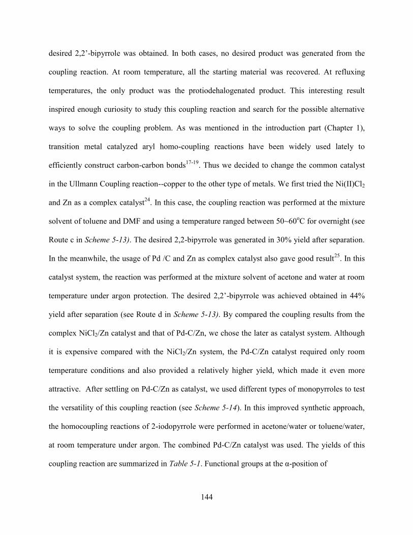
144
desired 2,2’-bipyrrole was obtained. In both cases, no desired product was generated from the
coupling reaction. At room temperature, all the starting material was recovered. At refluxing
temperatures, the only product was the protiodehalogenated product. This interesting result
inspired enough curiosity to study this coupling reaction and search for the possible alternative
ways to solve the coupling problem. As was mentioned in the introduction part (Chapter 1),
transition metal catalyzed aryl homo-coupling reactions have been widely used lately to
efficiently construct carbon-carbon bonds17-19
. Thus we decided to change the common catalyst
in the Ullmann Coupling reaction--copper to the other type of metals. We first tried the Ni(II)Cl2
and Zn as a complex catalyst24
. In this case, the coupling reaction was performed at the mixture
solvent of toluene and DMF and using a temperature ranged between 50~60oC for overnight (see
Route c in Scheme 5-13). The desired 2,2-bipyrrole was generated in 30% yield after separation.
In the meanwhile, the usage of Pd /C and Zn as complex catalyst also gave good result25
. In this
catalyst system, the reaction was performed at the mixture solvent of acetone and water at room
temperature under argon protection. The desired 2,2’-bipyrrole was achieved obtained in 44%
yield after separation (see Route d in Scheme 5-13). By compared the coupling results from the
complex NiCl2/Zn catalyst and that of Pd-C/Zn, we chose the later as catalyst system. Although
it is expensive compared with the NiCl2/Zn system, the Pd-C/Zn catalyst required only room
temperature conditions and also provided a relatively higher yield, which made it even more
attractive. After settling on Pd-C/Zn as catalyst, we used different types of monopyrroles to test
the versatility of this coupling reaction (see Scheme 5-14). In this improved synthetic approach,
the homocoupling reactions of 2-iodopyrrole were performed in acetone/water or toluene/water,
at room temperature under argon. The combined Pd-C/Zn catalyst was used. The yields of this
coupling reaction are summarized in Table 5-1. Functional groups at the α-position of
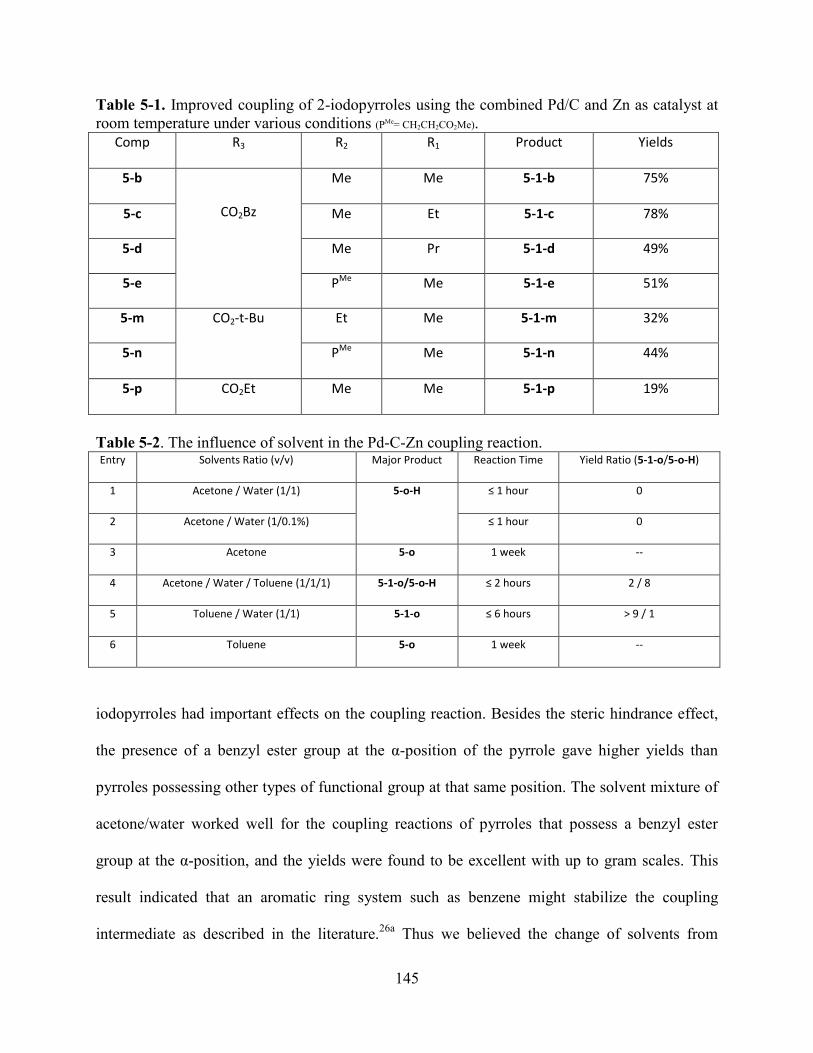
145
Table 5-1. Improved coupling of 2-iodopyrroles using the combined Pd/C and Zn as catalyst at
room temperature under various conditions (PMe= CH2CH2CO2Me).
Comp R3 R2 R1 Product Yields
5-b
CO2Bz
Me Me 5-1-b 75%
5-c Me Et 5-1-c 78%
5-d Me Pr 5-1-d 49%
5-e PMe Me 5-1-e 51%
5-m CO2-t-Bu Et Me 5-1-m 32%
5-n PMe Me 5-1-n 44%
5-p CO2Et Me Me 5-1-p 19%
Table 5-2. The influence of solvent in the Pd-C-Zn coupling reaction. Entry Solvents Ratio (v/v) Major Product Reaction Time Yield Ratio (5-1-o/5-o-H)
1 Acetone / Water (1/1) 5-o-H ≤ 1 hour 0
2 Acetone / Water (1/0.1%) ≤ 1 hour 0
3 Acetone 5-o 1 week --
4 Acetone / Water / Toluene (1/1/1) 5-1-o/5-o-H ≤ 2 hours 2 / 8
5 Toluene / Water (1/1) 5-1-o ≤ 6 hours > 9 / 1
6 Toluene 5-o 1 week --
iodopyrroles had important effects on the coupling reaction. Besides the steric hindrance effect,
the presence of a benzyl ester group at the α-position of the pyrrole gave higher yields than
pyrroles possessing other types of functional group at that same position. The solvent mixture of
acetone/water worked well for the coupling reactions of pyrroles that possess a benzyl ester
group at the α-position, and the yields were found to be excellent with up to gram scales. This
result indicated that an aromatic ring system such as benzene might stabilize the coupling
intermediate as described in the literature.26a
Thus we believed the change of solvents from
146
acetone/water to toluene/water would be helpful for the coupling reaction of other type
monopyrroles. Monopyrrole 5-o was used as starting material for the coupling reaction because it
was one the most reactive monopyrroles in the Ullmann-Coupling reaction. The results are
shown in Table 5-2.
The addition of toluene really helped to improve the coupling reaction; however the real
function served by benzene in the coupling reaction was still unknown. According to the
disappearance of starting material on TLC, in acetone/water system, reaction took place very fast
with pyrrole 5-o. However, only trace amounts of the desired 2,2’-bipyrrole 5-1-o could be
generated, while the protiodehalogenated product 5-o-H was obtained as the major product.
When organic solvent was changed from acetone to toluene, the yield of 5-o-H was dramatically
reduced; meanwhile the desired 2,2’-bipyrrole 5-1-o became the major product. Meanwhile, the
reaction rate was reduced. Water was found to play an important role in this reaction: the
presence of water increased the reaction rate and allowed room temperature performance of the
reaction. Although 5-1-o was one of the few example of 2,2’-bipyrroles that could be easily
obtained from room temperature Ullmann-Coupling reaction, it usually took around 24~48 hours
for this reaction to complete under Ullmann conditions. However, it usually took only several
hours for the completion of coupling 5-o under this improved condition. The absence of water,
no coupling reaction was observed and only starting materials 5-o was recovered even over a
long period (up to 4 days). The simple addition of only 0.1% of water into the acetone solution of
the reaction mixture led to the conversion of most starting material to 5-o-H within 2 hours.
The previous preparation of 5,5’-diformyl-2,2’-bipyrrole (see Figure 5-1) was achieved
from the Ullmann Coupling reaction in our group. Since it usually require high temperature,
functional groups such as aldehyde do not survive the Ullmann-Coupling conditions. Thus
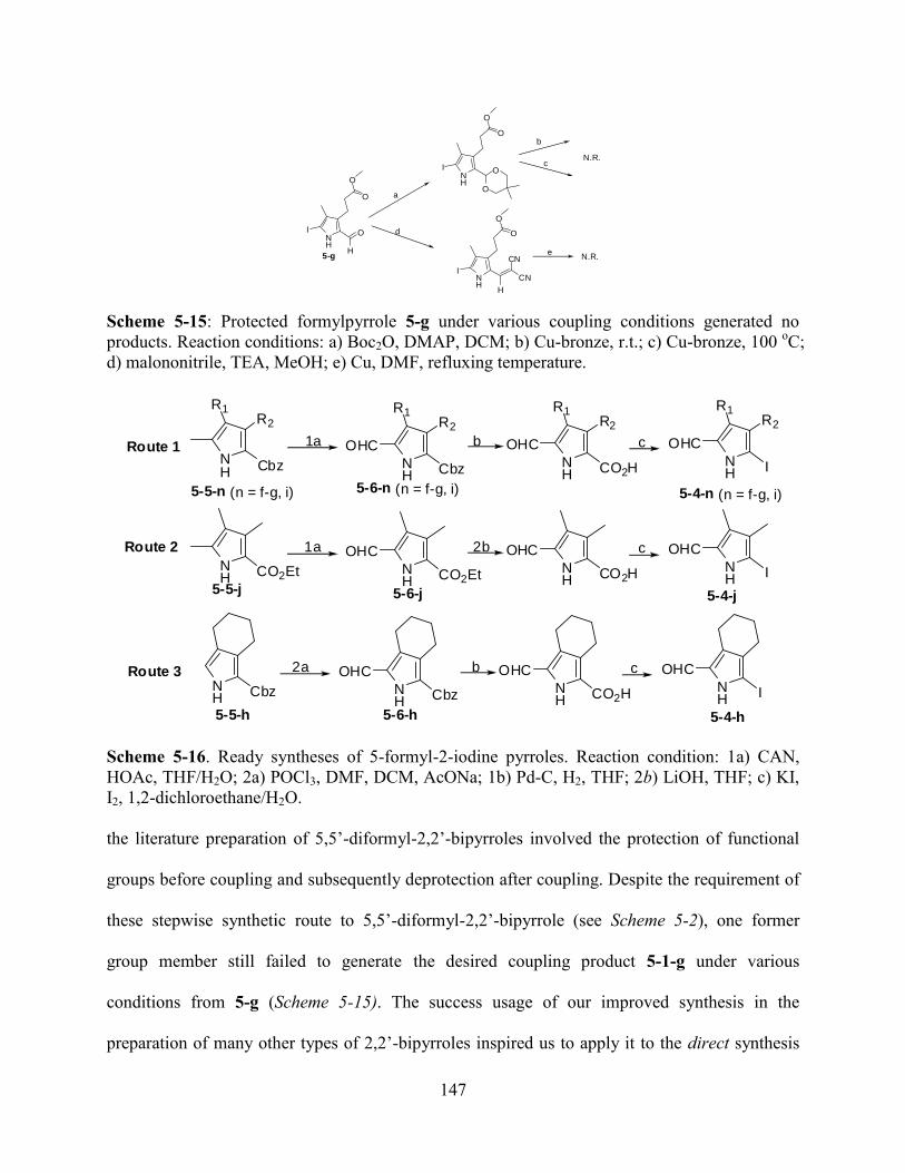
147
NH
I
O
5-g
O
O
H
NH
I
O
O
O
O
NH
I
O
O
H
CN
CN
a
d
N.R.
b
c
N.R.e
Scheme 5-15: Protected formylpyrrole 5-g under various coupling conditions generated no
products. Reaction conditions: a) Boc2O, DMAP, DCM; b) Cu-bronze, r.t.; c) Cu-bronze, 100 oC;
d) malononitrile, TEA, MeOH; e) Cu, DMF, refluxing temperature.
1a
NH
R2
Cbz
R1
NH
R2
Cbz
OHC
R1
NH
R2
CO2H
OHC
R1
NH
R2
I
OHC
R1
b c
2a
NH Cbz N
H Cbz
OHCNH CO2H
OHCNH I
OHCb c
1a
NH CO2Et N
H CO2Et
OHCNH CO2H
OHCNH I
OHC2b c
Route 1
Route 2
Route 3
5-4-n (n = f-g, i)
5-4-j
5-4-h
5-5-n (n = f-g, i)
5-5-j
5-5-h 5-6-h
5-6-j
5-6-n (n = f-g, i)
Scheme 5-16. Ready syntheses of 5-formyl-2-iodine pyrroles. Reaction condition: 1a) CAN,
HOAc, THF/H2O; 2a) POCl3, DMF, DCM, AcONa; 1b) Pd-C, H2, THF; 2b) LiOH, THF; c) KI,
I2, 1,2-dichloroethane/H2O.
the literature preparation of 5,5’-diformyl-2,2’-bipyrroles involved the protection of functional
groups before coupling and subsequently deprotection after coupling. Despite the requirement of
these stepwise synthetic route to 5,5’-diformyl-2,2’-bipyrrole (see Scheme 5-2), one former
group member still failed to generate the desired coupling product 5-1-g under various
conditions from 5-g (Scheme 5-15). The success usage of our improved synthesis in the
preparation of many other types of 2,2’-bipyrroles inspired us to apply it to the direct synthesis
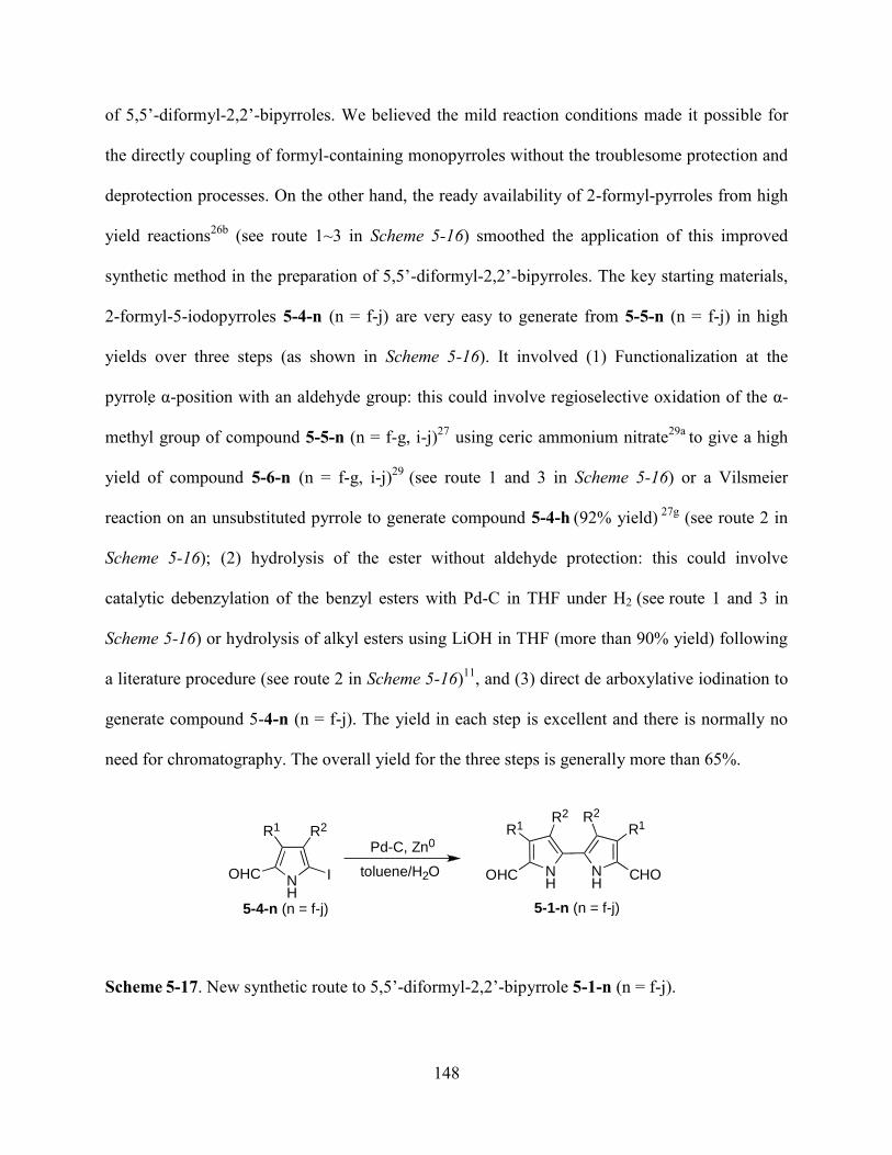
148
of 5,5’-diformyl-2,2’-bipyrroles. We believed the mild reaction conditions made it possible for
the directly coupling of formyl-containing monopyrroles without the troublesome protection and
deprotection processes. On the other hand, the ready availability of 2-formyl-pyrroles from high
yield reactions26b
(see route 1~3 in Scheme 5-16) smoothed the application of this improved
synthetic method in the preparation of 5,5’-diformyl-2,2’-bipyrroles. The key starting materials,
2-formyl-5-iodopyrroles 5-4-n (n = f-j) are very easy to generate from 5-5-n (n = f-j) in high
yields over three steps (as shown in Scheme 5-16). It involved (1) Functionalization at the
pyrrole α-position with an aldehyde group: this could involve regioselective oxidation of the α-
methyl group of compound 5-5-n (n = f-g, i-j)27
using ceric ammonium nitrate29a
to give a high
yield of compound 5-6-n (n = f-g, i-j)29
(see route 1 and 3 in Scheme 5-16) or a Vilsmeier
reaction on an unsubstituted pyrrole to generate compound 5-4-h (92% yield)
27g (see route 2 in
Scheme 5-16); (2) hydrolysis of the ester without aldehyde protection: this could involve
catalytic debenzylation of the benzyl esters with Pd-C in THF under H2 (see route 1 and 3 in
Scheme 5-16) or hydrolysis of alkyl esters using LiOH in THF (more than 90% yield) following
a literature procedure (see route 2 in Scheme 5-16)11
, and (3) direct de arboxylative iodination to
generate compound 5-4-n (n = f-j). The yield in each step is excellent and there is normally no
need for chromatography. The overall yield for the three steps is generally more than 65%.
Scheme 5-17. New synthetic route to 5,5’-diformyl-2,2’-bipyrrole 5-1-n (n = f-j).
5-1-n (n = f-j)
N H
N H
C H O
R 1 R 2 R 2 R 1
O H C N H
R 2 R 1
O H C I
P d - C , Z n 0
t o l u e n e / H 2 O
5-4-n (n = f-j)
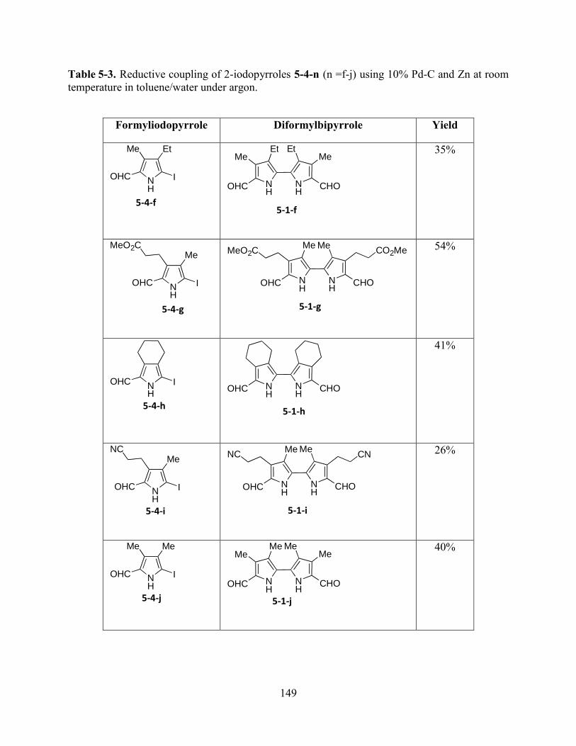
149
Table 5-3. Reductive coupling of 2-iodopyrroles 5-4-n (n =f-j) using 10% Pd-C and Zn at room
temperature in toluene/water under argon.
Formyliodopyrrole Diformylbipyrrole Yield
35%
54%
41%
26%
40%
N H
E t M e
O H C I
5-4-f
N H
M e
O H C I
M e O 2 C
5-4-g
N H
O H C I
5-4-h
N H
M e
O H C I
N C
5-4-i
N H
M e M e
O H C I
5-4-j
N H
N H
C H O
M e M e M e
M e
O H C
5-1-j
N H
N H
C H O
M e E t E t
M e
O H C
5-1-f
N H
N H
C H O
M e M e
O H C
C O 2 M e M e O 2 C
5-1-g
N H
N H
C H O O H C
5-1-h
N H
N H
C H O
M e M e
O H C
C N N C
5-1-i
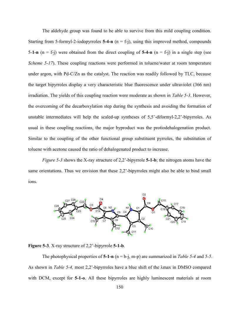
150
The aldehyde group was found to be able to survive from this mild coupling condition.
Starting from 5-formyl-2-iodopyrroles 5-4-n (n = f-j), using this improved method, compounds
5-1-n (n = f-j) were obtained from the direct coupling of 5-4-n (n = f-j) in a single step (see
Scheme 5-17). These coupling reactions were performed in toluene/water at room temperature
under argon, with Pd-C/Zn as the catalyst. The reaction was readily followed by TLC, because
the target bipyrroles display a very characteristic blue fluorescence under ultraviolet (366 nm)
irradiation. The yields of this coupling reaction were moderate as shown in Table 5-3. However,
the overcoming of the decarboxylation step during the synthesis and avoiding the formation of
unstable intermediates will help the scaled-up syntheses of 5,5’-diformyl-2,2’-bipyrroles. As
usual in these coupling reactions, the major byproduct was the protiodehalogenation product.
Similar to the coupling of the other functional group substituent pyrroles, the substitution of
toluene with acetone caused the ratio of dehalogenated product to increase.
Figure 5-3 shows the X-ray structure of 2,2’-bipyrrole 5-1-b; the nitrogen atoms have the
same orientations. Thus we envision that these 2,2’-bipyrroles might also be able to bind small
ions.
Figure 5-3. X-ray structure of 2,2’-bipyrrole 5-1-b.
The photophysical properties of 5-1-n (n = b-j, m-p) are summarized in Table 5-4 and 5-5.
As shown in Table 5-4, most 2,2’-bipyrroles have a blue shift of the λmax in DMSO compared
with DCM, except for 5-1-o. All these bipyrroles are highly luminescent materials at room
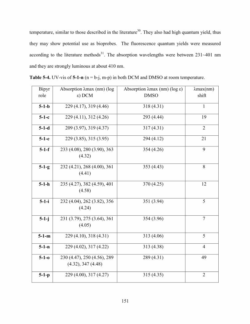
151
temperature, similar to those described in the literature30
. They also had high quantum yield, thus
they may show potential use as bioprobes. The fluorescence quantum yields were measured
according to the literature methods31
. The absorption wavelengths were between 231~401 nm
and they are strongly luminous at about 410 nm.
Table 5-4. UV-vis of 5-1-n (n = b-j, m-p) in both DCM and DMSO at room temperature.
Bipyr
role
Absorption λmax (nm) (log
ε) DCM
Absorption λmax (nm) (log ε)
DMSO
λmax(nm)
shift
5-1-b 229 (4.17), 319 (4.46) 318 (4.31) 1
5-1-c 229 (4.11), 312 (4.26) 293 (4.44) 19
5-1-d 209 (3.97), 319 (4.37) 317 (4.31) 2
5-1-e 229 (3.85), 315 (3.95) 294 (4.12) 21
5-1-f 233 (4.08), 280 (3.90), 363
(4.32)
354 (4.26) 9
5-1-g 232 (4.21), 268 (4.00), 361
(4.41)
353 (4.43) 8
5-1-h 235 (4.27), 382 (4.59), 401
(4.58)
370 (4.25) 12
5-1-i 232 (4.04), 262 (3.82), 356
(4.24)
351 (3.94) 5
5-1-j 231 (3.79), 275 (3.64), 361
(4.05)
354 (3.96) 7
5-1-m 229 (4.10), 318 (4.31) 313 (4.06) 5
5-1-n 229 (4.02), 317 (4.22) 313 (4.38) 4
5-1-o 230 (4.47), 250 (4.56), 289
(4.32), 347 (4.48)
289 (4.31) 49
5-1-p 229 (4.00), 317 (4.27) 315 (4.35) 2
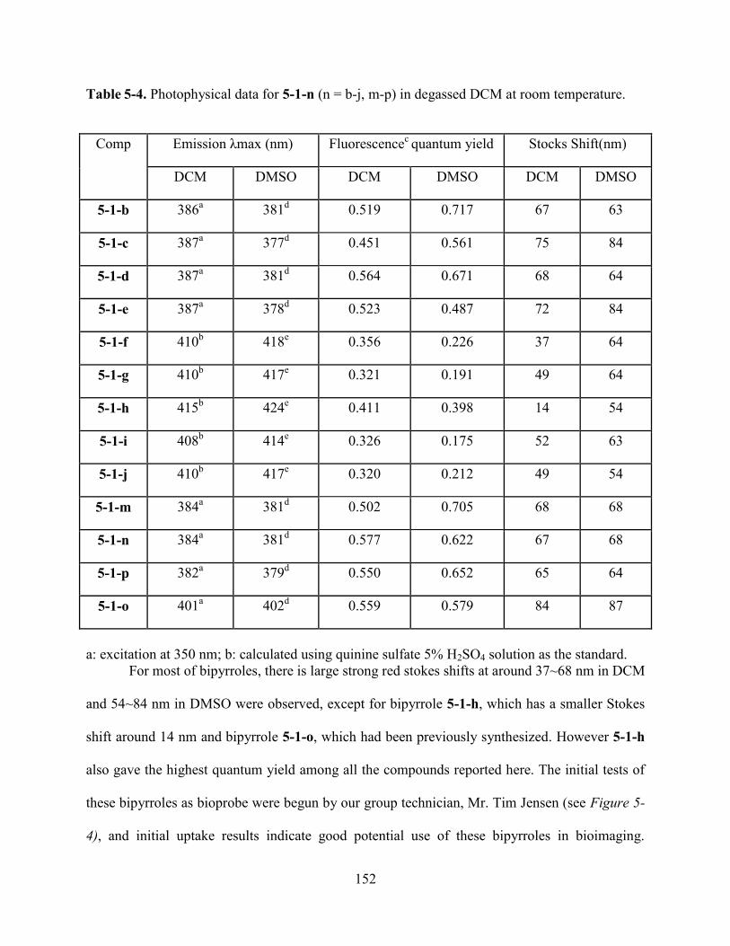
152
Table 5-4. Photophysical data for 5-1-n (n = b-j, m-p) in degassed DCM at room temperature.
Comp Emission λmax (nm) Fluorescencec quantum yield Stocks Shift(nm)
DCM DMSO DCM DMSO DCM DMSO
5-1-b 386a 381
d 0.519 0.717 67 63
5-1-c 387a 377
d 0.451 0.561 75 84
5-1-d 387a 381
d 0.564 0.671 68 64
5-1-e 387a 378
d 0.523 0.487 72 84
5-1-f 410b 418
e 0.356 0.226 37 64
5-1-g 410b 417
e 0.321 0.191 49 64
5-1-h 415b 424
e 0.411 0.398 14 54
5-1-i 408b 414
e 0.326 0.175 52 63
5-1-j 410b 417
e 0.320 0.212 49 54
5-1-m 384a 381
d 0.502 0.705 68 68
5-1-n 384a 381
d 0.577 0.622 67 68
5-1-p 382a 379
d 0.550 0.652 65 64
5-1-o 401a 402
d 0.559 0.579 84 87
a: excitation at 350 nm; b: calculated using quinine sulfate 5% H2SO4 solution as the standard.
For most of bipyrroles, there is large strong red stokes shifts at around 37~68 nm in DCM
and 54~84 nm in DMSO were observed, except for bipyrrole 5-1-h, which has a smaller Stokes
shift around 14 nm and bipyrrole 5-1-o, which had been previously synthesized. However 5-1-h
also gave the highest quantum yield among all the compounds reported here. The initial tests of
these bipyrroles as bioprobe were begun by our group technician, Mr. Tim Jensen (see Figure 5-
4), and initial uptake results indicate good potential use of these bipyrroles in bioimaging.
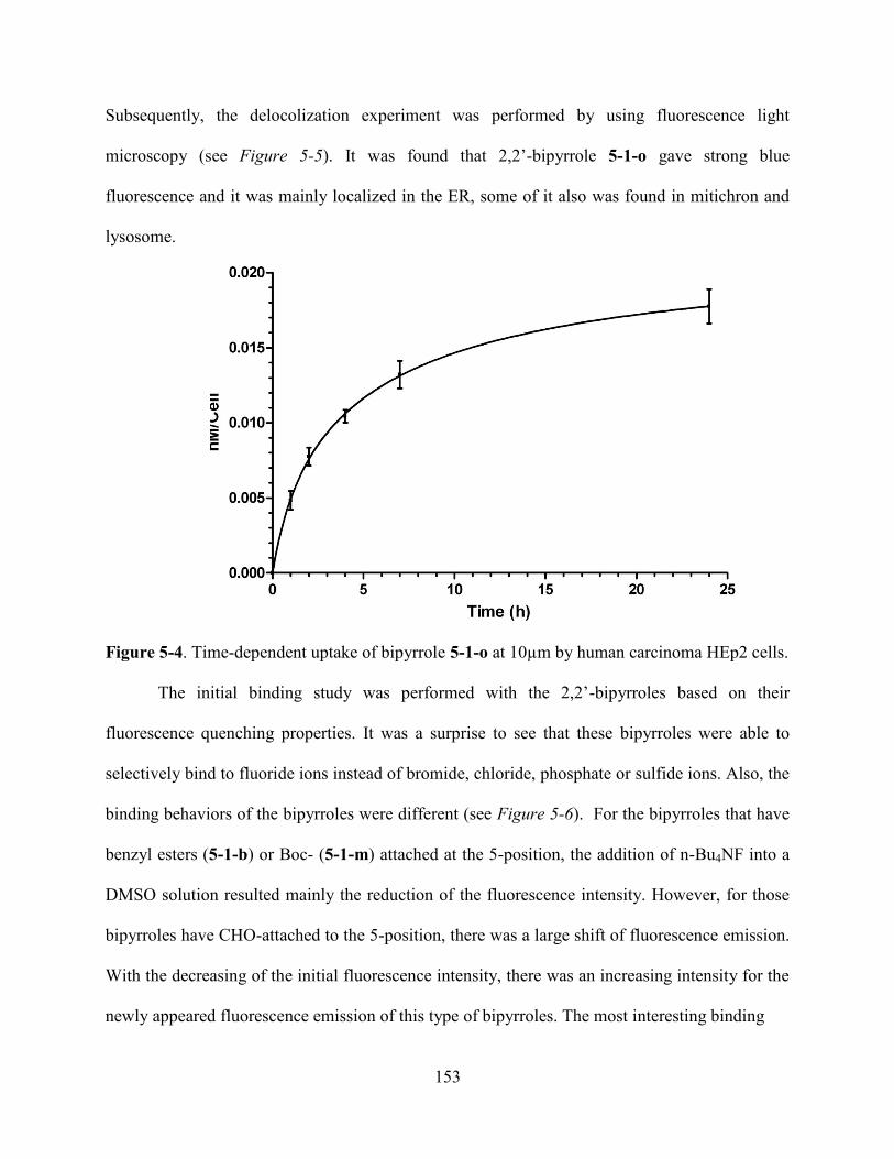
153
Subsequently, the delocolization experiment was performed by using fluorescence light
microscopy (see Figure 5-5). It was found that 2,2’-bipyrrole 5-1-o gave strong blue
fluorescence and it was mainly localized in the ER, some of it also was found in mitichron and
lysosome.
Figure 5-4. Time-dependent uptake of bipyrrole 5-1-o at 10µm by human carcinoma HEp2 cells.
The initial binding study was performed with the 2,2’-bipyrroles based on their
fluorescence quenching properties. It was a surprise to see that these bipyrroles were able to
selectively bind to fluoride ions instead of bromide, chloride, phosphate or sulfide ions. Also, the
binding behaviors of the bipyrroles were different (see Figure 5-6). For the bipyrroles that have
benzyl esters (5-1-b) or Boc- (5-1-m) attached at the 5-position, the addition of n-Bu4NF into a
DMSO solution resulted mainly the reduction of the fluorescence intensity. However, for those
bipyrroles have CHO-attached to the 5-position, there was a large shift of fluorescence emission.
With the decreasing of the initial fluorescence intensity, there was an increasing intensity for the
newly appeared fluorescence emission of this type of bipyrroles. The most interesting binding
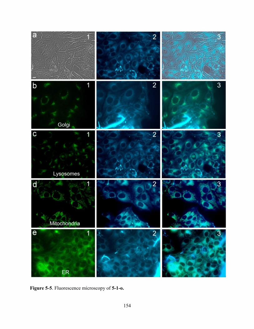
154
Figure 5-5. Fluorescence microscopy of 5-1-o.

155
350 400 450 500 550
-50
0
50
100
150
200
250
300
350
400R
ela
tive
In
ten
sity
Wavelength (nm)
0eq
6eq
50eq
150eq
200eq
250eq
300eq
400eq
500eq
1000eq
2000eq
3000eq
4000eq
5000eq
350 400 450 500 550 600
0
100
200
300
400
Re
lative
In
ten
sity
Wavelength (nm)
0eq
20eq
40eq
100eq
200eq
300eq
400eq
500eq
700eq
900eq
1100eq
1500eq
2000eq
2400eq
2800eq
350 400 450 500 550 600
0
200
400
600
800
1000
Rela
tive Inte
nsity
Wavelength (nm)
0eq
20eq
40eq
120eq
200eq
300eq
400eq
500eq
600eq
700eq
900eq
350 400 450 500 550
-50
0
50
100
150
200
250
300
350
Re
lative
In
ten
sity
Wavelength (nm)
0eq
200eq
400eq
600eq
800eq
1600eq
3000eq
6000eq
10000eq
350 400 450 500 550
0
100
200
300
400
500
Rela
tive Inte
nsity
Wavelength (nm)
0eq
1eq
2eq
10eq
50eq
100eq
200eq
400eq
600eq
800eq
1600eq
3000eq
6000eq
10000eq
350 400 450 500 550
-50
0
50
100
150
200
250
300
350
Re
lative
In
ten
sity
Wavelength (nm)
0eq
200eq
1000eq
3000eq
6000eq
10000eq
Figure 5-6. Fluorescence quenching upon addition of n-Bu4NF to DMSO solutions of 2,2’-
bipyrroles at r.t.. Top figures: 5-1-b (left, benzyl ester series) and 5-1-m (right, Boc series),
middle figures: 5-1-h (left) and 5-1-j (right), both are CHO series; bottom figures: 5-1-o (left,
CO2Et series), control experiment (right, no n-Bu4NF added).
156
behavior was observed for 5-1-o, which has an ethyl ester attached to the 5-position of the
bipyrrole. There was also a shift of fluorescence emission wavelength similar to that of 5-1-h and
5-1-j, but there was an increase of the fluorescence intensity instead of quenching for this
compound. The exact reason for this different fluorescence quenching behavior is still under
investigation. But it seems to be reasonable that besides the binding of bipyrrole to the fluoride
ion, a protonation/deprotonation process as described in the literature might also be involved. In
different bipyrrole systems, these two processes might have different contributions to the final
appearance of the fluorescence emission, which might result the different ―binding‖ behaviors
shown in Figure 5-6.
5.3 Conclusions and Future Work
In summary, we have developed an improved method for synthesis of 2,2’-bipyrroles
based on a Pd-C/zinc catalyzed homo-coupling in toluene/water at room temperature under argon.
The presence of water in the reaction system is critically important. These mild reaction
conditions allow functional groups such as aldehyde and nitrile to be carried through the
sequence intact. These bipyrroles are strongly luminescent and the new approach should provide
a ready access to new materials for light-emitting devices and bioimaging.
5.4 Experiment
5.4.1 General
All commercially available solvents and starting materials were used without further
purification. Silica gel (32-63 m) was used for flash column chromatography. All reactions
were monitored by TLC using 0.25 mm silica gel plates with or without UV indicator (60F-254).
Acetone (reagent plus, phenol free, ≥ 99.5%) and toluene (HPLC grade, Houston, TX) were

157
purchased from Sigma-Aldrich without further purification. Palladium, 10% on activated carbon,
reduced, dry powder was purchased from Strem Chemicals. Zinc dust, < 10 micron, 98+%, was
purchased from Sigma-Aldrich and activated according to the literature. 1H- and
13C-NMR
spectra were obtained on a DPX-250 or an ARX-300 Bruker spectrometer; chemical shifts ()
are given in ppm relative to CDCl3 (7.26 ppm), acetone-d6 (3.58 ppm, or DMSO-d6 (2.54 ppm).
MALDI-TOF mass spectra were obtained on an Applied Biosystems QSTAR XL. Electronic
absorption spectra were measured on a Perkin Elmer Lambda 35 UV-Vis spectrophotometer in
the 200-800 nm wavelength regions with 0.1 nm accuracy. Fluorescence spectra were measured
on a Perkin Elmer LS55 spectrometer in the 360-800 nm wavelength region with 1 nm accuracy.
Fluorescence quantum yields were measured using the standard method and quinine sulfate in
5% H2SO4 aqueous solution as the standard (quantum yield is 0.55), according to the literature.31
Monopyrroles used to make 2-iodopyrroles were synthesized according to the literature.27
Pyrrole 5-6-h was prepared in the same way as described in the literature27g
through a Vilsmeier
reaction on pyrrole 5c, the synthesis of which has been reported in the literature.27b
Pyrroles 5-4-j
were prepared from 5-6-j according to the literature.19
Pd-C (10%) was purchased from Strem
Chemicals. All the spectral data for CpoTMPn were in agreement with the literature6,21-22
. For
more information about these compounds, see scanned spectra at the end of this Chapter.
5.4.2 Total Synthesis of CpoTMPn
Preparation of 5-7-2
The preparation of 5-7-2 was achieved from the reaction of methoxypropionitrile (34 g, 0.4 mol),
ethyl bromoacetate (267.2 g, 1.6 mol) and activated zinc (104.7 g, 1.6 mol) in 500 ml freshly
distilled THF under argon condition. A vacuum was pulled over zinc dust for approximately 1
hour before adding freshly distilled THF. Then the reaction mixture was heated to reflux

158
temperature while stirring. To the refluxing solutions was added ethyl bromoacetate in a
dropwise manner through an addition funnel. Upon the appearance of a slightly green color in
the reaction mixture (usually around 0.8~1.0 mol), methoxypropionitrile (34 g, 0.4 mol) was
added all at once (through a syringe for small amount or through an addition funnel for large
amount). Then the rest of the ethyl bromoacetate was added to the reaction mixture in a dropwise
manner over a period of 1 hour. During this period, the color of the solution changed from green
to brown. Keeping adding until all the ethyl bromoacetate had been added into the reaction
mixture. After finishing addition of ethyl bromoacetate, the reaction mixture was left refluxing
for an additional 30 minutes. After cooling down to room temperature, the reaction mixture was
filtered through a Celite plug to get rid of any unreacted zinc powder. The solvent was then
removed under vacuum. The residue was deep yellow in color and was dissolved into EtOAc,
and subsequently 3M HCl (400 mL) was added. It was stirred at room temperature for a period
of 30 minutes. When stirring was stopped, EtOAc (400 mL) was added into the mixture to
extract the target compound from the aqueous solution. The organic phase was subsequently
washed with water, then with concentrated aqueous NaHCO3, and saturated NaCl solution. After
drying over anhydrous Na2SO4, the solvent was removed under vacuum. The residue, a reddish
liquid, was distilled under reduced pressure. The desired compound 5-7-2 was collected in the
95~110oC temperature range in 70% yield (52.6 g, 0.28 mol) as a colorless aromatic liquid. It
was found that high temperature should be avoided because the desired compound was destroyed
at high temperature, giving a highly sticky deep red gel.
Preparation of 5-7-1
Compound 5-7-1 was synthesized from 5-7-2 in two steps23
: 1) the formation of oxime; 2)
the Knorr reaction to form pyrrole. Oxime was generated from the reaction of 5-7-2 (33.2 g, 0.18
159
mol) with NaNO2 (13.34 g, 0.19 mol) in 143 mL HOAc. Dissolving 5-7-2 (33.2 g, 0.18 mol) in
ice-cold 143 mL HOAc and left it stir in ice-bath. Meanwhile, saturated aqueous NaNO2 (13.34 g,
0.19 mol) was prepared and placed into the ice-bath before adding into the ice-cool solution of 5-
7-2 (through pipette for small amount and addition funnel for large amount). This addition
process needed to be extremely slow to avoid the generation of toxic NOx gas. An addition
funnel was also connected to a bubble flask through tubes to control addition rate. After finishing
addition of NaNO2 solution, the ice-bath was removed and the reaction was continued to stir for
a period of 2 hours at room temperature. TLC is strongly recommended to follow this reaction.
The TLC eluting solvent was hexane/EtOAc (3/1). It should be noticed that the intermediate
oxime was more polar than desired 5-7-1, which showed polarity similar to the starting material
5-7-2. When TLC indicated the disappearance of oxime, the reaction was stopped. The resulting
solution was ready for the use in the next step—Knorr condensation to prepare 5-7-1. The Knorr
condensation reaction was performed in a three-necked large flask at temperature between
80~85oC. A water bath was strongly recommended rather than an oil-bath. The main reason was
the thermal sensitivity of this reaction and the strict temperature control could help the
achievement of high yield since large amount of heat was released over a short period. Water
bath provided great control of the temperature through constant addition of ice during the
reaction process. Ethyl acetoacetate (26.4 g, 0.16 mol) was dissolved in 26 mL HOAc in a three-
necked flask and the solution was heated to 80~85oC in a hot water bath. Activated zinc (25.4 g,
0.39 mol) was mixed well with NaOAc (31.8 g, 0.39 mol). This solid mixture was then added
slowly and frequently in small portion to the refluxing solution through funnel. The addition of
this mixed solid was alternated with the addition of oxime. Oxime was added to the mixture over
a period of 30 minutes, while the mixed solid was added over a period of 40 minutes. It was
160
important to have zinc always in excess throughout the reaction, which was easily achieved by
always allowing excess zinc powder in sight throughout the addition of oxime. After adding all
the reagents, the reaction mixture was stirred at 80~85oC for an additional period of 0.5~1 hour.
TLC was used to follow this reaction. When TLC indicated the disappearance of oxime from the
reaction mixture, the reaction was stopped. Recrystalization from ethanol water gave 5-7-1 in
60% yield (30.6 g, 0.11 mol) as a yellowish powder.
Preparation of 5-formyl-2-iodine-pyrrole
The synthesis of 5-formyl-2-iodopyrrole was achieved from the oxidation of 5-7-1
followed by iodination reaction as shown in Scheme 5-823a
. The preparation of 5-8-2 from 5-7-1
involved two steps: the oxidation and then the hydrolysis. In the oxidation step, 5-7-1 (5g, 0.02
mol) was dissolved in HOAc/Ac2O (16.6 ml/3.4 ml). The solution was placed in an ice-bath and
stirred at 0 oC. Under these conditions, Br2 (2.82 g, 0.91 mL) was added to the solution all at
once. Subsequently, SO2Cl2 (7.56g, 4.55 mL) was added slowly through addition funnel in the
dark over a period of 2 hours. Once half of the SO2Cl2 (2.5 mL) had been added, acetone was
added to the ice-bath to ensure the temperature remained below 0oC before addition of the rest of
the SO2Cl2. If the temperature was higher than 0 oC, the intermediate would be hydrolyzed by
HOAc during the oxidation process, which would resulted in incomplete oxidation and also ester
formation, thus lowering the yield of the desired acid.
After adding all of the SO2Cl2, the reaction mixture was refrigeratorated for a period of 4
hours. Then water at room temperature was added to the reaction mixture in a dropwise manner
until no more violent reaction could be observed. Then additional water was added very quickly.
The temperature was raised to 60 oC and kept there for a period of 30 minutes. Then the mixture
was poured it into cold water and set aside overnight. After filtration, it was ready for the
161
subsequent hydrolysis. In the hydrolysis step, the filtration residue was dissolved into 120 mL
mixture of acetone/water (v/v = 4/1) and heated to refluxing temperature. PH-paper indicated the
solution rapidly changed into acidic at around a period of 20 minutes refluxing. Then solid
NaHCO3 was added into the mixture solution; the generation of CO2 can be observed during this
process. Addition of solid NaHCO3 was stopped when no more CO2 was generated from the
reaction mixture. With the evaporation of acetone, the remaining product was separated over the
water layer as an oil form. Once the formation of two layers was observed in the refluxing flask,
the mixture was poured very slowly with constantly stirring into 100 mL cold water. Then let the
reaction mixture was occasionally stirred with glass rod for a period of 1 hour to ensure complete
precipitatation. After filtration, the solution was neutralized with 6 M HCl slowly under vigorous
stirring condition before keeping it at 0oC for a period of 8 hours at refrigerator. The resulting
fine solid was filtered and 5-8-2 was obtained in 45% yield (2.8 g, 0.009 mol). The iodination of
5-8-2 to generate 5-8-1 was performed in the mixture solvents of 1,2-dichloroethane/water with
the mixture of I2/KI as the iodination reagent. First NaHCO3 (2.96 g, 0.035 mol) was dissolved
into 16.5 mL water and heated to 50 oC. To this basic solution was added 5-8-2 (2.59 g, 0.008
mol) rapidly in several portions. When most of 5-8-2 was completed dissolved, 1,2-
dichloroethane (16.5 mL) was added and the temperature was raised to 71 oC. Then the saturated
aqueous solution of I2/KI (2.96 g/ 3.73 g) was added into the mixture over several minutes. The
reaction mixture was refluxed for an additional 40 minutes. Then Na2S2O3 was added to the
reaction mixture in portions to remove excess iodine. After separation and removal of the solvent
under vacuum, the desired product 5-8-1 was achieved in 90% yield (3.20 g, 0.008 mol).
162
Preparation of 5-1-a
The preparation of 5,5’-diformyl-2,2’-bipyrrole 5-1-a involved the 1) Ullmann-coupling
reaction, 2) the decarboxylation reaction and 3) Vilsmeier formylation reaction. The Ullmann-
coupling reaction was performed in DMI at room temperature with Cu-bronze as the coupling
reagent, under argon. To a round flask was added 5-8-1 (2.97 g, 0.0075 mol), vacuum was pulled
over the flask and then argon was applied to the system. After that, DMI (15.7 mL) was added.
Subsequently, Cu-bronze (2.82 g, 0.044 mol) was added into the reaction mixture all at once.
Then let the reaction to perform at room temperature over a period of 24~48 hours. The reaction
mixture was a suspended solution with greenish-brown color. TLC showed a strong blue
luminous spot under UV-radiation (366 nm). After adding water, the reaction mixture was
filtered through a Celite plug to remove precipitate and then washed with hot chloroform. The
organic phase was washed with aqueous 20% HNO3, water, and saturated NaHCO3. After
removing the solvents under vacuum, the residue was recrystallized from ethanol. After filtration,
the desired 5-9-2 was obtained as colorless needles in 60% yield (2.42 g, 0.0045 mol).
Following the formation of 5-9-2, hydrolysis to form the carboxylic acid was performed
in ethylene glycol at 200 oC with NaOH as the base, under argon. To ethylene glycol (130 mL)
was added 5-9-2 (2.42 g, 0.0045 mol) and NaOH (2.15 g, 0.05 mol). After NaOH was
completely dissolved, the reaction mixture was heated to 100 oC under argon. The mixture was
stirred at this temperature for a period of 30 minutes to achieve the hydrolysis of 5-9-2. TLC was
used to follow the hydrolysis reaction. Upon the disappearance of 5-9-2 and with the appearance
of a very polar new spot, the temperature was quickly raised to 200 oC for a period of 1 hour.
When TLC indicated the completion of decarboxylation from the disappearance of the polar spot,
the reaction mixture was poured into water and extracted with DCM. The organic phase was
163
quickly washed 8 times with water. After drying over Na2SO4, the solvent was removed and the
residue was put under vacuum for the subsequent use in the formylation reaction. The
formylation reaction used dry DMF as solvent; thus it was in excess amount. First, phosphoryl
chloride (6.14 g, 1.7 mL, 0.036 mol) was added into DMF (50 mL) under argon at 0 oC, and the
mixture was stirred at this temperature for a period of 15 minutes before adding the DMF
solution of the hydrolysis product (2.48 g, 0.01 mol). Then the temperature was raised to 60 oC
and the mixture was refluxed for 1 hour. It was then poured it into the aqueous NaOAC solution
and stirred at 85oC for 1 hour. The formation of a yellowish precipitate was observed. After
cooling down to room temperature and filtration, the residue was washed with cold water. After
drying over P4O10 under vacuum, after further recrystallization from THF, 5-1-a was obtained
44% yield (1.34 g, 0.0044 mol).
Preparation of 5-2
The preparation of symmetrical porphycene 5-2 was achieved using the McMurry
reductive coupling of 5-1-a6, 21-22
. Activated zinc metal (20g, 0.31 mol) was put into a flask and
vacuum was pulled over the flask, argon was then filled the system. Freshly distilled 800 mL
THF was then added. TiCl4 (16.5 mL, 0.15 mol) was added via syringe into the system all at
once. The solution turned blacking, and then brown. The reaction mixture was refluxed for a
period of 3 hours. Then the THF solution of 5-1-a (1.85 g, 6 mmol) was added to the reaction
mixture in a dropwise manner over a period of 10 minutes. TLC was used to strictly follow the
reaction and DCM was used as eluting solvent. The desired 5-2 gave a strong red-fluorescence
on TLC at 366nm (UV-radiation). After cooling to 0 oC, 300 mL 50% aqueous ammonium
solution was added over a period of 1 hour. After adding 600 mL DCM to the reaction mixture,
continued stirring for additional 15 minutes. Then the solution was poured into a Celite cake and
164
200 mL DCM was used to wash the cake. The organic layer was washed with water (300 mL).
After drying over Na2SO4, solvent was removed under vacuum and 5-2 obtained as blue powder
in 20% yield (325 mg, 0.6 mmol). The CpoTMPn was synthesized using PbO2 (40 mg, 0.167
mmol) as catalyst and reacting 5-2 (100 mg, 0.184 mmol) with hexanoic acid (2 mL, 0.016 mol)
in 15 mL dry DCM at room temperature under argon. After workup, CpoTMPn was obtained in
19.8% yield (24 mg, 0.037 mmol).
5.4.3 General Procedure for 2-Iodopyrrole 5-4-n (n = b-e, m-p)
Starting material pyrrole 5-5-n (n = b-e, m-p) was synthesized according to the
literature27
. 5-4-n (n = b-e, m-p) were obtained in two steps. Step 1. Hydrolysis: Pd-C (0.066 g, 6
mmol %) was added into a round bottom flask, to which was added the freshly distilled dry THF
10 mL to form a suspended solution of Pd-C. The mixture was stirred under a hydrogen
atmosphere for 20 minutes for the activation of Pd-C catalyst. Finally, 5-5-n (n = b-e, m-p) (1
mmol) was dissolved into freshly distilled dry THF 50 mL and added into the above solution
through syringe. After finishing adding all the reagents, the reaction mixture was left under a
hydrogen gas atmosphere for a period of 6-12 hours while TLC was used to follow this reaction.
The reaction was stopped when all starting material was consumed and a very polar spot was
observed on TLC. After finishing the reaction, the reaction mixture was passed through a Celite
cake to remove Pd-C and subsequently washed with THF several times. After drying over
anhydrous Na2SO4, solvent was removed under vacuum and the resulting carboxylic acid
pyrroles were ready for the next step without further characterization. Step 2. Iodination:
NaHCO3 (302.4 mg, 3.6 mmol) was added to 5 mL water and the solution was sonicated and
heated to 50oC in oil bath. After dissolving of NaHCO3, 5-5-n (n = b-e, m-p) (1 mmol) in 5 mL
1,2-dichloroethane was added and the reaction temperature was quickly raised to 70 oC. Shortly
165
after reaching 70 oC, a mixture of I2 /KI (279 mg, 1.1 mmol)/ (367 mg, 2.2 mmol) was dissolved
into 3 mL water and added into the reaction mixture all at once. Let it continue to stir at refluxing
conditions for a period of 1 hour. Upon completion of the reaction, it was cooled to room
temperature and sodium thiosulfate was added to the mixture in small portion under stirring
condition. Then the reaction mixture was poured into a separatory funeral and the organic layer
was collected. After drying over anhydrous Na2SO4, solvent was removed under vacuum and the
residue was recrystallization using methanol or ethanol/water.
Benzyl 5-iodo-3,4-dimethyl-pyrrole-2-carboxylate 5-4-b28g
: 81 % yield. mp 127-129 oC [lit
28g
mp 126-128 oC].
1H NMR (400 MHz, CDCl3) δ 8.90 (s, 1H), 7.43-7.35 (m, 5 H), 5.33 (s, 2H),
2.29 (s, 3H), 1.97 (s, 3H). ESI-MALDI Calcd for [C14H14INO2] m/z, 355.01, found: 355.10.
Benzyl 5-iodo-3-(3-methoxy-3-oxopropyl)-4-methyl-pyrrole-2-carboxylate 5-4-d28b
: 87 %
yield. mp 110-112 oC [lit
28b mp 110-111
oC].
1H NMR (250 MHz, CDCl3) δ 8.80 (s, 1H), 7.40-
7.38 (m, 5H), 5.30 (s, 2H), 3.64 (s, 3H), 3.10-3.04 (m, 2H), 2.53-2.47 (m, 2H), 2.00 (s, 3H). ESI-
MALDI Calcd for [C17H19INO2 + Na]+ m/z, 450.03, found: 449.97.
Benzyl 5-iodo-3-methyl-4-propyl-pyrrole-2-carboxylate 5-4-e28c
: 79 % yield. mp 118-120 oC
[lit28c
mp 119-120 oC].
1H NMR (250 MHz, CDCl3) δ 9.01 (s, 1H), 7.45-7.34 (m, 5H), 5.32 (s,
2H), 2.37-2.32 (m, 5H), 1.52-1.43 (m, 2H), 0.97-0.91 (m, 3H). 13
C NMR (250 MHz, CDCl3)
ppm 160.77, 136.58, 131.23, 129.02, 128.66, 127.66, 123.82, 74.13, 66.33, 28.86, 23.84, 14.27,
11.40. ESI-MALDI Calcd for [C16H18INO2] m/z, 383.22, found: 383.29.
Tert-butyl 3-ethyl-5-iodo-4-methyl-pyrrole-2-carboxylate 5-4-m28e
: 77 % yield. mp 105-107
oC [lit
28e mp 105-107
oC].
1H NMR (250 MHz, CDCl3) δ 9.49 (s, 1H), 3.68 (s, 3H), 3.06-2.99 (m,
2H), 2.54-2.47 (m, 2H), 1.97 (s, 3H), 1.56 (s, 9H). 13
C NMR (250 MHz, CDCl3) ppm 173.39,
166
159.88, 128.34, 125.49, 124.82, 81.27, 73.10, 51.43, 34.79, 28.34, 21.27, 11.69. ESI-MALDI
Calcd for C14H20INO4 m/z, 393.04, found: 392.90.
Diethyl 3-(2-ethoxyethyl)-5-iodo-pyrrole-2,4-dicarboxylate 5-4-o28h
: 83 % yield. mp 148-150
oC [lit
28h mp 150
oC].
1H NMR (250 MHz, CDCl3) δ 9.30 (s, 1H), 4.40-4.33 (m, 4H), 3.55-3.52
(m, 2H), 3.44-3.41 (m, 2H), 3.37 (s, 3H), 1.43-1.36 (m, 6H). ESI-MALDI Calcd for C14H20INO4
m/z, 393.04, found: 329.90.
Tert-butyl 3-ethyl-4,5-dimethyl-pyrrole-2-carboxylate 5-5-n28e
: 77 % yield. mp 105-107 oC
[lit28e
mp 105-107 oC].
1H NMR (300 MHz, CDCl3) δ 9.38 (s, 1H), 7.40-7.34 (m, 5H), 5.32 (s,
2H), 3.67 (s, 3H), 3.03-2.98 (m, 2H), 2.53-2.48 (m, 2H), 2.29 (s, 3H), 1.57 (s, 9H). 13
C NMR
(250 MHz, CDCl3) ppm 173.39, 159.88, 128.34, 125.49, 124.82, 81.27, 73.10, 51.43, 34.79,
28.34, 21.27, 11.69. ESI-MALDI Calcd for C14H20INO4 m/z, 393.04, found: 392.90.
5.3.3 General Procedure for Syntheses of Pyrrole 5-6-n (n = f-i): The following description
used pyrrole 5-5-i as a representative example for the preparation of pyrrole 5-6-n (n = f-g, i).
Pyrrole 5-5-i (2.0 g, 0.006 mole) was dissolved in THF (63 mL), and followed by addition of
HOAc(16 mL) and H2O (16 mL). Then 8.6 equiv of ceric ammonium nitrate (28.2 g, 0.052 mole)
was added to the mixture all in once. The reaction mixture was stirred at room temperature while
TLC was used to follow reaction progress. When there remained no starting material left, the
mixture was poured into 150 mL of water and extracted with DCM (100 mL) three times. The
organic layer was washed with water (100 mL) three times followed by saturated aqueous
NaHCO3 (100 mL). Then the organic extracts were combined and dried over anhydrous Na2SO4.
Finally the solution was concentrated under vacuum to remove the solvents. Recrystallization
from DCM/hexane gave a slight yellowish fine powder of 5-6-i in 89% yield (1.76 g).
167
Benzyl 3-Ethyl-5-formyl-4-methylpyrrole-2-carboxylate 5-6-f.27h,29b
91 % yield. Mp 86-87 °C
(lit.27h
mp 86-87 °C). 1H NMR (250 MHz, CDCl3) δ 9.76 (s, 1H), 9.53 (s, 1H), 7.44-7.33 (m, 5H),
5.34 (s, 2H), 2.79-2.70 (m, 2H), 2.30 (s, 3H), 1.23-1.17 (m, 3H). 13
C NMR (250 MHz, CDCl3)
ppm 179.80, 161.25, 137.21, 135.97, 129.06, 128.49, 128.93, 128.84, 127.10, 124.80, 67.04,
17.24, 16.84, 10.14. ESI-MS Calcd for C16H17NO3 m/z, 271.12, found: 271.18.
Benzyl 5-Formyl-4-(2-methoxycarbonylethyl)-3-methylpyrrole-2-carboxylate 5-6-g.27e,f,i
89% yield. Mp 79-80 °C (lit. 2i
mp 80-81 °C). 1H NMR (300 MHz, CDCl3) δ 9.81(s, 1H), 9.52 (s,
1H), 7.43-7.35 (m, 5H), 5.33 (s, 2H), 3.65 (s, 3H), 3.08-3.03 (m, 2H), 2.59-2.54 (m, 2H), 2.31 (s,
3H). 13
C NMR (250 MHz, DMSO- d6) ppm 180.49, 173.21, 161.20, 135.92, 132.36, 130.73,
128.99, 128.91, 128.76, 127.33, 124.75, 67.04, 52.08, 35.28, 19.25, 10.26. ESI- MS Calcd for
C18H19NO5 m/z, 329.13, found: 329.17.
Benzyl 4-(2-Cyanoethyl)-5-formyl-3-methylpyrrole-2-carboxylate 5-6-i. 87% yield. Mp 111-
113 °C. 1H NMR (250 MHz, CDCl3) δ 10.38 (s, 1H), 9.77 (s, 1H), 7.40-7.31 (m, 5H), 5.35 (s,
2H), 3.10-3.04 (m, 2H), 2.62-2.57 (m, 2H), 2.33 (s, 3H). 13
C NMR (250 MHz, CDCl3) ppm
180.27, 161.19, 135.73, 130.84, 129.43, 129.11, 128.96, 128.81, 127.68, 124.93, 119.36, 67.34,
20.25, 19.11, 10.35. ESI- MS Calcd for C17H16N2O3 m/z, 296.12, found: 296.15.
Benzyl 3:4-Butano-5-formylpyrrole-2-carboxylate 5-6-j. The starting material is readily
available pyrrole 5c. The procedure for the preparation of pyrrole 6c was different from the other
pyrroles 6. The Vilsmeier complex was prepared by adding 2 mL phosphoryl chloride to 20 mL
of dry DMF. Then benzyl 3:4-butanopyrrole-2-carboxylate 5c (2.55 g, 0.01 mol) was dissolved
in 20 mL of dry DMF and added slowly to the reaction mixture through a syringe under ice-bath
cooling conditions. The ice-bath was then removed and the mixture was refluxed for 45 min.
Then, 250 mL of aqueous NaHCO3 was added slowly until the pH reached 8. Stirring was
168
continued at 30oC until TLC indicated the complete hydrolysis of the intermediate imine salt.
The solution was extracted with DCM (100 mL) three times, and washed with water (100 mL)
three times, and finally dried over anhydrous Na2SO4 before removing the solvent under vacuum
to give the title compound 5-6-h as slightly yellow powder in 92% yield (2.61 g, 0.009 mol). Mp
111-112 °C. 1H NMR (250 MHz, CDCl3) δ 9.71 (s, 1H), 9.58 (s, 1H), 7.41-7.34 (m 5H), 5.34 (s,
2H), 2.84-2.81 (m, 4H), 1.80-1.78 (m, 4H). 13
C NMR (250 MHz, CDCl3) ppm 178.98, 160.68,
135.51, 131.91, 129.17, 128.86, 128.63, 128.42, 128.32, 123.19, 66.57, 22.72, 22.57, 22.36,
20.89. HRMS (ESI) Calcd for C17H18NO3 [M+H] +
284.1281, found: 284.1209.
5.4.4 General Procedure for Synthesis of Pyrroles 5-4-n (n = f-i)
Pyrroles 5-4-n (n = f-j) were obtained in two steps from pyrroles 5-6-n (n = f-i). A typical
preparation procedure involved two steps: hydrolysis and iodination. The hydrolysis was
monitored by TLC, there was a big polarity differences between the starting material and the
product. The following description using 5-4-g as a typical example to illustrate the general
procedure; the other 5,5’-diformyl-2,2’-bipyrroles were prepared in a similar manner.
5-Formyl-2-iodo-4-(2-methoxycarbonylethyl)-3-methylpyrrole 5-4-g. Step 1. Hydrolysis: Pd-
C (180 mg, 6%) was added into a 50 mL round bottom flask and 3 mL of freshly distilled dry
THF was added to form a suspension. This was stirred under a hydrogen atmosphere capped with
a balloon at room temperature for 20 min to active the Pd-C. Then, benzyl 5-formyl-4-(2-
methoxycarbonylethyl)-3-methylpyrrole-2-carboxylate 5-6-g (500 mg, 1.52 mmol) was
dissolved in 6 mL freshly distilled dry THF and added into the above solution through a syringe.
The mixture was stirred under the hydrogen atmosphere at room temperature for 6-12 h, using
TLC to follow the reaction; it was stopped when all the starting material has been transformed
into very polar spot on TLC. Upon completion of the reaction, the mixture was passed through a

169
Celite plug to remove Pd-C, followed by washing with THF (50 mL) three times. After removing
the combined solvent under vacuum, a grayish white powder of 5-formyl-4-(2-
methoxycarbonylethyl)-3-methylpyrrole-2-carboxylic acid was obtained in 97% yield (348.1
mg); it was used directly for the next iodination step without characterization. Step 2. Iodination:
NaHCO3 (453.6 mg 5.4 mmol) was added to 25 mL of water, into which the carboxylic acid
pyrrole (348.1 mg) obtained from the previous step was added, followed by sonication. Then the
reaction mixture was placed into a 50oC oil bath. Once carboxylic acid pyrrole was completely
dissolved, 25 mL of DCM was added to the reaction mixture and the temperature was quickly
raised to 70oC. Then, I2 (410.8 mg, 1.6 mmol) and KI (488.2 mg, 2.9 mmol) were dissolved in 15
mL of water and added to the reaction mixture all at once. The mixture was refluxed for 1 h,
using TLC to follow the reaction. The mixture was cooled and excess sodium thiosulfate was
added into the reaction mixture in small portions with stirring to remove excess iodine. The
reaction mixture was poured into a separatory funeral and the organic layer was collected. After
drying over anhydrous Na2SO4, the organic solvent was removed under vacuum. After
recrystallization from MeOH, a slightly yellow powder of 5-4-g was obtained in 85% yield
(401.4 mg). The overall yield of 5-4-g from 5-6-g was 82% (400mg, 1.25 mmol).
3-Ethyl-5-formyl-2-iodo-4-methylpyrrole 5-4-f.29b
85% yield. Mp 118-120 °C (lit.29b
mp 118-
120 °C). 1H NMR (250 MHz, CDCl3) δ 10.77 (s, 1H), 9.41 (s, 1H), 2.76-2.70 (m, 2H), 1.99 (s,
3H), 1.22-1.16 (m, 3H). 13
C NMR (250 MHz, CDCl3) ppm 176.53, 138.35, 133.38, 126.17,
83.80, 18.14, 16.89, 11.93. ESI- MS Calcd for C8H10INO m/z, 262.98, found: 263.05.
5-Formyl-2-iodo-4-(2-methoxycarbonylethyl)-3-methylpyrrole 5-4-g.29c
82% yield. Mp 91-
92 °C (lit.29c
mp 92 °C). 1H NMR (300 MHz, CDCl3) δ 10.66 (s, 1H), 9.41 (s, 1H), 3.64 (s, 3H),
3.06-3.00 (m, 2H), 2.57-2.51 (m, 2H), 1.96 (s, 3H). 13
C NMR (250 MHz, DMSO- d6) ppm
170
176.45, 172.63, 133.28, 132.84, 125.87, 82.60, 51.69, 35.06, 19.47, 11.46. ESI- MS Calcd for
C10H12INO3 m/z, 320.99, found: 321.06.
3:4-Butano-5-formyl-2-iodopyrrole 5-4-h. 84% yield. Mp 165-167 °C. 1H NMR (250 MHz,
CDCl3) δ 9.48 (br, 1H), 9.34 (s, 1H), 2.84-2.80 (m, 2H), 2.39-2.34 (m, 2H), 1.84-1.76 (m, 4H).
13C NMR (250 MHz, CDCl3) ppm 175.45, 133.53, 132.39, 128.57, 79.05, 23.01(2C), 22.66,
20.92. HRMS (ESI) Calcd for C9H11N2OI [M+H] +
275.9879, found: 275.9878.
4-(2-Cyanoethyl)-5-formyl-2-iodo-3-methylpyrrole 5-4-i. 81% yield. Mp 143-145 °C. 1H
NMR (250 MHz, CDCl3) δ 9.76 (s, 1H), 9.47 (s, 1H), 3.14-3.08 (m, 2H), 2.64-2.58 (m, 2H), 2.05
(s, 3H). 13
C NMR (250 MHz, CDCl3) ppm 175.81, 133.55, 129.49, 126.47, 118.58, 82.5, 20.41,
19.12, 11.64. HRMS (ESI) Calcd for C9H10N2OI [M+H] +
288.9832, found: 288.9834.
5.4.5 General procedure for reductive coupling of 5-4-n to generate 5-1-n (n =b-j, m-p)
Activated zinc was obtained by washing zinc dust with 3 M HCl and then filtering
through filter paper, after which it was washed successively with water, ethanol, and diethyl
ether, and then dried under vacuum. 5-4-b was used as an example to describe the general
procedure for reductive coupling of compounds 5-4-n (n = b-j, m-p). A mixture of Pd-C (10.0mg,
10%) and activated zinc powder (100 mg, 1.5 mmol) were placed in a dry 50 mL round bottom
flask. After removing air under vacuum, the flask was filled with argon. After that, 2 mL of
toluene was added to the reaction mixture which was stirred at ambient temperature under argon
protection for 15 mins before adding 2-iodopyrrole 5-4-b (132 mg, 0.5 mmol) dissolved in 8 mL
of toluene. Then 10 mL distillated water was added through a syringe into the reaction flask. The
reaction mixture was stirred vigorously at room temperature under argon. It is worth mention
that the Pd-C purchased from the other source or old catalyst would reduce the yield and in some
case even no reaction occurs. Also strict air-free and solvent adding sequence will also affect the
171
coupling yield. TLC was used to follow reaction, all of the five 5,5’-diformyl-2,2’-bipyrroles 5-
1-b display a characteristic blue fluorescence under UV irradiation (around 366 nm) on silica gel
TLC plates. Their DCM, acetone, DMSO and THF solutions also show strong blue luminescent
properties under the same wavelength illumination at ambient conditions, unlike the starting
material 5-4-b. Stop the reaction upon the disappearance of the starting material. DCM (40 mL)
was added and it was sonicated to form two layers. After removing the water layer, the remained
solution was filtered through a Celite plug, followed by washing with DCM (100 mL) three
times. The organic solvents were collected and dried over anhydrous Na2SO4, before evaporation
under vacuum. The pure target compounds were obtained by using silica gel column to separate
and eluted with EtOA /hexane (1: 2). Further purification could be performed by recrystallization
from DCM/hexane, or from ethanol.
5,5’-Bis(benzyoxyl)-3,3',4,4'-tetramethyl-2,2'-bipyrrole-2,2’-bipyrrole 5-1-b32
: After
recrystalizing from MeOH/DCM and washing with hexane (2 × 2 ml), pure 5-1-b was obtained
as a milk-white solid in 78 % yield (89.0 mg, 0.195 mmol). mp 218-220 oC [lit
32 mp 220
oC].
1H
NMR (250MHz, CDCl3) δ 9.08 (s, 2H), 7.39-7.34 (m, 10 H), 5.25 (s, 4H), 2.31 (s, 6H), 2.02 (s,
6H). 13
C NMR (250 MHz, CDCl3) ppm 161.42, 136.25, 128.51, 128.07, 128.03, 127.90, 124.90,
119.87, 119.00, 65.78, 10.67, 9.89. HRMS (ESI) Calcd for C28H29N2O4 [M+H]+: 457.2127,
found 457.2124.
5,5’-Bis(benzyoxyl)-3,3'-diethyl-4,4'-dimethyl-2,2'-bipyrrole 5-1-c: After recrystallizing from
DCM/hexane and washing with hexane (2 × 2 ml), pure 5-1-c was obtained as a milk-white solid
in 80 % yield (96.9 mg, 0.20 mmol). 1H NMR (300 MHz, CDCl3) δ 9.65 (s, 2H), 7.98 (s, 10H),
5.85 (s, 2H), 3.06-2.94 (m, 10H), 1.65 (s, 6H). 13
C NMR (300 MHz, CDCl3) ppm 161.35, 136.23,
172
128.50, 128.02, 127.08, 126.82, 124.18, 119.04, 65.74, 17.69, 15.42, 10.59. HRMS (ESI) Calcd
for C30H33N2O4 [M+H]+: 485.2440, found 485.2444.
5,5’-Bis(benzyoxyl)-4,4'-bis(3-methoxy-3-oxopropyl)-3,3'-dimethyl-2,2'-bipyrrole 5-1-d:
After recrystallizing from DCM/hexane and washing with hexane (2 × 2 ml), pure 5-1-d was
obtained as a milk-white solid in 65 % yield (97.5 mg, 0.16 mmol). 1H NMR (250 MHz, CDCl3)
9.21 (s, 2H), 7.37-7.33 (m, 10H), 5.23 (s, 4H), 3.63 (s, 6H), 3.07-3.01 (m, 4H), 2.54-2.47 (m, 4H,
2.05-2.00 (m, 6H). 13
C NMR (250 MHz, CDCl3) ppm 173.89, 161.28, 136.28, 130.82, 128.98,
128.75, 128.66, 125.30, 120.14, 119.45, 66.56, 51.91, 35.07, 21.11, 10.16. HRMS (ESI) Calcd
for C34H37N2O8 [M+H]+: 601.2550, found 601.2540.
5,5'-Bis(benzyloxy)-4,4'-dimethyl-3,3'-dipropyl-2,2'-bipyrrole 5-1-e: After recrystallizing
from MeOH/DCM and washing with hexane (2 × 2 ml), pure 4b was obtained as a milk-white
solid in 27 % yield (34.6 mg, 0.068 mmol). 1H NMR (250 MHz, CDCl3) 8.73 (s, 2H), 7.44-7.33
(m, 10H), 5.31 (s, 4H), 2.44-2.33 (m, 10H), 1.49-1.39 (m, 4H), 0.89-0.83 (m, 6H). 13
C NMR
(250 MHz, CDCl3) ppm 161.09, 136.24, 128.56, 128.13, 127.64, 124.85, 124.44, 118.90, 65.80,
29.70, 26.56, 24.07, 13.95, 10.65. HRMS (ESI) Calcd for [M+H] C32H37N2O4 [M+H]+:
513.2753, found 513.2759.
Di-tert-butyl 4,4'-diethyl-3,3'-dimethyl-2,2'-bipyrrole-5,5'-dicarboxylate 5-1-m: After
separating by using a silica gel column eluted with ethyl acetate and hexane, pure target 5-1-m
was obtained as a milk-white solid in 31% (32.3 mg, 0.078 mmol). 1H NMR (250 MHz, CDCl3)
δ 8.66 (s, 2H), 2.81-2.72 (m, 4H), 2.05 (s, 6H), 1.58 (s, 18H), 1.19-1.13 (m, 6H). 13
C NMR (250
MHz, CDCl3) ppm 161.37, 133.51, 124.49, 120.30, 118.95, 81.10, 28.85, 18.84, 15.52, 10.08.
HRMS (ESI) Calcd for C24H37N2O4 [M+H]+: 417.2753, found 417.2737.
173
5,5’-Di-4,4’-bis(2-methoxycarbonylethyl)-3,3’-dimethyl-2,2’-bipyrrole 5-1-n: After
separating by using a silica gel column eluted with ethyl acetate and hexane, pure target 11b was
obtained as a milk-white solid in 44% (58.5 mg, 0.11 mmol). 1H NMR (250 MHz, CDCl3) δ
8.75 (s, 2H), 3.67 (s, 6H), 3.08-3.01 (m, 2H), 2.60-2.54 (m, 2H), 2.05 (s, 6H), 1.56 (s, 18H). 13
C
NMR (250 MHz, CDCl3) ppm 173.59, 160.66, 128.92, 123.98, 120.55, 119.12, 81.12, 51.48,
34.90, 29.67, 20.77, 9.68. HRMS (ESI) Calcd for C28H41N2O8 [M+H]+: 533.2862, found
533.2854.
3,3',5,5'-tetracarboxylatethylester-4,4'-bis(2-ethoxyethyl)-2,2'-bipyrrole 5-1-o: After
recrystallization from DCM/hexane and washing with hexane (2 × 2 ml), pure 5-1-o was
obtained as a milk-white solid in 69% yield (92.5 mg, 0.17 mmol). 1H NMR (250 MHz, CDCl3)
δ 9.42 (s, 2H), 4.49-4.37 (m, 8H), 3.54-3.48 (s, 8H), 3.38 (s, 6H), 1.47-1.40 (s, 12H). FAB MS
Calcd for C26H36N2O10 m/z, 536.24, found: 536.30. MALDI-TOF MS Calcd for C26H36N2O10 m/z,
536.24, found: 536.41.
3,3’,4,4’-tetramethyl-5,5-dicarboxylatethylester -2,2’-bipyrrole 5-1-p: After separating by
using a silica gel column eluted with ethyl acetate and hexane, pure target 5-1-p was obtained as
a milk-white solid in 19% (63.1 mg, 0.19 mmol). 1H NMR (250 MHz, CDCl3) δ 8.97 (s, 2H),
4.34-4.25 (m, 4H), 2.31 (s, 6H), 2.05 (s, 6H), 1.38-1.32 (m, 6H). 13
C NMR (250 MHz, CDCl3)
ppm 161.70, 127.42, 124.55, 119.60, 119.25, 60.02, 14.47, 10.51, 9.87. MALDI-TOF MS Calcd
for C18H24N2O4 m/z, 332.17, found: 332.34.
3,3’-Diethyl-5,5-diformyl-4,4’-dimethyl-2,2’-bipyrrole 5-1-f9c
. After using the silica gel
column for the separation the title compound was obtained as a milk-white solid in 35% yield
(23.7 mg, 0.087 mmol). Mp 240-242 °C (lit. 9c
mp 241-242 °C). 1H NMR (250 MHz, CDCl3)
δ11.71(s, 2H), 9.61 (s, 2H), 2.74-2.71 (m, 4H), 1.95 (s, 6H), 1.56-1.10 (m, 6H). 13
C NMR (250
174
MHz, CDCl3) ppm 177.80, 135.93, 128.89, 128.18, 119.19, 16.93, 16.08, 9.31. HRMS (ESI)
Calcd for C16H21N2O2 [M+H] +
273.1597, found 273.1601.
5,5’-Diformyl-4,4’-bis(2-methoxycarbonylethyl)-3,3’-dimethyl-2,2’-bipyrrole 5-1-g. After
separating by using the silica gel column the title compound was obtained as a milk-white solid
in 54% yield (52.4 mg, 0.13 mmol). Mp >330 °C (dec.). 1H NMR (250 MHz, DMSO-d6) δ 11.7
(s, 2H), 9.60(s, 2H), 3.57 (s, 6H), 3.00-2.95 (m, 4H), 2.58-2.48 (m, 4H), 1.93 (s, 6H). 13
C NMR
(250 MHz, CDCl3) ppm 178.30, 172.61, 131.69, 129.32, 127.79, 119.70, 51.30, 34.51, 19.22,
9.30. HRMS (ESI) Calcd for C20H25N2O6 [M+H] +
389.1707, found 389.1707.
3:4, 3’:4’-Bisbutano-5,5’-diformyl-2,2’-bipyrrole 5-1-h. After separating by using the silica
gel column the title compound was obtained as a milk-white solid in 41% yield (30.3 mg, 0.10
mmol). Mp >310 °C (dec.). 1H NMR (300 MHz, DMSO-d6) δ 11.50 (s, 2H), 9.54 (s, 2H), 2.80
(m, 4H), 2.50 (m, 4H), 1.67 (br, 8H). 13
C NMR (250 MHz, DMSO-d6) ppm 177.26, 128.38 (2C),
126.85, 121.55, 22.94, 22.37, 21.76, 21.22. HRMS (ESI) Calcd for C18H21N2O2 [M+H]+
297.1597, found 297.1605.
4,4’-Bis(2-cyanoethyl)-5,5’-diformyl-3,3’-dimethyl-2,2’-bipyrrole 5-1-i. After separating by
using the silica gel column the title compound was obtained as a milk-white solid in 26% yield
(20.9 mg, 0.065 mmol). Mp >330 °C (dec.). 1H NMR (250 MHz, acetone-d6) δ 9.71 (s, 2H), 9.65
(s, 2H), 3.18-3.12 (m, 4H), 2.71-2.65 (m, 4H), 2.18 (s, 6H). 13
C NMR (250 MHz, acetone-d6)
ppm 178.86, 131.00, 130.71, 128.28, 121.43, 120.21, 20.79, 18.99, 9.72. HRMS (ESI) Calcd for
C18H19N4O2 [M+H]+
323.1502, found 323.1504.
5,5’-Diformyl-3,3’,4,4’-tetramethyl-2,2’-bipyrrole 5-1-j9a
. After separating by using the silica
gel column the title compound was obtained as a milk-white solid in 40% yield (24.4 mg, 0.10
mmol). Mp >307°C (dec.) [lit. 9a
mp 305-307 °C (dec.)]. 1H NMR (250 MHz, DMSO-d6) δ 11.66
175
(s, 2H), 9.61 (s, 2H), 2.25 (s, 6H), 1.91 (s, 6H). 13
C NMR (250 MHz, DMSO-d6) ppm 177.86,
129.53(2C), 128.06, 119.89, 9.44, 9.00. HRMS (ESI) Calcd for C14H17N2O2 [M+H] +
245.1284,
found 245.1289.
5.5 References
1 Dougherty, T. J. Photochem. Photobiol. 1987, 46, 569.
2 Gomer, C. J. Photochem. Photobiol. 1991, 54, 1093; b) Morgan, A. R.; Rampersaud, A.;
Garbo, G. M.; Keck, R. W.;Selman, S. H., J. Med. Chem. 1989, 32, 904.
3 Kadish, K. M.; Smith, K. M.; Guilard, R. Eds., In Porphyrin Handbook, Academic Press, San
Diego, San Francisco, New York, Boston, London, Sydney, Tokyo, Volume 2, 2000.
4 Vogel, E.; Köcher, M.; Schmickler, H.; Lex, J. Angew. Chem. Int. ed. Engl. 1986, 25, 257.
5 Aramendia, P. F.; Redmond, R. W.; Nonell, S.; Schuster, W.; Braslavsky, S. E.; Schaffner, K.
Photochem. Photobiol. 1986, 44, 555.
6 Richert, C.; Wessels, J. M.; Müller, M.; kisters, M.; benninghaus, T.; Goetz, A. E. J. Med.
Chem. 1994, 37, 2797.
7 Vogel, E.; Kocher, M.; Schmickler, H.; Lex, J. Angew. Chem., Int. Ed. Engl. 1986, 25, 257; b)
Sessler, J. L.; Weghorn, S. J. Expanded, Contracted and Isomeric Porphyrins, Pergamon:
New York, 1997.
8 Vogel, E.; Balci, M.; Kakumanu, P.; Kogh, P.; Lex, J.; Ermer, O. Angew. Chem., Int. Ed.
Engl. 1987, 26, 928; b) Nonell, S.; Bou, N.; Borrell, J. I.; Teixido, J.; Villanueva, A.;
Juarranz, A,; Canete, M. Teterhedron Lett. 1995, 36, 3405; c) Gavalda, A.; Borrell, J. I.;
Teixido, J.; Nonell, S.; Arad, O.; Grau, R.; Canete, M.; Juarranz, A,; Villanueva, A.; Stockert,
J. C. J. Porphyrins Phthalocyanines 2001, 5, 846; d) Hayashi, T.; Dejima, H.; Matsuo, T.;
Sato, H.; Murata, D.; Hisaeda, Y. J. Am. Chem. Soc. 2002, 124, 11226; e) Hayashi, T.;
Nakashima, Y.; Ito, K.; Ikegami, T.; Aritome, I.; Suzuki, A.; Hisaeda, Y. Org. Lett. 2003, 5,
2845.
9 Bauer, V. J.; Clive, D. L. J.; Dolphin, D.; Paine III, J. B.; Harris, F. L.; King, M. M.; Loder,
J.; Wang, S-W. C.; Woodward, R. B. J. Am. Chem. Soc. 1983, 105, 6429; b) Broadhurst, M.
J.; Grigg, R.; Johnson, A. W. J. Chem. Soc., Perkin Trans. 1 1972, 2111; c) Sessler, J. L.;
Cyr, M. J.; Lynch, V.; McGhee, E.; Ibers, J. A. J. Am. Chem. Soc. 1990, 112, 2810; d)
Schevchuk, S. V.; Davis, J. M.; Sessler, J. L. Tetrahedron Lett. 2001, 42, 2447.
10 Sessler, J. L.; An, D.; Cho, W-S.; Lynch, V. Angew. Chem., Int. Ed. Engl. 2003, 42, 2278; b)
Setsune, J.-I.; Tsukajima, A.; Watanabe, J. Tetrahedron Lett. 2006, 47, 1817; c) Geier, G. R.
III.; Grindrod, S. C. J. Org. Chem. 2004, 69, 6404; d) Decréau, R. A.; Collman, J. P.
Tetrahedron Lett. 2003, 44, 3323.
176
11 The Porphyrin Handbook, Kadish, K. M.; Smith, K. M.; Guilard, R., Eds.; Academic Press:
San Diego, 2000; Vol 6; b) Gale, P. A.; Anzenbacher, P., Jr.; Sessler, J. L.; Coord. Chem.
Rev. 2001, 222, 57; c) Kleemann, A.; Engl, J.; Kutscher, B.; Reichert, D. Pharmaceutical
Substance: Synthesis, Patents, Applications, 4th
ed.; Georg Thieme: Stuttgart, 2001.
12 Ceacareanu, D. M.; Gerstenberger, M. R. C.; Haas, A. J. Heterocycl. Chem. 1985, 22, 281; b)
Wasserman, H. H.; Petersen, A. K.; Xia, M.; Wang, J. Tetrahedron Lett. 1999, 40, 7567; c)
Oda, K.; Sakai, M.; Ohno, K.; Machida, M. Heterocycles 1999, 40, 277; d) Dohi, T.;
Morimoto, K.; Maruyama, A.; Kita, Y. Org. Lett. 2006, 8, 2007.
13 Sessler, J. L.; Hoehner, M. C.; Synlett 1994, 3, 211; b) Vogel, E.; Deponte, R. US Patent,
5,756,724; c) Skowronek, P.; Lightner, D. A. Monatsh. Chem. 2003, 134, 889; d) Furstner,
A.; Radkowski, K.; Peters, H. Angew. Chem., Int. Ed. Engl. 2005, 44, 2777.
14 Arad, O.; Morros, J.; Batllori, X.; Teixido, J.; Nonell, S.; Borrell, J. I. Org. Lett. 2006, 8, 847.
15 Hassan, J.; Sevignon, M.; Gozzi, C.; Schulz, E.; Lemaire, M. Chem. Rev. 2002, 102, 1359. (b)
Grigg, R.; Johnson, A. W.; Wasley, J. W. F. J. Chem. Soc. 1963, 359. (c) Zhang, S.; Zhang,
D.; Liebeskind, L. S. J. Org. Chem. 1997, 62, 2312.
16 Hassan, J.; Sevigon, M.; Gozzi, C.; Schulz, E.; Lemaire, M. Chem. Rev. 2002, 102, 1359; b)
Shezad, N.; Clifford, A. A.; Rayner, C. M. Green Chem. 2002, 4, 64.
17 Venkatraman, S.; Li, C-J. Org. Lett. 1999, 1, 1133 ; b) Venkatraman, S.; Li, C-J.
Tetrahedron Lett. 2000, 41, 4831.
18 Trost, B. M. Accounts Chem. Res. 1990, 23, 34; b) Stille, J. F. Angew. Chem. Int. Ed. Engl.
1986, 75, 508; c) Miyaura, N.; Suzuki, A. Chem. Rev. 1995, 95, 2457; d) Heck, R. H.
Accounts Chem. Res. 1979, 12, 146; e) Sonogashira, K. In Comprehensive Organic Synthesis;
Trost, B. M., Ed.; Pergamon; Oxford, 1991; f) Bamfield, P.; Quan. P. M. Synthesis, 1978,
537.
19 Cho, D. H.; Lee, J. H.; Kim, B. H. J. Org. Chem. 1999, 64, 8048.
20 Tolendano, H.; Edrei, R.; Kimel, S., J. Photochem. Photobiol, B: Biology, 1998, 42, 20.
21 Vogel, E.; Emanuel, M.; Martin, H.; Otto, C.; Alexander, D., US patent: WO/1996/031452,
1996.
22 Szeimies, R.-M.; Karrer, S.; Abels, C.; Steeinbach, P.; Fickweiler, S.; messmann, H.;
Baeumler, W.; Landthaler, M. J. Photochem. Photobiol. B, 1996, 34, 67.
23 Jones, R. A.;Bean, G. P., In The chemistry of pyrroles, Academic Press, London, New York,
1977; b) Dolphin, D., In The Porphyrins, Academic Press, New York, 1978.
24 Dorff, P. H.; Chiu, G.; Goldstein, S. W.; Morgan, B. P. Tetrahedron Lett. 1998, 39, 3375.
25 Venkatraman S.; Li, C.-J. Org. Lett. 1999, 1, 1133.
177
26 Itahara, T. J. Org. Chem. 1985, 50, 5272.
27 Kleinspehn, G. G. J. Am. Chem. Soc., 1955, 77, 1546; b) John, K. D.; Pearce, W. A.; Pyke, S.
M. J. Porphyrins Phthalocyanines, 2002, 6, 661; c) Takaya, H.; Kojima, S.; Murahashi, S. -
I. Org. Lett., 2001, 3, 421; d) Smith, K. M.; Martynenko, Z.; Pandey. R. K.; Tabba, H. D. J.
Org. Chem., 1983, 48, 4296; e) Smith, K. M.; Milgrom, L. R.; Kenner, G. W. J. Chem. Soc.,
Perkin Trans. 1, 1981, 2065; f) Smith, K. M.; Pandey, R. K. J. Chem. Soc., Perkin Trans. 1
1987, 1229; g) Smith, K. M.; Minnetian, O. M. J. Org. Chem. 1985, 50, 2073; h) Jackson, A.
H.; Kenner, G. W.; Warburton, D. J. Chem. Soc. 1965, 1328; i) Ringuet, M.; Gagnon, J. Can.
J. Chem. 1985, 63, 2420.
28 (a) Smith, K. M.; Minnetian, O. M. J. Org. Chem. 1985, 50, 2073-2080. (b) Cox, M. T.;
Jackson, A. H.; Kenner, G. W. J. Chem. Soc. C, 1971, 1974-1981. (c) Al-Hazimi, H. M. G.;
Jackson, A. H.; Knight, D. W.; Lash, T. D. J. Chem. Soc. Perkin Trans 1, 1987, 265. (d)
Jackson, A. H.; Kenner, G. W.; Smith, K. M. J. Chem. Soc. C, 1971, 502. (e) Clelow, P. J. J.
Chem. Soc. Perkin Trans 1, 1990, 1925. (f) Paine, J. B. III. In The porphyrins; Dolphine, D.
Ed.; Academic; New York, 1978; Vol. 1, Chapter 4, pp101-234. (g) Grigg, R.; Jackson, A.
W. J. Chem. Soc. 1964, 3315. (h) Richert, C.; Wessels, J. M.; Müller, M.; Kisters, M.;
Benninghaus, T.; Goetz, A. E. J. Med. Chem. 1994, 37, 2797.
29 (a) Thyrann, T.; Lightner, D. A. Tetrahedron Lett. 1995, 36, 4345. (b) Tu, B.; Lightner, D. A.
J. Heterocycl. Chem. 2003, 40, 707; b) Jacobi, P. A.; Pippin, D. Org. Lett. 2001, 3, 827.
30 Kuo, W.-J.; Chen, Y-H.; Jeng, R.-J.; Chan, L.-H.; Lin, W-P.; Yang, Z.-M. Tetrahedron,
2007, 63, 7086; b) Che, C-M.; Wan, C-W.; Lin, W-Z.; Yu, W-Y.; Zhou, Z-Y.; Lai, W. -Y.;
Lee, S. -T. Chem. Commun. 2001, 721.
31 Fery-Forgues, S.; Lavabre, D. J. Chem. Ed. 1999, 76, 1260.
32 Heinz, F. Monatshefte Fuer Chemie, 1988, 119(2), 247-252.
178
CHAPTER 6. THE UNIQUE REGIOCHEMISTRY OF MONO-(L)-
ASPARTYLCHLORIN-E6
6.1 Introduction
PDT is a binary cancer therapy that relies on the selective uptake of a photosensitizer into
tumor tissues, followed by generation of singlet oxygen and other cytotoxic species upon
irradiation with light of an appropriate wavelength1-3
. Photofrin® (porfimer sodium) has been
commercially developed and approved in more than 40 countries as a 1st generation
photosensitizer. It has limited application in the PDT treatment of cancers, due to the following
factors: it has low absorption of light within the ―therapeutic window‖ (600-800 nm) and it is
slow to clear from skin, resulting in residual patient photosensitivity4. To extend the application
of photosensitizers in PDT, so-called 2nd
-generation photosensitizers, such as LS-11 (6-10) has
been developed recently. LS-11 is the latest name for mono-(L)-aspartylchlorin-e6, which
previously had been also known as Talaporfin sodium, NPe6 and MACE. As a chlorophyll
derivative, LS-11 has been used in advanced-stage clinical trials for PDT. LS-11 has a strong
characteristic absorption maximum at 666 nm (solvent dependent), ability to generate cytotoxic
singlet oxygen in high yields upon irradiation, and enjoys rapid clearance from normal tissue4-5
.
Moreover, it has increased stability and amphiphilicity compared with many synthetic chlorins
tested as PDT sensitizers.
A patent search identifies LS-11 (at that time ―NPe6‖ or Talaporfin sodium) as the 173-
aspartyl derivative, though the option for a mixture with other regioisomers was left open in the
patent itself6. Surprisingly, in 1998 a very thorough 2D NMR study was published claiming that
LS-11 is actually the 152-regioisomer
7 (6-10). Unfortunately, the conclusions in this paper were
not universally accepted due to the ambiguous NMR spectra of the aggregated compound8 in
179
water. Additionally, the result was counter-intuitive from a mechanistic perspective since the
propionic side chain is the most reactive and also the least steric hindered position compared
with the acetic and formic analogs. Although this paper also reported the syntheses and
spectroscopic evaluation of the 173- and 13
1-positional isomers of 15
2-LS-11, the yields were
low and the synthetic routes to these regioisomers were ambiguous. As a result, most papers ever
since 1998 reported LS-11 to be either the 152-or the 17
3-aspartyl derivatives, most often the
latter. The identity of LS-11 has remained a matter of conjecture and the distributors appear to
have remained silent on the critically important structural issue raised by Gomi et al.7.
Meanwhile, subsequent to the initial patent, our group had also synthesized and biologically
studied this molecule as well as an over-reacted di-(L)-aspartylchlorin-e6 (―DACE‖) that was
correctly identified as the 173,15
2-diaspartyl compound (6-8)
9. Recently, our group also reported
the unambiguous improved syntheses of the 131-, 15
2- and 17
3-aspartyl regioisomers of LS-11,
as their tetramethyl esters10
. The transfer of free acids into esters provided good solubility and
avoided the formation of aggregates in solution; thus monomeric NMR spectra were obtained. In
the meanwhile, we were also able to obtain the X-ray structure of the tetramethyl ester of
authentic LS-11, which was prepared from the coupling of commercially available chlorin-e6
with aspartic acid derivatives using peptide coupling reagents, such as DCC. Our results are in
agreement with the recent, though unaccepted, report7 that aspartyl group is attached to the 15
2-
side chain position in chlorin-e6. With the absence of carboxylic protecting groups during the
synthesis of the commercial material, which is formed clearly as one pure regioisomer, it is
really surprising to see such a highly regioselective coupling to form uniquely the 152-positional
LS-11 derivative (6-10). Considering the fact that chlorin-e6 possesses no less than three
carboxylic acid functional groups, all of which are able to undergo amino-acid coupling reactions,
180
if there is selective formation of one unique regioisomer, then it should be the 173-regioisomer of
LS-11 instead of 152-aspartyl isomer. However, the regioisomer isolated from experiment is
indeed the 152-positional LS-11
7, 10 instead of that at 17
3. It seems to us that the reaction should
have an unknown complexity in the pathway which guides the efficient coupling of amino acid
at the specifically unfavorable 152-position. To discover this pathway, we designed reactions and
explored MALDI-TOF and NMR techniques. In the meanwhile, we achieved a key improvement
in synthesizing the 152-positional LS-11, in which organic base was found to play an important
role during the coupling.
When using H-Lys (Boc)-OtBu instead of L-aspartic acid, the 152-positional Lys-
substituted chlorin-e6 was also achieved in good yield with high regio-selectivity.
6.2 Results and Discussion
We hypothesized that the highly regioselective formation of the 152-positional
regioisomer was due to the formation of a seven-member anhydride ring intermediate, which has
high reactivity and can easily undergo a ring opening reaction upon addition of a nucleophilic
reagent, such as the L-aspartic acid dimethy lester in the absence of a peptide coupling agent (see
Scheme 6-1). To substantiate our rationale, first we designed a reaction to prove that cyclic
anhydride ring do form for 6-1 under the coupling condition before the addition of nucleophilic
reagent. The designed reaction was performed by coupling pure chlorin-e6 with the peptide
coupling reagent DCC6,10
. In this coupling reaction, 1 equivalent of chlorine-e6 was suspended in
dry DCM solution under an argon atmosphere. One equivalent of DCC and 1 equivalent of
DMAP were dissolved in dry DCM and were added to a DCM solution of chlorin-e6 at room
temperature under an argon atmosphere. It took around 5-10 minutes for chlorin-e6 to become
completely dissolved after adding DCC/DMAP. In the meanwhile, the solution changed into a

181
brown color. It took a round 30 minutes before TLC indicated the formation of two new spots,
which changed color on the TLC plate upon standing. The initial color was brown and
subsequently it changed into green. The minor of the two products has Rf value at 0.8 and the
major product has Rf value at 0.4 when 10% acetone/DCM was used as the eluting solvent.
MALDI mass spectra of the reaction mixture gave two peaks at MW 785 and 578 (see Figure 6-
1).
N
NHN
HN
Me Me
Et
Me
Me
O OH
O
OH
N
NHN
HN
Me Me
Et
Me
Me
O
O
OH
L-aspartic acid
DCC/ DMAP
N
NHN
HN
Me Me
Et
Me
Me
OO
OH OH
MeO2C CO2Me
CH2N2 /DCM
Chlorin e6 6-1
6-46-5
OH
O
OO
NH
O
N
NHN
HN
Me Me
Et
Me
Me
OO
OMe OMe
MeO2C CO2Me
NH
O

182
Scheme 6-1. Rationale for the unique formation of 6-5.
Figure 6-1. MALDI-TOF mass spectrum of the reaction mixture (left) and 6-2 (right) with CCA.
CH2N2 /DCM
6-1 6-6
N
NHN
HN
Me Me
Et
Me
Me
O
O
OH OO
N
NHN
HN
Me Me
Et
Me
Me
O
O
OMe OO
Scheme 6-2. Methylation of 6-1 using diazomethane in DCM to generate 6-6.
The separation was performed using silica gel TLC, with 10% acetone/DCM as the
eluting solvent. However, due to their high reactivity, only tiny amount of pure products could be
isolated. Thus only MALDI and UV-vis studies were performed with them. The MALDI-TOF
spectrum gave a peak at 578 for the isolated polar compound, which matched with the formation
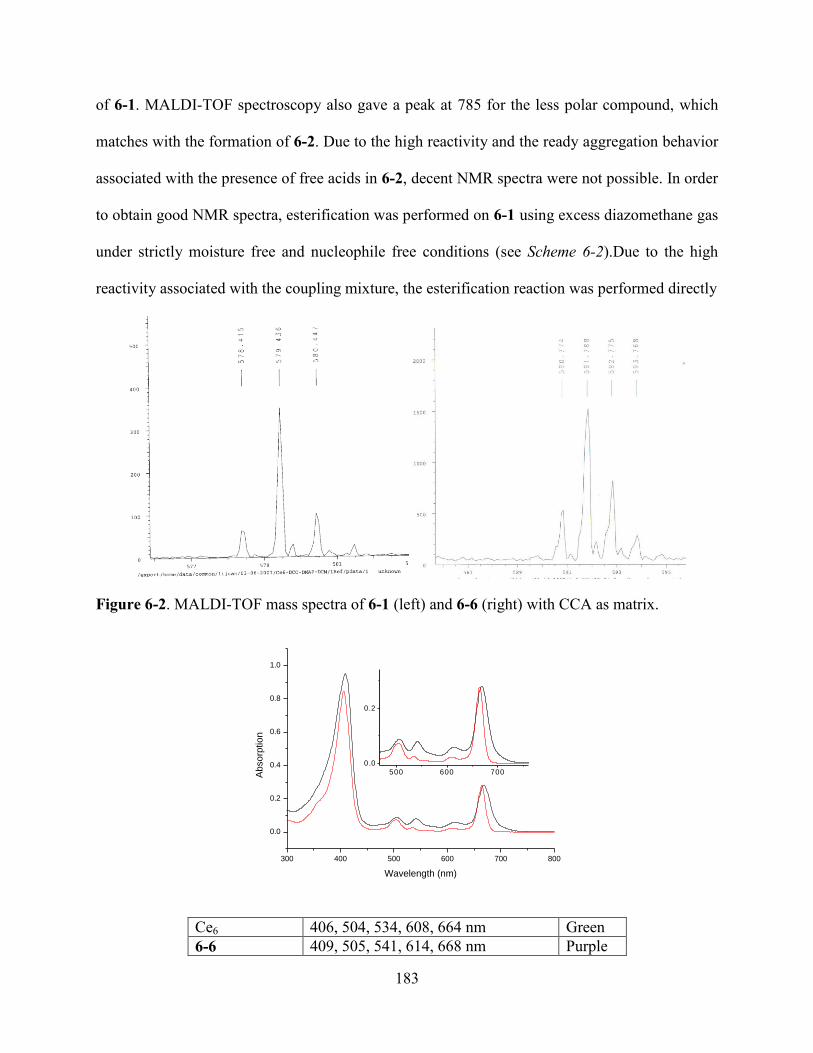
183
of 6-1. MALDI-TOF spectroscopy also gave a peak at 785 for the less polar compound, which
matches with the formation of 6-2. Due to the high reactivity and the ready aggregation behavior
associated with the presence of free acids in 6-2, decent NMR spectra were not possible. In order
to obtain good NMR spectra, esterification was performed on 6-1 using excess diazomethane gas
under strictly moisture free and nucleophile free conditions (see Scheme 6-2).Due to the high
reactivity associated with the coupling mixture, the esterification reaction was performed directly
Figure 6-2. MALDI-TOF mass spectra of 6-1 (left) and 6-6 (right) with CCA as matrix.
300 400 500 600 700 800
0.0
0.2
0.4
0.6
0.8
1.0
500 600 700
0.0
0.2
Ab
so
rptio
n
Wavelength (nm)
Ce6 406, 504, 534, 608, 664 nm Green
6-6 409, 505, 541, 614, 668 nm Purple
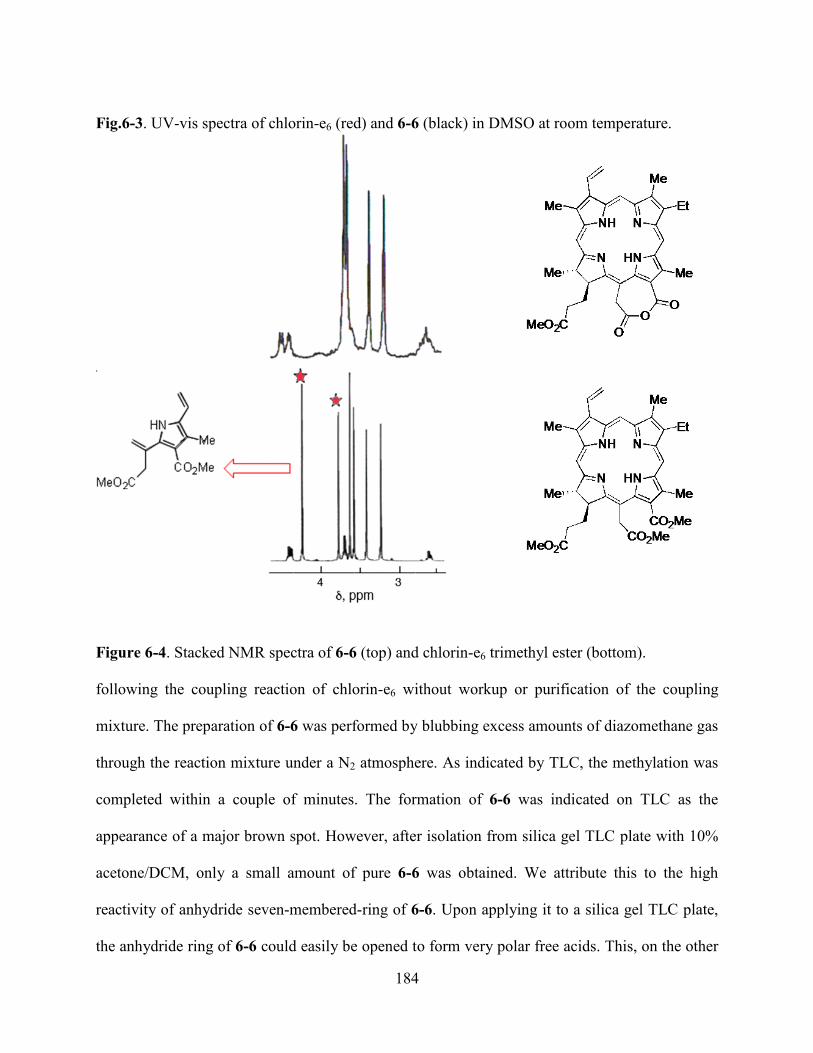
184
Fig.6-3. UV-vis spectra of chlorin-e6 (red) and 6-6 (black) in DMSO at room temperature.
Figure 6-4. Stacked NMR spectra of 6-6 (top) and chlorin-e6 trimethyl ester (bottom).
following the coupling reaction of chlorin-e6 without workup or purification of the coupling
mixture. The preparation of 6-6 was performed by blubbing excess amounts of diazomethane gas
through the reaction mixture under a N2 atmosphere. As indicated by TLC, the methylation was
completed within a couple of minutes. The formation of 6-6 was indicated on TLC as the
appearance of a major brown spot. However, after isolation from silica gel TLC plate with 10%
acetone/DCM, only a small amount of pure 6-6 was obtained. We attribute this to the high
reactivity of anhydride seven-membered-ring of 6-6. Upon applying it to a silica gel TLC plate,
the anhydride ring of 6-6 could easily be opened to form very polar free acids. This, on the other
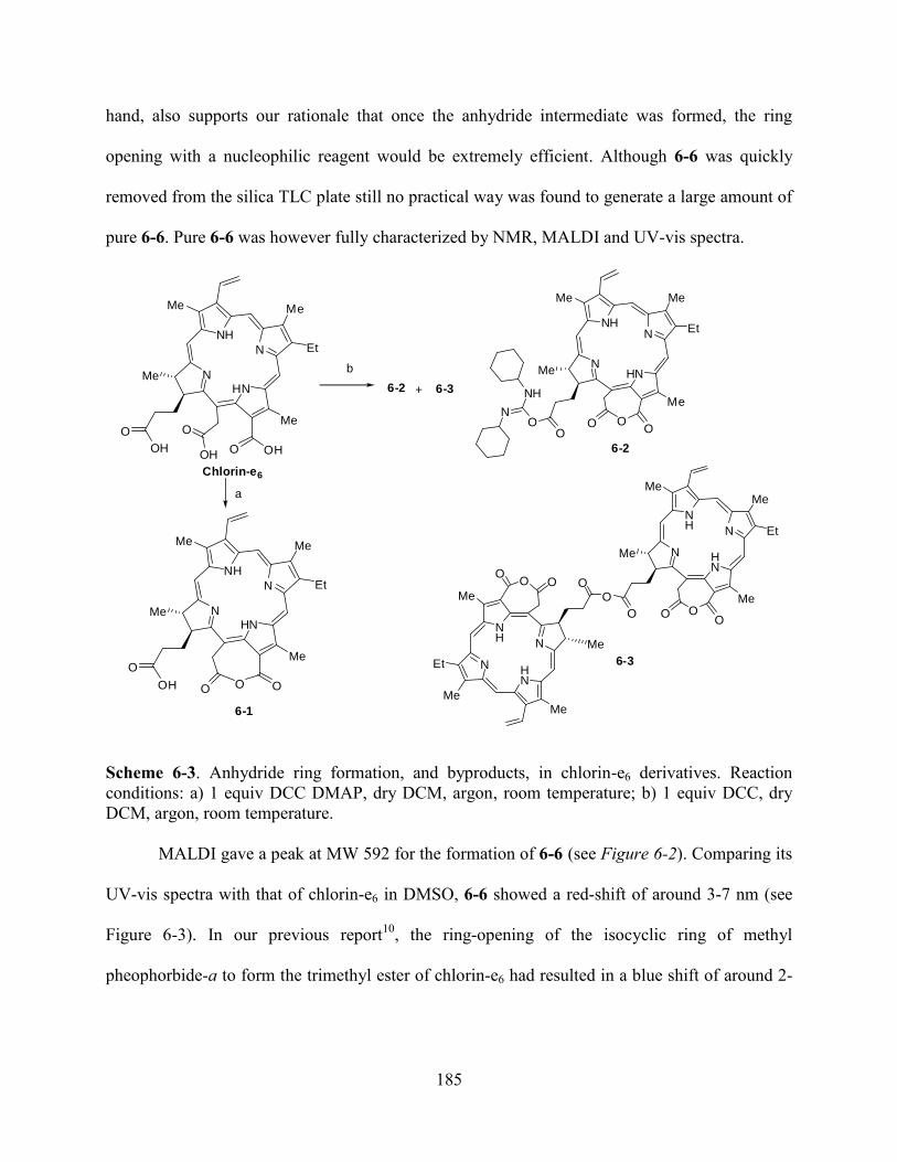
185
hand, also supports our rationale that once the anhydride intermediate was formed, the ring
opening with a nucleophilic reagent would be extremely efficient. Although 6-6 was quickly
removed from the silica TLC plate still no practical way was found to generate a large amount of
pure 6-6. Pure 6-6 was however fully characterized by NMR, MALDI and UV-vis spectra.
N
NH
N
HN
Me Me
Et
Me
Me
O OH
O
OH
N
NH
N
HN
Me Me
Et
Me
Me
O
O
OH
N
NHN
HN
Me Me
Et
Me
Me
OOO
N
NH
a
b
N
NH N
HN
MeMe
Et
Me
Me
OO
N
HN
N
NH
MeMe
Et
Me
OO
O
Me
+
Chlorin-e6
6-2
6-3
6-1
OO
O O
OO
OH
O
OO
6-2 6-3
Scheme 6-3. Anhydride ring formation, and byproducts, in chlorin-e6 derivatives. Reaction
conditions: a) 1 equiv DCC DMAP, dry DCM, argon, room temperature; b) 1 equiv DCC, dry
DCM, argon, room temperature.
MALDI gave a peak at MW 592 for the formation of 6-6 (see Figure 6-2). Comparing its
UV-vis spectra with that of chlorin-e6 in DMSO, 6-6 showed a red-shift of around 3-7 nm (see
Figure 6-3). In our previous report10
, the ring-opening of the isocyclic ring of methyl
pheophorbide-a to form the trimethyl ester of chlorin-e6 had resulted in a blue shift of around 2-
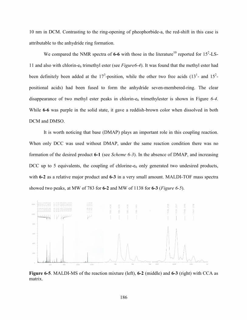
186
10 nm in DCM. Contrasting to the ring-opening of pheophorbide-a, the red-shift in this case is
attributable to the anhydride ring formation.
We compared the NMR spectra of 6-6 with those in the literature10
reported for 152-LS-
11 and also with chlorin-e6 trimethyl ester (see Figure6-4). It was found that the methyl ester had
been definitely been added at the 173-position, while the other two free acids (13
1- and 15
2-
positional acids) had been fused to form the anhydride seven-membered-ring. The clear
disappearance of two methyl ester peaks in chlorin-e6 trimethylester is shown in Figure 6-4.
While 6-6 was purple in the solid state, it gave a reddish-brown color when dissolved in both
DCM and DMSO.
It is worth noticing that base (DMAP) plays an important role in this coupling reaction.
When only DCC was used without DMAP, under the same reaction condition there was no
formation of the desired product 6-1 (see Scheme 6-3). In the absence of DMAP, and increasing
DCC up to 5 equivalents, the coupling of chlorine-e6 only generated two undesired products,
with 6-2 as a relative major product and 6-3 in a very small amount. MALDI-TOF mass spectra
showed two peaks, at MW of 783 for 6-2 and MW of 1138 for 6-3 (Figure 6-5).
Figure 6-5. MALDI-MS of the reaction mixture (left), 6-2 (middle) and 6-3 (right) with CCA as
matrix.
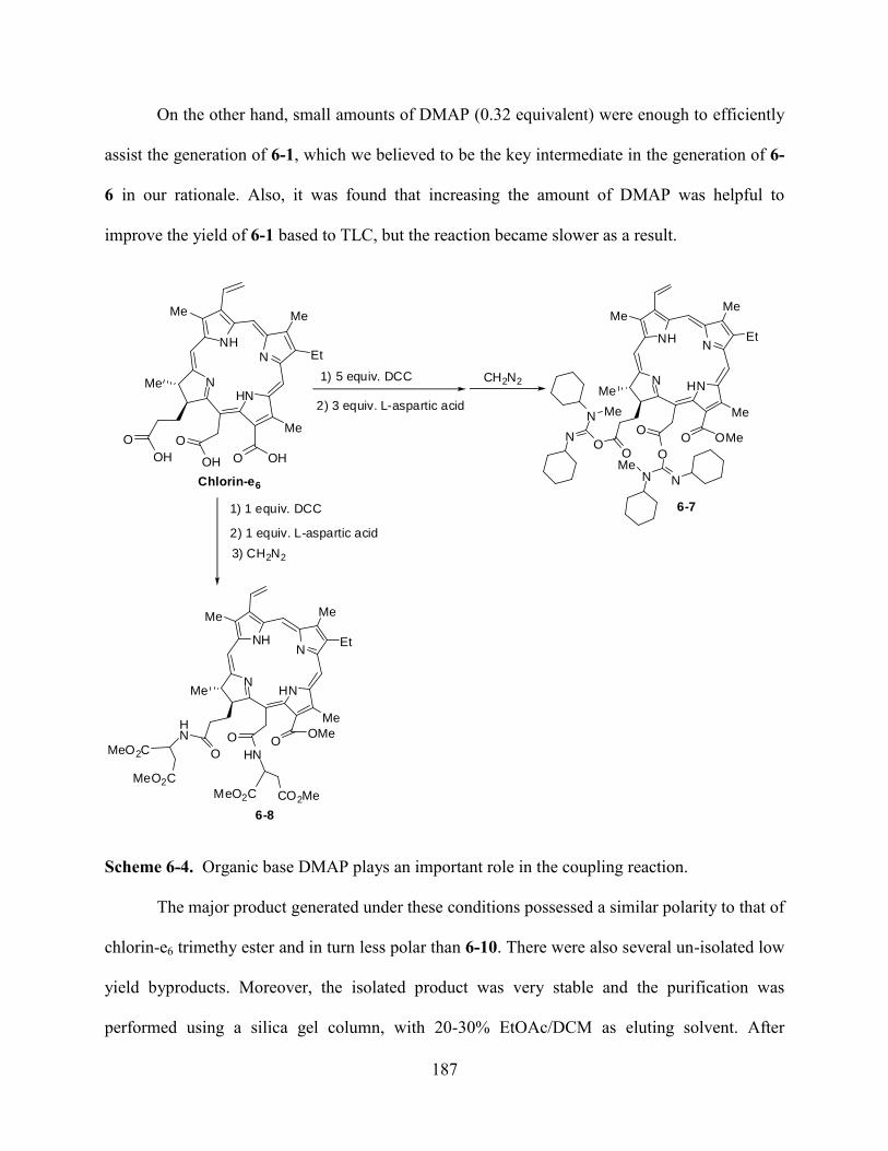
187
On the other hand, small amounts of DMAP (0.32 equivalent) were enough to efficiently
assist the generation of 6-1, which we believed to be the key intermediate in the generation of 6-
6 in our rationale. Also, it was found that increasing the amount of DMAP was helpful to
improve the yield of 6-1 based to TLC, but the reaction became slower as a result.
N
NHN
HN
MeMe
Et
Me
Me
O
OO
N
N
NN
OMe
N
NHN
HN
Me Me
Et
Me
Me
O OH
O
OH
2) 3 equiv. L-aspartic acid
1) 5 equiv. DCC CH2N2
1) 1 equiv. DCC
2) 1 equiv. L-aspartic acid
3) CH2N2
N
NHN
HN
Me Me
Et
Me
Me
OO
HN OMe
MeO2C
MeO2C
HN
MeO2C CO2Me
Chlorin-e6
6-7
6-8
O
OH
OO
O
Me
Me
Scheme 6-4. Organic base DMAP plays an important role in the coupling reaction.
The major product generated under these conditions possessed a similar polarity to that of
chlorin-e6 trimethy ester and in turn less polar than 6-10. There were also several un-isolated low
yield byproducts. Moreover, the isolated product was very stable and the purification was
performed using a silica gel column, with 20-30% EtOAc/DCM as eluting solvent. After
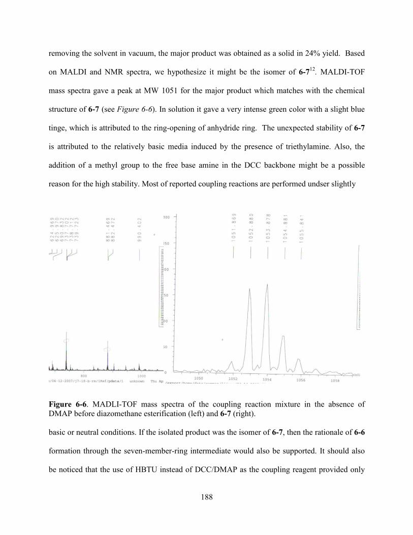
188
removing the solvent in vacuum, the major product was obtained as a solid in 24% yield. Based
on MALDI and NMR spectra, we hypothesize it might be the isomer of 6-712
. MALDI-TOF
mass spectra gave a peak at MW 1051 for the major product which matches with the chemical
structure of 6-7 (see Figure 6-6). In solution it gave a very intense green color with a slight blue
tinge, which is attributed to the ring-opening of anhydride ring. The unexpected stability of 6-7
is attributed to the relatively basic media induced by the presence of triethylamine. Also, the
addition of a methyl group to the free base amine in the DCC backbone might be a possible
reason for the high stability. Most of reported coupling reactions are performed undser slightly
Figure 6-6. MADLI-TOF mass spectra of the coupling reaction mixture in the absence of
DMAP before diazomethane esterification (left) and 6-7 (right).
basic or neutral conditions. If the isolated product was the isomer of 6-7, then the rationale of 6-6
formation through the seven-member-ring intermediate would also be supported. It should also
be noticed that the use of HBTU instead of DCC/DMAP as the coupling reagent provided only
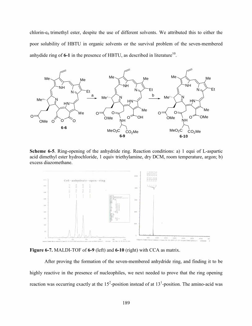
189
chlorin-e6 trimethyl ester, despite the use of different solvents. We attributed this to either the
poor solubility of HBTU in organic solvents or the survival problem of the seven-membered
anhydide ring of 6-1 in the presence of HBTU, as described in literature10
.
6-6
N
NHN
HN
Me Me
Et
Me
Me
OO
OMe
a
NH
CO2MeMeO2C
N
NHN
HN
Me Me
Et
Me
Me
OO
OMe
b
OH
6-9 6-10
OO
O
NH
CO2MeMeO2C
N
NHN
HN
Me Me
Et
Me
Me
OO
OMe OMeO
Scheme 6-5. Ring-opening of the anhydride ring. Reaction conditions: a) 1 equi of L-aspartic
acid dimethyl ester hydrochloride, 1 equiv triethylamine, dry DCM, room temperature, argon; b)
excess diazomethane.
Figure 6-7. MALDI-TOF of 6-9 (left) and 6-10 (right) with CCA as matrix.
After proving the formation of the seven-membered anhydride ring, and finding it to be
highly reactive in the presence of nucleophiles, we next needed to prove that the ring opening
reaction was occurring exactly at the 152-position instead of at 13
1-position. The amino-acid was
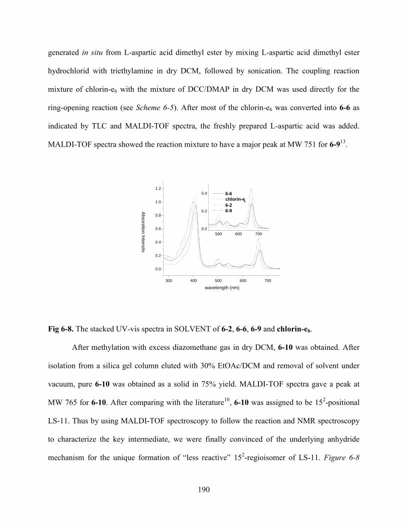
190
generated in situ from L-aspartic acid dimethyl ester by mixing L-aspartic acid dimethyl ester
hydrochlorid with triethylamine in dry DCM, followed by sonication. The coupling reaction
mixture of chlorin-e6 with the mixture of DCC/DMAP in dry DCM was used directly for the
ring-opening reaction (see Scheme 6-5). After most of the chlorin-e6 was converted into 6-6 as
indicated by TLC and MALDI-TOF spectra, the freshly prepared L-aspartic acid was added.
MALDI-TOF spectra showed the reaction mixture to have a major peak at MW 751 for 6-913
.
Fig 6-8. The stacked UV-vis spectra in SOLVENT of 6-2, 6-6, 6-9 and chlorin-e6.
After methylation with excess diazomethane gas in dry DCM, 6-10 was obtained. After
isolation from a silica gel column eluted with 30% EtOAc/DCM and removal of solvent under
vacuum, pure 6-10 was obtained as a solid in 75% yield. MALDI-TOF spectra gave a peak at
MW 765 for 6-10. After comparing with the literature10
, 6-10 was assigned to be 152-positional
LS-11. Thus by using MALDI-TOF spectroscopy to follow the reaction and NMR spectroscopy
to characterize the key intermediate, we were finally convinced of the underlying anhydride
mechanism for the unique formation of ―less reactive‖ 152-regioisomer of LS-11. Figure 6-8
300 400 500 600 700
0.0
0.2
0.4
0.6
0.8
1.0
1.2
500 600 700 0.0
0.2
0.4
wavelength (nm)
6-6 chlorin-e
6 6-2 6-9 A
bsorp
tion In
tensity

191
shows the stacked UV-vis spectra of 6-2, 6-6, 6-9 and the starting material chlorin-e6. The small
red-shift presented in both 6-2 and 6-6 is attributed to the formation of the seven-membered
anhydride ring intermediate which was absent in both 6-9 and chlorin-e6.
N
NHN
HN
Me Me
Et
Me
Me
O OH
O
OH
N
NHN
HN
Me Me
Et
Me
Me
OO
OMe OMeNH
Boc
Chlorin-e6
CH2N2a
b
NHBoc
6-11
OH
O O
Scheme 6-7. Formation of a lysine derivative of chlorin-e6. Reaction condition: a) 1 equiv
DCC/DMAP (1/1), dry DCM, argon, room temperature; b) 1 equiv H-Lys (Boc)-OtBu.HCl, 1
equiv triethylamine, dry DCM, argon, room temperature.
After discovering the underlying mechanism for the unique formation of the 152-
regioisomer of chlorine-e6, we also improved its synthesis based on a two-step one-pot reaction.
In this improved synthetic route, MALDI-TOF spectra and TLC were used to detect the
formation of anhydride intermediate before addition of L-aspartic acid dimethyl ester. By
dividing the previous one-step one-port reaction6,10
into a two-step one-port reaction, the
selectivity of the coupling was improved. We also used H-Lys(Boc)-OtBu hydrochloride in
place of L-aspartic acid dimethyl ester hydrochloride to test of the versatility of the mechanism
and of our improved synthesis (see Scheme 6-6). The reaction was performed by using 1
equivalent of DCC and 1 equivalent of DMAP as the coupling reagent to react with chlorin-e6 in
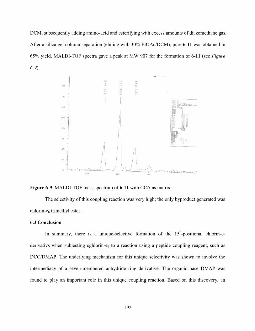
192
DCM, subsequently adding amino-acid and esterifying with excess amounts of diazomethane gas.
After a silica gel column separation (eluting with 30% EtOAc/DCM), pure 6-11 was obtained in
65% yield. MALDI-TOF spectra gave a peak at MW 907 for the formation of 6-11 (see Figure
6-9).
Figure 6-9. MALDI-TOF mass spectrum of 6-11 with CCA as matrix.
The selectivity of this coupling reaction was very high; the only byproduct generated was
chlorin-e6 trimethyl ester.
6.3 Conclusion
In summary, there is a unique-selective formation of the 152-positional chlorin-e6
derivativs when subjecting cghlorin-e6 to a reaction using a peptide coupling reagent, such as
DCC/DMAP. The underlying mechanism for this unique selectivity was shown to involve the
intermediacy of a seven-membered anhydride ring derivative. The organic base DMAP was
found to play an important role in this unique coupling reaction. Based on this discovery, an
193
improved synthesis of 152-positional chlorin-e6 was developed, namely a two-step one-pot
process and the yield was good. The versatility of this coupling reaction was also tested.
6.4 Experiment
6.4.1 General
Silica gel (32–63 mm) was used for flash column chromatography. All reactions were monitored
and some highly reactive compounds were separated by TLC using 0.25 mm silica gel plates
with or without UV indicator (60F-254). 1H NMR spectra were obtained on either a DPX-250,
ARX-300 or Varian VS-700 spectrometer. Chemical shifts (δ) are given in ppm relative to
CD2Cl2 (5.32), CDCl3 (7.27) or DMSO-d6 (2.50) as indicated. Mass spectra were obtained on an
Applied Biosystems QSTAR XL instrument. All solvents were obtained from Fisher Scientific
(HPLC grade, Houston, TX) and were used without further purification unless indicated. DMSO
(Biotech grade solvent, 99.8%) was purchased from Sigma–Aldrich and used without further
purification.
6.4.2 Preparation of diazomethane gas (small scale)
The apparatus consists of a Buckner-type conical flask (A), which was equipped with a magnetic
bar and a rubber stopper, and another Erlenmeyer flask (B), which was connected to the conical
flask (A) through polythene tubing. The stopper on the conical flask (A) has two holes, the first
one is closed with a rubber septum and the second one is used to hold polythene tubing for the
connection to Erlenmeyer flask (B). For example, the typical preparation of approximately 59
mmol diazomethane preceeds as follows: Diazald (18g, 84 mmol) was weighed directly into
flask (A) and ethanol (110 mL, 5 times its weight of diazald) was added. The flask was cooled to
0 oC by using an ice-bath. Flask (B) has the reaction mixture readily for the methylation reaction
in dry DCM. There must be a good stream of nitrogen gas continuously pasings through the

194
whole system. Concentrated aqueous solution of sodium hydroxide was added dropwise through
a plastic pipette inserted into the hole on the rubber stopper of flask (A). Upon the complete
dissolving of diazald, sodium hydroxide was added at a rate of 1 mL every 30 sec. After a few
seconds yellow diazomethane gas can be found to pass into flask (B), where the desired
methylation reaction occured. Continuing adding sodium hydroxide solution until the
disappearance of yellow colour in flask (A) is observed. TLC monitoring of the solution in flask
(B) showed complete methylation of the acid. The methylation reaction could complete within
couple of minutes.
6.4.3 Preparation of 6-2 and 6-3
Ce6 (60 mg, 0.1 mmol) was suspended in dry DCM 25 mL under Argon. Concentrated DCC (1
equiv) in dry DCM 5 mL was added into the reaction mixture. After 5-10 minutes, chlorin-e6
became completely dissolved and the solution showed a brown color. 30 Minutes later, when
using TLC to follow the reaction, two new brown spots (initial brown in color, later changing to
green) appeared at Rf 0.9 (6-3, minor) and 0.8 (6-2, major) (as indicate from MALDI-MS
spectroscopy) when 10% acetone/DCM was used as eluting solvent. Extending the reaction time
(it usually took 3-4 hours), only a small amount of chlorin-e6 remained according to TLC used to
follow the reaction. The MALDI spectrum of the reaction mixture gave a major peak at MW 784
for 6-2 and minor peak at MW 1138 for 6-3.
6.4.4 The preparation of 6-1 and 6-6
Chlorin-e6 (60 mg, 0.1 mmol) was suspended in dry DCM 25 mL under argon. Concentrated
DCC (1 equiv) in dry DCM solution 5 mL and DMAP (0.32 equivalent) were added into the
reaction mixture. After 4 h, TLC (10% acetone/DCM as eluting solvent) showed two major
brown sports: the first one at Rf 0.8 (6-2, minor) and a second one at 0.4 (6-1, major). In
195
agreement with TLC, the MALDI spectrum of the reaction mixture gave two major peaks, at 578
(6-1, major) and 785 (6-2, minor). No anhydride dimer (6-3) was detected under these conditions.
The separation of 6-1 and 6-2 from the reaction mixtures was performed on TLC plates (silica
gel) very quickly. Small amounts of each substance were obtained and identified by MALDI
mass spectra. However, the separation yield was very low due to decomposition of the
compounds. Both UV-vis spectra of the DCC-adduct and anhydride-acid showed several nm red-
shifts compared with chlorin-e6 trimethyl ester, due to the formation of an extra seven-membered
ring on the chlorin.
The methylation of 6-1 to form 6-6 was performed by bubbling diazomethane gas into the DCM
reaction mixture of 6-1. TLC gave a major brown spot for 6-6. Separation was performed on
TLC plates (silica gel). The desired 6-6 was fully characterized by MALDI-TOF, NMR and UV-
vis spectroscopy.
6-6: MALDI-TOF Calcd for C35H36N4O5, 592.7. Found 592.8. UV-Vis (max nm/DMSO): 409,
505, 541, 614, 668; 1H-NMR (CDCl3, 300 MHz) 9.57 (1H, s), 9.29 (1H, s), 8.58 (1H, s), 7.98-
7.93 (1H, m), 6.35 (1H, dd, J = 17.5 Hz), 6.18-6.16 (1H, dd, J = 11.9 Hz), 5.43 (2H, br), 4.53
(1H, t, J = 8.4 Hz), 4.44 (1H, tr J = 14.7), 3.70-3.67 (8H, m), 3.38 (3H, s), 3.18 (3H, s), 2.70-
2.67 (1H, m), 2.60-2.59 (1H, m), 2.38-2.36 (1 H, m), 1.99-1.97 (1H, m), 1.75-1.74 and 1.70-1.68
(6H, m), -0.42 (1H, s), -0.52 (1H, s).
6-7: MALDI-TOF Calcd for C63H86N8O6,1051.4. Found 1051.9. 1H-NMR (DMSO-d6, 300
MHz ) 9.74 (1H, s), 9.41 (1H, d, J = 6.0 Hz), 9.13 (1H, t, J = 9.72), 8.20-8.11 (3H, m), 6.41(1H,
d, J = 17.8 Hz), 6.13 (1H, d, J = 11.4 Hz), 4.56 (1H, d, J = 6.8 Hz), 4.06 (1H, tetra, J = 7.11 Hz),
3.96 (2H, d, J = 11.0 Hz), 3.84 (2H, s), 3.67-3.62 (6H, d, J = 13.7 Hz), 3.50-3.47 (3H, d, J = 9.8

196
Hz), 3.38 (3H, s), 3.14 (3H, s), 2.53 (3H, s), 2.01 (2H, s), 1.82-1.71 and 1.63-1.59 (10 H, br),
1.34-1.05 (10H, br), -1.08-1.13 (2H, m).
6-8: MALDI-TOF Calcd for C63H86N8O6, 1051.4. Found 1051.4. 1H-NMR (CD2Cl2, 400 MHz )
9.74 (1H, s), 9.41 (1H, s), 9.13-9.09 (1H, m), 8.19-8.11 (3H, m), 6.41 (2H, d, J = 18 Hz), 6.13
(1H, d, J = 12 Hz), 3.96-3.92 (1H, m), 3.83 (2H, s), 3.67-3.62 (2H, m), 3.47 (2H, s), 3.38 (6H, s),
3.14 (3H, s), 2.53 (3H, s), 1.82 (2H, s), 1.60-1.59 (12H, m), 1.22-1.17 (10H, s), -1.08- -1.13 (2H,
m).
6-9: MALDI-TOF Calcd for C41H47N5O9, 753.8. Found 753.5. UV-Vis (max nm/DMSO): 402,
501, 530, 609, 664.
6-10: MALDI-TOF Calcd for C42H49N5O9, 767.9. Found 767.9. 1H-NMR (CD2Cl2, 400 MHz )
9.71 (1H, s), 9.56 (1H, s), 8.79 (1H, s), 8.07-8.02 (1H, m), 6.36 (1H, d, J = 16 Hz), 6.15 (1H, d, J
= 12 Hz), 5.30 (1H, s), 4.89 (1H, s), 4.53-4.50 (2H, m), 4.32 (2H, s), 3.80-3.77 (2H, m), 3.60 (5H,
m), 3.48 (2H, s), 3.43 (2H, m),3.30 (2H, s), 3.13 (2H, s), 2.90 (2H, s), 2.33 (2H, s), 2.07 (1H, s),
1.78-1.71 (6H, m), 1.30-1.29 (2H, m), -1.26 (1H, s), -1.38 (1H, s).
6-11: MALDI-TOF Calcd for C51H68N6O9, 909.1. Found 909.1. 1H-NMR (CDCl3, 300 MHz )
9.73 (1H, s), 9.58 (1H, s), 8.78 (1H, s), 8.10-8.01 (1H, m), 6.39 (1H, d, J = 17.8 Hz), 6.17 (1H, d,
J = 11.4 Hz), 5.22 (2H, s), 4.53 (3H, s), 4.27 (3H, s), 3.80 (2H, s), 3.59 (3H, s), 3.49 (3H, s),
3.30 (2H, s), 2.91 (2H, s), 2.58 (1H, s), 2.22 (1H, s), 2.06 (2H, s), 1.88 (6H, s) 1.44 (9H, s), 1.29
(9H, s), -1.30 (1H, s), -1.43 (1H, s).
6.5 References
1 Dougherty, T. J.; Gomer, C. J.; Henderson B. W.; Jori, G.; Kessel D.; Korbelik, M.; Moan, J.;
Peng, Q. J. Natl. Cancer Inst. 1998, 90, 889-905.
2 Pandey, R. K.; Zheng, G. In The Porphyrin Handbook, Kadish, K. M., Smith, K. M., Guilard,
R., Eds.; Academic Press, Boston; 2000, Vol. 6, pp 157.
197
3 Vicente, M. G. H. Current Medicinal Chemistry, Anti-Cancer Agents 1, 2001, 175.
4 Spikes, J. D.; Bommer, J. C. In Chlorophylls, Scheer, H., Eds; CRC Press, Boston; 1996, pp
1181.
5 Song, L. -M. W.; Wang, K. K.; Zinsmeister, A. R. Cancer, 1998, 82, 421.
6 Bommer, J. C.; Ogden, B. F. U. S. Patent, 1987, 4,693,885.
7 Gomi, S.; Nishizuka, T.; Ushiroda, O.; Uchida, N.; Takahashi, H.; Sumi, S.
Heterocycles,1998, 48, 2231.
8 Abraham, R. J.; Rowan, A. E. In Chlorophylls; Scheer, H., Eds.; CRC Press: Boston; 1996,
pp 797.
9 Roberts, W. G.; Shiau, F. -Y.; Nelson, J. S.; Smith, K. M.; Berns, M. W. J. Natl. Cancer Inst.
1988, 80, 330.
10 Hargus J. A.; Fronczek, F. R.; Vicente, M. G. H.; Smith, K. M. Photochem. Photobio. 2007,
83, 1006.
198
APPENDIX: LETTERS OF PERMISSION
From: Lijuan Jiao [mailto:[email protected]]
Sent: 09 July 2007 17:11
To: CONTRACTS-COPYRIGHT (shared)
Subject: Ask for permission to use my publication for my dissertation
Dear Sir/ Madam:
I am a graduate student from Louisiana State University and I am graduating this summer. I
am writing to ask for the permission to use the materials described in my publication for the
usage in my dissertation. The Title of the publication is "Benzoporphyrins via an olefin ring-
closure metathesis methodology" and it was published in 2006 in the Journal "Chem. Commun."
The page number is from 3900-3902.
Thanks for your time and your help.
Sincerely:
Lijuan Jiao
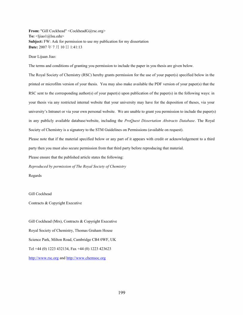
199
From: "Gill Cockhead" <[email protected]>
To: <[email protected]>
Subject: FW: Ask for permission to use my publication for my dissertation
Date: 2007 年 7 月 10 日 1:41:13
Dear Lijuan Jiao:
The terms and conditions of granting you permission to include the paper in you thesis are given below.
The Royal Society of Chemistry (RSC) hereby grants permission for the use of your paper(s) specified below in the
printed or microfilm version of your thesis. You may also make available the PDF version of your paper(s) that the
RSC sent to the corresponding author(s) of your paper(s) upon publication of the paper(s) in the following ways: in
your thesis via any restricted internal website that your university may have for the deposition of theses, via your
university’s Intranet or via your own personal website. We are unable to grant you permission to include the paper(s)
in any publicly available database/website, including the ProQuest Dissertation Abstracts Database. The Royal
Society of Chemistry is a signatory to the STM Guidelines on Permissions (available on request).
Please note that if the material specified below or any part of it appears with credit or acknowledgement to a third
party then you must also secure permission from that third party before reproducing that material.
Please ensure that the published article states the following:
Reproduced by permission of The Royal Society of Chemistry
Regards
Gill Cockhead
Contracts & Copyright Executive
Gill Cockhead (Mrs), Contracts & Copyright Executive
Royal Society of Chemistry, Thomas Graham House
Science Park, Milton Road, Cambridge CB4 0WF, UK
Tel +44 (0) 1223 432134, Fax +44 (0) 1223 423623
http://www.rsc.org and http://www.chemsoc.org
200
From: Lijuan Jiao [mailto:[email protected]]
Sent: 09 July 2007 17:36
To: Rights and Permissions (ELS)
Subject: Request the permission to use my publications for my dissertation
Dear Sir/ Madam:
I am a graduate student from the Chemistry Department of Louisiana State University and I
am graduating this summer. I am writing to request the permission to use the materials described
in the following two papers (I am the first author for both of papers) for the usage in my
dissertation.
Paper 1: "Syntheses and properties of functionalized oxacalix[4]arene porphyrins", published in
the Journal "Tetrahedron" in 2007, and the volume is 63 and page numbers are from 4011-4017.
Paper 2: "beta-beta Linked cofacial bis-porphyrins", published in the Journal "tetrahedron
Letters" in 2006. The Volume number is 47 and the page numbers are from 501-504.
Thanks for your time and your kind help.
Sincerely:
Lijuan Jiao
201
From: "Rights and Permissions (ELS)" <[email protected]>
To: "Lijuan Jiao" <[email protected]>
Subject: RE: Request the permission to use my publications for my dissertation
Date: 2007 年 7 月 11 日 4:53:06
Dear Lijuan Jiao
Thank you for your e-mail.
Elsevier is pleased to announce our partnership with Copyright Clearance Center’s Rightslink service. With
Rightslink ® it’s faster and easier than ever before to obtain permission to use and republish material from Elsevier.
In this instance the link to the requested material is http://dx.doi.org/10.1016/j.tetlet.2005.11.065
For future requests simply visit: http://www.sciencedirect.com/ and locate your desired content.
Click on Permissions within the table of contents or in the tool-box to the right of the online article to open the
following page:
1. Select the way you would like to reuse the content
2. Create an account if you haven’t already
3. Accept the terms and conditions and you’re done
Please contact Rightslink Customer Care with any questions or comments concerning this service:
Copyright Clearance Center
Rightslink Customer Care
Tel (toll free): 877/622-5543
Tel: 978/777-9929
E-mail: mailto:[email protected]
Web: http://www.rightslink.com

202
VITA
Lijuan Jiao was born on December 10, 1977, in Guoyang county, Anhui province, China.
She enrolled in Shandong Univeristy, Jinan, China, in 1996. From Shandong University, she
earned her Bachelor of Science degree in chemistry in June of 2000. For her master degree study
in chemistry, she enrolled in University of Science &Technology of China, Hefei, China, where
she earned her Master of Science degree in chemistry in June of 2003. In August of 2003, with
the financial support from the Department of Chemistry of Louisiana State Univeristy (LSU), she
came to LSU to begin her doctoral degree study in chemistry and soon joined Professor Kevin M.
Smith’s research group to begin doing research in porphyrin chemistry.


![[Derivatives Consulting Group] Introduction to Equity Derivatives](https://img.pdfslide.us/doc/110x75/5525eed15503467c6f8b4b12/derivatives-consulting-group-introduction-to-equity-derivatives.jpg)