Embed Size (px)
Citation preview

Accepted Manuscript
Synthesis and evaluation of frentizole-based indolyl thiourea analogues as
MAO/ABAD inhibitors for Alzheimer’s disease treatment
Lukas Hroch, Patrick Guest, Ondrej Benek, Ondrej Soukup, Jana Janockova,
Rafael Dolezal, Kamil Kuca, Laura Aitken, Terry K. Smith, Frank Gunn-Moore,
Dominykas Zala, Rona R. Ramsay, Kamil Musilek
PII: S0968-0896(16)31452-3
DOI: http://dx.doi.org/10.1016/j.bmc.2016.12.029
Reference: BMC 13456
To appear in: Bioorganic & Medicinal Chemistry
Received Date: 30 November 2016
Accepted Date: 18 December 2016
Please cite this article as: Hroch, L., Guest, P., Benek, O., Soukup, O., Janockova, J., Dolezal, R., Kuca, K., Aitken,
L., Smith, T.K., Gunn-Moore, F., Zala, D., Ramsay, R.R., Musilek, K., Synthesis and evaluation of frentizole-based
indolyl thiourea analogues as MAO/ABAD inhibitors for Alzheimer’s disease treatment, Bioorganic & Medicinal
Chemistry (2016), doi: http://dx.doi.org/10.1016/j.bmc.2016.12.029
This is a PDF file of an unedited manuscript that has been accepted for publication. As a service to our customers
we are providing this early version of the manuscript. The manuscript will undergo copyediting, typesetting, and
review of the resulting proof before it is published in its final form. Please note that during the production process
errors may be discovered which could affect the content, and all legal disclaimers that apply to the journal pertain.

1
Synthesis and evaluation of frentizole-based indolyl thiourea analogues as MAO/ABAD inhibitors for Alzheimer’s
disease treatment
Lukas Hrocha,b, Patrick Guestc, Ondrej Benekb,d, Ondrej Soukupb, Jana Janockovab, Rafael Dolezalb,d,e,
Kamil Kucab,d, Laura Aitkenc, Terry K. Smithc, Frank Gunn-Moorec, Dominykas Zalac, Rona R. Ramsayc*,
Kamil Musilekb,d*
a Charles University in Prague, Faculty of Pharmacy in Hradec Kralove, Department of Pharmaceutical Chemistry and
Drug Control, Heyrovskeho 1203, 500 05 Hradec Kralove, Czech Republic
b University Hospital, Biomedical Research Center, Sokolska 581, 500 05 Hradec Kralove, Czech Republic.
c University of St. Andrews, School of Biology, Medical and Biological Sciences Building or Biomedical Sciences Research
Complex, North Haugh, St. Andrews KY16 9ST, United Kingdom
d University of Hradec Kralove, Faculty of Science, Department of Chemistry, Rokitanskeho 62, 500 03 Hradec Kralove,
Czech Republic; [email protected]
e University of Hradec Kralove, Faculty of Informatics and Management, Center for Basic and Applied Research,
Rokitanskeho 62, 500 03 Hradec Kralove, Czech Republic
Abstract
Alzheimer’s disease (AD) is a neurodegenerative disorder associated with an excessive accumulation of
amyloid-beta peptide (Aβ). Based on the multifactorial nature of AD, preparation of multi-target-directed
ligands presents a viable option to address more pathological events at one time. A novel class of
asymmetrical disubstituted indolyl thioureas have been designed and synthesized to interact with
monoamine oxidase (MAO) and/or amyloid-binding alcohol dehydrogenase (ABAD). The design combines
the features of known MAO inhibitors scaffolds (e.g. rasagiline or ladostigil) and a frentizole moiety with
potential to interact with ABAD. Evaluation against MAO identified several compounds that inhibited in the
low to moderate micromolar range. The most promising compound (19) inhibited human MAO-A and MAO-
B with IC50 values of 6.34 μM and 0.30 μM, respectively. ABAD activity evaluation did not show any highly
potent compound, but the compound series allowed identification of structural features to assist the future
development of ABAD inhibitors. Finally, several of the compounds were found to be potent inhibitors of
horseradish peroxidase (HRP), preventing the use of the Amplex™ Red assay to detect hydrogen peroxide
produced by MAO, highlighting the need for serious precautions when using an enzyme-coupled assay.

2
Keywords
Alzheimer's disease (AD); monoamine oxidase (MAO); amyloid-beta peptide (Aβ); mitochondrial
amyloid-binding alcohol dehydrogenase (ABAD); 17β-hydroxysteroid dehydrogenase type 10 (17β-HSD10);
horseradish peroxidase (HRP).
1. Introduction
Alzheimer’s disease (AD) is the most common cause of senile dementia and is characterized by a
progressive impairment of cognitive functions including memory, language and motor skills.1,2 Though the
aetiology of AD still remains unclear, there are several established factors contributing to the progress of
AD including e.g. alteration in neurotransmitter systems, deposition of amyloid beta peptide (Aβ), tau-
protein hyperphoshorylation with tangle formation, oxidative stress and mitochondrial dysfunction.3,4
Most of the currently marketed drugs (namely donepezil, rivastigmine, galanthamine) prevent the
degradation of acetylcholine via the inhibition of acetylcholinesterase to sustain sufficient levels in the face
of the decreased numbers of cholinergic neurons in AD.5 This approach is only effective for symptomatic
treatment of mild and moderate cognitive decline and does not provide prevention of disease progression.6
Alongside the well-established dysregulation of cholinergic system, the impairment of other
neurotransmitter systems (e.g. dopaminergic or serotonergic) is also present in the brain of AD patients,
causing behavioural syndromes in AD patients.7 Thus, monoamine oxidases (MAO), enzymes responsible
for the metabolism of monoamine neurotransmitters, were suggested as possible targets for AD
treatment.8 MAO occurs in two isoforms (MAO-A and MAO-B) with different substrate and inhibitor
specificities.8–10 Alterations of both MAO-A and MAO-B activities (elevated enzyme levels or changes in
expression in various brain regions) were reported in AD patient brains.11–13 While elevated levels of MAO-B
are well-described, changes in MAO-A activity are rather more complex and unclear.14 In the periphery,
inhibition of MAO-A can contribute to potential side effects, in particular to tyramine-induced hypertension
(also referred as the “cheese effect”). It is induced by dietary amines after the irreversible inhibition of
intestinal MAO-A where the inhibitor is covalently bound to the enzyme (via hydrazine or propargyl-amino
moiety).8 Furthermore, elevated levels of the MAO isoforms can also generate toxic metabolites (e.g.
aldehydes or hydrogen peroxide),15 which consequently promote already ongoing oxidative stress in AD.16
Taken together, targeting both MAO-A and MAO-B could potentially contribute to therapeutic effects in AD
treatment.
Besides the neurotransmission anomalies, one of the most widely accepted AD hypothesis focuses on
the pathological production of Aβ leading to formation of insoluble extracellular Aβ plaques.17,18 Intra-
mitochondrial Aβ deposition has been associated with mitochondrial dysfunction and impaired synaptic
trafficking resulting in neuronal damage.19,20 Thus, Aβ interactions with mitochondrial proteins may become

3
therapeutic targets for cellular protection in AD.21 Several mitochondrial proteins have been shown to
interact directly with Aβ, possibly influencing mitochondrial metabolism.22 The amyloid-binding alcohol
dehydrogenase (ABAD) enzyme, also known as 17β-hydroxysteroid dehydrogenase type 10 (17β-HSD10), is
one such Aβ binding protein.23 The Aβ-ABAD interaction promotes the enzyme to undergo functional
changes, resulting in altered enzymatic activity, elevated levels of toxic metabolites, enhanced oxidative
stress and mitochondrial and synaptic dysfunction.24–26 Studies using an ABAD decoy peptide consisting of
the ABAD amino acid residues responsible for interaction with Aβ, have been shown to disrupt the
interaction between ABAD and Aβ, reducing Aβ-induced mitochondrial and neuronal toxicity and improving
learning and memory in AD mice models.25,27 Further, the direct inhibition of the ABAD enzyme has been
shown to have neuroprotective effects in terms of reducing cell stress via decreased generation of reactive
oxygen species, improved mitochondrial respiration, and stabilization of estradiol levels.28,29 Thus both the
direct inhibition of ABAD enzyme and the disruption of the Aβ-ABAD interaction present possible
approaches for AD treatment.29
Due to the multifactorial nature of AD aetiology, the question whether a single-target-directed drug
could yield therapeutic effect in AD treatment has led scientists to focus still more on the emerging multi-
target-directed ligand (MTDL) approach.30 Simultaneous modulation of several pathological events of the
ongoing disease may then contribute to therapeutic effect of progressing AD.31 The MTDL approach has
been employed by many research groups32–35 and has shown success, for example in the case of the
compound ladostigil36 which has reached clinical trials.37 Exploiting the advantage of conjunctive approach,
we designed a series of compounds combining known scaffolds of MAO and ABAD inhibitors to introduce
MAO inhibitory activity into frentizole moiety and explore structural inhibitory features of both MAO and
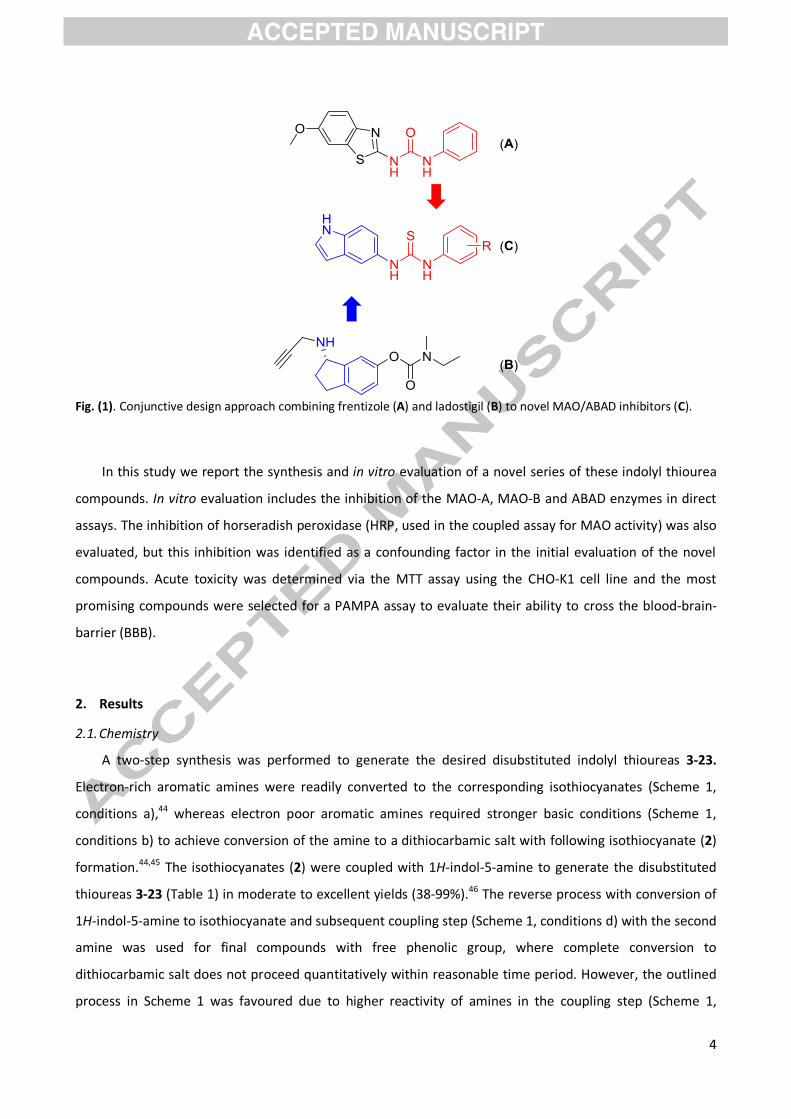
ABAD scaffolds. Xie et al. observed that the FDA approved drug, frentizole (Fig. 1: A), acted as a weak
inhibitor of the Aβ-ABAD interaction (IC50 = 200 μM) and described a novel class of frentizole-based
benzothiazolyl (thio)ureas acting as inhibitors of the Aβ-ABAD interaction.38 Relatively minimal
modifications of the distal phenyl ring substitution also allowed phosphonate analogues of the same
scaffold to act as direct ABAD inhibitors, yielding two compounds that inhibited ABAD with IC50 values of
52.7 μM and 341.9 μM.39,40 Recently, low micromolar scale ABAD inhibitors were reported.41 Furthermore,
a fused bicyclic system comprised of 5- and 6-membered rings (i.e. indane) represents core scaffold of
several MAO inhibitors (e.g. rasagiline or ladostigil). The carbocyclic system is not exclusive for MAO
inhibition, since other bicyclic and heterocyclic scaffolds have been reported to inhibit MAO.35,42 In this
case, the aminoindane moiety has been replaced with an isosteric indole ring (Fig. 1: B) and further
incorporated into frentizole scaffold (Fig. 1: A). The frentizole urea linker was changed to the analogous
thiourea linker since the similar thiocarbonyl scaffold was previously identified to be beneficial for ABAD
inhibition.43 The distal phenyl ring presented in frentizole was modified by various substitution patterns
that have been introduced to investigate structure and activity relationship of this scaffold (Fig. 1: C).
4
Fig. (1). Conjunctive design approach combining frentizole (A) and ladostigil (B) to novel MAO/ABAD inhibitors (C).
In this study we report the synthesis and in vitro evaluation of a novel series of these indolyl thiourea
compounds. In vitro evaluation includes the inhibition of the MAO-A, MAO-B and ABAD enzymes in direct
assays. The inhibition of horseradish peroxidase (HRP, used in the coupled assay for MAO activity) was also
evaluated, but this inhibition was identified as a confounding factor in the initial evaluation of the novel
compounds. Acute toxicity was determined via the MTT assay using the CHO-K1 cell line and the most
promising compounds were selected for a PAMPA assay to evaluate their ability to cross the blood-brain-
barrier (BBB).
2. Results
2.1. Chemistry
A two-step synthesis was performed to generate the desired disubstituted indolyl thioureas 3-23.
Electron-rich aromatic amines were readily converted to the corresponding isothiocyanates (Scheme 1,
conditions a),44 whereas electron poor aromatic amines required stronger basic conditions (Scheme 1,
conditions b) to achieve conversion of the amine to a dithiocarbamic salt with following isothiocyanate (2)
formation.44,45 The isothiocyanates (2) were coupled with 1H-indol-5-amine to generate the disubstituted
thioureas 3-23 (Table 1) in moderate to excellent yields (38-99%).46 The reverse process with conversion of
1H-indol-5-amine to isothiocyanate and subsequent coupling step (Scheme 1, conditions d) with the second
amine was used for final compounds with free phenolic group, where complete conversion to
dithiocarbamic salt does not proceed quantitatively within reasonable time period. However, the outlined
process in Scheme 1 was favoured due to higher reactivity of amines in the coupling step (Scheme 1,

5
conditions d), resulting in higher overall yields. The procedure also allowed compounds to be more readily
purified by subsequent column chromatography rendering final compounds (3-23). The experimental
section gives a detailed description of which procedure was used for each amine.
Scheme 1. Synthesis of indolyl thioureas 3-23. Reagents and conditions: (a) CS2, Et3N, r.t. (b) NaH, CS2, reflux; (c)
Boc2O, DMAP, 0°C to r.t.; (d) 1H-indole-5-amine, DCM, r.t.
Table 1. Substitution pattern and isolated yields of indolyl thioureas (3-23).
Comp. Ar Yield (%)
Comp. Ar
Yield (%)
Comp. Ar
Yield (%)
3
90
10
93
17
85
4
61
11
99
18
99
5
92
12
89
19
38
6
92
13
99
20
99
7
84
14
80
21
66
8
99
15
97
22
90
9
89
16
99
23
93
6
2.2. Biological activity
2.2.1. Controls in the coupled assay for MAO reveal HRP inhibition
An initial screen using a coupled fluorescence assay to assess MAO inhibition by the novel indole
thiourea compounds suggested potent inhibition in the micromolar range that was not replicated in the
direct spectrophotometric assay. The coupled assay uses HRP to detect the second MAO product, H2O2,
resulting in the conversion of 10-acetyl-3,7-dihydroxyphenoxazine to resorufin.47–49 The compounds did not
quench the fluorescence of resorufin but most of them inhibited HRP by greater than 30% at 50 µM, the
most potent by >80%. The inhibition of HRP by each compound at 5 or 50 µM is given as Supplementary
Information (Table S1). The most potent HRP inhibitors, all with IC50 values of around 1 µM, always
possessed a phenolic group, either in the 4-position (6, 17, 21 and 23) or 2-position (4) of the phenyl ring.
The phenolic group has previously been associated with HRP inhibition, however on a flavonoid scaffold.50
In light of these findings, we point out a potential flaw of the coupled Ampliflu™ Red assay, which may
produce false positive results. In this study, the HRP inhibition at 1 µM prevented the use of the coupled
assay to evaluate MAO inhibition.
2.2.2. Monoamine oxidase activity
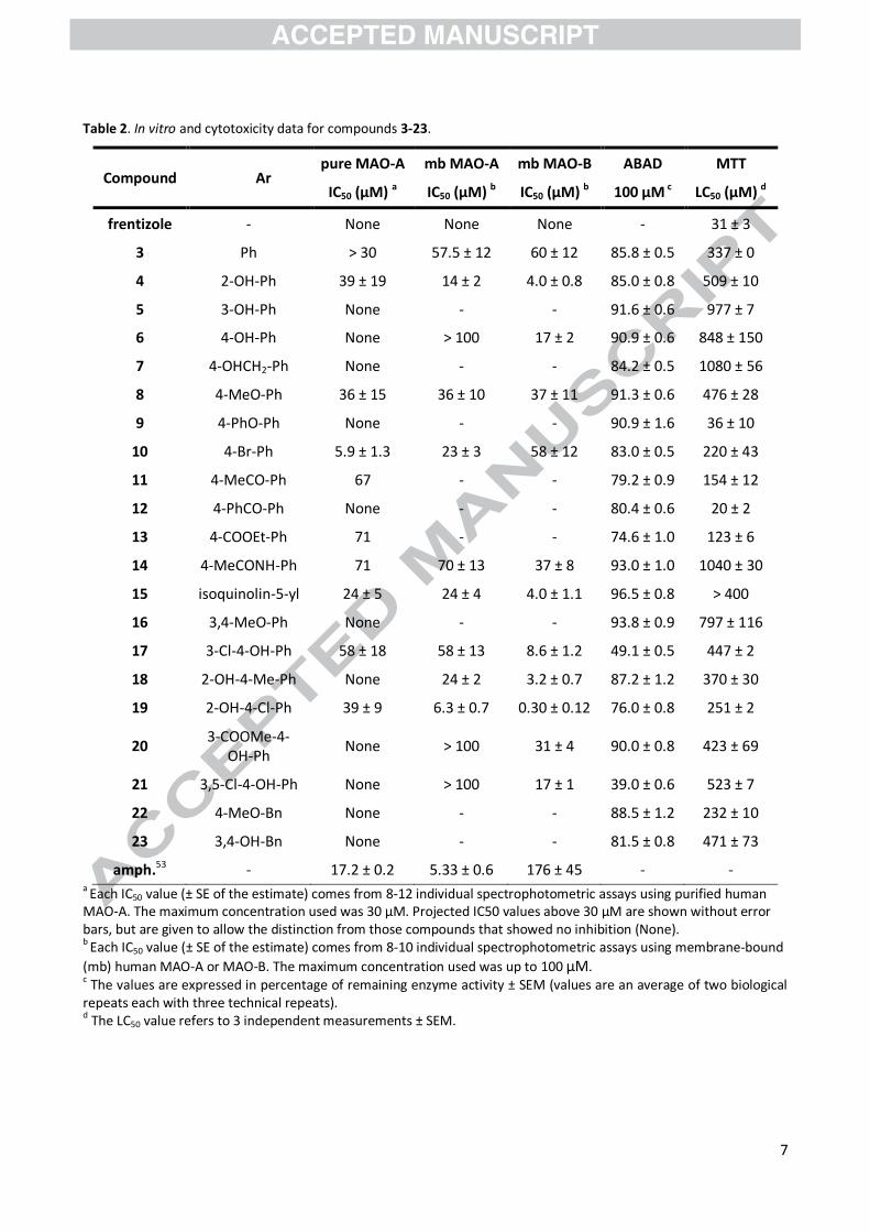
To avoid the observed interference issues with the coupled assay, all compounds were evaluated using
the kynuramine spectrophotometric assay.51 All compounds were soluble in assay buffer at 100 μM but
absorbed light at the wavelength (314 nm) of the kynuramine assay, limiting the maximum concentration
to 30 µM for the initial screen. Compounds selected for further study could be used up to 100 μM. IC50
values were measured for all compounds using purified MAO-A as shown in Table 2. Several compounds
displayed moderate to low micromolar range of inhibition, allowing selection of compounds for evaluation
against membrane-bound MAO-A and MAO-B. IC50 values for the selected compounds are given in Table 2.
The selectivity index calculated based on the IC50 values showed a small selectivity for MAO-B (6, 15, 17, 18
and 19).
2.2.3. ABAD activity
Compounds 3-23 were screened at a fixed 100 µM concentration to evaluate their inhibitory effect on
ABAD activity with modified spectrophotometric assay.26 The majority of the compounds did not
significantly inhibit ABAD function. However, compounds 17 and 21 induced a decrease in ABAD activity of
51% or 61%, respectively (Table 2). Both compounds possess a phenolic group, which appears to be a key
feature for displaying inhibition of ABAD. Based on the small effects observed in the 100 µM screen, the
determination of IC50 was not possible since the concentration range needed would exceed the limits of
assay.
7
Table 2. In vitro and cytotoxicity data for compounds 3-23.
Compound Ar pure MAO-A mb MAO-A mb MAO-B ABAD MTT
IC50 (µM) a IC50 (µM) b IC50 (µM) b 100 µM c LC50 (µM) d
frentizole - None None None - 31 ± 3
3 Ph > 30 57.5 ± 12 60 ± 12 85.8 ± 0.5 337 ± 0
4 2-OH-Ph 39 ± 19 14 ± 2 4.0 ± 0.8 85.0 ± 0.8 509 ± 10
5 3-OH-Ph None - - 91.6 ± 0.6 977 ± 7
6 4-OH-Ph None > 100 17 ± 2 90.9 ± 0.6 848 ± 150
7 4-OHCH2-Ph None - - 84.2 ± 0.5 1080 ± 56
8 4-MeO-Ph 36 ± 15 36 ± 10 37 ± 11 91.3 ± 0.6 476 ± 28
9 4-PhO-Ph None - - 90.9 ± 1.6 36 ± 10
10 4-Br-Ph 5.9 ± 1.3 23 ± 3 58 ± 12 83.0 ± 0.5 220 ± 43
11 4-MeCO-Ph 67 - - 79.2 ± 0.9 154 ± 12
12 4-PhCO-Ph None - - 80.4 ± 0.6 20 ± 2
13 4-COOEt-Ph 71 - - 74.6 ± 1.0 123 ± 6
14 4-MeCONH-Ph 71 70 ± 13 37 ± 8 93.0 ± 1.0 1040 ± 30
15 isoquinolin-5-yl 24 ± 5 24 ± 4 4.0 ± 1.1 96.5 ± 0.8 > 400
16 3,4-MeO-Ph None - - 93.8 ± 0.9 797 ± 116
17 3-Cl-4-OH-Ph 58 ± 18 58 ± 13 8.6 ± 1.2 49.1 ± 0.5 447 ± 2
18 2-OH-4-Me-Ph None 24 ± 2 3.2 ± 0.7 87.2 ± 1.2 370 ± 30
19 2-OH-4-Cl-Ph 39 ± 9 6.3 ± 0.7 0.30 ± 0.12 76.0 ± 0.8 251 ± 2
20 3-COOMe-4-
OH-Ph None > 100 31 ± 4 90.0 ± 0.8 423 ± 69
21 3,5-Cl-4-OH-Ph None > 100 17 ± 1 39.0 ± 0.6 523 ± 7
22 4-MeO-Bn None - - 88.5 ± 1.2 232 ± 10
23 3,4-OH-Bn None - - 81.5 ± 0.8 471 ± 73
amph.53 - 17.2 ± 0.2 5.33 ± 0.6 176 ± 45 - - a Each IC50 value (± SE of the estimate) comes from 8-12 individual spectrophotometric assays using purified human MAO-A. The maximum concentration used was 30 µM. Projected IC50 values above 30 µM are shown without error bars, but are given to allow the distinction from those compounds that showed no inhibition (None). b Each IC50 value (± SE of the estimate) comes from 8-10 individual spectrophotometric assays using membrane-bound
(mb) human MAO-A or MAO-B. The maximum concentration used was up to 100 µM. c The values are expressed in percentage of remaining enzyme activity ± SEM (values are an average of two biological repeats each with three technical repeats). d The LC50 value refers to 3 independent measurements ± SEM.

8
2.2.4. Cytotoxicity
To evaluate acute cytotoxicity, all compounds were screened using the standard MTT assay with the
CHO-K1 cell line which is commonly used for cytotoxicity screening.52 The observed toxicity was expressed
as LC50 values (Table 2). The majority of the compounds showed very low levels of toxicity with an LC50
value over 200 µM compared to parent frentizole, producing valuable results for further investigation. Two
compounds (9 and 12) showed elevated toxicity (LC50 ~20-40 µM) but these compounds didn’t show any
beneficial biological activity.
2.2.5. Blood-brain barrier permeation and physicochemical properties
Penetration across the BBB is an essential property for compounds targeting the CNS. In order to
predict passive blood-brain penetration of most promising compounds prepared in the current study (10,
17, 21), a modification of the parallel artificial membrane permeation assay (PAMPA) has been used based
on reported protocol.54 The penetration value (Pe) was derived mathematically from time-dependent
changes of the selected compounds’ concentrations in two aqueous phases separated by the artificial
membrane (see Experimental section). Compounds 10 and 17 showed Pe values greater than 4, which
indicate sufficient passive transition of the compounds through the BBB (Table 3).55,56
For the physicochemical properties, the number of hydrogen bond donors (HBD), number of hydrogen
bond acceptors (HBA), topological polar surface area (TPSA), logarithm of the n-octanol-water partition
coefficient for non-ionized species (ClogP) and the n-octanol-water distribution coefficient (ClogD)
reflecting the ratio of the ionic forms at pH = 7.4 have been calculated in ACDLabs PhysChem Suite 12.0 and
compared with the experimental values of Pe and chromatographic capacity factors k. The three
compounds 10, 17 and 21 do not violate any of the Lipinski’s rules of five, although Pe values (Table 4)
apparently follow their own trend showing somewhat ambiguous relationship with k and the in silico
molecular descriptors. The most striking deviation is exhibited by compound 21 that has a low penetration
rate Pe despite its sufficient hydrophobicity as implied by relatively high values of ClogP, ClogD and k. This
finding may results from overall physicochemical effect of 3,5-dichloro-4-hydroxy aromatic moiety (see
Table 1) which renders 21 the most ionisable compound at the physiological pH of 7.4 in comparison to 10
and 17. Therefore, it should be noted that the PAMPA (predictive) assay cannot be simplified by high
performance liquid chromatography analysis or basic molecular descriptors (HBD/HBA, TPSA, ClogP/D), but
it can be replaced in vivo experiments when the predicted values should be verified.
Table 3. Physical chemical properties of selected compounds.
Comp. Pe ± SEMa k ± SEMb HBD/HBAc TPSAc ClogPc ClogD7.4c
10 6.9 ± 0.6 4.355 ± 0.000 3/3 71.9 3.57 3.57
17 7.3 ± 0.2 3.760 ± 0.000 4/4 92.2 3.16 3.12
21 3.0 ± 0.3 3.975 ± 0.000 4/4 92.2 4.14 3.61

9
a Prediction of blood-brain barrier penetration of drugs expressed as Pe ± SEM (*10-6 cm.s-1). High BBB permeation predicted for Pe > 4; BBB permeation uncertain for Pe between 2.0 and 4.0; low BBB permeation predicted for < 2.0 b Capacity factors k determined by a gradient HPLC method on a reverse C18 stationary phase working in acid polar
organic mode (pH ~3.5). The values of k given as mean ± SEM of 24 measurements. c Calculated in ACDLabs PhysChem Suite 12.0
2.3. Molecular modelling
A molecular docking study was performed in an attempt to identify the binding modes of compound 19
within the MAO-A and MAO-B active sites. Flexible docking was performed using AutoDock Vina 1.1.2 and
the 2Z5X (i.e. MAO-A) and 2V5Z (i.e. MAO-B) protein X-ray structures from the Protein Databank. Figure 2
shows the top-scored docking pose of compound 19 (-10.3 kcal/mol) located within the MAO-A cavity.
Compound 19 was predicted to bind below the flavin moiety of the FAD cofactor. The indole five-
membered ring of 19 displays alignment with Tyr407 phenyl ring supporting the formation of π-π stacking
interaction (3.8 Å). The orientation of the N-indole hydrogen towards oxygen of Tyr444 phenolic group
suggests further indole ring stabilization via hydrogen bonding (2.1 Å). A secondary weak hydrogen bond
may also be formed between the thiourea nitrogen and amid hydrogen of Gln215 which are in near
proximity (2.2 Å). Lastly, the distant 4-chloro-2-hydroxyphenyl ring of 19 is oriented towards the aromatic
cycle of Phe208 suggesting the potential for a weak T-shaped π-π interaction (3.9 Å), where the para
positioned chlorine points to a hydrophobic cavity formed by Leu97, Ala111 and Ile325 (pocket surface not
shown for clarity of the figure). More or less similar interactions were revealed in the top-scored mode of
compound 19 within the active site of MAO-B (-10.3 kcal/mol) (Fig. 3). In contrast to MAO-A, the indole
moiety of 19 was stacked between Tyr435 (3.9 Å) and Tyr398 (3.7 Å) without involvement of the indole
hydrogen in any polar interactions. Interestingly, the 2-hydroxy moiety of 19 was stabilized by H-bond
interactions with hydroxyl of Tyr326 (2.5 Å) and terminal amide hydrogen of Gln206 (2.9 Å). Although the
best docking scores of compound 19 in MAO-A/B do not suggest any significant binding preference for
either of the enzymes, the calculations provided 9 energetically similar binding modes of compound 19 in
MAO-B cavity, whilst in MAO-A cavity only 2 binding modes were found. However, the reason for the lack
of thermodynamic explanation for the different IC50 values for MAO-A and MAO-B is still not obvious.
Compounds can often bind with either end of the molecule close to the flavin in MAO,57 or in rotated
configuration.58 In the latter article, molecular dynamics were used to understand a change in affinity, but
that is beyond the scope of this exploratory study for these relatively weak inhibitors of MAO.

10
Fig. (2). Superimposition of 19 (-10.3 kcal/mol) in the active site MAO-A (PDB ID: 2Z5X). The ligand is shown as blue sticks, selected MAO-A residues as magenta sticks, flavin cofactor as yellow sticks and the backbone as light grey cartoon. For the sake of clarity, only four of residues are displayed.
Fig. (3). Superimposition of 19 (-10.3 kcal/mol) in the active site MAO-B (PDB ID: 2V5Z). Ligand is shown as blue sticks, selected MAO-B residues as magenta sticks, flavin cofactor as yellow sticks and the backbone as light grey cartoon. For the sake of clarity, only four of residues are displayed.
11
3. Structure-activity relationship
Initial evaluation of the compounds on the activity of purified MAO-A identified only compound 10
(para-Br) as giving significant inhibition, with an IC50 value of 5.9 µM for purified human MAO-A, but several
other (mainly para) substitutions were found on µM level (4, 8, 15, 17, 19). On the other hand, the
introduction of a second phenyl ring connected via an ether (9) or a carbonyl moiety (12) resulted in the
loss of activity, indicating the space limitation for the favoured para substituent. In contrast, isoquinolin-5-
yl moiety (15) displayed promising inhibition.
For the selected compounds studied on both membrane-bound MAO-A and B, non-selective inhibition
was observed only for the parent compound 3 (IC50 around 60 µM), and with the para-methoxy substituent
8 (IC50 around 36 µM). All other compounds were more selective for MAO-B. MAO-A inhibition was
retained for ortho-hydroxyl-containing compounds (4, 19) particularly with additional chlorine in the para
(19) or meta position (17, 19) and also for small lipophilic substitutions such as methoxy moiety (8),
although with higher IC50. The substituents generally improved inhibition of MAO-B. In the para position,
the hydroxy moiety improved inhibition of MAO-B by 4-fold (17 versus 3). The ortho hydroxyl (4) improved
binding to MAO-B by 15-fold (compared to only 4-fold on MAO-A) giving an IC50 decrease to 4 µM. With the
ortho-hydroxyl present, para substitution with chlorine improved MAO-B inhibition to 0.3 µM (19), 10-fold
better than a methyl substituent (18). In contrast, the para bromine moiety was favoured for MAO-A but
not MAO-B inhibition (10 versus 3).
For inhibition of ABAD activity, compounds 17 and 21 were the most potent with a 4-hydroxy
substitution and additionally chlorine on the distal phenyl ring. The 4-hydroxy moiety alone (6) gave
inhibition comparable with other positioned isomers (4 and 5) and not much better than 3 without the
hydroxyl group. The introduction of chlorine into the C-3 position (17) increased the inhibitory effect.
Additionally, a second chlorine present in the C-5 position (21) further increased the inhibitory effect, which
is in agreement with recently reported findings.41 Functional groups other than chlorine (20 and 23) were
not beneficial. Comparing the discussed compounds with the previously published
urea/thiourea/phosphonate series, it shows that the inhibition ability doesn’t vary significantly with the
linker changes.38–40 On the other hand, the bicyclic aromatic scaffold using substituted benzothiazole or
other moiety might be a crucial factor. In this case, the introduction of indole moiety seems to be rather
unfavourable for ABAD inhibitory ability if compared to previously used benzothiazoles.38–41 This fact might
be related to either its flipped position and/or missing substitution of the indolyl scaffold, which will be the
matter of further investigation.
Taken together, the best MAO and ABAD inhibitors from presented series of compounds do not
possess the same structural features required for inhibition of both enzymes, when ABAD inhibition seems
to be restricted to the 3-chlorine-4-hydroxy or 3,5-chlorine-4-hydroxy scaffold. The presence of the
phenolic moiety particularly influenced the activity on the MAO targets. An improved MAO inhibitory ability

12
(with a degree of MAO-B selectivity) was shown in a decrease of IC50 with presence of ortho-hydroxy group
(3 versus 4), with an additional para-substitution with chlorine pushing the IC50 to low micromolar (MAO-A)
and high nanomolar values (MAO-B) for compound 19. In contrast, the most active ABAD inhibitors have
the phenolic group restricted to the para-position of the distal phenyl ring, with improvement due to meta-
substitution (single or double chlorine moiety).
Importantly, the majority of the tested MAO/ABAD inhibitors shared a low cytotoxic profile (usually
one order of magnitude better) compared to parent compound frentizole. Only two compounds (9 and 12)
showed higher cytotoxicity, possibly associated with their increased lipophilicity, since they contain an
additional phenyl ring connected via ether or carbonyl linker. However, compounds 9 and 12 did not show
biological activity valuable for further investigation.
4. Conclusion
In summary, a novel class of disubstituted thioureas was synthesized and evaluated for MAO-A, MAO-B
and ABAD inhibitory ability and a cytotoxicity profile. Some compounds showed MAO inhibitory activity in
the micromolar range (mostly with modest MAO-B selectivity), expanding the pool of known MAO
inhibitors scaffolds. In case of ABAD, the molecular design was only partially successful in retaining ABAD
inhibitory ability, with only two compounds showed promising structural features for ABAD inhibition. The
majority of the compounds also exhibited HRP inhibitory properties, demonstrating the limitations for the
use of the HRP-coupled assay, particularly with compounds possessing phenolic groups. Therefore, we
emphasize caution when using coupled enzymatic reactions such as the Amplex™/Ampliflu™ Red assay, and
the need to validate positive results with direct or different assays to avoid misconceptions in data
interpretation.
Conflict of interest
The authors confirm that this article content has no conflict of interest.
Acknowledgement
This work was supported by the Ministry of Health of the Czech Republic (no. NV15-28967A), the
Charles University in Prague (SVV 260 291), COST Action CM1103 (STSM 15879 and 17487) and CA15135,
University of Hradec Kralove (Faculty of Informatics and Management, project Excellence 2015), University
of St Andrews (undergraduate project funding to D.Z.), Biotechnology and Biological Sciences Research
Council (BBSRC; no. BB/J01446X/1), the Alzheimer’s Society and the Barcopel Foundation.

13
5. Experimental section
5.1. Synthesis
5.1.1. Chemicals and instrumentation
All reagents and solvents were purchased from commercial sources (Sigma Aldrich, Merck) and they
were used without any further purification. Thin-layer chromatography for reaction monitoring was
performed on Merck aluminium sheets, silica gel 60 F254. NMR spectra (1H and 13C) were acquired at
500/125 MHz on a Varian S500 spectrometer or at 300/75 MHz on a Varian Gemini 300 spectrometer.
Chemical shifts δ are given in ppm and referenced to the signal center of solvent peaks DMSO-d6 (δ
2.50 ppm and 39.52 ppm for 1H and 13C, respectively). Coupling constants are expressed in Hz. High
resolution mass spectra (HRMS) were recorded by coupled LCMS system consisting of Dionex UltiMate
3000 analytical LC system and Q Exactive Plus hybrid quadrupole-orbitrap spectrometer. As an ion-source,
heated electro-spray ionization (HESI) was utilized (setting: sheath gas flow rate 40, aux gas flow rate 10,
sweep gas flow rate 2, spray voltage 3.2 kV, capillary temperature 350°C, aux gas temperature 300°C, S-lens
RF level 50. Positive ions were monitored in the range of 100-1500 m/z with the resolution set to 140 000.
Obtained mass spectra were processed in Xcalibur 3.0.63 software. Uncalibrated purity > 95% at 254nm
was confirmed for all the studied compounds by HPLC. Elemental analyses were carried out with CE
Instruments EA-1110 CHN (CE Instruments, Wigan, UK). Melting points were determined on a Stuart SMP30
melting point apparatus and are uncorrected.
5.1.2. General procedure for the synthesis of isothiocyanates for electron rich aromatic amines (2).
This procedure was employed to generate corresponding isothiocyanates for final thioureas 3-9 and
14-23. The reverse process, where 1H-indole-5-isothiocyanate was firstly generated followed by
subsequent coupling with corresponding amine, was used for final thioureas with free phenolic groups (4-7,
17, 19, 21 and 23).
An amine (1; 3 mmol) was dissolved in THF (5 mL). While stirring, CS2 (30 mmol, 2.28 g, 1.80 mL) and
Et3N (3 mmol, 0.30 g, 0.42 mL) were added. After the complete conversion to dithiocarbamic acid salt
(monitored via TLC, generally within 30-60 min), the reaction mixture was cooled on an ice bath with
immediate addition of Boc2O (2.97 mmol, 0.65 g, 1 mL THF solution) and DMAP (0.03 mmol, 11 mg, 0.5 mL
THF solution). Complete consumption of dithiocarbamic acid salt proceeded within 15-60 min. Solvent and
other volatiles were removed under reduced pressure yielding isothiocyanate (2) quantitatively (TLC) and
used in next step without further purification.44
5.1.3. General procedure for the synthesis of isothiocyanates electron poor aromatic amines (2).
This procedure was employed to generate corresponding isothiocyanates for final thioureas 10-13.
14
An amine (1; 3 mmol) was dissolved in THF (10 mL) and cooled on an ice bath. NaH (60% in mineral oil;
1.5 mmol, 0.18 g) was added and mixture was stirred for next 10 min on an ice bath. CS2 (9 mmol, 0.69 g,
0.54 mL) was added drop-wise and the reaction was allowed to reach room temperature. The mixture was
refluxed for 18 h and then cooled on an ice bath. Boc2O (2.97 mmol, 0.65 g, 1 mL THF solution) and DMAP
(0.03 mmol, 11 mg, 0.5 mL THF solution) were added. The mixture was stirred for next 60 min at room
temperature. The solution was acidified with 1 N HCl (15 mL) and extracted with Et2O (3×20 mL). The
combined organic layers were dried (Na2SO4) and the solvent was removed under reduced pressure. The
residue was purified by column chromatography (silica gel, heptane-EtOAc, 5:1) to afford isothiocyanate
(2), which was directly used in next step.44,45
5.1.4. General procedure for the synthesis of 1-aryl-3-(1H-indol-5-yl)thiourea (3-23):
The aromatic amine (1 mmol) was dissolved in DCM (5 mL). Solution of isothiocyanate (1 mmol) in
DCM (2 mL) was added drop-wise and the mixture was stirred for 20 h at room temperature.46 Solvent was
removed under reduced pressure and the residue was purified by column chromatography (silica gel,
CHCl3-MeOH) to yield corresponding product. Compounds were recrystallized from Et2O-EtOAc to yield final
thiourea.
1-(1H-indol-5-yl)-3-phenylthiourea (3)
Light-yellow solid, yield 0.24 g (90%), mp 159-160 °C. 1H NMR (500 MHz, DMSO-d6) δ 11.10 (s, 1H), 9.67
(s, 1H), 9.45 (s, 1H), 7.55 (d, J = 1.5 Hz, 1H), 7.50 (d, J = 7.5 Hz, 2H), 7.39 – 7.34 (m, 2H), 7.33 – 7.28 (m, 2H),
7.13 – 7.05 (m, 2H), 6.45 – 6.40 (m, 1H). 13C NMR (125 MHz, DMSO-d6) δ 179.96, 139.81, 133.88, 130.54,
128.21, 127.53, 126.06, 124.12, 123.78, 119.47, 116.40, 111.23, 101.28. HRMS (HESI) calcd for C15H14N3S
[M+H]+ 268.09029, found 268.09042. Anal. calcd for C15H13N3S: C, 67.39; H, 4.90; N, 15.72; S, 11.99. Found
C, 67.15; H, 5.04; N, 15.49; S, 12.05.
1-(2-hydroxyphenyl)-3-(1H-indol-5-yl)thiourea (4)
Beige solid, yield 0.17 g (61%), mp 137-138 °C. 1H NMR (300 MHz, DMSO-d6) δ 11.15 (s, 1H), 9.86 (s,
1H), 9.75 (s, 1H), 8.76 (s, 1H), 8.14 (d, J = 7.7 Hz, 1H), 7.58 (s, 1H), 7.45 – 7.32 (m, 2H), 7.08 (d, J = 8.5 Hz,
1H), 6.99 – 6.89 (m, 1H), 6.87 – 6.71 (m, 2H), 6.44 (s, 1H). 13C NMR (75 MHz, DMSO-d6) δ 178.95, 148.80,
134.04, 129.96, 127.66, 127.14, 126.29, 124.81, 124.25, 119.56, 118.41, 116.71, 115.09, 111.53, 101.35.
HRMS (HESI) calcd for C15H14N3OS [M+H]+ 284.08521, found 284.08502. Anal. calcd for C15H13N3OS: C, 63.58;
H, 4.62; N, 14.83; S, 11.31. Found C, 63.25; H, 4.88; N, 14.51; S, 11.27.
1-(3-hydroxyphenyl)-3-(1H-indol-5-yl)thiourea (5)
Off-white solid, yield 0.26 g (92%), mp 102-104 °C. 1H NMR (500 MHz, DMSO-d6) δ 11.08 (s, 1H), 9.57
(s, 1H), 9.39 (s, 2H), 7.54 (d, J = 1.6 Hz, 1H), 7.38 – 7.33 (m, 2H), 7.11 – 7.05 (m, 2H), 7.04 (t, J = 2.1 Hz, 1H),
6.87 (dd, J = 8.0, 1.1 Hz, 1H), 6.53 – 6.49 (m, 1H), 6.44 – 6.40 (m, 1H). 13C NMR (125 MHz, DMSO-d6) δ
15
179.69, 157.32, 140.71, 133.83, 130.71, 129.00, 127.48, 126.02, 119.53, 116.37, 114.00, 111.26, 111.12,
110.42, 101.26. HRMS (HESI) calcd for C15H14N3OS [M+H]+ 284.08521, found 284.08505. Anal. calcd for
C15H13N3OS: C, 63.58; H, 4.62; N, 14.83; S, 11.31. Found C, 63.23; H, 4.55; N, 14.56; S, 11.02.
1-(4-hydroxyphenyl)-3-(1H-indol-5-yl)thiourea (6)
Off-white solid, yield 0.26 g (92%), mp 188-190 °C. 1H NMR (500 MHz, DMSO-d6) δ 11.07 (s, 1H), 9.40
(s, 1H), 9.30 (br s, 1H), 9.15 (s, 1H), 7.52 (d, J = 1.6 Hz, 1H), 7.37 – 7.32 (m, 2H), 7.22 – 7.15 (m, 2H), 7.05
(dd, J = 8.6, 2.0 Hz, 1H), 6.75 – 6.68 (m, 2H), 6.44 – 6.39 (m, 1H). 13C NMR (125 MHz, DMSO-d6) δ 180.31,
154.70, 133.85, 130.86, 130.69, 127.52, 126.47, 126.00, 119.66, 116.53, 114.85, 111.17, 101.27. HRMS
(HESI) calcd for C15H14N3OS [M+H]+ 284.08521, found 284.08514. Anal. calcd for C15H13N3OS: C, 63.58; H,
4.62; N, 14.83; S, 11.31. Found C, 63.29; H, 4.83; N, 14.61; S, 11.58.
1-(4-(hydroxymethyl)phenyl)-3-(1H-indol-5-yl)thiourea (7)
Light-brown solid, yield 0.25 g (84%), mp 151-152 °C. 1H NMR (500 MHz, DMSO-d6) δ 11.10 (s, 1H), 9.62
(s, 1H), 9.41 (s, 1H), 7.55 (s, 1H), 7.46 – 7.40 (m, 2H), 7.39 – 7.33 (m, 2H), 7.29 – 7.22 (m, 2H), 7.08 (dd, J =
8.6, 1.8 Hz, 1H), 6.43 (s, 1H), 5.14 (t, J = 5.7 Hz, 1H), 4.46 (d, J = 5.7 Hz, 2H). 13C NMR (125 MHz, DMSO-d6) δ
180.04, 138.48, 138.32, 133.89, 130.58, 127.54, 126.44, 126.07, 123.74, 119.53, 116.44, 111.24, 101.30,
62.64. HRMS (HESI) calcd for C16H16N3OS [M+H]+ 298.10086, found 298.10074. Anal. calcd for C16H15N3OS: C,
64.62; H, 5.08; N, 14.13; S, 10.78. Found C, 64.36; H, 5.04; N, 13.92; S, 11.02.
1-(1H-indol-5-yl)-3-(4-methoxyphenyl)thiourea (8)
Off-white solid, yield 0.30 g (99%), mp 157-158 °C. 1H NMR (500 MHz, DMSO-d6) δ 11.09 (s, 1H), 9.50
(s, 1H), 9.24 (s, 1H), 7.53 (d, J = 1.8 Hz, 1H), 7.39 – 7.30 (m, 4H), 7.06 (dd, J = 8.6, 1.8 Hz, 1H), 6.92 – 6.85 (m,
2H), 6.42 (s, 1H), 3.74 (s, 3H). 13C NMR (125 MHz, DMSO-d6) δ 180.29, 156.38, 133.86, 132.54, 130.59,
127.53, 126.18, 126.02, 119.57, 116.48, 113.46, 111.21, 101.26, 55.19. HRMS (HESI) calcd for C16H16N3OS
[M+H]+ 298.10086, found 298.10068. Anal. calcd for C16H15N3OS: C, 64.62; H, 5.08; N, 14.13; S, 10.78. Found
C, 64.40; H, 5.10; N, 13.98; S, 10.98.
1-(1H-indol-5-yl)-3-(4-phenoxyphenyl)thiourea (9)
Beige solid, yield 0.32 g (89%), mp 180-181 °C. 1H NMR (500 MHz, DMSO-d6) δ 11.10 (s, 1H), 9.64 (s,
1H), 9.42 (s, 1H), 7.53 (d, J = 1.1 Hz, 1H), 7.49 – 7.44 (m, 2H), 7.41 – 7.33 (m, 4H), 7.12 (t, J = 7.4 Hz, 1H),
7.06 (dd, J = 8.6, 1.9 Hz, 1H), 7.01 – 6.94 (m, 4H), 6.44 – 6.40 (m, 1H). 13C NMR (125 MHz, DMSO-d6) δ
180.13, 157.07, 152.97, 135.38, 133.89, 130.53, 129.97, 127.53, 126.06, 125.97, 123.15, 119.52, 118.66,
118.15, 116.47, 111.23, 101.27. HRMS (HESI) calcd for C21H18N3OS [M+H]+ 360.11651, found 360.11618.
Anal. calcd for C21H17N3OS: C, 70.17; H, 4.77; N, 11.69; S, 8.92. Found C, 69.88; H, 4.96; N, 11.47; S, 9.19.
1-(4-bromophenyl)-3-(1H-indol-5-yl)thiourea (10)
Beige solid, yield 0.32 g (93%), mp 190-191 °C. 1H NMR (500 MHz, DMSO-d6) δ 11.11 (s, 1H), 9.78 (s,
1H), 9.52 (s, 1H), 7.53 (d, J = 1.4 Hz, 1H), 7.48 (s, 4H), 7.39 – 7.34 (m, 2H), 7.06 (dd, J = 8.6, 2.0 Hz, 1H), 6.44
16
– 6.41 (m, 1H). 13C NMR (125 MHz, DMSO-d6) δ 179.90, 139.33, 133.93, 130.94, 130.36, 127.54, 126.11,
125.73, 119.40, 116.41, 116.04, 111.29, 101.30. HRMS (HESI) calcd for C15H13BrN3S [M+H]+ 346.00081,
found 346.00082. Anal. calcd for C15H12BrN3S: C, 52.03; H, 3.49; N, 12.14; S, 9.26. Found C, 52.13; H, 3.40; N,
12.25; S, 9.48.
1-(4-acetylphenyl)-3-(1H-indol-5-yl)thiourea (11)
Light-yellow solid, yield 0.33 g (99%), mp 163-164 °C. 1H NMR (500 MHz, DMSO-d6) δ 11.12 (s, 1H), 9.99
(s, 1H), 9.89 (s, 1H), 7.93 – 7.88 (m, 2H), 7.76 – 7.71 (m, 2H), 7.58 (s, 1H), 7.40 – 7.35 (m, 2H), 7.09 (dd, J =
8.6, 1.9 Hz, 1H), 6.45 – 6.41 (m, 1H), 2.54 (s, 3H). 13C NMR (125 MHz, DMSO-d6) δ 196.57, 179.59, 144.59,
133.94, 131.82, 130.40, 128.74, 127.51, 126.14, 121.66, 119.26, 116.24, 111.28, 101.31, 26.48. HRMS (HESI)
calcd for C17H16N3OS [M+H]+ 310.10086, found 310.10062. Anal. calcd for C17H15N3OS: C, 66.00; H, 4.89; N,
13.58; S, 10.36. Found C, 66.25; H, 5.11; N, 13.43; S, 10.11.
1-(4-benzoylphenyl)-3-(1H-indol-5-yl)thiourea (12)
White solid, yield 0.33 g (89%), mp 198-199 °C. 1H NMR (500 MHz, DMSO-d6) δ 11.13 (s, 1H), 9.99 (s,
1H), 9.91 (s, 1H), 7.80 – 7.75 (m, 2H), 7.74 – 7.69 (m, 4H), 7.66 (t, J = 7.4 Hz, 1H), 7.60 – 7.53 (m, 3H), 7.41 –
7.35 (m, 2H), 7.10 (dd, J = 8.6, 1.4 Hz, 1H), 6.46 – 6.42 (m, 1H). 13C NMR (125 MHz, DMSO-d6) δ 194.58,
179.58, 144.38, 137.54, 133.95, 132.23, 131.51, 130.41, 129.34, 128.47, 127.52, 126.16, 121.65, 119.26,
116.25, 111.28, 101.32. HRMS (HESI) calcd for C22H18N3OS [M+H]+ 372.11651, found 372.11627. Anal. calcd
for C22H17N3OS: C, 71.14; H, 4.61; N, 11.31; S, 8.63. Found C, 70.92; H, 4.67; N, 11.13; S, 8.32.
Ethyl 4-(3-(1H-indol-5-yl)thioureido)benzoate (13)
Off-white solid, yield 0.36 g (99%), mp 149-150 °C. 1H NMR (500 MHz, DMSO-d6) δ 11.12 (s, 1H), 9.94
(s, 1H), 9.82 (br s, 1H), 7.93 – 7.86 (m, 2H), 7.75 – 7.69 (m, 2H), 7.57 (s, 1H), 7.41 – 7.33 (m, 2H), 7.09 (dd, J
= 8.6, 1.6 Hz, 1H), 6.46 – 6.41 (m, 1H), 4.29 (q, J = 7.1 Hz, 2H), 1.31 (t, J = 7.1 Hz, 3H). 13C NMR (125 MHz,
DMSO-d6) δ 179.64, 165.38, 144.56, 133.96, 130.36, 129.50, 127.52, 126.15, 124.41, 121.92, 119.30,
116.31, 111.29, 101.32, 60.41, 14.22. HRMS (HESI) calcd for C18H18N3O2S [M+H]+ 340.11142, found
340.11133. Anal. calcd for C18H17N3O2S: C, 63.70; H, 5.05; N, 12.38; S, 9.45. Found C, 63.53; H, 5.12; N,
12.13; S, 9.68.
N-(4-(3-(1H-indol-5-yl)thioureido)phenyl)acetamide (14)
Beige solid, yield 0.26 g (80%), mp 172-174 °C. 1H NMR (500 MHz, DMSO-d6) δ 11.09 (s, 1H), 9.90 (s,
1H), 9.56 (s, 1H), 9.33 (s, 1H), 7.58 – 7.46 (m, 3H), 7.42 – 7.29 (m, 4H), 7.06 (d, J = 8.4 Hz, 1H), 6.42 (s, 1H),
2.03 (s, 3H). 13C NMR (125 MHz, DMSO-d6) δ 180.04, 168.05, 135.92, 134.71, 133.89, 130.58, 127.54,
126.05, 124.71, 119.58, 118.91, 116.50, 111.22, 101.29, 23.92. HRMS (HESI) calcd for C17H17N4OS [M+H]+
325.11176, found 325.11176. Anal. calcd for C17H16N4OS: C, 62.94; H, 4.97; N, 17.27; S, 9.88. Found C,
62.71; H, 5.01; N, 16.99; S, 10.12.
17
1-(1H-indol-5-yl)-3-(isoquinolin-5-yl)thiourea (15)
Beige solid, yield 0.31 g (97%), mp 213-214 °C. 1H NMR (300 MHz, DMSO-d6) δ 11.13 (s, 1H), 9.85 (s,
1H), 9.61 (s, 1H), 9.33 (s, 1H), 8.55 (d, J = 5.6 Hz, 1H), 8.01 (d, J = 7.8 Hz, 1H), 7.89 – 7.74 (m, 2H), 7.72 – 7.56
(m, 2H), 7.46 – 7.30 (m, 2H), 7.15 (d, J = 8.1 Hz, 1H), 6.45 (s, 1H). 13C NMR (75 MHz, DMSO-d6) δ 181.64,
152.47, 142.78, 135.16, 134.09, 132.70, 130.47, 129.40, 128.96, 127.62, 127.14, 126.16, 125.92, 119.79,
116.95, 116.38, 111.39, 101.37. HRMS (HESI) calcd for C18H15N4S [M+H]+ 319.10119, found 319.10117. Anal.
calcd for C18H14N4S: C, 67.90; H, 4.43; N, 17.60; S, 10.07. Found C, 67.67; H, 4.24; N, 17.33; S, 9.80.
1-(3,4-dimethoxyphenyl)-3-(1H-indol-5-yl)thiourea (16)
Off-white solid, yield 0.34 g (99%), mp 152.5-153.5 °C. 1H NMR (500 MHz, DMSO-d6) δ 11.09 (s, 1H),
9.49 (s, 1H), 9.27 (s, 1H), 7.52 (d, J = 1.6 Hz, 1H), 7.38 – 7.32 (m, 2H), 7.14 (d, J = 2.3 Hz, 1H), 7.06 (dd, J =
8.6, 2.0 Hz, 1H), 6.93 (dd, J = 8.6, 2.3 Hz, 1H), 6.89 (d, J = 8.6 Hz, 1H), 6.44 – 6.40 (m, 1H), 3.74 (s, 3H), 3.73
(s, 3H). 13C NMR (125 MHz, DMSO-d6) δ 180.04, 148.23, 146.01, 133.89, 132.72, 130.61, 127.52, 126.03,
119.66, 116.59, 116.54, 111.53, 111.19, 109.54, 101.27, 55.70, 55.47. HRMS (HESI) calcd for C17H18N3O2S
[M+H]+ 328.11142, found 328.11111. Anal. calcd for C17H17N3O2S: C, 62.37; H, 5.23; N, 12.83; S, 9.79. Found
C, 62.11; H, 5.37; N, 12.59; S, 10.01.
1-(3-chloro-4-hydroxyphenyl)-3-(1H-indol-5-yl)thiourea (17)
Light-brown solid, yield 0.27 g (85%), mp 152-153 °C. 1H NMR (500 MHz, DMSO-d6) δ 11.09 (s, 1H),
10.02 (s, 1H), 9.58 (s, 1H), 9.22 (s, 1H), 7.51 (d, J = 1.9 Hz, 1H), 7.44 (d, J = 2.5 Hz, 1H), 7.38 – 7.32 (m, 2H),
7.14 (dd, J = 8.7, 2.5 Hz, 1H), 7.04 (dd, J = 8.6, 1.9 Hz, 1H), 6.90 (d, J = 8.7 Hz, 1H), 6.45 – 6.40 (m, 1H). 13C
NMR (125 MHz, DMSO-d6) δ 180.29, 150.25, 133.93, 131.84, 130.44, 127.56, 126.47, 126.07, 124.97,
119.59, 118.59, 116.59, 115.90, 111.27, 101.30. HRMS (HESI) calcd for C15H13ClN3OS [M+H]+ 318.04624,
found 318.04614. Anal. calcd for C15H12ClN3OS: C, 56.69; H, 3.81; N, 13.22; S, 10.09. Found C, 56.91; H, 4.01;
N, 12.95; S, 10.29.
1-(2-hydroxy-4-methylphenyl)-3-(1H-indol-5-yl)thiourea (18)
Light-yellow solid, yield 0.27 g (99%), mp 143.5-144.5 °C. 1H NMR (500 MHz, DMSO-d6) δ 11.13 (s, 1H),
9.74 (s, 1H), 9.60 (br s, 1H), 8.68 (br s, 1H), 7.89 (d, J = 7.9 Hz, 1H), 7.57 (s, 1H), 7.43 – 7.34 (m, 2H), 7.08 (dd,
J = 8.6, 1.8 Hz, 1H), 6.65 (s, 1H), 6.58 (d, J = 8.2 Hz, 1H), 6.43 (s, 1H), 2.20 (s, 3H). 13C NMR (125 MHz, DMSO-
d6) δ 179.03, 148.95, 134.35, 134.00, 130.08, 127.64, 126.23, 124.55, 124.48, 119.56, 119.05, 116.66,
115.79, 111.46, 101.32, 20.71. HRMS (HESI) calcd for C16H16N3OS [M+H]+ 298.10086, found 298.10065. Anal.
calcd for C16H15N3OS: C, 64.62; H, 5.08; N, 14.13; S, 10.78. Found C, 64.48; H, 5.18; N, 13.89; S, 10.94.
1-(4-chloro-2-hydroxyphenyl)-3-(1H-indol-5-yl)thiourea (19)
Beige solid, yield 0.12 g (38%), mp 150-151 °C. 1H NMR (500 MHz, DMSO-d6) δ 11.14 (s, 1H), 10.31 (s,
1H), 9.93 (s, 1H), 8.76 (br s, 1H), 8.14 (d, J = 7.9 Hz, 1H), 7.58 (s, 1H), 7.47 – 7.31 (m, 2H), 7.08 (d, J = 7.8 Hz,
1H), 6.93 – 6.73 (m, 2H), 6.44 (s, 1H). 13C NMR (125 MHz, DMSO-d6) δ 179.12, 150.04, 134.05, 129.92,
128.12, 127.62, 126.42, 126.27, 125.58, 119.51, 118.11, 116.69, 114.75, 111.48, 101.33. HRMS (HESI) calcd
18
for C15H13ClN3OS [M+H]+ 318.04624, found 318.04617. Anal. calcd for C15H12ClN3OS: C, 56.69; H, 3.81; N,
13.22; S, 10.09. Found C, 56.44; H, 3.56; N, 13.39; S, 10.38.
Methyl 5-(3-(1H-indol-5-yl)thioureido)-2-hydroxybenzoate (20)
White solid, yield 0.35 g (99%), mp 137-138 °C. 1H NMR (500 MHz, DMSO-d6) δ 11.11 (s, 1H), 10.36 (s,
1H), 9.67 (s, 1H), 9.29 (s, 1H), 7.81 (d, J = 2.3 Hz, 1H), 7.57 (dd, J = 8.8, 2.5 Hz, 1H), 7.51 (s, 1H), 7.40 – 7.33
(m, 2H), 7.07 – 7.01 (m, 1H), 6.94 (d, J = 8.8 Hz, 1H), 6.43 (s, 1H), 3.89 (s, 3H). 13C NMR (125 MHz, DMSO-d6)
δ 180.47, 168.93, 157.17, 134.00, 133.29, 131.58, 130.24, 127.61, 126.21, 126.10, 119.56, 116.92, 116.65,
112.18, 111.37, 101.31, 52.48. HRMS (HESI) calcd for C17H16N3O3S [M+H]+ 342.09069, found 342.09082.
Anal. calcd for C17H15N3O3S: C, 59.81; H, 4.43; N, 12.31; S, 9.39. Found C, 59.68; H, 4.76; N, 12.02; S, 9.71.
1-(3,5-dichloro-4-hydroxyphenyl)-3-(1H-indol-5-yl)thiourea (21)
Beige solid, yield 0.22 g (66%), mp 174-175 °C. 1H NMR (500 MHz, DMSO-d6) δ 11.11 (s, 1H), 9.94 (s,
1H), 9.79 (s, 1H), 9.34 (s, 1H), 7.50 (d, J = 0.9 Hz, 1H), 7.46 (s, 2H), 7.40 – 7.32 (m, 2H), 7.03 (dd, J = 8.6, 1.8
Hz, 1H), 6.44 – 6.40 (m, 1H). 13C NMR (125 MHz, DMSO-d6) δ 180.14, 145.94, 134.01, 132.68, 130.16,
127.59, 126.13, 124.89, 121.36, 119.50, 116.62, 111.37, 101.32. HRMS (HESI) calcd for C15H12Cl2N3OS
[M+H]+ 352.00726, found 352.00729. Anal. calcd for C15H11Cl2N3OS: C, 51.15; H, 3.15; N, 11.93; S, 9.10.
Found C, 51.41; H, 3.37; N, 11.68; S, 9.35.
1-(1H-indol-5-yl)-3-(4-methoxybenzyl)thiourea (22)
Beige solid, yield 0.28 g (90%), mp 155-156 °C. 1H NMR (500 MHz, DMSO-d6) δ 11.11 (s, 1H), 9.42 (s,
1H), 7.69 (br s, 1H), 7.44 (s, 1H), 7.39 – 7.33 (m, 2H), 7.25 (d, J = 8.5 Hz, 2H), 6.95 (dd, J = 8.5, 1.4 Hz, 1H),
6.88 (d, J = 8.5 Hz, 2H), 6.42 (s, 1H), 4.64 (d, J = 5.4 Hz, 2H), 3.73 (s, 3H). 13C NMR (125 MHz, DMSO-d6) δ
180.97, 158.16, 134.02, 131.38, 129.73, 128.66, 127.72, 126.17, 119.62, 116.84, 113.56, 111.60, 101.30,
55.03, 46.78. HRMS (HESI) calcd for C17H18N3OS [M+H]+ 312.11651, found 312.11636. Anal. calcd for
C17H17N3OS: C, 65.57; H, 5.50; N, 13.49; S, 10.30. Found C, 65.29; H, 5.60; N, 13.37; S, 10.40.
1-(3,4-dihydroxybenzyl)-3-(1H-indol-5-yl)thiourea (23)
Light-brown solid, yield 0.29 g (93%), mp 50-51 °C. 1H NMR (500 MHz, DMSO-d6) δ 11.10 (s, 1H), 9.38
(s, 1H), 8.85 (br s, 1H), 8.71 (br s, 1H), 7.57 (br s, 1H), 7.45 (s, 1H), 7.39 – 7.31 (m, 2H), 6.96 (dd, J = 8.5, 1.8
Hz, 1H), 6.74 (d, J = 1.7 Hz, 1H), 6.66 (d, J = 8.0 Hz, 1H), 6.56 (dd, J = 8.0, 1.5 Hz, 1H), 6.42 – 6.38 (m, 1H),
4.54 (d, J = 5.1 Hz, 2H). 13C NMR (125 MHz, DMSO-d6) δ 180.83, 145.01, 144.16, 133.97, 130.09, 129.85,
127.70, 126.17, 119.58, 118.35, 116.72, 115.24, 114.99, 111.55, 101.30, 47.13. HRMS (HESI) calcd for
C16H16N3O2S [M+H]+ 314.09577, found 314.09558. Anal. calcd for C16H15N3O2S: C, 61.32; H, 4.82; N, 13.41; S,
10.23. Found C, 61.05; H, 5.03; N, 13.12; S, 10.36.

19
5.2. In vitro evaluation
Ampliflu™ Red (10-acetyl-3,7-dihydroxyphenoxazine), kynuramine, horseradish peroxidase, human
MAO-A and MAO-B (expressed in insect cell membranes) were purchased from Sigma-Aldrich. Human
MAO-A from heterologous expression in yeast was purified as previously reported.51 Compounds were
dissolved in DMSO and stored in glass vials at 4°C in the dark. Control and inhibitor assays contained the
same amount of DMSO (1% (v/v)). A multi-mode microplate reader (Molecular Devices FilterMax F5) was
used for fluorometric assays with HRP and Ampliflu™ Red. A Uvikon 2101 spectrophotometer was used for
absorbance assays with MAO-A and MAO-B. The IC50 values for selected compounds were determined by
plotting remaining activity of enzyme against the log of compound concentration. Data analysis was
performed with GraphPad Prism 5 software using the three parameter equation.
5.2.1. Fluorescence quenching and horseradish peroxidase activity assay
No fluorescence quenching of resorufin (1 µM and 10 µM) was observed in the presence of tested
compounds (50 µM). Additionally, an inhibitory effect on HRP was evaluated using modified Ampliflu™ Red,
where a direct HRP substrate (H2O2) was used instead of in-situ MAO-generated H2O2.47,49,59 The assay
mixture with a final volume of 200 µL 50 mM potassium phosphate buffer (pH 7.4) in the well contained;
H2O2 (20 µM), Ampliflu™ Red (20 µM) and HRP (0.02 U/mL) and a single compound of interest (50 µM or
5 µM, 1% DMSO (v/v)). The control wells contained same amount of DMSO (1% (v/v)) without any inhibitor.
Fluorometric determination of the remaining HRP activity was followed by the fluorescence of the resorufin
formed (excitation at 535 nm, emission at 595 nm) at 30°C.
5.2.2. Monoamine oxidase activity assay
The spectrophotometric assays for purified human MAO-A and for membrane-bound human MAO-
A/MAO-B are based on the conversion of kynuramine to 4-hydroxyquinoline using reported conditions.51,60
The spectrophotometric assay for purified human MAO-A activity in a final volume of 200 µL 50 mM
potassium phosphate buffer (pH 7.4) contained kynuramine (0.3 mM, equivalent to 2×KM) as the substrate
and purified MAO-A (52 nM). The absorbance change at 30°C was followed at 314 nm in the Uvikon 2101
spectrophotometer
The spectrophotometric assay for membrane-bound human MAO-A and MAO-B was measured at 314
nm and 30°C in a Uvikon 2101 spectrophotometer. In a final volume of 0.4 mL 50 mM potassium phosphate
pH 7.5, kynuramine was fixed at 2×KM and inhibitor concentration varied over 4 orders of magnitude.
Membrane-bound MAO-A was used at 1/4000 and MAO-B at 1/2000 of the supplied suspension. The final
kynuramine concentrations were 60 μM for MAO-A and 30 μM for MAO-B. The linear rates from 8-10
assays were plotted against the log of inhibitor concentration and analysed using non-linear regression (3-
parameter equation) in GraphPad Prism 4 software with the bottom fixed at zero. The IC50 values are
shown with the standard error of the parameter estimate.

20
5.2.3. ABAD activity assay
A modified version of ABAD activity assay has been used to determine inhibitory effect of discussed
compounds.26 In-house assay reaction conditions consisted of ABAD enzyme (0.5 μg/mL), NADH (250 μM),
acetoacetyl-CoA (120 μM) and a single compound of interest (100 μM, 1% DMSO (v/v)). Solutions were
prepared in assay buffer (10 mM HEPES buffer, 0.5% (w/v) gelatin (porcine skin), pH 7.4 at 37°C). Control
solutions containing an equivalent concentration of DMSO (1% (v/v)) were also prepared and run
concurrently. Reaction progression was measured via a decrease in NADH absorbance at 340 nm using a
SpectraMAX M2e spectrophotometer. The reaction period was gated to yield steady state conditions (R2 >
0.9). Assay validity was demonstrated on compound AG1805143 and experimental data were included in
Supplementary Information (Fig. S1).
5.3. Cell viability assessment
Standard MTT assay (Sigma Aldrich) was used according to the manufacturer’s protocol on the CHO-K1
cell line (Chinese hamster ovary, ECACC, Salisbury, UK) in order to compare the cytotoxic effect of studied
compounds. The cells were cultured according to ECACC recommended conditions and seeded in a density
of 8000 per well as was described earlier.52 Briefly, tested compounds were dissolved in DMSO and
subsequently in the growth medium (F-12) supplemented with 10% FBS and 1% PEN/STREP so that the final
concentration of DMSO did not exceed 0.5% (v/v). Cells were exposed to the tested compounds in the
medium (100 µL) for 24 hours. Then this medium was replaced by the medium containing 10 μM of MTT
(100 µL) and cells were allowed to produce formazan for another approximately 3 h under surveillance.
Thereafter, medium with MTT was removed and crystals of formazan were dissolved in DMSO (100 µL). Cell
viability was assessed spectrophotometrically by the amount of formazan produced. Absorbance was
measured at 570 nm, with 650 nm as a reference wavelength on Synergy HT reader (BioTek, USA). LC50 was
then calculated from the control - subtracted triplicates using non-linear regression (four parameters) of
GraphPad Prism 5 software. Final LC50 and SEM value was obtained as a mean of 3 independent
measurements. MTT assay was validated with simultaneous tacrine LC50 determination, which is in
agreement with those reported in literature.49 Triton X (0.1%) has been used as negative control for MTT
assay, confirming 100% of cells death.
5.4. Prediction of blood-brain barrier permeation
5.4.1. Chemicals and instrumentation
In LC-UV-MS analyses, acetonitrile, formic acid, both of LC-MS grade purity (Sigma Aldrich), and
ultrapure water of ASTM I type (resistance 18.2 MΩ.cm at 25°C) prepared by Barnstead Smart2Pure 3
UV/UF (ThermoFisher Scientific, Bremen, Germany) apparatus were used for preparation of mobile phases.

21
5.4.2. Parallel artificial membrane permeation assay
A modification of the parallel artificial membrane permeation assay (PAMPA) has been used based on
reported protocol.54 The filter membrane of the donor plate was coated with PBL (Polar Brain Lipid, Avanti,
USA) in dodecane (4 µL of 20 mg/mL PBL in dodecane) and the acceptor well was filled with 300 µL of PBS
pH 7.4 buffer (VD). Tested compound were dissolved first in DMSO and then diluted with PBS pH 7.4 to
reach the final concentration 25 µM in the donor well. Concentration of DMSO did not exceed 0.5% (V/V) in
the donor solution. 300 µL of the donor solution was added to the donor wells (VA) and the donor filter
plate was carefully put on the acceptor plate so that the coated membrane was “in touch” with both donor
solution and acceptor buffer. Test compound diffused from the donor well through the lipid membrane
(Area = 0.28 cm2) to the acceptor well. The concentration of the drug in both donor and the acceptor wells
was assessed after 3, 4, 5 and 6 hours of incubation in quadruplicate using HPLC. Concentration of the
compounds was calculated from the standard curve and expressed as the permeability (Pe) according the
following equation.55,56
5.4.3. HPLC analysis
HPLC analysis of the donor and acceptor solutions from the PAMPA assay was carried out using a
Dionex UltiMate 3000 analytical system equipped with a Waters Atlantis dC18 (2.1 x 100 mm/3 µm) column
as the stationary phase. The elution was performed by a linear gradient method utilizing ultra-pure water
(MPA) and acetonitrile (MPB), both acidified with 0.1% (v) of formic acid. For the elution, following simple
program was developed: 0-1.0 min 10% MPB, 1.0-4.0 min 10-100% MPB, 4.0-5.0 min 100% MPB, 5.0-
5.0 min 10% MPB, 5.0-7.5 min 10% MPB.52 This 7.5 min long analytical method with mobile phase flow-rate
set to 0.4 mL/min and injection volume to 5 µL enabled to achieve relatively high sensitivity in UV-detection
at the wavelength of 278 nm, which was desirable especially for quantification of the analytes in the
acceptor well. In order to determine the unknown concentrations of the studied compounds in the donor
and acceptor solutions, an 8-level calibration measurement spanning range of 0.1 – 50 µg/mL was carried
out. The linearity of calibration for all compounds was confirmed with R2 > 0.999. Processing of UV-
chromatograms and quantification was carried out in Chromeleon 6.80. This HPLC method was also
employed for uncalibrated purity evaluation (at the wavelength of 254 nm) and for HRMS determination of
the studied compounds by coupling the analytical system with a Q Exactive Plus mass spectrometer. The
obtained mass spectra were processed in Xcalibur 3.0.63 software.

22
5.5. Molecular docking
Structure models of MAO-A (PDB ID: 2Z5X, resolution: 2.2 Å) and MAO-B (PDB ID: 2V5Z, resolution:
1.6 Å) have been obtained from online PDB database (www.rcsb.org). The protein structure preparation
process included removal of ligands, residual waters and additive co-crystallized molecules, merging of non-
polar hydrogens, addition of polar hydrogens and assignment of default Gastaiger charges. Further protein
structure processing for flexible molecular docking was performed with MGL Tools Receptor Python scripts.
A cavity in 2Z5X was approximated by a docking grid box with 35 Å long edge centered at N5 FAD atom
(x = 34.176, y = 31.342, z = -14.785). Analogically, a grid box of the same size was centred at x = 52.895, y
= 158.664, z = 27.672 within MAO-B 3D model. The ligand 19 was drawn in HyperChem 8.0 and
geometrically optimized with PM3 semi-empirical method employing Polak-Ribiere conjugate gradient
algorithm. The structure of ligand was further processed utilizing MGL Tools Ligand Python scripts.
Molecular docking runs were performed in AutoDock Vina (v1.1.2) using 24 CPUs in parallel with the
exhaustiveness parameter set to 128.61 Several of the cavity forming residues (MAO-A: Tyr69, Leu97, Gln99,
Val101, Phe173, Ile180, Asn181, Ile207, Phe208, Ser209, Val210, Gln215, Lys305, Cys323, Met324, Ile325,
Glu327, Ile335, Thr336, Leu337, Met350, Phe352, Tyr407, Tyr444; MAO-B: Ser59, Tyr60, Gln65, His90,
Phe99, Phe103, Trp119, Leu164, Leu167, Phe168, Leu171, Cys172, Ile198, Ile199, Ser200, Gln206, Lys296,
Ile316, Tyr326, Phe343, Tyr398, Tyr435) were assigned as flexible ones. Flexible docking was repeated 10
times for both enzymes and the top-scored poses were visually inspected with figures generation using
PyMOL (v1.7). In each docking run, 9 binding modes with the lowest binding energies were stored to
evaluate all significantly contributing ligand-enzyme interactions.
References
1. Goedert, M.; Spillantini, M. G. Science (80-. ). 2006, 314, 777–781.
2. De-Paula, V.; Radanovic, M.; Diniz, B. S.; Forlenza, O. V In Protein Aggregation and Fibrillogenesis in Cerebral and Systemic Amyloid Disease; Harris, J. R., Ed.; Springer Netherlands, 2012; Vol. 65, pp. 329–352.
3. Blennow, K.; de Leon, M. J.; Zetterberg, H. Lancet 2006, 368, 387–403.
4. Barage, S. H.; Sonawane, K. D. Neuropeptides 2015, 52, 1–18.
5. van Marum, R. J. Fundam. Clin. Pharmacol. 2008, 22, 265–274.
6. Munoz-Torrero, D. Curr. Med. Chem. 2008, 15, 2433–2455.
7. Dringenberg, H. C. Behav. Brain Res. 2000, 115, 235–249.
8. Youdim, M. B. H.; Edmondson, D.; Tipton, K. F. Nat. Rev. Neurosci. 2006, 7, 295–309.
9. Shih, J. C.; Chen, K.; Ridd, M. J. Annu. Rev. Neurosci. 1999, 22, 197–217.
10. Ramsay, R. R. Monoamine Oxidases: The Biochemistry of the Proteins As Targets in Medicinal Chemistry and Drug Discovery. Curr. Top. Med. Chem. 2012, 12, 2189–2209.
11. Sparks, D. L.; Woeltz, V. M.; Markesbery, W. R. Arch. Neurol. 1991, 48, 718–721.

23
12. Jossan, S. S.; Gillberg, P. G.; Gottfries, C. G.; Karlsson, I.; Oreland, L. Neuroscience 1991, 45, 1–12.
13. Emilsson, L.; Saetre, P.; Balciuniene, J.; Castensson, A.; Cairns, N.; Jazin, E. E. Increased monoamine oxidase messenger RNA expression levels in frontal cortex of Alzheimer’s disease patients; 2002; Vol. 326.
14. Kennedy, B. P.; Ziegler, M. G.; Alford, M.; Hansen, L. A.; Thal, L. J.; Masliah, E. J. Neural Transm. 2003, 110, 789–801.
15. Burke, W. J.; Li, S. W.; Schmitt, C. A.; Xia, P.; Chung, H. D.; Gillespie, K. N. Brain Res. 1999, 816, 633–637.
16. Yan, M. H.; Wang, X.; Zhu, X. Free Radic. Biol. Med. 2013, 62, 90–101.
17. Hardy, J. A.; Higgins, G. A. Science (80-. ). 1992, 256, 184–185.
18. Bayer, T. A.; Wirths, O. Front. Aging Neurosci. 2010, 2, 10.
19. Du, H.; Guo, L.; Yan, S. Q.; Sosunov, A. A.; McKhann, G. M.; Yan, S. S. Proc. Natl. Acad. Sci. U. S. A. 2010, 107, 18670–18675.
20. Manczak, M.; Anekonda, T. S.; Henson, E.; Park, B. S.; Quinn, J.; Reddy, P. H. Hum. Mol. Genet. 2006, 15, 1437–1449.
21. Du, H.; Guo, L.; Yan, S. S. Antioxid. Redox Signal. 2012, 16, 1467–1475.
22. Benek, O.; Aitken, L.; Hroch, L.; Kuca, K.; Gunn-Moore, F.; Musilek, K. Curr. Med. Chem. 2015, 22, 1056–1085.
23. Yan, S. D.; Fu, J.; Soto, C.; Chen, X.; Zhu, H. J.; AlMohanna, F.; Collison, K.; Zhu, A. P.; Stern, E.; Saido, T.; Tohyama, M.; Ogawa, S.; Roher, A.; Stern, D.; Du Yan, S.; Fu, J.; Soto, C.; Chen, X.; Zhu, H. J.; Al-Mohanna, F.; Collison, K.; Zhu, A. P.; Stern, E.; Saido, T.; Tohyama, M.; Ogawa, S.; Roher, A.; Stern, D. Nature 1997, 389, 689–695.
24. Lustbader, J. W.; Cirilli, M.; Lin, C.; Xu, H. W.; Takuma, K.; Wang, N.; Caspersen, C.; Chen, X.; Pollak, S.; Chaney, M.; Trinchese, F.; Liu, S. M.; Gunn-Moore, F.; Lue, L. F.; Walker, D. G.; Kuppusamy, P.; Zewier, Z. L.; Arancio, O.; Stern, D.; Yan, S. S. D.; Wu, H. Science (80-. ). 2004, 304, 448–452.
25. Yao, J.; Du, H.; Yan, S. Q.; Fang, F.; Wang, C. D.; Lue, L. F.; Guo, L.; Chen, D.; Stern, D. M.; Gunn-Moore, F.; Chen, J. X.; Arancio, O.; Yan, S. S. J. Neurosci. 2011, 31, 2313–2320.
26. Yan, S. D.; Shi, Y. G.; Zhu, A. P.; Fa, J.; Zhu, H. J.; Zhu, Y. C.; Gibson, L.; Stern, E.; Collison, K.; Al-Mohanna, F.; Ogawa, S.; Roher, A.; Clarke, S. G.; Stern, D. M. J. Biol. Chem. 1999, 274, 2145–2156.
27. Takuma, K.; Yao, J.; Huang, J. M.; Xu, H. W.; Chen, X.; Luddy, J.; Trillat, A. C.; Stern, D. M.; Arancio, O.; Yan, S. S. D. FASEB J. 2005, 19, 597–598.
28. Lim, Y. A.; Grimm, A.; Giese, M.; Mensah-Nyagan, A. G.; Villafranca, J. E.; Ittner, L. M.; Eckert, A.; Gotz, J. PLoS One 2011, 6, 12.
29. Marques, A. T.; Fernandes, P. A.; Ramos, M. J. Mini-Reviews Med. Chem. 2009, 9, 1002–1008.
30. Youdim, M. B. H.; Buccafusco, J. J. Trends Pharmacol. Sci. 2005, 26, 27–35.
31. Cavalli, A.; Bolognesi, M. L.; Minarini, A.; Rosini, M.; Tumiatti, V.; Recanatini, M.; Melchiorre, C. J. Med. Chem. 2008, 51, 347–372.
32. Luo, W.; Li, Y. P.; He, Y.; Huang, S. L.; Tan, J. H.; Ou, T. M.; Li, D.; Gu, L. Q.; Huang, Z. S. Bioorg. Med. Chem. 2011, 19, 763–770.
33. Fang, L.; Appenroth, D.; Decker, M.; Kiehmopf, M.; Roegler, C.; Deufel, T.; Fleck, C.; Peng, S. X.; Zhang, Y.; Lehmann, J. J. Med. Chem. 2008, 51, 713–716.
34. Marco-Contelles, J.; Leon, R.; de los Rios, C.; Guglietta, A.; Terencio, J.; Lopez, M. G.; Garcia, A. G.;

24
Villarroya, M. J. Med. Chem. 2006, 49, 7607–7610.
35. Bolea, I.; Juarez-Jimenez, J.; de los Rios, C.; Chioua, M.; Pouplana, R.; Luque, F. J.; Unzeta, M.; Marco-Contelles, J.; Samadi, A. J. Med. Chem. 2011, 54, 8251–8270.
36. Sterling, J.; Herzig, Y.; Goren, T.; Finkelstein, N.; Lerner, D.; Goldenberg, W.; Miskolczi, I.; Molnar, S.; Rantal, F.; Tamas, T.; Toth, G.; Zagyva, A.; Zekany, A.; Lavian, G.; Gross, A.; Friedman, R.; Razin, M.; Huang, W.; Krais, B.; Chorev, M.; Youdim, M. B.; Weinstock, M. J. Med. Chem. 2002, 45, 5260–5279.
37. Youdim, M. H.; Amit, T.; Bar-Am, O.; Weinreb, O.; Yogev-Falach, M. Neurotox. Res. 2006, 10, 181–192.
38. Xie, Y. L.; Deng, S. M.; Chen, Z. Z.; Yan, S. D.; Landry, D. W. Bioorg. Med. Chem. Lett. 2006, 16, 4657–4660.
39. Valasani, K. R.; Hu, G.; Chaney, M. O.; Yan, S. S. Chem. Biol. Drug Des. 2013, 81, 238–249.
40. Valasani, K. R.; Sun, Q.; Hu, G.; Li, J.; Du, F.; Guo, Y.; Carlson, E. A.; Gan, X.; Yan, S. S. Curr. Alzheimer Res. 2014, 11, 9.
41. Hroch, L.; Benek, O.; Guest, P.; Aitken, L.; Soukup, O.; Janockova, J.; Musil, K.; Dohnal, V.; Dolezal, R.; Kuca, K.; Smith, T. K.; Gunn-Moore, F.; Musilek, K. Bioorg. Med. Chem. Lett. 2016, 26, 3675–3678.
42. Perez, V.; Marco, J. L.; Fernandez-Alvarez, E.; Unzeta, M. Br. J. Pharmacol. 1999, 127, 869–876.
43. Kissinger, C. R.; Rejto, P. A.; Pelletier, L. A.; Thomson, J. A.; Showalter, R. E.; Abreo, M. A.; Agree, C. S.; Margosiak, S.; Meng, J. J.; Vanderpool, R.; Li, B.; Tempczyk-Russell, A.; Villafranca, J. E. J. Mol. Biol. 2004, 342, 943–952.
44. Munch, H.; Hansen, J. S.; Pittelkow, M.; Christensen, J. B.; Boas, U. Tetrahedron Lett. 2008, 49, 3117–3119.
45. Wong, R.; Dolman, S. J. J. Org. Chem. 2007, 72, 3969–3971.
46. Busacca, C. A.; Milligan, J. A.; Rattanangkool, E.; Ramavarapu, C.; Chen, A.; Saha, A. K.; Li, Z.; Lee, H.; Geib, S. J.; Wang, G.; Senanayake, C. H.; Wipf, P. J. Org. Chem. 2014, 79, 9878–87.
47. Zhou, M.; Diwu, Z.; Panchuk-Voloshina, N.; Haugland, R. P. Anal. Biochem. 1997, 253, 162–8.
48. Holt, A.; Palcic, M. M. Nat. Protoc. 2006, 1, 2498–2505.
49. Benek, O.; Soukup, O.; Pasdiorova, M.; Hroch, L.; Sepsova, V.; Jost, P.; Hrabinova, M.; Jun, D.; Kuca, K.; Zala, D.; Ramsay, R. R.; Marco-Contelles, J.; Musilek, K. ChemMedChem 2016, 11, 1264–1269.
50. Kabeya, L. M.; de Marchi, A. A.; Kanashiro, A.; Lopes, N. P.; da Silva, C.; Pupo, M. T.; Lucisano-Valima, Y. M. Bioorg. Med. Chem. 2007, 15, 1516–1524.
51. Tan, A. K.; Ramsay, R. R. Biochemistry 1993, 32, 2137–2143.
52. Malinak, D.; Dolezal, R.; Marek, J.; Salajkova, S.; Soukup, O.; Vejsova, M.; Korabecny, J.; Honegr, J.; Penhaker, M.; Musilek, K.; Kuca, K. Bioorg. Med. Chem. Lett. 2014, 24, 5238–5241.
53. Lühr, S.; Vilches-Herrera, M.; Fierro, A.; Ramsay, R. R.; Edmondson, D. E.; Reyes-Parada, M.; Cassels, B. K.; Iturriaga-Vásquez, P. Bioorg. Med. Chem. 2010, 18, 1388–1395.
54. Di, L.; Kerns, E. H.; Fan, K.; McConnell, O. J.; Carter, G. T. Eur. J. Med. Chem. 2003, 38, 223–232.
55. Sugano, K.; Hamada, H.; Machida, M.; Ushio, H. J. Biomol. Screen. 2001, 6, 189–196.
56. Wohnsland, F.; Faller, B. J. Med. Chem. 2001, 44, 923–930.
57. Jones, T. Z. E.; Fleming, P.; Eyermann, C. J.; Gravestock, M. B.; Ramsay, R. R. Biochem. Pharmacol. 2005, 70, 407–416.
58. Juárez-Jiménez, J.; Mendes, E.; Galdeano, C.; Martins, C.; Silva, D. B.; Marco-Contelles, J.; do Carmo
25
Carreiras, M.; Luque, F. J.; Ramsay, R. R. Biochim. Biophys. Acta - Proteins Proteomics 2014, 1844, 389–397.
59. Violante-Mota, F.; Tellechea, E.; Moran, J. F.; Sarath, G.; Arredondo-Peter, R. Phytochemistry 2010, 71, 21–6.
60. Weissbach, H.; Smith, T. E.; Daly, J. W.; Witkop, B.; Udenfriend, S. J. Biol. Chem. 1960, 235, 1160–1163.
61. Trott, O.; Olson, A. J. J. Comput. Chem. 2010, 31, 455–461.
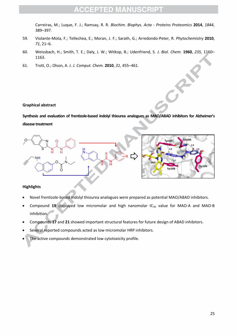
Graphical abstract
Synthesis and evaluation of frentizole-based indolyl thiourea analogues as MAO/ABAD inhibitors for Alzheimer’s
disease treatment
Highlights
Novel frentizole-based indolyl thiourea analogues were prepared as potential MAO/ABAD inhibitors.
Compound 19 displayed low micromolar and high nanomolar IC50 value for MAO-A and MAO-B
inhibition.
Compounds 17 and 21 showed important structural features for future design of ABAD inhibitors.
Several reported compounds acted as low micromolar HRP inhibitors.
The active compounds demonstrated low cytotoxicity profile.


![Varioloid A, a new indolyl-6,10b-dihydro-5aH-[1]benzofuro](https://img.pdfslide.us/doc/110x75/625adcddb4967102467e62cf/varioloid-a-a-new-indolyl-610b-dihydro-5ah-1benzofuro-.jpg)