Embed Size (px)
Citation preview
i
v
Synthesis and Characterization of Second- Generation Iron Pro-chelators
by
Lissette Perez
Department of Chemistry Duke University
Date:_______________________ Approved:
___________________________ Katherine J. Franz, Supervisor
___________________________
Alvin Crumbliss
___________________________ Richard MacPhail
Thesis submitted in partial fulfillment of the requirements for the degree of Master of Science in the Department of
Chemistry in the Graduate School of Duke University
2010
ABSTRACT
Synthesis and Characterization of Second- Generation Iron Pro-chelators
by
Lissette Perez
Department of Chemistry Duke University
Date:_______________________ Approved:
___________________________ Katherine J. Franz, Supervisor
___________________________
Alivn Crumbliss
___________________________ Richard MacPhail
An abstract of a thesis submitted in partial fulfillment of the requirements for the degree
of Master of Science in the Department of Chemistry in the Graduate School
of Duke University
2010

Copyright by Lissette Perez
2010
iv
Abstract Excessive amounts of labile metals such as iron, zinc, and copper have been
found in the brains of patients diagnosed with various neurodegenerative diseases,
which include Alzheimer’s and Parkinson’s disease. Iron and copper can participate in
the Haber-Weiss cycle forming reactive oxygen species, ROS, which can induce cell
death. In vivo, these labile metals act catalytically regenerating additional ROS.
It has recently become evident that these neurodegenerative diseases are
complex and have various targets for therapy. Described herein are the newest
approaches to tackling those multifaceted issues using multifunctional chelators as well
as an in depth look at a specific class of iron binding pro-chelators. Researchers have
taken advantage of properties that increase cell and blood brain barrier, BBB,
permeability, increase antioxidant capabilities, increased selectivity for metals in
damage prone environments, lower Aβ peptide aggregation, and inhibit key enzymes
associated with disease pathways.
Our research group has described sets of masked chelators, which have the
potential for selectivity binding iron and copper in damage prone environments. QBP, a
hydroxyquinoline-based pro-chelator was recently shown to inhibit further Aβ
aggregation and diminish copper’s reactivity while BSIH, a salicylaldhyde isonicotinic
hydrazone pro-chelator, has been shown to react selectively with hydrogen peroxide
revealing an active chelator able to bind iron. Described in this thesis is a set of iron pro-
chelator derivatives of the parent compound, BSIH. These masked chelators can
potentially reveal high affinity metal ligands when oxidized by H2O2. Unlike traditional
iron chelators, which can disrupt iron homeostasis, these pro-chelators have a protective
mask consisting of various boronic esters that react and reveal a phenolic oxygen

v
selectively by H2O2. Studies on kinetics of oxidation by H2O2, solubility, iron affinity,
and stability in various conditions have been established.
vi
Contents
Abstract..........................................................................................................................................iv
List of Tables ................................................................................................................................. ix
List of Figures ................................................................................................................................ x
Schemes........................................................................................................................................xiii
1. Minding Metals: Tailoring multifunctional chelating agents for neurodegenerative diseases ........................................................................................................................................... 1
1.1 Introduction....................................................................................................................... 1
1.2 Increasing uptake into cells and across the blood brain barrier ................................ 4
1.2.1 Multifunctional agents that target specific transporters or receptors.................. 5
1.2.1.1 Carbohydrate conjugates to facilitate BBB uptake .......................................... 5
1.2.1.2 Iron binding peptides that target neuronal receptors..................................... 7
1.2.1.3 Chelators with moieties that target integrin receptors ................................... 9
1.2.1.4 Nanoparticles as chelator carriers.................................................................... 10
1.2.2 Strategies for increasing passive diffusion ............................................................ 11
1.2.2.3 Metal chelators with increased lipophilicity .................................................. 11
1.3 Prochelators for triggered activation against oxidative stress ................................. 13
1.3.1 Selectivity based on chelator reactivity .................................................................. 14
1.3.1.1 Masked iron chelators ....................................................................................... 14
1.3.1.2 Photo-caged iron chelators ............................................................................... 14
1.3.1.3 Iron chelators activated by hydroxyl radicals................................................ 15
1.4 Chelator–antioxidant hybrid molecules ...................................................................... 16
1.4.1 L-DOPA/antioxidant hybrid molecules................................................................. 16
1.4.2 Radical scavaging/ iron binding hybrid molecules ............................................. 17
1.5 Chelators that target Aβ ................................................................................................. 18

vii
1.5.1 Amyloid-binding metal chelators ........................................................................... 18
1.5.2 Prochelators activated by Cu–Aβ ............................................................................ 20
1.5.3 Artificial peptidases to break up Aβ peptides....................................................... 21
1.6 Metal chelators with enzyme inhibitory activity ....................................................... 23
1.6.1 Monoamine oxidase (MAO) dual action agents ................................................... 23
1.6.2 Acetylcholinesterase (AChE) triple action agents................................................. 24
1.7 Conclusions ..................................................................................................................... 26
2. Prochelator, BSIH, Strategy for Inhibiting Oxidative Stress via Iron Chelation ............ 28
2.1 Background and Significance........................................................................................ 28
2.2 Experimental Section...................................................................................................... 32
2.2.1 Materials and Methods ............................................................................................. 32
2.2.2 Synthesis ..................................................................................................................... 32
2.2.2.1 Synthesis of Pro-chelators ................................................................................. 32
2.2.2.2 Synthesis of Chelators ....................................................................................... 34
2.2.2.3 Synthesis of bis-Iron Complexes...................................................................... 36
2.3 Results .............................................................................................................................. 37
2.3.1 Solubility of pro-chelators/chelators in various solutions.................................. 37
2.3.2 DEA-BSIH stability studies by 1H-NMR ................................................................ 38
2.3.3 Determination of unknown contaminant in BSIH spectra .................................. 39
2.3.4 Determination of stability in 100 mM NaOH, conditions needed for solubility purposes............................................................................................................................... 40
2.3.5 Determination of stability in minimal essential media, MEM, conditions for future cell studies................................................................................................................ 41
2.3.6 Kinetics of Oxidation of Pro-Chelators to Chelators ............................................ 42
2.3.7 Iron binding stability of the chelators..................................................................... 44
2.3.7.1 Standardization of the iron perchlorate solution .......................................... 44
viii
2.3.7.2 Determination of Apparent Stability Constant for Complex Formation with Iron .......................................................................................................................... 44
2.3 Discussion: ....................................................................................................................... 48
2.3.1 Synthetic Strategy ...................................................................................................... 48
2.3.2 Stability and Solubility of the Chelators/Pro-Chelators...................................... 51
2.3.3 Pro-Chelator Rates of Activation: The Oxidation of the Pro-chelator to Chelator................................................................................................................................ 52
2.3.4 Apparent Stability Constants of Chelators at pH 7.4 with Iron(III) ................... 52
2.3.5 Summary and Conclusions ...................................................................................... 53
Appendix A.................................................................................................................................. 55
Determination of stability of pro-chelators/chelators in 100 mM NaOH, conditions needed for solubility purposes ........................................................................................... 55
Determination of stability in minimal essential media, MEM, conditions for future cell studies.............................................................................................................................. 571H-NMR Spectra of Chelators and Pro-Chelators............................................................ 59
Appendix B .................................................................................................................................. 67
Determining a pFe value ..................................................................................................... 67
Determining pFe value of EDTA at pH 7.4....................................................................... 69
αEDTA Calculations: .............................................................................................................. 69
€
αFe 3
+ Calculations:............................................................................................................... 70
αFeEDTA Calculations:............................................................................................................ 71
Log K’FeEDTA Determination.................................................................................................. 71
pFe for Fe-EDTA ................................................................................................................... 71
References..................................................................................................................................... 72
ix
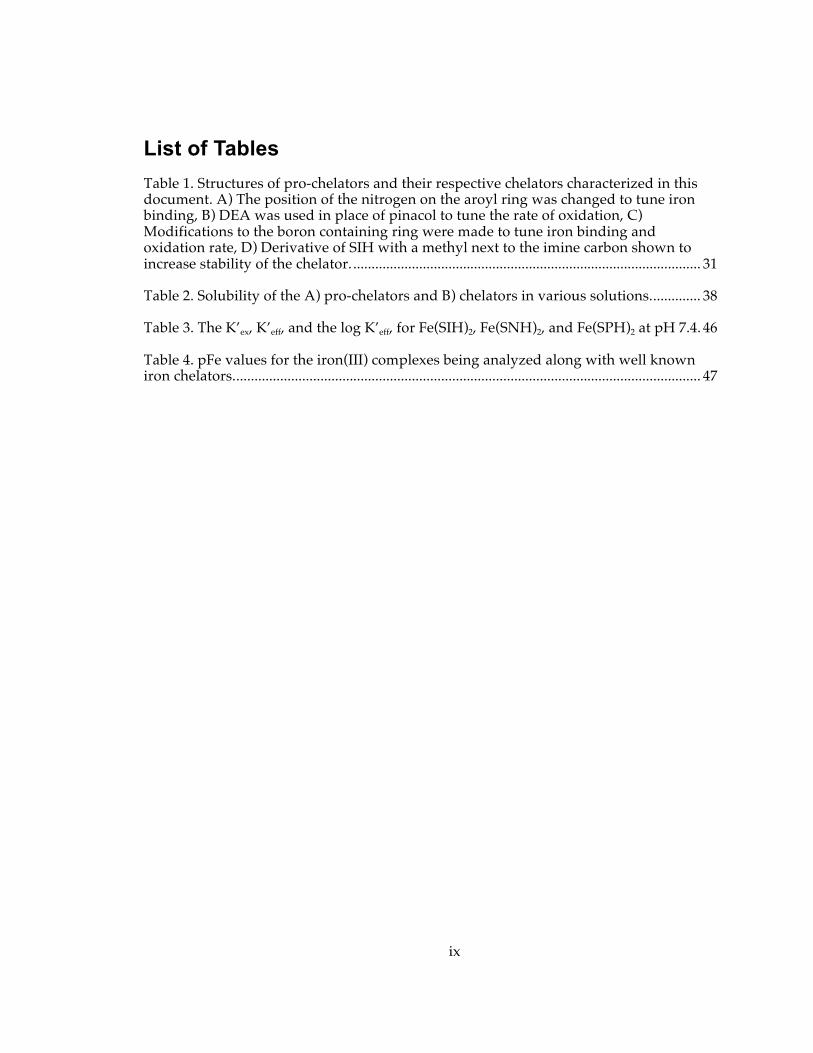
List of Tables Table 1. Structures of pro-chelators and their respective chelators characterized in this document. A) The position of the nitrogen on the aroyl ring was changed to tune iron binding, B) DEA was used in place of pinacol to tune the rate of oxidation, C) Modifications to the boron containing ring were made to tune iron binding and oxidation rate, D) Derivative of SIH with a methyl next to the imine carbon shown to increase stability of the chelator. ............................................................................................... 31
Table 2. Solubility of the A) pro-chelators and B) chelators in various solutions.............. 38
Table 3. The K’ex, K’eff, and the log K’eff, for Fe(SIH)2, Fe(SNH)2, and Fe(SPH)2 at pH 7.4. 46
Table 4. pFe values for the iron(III) complexes being analyzed along with well known iron chelators................................................................................................................................ 47
x
List of Figures Figure 1: Structures of select medicinal iron chelators. ........................................................... 3
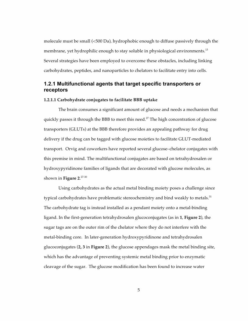
Figure 2: Top: structures of metal ion chelators containing pendant or masking carbohydrate groups. Bottom: schematic example of cellular uptake by glucose transporters of prochelator 3, followed by intracellular enzymatic activation and metal binding. ........................................................................................................................................... 6
Figure 3: Neuroprotective NAP peptides modified with bis-hydroxamic acid (4) or hydroxyquinoline (5) groups; and amino acid derivative of 8-hydroxyquinoline (6). ....... 8
Figure 4: A trifunctional chelator composed of: (A) an ABIR-binding peptide to facilitate entry through cell membranes, (B) cypate as a near-IR probe for detection, and (C) DFO as the iron chelator.36................................................................................................................... 10
Figure 5: Nanoparticle carriers for penicillamine (8) and deferiprone (9) analogues. ...... 11
Figure 6: Acetoxymethyl protecting groups (highlighted in red) provide a lipophilic ferrichrome analogue (10) that is converted to a hydrophilic version (black) by intracellular esterases.43 .............................................................................................................. 12
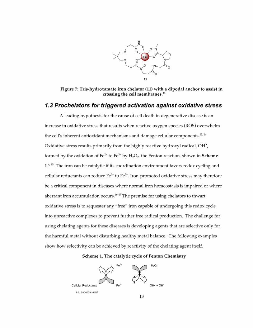
Figure 7: Tris-hydroxamate iron chelator (11) with a dipodal anchor to assist in crossing the cell membranes.44 .................................................................................................................. 13
Figure 8: Exposure to UVA light induces release of the photoactive protecting group (colored in red) on 13 to generate the active chelator SIH available for metal binding.... 15
Figure 9: The tetradentate aminocarboxylate 14 binds iron weakly, but increases affinity following hydroxylation (highlighted in red) in the presence of H2O2 and a reductant. [X= coordinating solvent]. In a similar fashion, the aromatic rings in 15 can also be hydroxylated. In this case, only modifications at the ortho positions (highlighted in red) result in compounds with improved iron affinity.................................................................. 16
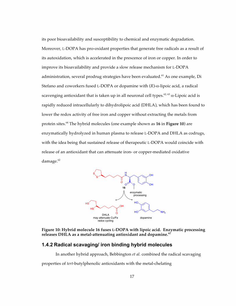
Figure 10: Hybrid molecule 16 fuses L-DOPA with lipoic acid. Enzymatic processing releases DHLA as a metal-attenuating antioxidant and dopamine.62 ................................. 17
Figure 11: Structure of BHT antioxidant (red) fused to hydroxypyridinone chelator (blue). ............................................................................................................................................ 18
Figure 12: Examples of chelators that incorporate features of thioflavin-T as an amyloid-directing group. XH1 (18) contains an aminocarboxylate binding site, whereas HBTI (19) uses an O/N bidentate unit reminiscent of clioquinol. ......................................................... 19
Figure 13: The H2O2 generated from Cu-Aβ species in the presence of O2 and ascorbic acid unmasks prochelator QBP (20) to release 8-hydroxyquinoline that extracts Cu2+ from Aβ and prevents further redox-cycling and Aβ aggregation...................................... 21
xi
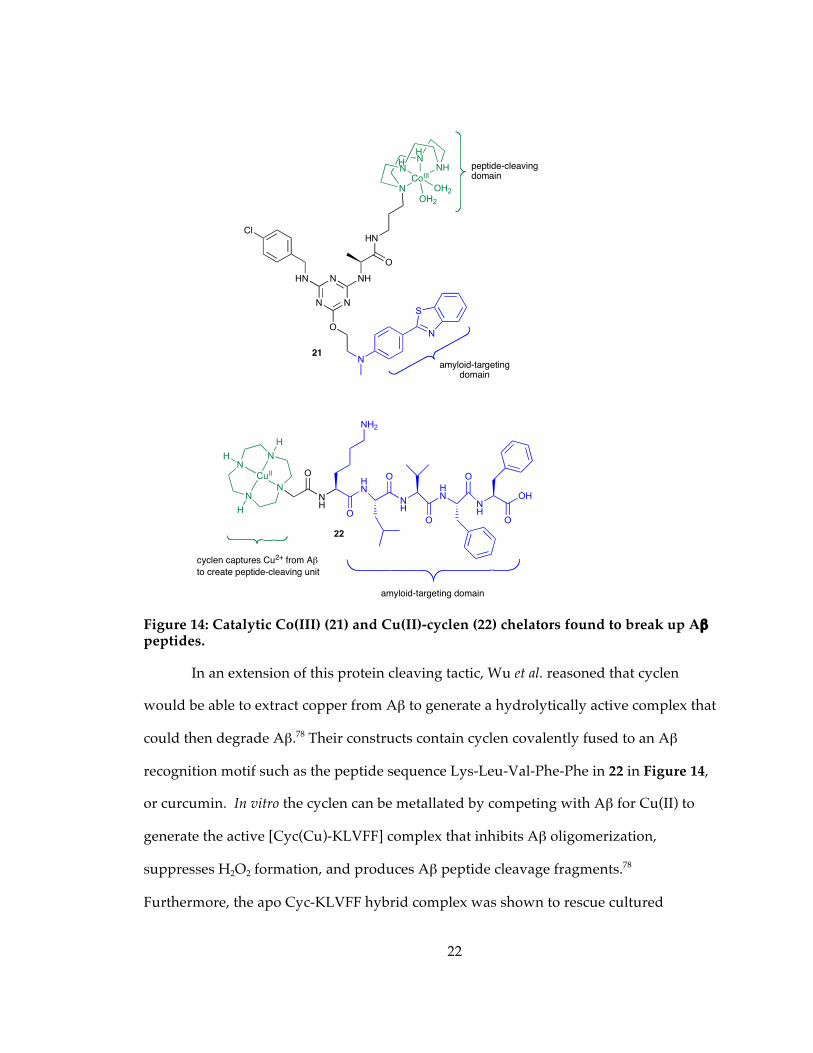
Figure 14: Catalytic Co(III) (21) and Cu(II)-cyclen (22) chelators found to break up Aβ peptides......................................................................................................................................... 22
Figure 15: Structures of bifunctional metal-ion chelators, M-30 (23) and HLA-20 (24) that contain an N-propargylamine moiety found in MAO inhibitors like Seleginline and Rasagiline. .................................................................................................................................... 23
Figure 16: A bis-tacrine molecule (25) where the linker can interact with metal ions. The tacrine units are colored in red. 25 has been found to inhibit AChE activity, reverse AChE-induced amyloid fibrillogenesis, and bind metal ions. ............................................. 25
Figure 17: The multifunctional prochelator 26 combines structural features of HLA-20 (24, see Figure 15) and AChE inhibitors rivastigmine and donepezil. The carbamyl group is cleaved by AChE to release the 8-hydroxyquinoline metal binding group (highlighted in bold). .................................................................................................................. 26
Figure18: 1H-NMR spectra of (a) DEA, (b) DEA-BSIH in 20 mM NaHPO4 buffer at pH 7.4 after 3 h (c) DEA-BSIH in 20 mM NaHPO4 buffer at pH 7.4 after 45 h ............................... 39
Figure 19. 1H-NMR of BSIH. A) BSIH in d6-DMSO. B) Immediately following collection of spectra A, the sample was spiked with D2O and a second set of spectra were collected. The peak at 8.5 ppm clearly disappears in the presence of D2O indicating the boronate species of BSIH. ........................................................................................................................... 40
Figure 20: UV-vis spectra of 50 µM solutions of FMDOP-Bpin-IH and DEA-BSIH in 100 mM NaOH before and after freezing at -20 C. The spectra of the other pro-chelators/chelators examined can be found in Appendix A................................................ 41
Figure 21: 50 µM solutions of the pro-chelator, DEA-BSIH, and it’s respective chelator, SIH, in MEM at pH 7.4 and 37 °C. The spectra of the other pro-chelators/chelators examined can be found in the supplementary section. ......................................................... 42
Figure 22. The oxidation of DEA-BSIH to SIH using H2O2 in 20 mM NaHPO4 at pH 7.4.42
Figure 23. The oxidation of pro-chelator, DEA-BSIH, to its corresponding chelator, SIH, in the presence of 5 mM H2O2. The conversion was evaluated in 20 mM sodium phosphate buffer at ph 7.4. ........................................................................................................ 43
Figure 24. The second order rate constant for DEA-BSIH being oxidized to SIH, Figure 22. The graph shows the plot of kobs vs H2O2 concentration for the oxidation of 50 µM BSIH to SIH in 20 mM NaHPO4 buffer at pH 7.4................................................................... 44
Figure 25. Absorbance of Fe(SNH)2 with varying amounts of EDTA in a 50:50 MeOH: 10 mM HEPES/100 mM NaCl solution. ....................................................................................... 47
Figure 27. Possible structures of BAHAP-INH...................................................................... 49
Figure 28. UV-vis spectra of 50 µM solutions of FMDOP-Bpin-IH, DEA-BSIH, BSNH, BSPH, and BSIH in 100 mM NaOH before and after freezing at -20 C. Spectra of FMDOP-Bpin-IH and DEA-BSIH can also be found in the document in Figure 20.......... 55
xii
Figure 29. UV-vis spectra of 50 µM solutions of SNH, SPH, SIH, FMDOP-IH and HAP-INH in 100 mM NaOH before and after freezing at -20 C. ................................................... 56
Figure 30. 50 µM solutions of the pro-chelators, BSNH, BSPH, DEA-BSIH, and FMDOP-Bpin-IH in MEM at pH 7.4 and 37 °C. Spectra of DEA-BSIH can also be found in the document in Figure 21. ............................................................................................................... 57
Figure 31. 50 µM solutions of the chelators, SNH, SPH, FMDOP-IH, and HAP-INH in MEM at pH 7.4 and 37 °C. Spectra of SIH can also be found in the document in Figure 21.................................................................................................................................................... 58
xiii
Schemes Scheme 1. The catalytic cycle of Fenton Chemistry................................................................ 13
Scheme 2. The masked chelator, BSIH (12), binds Fe3+ only following deprotection by H2O2.50............................................................................................................................................ 29
Scheme 3. Two possible routes for the synthesis of the pro-chelators. ............................... 49
Scheme 4. Possible route to synthesis of BHAP-INH using a selected diol. ...................... 50
1
1. Minding Metals: Tailoring multifunctional chelating agents for neurodegenerative diseases
1.1 Introduction Iron, copper, and zinc play complicated roles in human health and disease.
While all three are essential nutrients utilized as various protein cofactors, their
misappropriation within cells and tissues can lead to significant damage. Biological
systems tightly regulate these metals at both the systemic level of absorption and
distribution as well as the cellular level of storage, recycling, and utilization so that the
organism meets metabolic demand without over accumulation (for recent reviews of
iron, copper, and zinc homeostasis see references1-3). Excess metal that is not
appropriately contained by the proteins or vesicles that utilize, store, or transport it
becomes available for unintentional and potentially toxic reactivity. One of the dangers
of redox-active metals like iron and copper is their ability to promote the formation of
highly toxic hydroxyl radicals that oxidize biomolecules and subsequently lead to cell
death.4, 5 Even in the absence of redox activity, metal cations like Zn2+ and Cu2+ may also
cause damage by inducing aberrant protein aggregation.6, 7
Both kinds of metal-promoted damage, oxidative stress and protein misfolding,
have been linked to Parkinson’s, Alzheimer’s, and other neurodegenerative diseases.8-13
Furthermore, there is increasing recognition that these diseases are associated with
localized accumulation of metal ions in disease-affected regions.14 Iron-induced
oxidative stress in particular is speculated to play a role in a wide variety of progressive
inflammatory and degenerative diseases, ranging from atherosclerosis and aging to
diabetes and macular degeneration.15 The emphasis here is on localized misregulation, as
Chapter 1 is a slightly modified version of a review recently been published in Dalton Transactions. L.R. Perez and K.J. Franz, Dalton Transactions 2010, 39, 2177-2187
2
these diseases are not characterized by systemic metal imbalances, as is the case with
traditional iron overload diseases like hemochromatosis and thalassemia.
Inhibiting metal-promoted damage by using small molecule chelating agents is a
promising strategy for treating diseases associated with localized metal accumulation.10,
16-18 The target pool of metal ions in these diseases, however, is significantly different
from diseases of systemic metal overload where the target metal is plentiful and more
readily available for sequestration. Furthermore, our current understanding of metal
homeostasis in the central nervous system is limited, which further constrains the
interpretation of the effects of manipulating metals in the brain.19 Designing chelators to
target localized metal imbalance therefore presents unique challenges. Generalized
metal chelation can indeed protect against acute oxidative stress or inhibit protein
aggregation, but long-term treatment with metal chelators poses serious risks associated
with depletion or redistribution of healthy metal ions, inhibition of metalloenzymes, or
other unintended consequences.
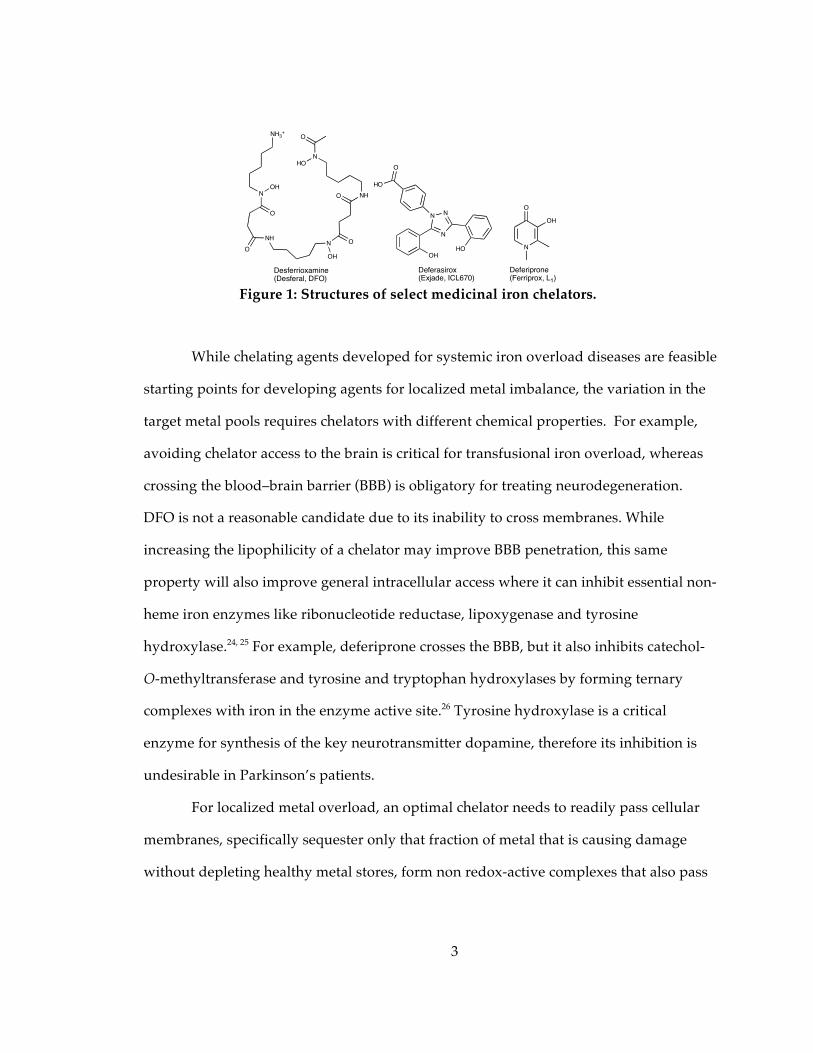
Research on medicinal iron chelators over the last 40 years has been dominated
by the search for alternatives to desferrioxamine B (DFO, see Figure 1), the naturally
occurring siderophore that has been used clinically since the 1970s for treating
transfusional iron overload diseases.20 DFO is a hydrophilic, hexadentate chelator that is
not absorbed by the gut and has a short plasma half-life, requiring it to be administered
daily in large doses via subcutaneous transfusion. Extensive research to identify
alternatives has lead to the commercial development of two orally available agents, the
bidentate hydroxypyridinone known as L1 or deferiprone, and the tridententate chelator
deferasirox, or Exjade (Figure 1).21-23 The targets of all these drugs are primarily non-
transferrin bound iron in the plasma, followed by intracellular iron stores in the liver,
heart and endocrine tissues.
3
While chelating agents developed for systemic iron overload diseases are feasible
starting points for developing agents for localized metal imbalance, the variation in the
target metal pools requires chelators with different chemical properties. For example,
avoiding chelator access to the brain is critical for transfusional iron overload, whereas
crossing the blood–brain barrier (BBB) is obligatory for treating neurodegeneration.
DFO is not a reasonable candidate due to its inability to cross membranes. While
increasing the lipophilicity of a chelator may improve BBB penetration, this same
property will also improve general intracellular access where it can inhibit essential non-
heme iron enzymes like ribonucleotide reductase, lipoxygenase and tyrosine
hydroxylase.24, 25 For example, deferiprone crosses the BBB, but it also inhibits catechol-
O-methyltransferase and tyrosine and tryptophan hydroxylases by forming ternary
complexes with iron in the enzyme active site.26 Tyrosine hydroxylase is a critical
enzyme for synthesis of the key neurotransmitter dopamine, therefore its inhibition is
undesirable in Parkinson’s patients.
For localized metal overload, an optimal chelator needs to readily pass cellular
membranes, specifically sequester only that fraction of metal that is causing damage
without depleting healthy metal stores, form non redox-active complexes that also pass
N
O
OH
OH
N
NN
HO
O
HO
NH3+
N
NH
OH
O
ON
OH
O NH
N
O
O
HO
Desferrioxamine(Desferal, DFO)
Deferiprone (Ferriprox, L1)
Deferasirox(Exjade, ICL670)
Figure 1: Structures of select medicinal iron chelators.
4
cellular membranes for elimination, avoid redistribution of iron or other metals within a
cell or between cells, and be non-toxic.
It is now becoming apparent that the next generation of therapeutically relevant
metal chelators must have multiple functions to be most effective. Some noteworthy
advances in these multifunctional metal chelating agents involve functions that increase
cell and BBB permeability, increase anti-oxidant capabilities, increase selectivity for
metals in damage-prone environments, lower Aβ peptide aggregation, and inhibit key
enzymes associated with specific disease pathways. Chapter one will highlight recent
advances and clever strategies in the design of multifaceted metal chelating agents
targeted against neurodegenerative disease. Chapter two of this document will focus
more specifically on masked iron pro-chelators, which are activated in the presence of
H2O2.
1.2 Increasing uptake into cells and across the blood brain barrier
If metal chelating agents are to be used to treat neurodegenerative disease, a
prerequisite is that they must bypass the blood–brain barrier, a continuous layer of
endothelial cells connected by tight junctions where the bloodstream meets the neural
tissue.13 Its function is to restrict high molecular weight compounds and most ions from
entering the brain while allowing essential nutrients to pass through, thereby blocking
entry of neurotoxic materials while minimizing fluctuations in levels of vital substances
in the central nervous system. As mentioned previously, abnormally elevated levels of
metals have been found localized in the brains of patients with neurodegenerative
diseases.14 In order to sequester potentially damaging sources of aberrant metal ions,
chelating agents must be able to pass through the BBB; however, creating a compound
capable of permeating the BBB is not trivial. Conventional wisdom posits that the
5
molecule must be small (<500 Da), hydrophobic enough to diffuse passively through the
membrane, yet hydrophilic enough to stay soluble in physiological environments.13
Several strategies have been employed to overcome these obstacles, including linking
carbohydrates, peptides, and nanoparticles to chelators to facilitate entry into cells.
1.2.1 Multifunctional agents that target specific transporters or receptors
1.2.1.1 Carbohydrate conjugates to facilitate BBB uptake
The brain consumes a significant amount of glucose and needs a mechanism that
quickly passes it through the BBB to meet this need.27 The high concentration of glucose
transporters (GLUTs) at the BBB therefore provides an appealing pathway for drug
delivery if the drug can be tagged with glucose moieties to facilitate GLUT-mediated
transport. Orvig and coworkers have reported several glucose–chelator conjugates with
this premise in mind. The multifunctional conjugates are based on tetrahydrosalen or
hydroxypyridinone families of ligands that are decorated with glucose molecules, as
shown in Figure 2.27-30
Using carbohydrates as the actual metal binding moiety poses a challenge since
typical carbohydrates have problematic stereochemistry and bind weakly to metals.31
The carbohydrate tag is instead installed as a pendant moiety onto a metal-binding
ligand. In the first-generation tetrahydrosalen glucoconjugates (as in 1, Figure 2), the
sugar tags are on the outer rim of the chelator where they do not interfere with the
metal-binding core. In later-generation hydroxypyridinone and tetrahydrosalen
glucoconjugates (2, 3 in Figure 2), the glucose appendages mask the metal binding site,
which has the advantage of preventing systemic metal binding prior to enzymatic
cleavage of the sugar. The glucose modification has been found to increase water
6
solubility, minimize toxicity, and shows promise for specific targeting of these
compounds.27, 30
In vivo, these glucoconjugate pro-ligands are anticipated to gain entry into the
brain via glucose transporters on the surface of endothelial cells on the BBB.27 Once
inside cells, the glucose masking group would be enzymatically cleaved to release the
active chelating agent, as shown at the bottom of Figure 2. As a proof-of-principle, it
was shown that both a broad-spectrum glucosidase from Agrobacterium faecalis and a rat
brain homogenate as a model of glucosidase activity convert the glucoconjugate
prochelators into their metal-binding hydroxypyridinone or tetrahydrosalen versions.28,
30
OHOHO
OHO
OH
OH
NO
OHOH
HOO
OH
HO
N
N N
OHOHO
OHO
OH
OOH
OHHO
O
OHN
OOHOHO
OHO
OH
1
2
3
Figure 2: Top: structures of metal ion chelators containing pendant or masking carbohydrate groups. Bottom: schematic example of cellular uptake by glucose transporters of prochelator 3, followed by intracellular enzymatic activation and metal binding.
7
A rat brain perfusion experiment with a radiolabeled hydroxypryidinone
glucoconjugate demonstrated adequate cerebral uptake, showing that these compounds
are indeed taken across the BBB.28
Once the carbohydrate groups are cleaved by β-glucosidases, the released
tetrahydrosalen or hydroxypyridinone chelators are available to bind metals. In
addition, both classes of compounds are effective radical-scavenging antioxidants.28, 29 In
vitro studies show that both chelators compete with amyloid-beta (Aβ) peptides for
binding Cu2+ and Zn2+, and decrease Aβ1-40 peptide aggregation induced by these
metals.27-30 The ability of 1 to prevent Aβ peptide aggregation was comparable to that of
EDTA despite its lower affinity for Cu2+. This observation could be attributed to the
increased lipophilicity and/or intercalating ability of the tetrahydrosalen compound
into Aβ peptide aggregates.30
1.2.1.2 Iron binding peptides that target neuronal receptors
Functionalizing neuropeptides with metal chelating groups is another strategy to
target agents to specific brain locations.32 Vasoactive intestinal peptide, VIP, is widely
distributed in areas of the brain associated with learning and memory and is associated
with specific neuronal receptors in the brain.33 An eight amino acid peptide abbreviated
as NAP with amino acid sequence Asn-Ala-Pro-Val-Ser-Ile-Pro-Gln is the smallest active
portion of activity-dependent neuroprotective protein, ADNP, which is a glial cell
mediator of VIP-induced neuroprotection.32 NAP has demonstrated potent
neuroprotection in both cell culture and animal models of neurodegeneration and is
capable of crossing the BBB.34 In order to create a multifunctional agent that would take
advantage of the targeting ability of the NAP neuropeptide to sites of
neurodegeneration and expand its antioxidant potential, Fridkin and coworkers
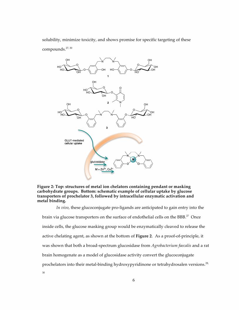
modified the native peptide by adding hydroxamate (4) or 8-hydroxyquinoline (5)
8
moieties to derivatives of NAP, as shown in Figure 3.32, 34 The modified peptides form
stable metal ion complexes with Fe2+/3+, Cu2+ and Zn2+ in water, pH 5–7, whereas the
parent NAP peptide shows no binding affinity to these metal ions. The peptides
containing two hydroxamate units inhibit lipid peroxidation and iron-catalyzed
hydroxyl radical formation in vitro and protect neuroblastoma cells against oxidative
stress induced by exogenous hydrogen peroxide.34 Although not yet tested, these
peptides could allow for transport and localization of therapeutic chelators into the
brain.
Figure 3: Neuroprotective NAP peptides modified with bis-hydroxamic acid (4) or hydroxyquinoline (5) groups; and amino acid derivative of 8-hydroxyquinoline (6).
An alternative strategy for targeting chelators into the brain is to modify them
with a neutral amino acid carrier group that would facilitate uptake by system L, a
known brain uptake pathway for hydrophilic amino acids including L-DOPA. Fridkin
and coworkers therefore synthesized and studied a bifunctional iron chelator, M10 (6),
that fuses an 8-hydroxyquinoline unit with an alanine amino acid, as shown in Figure
3.35 They have shown that 6 is a metal chelator that exibits free radical scavenging
properties and is water soluble. The hydrophilicity of 6 may prevent its passive
diffusion into cells, which would avert its interference with normal, systemic metal
N
OH
H2N
HO
OS
N
OH
6
4
5
Ala-Pro-Val-Ser-Ile-Pro NH
H2N
O
HN O
OH
NH2
O
NHO
OH
Asn-Ala-Pro-Cys-Ser-Ile-Pro-Glu
9
metabolism. It is hoped that the carrier group would thus allow selective targeting of the
agent into the brain while minimizing effects on systemic metal balance.35
1.2.1.3 Chelators with moieties that target integrin receptors
Overexpression of the cell surface avb3 integrin receptor (ABIR) has been found
on activated endothelial cells in the neovasculature of tumors and has been associated
with tumor growth, invasion, and metastasis.36 Fluorescent probes and anticancer
agents have previously been shown to be taken up when coupled to ABIR-binding
peptides.37, 38 In order to use this approach to improve the uptake of metal chelators,
Achilefu and coworkers synthesized a tri-functional agent consisting of a DFO chelating
unit, a near-infrared cypate fluorescent probe, and a cyclic RGD peptide that is known to
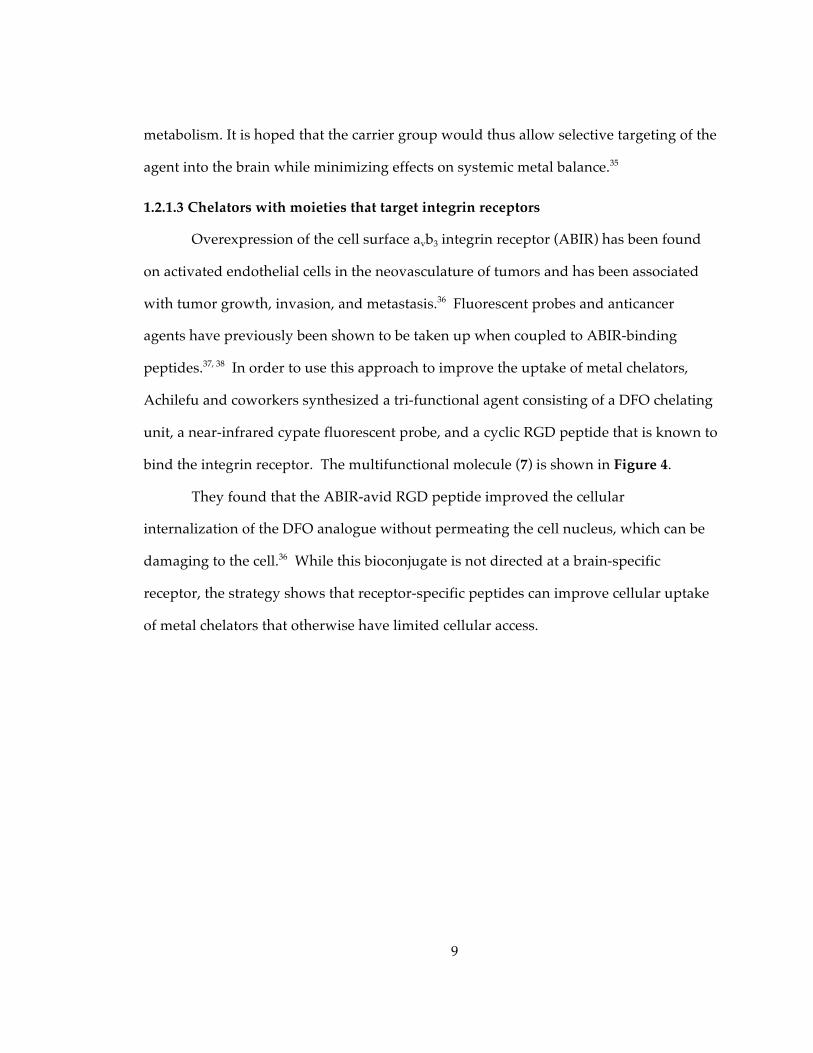
bind the integrin receptor. The multifunctional molecule (7) is shown in Figure 4.
They found that the ABIR-avid RGD peptide improved the cellular
internalization of the DFO analogue without permeating the cell nucleus, which can be
damaging to the cell.36 While this bioconjugate is not directed at a brain-specific
receptor, the strategy shows that receptor-specific peptides can improve cellular uptake
of metal chelators that otherwise have limited cellular access.
10
Figure 4: A trifunctional chelator composed of: (A) an ABIR-binding peptide to facilitate entry through cell membranes, (B) cypate as a near-IR probe for detection, and (C) DFO as the iron chelator.36
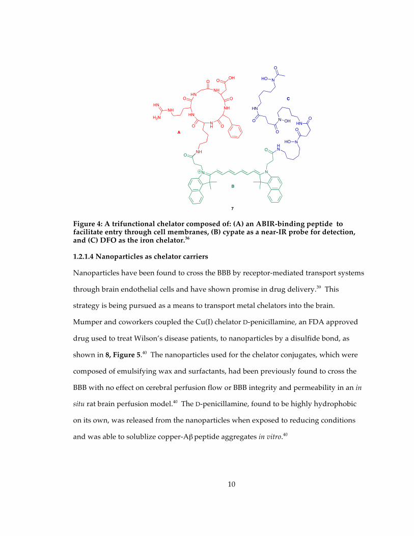
1.2.1.4 Nanoparticles as chelator carriers
Nanoparticles have been found to cross the BBB by receptor-mediated transport systems
through brain endothelial cells and have shown promise in drug delivery.39 This
strategy is being pursued as a means to transport metal chelators into the brain.
Mumper and coworkers coupled the Cu(I) chelator D-penicillamine, an FDA approved
drug used to treat Wilson’s disease patients, to nanoparticles by a disulfide bond, as
shown in 8, Figure 5.40 The nanoparticles used for the chelator conjugates, which were
composed of emulsifying wax and surfactants, had been previously found to cross the
BBB with no effect on cerebral perfusion flow or BBB integrity and permeability in an in
situ rat brain perfusion model.40 The D-penicillamine, found to be highly hydrophobic
on its own, was released from the nanoparticles when exposed to reducing conditions
and was able to solublize copper-Aβ peptide aggregates in vitro.40
NHO
N N
O
HN
7
A
B
N
O
HO
HN
ON OH
O
HN
ONH
HN
HN
NH
NH
O
O
OOH
O
O
NH
HN
H2N
O
C
NHO
O
11
Figure 5: Nanoparticle carriers for penicillamine (8) and deferiprone (9) analogues.
With a similar concept in mind, Smith and coworkers devised polystyrene
nanoparticles decorated with pyridinone chelators, as shown for Nano-N2PY, 9, in
Figure 5.41, 42 In this case, conjugation of the chelators to the nanoparticle does not alter
its metal chelating ability. The Nano-N2PY conjugates were shown to inhibit Aβ
aggregation in vitro and to protect neuronal cells from Aβ -associated neurotoxicity.42
1.2.2 Strategies for increasing passive diffusion
1.2.2.3 Metal chelators with increased lipophilicity
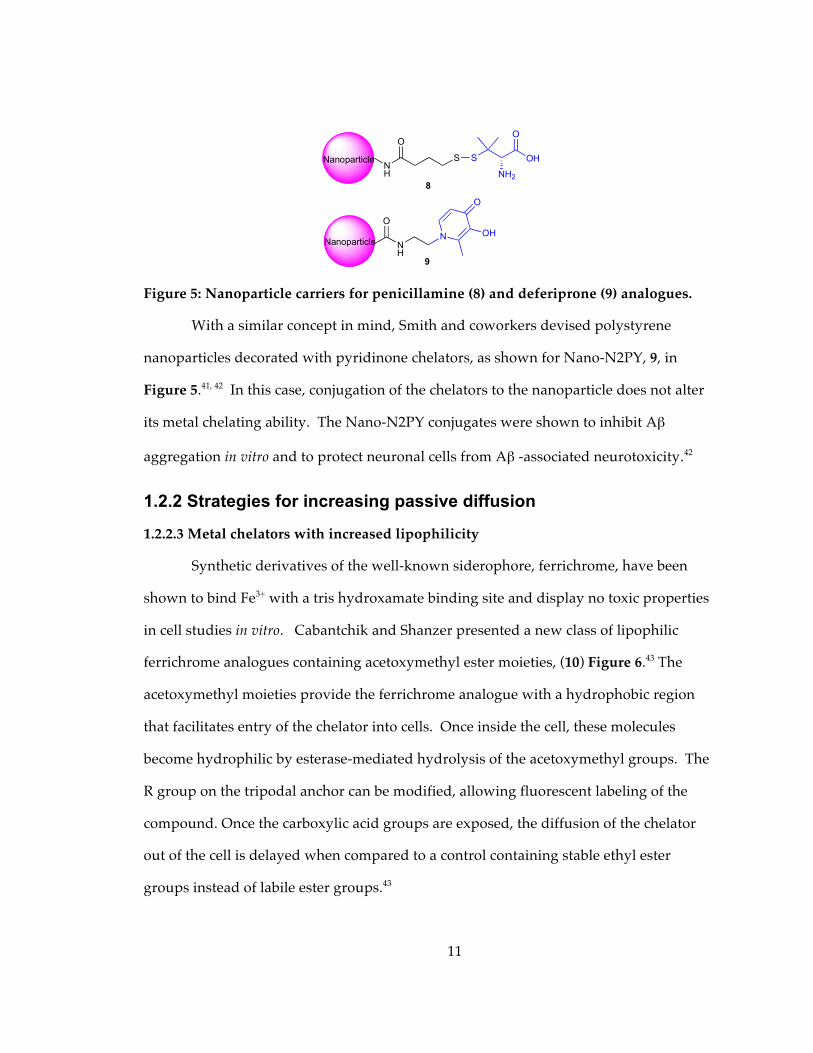
Synthetic derivatives of the well-known siderophore, ferrichrome, have been
shown to bind Fe3+ with a tris hydroxamate binding site and display no toxic properties
in cell studies in vitro. Cabantchik and Shanzer presented a new class of lipophilic
ferrichrome analogues containing acetoxymethyl ester moieties, (10) Figure 6.43 The
acetoxymethyl moieties provide the ferrichrome analogue with a hydrophobic region
that facilitates entry of the chelator into cells. Once inside the cell, these molecules
become hydrophilic by esterase-mediated hydrolysis of the acetoxymethyl groups. The
R group on the tripodal anchor can be modified, allowing fluorescent labeling of the
compound. Once the carboxylic acid groups are exposed, the diffusion of the chelator
out of the cell is delayed when compared to a control containing stable ethyl ester
groups instead of labile ester groups.43
O
NH
N OH
O
S
O
OH
NH28
9
Nanoparticle
Nanoparticle
O
NH
S
12
Figure 6: Acetoxymethyl protecting groups (highlighted in red) provide a lipophilic ferrichrome analogue (10) that is converted to a hydrophilic version (black) by intracellular esterases.43
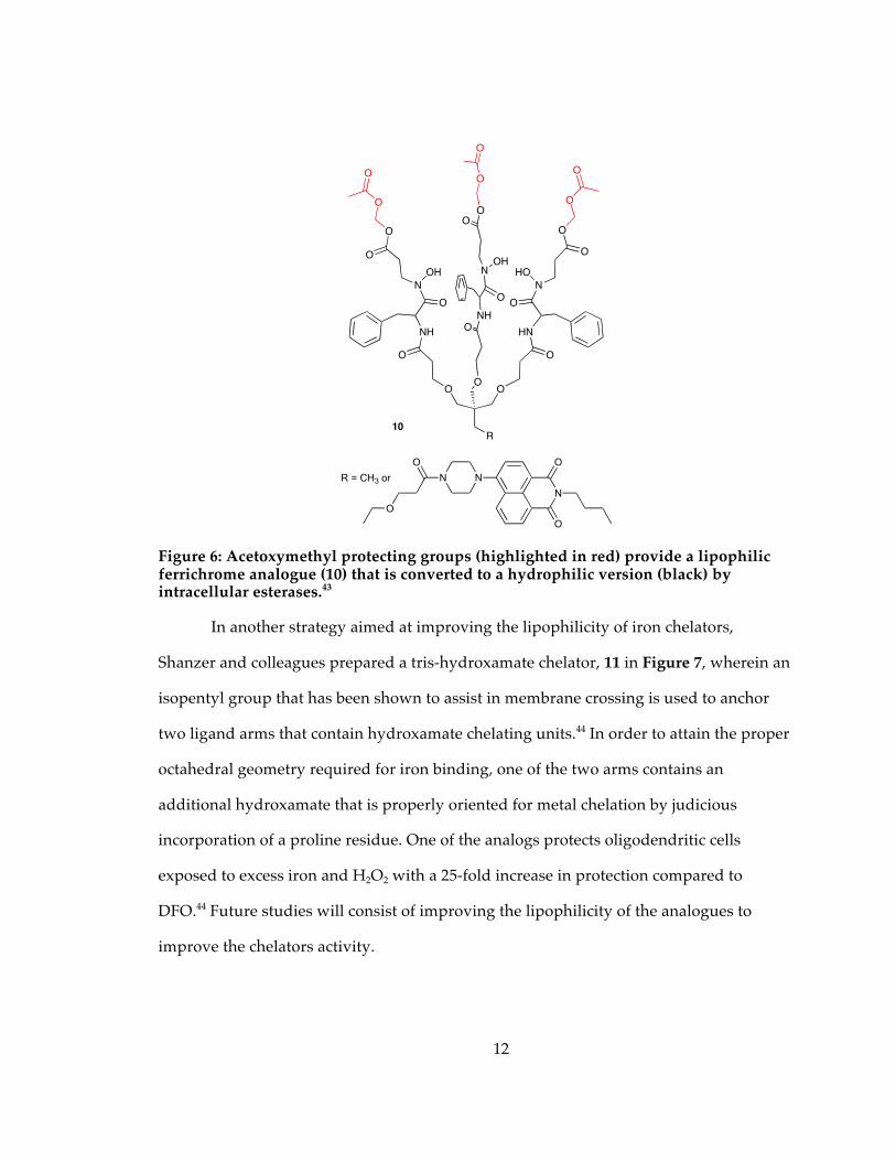
In another strategy aimed at improving the lipophilicity of iron chelators,
Shanzer and colleagues prepared a tris-hydroxamate chelator, 11 in Figure 7, wherein an
isopentyl group that has been shown to assist in membrane crossing is used to anchor
two ligand arms that contain hydroxamate chelating units.44 In order to attain the proper
octahedral geometry required for iron binding, one of the two arms contains an
additional hydroxamate that is properly oriented for metal chelation by judicious
incorporation of a proline residue. One of the analogs protects oligodendritic cells
exposed to excess iron and H2O2 with a 25-fold increase in protection compared to
DFO.44 Future studies will consist of improving the lipophilicity of the analogues to
improve the chelators activity.
OOO
NH
N
O
O
OH
O
O
O
O
HN
N
O
O
HO
O
O
O
O
NH
N
O
O
OH
O
O
O
O
R
R = CH3 or
O
N
O
N
N
O
O
10
13
Figure 7: Tris-hydroxamate iron chelator (11) with a dipodal anchor to assist in crossing the cell membranes.44
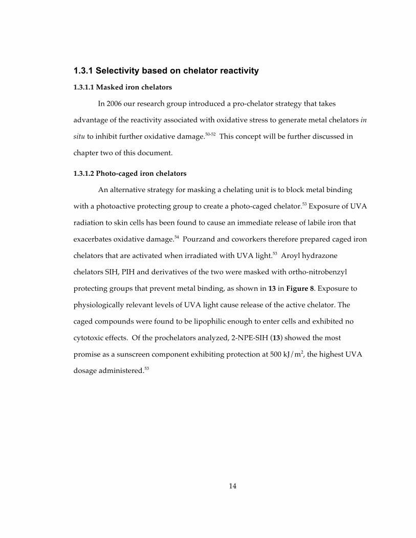
1.3 Prochelators for triggered activation against oxidative stress A leading hypothesis for the cause of cell death in degenerative disease is an
increase in oxidative stress that results when reactive oxygen species (ROS) overwhelm
the cell’s inherent antioxidant mechanisms and damage cellular components.13, 14
Oxidative stress results primarily from the highly reactive hydroxyl radical, OH•,
formed by the oxidation of Fe2+ to Fe3+ by H2O2, the Fenton reaction, shown in Scheme
1.4, 45 The iron can be catalytic if its coordination environment favors redox cycling and
cellular reductants can reduce Fe3+ to Fe2+. Iron-promoted oxidative stress may therefore
be a critical component in diseases where normal iron homeostasis is impaired or where
aberrant iron accumulation occurs.46-49 The premise for using chelators to thwart
oxidative stress is to sequester any “free” iron capable of undergoing this redox cycle
into unreactive complexes to prevent further free radical production. The challenge for
using chelating agents for these diseases is developing agents that are selective only for
the harmful metal without disturbing healthy metal balance. The following examples
show how selectivity can be achieved by reactivity of the chelating agent itself.
Scheme 1. The catalytic cycle of Fenton Chemistry
Fe2+ H2O2
Cellular Reductants Fe3+ OH + OH-
i.e. ascorbic acid
O
N
O
O
HN
O
O
NO
N
O
HNO
O
O
11
Fe
14
1.3.1 Selectivity based on chelator reactivity
1.3.1.1 Masked iron chelators
In 2006 our research group introduced a pro-chelator strategy that takes
advantage of the reactivity associated with oxidative stress to generate metal chelators in
situ to inhibit further oxidative damage.50-52 This concept will be further discussed in
chapter two of this document.
1.3.1.2 Photo-caged iron chelators
An alternative strategy for masking a chelating unit is to block metal binding
with a photoactive protecting group to create a photo-caged chelator.53 Exposure of UVA
radiation to skin cells has been found to cause an immediate release of labile iron that
exacerbates oxidative damage.54 Pourzand and coworkers therefore prepared caged iron
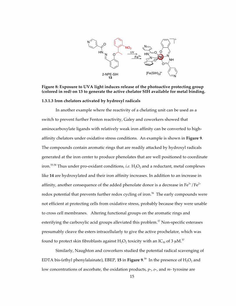
chelators that are activated when irradiated with UVA light.53 Aroyl hydrazone
chelators SIH, PIH and derivatives of the two were masked with ortho-nitrobenzyl
protecting groups that prevent metal binding, as shown in 13 in Figure 8. Exposure to
physiologically relevant levels of UVA light cause release of the active chelator. The
caged compounds were found to be lipophilic enough to enter cells and exhibited no
cytotoxic effects. Of the prochelators analyzed, 2-NPE-SIH (13) showed the most
promise as a sunscreen component exhibiting protection at 500 kJ/m2, the highest UVA
dosage administered.53
15
Figure 8: Exposure to UVA light induces release of the photoactive protecting group (colored in red) on 13 to generate the active chelator SIH available for metal binding.
1.3.1.3 Iron chelators activated by hydroxyl radicals
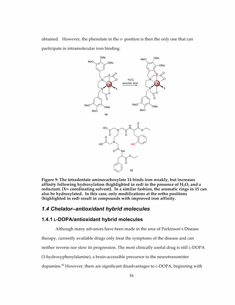
In another example where the reactivity of a chelating unit can be used as a
switch to prevent further Fenton reactivity, Galey and coworkers showed that
aminocarboxylate ligands with relatively weak iron affinity can be converted to high-
affinity chelators under oxidative stress conditions. An example is shown in Figure 9.
The compounds contain aromatic rings that are readily attacked by hydroxyl radicals
generated at the iron center to produce phenolates that are well positioned to coordinate
iron.55-58 Thus under pro-oxidant conditions, i.e. H2O2 and a reductant, metal complexes
like 14 are hydroxylated and their iron affinity increases. In addition to an increase in
affinity, another consequence of the added phenolate donor is a decrease in Fe3+/Fe2+
redox potential that prevents further redox cycling of iron.56 The early compounds were
not efficient at protecting cells from oxidative stress, probably because they were unable
to cross cell membranes. Altering functional groups on the aromatic rings and
esterifying the carboxylic acid groups alleviated this problem.57 Non-specific esterases
presumably cleave the esters intracellularly to give the active prochelator, which was
found to protect skin fibroblasts against H2O2 toxicity with an IC50 of 3 µM.57
Similarly, Naughton and coworkers studied the potential radical scavenging of
EDTA bis-(ethyl phenylalainate), EBEP, 15 in Figure 9.59 In the presence of H2O2 and
low concentrations of ascorbate, the oxidation products, p-, o-, and m- tyrosine are
NHN
O
NO
2-NPE-SIH13
NO2
Fe3+UV
N
NH
N
O
O
FeNHN
N
O
O
[Fe(SIH)2]+
Fe
16
obtained. However, the phenolate in the o- position is then the only one that can
participate in intramolecular iron binding.
Figure 9: The tetradentate aminocarboxylate 14 binds iron weakly, but increases affinity following hydroxylation (highlighted in red) in the presence of H2O2 and a reductant. [X= coordinating solvent]. In a similar fashion, the aromatic rings in 15 can also be hydroxylated. In this case, only modifications at the ortho positions (highlighted in red) result in compounds with improved iron affinity.
1.4 Chelator–antioxidant hybrid molecules
1.4.1 L-DOPA/antioxidant hybrid molecules
Although many advances have been made in the area of Parkinson’s Disease
therapy, currently available drugs only treat the symptoms of the disease and can
neither reverse nor slow its progression. The most clinically useful drug is still L-DOPA
(3-hydroxyphenylalanine), a brain-accessible precursor to the neurotransmitter
dopamine.60 However, there are significant disadvantages to L-DOPA, beginning with
O
X
O
H2O2
N
N
O
O
X
Fe
OMe
MeOOMe
OMe
MeO
MeO
O
X
O
N
N
O
O
Fe
OMeMeO
OMe
OMe
MeO
MeO
O
ascorbic acid
14
N
NHO
O
HO
O
NHO
O
O
HN
O
O
O
15
HO
17
its poor bioavailability and susceptibility to chemical and enzymatic degradation.
Moreover, L-DOPA has pro-oxidant properties that generate free radicals as a result of
its autoxidation, which is accelerated in the prescence of iron or copper. In order to
improve its bioavailability and provide a slow release mechanism for L-DOPA
administration, several prodrug strategies have been evaluated.61 As one example, Di
Stefano and coworkers fused L-DOPA or dopamine with (R)-α-lipoic acid, a radical
scavenging antioxidant that is taken up in all neuronal cell types.62, 63 α-Lipoic acid is
rapidly reduced intracellularly to dihydrolipoic acid (DHLA), which has been found to
lower the redox activity of free iron and copper without extracting the metals from
protein sites.64 The hybrid molecules (one example shown as 16 in Figure 10) are
enzymatically hydrolyzed in human plasma to release L-DOPA and DHLA as codrugs,
with the idea being that sustained release of therapeutic L-DOPA would coincide with
release of an antioxidant that can attenuate iron- or copper-mediated oxidative
damage.62
Figure 10: Hybrid molecule 16 fuses L-DOPA with lipoic acid. Enzymatic processing releases DHLA as a metal-attenuating antioxidant and dopamine.62
1.4.2 Radical scavaging/ iron binding hybrid molecules
In another hybrid approach, Bebbington et al. combined the radical scavaging
properties of tert-butylphenolic antioxidants with the metal-chelating
HN
S
S
OOO
OH
OH
OH
HS
HS
ONH2HO
HO
DHLAmay attenuate Cu/Fe
redox cyclingdopamine
enzymatic processing
16

18
hydroxypyridinone unit of deferiprone; an example of one of these hybrid molecules is
shown as 17 in Figure 11.65, 66 The dual-action agents were shown to inhibit lipid
peroxidation in rat brain homogenates and protect cells against toxicity induced by ROS-
generating iodoacetate.65 Some of the derivatives showed superior neuroprotection
compared to dual administration of antioxidants like BHT (butylhydroxytoluene) or
Trolox and deferiprone.65
Figure 11: Structure of BHT antioxidant (red) fused to hydroxypyridinone chelator (blue).
1.5 Chelators that target Aβ
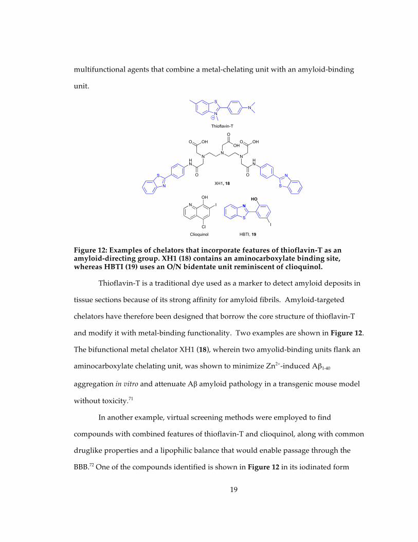
1.5.1 Amyloid-binding metal chelators
A diagnostic feature of Alzheimer’s disease includes the formation of
extracellular plaques composed primarily of aggregated amyloid beta peptide (Aβ).67
Metal ions, particularly Cu+/2+ and Zn2+ but also Fe2+/3+, accelerate Aβ peptide
aggregation in vitro and have been found at elevated levels in neurotoxic oligomers.68
The notion that metal chelation could be a promising treatment option for Alzheimer’s
was encouraged by promising phase IIa clinical trials of clioquinol (Figure 12), a
derivative of 8-hydroxyquinoline.69, 70 However, as expressed in the recurrent theme of
this review, it is challenging to use general chelating agents to mitigate damaging effects
of some metal ions without disturbing the beneficial properties of others. In an attempt
to target metal-binding agents directly to amyloid fibrils, several groups have developed
N
OHO
HO
17
19
multifunctional agents that combine a metal-chelating unit with an amyloid-binding
unit.
Figure 12: Examples of chelators that incorporate features of thioflavin-T as an amyloid-directing group. XH1 (18) contains an aminocarboxylate binding site, whereas HBTI (19) uses an O/N bidentate unit reminiscent of clioquinol.
Thioflavin-T is a traditional dye used as a marker to detect amyloid deposits in
tissue sections because of its strong affinity for amyloid fibrils. Amyloid-targeted
chelators have therefore been designed that borrow the core structure of thioflavin-T
and modify it with metal-binding functionality. Two examples are shown in Figure 12.
The bifunctional metal chelator XH1 (18), wherein two amyolid-binding units flank an
aminocarboxylate chelating unit, was shown to minimize Zn2+-induced Aβ1-40
aggregation in vitro and attenuate Aβ amyloid pathology in a transgenic mouse model
without toxicity.71
In another example, virtual screening methods were employed to find
compounds with combined features of thioflavin-T and clioquinol, along with common
druglike properties and a lipophilic balance that would enable passage through the
BBB.72 One of the compounds identified is shown in Figure 12 in its iodinated form
S
N
N
Thioflavin-T
N
OH
Cl
I
Clioquinol
S
N
HN
O
N
O OH
N
OH
O
N
S
N
OHO
O
HN
XH1, 18
N
S
HO
I
HBTI, 19
20
(HBTI, 19), which could permit its use as a non-invasive imaging agent.72 HBTI is
comparable to clioquinol in its ability to reduce both Zn2+ and Cu2+-induced Aβ
aggregation, as confirmed by the Aβ aggregation turbity assay.72 In addition, fluorescent
measurements of HBTI demonstrate significant changes in the presence of amyloid
fibrils, indicating that the compound is intercalating within the peptide aggregates as
envisioned in the design of the multifunctional agent.72
1.5.2 Prochelators activated by Cu–Aβ
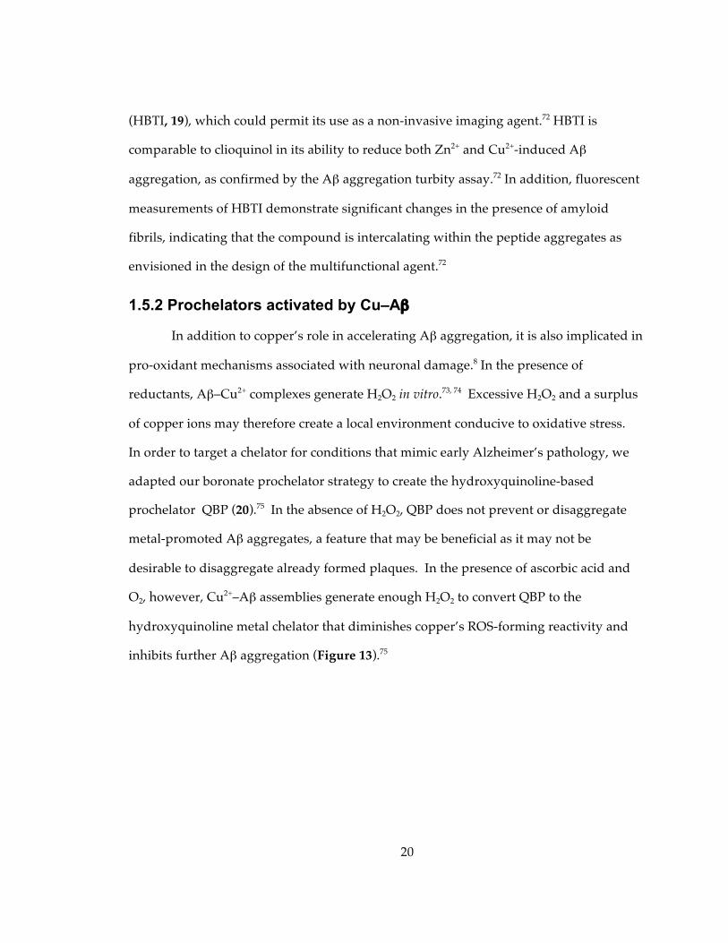
In addition to copper’s role in accelerating Aβ aggregation, it is also implicated in
pro-oxidant mechanisms associated with neuronal damage.8 In the presence of
reductants, Aβ–Cu2+ complexes generate H2O2 in vitro.73, 74 Excessive H2O2 and a surplus
of copper ions may therefore create a local environment conducive to oxidative stress.
In order to target a chelator for conditions that mimic early Alzheimer’s pathology, we
adapted our boronate prochelator strategy to create the hydroxyquinoline-based
prochelator QBP (20).75 In the absence of H2O2, QBP does not prevent or disaggregate
metal-promoted Aβ aggregates, a feature that may be beneficial as it may not be
desirable to disaggregate already formed plaques. In the presence of ascorbic acid and
O2, however, Cu2+–Aβ assemblies generate enough H2O2 to convert QBP to the
hydroxyquinoline metal chelator that diminishes copper’s ROS-forming reactivity and
inhibits further Aβ aggregation (Figure 13).75
21
Figure 13: The H2O2 generated from Cu-Aβ species in the presence of O2 and ascorbic acid unmasks prochelator QBP (20) to release 8-hydroxyquinoline that extracts Cu2+ from Aβ and prevents further redox-cycling and Aβ aggregation.
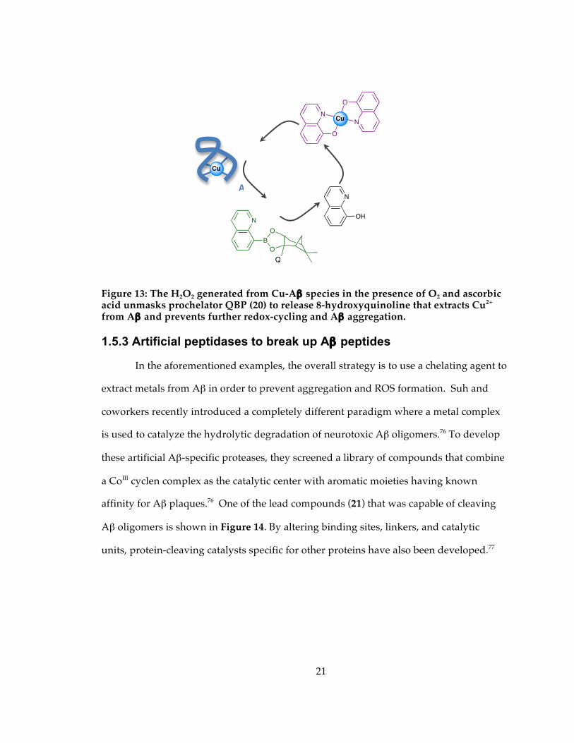
1.5.3 Artificial peptidases to break up Aβ peptides
In the aforementioned examples, the overall strategy is to use a chelating agent to
extract metals from Aβ in order to prevent aggregation and ROS formation. Suh and
coworkers recently introduced a completely different paradigm where a metal complex
is used to catalyze the hydrolytic degradation of neurotoxic Aβ oligomers.76 To develop
these artificial Aβ-specific proteases, they screened a library of compounds that combine
a CoIII cyclen complex as the catalytic center with aromatic moieties having known
affinity for Aβ plaques.76 One of the lead compounds (21) that was capable of cleaving
Aβ oligomers is shown in Figure 14. By altering binding sites, linkers, and catalytic
units, protein-cleaving catalysts specific for other proteins have also been developed.77
O
2, e-
H
2O2
A
�
N
O
N
O
CuCu
Cu
N
BO
O
N
OH
Q
BP (20)
22
Figure 14: Catalytic Co(III) (21) and Cu(II)-cyclen (22) chelators found to break up Aβ peptides.
In an extension of this protein cleaving tactic, Wu et al. reasoned that cyclen
would be able to extract copper from Aβ to generate a hydrolytically active complex that
could then degrade Aβ.78 Their constructs contain cyclen covalently fused to an Aβ
recognition motif such as the peptide sequence Lys-Leu-Val-Phe-Phe in 22 in Figure 14,
or curcumin. In vitro the cyclen can be metallated by competing with Aβ for Cu(II) to
generate the active [Cyc(Cu)-KLVFF] complex that inhibits Aβ oligomerization,
suppresses H2O2 formation, and produces Aβ peptide cleavage fragments.78
Furthermore, the apo Cyc-KLVFF hybrid complex was shown to rescue cultured
CoIII
OH2
OH2
NH
N
N
N
HN
N
N N
ON
S
HN
Cl
O
NH
N21
HH
amyloid-targetingdomain
peptide-cleavingdomain
O
NH
NH2
HN
O
O
NH
O
HN
NH
O
O
OH
22
N
N
N
N
CuII
H
H
H
amyloid-targeting domain
cyclen captures Cu2+ from A!
to create peptide-cleaving unit
23
neurons from Aβ/Cu-induced toxicity, suggesting that it is able to acquire Cu(II) in situ
to generate the active compound.78
1.6 Metal chelators with enzyme inhibitory activity
1.6.1 Monoamine oxidase (MAO) dual action agents
The substantia nigra is a small part of the basal ganglia which is important for
movement, reward, and addiction and is the region most affected in Parkinson’s disease.
In addition to elevated levels of iron in the substantia nigra, the brains of Parkinson’s
and Alzheimer’s patients also show increased activity of monoamine oxidases (MAO),
enzymes that oxidatively degrade neurotransmitters like dopamine and generate H2O2
as a byproduct.79 These factors, combined with diminished antioxidant stores of
glutathione, lead to a localized environment primed for oxidative stress. In order to
increase the effectiveness of iron chelation as a strategy against these diseases,
multifunctional agents have been developed that combine iron chelation ability with
MAO inhibition.79, 80
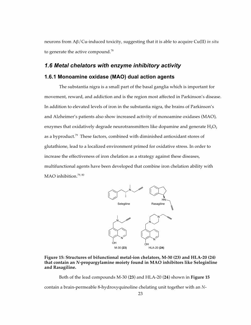
Figure 15: Structures of bifunctional metal-ion chelators, M-30 (23) and HLA-20 (24) that contain an N-propargylamine moiety found in MAO inhibitors like Seleginline and Rasagiline.
Both of the lead compounds M-30 (23) and HLA-20 (24) shown in Figure 15
contain a brain-permeable 8-hydroxyquinoline chelating unit together with an N-
N
N
OH
M-30 (23)
N
N
OH
N
HLA-20 (24)
N
Selegiline
HN
Rasagiline
24
propargylamine moiety. The latter has been found to be responsible for the
neuroprotective effects of rasagiline and selegiline, two MAO inhibitors used clinically
for treating Parkinson’s patients.81 In vitro the bifunctional agents coordinate Fe3+ to give
3:1 ligand:metal complexes that inhibit lipid peroxidation at levels comparable to
desferrioxamine, while also showing modest MAO inhibitory activity and reasonable
cell permeability.80, 82 The compounds show significant protective effects in cell culture
models used for studying neuronal oxidative stress and show a wide range of
pharmacological activities that include regulation of the amyloid precursor protein
(APP) and reduction of Aβ peptide levels.82-84 The propargyl substituent on the chelator
plays a crucial role in the multifaceted activity of M-30 and HLA-20 that make these
compounds promising for future development.85
1.6.2 Acetylcholinesterase (AChE) triple action agents
Acetylcholinesterase (AChE) is the enzyme responsible for breaking down
acetylcholine, a neurotransmitter that is in short supply in Alzheimer’s disease. Classical
inhibitors of AChE, like tacrine, the first drug approved for Alzheimer’s, block the active
site in order to normalize cholinergic levels and improve cognitive function.86 They do
not, however, address the underlying pathology. In addition to its hydrolysis active site,
AChE also contains a peripheral anionic site that has been identified as being
responsible for promoting Aβ fibrillization. Dual binding site inhibitors that block both
the active site and the peripheral anionic site are being sought as they potentially could
alleviate cognitive symptoms associated with acetylcholine deficiency while
simultaneously reducing Aβ fibrillization, which is believed to be central to the disease
mechanism.86
In order to take this concept one step further, Bolognesi et al. modified a dual-
action AChE inhibitor with a metal chelating function to create the triple action agent 25
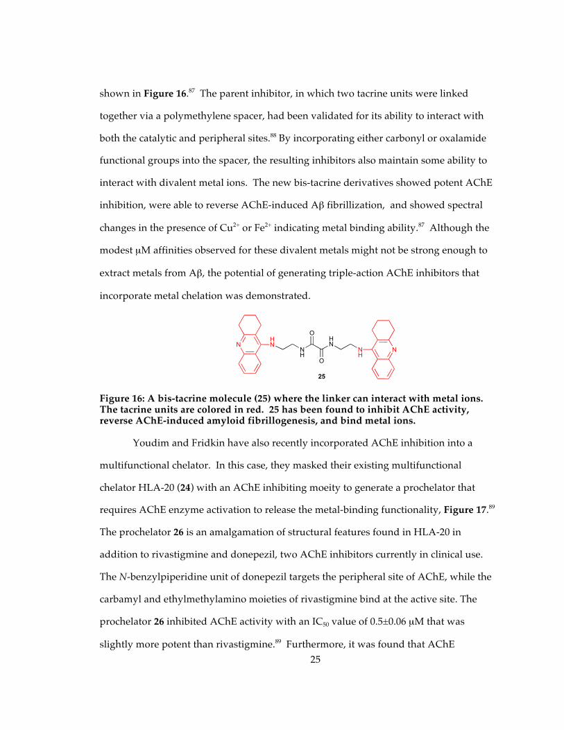
25
shown in Figure 16.87 The parent inhibitor, in which two tacrine units were linked
together via a polymethylene spacer, had been validated for its ability to interact with
both the catalytic and peripheral sites.88 By incorporating either carbonyl or oxalamide
functional groups into the spacer, the resulting inhibitors also maintain some ability to
interact with divalent metal ions. The new bis-tacrine derivatives showed potent AChE
inhibition, were able to reverse AChE-induced Aβ fibrillization, and showed spectral
changes in the presence of Cu2+ or Fe2+ indicating metal binding ability.87 Although the
modest µM affinities observed for these divalent metals might not be strong enough to
extract metals from Aβ, the potential of generating triple-action AChE inhibitors that
incorporate metal chelation was demonstrated.
Figure 16: A bis-tacrine molecule (25) where the linker can interact with metal ions. The tacrine units are colored in red. 25 has been found to inhibit AChE activity, reverse AChE-induced amyloid fibrillogenesis, and bind metal ions.
Youdim and Fridkin have also recently incorporated AChE inhibition into a
multifunctional chelator. In this case, they masked their existing multifunctional
chelator HLA-20 (24) with an AChE inhibiting moeity to generate a prochelator that
requires AChE enzyme activation to release the metal-binding functionality, Figure 17.89
The prochelator 26 is an amalgamation of structural features found in HLA-20 in
addition to rivastigmine and donepezil, two AChE inhibitors currently in clinical use.
The N-benzylpiperidine unit of donepezil targets the peripheral site of AChE, while the
carbamyl and ethylmethylamino moieties of rivastigmine bind at the active site. The
prochelator 26 inhibited AChE activity with an IC50 value of 0.5±0.06 µM that was
slightly more potent than rivastigmine.89 Furthermore, it was found that AChE
NNH
HN
NH
25
O
O
NHN
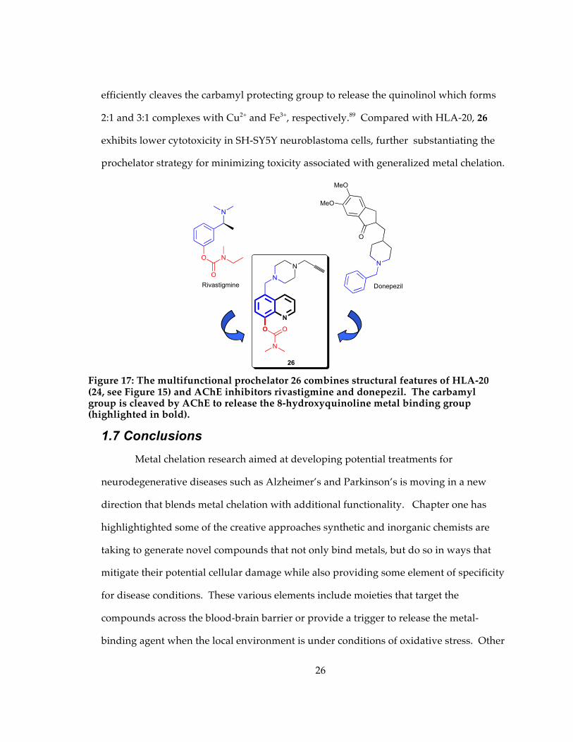
26
efficiently cleaves the carbamyl protecting group to release the quinolinol which forms
2:1 and 3:1 complexes with Cu2+ and Fe3+, respectively.89 Compared with HLA-20, 26
exhibits lower cytotoxicity in SH-SY5Y neuroblastoma cells, further substantiating the
prochelator strategy for minimizing toxicity associated with generalized metal chelation.
1.7 Conclusions Metal chelation research aimed at developing potential treatments for
neurodegenerative diseases such as Alzheimer’s and Parkinson’s is moving in a new
direction that blends metal chelation with additional functionality. Chapter one has
highlightighted some of the creative approaches synthetic and inorganic chemists are
taking to generate novel compounds that not only bind metals, but do so in ways that
mitigate their potential cellular damage while also providing some element of specificity
for disease conditions. These various elements include moieties that target the
compounds across the blood-brain barrier or provide a trigger to release the metal-
binding agent when the local environment is under conditions of oxidative stress. Other
N
O
O
N
Rivastigmine
MeO
MeO
O
N
Donepezil
N
N
O
N
26
N
O
Figure 17: The multifunctional prochelator 26 combines structural features of HLA-20 (24, see Figure 15) and AChE inhibitors rivastigmine and donepezil. The carbamyl group is cleaved by AChE to release the 8-hydroxyquinoline metal binding group (highlighted in bold).
27
design strategies are used to create hybrid molecules that combine metal chelation with
either antioxidant or enzyme inhibitory moieties, or domains that target the chelator to
amyloid fibrils. Many of these compounds are at a very early stage of development, and
significant work is still required to test these strategies in cell and animal models of
disease. The concepts, however, may have broad applicability in a range of conditions,
including cancer and bacterial and viral infection, where interference in cellular metal
regulation via targeted metal chelation may provide a therapeutic angle.90
28
2. Prochelator, BSIH, Strategy for Inhibiting Oxidative Stress via Iron Chelation
2.1 Background and Significance As previously stated, research on iron chelation therapy agents has moved beyond
binding metals alone. Researchers have realized that it is necessary to functionalize the
chelator to not only bind metals but also to perform other functions, for instance, to
prevent oxidative damage from occurring.50, 51, 91, 92 By making chelators that act
selectively on certain tissues, their effects might be enhanced while toxicity is reduced.93
Our group has designed masked iron chelators that bind iron selectively when activated
in the presence of H2O2. SIH, salicylaldehyde isonicotinic acid hydrazone, is a well
known aroylhydrazone chelator that shows promise for chelation therapy because of
favorable attributes of membrane permeability and high affinity iron binding.21, 50, 93, 94
The masked chelator, BSIH, is modeled after SIH, differing by a boronic pinacol ester
masking the phenolic oxygen that is essential to iron binding, Scheme 2.

29
Scheme 2. The masked chelator, BSIH (12), binds Fe3+ only following deprotection by H2O2.50
In its masked form, BSIH has little to no affinity for metal ions. Scheme 2 shows
that the boronic ester on BSIH reacts with hydrogen peroxide to convert BSIH to SIH,
thus allowing the chelator to bind iron and prevent hydroxyl radical formation.50 BSIH
itself shows protection against cell death induced by hydrogen peroxide in cultured
retinal pigment epthelial cells.52 Importantly, prolonged and repetitive exposure of
healthy cells to BSIH was not toxic, whereas similar treatment with SIH or DFO caused
cell death, presumably due to metal depletion.52 Several analogs of BSIH have been
studied to tune properties including lipophilicity, iron binding affinity, and the rate of
hydrogen peroxide dependent unmasking of the prochelator.51 It was found that
modifications made to the aroyl ring of the chelators tune their iron affinity, whereas
modifications on the boron-containing ring of the pro-chelators attenuate their reaction
rates with hydrogen peroxide. Specifically, a methoxy derivative pro-chelator (p-
OMe)BASIH was found to react with hydrogen peroxide nearly 5 times faster than the
NHN
B
NO
O O H2O2 NHN
NO
OH
SIH
N
NH
N
O
O
FeNHN
N
O
O
[Fe(SIH)2]+
Fe3+
Fe
Fe3+,
other Mn+
BSIH
✗
30
chloro derivative (m-Cl)BASIH.51 The rate of oxidation from the pro-chelator to chelator
as well as the metal binding affinity of the chelator influenced the overall ability of these
molecules to inhibit hydroxyl radical formation catalyzed by iron or copper in the
presence of hydrogen peroxide and ascorbic acid. This finding suggests that the pro-
chelator strategy may be effective for turning on metal chelation only under oxidative
stress conditions.
At this time a set of second-generation pro-chelators and their respective
chelators have been synthesized, and are listed in Table 1. Compared to the first
generation pro-chelator/chelator, BSIH/SIH, changes have been made to the ligands,
where the position of the nitrogen on the aroyl ring was changed to the meta
(BSNH/SNH) and ortho (BSPH/SPH) position with respect to the hydrazone as
compared to BSIH, which has the nitrogen in the para position, Table 1A. These
modifications have been made to study the effect of the nitrogen’s position on the aroyl
ring with respect to the affinity of the ligand to the iron. In addition, a pro-chelator was
synthesized with a diethanolamine boronic ester mask, replacing the pinacol boronic
ester moiety (DEA-BSIH) from BSIH, Table 1B. The decision to change the pinacol
boronic ester to a diethanolamine ester was made to study changes in the rates of
oxidation to the chelator form. A pro-chelator and its respective chelator have also been
synthesized with modifications to the ring containing the B- or OH- (FMDOP-Bpin-IH/
FMDOP-IH) to examine changes in iron affinity and/or the rate of oxidation of the
boronic ester with H2O2, Table 1C. In order to increase the stability of the well-known
chelator, SIH, a derivative chelator was synthesized with a methyl group on the imine
carbon of SIH (HAP-INH), Table 1D. The solubility of the pro-chelators and chelators
has been determined in de-ionized water, 20 mM sodium phosphate buffer at pH 7.4,
92% MEM: 8% DMSO, MEM, and 100 mM NaOH. The stability of the pro-chelators/
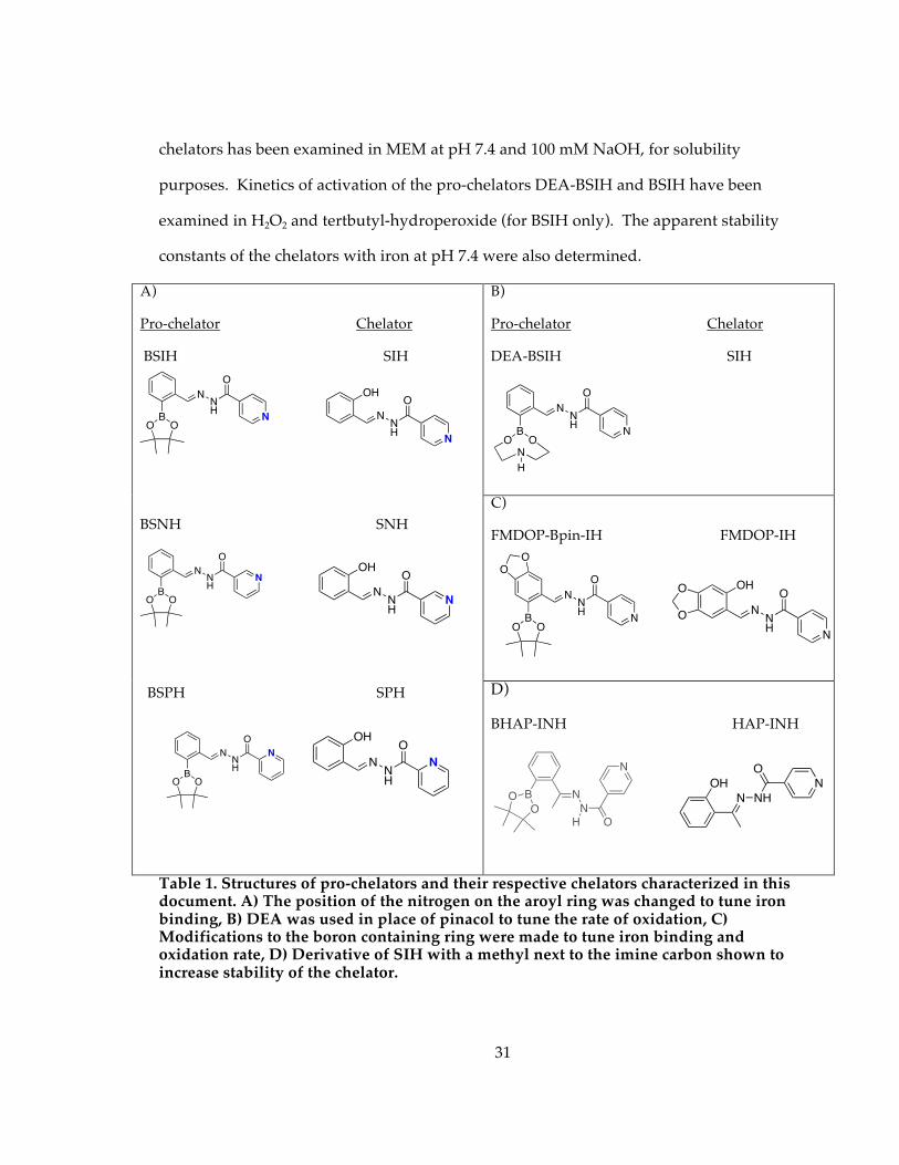
31
chelators has been examined in MEM at pH 7.4 and 100 mM NaOH, for solubility
purposes. Kinetics of activation of the pro-chelators DEA-BSIH and BSIH have been
examined in H2O2 and tertbutyl-hydroperoxide (for BSIH only). The apparent stability
constants of the chelators with iron at pH 7.4 were also determined.
B)
Pro-chelator Chelator
DEA-BSIH SIH
C)
FMDOP-Bpin-IH FMDOP-IH
A)
Pro-chelator Chelator
BSIH SIH
BSNH SNH
BSPH SPH
D)
BHAP-INH HAP-INH
Table 1. Structures of pro-chelators and their respective chelators characterized in this document. A) The position of the nitrogen on the aroyl ring was changed to tune iron binding, B) DEA was used in place of pinacol to tune the rate of oxidation, C) Modifications to the boron containing ring were made to tune iron binding and oxidation rate, D) Derivative of SIH with a methyl next to the imine carbon shown to increase stability of the chelator.
BOO
NNH
O
N NNH
O
N
OH
BOO
NNH
O
N
NNH
O
N
OH
BOO
NNH
O
N
NNH
O
N
OH
BOO
NNH
O
N
N
H
NNH
O
N
OHO
OBOO
NNH
O
N
O
O
N NH
O
NOHB
O
O N
N
O
N
H
32
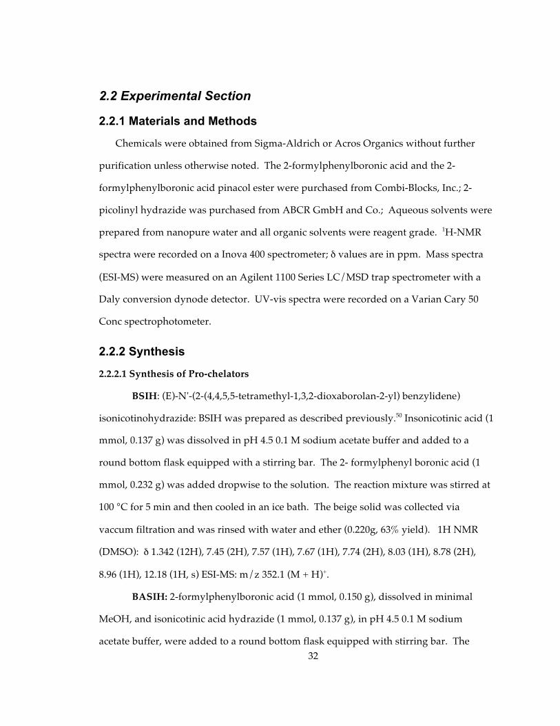
2.2 Experimental Section
2.2.1 Materials and Methods
Chemicals were obtained from Sigma-Aldrich or Acros Organics without further
purification unless otherwise noted. The 2-formylphenylboronic acid and the 2-
formylphenylboronic acid pinacol ester were purchased from Combi-Blocks, Inc.; 2-
picolinyl hydrazide was purchased from ABCR GmbH and Co.; Aqueous solvents were
prepared from nanopure water and all organic solvents were reagent grade. 1H-NMR
spectra were recorded on a Inova 400 spectrometer; δ values are in ppm. Mass spectra
(ESI-MS) were measured on an Agilent 1100 Series LC/MSD trap spectrometer with a
Daly conversion dynode detector. UV-vis spectra were recorded on a Varian Cary 50
Conc spectrophotometer.
2.2.2 Synthesis
2.2.2.1 Synthesis of Pro-chelators
BSIH: (E)-N'-(2-(4,4,5,5-tetramethyl-1,3,2-dioxaborolan-2-yl) benzylidene)
isonicotinohydrazide: BSIH was prepared as described previously.50 Insonicotinic acid (1
mmol, 0.137 g) was dissolved in pH 4.5 0.1 M sodium acetate buffer and added to a
round bottom flask equipped with a stirring bar. The 2- formylphenyl boronic acid (1
mmol, 0.232 g) was added dropwise to the solution. The reaction mixture was stirred at
100 °C for 5 min and then cooled in an ice bath. The beige solid was collected via
vaccum filtration and was rinsed with water and ether (0.220g, 63% yield). 1H NMR
(DMSO): δ 1.342 (12H), 7.45 (2H), 7.57 (1H), 7.67 (1H), 7.74 (2H), 8.03 (1H), 8.78 (2H),
8.96 (1H), 12.18 (1H, s) ESI-MS: m/z 352.1 (M + H)+.
BASIH: 2-formylphenylboronic acid (1 mmol, 0.150 g), dissolved in minimal
MeOH, and isonicotinic acid hydrazide (1 mmol, 0.137 g), in pH 4.5 0.1 M sodium
acetate buffer, were added to a round bottom flask equipped with stirring bar. The
33
reaction mixture was stirred in the flask for 6 minutes at 100 °C and then cooled in an ice
bath. A white product precipitated out of solution and was filtered with DI-water and
collected via vacuum filtration (0.495 g, 92% yield). 1H-NMR (CD3OD 400 MHz): δ 8.75
(2H), 8.46 (1H), 7.88 (2H), 7.66 (1H), 7.44 (2H), 7.39 (1H); ESI-MS: m/z 270.0 (M + H)+.
DEA-BSIH: (E)-N'-(2-(1,3,6,2-dioxazaborocan-2 yl) benzylidene)isonicotino-
hydrazide: BASIH (1mmol, 0.2692 g) and diethanolamine, DEA, (1.2 mmol, 0.1262 g)
were dissolved in minimal DMF and combined in a round bottom flask. The reaction
was stirred for 20 min at room temperature. The white precipitate was collected by
vacuum filtration, washed with cold DMF and dried in vacuo to yield a white precipitate.
(0.200 g, 59.2% yield) 1H-NMR (D2O 400 MHz): δ 8.61 (2H), 7.714 (1H), 7.52 (1H), 7.49
(2H), 7.43 (1H), 7.41 (1H), 7.38 (1H), 3.84 (4H), 3.17 (4H). ESI-MS: m/z 339.1 (M + H)+.
BSNH:(E)-N-(2-(4,4,5,5-tetramethyl-1,3,2-dioxaborolan-2-yl)benzylidene
nicotinohydrazide: The 2-formylphenyl boronic acid pinacol ester (1 mmol, 0.2328 g)
was added to a nearly saturated solution of nicotinic acid (1 mmol, 0.1378 g) in pH 4.5
0.1 M sodium acetate buffer. The reaction was stirred for 12 min at 100 °C and then
cooled in an ice bath. The beige solid precipitate was collected by vacuum filtration.
(0.195 g, 55%) 1H-NMR (d6-DMSO 400MHz ): δ 12.148 (1H), 9.057 (1H), 8.947 (1H), 8.778
(1H), 8.258 (1H), 8.040 (1H), 7.743 (1H), 7.564 (2H), 7.442 (1H), 1.343 (12H). ESI-MS: m/z
352.1 (M + H)+.
BSPH: (E)-N-(2-(4,4,5,5-tetramethyl-1,3,2-diocaborolan-2-yl) benzylidene)
picolinohydrazide: A similar procedure to the one above was performed except the
nicotinic acid was replaced by 2-picolinyl hydrazide and the reaction was stirred for 10
min at 100 °C. A beige solid was collected at 53% yield. (0.186 g). 1H-NMR(d6-
DMSO400MHz): δ 12.239 (1H), 9.014 (1H), 8.121 (1H), 8.061 (1H), 8.004 (1H), 7.694 (2H),
7.553 (1H), 7.432 (1H), 1.346 (12H). ESI-MS: m/z 352.1 (M + H)+.
34
BAHAP-INH: (1-Hydroxy-4-methyl-1H-benzo[d][1,2,3]diazaborinin-2-yl)-
pyridin-4-yl-methanone: Isonicotinic acid (2 mmol, 0.328 g) and 2-acetylphenylboronic
acid (2 mmol, 0.274 g) in ethanol were added to a round bottom flask equipped with a
stirring bar followed by a few drops of glacial acetic acid. The reaction mixture was
heated for 6 minutes at 100 °C and was then cooled to room temperature. The white
solid product was rinsed with ethanol and filtered via vacuum filtration. (0.431 g, 81%
yield). 1H-NMR(d6-DMSO 400MHz): δ 8.73 (2H), 7.78 (2H), 7.67 (2H), 7.56 (2H); ESI-MS:
m/z 266.0 (M + H)+
FMDOP-Bpin ester-IH: (E)-N-((5-(4,4,5,5-tetramethyl-1,3,2-diozaborolan-
2yl)benzoic[d][1,3]dioxol-6-yl)methylene)isonicotinohydrazide: 2-formyl-4,5-
methylenedioxyphenylboronic acid (1 mmol, 0.1942 g) was diluted in minimal methanol
and the isonicotinic acid hydrazide (1 mmol, 0.1378 g) was dissolved in pH 4.5 0.1 M
sodium acetate buffer. The reactants were combined in a round bottom flask and stirred
for 6 min at 100 °C. The solid white boronic acid hydrazone (FMDOP-BAH) was cooled
in ice and collected by vacuum filtration. (0.274 g, 88%). 1H-NMR (CD3OD 400 MHz): δ
8.752 (2H), 8.391 (1H), 7.875 (2H), 7.736 (1H), 6.859 (2H)
A portion of the FMDOP-BAH (0.24 mmol, 0.078 g) and pinacol (0.3 mmol,
0.0334 g) were then dissolved in DMF and added to a round bottom flask. The reaction
stirred for 21 h at room temperature and was then collected by vacuum filtration. The
filtrate was collected and dried in vacuo. (0.076 g, 82%) 1H-NMR (DMSO 400 MHz): δ
12.150 (1H), 8.956 (1 H), 8.785 (2 H), 7.807 (2 H), 7.523 (1 H), 7.134 (1 H), 6.119 (2 H) 1.325
(12 H) ESI-MS: m/z 396.1 (M + H)+
2.2.2.2 Synthesis of Chelators
SIH: (E)-N'-(2-hydroxybenzylidene)isonicotinohydrazide: SIH was prepared as
described previously by a Schiff-base condensation.50, 91, 94, 95 The isonicotinic acid
35
hydrazide (1 mmol, 0.137 g) was dissolved in 0.1 M pH 4.5 sodium acetate buffer and
stirred at 100 °C over an oil bath. The salicylaldehyde (1 mmol, 0.122 g), a clear oil, was
immediately added dropwise to the round bottom flask and the reaction mixture stirred
for 5 min 100 °C. The white product precipitated out of solution and was collected via
vacuum filtration and washed with water. H NMR (DMSO): δ 6.933 (2H, m), 7.319 (1H,
td, J = 1.68, J = 6.69), 7.608 (1H, dd, J = 1.49, J = 7.67), 7.846 (2H, dd, J = 1.65, J = 4.4),
8.682(1H, s), 8.802 (2 H, dd, J = 1.59 , J = 4.4), 11.075 (1H, s), 12.298 (1H, s). MS (ESI): m/z
242.1 (M + H+), 264.1 (M + Na+), 240.1 (M – H+).UV-vis (1:1 MeOH: 20 mM sodium
phosphate buffer) nm (M-1 cm-1): 214 (16,839), 286 (16,455), 332 (13,934).
SNH: (E)-N-(2-hydroxybenzylidene)nicotinohydrazide: Both the salicylaldehyde
(2 mmol, 0.2442 g) and the nicotinic hydrazide (2 mmol, 0.2762 g) were dissolved in pH
4.5 0.1 M sodium acetate buffer and combined in a round bottom flask. The reaction was
stirred for 8 min at 95°C and then cooled over ice. The white solid was collected by
vacuum filtration. (0.506 g, 105%). (Further purification is necessary) 1H-NMR (d6-
DMSO 400 MHz): δ 12.245 (1H), 11.139 (1H), 9.909 (1H), 8.785 (1H), 8.652 (1H), 8.288
(1H), 7.603 (2H), 7.316 (1H), 6.933 (2H). ESI-MS: m/z 242.1. (M + H)+; UV-vis (1:1
MeOH: 20 mM sodium phosphate buffer) nm (M-1 cm-1): 214 (16,267), 286 (18,660),
332 (12,985).
SPH: (E)-N-(2-hydroxybenzylidene)picolinohydrazide: A simlar procedure to the
one above was performed except the nicotinic hydrazide was replaced by 2-picolinyl
hydrazide and the reaction was stirred for 10 min. A white solid was collected ( 0.432 g,
90%). 1H-NMR (d6-DMSO 400 MHz): δ 12.506 (1H), 11.418 (1H), 8.839 (1H), 8.741 (1H),
8.150 (1H), 8.073 (1H), 7.690 (1H), 7.484 (1H), 7.316 (1H), 6.933 (2H). ESI-MS: m/z 242.1
(M + H)+; UV-vis (1:1 MeOH: 20 mM sodium phosphate buffer) nm (M-1 cm-1): 214
(21,395), 286 (19,197), 332 (18,204).
36
FMDOP-IH:(E)-N-((5-hydroxybenzoi[d][1,3]dioxol-6-yl)methylene)
isocotinohydrazide: A portion of FMDOP-BAH (0.5 mmol, 0.1630 g) was added to 15 mL
methanol in a round bottom flask. Hydrogen peroxide (50%) (11 mmol, 623 uL) was
added dropwise and the reaction mixture stirred for 2 h. (0.109 g, 77%) 1H-NMR
(CD3OD 400 MHz): δ 8.753 (2H), 8.462 (2H), 7.889 (2H), 6.971 (1H), 6.476 (1H), 5.951 (2H).
ESI-MS: m/z 286.1 (M + H)+. UV-vis (1:1 MeOH: 20 mM sodium phosphate buffer)
nm (M-1 cm-1): 214 (20,476), 263 (15,312), 363 (20,534).
HAP-INH: (E)-N'-(1-(2-hydroxyphenyl)ethylidene)isonicotinohydrazide: HAP-
INH was prepared in a similar way that has been described previously.96 The 2-
hydroxyacetophenone (1 mmol, 0.1362 g) was dissolved in ethanol and the isonictotinic
acid hydrazide was dissolved in ethanol and catalytic amounts of glacial acetic acid.
The reaction was stirred at 90 °C for 19h and monitored by TLC (6:4 hexanes: ethyl
acetate). The reaction mixture was then cooled in an ice bath and the white powder was
filtered and rinsed with cold ethanol. (0.177 g, 69%) NMR (CD3OD 400 MHz):
δ 8.762 (2 Η), 7.908 (2 Η), 7.664 (1Η), 7.318 (1Η), 6.954 (2). ESI-MS: m/z 256 (M + H)+;UV-
vis (1:1 MeOH: 20 mM sodium phosphate buffer) nm (M-1 cm-1): 214 (26,364), 286
(14,425), 332 (10,962).
2.2.2.3 Synthesis of bis-Iron Complexes
[Fe(SIH)2](NO3): [Fe(SIH)2]+ was synthesized using a procedure previously
described.50, 97 SIH (1.72 mmol, 0.4150 g) was dissolved in 125 mL of MeOH and added
to a round bottom flask equipped with stirring bar and refluxed. Fe(NO3)3•9H2O was
dissolved in 30 mL of MeOH and added dropwise into the refluxing solution. After 1
hour of refluxing at 90 °C, the solution was cooled and the black precipitate was
collected via vacuum filtration, washed with ethanol and diethyl ether. MS (ESI): m/z
37
533.9 (M – 2H+), 536.1 (M + H+). In revisiting the [Fe(SIH)2]+ complex, the extinction
coefficient values were adjusted to the following: UV-vis (1:1 MeOH: 20 mM sodium
phosphate buffer) nm (M-1 cm-1): 294 (31,859), 332 (21,679), 450 (5428.1).
[Fe(SNH)2](NO3): [Fe(SNH)2]+ was synthesized using the same procedure used
for [Fe(SIH)2]+ . MS (ESI): m/z 533.9 (M – 2H+), 536.1 (M + H+). UV-vis (1:1 MeOH:
20 mM sodium phosphate buffer) nm (M-1 cm-1): 450 (4952.2)
[Fe(SPH)2](NO3): [Fe(SPH)2]+ was synthesized using the same procedure used
for [Fe(SIH)2]+ . MS (ESI): m/z 534.0 (M – 2H+), 536.2 (M + H+). UV-vis (1:1 MeOH:
20 mM sodium phosphate buffer) nm (M-1 cm-1): 450 (4689.4)
[Fe(FMDOP-IH)2](NO3): [Fe(FMDOP-IH)2]+ was synthesized using the same
procedure used for [Fe(SIH)2]+ . MS (ESI): m/z 624.2 (M + H+), 622.0 (M - 2H+).
[Fe(HAP-INH)2](NO3): [Fe(HAP-INH)2]+ was synthesized using the same
procedure used for [Fe(SIH)2]+ . MS (ESI): m/z 564.2 (M + H+), 562.0 (M - 2H+).
2.3 Results The solubility and stability of the pro-chelators and chelators were analyzed in
various solvents. The oxidation of BSIH and DEA-BSIH to SIH has been evaluated in
H2O2 and tertbutyl-hydroperoxide (for BSIH only). The conditional stability constants of
the chelators with iron have also been evaluated at pH 7.4.
2.3.1 Solubility of pro-chelators/chelators in various solutions
The solubility of the pro-chelators/chelators was analyzed in either de-ionized
water, 20 mM sodium phosphate buffer at pH 7.4, 92% MEM: 8% DMSO, MEM, or 100
mM NaOH. To determine solubility, small aliquots of solvent were added to the solid
pro-chelator and the solution was placed in the ultrasonic bath. Solubility of the
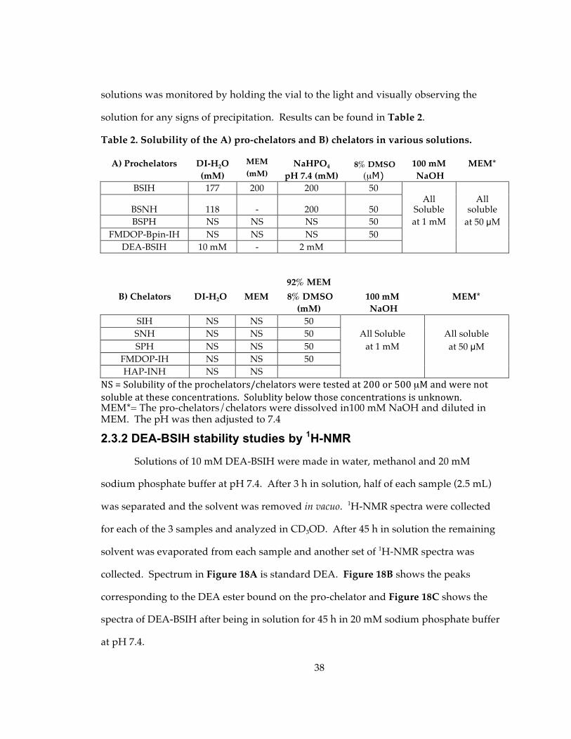
38
solutions was monitored by holding the vial to the light and visually observing the
solution for any signs of precipitation. Results can be found in Table 2.
Table 2. Solubility of the A) pro-chelators and B) chelators in various solutions.
A) Prochelators DI-H2O MEM NaHPO4 8% DMSO 100 mM MEM* (mM) (mM) pH 7.4 (mM) (µM) NaOH
BSIH 177 200 200 50
BSNH 118 - 200 50 All
Soluble All
soluble BSPH NS NS NS 50 at 1 mM at 50 µM
FMDOP-Bpin-IH NS NS NS 50 DEA-BSIH 10 mM - 2 mM
92% MEM B) Chelators DI-H2O MEM 8% DMSO 100 mM MEM*
(mM) NaOH SIH NS NS 50
SNH NS NS 50 All Soluble All soluble SPH NS NS 50 at 1 mM at 50 µM
FMDOP-IH NS NS 50 HAP-INH NS NS
NS=Solubilityoftheprochelators/chelatorsweretestedat200or500µMandwerenotsolubleattheseconcentrations.Solublitybelowthoseconcentrationsisunknown.MEM*= The pro-chelators/chelators were dissolved in100 mM NaOH and diluted in MEM. The pH was then adjusted to 7.4
2.3.2 DEA-BSIH stability studies by 1H-NMR
Solutions of 10 mM DEA-BSIH were made in water, methanol and 20 mM
sodium phosphate buffer at pH 7.4. After 3 h in solution, half of each sample (2.5 mL)
was separated and the solvent was removed in vacuo. 1H-NMR spectra were collected
for each of the 3 samples and analyzed in CD3OD. After 45 h in solution the remaining
solvent was evaporated from each sample and another set of 1H-NMR spectra was
collected. Spectrum in Figure 18A is standard DEA. Figure 18B shows the peaks
corresponding to the DEA ester bound on the pro-chelator and Figure 18C shows the
spectra of DEA-BSIH after being in solution for 45 h in 20 mM sodium phosphate buffer
at pH 7.4.

39
2.3.3 Determination of unknown contaminant in BSIH spectra
It was noticed that upon using the same reaction parameters on a continuous
basis for the synthesis of BSIH, a peak at 8.5 ppm appeared at different intensities using
1H-NMR, Figure 19A. A simple 1H-NMR experiment was performed where the
spectrum of BSIH in d6-DMSO was collected. Immediately following, a couple of drops
of D2O were added to the sample and a second spectrum was collected, Figure 19B. It is
clear that upon the addition of the D2O the peak at 8.5 ppm disappears. This finding
confirms our hypothesis that the impurity is actually a proton from the hydroxyl group
of the boronate species of BSIH.
Figure18: 1H-NMR spectra of (a) DEA, (b) DEA-BSIH in 20 mM NaHPO4 buffer at pH 7.4 after 3 h (c) DEA-BSIH in 20 mM NaHPO4 buffer at pH 7.4 after 45 h
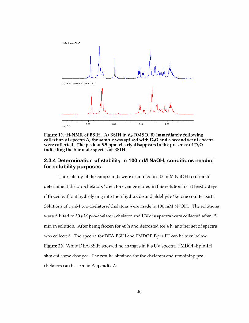
40
Figure 19. 1H-NMR of BSIH. A) BSIH in d6-DMSO. B) Immediately following collection of spectra A, the sample was spiked with D2O and a second set of spectra were collected. The peak at 8.5 ppm clearly disappears in the presence of D2O indicating the boronate species of BSIH.
2.3.4 Determination of stability in 100 mM NaOH, conditions needed for solubility purposes
The stability of the compounds were examined in 100 mM NaOH solution to
determine if the pro-chelators/chelators can be stored in this solution for at least 2 days
if frozen without hydrolyzing into their hydrazide and aldehyde/ketone counterparts.
Solutions of 1 mM pro-chelators/chelators were made in 100 mM NaOH. The solutions
were diluted to 50 µM pro-chelator/chelator and UV-vis spectra were collected after 15
min in solution. After being frozen for 48 h and defrosted for 4 h, another set of spectra
was collected. The spectra for DEA-BSIH and FMDOP-Bpin-IH can be seen below,
Figure 20. While DEA-BSIH showed no changes in it’s UV spectra, FMDOP-Bpin-IH
showed some changes. The results obtained for the chelators and remaining pro-
chelators can be seen in Appendix A.
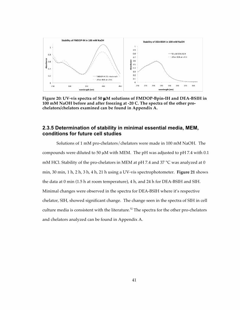
41
Figure 20: UV-vis spectra of 50 µM solutions of FMDOP-Bpin-IH and DEA-BSIH in 100 mM NaOH before and after freezing at -20 C. The spectra of the other pro-chelators/chelators examined can be found in Appendix A.
2.3.5 Determination of stability in minimal essential media, MEM, conditions for future cell studies
Solutions of 1 mM pro-chelators/chelators were made in 100 mM NaOH. The
compounds were diluted to 50 µM with MEM. The pH was adjusted to pH 7.4 with 0.1
mM HCl. Stability of the pro-chelators in MEM at pH 7.4 and 37 °C was analyzed at 0
min, 30 min, 1 h, 2 h, 3 h, 4 h, 21 h using a UV-vis spectrophotometer. Figure 21 shows
the data at 0 min (1.5 h at room temperature), 4 h, and 24 h for DEA-BSIH and SIH.
Minimal changes were observed in the spectra for DEA-BSIH where it’s respective
chelator, SIH, showed significant change. The change seen in the spectra of SIH in cell
culture media is consistent with the literature.52 The spectra for the other pro-chelators
and chelators analyzed can be found in Appendix A.
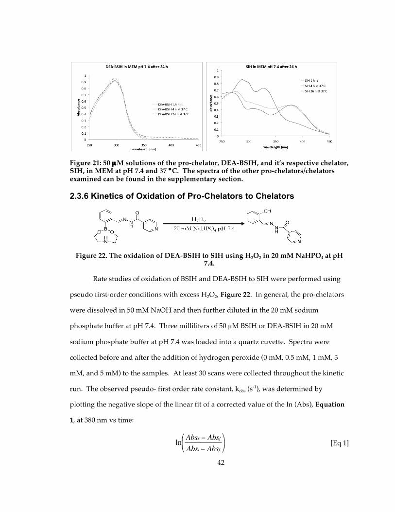
42
Figure 21: 50 µM solutions of the pro-chelator, DEA-BSIH, and it’s respective chelator, SIH, in MEM at pH 7.4 and 37 °C. The spectra of the other pro-chelators/chelators examined can be found in the supplementary section.
2.3.6 Kinetics of Oxidation of Pro-Chelators to Chelators
Figure 22. The oxidation of DEA-BSIH to SIH using H2O2 in 20 mM NaHPO4 at pH 7.4.
Rate studies of oxidation of BSIH and DEA-BSIH to SIH were performed using
pseudo first-order conditions with excess H2O2, Figure 22. In general, the pro-chelators
were dissolved in 50 mM NaOH and then further diluted in the 20 mM sodium
phosphate buffer at pH 7.4. Three milliliters of 50 µM BSIH or DEA-BSIH in 20 mM
sodium phosphate buffer at pH 7.4 was loaded into a quartz cuvette. Spectra were
collected before and after the addition of hydrogen peroxide (0 mM, 0.5 mM, 1 mM, 3
mM, and 5 mM) to the samples. At least 30 scans were collected throughout the kinetic
run. The observed pseudo- first order rate constant, kobs (s-1), was determined by
plotting the negative slope of the linear fit of a corrected value of the ln (Abs), Equation
1, at 380 nm vs time:
€
ln Absx − AbsfAbsi − Absf
[Eq 1]
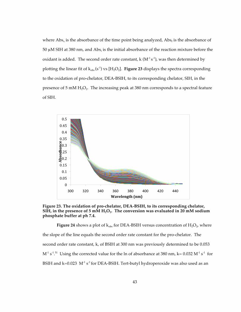
43
where Absx is the absorbance of the time point being analyzed, Absf is the absorbance of
50 µM SIH at 380 nm, and Absi is the initial absorbance of the reaction mixture before the
oxidant is added. The second order rate constant, k (M-1 s-1), was then determined by
plotting the linear fit of kobs (s-1) vs [H2O2]. Figure 23 displays the spectra corresponding
to the oxidation of pro-chelator, DEA-BSIH, to its corresponding chelator, SIH, in the
presence of 5 mM H2O2. The increasing peak at 380 nm corresponds to a spectral feature
of SIH.
Figure 23. The oxidation of pro-chelator, DEA-BSIH, to its corresponding chelator, SIH, in the presence of 5 mM H2O2. The conversion was evaluated in 20 mM sodium phosphate buffer at ph 7.4.
Figure 24 shows a plot of kobs for DEA-BSIH versus concentration of H2O2, where
the slope of the line equals the second order rate constant for the pro-chelator. The
second order rate constant, k, of BSIH at 300 nm was previously determined to be 0.053
M-1 s-1.51 Using the corrected value for the ln of absorbance at 380 nm, k= 0.032 M-1 s-1 for
BSIH and k=0.023 M-1 s-1 for DEA-BSIH. Tert-butyl hydroperoxide was also used as an
00.050.10.150.20.250.30.350.40.450.5
300 320 340 360 380 400 420 440
Absorbance
Wavelength(nm)
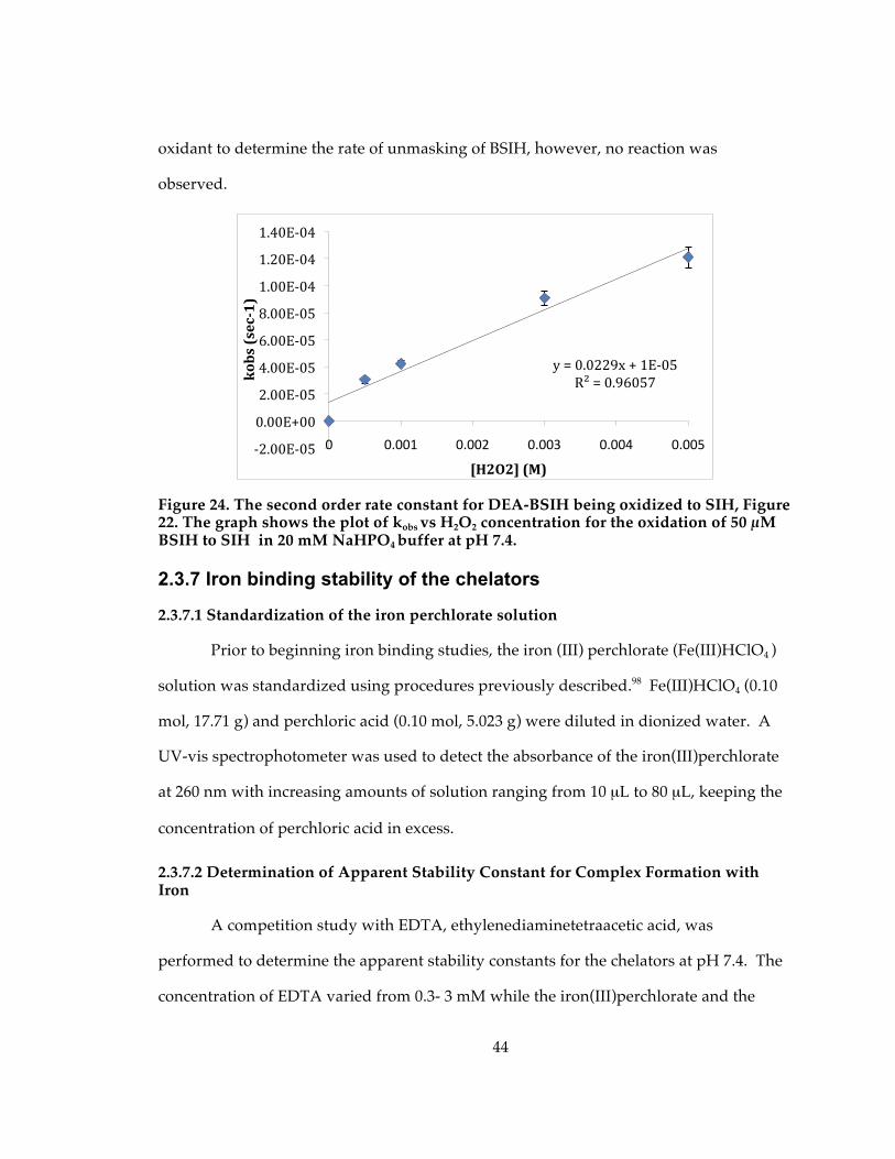
44
oxidant to determine the rate of unmasking of BSIH, however, no reaction was
observed.
Figure 24. The second order rate constant for DEA-BSIH being oxidized to SIH, Figure 22. The graph shows the plot of kobs vs H2O2 concentration for the oxidation of 50 µM BSIH to SIH in 20 mM NaHPO4 buffer at pH 7.4.
2.3.7 Iron binding stability of the chelators
2.3.7.1 Standardization of the iron perchlorate solution
Prior to beginning iron binding studies, the iron (III) perchlorate (Fe(III)HClO4 )
solution was standardized using procedures previously described.98 Fe(III)HClO4 (0.10
mol, 17.71 g) and perchloric acid (0.10 mol, 5.023 g) were diluted in dionized water. A
UV-vis spectrophotometer was used to detect the absorbance of the iron(III)perchlorate
at 260 nm with increasing amounts of solution ranging from 10 µL to 80 µL, keeping the
concentration of perchloric acid in excess.
2.3.7.2 Determination of Apparent Stability Constant for Complex Formation with Iron
A competition study with EDTA, ethylenediaminetetraacetic acid, was
performed to determine the apparent stability constants for the chelators at pH 7.4. The
concentration of EDTA varied from 0.3- 3 mM while the iron(III)perchlorate and the
y=0.0229x+1E‐05R²=0.96057
‐2.00E‐05
0.00E+00
2.00E‐05
4.00E‐05
6.00E‐05
8.00E‐05
1.00E‐04
1.20E‐04
1.40E‐04
0 0.001 0.002 0.003 0.004 0.005
kobs(sec1)
[H2O2](M)
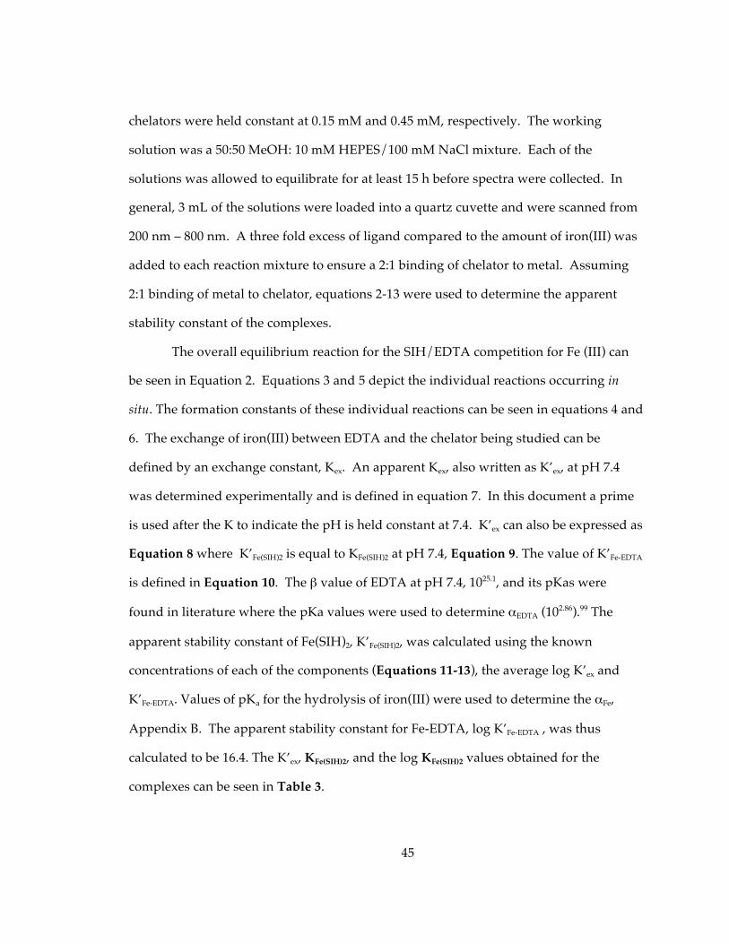
45
chelators were held constant at 0.15 mM and 0.45 mM, respectively. The working
solution was a 50:50 MeOH: 10 mM HEPES/100 mM NaCl mixture. Each of the
solutions was allowed to equilibrate for at least 15 h before spectra were collected. In
general, 3 mL of the solutions were loaded into a quartz cuvette and were scanned from
200 nm – 800 nm. A three fold excess of ligand compared to the amount of iron(III) was
added to each reaction mixture to ensure a 2:1 binding of chelator to metal. Assuming
2:1 binding of metal to chelator, equations 2-13 were used to determine the apparent
stability constant of the complexes.
The overall equilibrium reaction for the SIH/EDTA competition for Fe (III) can
be seen in Equation 2. Equations 3 and 5 depict the individual reactions occurring in
situ. The formation constants of these individual reactions can be seen in equations 4 and
6. The exchange of iron(III) between EDTA and the chelator being studied can be
defined by an exchange constant, Kex. An apparent Kex, also written as K’ex, at pH 7.4
was determined experimentally and is defined in equation 7. In this document a prime
is used after the K to indicate the pH is held constant at 7.4. K’ex can also be expressed as
Equation 8 where K’Fe(SIH)2 is equal to KFe(SIH)2 at pH 7.4, Equation 9. The value of K’Fe-EDTA
is defined in Equation 10. The β value of EDTA at pH 7.4, 1025.1, and its pKas were
found in literature where the pKa values were used to determine αEDTA (102.86).99 The
apparent stability constant of Fe(SIH)2, K’Fe(SIH)2, was calculated using the known
concentrations of each of the components (Equations 11-13), the average log K’ex and
K’Fe-EDTA. Values of pKa for the hydrolysis of iron(III) were used to determine the αFe,
Appendix B. The apparent stability constant for Fe-EDTA, log K’Fe-EDTA , was thus
calculated to be 16.4. The K’ex, KFe(SIH)2, and the log KFe(SIH)2 values obtained for the
complexes can be seen in Table 3.
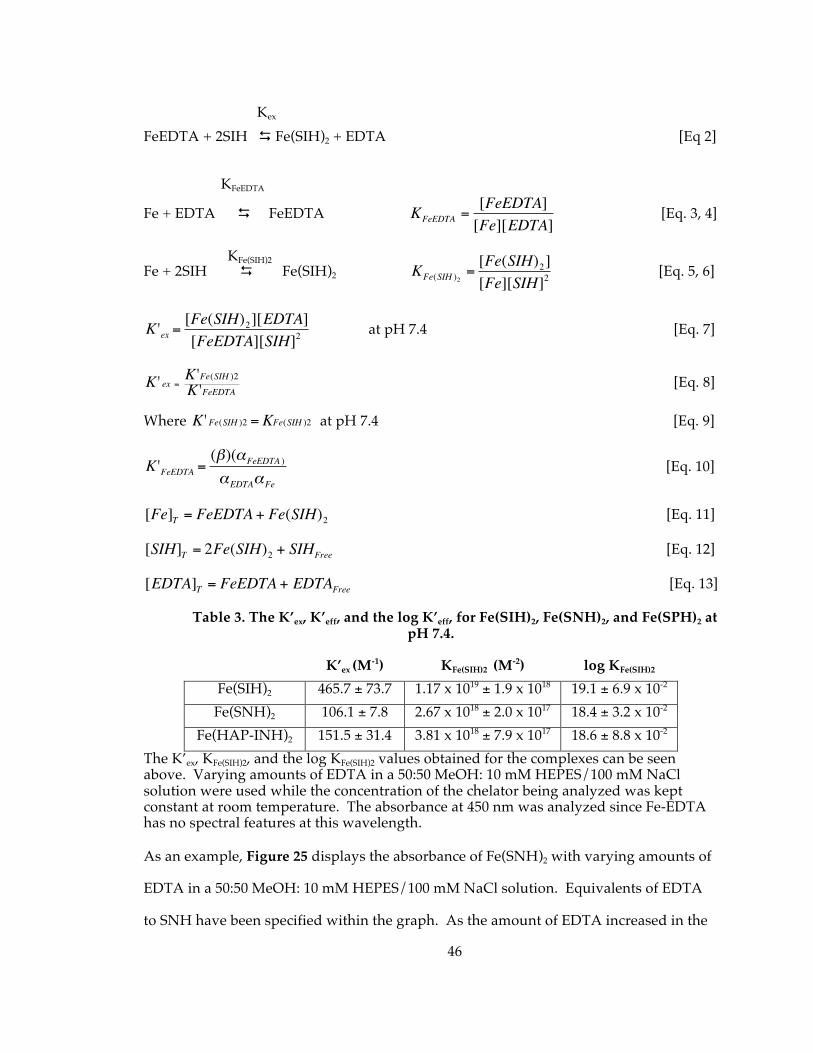
46
FeEDTA + 2SIH Fe(SIH)2 + EDTA [Eq 2]
Fe + EDTA FeEDTA
€
KFeEDTA =[FeEDTA][Fe][EDTA]
[Eq. 3, 4]
Fe + 2SIH Fe(SIH)2
€
KFe(SIH )2=[Fe(SIH)2][Fe][SIH]2
[Eq. 5, 6]
€
K 'ex =[Fe(SIH)2][EDTA][FeEDTA][SIH]2
at pH 7.4 [Eq. 7]
€
K ' ex =K 'Fe(SIH )2K 'FeEDTA [Eq. 8]
Where
€
K ' Fe(SIH )2 = KFe(SIH )2 at pH 7.4 [Eq. 9]
€
K 'FeEDTA =(β)(αFeEDTA )
αEDTAαFe
[Eq. 10]
€
[Fe]T = FeEDTA + Fe(SIH)2 [Eq. 11]
€
[SIH]T = 2Fe(SIH)2 + SIHFree [Eq. 12]
€
[EDTA]T = FeEDTA + EDTAFree [Eq. 13]
Table 3. The K’ex, K’eff, and the log K’eff, for Fe(SIH)2, Fe(SNH)2, and Fe(SPH)2 at pH 7.4.
K’ex (M-1) KFe(SIH)2 (M-2) log KFe(SIH)2
Fe(SIH)2 465.7 ± 73.7 1.17 x 10¹⁹ ± 1.9 x 10¹⁸ 19.1 ± 6.9 x 10⁻²
Fe(SNH)2 106.1 ± 7.8 2.67 x 10¹⁸ ± 2.0 x 10¹⁷ 18.4 ± 3.2 x 10⁻² Fe(HAP-INH)2 151.5 ± 31.4 3.81 x 10¹⁸ ± 7.9 x 10¹⁷ 18.6 ± 8.8 x 10⁻²
The K’ex, KFe(SIH)2, and the log KFe(SIH)2 values obtained for the complexes can be seen above. Varying amounts of EDTA in a 50:50 MeOH: 10 mM HEPES/100 mM NaCl solution were used while the concentration of the chelator being analyzed was kept constant at room temperature. The absorbance at 450 nm was analyzed since Fe-EDTA has no spectral features at this wavelength. As an example, Figure 25 displays the absorbance of Fe(SNH)2 with varying amounts of
EDTA in a 50:50 MeOH: 10 mM HEPES/100 mM NaCl solution. Equivalents of EDTA
to SNH have been specified within the graph. As the amount of EDTA increased in the
Kex
KFeEDTA
KFe(SIH)2
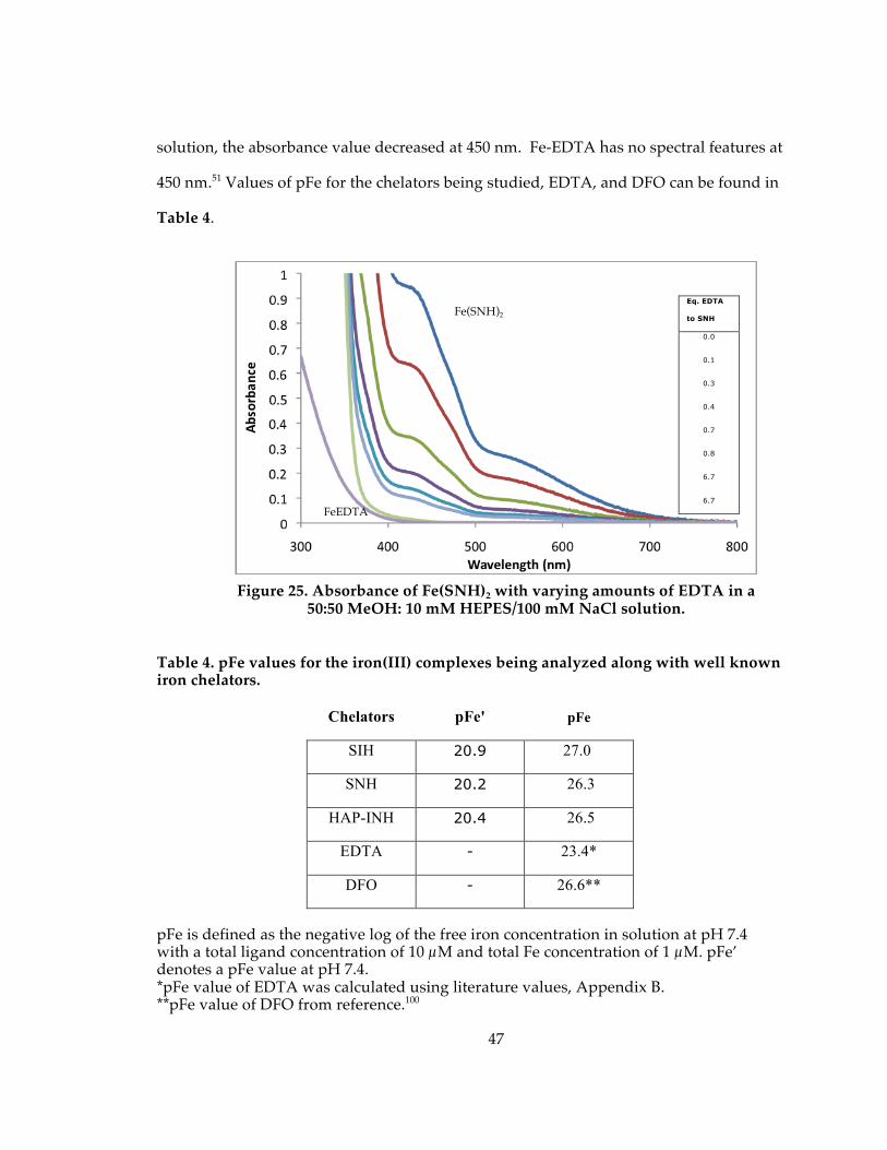
47
solution, the absorbance value decreased at 450 nm. Fe-EDTA has no spectral features at
450 nm.51 Values of pFe for the chelators being studied, EDTA, and DFO can be found in
Table 4.
Table 4. pFe values for the iron(III) complexes being analyzed along with well known iron chelators.
Chelators pFe' pFe
SIH 20.9 27.0
SNH 20.2 26.3
HAP-INH 20.4 26.5
EDTA - 23.4*
DFO - 26.6**
pFe is defined as the negative log of the free iron concentration in solution at pH 7.4 with a total ligand concentration of 10 µM and total Fe concentration of 1 µM. pFe’ denotes a pFe value at pH 7.4. *pFe value of EDTA was calculated using literature values, Appendix B. **pFe value of DFO from reference.100
Eq. EDTA
to SNH
0.0
0.1
0.3
0.4
0.7
0.8
6.7
6.7
Fe(SNH)2
FeEDTA
Figure 25. Absorbance of Fe(SNH)2 with varying amounts of EDTA in a 50:50 MeOH: 10 mM HEPES/100 mM NaCl solution.
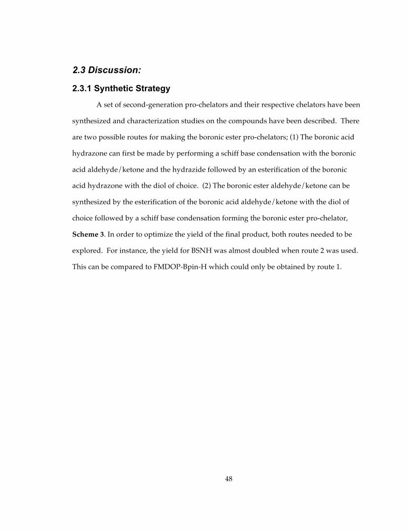
48
2.3 Discussion:
2.3.1 Synthetic Strategy
A set of second-generation pro-chelators and their respective chelators have been
synthesized and characterization studies on the compounds have been described. There
are two possible routes for making the boronic ester pro-chelators; (1) The boronic acid
hydrazone can first be made by performing a schiff base condensation with the boronic
acid aldehyde/ketone and the hydrazide followed by an esterification of the boronic
acid hydrazone with the diol of choice. (2) The boronic ester aldehyde/ketone can be
synthesized by the esterification of the boronic acid aldehyde/ketone with the diol of
choice followed by a schiff base condensation forming the boronic ester pro-chelator,
Scheme 3. In order to optimize the yield of the final product, both routes needed to be
explored. For instance, the yield for BSNH was almost doubled when route 2 was used.
This can be compared to FMDOP-Bpin-H which could only be obtained by route 1.
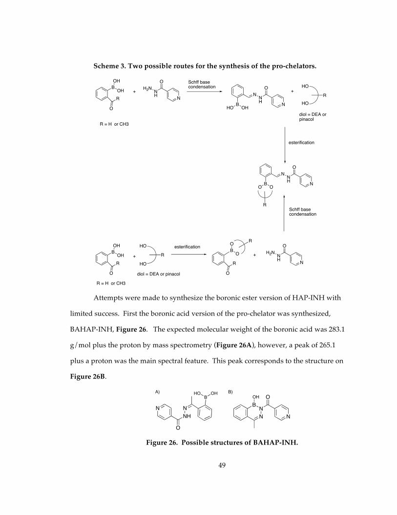
49
Scheme 3. Two possible routes for the synthesis of the pro-chelators.
Attempts were made to synthesize the boronic ester version of HAP-INH with
limited success. First the boronic acid version of the pro-chelator was synthesized,
BAHAP-INH, Figure 26. The expected molecular weight of the boronic acid was 283.1
g/mol plus the proton by mass spectrometry (Figure 26A), however, a peak of 265.1
plus a proton was the main spectral feature. This peak corresponds to the structure on
Figure 26B.
Figure 26. Possible structures of BAHAP-INH.
BOH
OH
R
O
R = H or CH3
+
N
NH
H2N
O Schff base condensation
BOHHO
NNH
O
N
+HO
HO
R
diol = DEA or pinacol
esterification
BOO
NNH
O
N
R
BOH
OH
R
O
+
HO
R
diol = DEA or pinacol
esterification
R
O
B
O
O
R
N
NH
H2N
O
+
HO
Schff base condensation
R = H or CH3
NH
N
O
N
BOHHOA)
BN
N
O
N
OH
B)
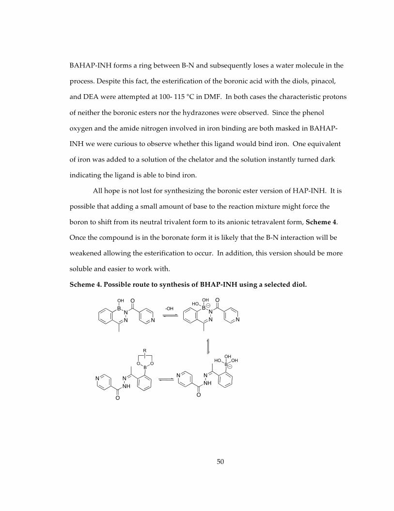
50
BAHAP-INH forms a ring between B-N and subsequently loses a water molecule in the
process. Despite this fact, the esterification of the boronic acid with the diols, pinacol,
and DEA were attempted at 100- 115 °C in DMF. In both cases the characteristic protons
of neither the boronic esters nor the hydrazones were observed. Since the phenol
oxygen and the amide nitrogen involved in iron binding are both masked in BAHAP-
INH we were curious to observe whether this ligand would bind iron. One equivalent
of iron was added to a solution of the chelator and the solution instantly turned dark
indicating the ligand is able to bind iron.
All hope is not lost for synthesizing the boronic ester version of HAP-INH. It is
possible that adding a small amount of base to the reaction mixture might force the
boron to shift from its neutral trivalent form to its anionic tetravalent form, Scheme 4.
Once the compound is in the boronate form it is likely that the B-N interaction will be
weakened allowing the esterification to occur. In addition, this version should be more
soluble and easier to work with.
Scheme 4. Possible route to synthesis of BHAP-INH using a selected diol.
BN
N
O
N
OH
BN
N
O
N
OH
-OH
HO
NH
N
O
N
BOHHO
OH
NH
N
O
N
BOO
R
51
2.3.2 Stability and Solubility of the Chelators/Pro-Chelators
In the near future, our group would like to examine whether the pro-chelators
mentioned in this document can be effective at protecting cells from oxidative damage.
Therefore solubility and stability of these materials in biologically relevant solutions is
necessary. The solubility of the pro-chelators/ chelators was determined in water,
sodium phosphate buffer, 100 mM NaOH, MEM and 92% MEM: 8% DMSO. DEA-BSIH
is the only pro-chelator of the set examined in this document that is soluble in water at
high concentrations, 10 mM. An 1H-NMR experiment was performed to assess whether
the diethanolamine, DEA, on the pro-chelator was stable in water, methanol, and
sodium phosphate buffer. As can be seen in Figure 18, after 3 and 45 hours in solutions
of water, methanol, and sodium phosphate buffer no indication of unattached DEA is
visible by 1H-NMR spectra. This indicates that the DEA diol is bound to the boron on
the pro-chelator.
Dissolving some of the pro-chelators/chelators directly in MEM was
problematic. Although BSIH is soluble in MEM at 200 µM, neither BSPH nor FMDOP-
Bpin-IH was soluble up to 50 µM. In order to get all the chelators and pro-chelators
dissolved in the same solvent system, a solution of 92% MEM: 8% DMSO was used since
both the chelators and pro-chelators were soluble in DMSO. All of the pro-chelators
examined were soluble in the 92% MEM: 8% DMSO solution, however, once MEM was
introduced to the chelator DMSO solutions, SIH, SPH, and FMDOP-IH came out of
solution. Clearly, another method needed to be employed.
Previously, our group has used solutions of 100 mM NaOH to dissolve pro-
chelators/chelators to make less concentrated solutions in MEM and sodium phosphate
buffer. Solutions of 1 mM pro-chelators/chelators were made in 100 mM NaOH and all
were soluble. The stability of the compounds was examined in the NaOH solution to
52
find out if the pro-chelators/chelators can be stored in this solution for at least 2 days if
frozen without hydrolyzing into their hydrazide and aldehyde/ketone counterparts.
While BSIH, BSNH, and BSPH show minimal change in their UV spectra, DEA-BSIH
shows no change in the spectra even after sitting at room temperature for 4 h. However,
FMDOP-Bpin-IH shows some spectral change indicating that one would want to make
up fresh solutions daily when working with it. In MEM at pH 7.4 the pro-chelators
showed minimal changes in their UV spectra while the chelators, SNH, SPH, and
FMDOP-IH showed dramatic changes in their spectra. HAP-INH showed minimal UV
spectral changes. This suggests that adding a methyl group to the imine carbon of SIH
stabilizes the hydrazone in MEM.
2.3.3 Pro-Chelator Rates of Activation: The Oxidation of the Pro-chelator to Chelator
Rate studies of oxidation were performed for BSIH and DEA-BSIH using various
concentrations of H2O2 under pseudo first order reaction conditions. The second order
rate constant, k, of BSIH was previously determined to be 0.053 M-1 s-1 at 300 nm.51
Using the corrected value for the ln of absorbance at 380 nm, Equation 1, the second
order rate constants were determined to be k=0.032 M-1 s-1 for BSIH and k=0.023 M-1 s-1
for DEA-BSIH . Therefore changing the diol mask from pinnacol to diethanolamine
caused little change in the rate of unmasking. It is also important to note that SIH was
monitored for three hours in the presence of H2O2 to determine whether the peroxide
would induce degradation of the chelator. No change in absorbance was observed over
the 3 hour course.
2.3.4 Apparent Stability Constants of Chelators at pH 7.4 with Iron(III)
EDTA is a well known metal chelator with known spectral features and an
affinity for iron(III) which has been well documented. Therefore, it was a good choice
53
for the competition between iron(III) and the new chelators being characterized. To
obtain an apparent stability constant, also known at the conditional or effective stability
constants, a buffered solution at pH 7.4 at room temperature was employed. The log of
the apparent stability constants, log K’, for Fe(SIH)2, Fe(SNH)2, and Fe(HAP-INH)2 were
found to be 19.1, 18.4, 18.6, respectively, at the specified conditions. However, it is often
more useful to compare pFe values (-log[Feaq3+]) than directly comparing apparent
stability constants to different ligands because of differences in pKa’s and denticities. A
pFe value measures the amount of uncomplexed iron that would be available at typical
conditions of pH 7.4 with total ligand and iron concentrations of 10 µM and 1 µM,
respectively. Values of pFe for SIH have been reported previously and range from 23-
50. Issues with solubility and hydrolysis of the chelator at high and low pH values
during potentiometric titrations could explain the wide ranges of pFe values reported
for SIH. Using the technique of a competition between our ligands and EDTA, values of
pFe for SIH, SNH, and HAP-INH were found to be 27.0, 26.3, and 26.5, respectively. For
comparison, pFe values of EDTA and DFO are 23.4 and 26.6, respectively. Between the
chelators analyzed herein, changing the position of the nitrogen of the aroyl ring to the
meta position and adding a methyl group to the imine carbon of SIH seem to have
minimal effects on the stability of the metal complexes.
2.3.5 Summary and Conclusions
A group of second generation iron pro-chelators have been described which are
derivatives of the parent compound, BSIH. These masked chelators can potentially
reveal high affinity metal ligands when oxidized by H2O2. Unlike traditional iron
chelators, which can disrupt iron homeostasis, these pro-chelators have a protective
mask consisting of various boronic esters that react and reveal a phenolic oxygen
selectively by H2O2. Compared to the first generation pro-chelator/chelator pair,
54
BSIH/SIH, changes have been made to the ligands to study solubility, changes in iron
binding stability, and rates of oxidation. Dissolving the pro-chelators/chelators in base
before further diluting in cell culture media or buffer was found to increase the
solubility of the compounds significantly. Concentrations as low as 50 µM were
achieved whereas without adding base most would not go into solution or would
precipitate out. A noteworthy exception was the DEA-BSIH prochelator. This BSIH
derivative was soluble in water even at 10 mM. The boronic ester of this prochelator
showed stability in water, methanol, and 20 mM sodium phosphate buffer even after 45
hours in solution.
Upon further examination of BSIH a peak at 8.5 ppm in the 1H- NMR, which was
originally thought to be a contaminate, the peak turned out to be a proton
corresponding to the boronate version of BSIH. Depending on the amount of water in
the sample as well as the pH of a solution, this peak will vary in size.
Kinetics of oxidation of BSIH and DEA-BSIH were performed in H2O2 and
minimal differences were found in the rates of unmasking. BSIH was also exposed to
tert-butyl hydroperoxide with no reaction observed.
The apparent stability constants and pFe values of SIH, SNH, and HAP-INH
were determined and found to be comparable to DFO. Changing the position of the
nitrogen of the aroyl ring to the meta position (SNH) and adding a methyl group to the
imine carbon of SIH (HAP-INH) seem to have minimal effects on the stability of the
metal complexes.
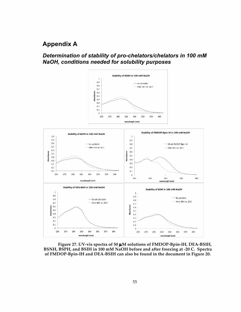
55
Appendix A
Determination of stability of pro-chelators/chelators in 100 mM NaOH, conditions needed for solubility purposes
Figure 27. UV-vis spectra of 50 µM solutions of FMDOP-Bpin-IH, DEA-BSIH, BSNH, BSPH, and BSIH in 100 mM NaOH before and after freezing at -20 C. Spectra of FMDOP-Bpin-IH and DEA-BSIH can also be found in the document in Figure 20.
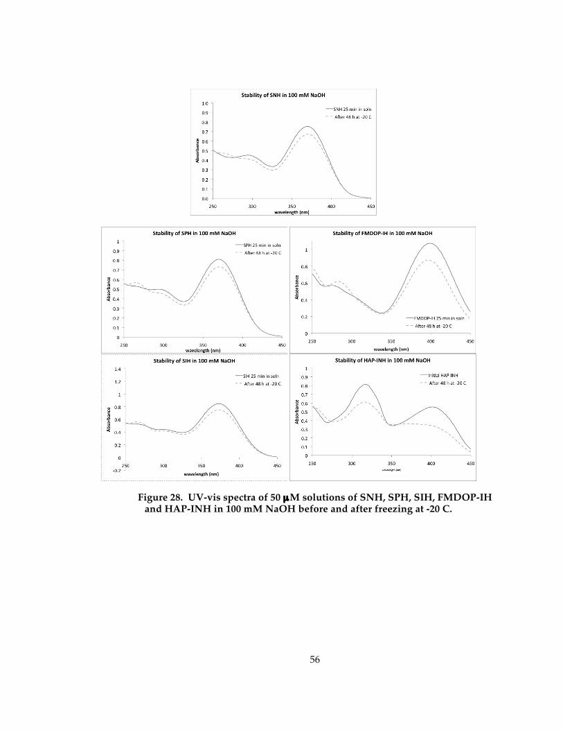
56
Figure 28. UV-vis spectra of 50 µM solutions of SNH, SPH, SIH, FMDOP-IH and HAP-INH in 100 mM NaOH before and after freezing at -20 C.
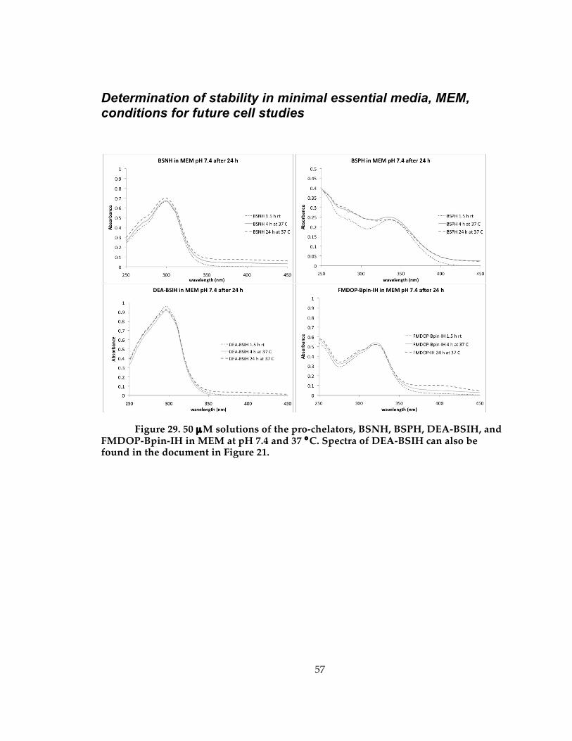
57
Determination of stability in minimal essential media, MEM, conditions for future cell studies
Figure 29. 50 µM solutions of the pro-chelators, BSNH, BSPH, DEA-BSIH, and FMDOP-Bpin-IH in MEM at pH 7.4 and 37 °C. Spectra of DEA-BSIH can also be found in the document in Figure 21.
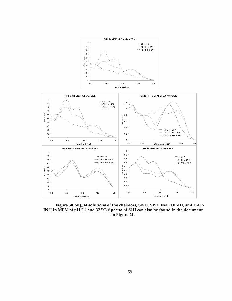
58
Figure 30. 50 µM solutions of the chelators, SNH, SPH, FMDOP-IH, and HAP-INH in MEM at pH 7.4 and 37 °C. Spectra of SIH can also be found in the document
in Figure 21.
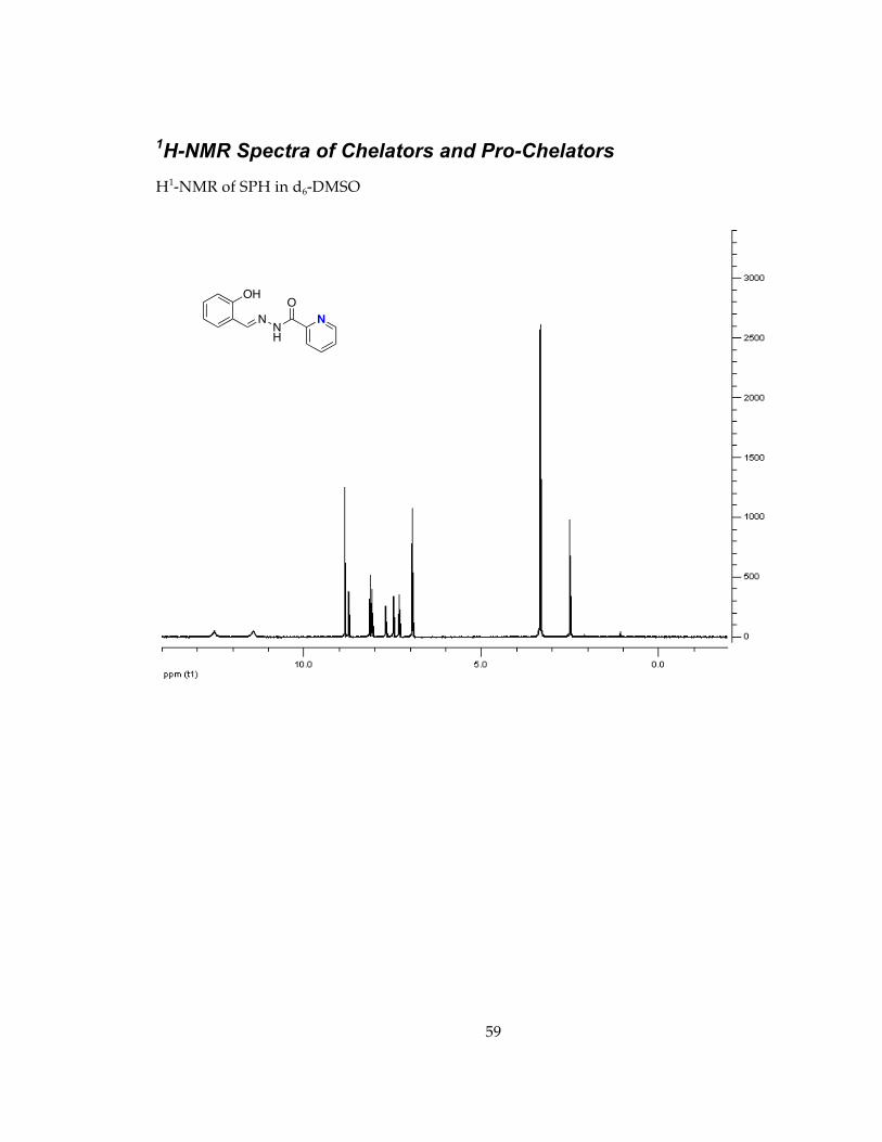
59
1H-NMR Spectra of Chelators and Pro-Chelators H1-NMR of SPH in d6-DMSO
NNH
O
N
OH
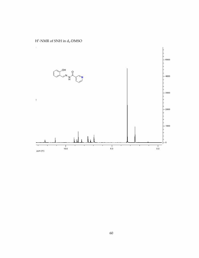
60
H1-NMR of SNH in d6-DMSO
NNH
O
N
OH
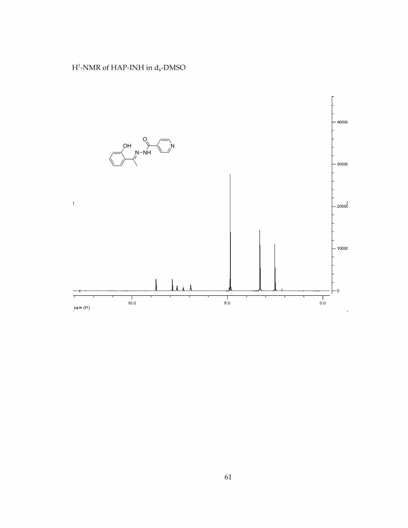
61
H1-NMR of HAP-INH in d6-DMSO
N NH
O
NOH
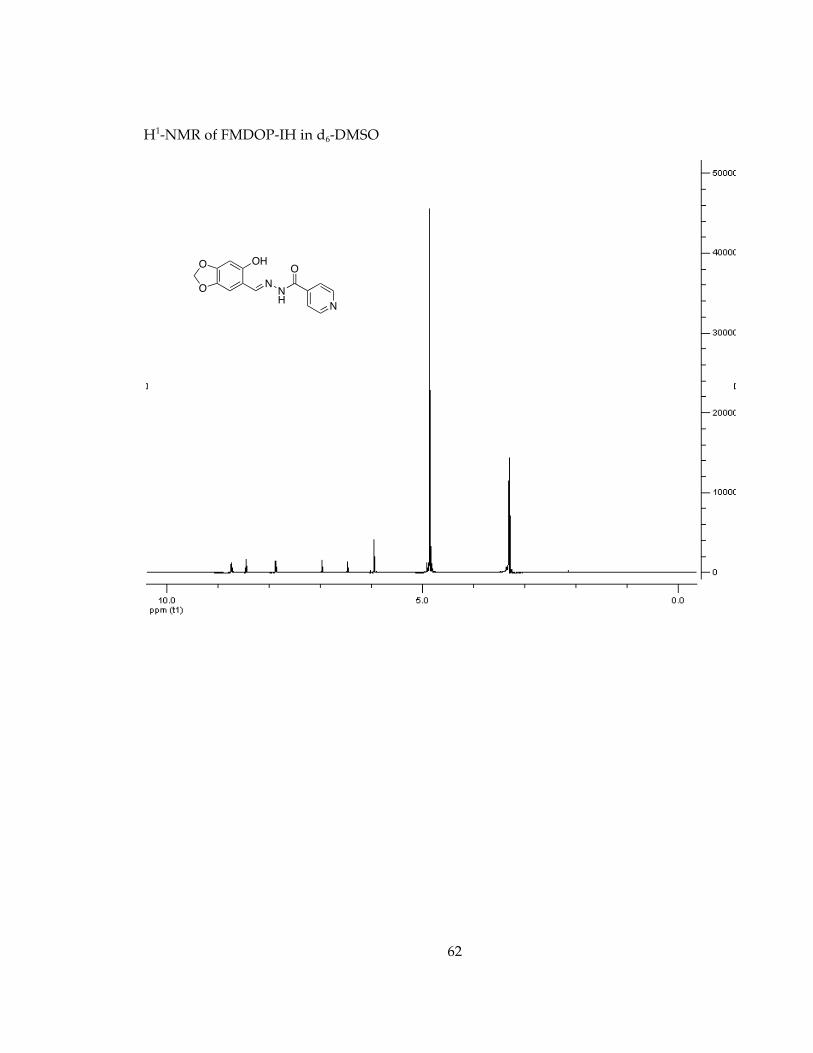
62
H1-NMR of FMDOP-IH in d6-DMSO
NNH
O
N
OHO
O
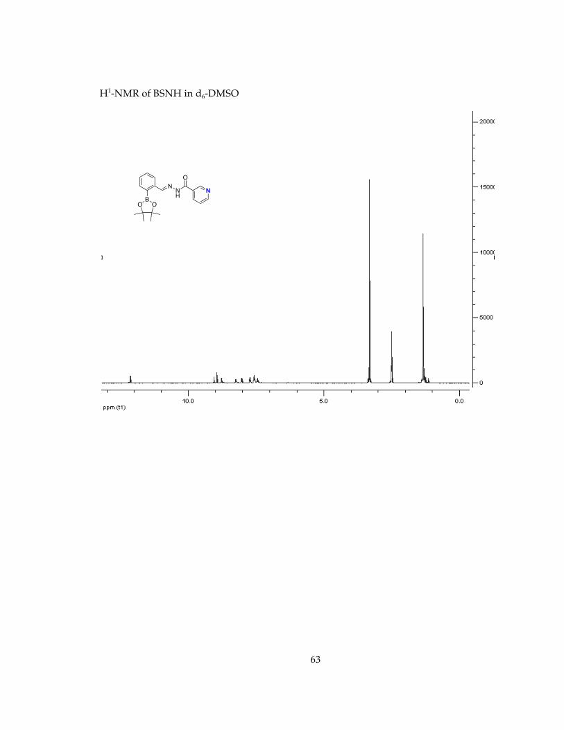
63
H1-NMR of BSNH in d6-DMSO
BOO
NNH
O
N
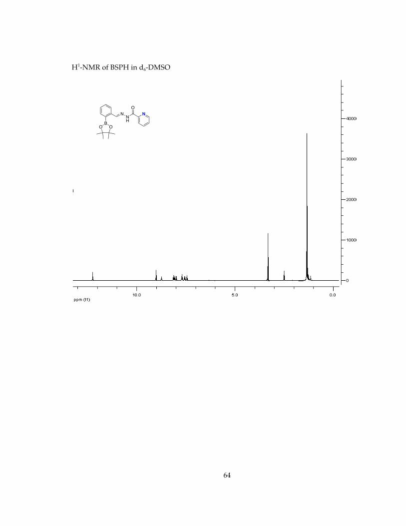
64
H1-NMR of BSPH in d6-DMSO
BOO
NNH
O
N
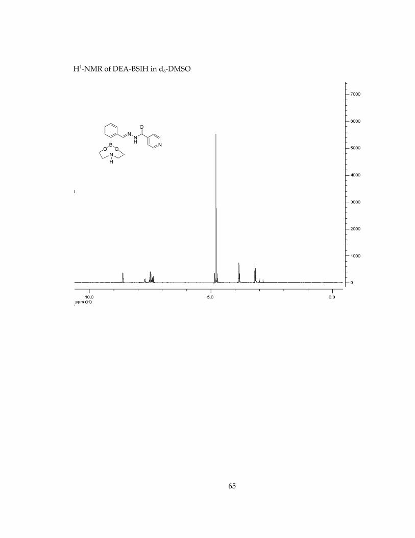
65
H1-NMR of DEA-BSIH in d6-DMSO
BOO
NNH
O
N
N
H
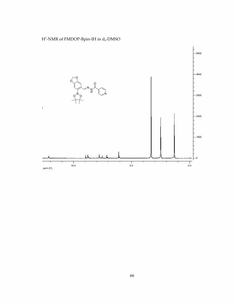
66
H1-NMR of FMDOP-Bpin-IH in d6-DMSO
BOO
NNH
O
N
O
O
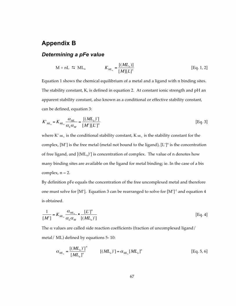
67
Appendix B
Determining a pFe value
M + nL MLn
€
KMLn=[(MLn )][M][L]n
[Eq. 1, 2]
Equation 1 shows the chemical equilibrium of a metal and a ligand with n binding sites.
The stability constant, K, is defined in equation 2. At constant ionic strength and pH an
apparent stability constant, also known as a conditional or effective stability constant,
can be defined, equation 3:
€
K 'MLn = KMLn
αML
αLαM
=[(MLn )'][M '][L']n
[Eq. 3]
where K’
€
MLn is the conditional stability constant, K
€
MLn is the stability constant for the
complex, [M’] is the free metal (metal not bound to the ligand), [L’]n is the concentration
of free ligand, and [(MLn)’] is concentration of complex. The value of n denotes how
many binding sites are available on the ligand for metal binding; ie. In the case of a bis
complex, n = 2.
By definition pFe equals the concentration of the free uncomplexed metal and therefore
one must solve for [M’]. Equation 3 can be rearranged to solve for [M’]-1 and equation 4
is obtained.
€
1[M ']
= KMLn
αMLn
αLαM
•[L']n
[(MLn )'] [Eq. 4]
The α values are called side reaction coefficients (fraction of uncomplexed ligand/
metal/ ML) defined by equations 5- 10:
€
αMLn=[(MLn )'][MLn ]
n
n
€
[(MLn )'] =αMLn[MLn ]
n [Eq. 5, 6]
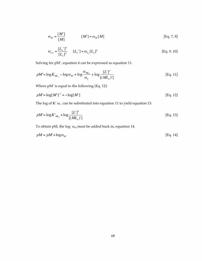
68
€
αM =[M '][M]
€
[M '] =αM [M] [Eq. 7, 8]
€
αLn =[Ln ']
n
[Ln ]n
€
[Ln '] =αLn[Ln ]
n [Eq. 9, 10]
Solving for pM’, equation 4 can be expressed as equation 11.
€
pM '= logKMLn− logαM + log
αMLn
αL
+ log [L']n
[(MLn )'] [Eq. 11]
Where pM’ is equal to the following (Eq. 12):
€
pM '= log[M ']−1 = −log[M '] [Eq. 12]
The log of K’
€
MLn can be substituted into equation 11 to yield equation 13.
€
pM '= logK 'MLn + log [L']n
[(MLn )'] [Eq. 13]
To obtain pM, the log αM must be added back in, equation 14.
€
pM = pM '+ logαM [Eq. 14]
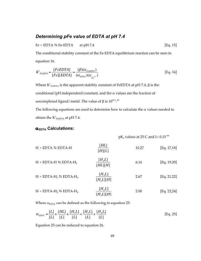
69
Determining pFe value of EDTA at pH 7.4
Fe + EDTA Fe-EDTA at pH 7.4 [Eq. 15]
The conditional stability constant of the Fe-EDTA equilibrium reaction can be seen in
equation 16.
€
K 'FeEDTA =[FeEDTA][Fe][EDTA]
=(β)(αFeEDTA )(αEDTA )(αFe 3
+ ) [Eq. 16]
Where K’FeEDTA is the apparent stability constant of FeEDTA at pH 7.4, β is the
conditional (pH independent) constant, and the α values are the fraction of
uncomplexed ligand/metal. The value of β is 1025.1.99
The following equations are used to determine how to calculate the α values needed to
obtain the K’FeEDTA at pH 7.4.
αEDTA Calculations:
pKa values at 25 C and I= 0.15 99
H + EDTA EDTA-H
€
[HL][H][L]
10.27 [Eq. 17,18]
H + EDTA-H EDTA-H2
€
[H2L][HL][H]
6.16 [Eq. 19,20]
H + EDTA-H2 EDTA-H3
€
[H3L][H2L][H]
2.67 [Eq. 21,22]
H + EDTA-H3 EDTA-H4
€
[H4L][H3L][H]
2.00 [Eq. 23,24]
Where αEDTA can be defined as the following in equation 25:
€
αEDTA =[L][L]
+[HL][L]
+[H2L][L]
+[H3L][L]
+[H4L][L]
[Eq. 25]
Equation 25 can be reduced to equation 26.
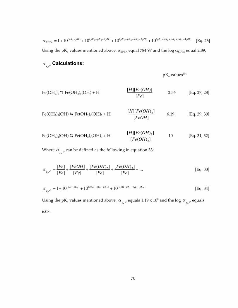
70
€
αEDTA =1+10(pK1− pH ) +10(pK1 + pK2−2pH ) +10( pK1 + pK2 + pK3−3pH ) +10(pK1 + pK2 + pK3 + pK4 −4 pH ) [Eq. 26]
Using the pKa values mentioned above, αEDTA equal 784.97 and the log αEDTA equal 2.89.
€
αFe 3
+ Calculations:
pKa values101
Fe(OH2)6 Fe(OH2)5(OH) + H
€
[H][Fe(OH)][Fe]
2.56 [Eq. 27, 28]
Fe(OH2)5(OH) Fe(OH2)4(OH)2 + H
€
[H][Fe(OH)2][FeOH]
6.19 [Eq. 29, 30]
Fe(OH2)4(OH) Fe(OH2)3(OH)3 + H
€
[H][Fe(OH)3][Fe(OH)2]
10 [Eq. 31, 32]
Where
€
αFe 3
+ can be defined as the following in equation 33:
€
αFe 3
+ =[Fe][Fe]
+[FeOH][Fe]
+[Fe(OH)2][Fe]
+[Fe(OH)3][Fe]
+ ... [Eq. 33]
€
αFe 3
+ =1+10(pH− pK1 ) +10(2pH− pK1− pK2 ) +10(3pH− pK1− pK2− pK3 ) [Eq. 34]
Using the pKa values mentioned above,
€
αFe 3
+ equals 1.19 x 10⁶ and the log
€
αFe 3
+ equals
6.08.
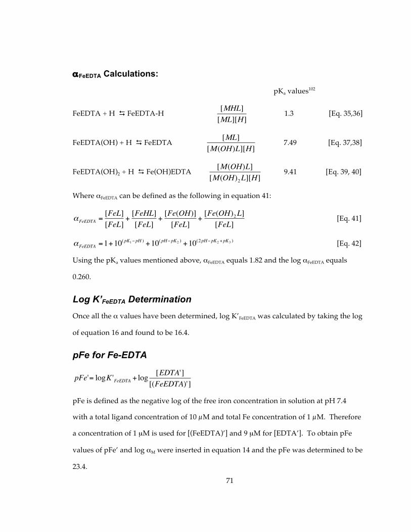
71
αFeEDTA Calculations:
pKa values102
FeEDTA + H FeEDTA-H
€
[MHL][ML][H]
1.3 [Eq. 35,36]
FeEDTA(OH) + H FeEDTA
€
[ML][M(OH)L][H]
7.49 [Eq. 37,38]
FeEDTA(OH)2 + H Fe(OH)EDTA
€
[M(OH)L][M(OH)2L][H]
9.41 [Eq. 39, 40]
Where αFeEDTA can be defined as the following in equation 41:
€
αFeEDTA =[FeL][FeL]
+[FeHL][FeL]
+[Fe(OH)][FeL]
+[Fe(OH)2L][FeL]
[Eq. 41]
€
αFeEDTA =1+10(pK1− pH ) +10(pH− pK2 ) +10(2pH− pK2 + pK3 ) [Eq. 42]
Using the pKa values mentioned above, αFeEDTA equals 1.82 and the log αFeEDTA equals
0.260.
Log K’FeEDTA Determination Once all the α values have been determined, log K’FeEDTA was calculated by taking the log
of equation 16 and found to be 16.4.
pFe for Fe-EDTA
€
pFe'= logK 'FeEDTA + log [EDTA'][(FeEDTA)']
pFe is defined as the negative log of the free iron concentration in solution at pH 7.4
with a total ligand concentration of 10 µM and total Fe concentration of 1 µM. Therefore
a concentration of 1 µM is used for [(FeEDTA)’] and 9 µM for [EDTA’]. To obtain pFe
values of pFe’ and log αM were inserted in equation 14 and the pFe was determined to be
23.4.
72
References 1. Andrews, N. C.; Schmidt, P. J., Iron Homeostasis. Ann. Rev. Physiol. 2007, 69 (1), 69-85. 2. Cousins, R. J.; Liuzzi, J. P.; Lichten, L. A., Mammalian Zinc Transport, Trafficking, and Signals. J. Biol. Chem. 2006, 281 (34), 24085-24089. 3. Kim, B.-E.; Nevitt, T.; Thiele, D. J., Mechanisms for copper acquisition, distribution and regulation. Nat Chem Biol 2008, 4 (3), 176-185. 4. Pierre, J. L.; Fontecave, M., Iron and activated oxygen species in biology: The basic chemistry. Biometals 1999, 12, 195-199. 5. Prousek, J., Fenton chemistry in biology and medicine *.(Henry John Horstman Fenton). Pure Appl. Chem. 2007, 79 (12), 2325(14). 6. Faller, P.; Hureau, C., Bioinorganic chemistry of copper and zinc ions coordinated to amyloid-[small beta] peptide. Dalton Transactions 2009, (7), 1080-1094. 7. Roychaudhuri, R.; Yang, M.; Hoshi, M. M.; Teplow, D. B., Amyloid {beta}-Protein Assembly and Alzheimer Disease. Journal of Biological Chemistry 2009, 284 (8), 4749-4753. 8. Castellani, R. J.; Honda, K.; Zhu, X. W.; Cash, A. D.; Nunomura, A.; Perry, G.; Smith, M. A., Contribution of redox-active iron and copper to oxidative damage in Alzheimer disease. Ageing Research Reviews 2004, 3 (3), 319-326. 9. Gaggelli, E.; Kozlowski, H.; Valensin, D.; Valensin, G., Copper Homeostasis and Neurodegenerative Disorders (Alzheimer's, Prion, and Parkinson's Diseases and Amyotrophic Lateral Sclerosis). Chem. Rev. 2006, 106 (6), 1995-2044. 10. Crichton, R. R.; Dexter, D. T.; Ward, R. J., Metal based neurodegenerative diseases--From molecular mechanisms to therapeutic strategies. Coordination Chemistry Reviews 2008, 252 (10-11), 1189-1199. 11. Crichton, R. R.; Ward, R. J., Metal-based neurodegeneration: from molecular mechanisms to therapeutic strategies. Wiley: Hoboken, NJ, 2006. 12. Halliwell, B., Oxidative stress and neurodegeneration: where are we now? J. Neurochem. 2006, 97 (6), 1634-1658. 13. Barnham, K. J.; Masters, C. L.; Bush, A. I., Neurodegenerative diseases and oxidative stress. Nat. Rev. Drug Disc. 2004, 3 (3), 205-214. 14. Zecca, L.; Youdim, M. B. H.; Riederer, P.; Conner, J. R.; Crichton, R. R., Iron, brain aging and neurodegenerative disorders. Nat. Rev. Neurosci. 2004, 5, 863-873. 15. Kell, D., Iron behaving badly: inappropriate iron chelation as a major contributor to the aetiology of vascular and other progressive inflammatory and degenerative diseases. BMC Medical Genomics 2009, 2 (1), 2.
73
16. Gaeta, A.; Hider, R. C., The crucial role of metal ions in neurodegeneration: the basis for a promising therapeutic strategy. British Journal of Pharmacology 2005, 146 (8), 1041-1059. 17. Richardson, D. R., Novel chelators for central nervous system disorders that involve alterations in the metabolism of iron and other metal ions. Annals of the New York Academy of Sciences 2004, 1012, 326-341. 18. Youdim, M. B. H.; Stephenson, G.; Ben Shachar, D., Ironing iron out in Parkinson's disease and other neurodegenerative diseases with iron chelators - A lesson from 6-hydroxydopamine and iron chelators, desferal and VK-28. Annals of the New York Academy of Sciences 2004, 1012, 306-325. 19. Madsen, E.; Gitlin, J. D., Copper and Iron Disorders of the Brain. Annu. Rev. Neurosci. 2007, 30 (1), 317-337. 20. Tam, T. F.; Leung-Toung, R.; Li, W. R.; Wang, Y. S.; Karimian, K.; Spino, M., Iron chelator research: Past, present, and future. Current Medicinal Chemistry 2003, 10 (12), 983-995. 21. Kalinowski, D. S.; Richardson, D. R., The Evolution of Iron Chelators for the Treatment of Iron Overlad Disease and Cancer. Pharmacol. Rev. 2005, 57, 547-583. 22. Kontoghiorghes, G. J.; Pattichis, K.; Neocleous, K.; Kolnagou, A., The design and development of deferiprone (L1) and other iron chelators for clinical use: Targeting methods and application prospects. Current Medicinal Chemistry 2004, 11 (16), 2161-2183. 23. Liu, Z. D.; Hider, R. C., Design of iron chelators with therapeutic application. Coordination Chemistry Reviews 2002, 232 (1-2), 151-171. 24. Liu, Z. D.; Kayyali, R.; Hider, R. C.; Porter, J. B.; Theobald, A. E., Design, synthesis, and evaluation of novel 2-substituted 3-hydroxypyridin-4-ones: Structure-activity investigation of metalloenzyme inhibition by iron chelators. Journal of Medicinal Chemistry 2002, 45 (3), 631-639. 25. Liu, Z. D.; Lockwood, M.; Rose, S.; Theobald, A. E.; Hider, R. C., Structure-activity investigation of the inhibition of 3-hydroxypyridin-4-ones on mammalian tyrosine hydroxylase. Biochem. Pharmacol. 2001, 61 (3), 285-290. 26. Waldmeier, P. C.; Buchle, A. M.; Steulet, A. F., Inhibition of catechol-o-methyltransferase (COMT1) as well as tyrosine and tryptophan hydroxylase by the orally-active iron chelator, 1,2-dimethyl-3-hydroxypyridin-4-one (L1, CP20), in rat brain in vivo. Biochem. Pharmacol. 1993, 45 (12), 2417-2424. 27. Storr, T.; Merkel, M.; Song-Zhao, G. X.; Scott, L. E.; Green, D. E.; Bowen, M. L.; Thompson, K. H.; Patrick, B. O.; Schugar, H. J.; Orvig, C., Synthesis, Characterization, and Metal Coordinating Ability of Multifunctional Carbohydrate-Containing Compounds for Alzheimer's Therapy. J. Am. Chem. Soc. 2007, 129 (23), 7453–7463.
74
28. Schugar, H.; Green, D. E.; Bowen, M. L.; Scott, L. E.; Storr, T.; Bohmerle, K.; Thomas, F.; Allen, D. D.; Lockman, P. R.; Merkel, M.; Thompson, K. H.; Orvig, C., Combating Alzheimer's disease with multifunctional molecules designed for metal passivation. Angewandte Chemie-International Edition 2007, 46 (10), 1716-1718. 29. Scott, L. E.; Page, B. D. G.; Patrick, B. O.; Orvig, C., Altering pyridinone N-substituents to optimise activity as potential prodrug for Alzheimer's disease. Dalton Trans. 2008, 6364-6367. 30. Storr, T.; Scott, L. E.; Bowen, M. L.; Green, D. E.; Thompson, K. H.; Schugar, H. J.; Orvig, C., Glycosylated tetrahydrosalens as multifunctional molecules for Alzheimer's therapy. Dalton Transactions 2009, (16), 3034-3043. 31. Storr, T.; Thompson, K. H.; Orvig, C., Design of targeting ligands in medicinal inorganic chemistry Chem. Soc. Rev. 2006, 35, 534 - 544. 32. Zheng, H.; Blat, D.; Fridkin, M., Novel neuroprotective neurotrophic NAP analogs targeting metal toxicity and oxidative stress: potential candidates for the control of neurodegenerative diseases. Journal of Neural Transmission 2006, 71, 163-172. 33. Gozes, I.; Lilling, G.; Glazer, R.; Ticher, A.; Ashkenazi, I. E.; Davidson, A.; Rubinraut, S.; Fridkin, M.; Brenneman, D. E., Superactive Lipophilic Peptides Discriminate Multiple Vasoactive Intestinal Peptide Receptors. Journal of Pharmacology and Experimental Therapeutics 1995, 273, 161-167. 34. Blat, D.; Weiner, L.; Youdim, M. B. H.; Fridkin, M., A Novel Iron-Chelating Derivative of the Neuroprotective Peptide NAPVSIPQ Shows Superior Antioxidant and Antineurodegenerative Capabilities. J. Med. Chem. 2008, 51 (1), 126-134. 35. Zheng, H.; Youdim, M. B. H.; Weiner, L. M.; Fridkin, M., Novel potential neuroprotective agents with both iron chelating and amino acid-based derivatives targeting central nervous system neurons Biochemical Pharmacology 2005, 70 (11), 1642-1652. 36. Ye, Y.; Bloch, S.; Xu, B.; Achilefu, S., Novel near-infrared fluorescent integrin-targeted DFO analogue. Bioconjugate Chemistry 2008, 19 (1), 225-234. 37. Chen, X.; Sievers, E.; Hou, Y.; Park, R.; Tohme, M.; Bart, R.; Bremner, R.; Bading, J. R.; Conti, P. S., Integrin alpha v beta 3-targeted imaging of lung cancer. Neoplasia 2005, 7, 271-279. 38. Hsu, A. R.; Hou, L. C.; Veeravagu, A.; Greve, J. M.; Vogel, H.; Tse, V.; Chen, X., In Vivo Near-infrared Fluorecence Imaging of Integrin alphavbeta3 in an Orthotopic Glioblastoma Model. Molecular Imaging and Biology 2006, 8 (6), 315-323. 39. Faraji, A. H.; Wipf, P., Nanoparticles in cellular drug delivery. Bioorganic & Medicinal Chemistry 2009, 17 (8), 2950-2962. 40. Cui, Z.; Lockman, P. R.; Atwood, C. S.; Hsu, C.-H.; Gupte, A.; Allen, D. D.; Mumper, R. J., Novel D-penicillamine carrying nanoparticles for metal chelation therapy
75
in Alzheimer's and other CNS diseases. European Journal of Pharmaceutics and Biopharmaceutics 2005, 59 (2), 263-272. 41. Liu, G.; Men, P.; Harris, P. L. R.; Rolston, R. K.; Perry, G.; Smith, M. A., Nanoparticle iron chelators: A new therapeutic approach in Alzheimer disease and other neurologic disorders associated with trace metal imbalance. Neuroscience Letters 2006, 406 (3), 189-193. 42. Liu, G.; Men, P.; Kudo, W.; Perry, G.; Smith, M. A., Nanoparticle-chelator conjugates as inhibitors of amyloid-[beta] aggregation and neurotoxicity: A novel therapeutic approach for Alzheimer disease. Neuroscience Letters 2009, 455 (3), 187-190. 43. Meijler, M. M.; Arad-Yellin, R.; Cabantchik, Z. I.; Shanzer, A., Synthesis and Evaluation of Iron Chelators with Masked Hydrophilic Moieties J. Am. Chem. Soc. 2002, 124 (43), 12666–12667. 44. Yavin, E.; Kikkiri, R.; Gil, S.; Arad-Yellin, R.; Yavin, E.; Shanzer, A., Synthesis and biological evaluation of lipophilic iron chelators as protective agents from oxidative stress. Org. Biomol. Chem. 2005, 3, 2685 - 2687. 45. Prousek, J., Fenton chemistry in biology and medicine Pure and Applied Chemistry 2007, 79 (12), 2325-2338. 46. Valko, M.; Morris, H.; Cronin, M. T. D., Metals, Toxicity and Oxidative Stress. Curr. Med. Chem. 2005, 12, 1161-1208. 47. Chiueh, C. C., Iron overload, oxidative stress, and axonal dystrophy in brain disorders. Pediatric Neurology 2001, 25 (2), 138-147. 48. Ke, Y.; Qian, Z. M., Iron misregulation in the brain: a primary cause of neurodegenerative disorders. Lancet Neurology 2003, 2 (4), 246-253. 49. Rouault, T. A., Iron on the brain. Nature Genetics 2001, 28 (4), 299-300. 50. Charkoudian, L. K.; Pham, D. M.; Franz, K. J., A Pro-Chelator Triggered by Hydrogen Peroxide Inhibits Iron-Promoted Hydroxyl Radical Formation. J. Am. Chem. Soc. 2006, 128 (38), 12424-12425. 51. Charkoudian, L. K., Pham, D. M., Kwon, A. M., Vangeloff, A. D., Franz, K. J., Modifications of Boronic Ester Pro-Chelators Triggered by Hydrogen Peroxide Tune Reactivity to Inhibit Metal-Promoted Oxidative Stress. Dalton Trans. 2007, 5031-5042. 52. Charkoudian, L. K., Dentchev, T., Lukinova, N., Wolkow, N. Dunaief, J. L., Franz, K. J., Iron prochelator BSIH protects retinal pigment epithelial cells against cell death induced by hydrogen peroxide. Journal of Inorganic Biochemistry 2008, 102, 2130-2135. 53. Yiakouvaki, A.; Savovic, J.; Al-Qenaei, A.; Dowden, J.; Pourzand, C., Caged-Iron Chelators a Novel Approach towards Protecting Skin Cells against UVA-Induced Necrotic Cell Death. Journal of Investigative Dermatology 2006, 126, 2287–2295.
76
54. Pourzand, C.; Watkin, R. D.; Brown, J. E.; Tyrrell, R. M., Ultraviolet A radiation induces immediate release of iron in human primary skin fibroblasts: The role of ferritin. Proceedings of the National Academy of Sciences of the United States of America 1999, 96 (12), 6751-6756. 55. Galey, J.-B.; Dumats, J.; Beck, I.; Fernandez, B.; Hocquaux, M., N,N'-Bis-Dibenzyl Ethylenediaminediacetic Acid (Dbed): a Site-Specific Hydroxyl Radical Scavenger Acting as an ‚ÄúOxidative Stress Activatable‚Äù Iron Chelator <i>In Vitro</i>. Free Radical Research 1995, 22 (1), 67 - 86. 56. Serratrice, G.; Galey, J. B.; Saint Aman, E.; Dumats, J., Iron(III) Complexation by New Aminocarboxylate Chelators - Thermodynamic and Kinetic Studies. European Journal of Inorganic Chemistry 2001, (2), 471-479. 57. Galey, J.-B.; Destrée, O.; Dumats, J.; Génard, S.; Tachon, P., Protection against Oxidative Damage by Iron Chelators: Effect of Lipophilic Analogues and Prodrugs of N,N'-Bis(3,4,5-trimethoxybenzyl)ethylenediamine- N,N'-diacetic Acid (OR10141). J. Med. Chem. 2000, 43 (7), 1418–1421. 58. Galey, J.-B.; Dumats, J.; Genard, S.; Destree, O.; Pichaud, P.; Cctroux, P.; Marrot, L.; Beck, I.; Fernandez, B.; Barre, G.; Seite, M.; Hussler, G.; Hocquaux, M., N,N'-bis-(3,4,5-trimethoxybenzyl) ethylenediamine N,N'-diacetic acid as a new iron chelator with potential medicinal applications against oxidative stress. Biochemical Pharmacology 1996, 51 (2), 103-115. 59. Naughton, D. P.; Grootveld, M., EDTA Bis-(ethyl phenylalaninate): A Novel Transition Metal-Ion Chelating Hydroxyl Radical Scavenger with a Potential Anti-inflammatory Role. Bioorganic & Medicinal Chemistry Letters 2001, 11, 2573-2575. 60. Di Stefano, A.; Sozio, P.; Iannitelli, A.; Cerasa, L. S., New drug delivery strategies for improved Parkinson's disease therapy. Expert Opinion on Drug Delivery 2009, 6 (4), 389-404. 61. Di Stefano, A.; Sozio, P.; Cerasa, L. S., Antiparkinson Prodrugs. Molecules 2008, 13, 46-68. 62. Di Stefano, A.; Sozio, P.; Cocco, A.; Iannitelli, A.; Santucci, E.; Costa, M.; Pecci, L.; Nasuti, C.; Cantalamessa, F.; Pinnen, F., L-Dopa- and Dopamine-(R)-alpha-Lipoic Acid Conjugates as Multifunctional Codrugs with Antioxidant Properties. Journal of Medicinal Chemistry 2006, 49 (4), 1486-1493. 63. Packer, L.; Tritschler, H. J.; Wessel, K., Neuroprotection by the Metabolic Antioxidant -Lipoic Acid. Free Radical Biology and Medicine 1997, 22 (1-2), 359-378. 64. Suh, J. H.; Ben-Zhan, Z.; DeSzoeke, E.; Frei, B.; Hagen, T. M., Dihydrolipoic acid lowers the redox activity of transition metal ions but does not remove them from the active site of enzymes. Redox Report 2004, 9 (1), 57-61. 65. Bebbington, D.; Monck, N. J. T.; Gaur, S.; Palmer, A. M.; Benwell, K.; Harvey, V.; Malcolm, C. S.; Porter, R. H. P., 3,5-Disubstituted-4-hydroxyphenyls Linked to 3-
77
Hydroxy-2-methyl- 4(1H)-pyridinone: Potent Inhibitors of Lipid Peroxidation and Cell Toxicity. Journal of Medicinal Chemistry 2000, 43 (15), 2779-2782. 66. Bebbington, D.; Dawson, C. E.; Gaur, S.; Spencer, J., Prodrug and covalent linker strategies for the solubilization of dual-Action antioxidants/Iron chelators. Bioorganic & Medicinal Chemistry Letters 2002, 12 (22), 3297-3300. 67. Rauk, A., Why is the amyloid beta peptide of Alzheimer's disease neurotoxic? Dalton Trans. 2008, 1273-1282. 68. Bush, A. I., The metallobiology of Alzheimer's disease. Trends in Neurosciences 2003, 26 (4), 207-214. 69. Adlard, P. A.; Cherny, R. A.; Finkelstein, D. I.; Gautier, E.; Robb, E.; Cortes, M.; Volitakis, I.; Liu, X.; Smith, J. P.; Perez, K.; Laughton, K.; Li, Q.-X.; Charman, S. A.; Nicolazzo, J. A.; Wilkins, S.; Deleva, K.; Lynch, T.; Kok, G.; Ritchie, C. W.; Tanzi, R. E.; Cappai, R.; Masters, C. L.; Barnham, K. J.; Bush, A. I., Rapid Restoration of Cognition in Alzheimer's Transgenic Mice with 8-Hydroxy Quinoline Analogs Is Associated with Decreased Interstitial A[beta]. Neuron 2008, 59 (1), 43-55. 70. Ritchie, C. W.; Bush, A. I.; Mackinnon, A.; Macfarlane, S.; Mastwyk, M.; MacGregor, L.; Kiers, L.; Cherny, R.; Li, Q.-X.; Tammer, A.; Carrington, D.; Mavros, C.; Volitakis, I.; Xilinas, M.; Ames, D.; Davis, S.; Beyreuther, K.; Tanzi, R. E.; Masters, C. L., Metal-Protein Attenuation With Iodochlorhydroxyquin (Clioquinol) Targeting A{beta} Amyloid Deposition and Toxicity in Alzheimer Disease: A Pilot Phase 2 Clinical Trial. Arch Neurol 2003, 60 (12), 1685-1691. 71. Dedeoglu, A.; Cormier, K.; Payton, S.; Tseitlin, K. A.; Kremsky, J. N.; Lai, L.; Li, X.; Moir, R. D.; Tanzi, R. E.; Bush, A. I.; Kowall, N. W.; Rogers, J. T.; Huang, X., Preliminary Studies of a Novel Bifunctional Metal Chelator Targeting Alzheimer's Amyloidogonesis. Experimental Gerontology 2004, 39, 1641-1649. 72. Rodriguez-Rodriguez, C.; Sanchez de Groot, N.; Rimola, A.; Alvarez-Larena, A.; Lloveras, V.; Vidal-Gancedo, J.; Ventura, S.; Vendrell, J.; Sodupe, M.; Gonzalez-Duarte, P., Design, Selection, and Characterization of Thioflavin-Based Intercalation Compounds with Metal Chelating Properties for Application in Alzheimer’s Disease. Journal of the American Chemical Society 2009, 131 (4), 1436-1451. 73. Opazo, C.; Huang, X.; Cherny, R. A.; Moir, R. D.; Roher, A. E.; White, A. R.; Cappai, R.; Masters, C. L.; Tanzi, R. E.; Inestrosa, N. C.; Bush, A. I., Metalloenzyme-like Activity of Alzheimer's Disease beta -Amyloid. Cu-Dependent Catalytic Conversion of Dopamine, Cholesterol, and Biological Reducing Agents to Neurotoxic H2O2. Journal of Biological Chemistry 2002, 277 (43), 40302-40308. 74. Tabner, B. J.; El-Agnaf, O. M. A.; Turnbull, S.; German, M. J.; Paleologou, K. E.; Hayashi, Y.; Cooper, L. J.; Fullwood, N. J.; Allsop, D., Hydrogen Peroxide Is Generated during the Very Early Stages of Aggregation of the Amyloid Peptides Implicated in Alzheimer Disease and Familial British Dementia. Journal of Biological Chemistry 2005, 280 (43), 35789-35792.
78
75. Dickens, M. G.; Franz, K. J., A Prochelator Activated by Hydrogen Peroxide Prevents Metal-Induced Amyloid Beta Aggregation. submitted for publication 2009. 76. Suh, J.; Yoo, S.; Kim, M.; Jeong, K.; Ahn, J. Y.; Kim, M. S.; Chae, P. S.; Lee, T. Y.; Lee, J.; Jang, Y. A.; Ko, E. H., Cleavage agents for soluble oligomers of amyloid beta peptides. Angewandte Chemie-International Edition 2007, 46 (37), 7064-7067. 77. Suh, J.; Chei, W. S., Metal complexes as artificial proteases: toward catalytic drugs. Current Opinion in Chemical Biology 2008, 12 (2), 207-213. 78. Wu, W.; Lei, P.; Liu, Q.; Hu, J.; Gunn, A. P.; Chen, M.; Rui, Y.; Su, X.; Xie, Z.; Zhao, Y.; Bush, A. I.; Li, Y., Sequestration of Copper from {beta}-Amyloid Promotes Selective Lysis by Cyclen-Hybrid Cleavage Agents. Journal of Biological Chemistry 2008, 283 (46), 31657-31664. 79. Youdim, M. B. H.; Fridkin, M.; Zheng, H., Novel bifunctional drugs targeting monoamine oxidase inhibition and iron chelation as an approach to neuroprotection in Parkinson’s disease and other neurodegenerative diseases. Journal of Neural Transmission 2004, 111 (10-11), 1455-1471. 80. Zheng, H.; Gal, S.; Weiner, L. M.; Bar-Am, O.; Warshawsky, A.; Fridkin, M.; Youdim, M. B. H., Novel multifunctional neuroprotective iron chelator-monoamine oxidase inhibitor drugs for neurodegenerative diseases: in vitro studies on antioxidant activity, prevention of lipid peroxide formation and monoamine oxidase inhibition Journal of Neurochemistry 2005, 95, 68-78. 81. Youdim, M. B. H., The Path from Anti Parkinson Drug Selegiline and Rasagiline to Multifunctional Neuroprotective Anti Alzheimer Drugs Ladostigil and M30. Current Alzheimer Research 2006, 3 (5), 541-550. 82. Zheng, H.; Weiner, L. M.; Bar-Am, O.; Epsztejn, S.; Cabantchik, Z. I.; Warshawsky, A.; Youdim, M. B. H.; Fridkin, M., Design, synthesis, and evaluation of novel bifunctional iron-chelators as potential agents for neuroprotection in Alzheimer’s, Parkinson’s, and other neurodegenerative diseases Bioorganic & Medicinal Chemistry Letters 2005, 13 (3), 773-783. 83. Avramovich-Tirosh, Y.; Amit, T.; Bar-Am, O.; Zheng, H.; Fridkin, M.; Youdim, M. B. H., Therapeutic targets and potential of the novel brain- permeable multifunctional iron chelator-monoamine oxidase inhibitor drug, M-30, for the treatment of Alzheimer’s disease Journal of Neurochemistry 2007, 100 (2), 490-502. 84. Avramovich-Tirosh, Y.; Rezrlichenko, D.; Amit, T.; Zheng, H.; Fridkin, M.; Weinreb, O.; Mandel, S.; Youdim, M. B. H., Neurorescue activity, APP regulation and amyloid-beta peptide reduction by novel multi-functional brain permeable iron- chelating- antioxidants, m-30 and green tea polyphenol, EGCG. Current Alzheimer Research 2007, 4 (4), 403-411. 85. Mandel, S.; Amit, T.; Bar-Am, O.; Youdim, M. B. H., Iron dysregulation in Alzheimer's disease: Multimodal brain permeable iron chelating drugs, possessing
79
neuroprotective-neurorescue and amyloid precursor protein-processing regulatory activities as therapeutic agents. Progress in Neurobiology 2007, 82 (6), 348-360. 86. Cavalli, A.; Bolognesi, M. L.; Minarini, A.; Rosini, M.; Tumiatti, V.; Recanatini, M.; Melchiorre, C., Multi-target-Directed Ligands to Combat Neurodegenerative Diseases. Journal of Medicinal Chemistry 2008, 51 (3), 347-372. 87. Bolognesi, M. L.; Cavalli, A.; Valgimigli, L.; Bartolini, M.; Rosini, M.; Andrisano, V.; Recanatini, M.; Melchiorre, C., Multi-Target-Directed Drug Design Strategy: From a Dual Binding Site Acetylcholinesterase Inhibitor to a Trifunctional Compound against Alzheimer’s Disease. J. Med. Chem. 2007, 50 (26), 6446-6449. 88. Pang, Y.-P.; Quiram, P.; Jelacic, T.; Hong, F.; Brimijoin, S., Highly Potent, Selective, and Low Cost Bis-tetrahydroaminacrine Inhibitors of Acetylcholinesterase. Journal of Biological Chemistry 1996, 271 (39), 23646- 23649. 89. Zheng, H.; Youdim, M. B. H.; Fridkin, M., Site-Activated Multifunctional Chelator with Acetylcholinesterase and Neuroprotective‚àíNeurorestorative Moieties for Alzheimer‚Äôs Therapy. Journal of Medicinal Chemistry 2009, 52 (14), 4095-4098. 90. Ba, L. A.; Doering, M.; Burkholz, T.; Jacob, C., Metal trafficking: from maintaining the metal homeostasis to future drug design. Metallomics 2009, 1 (4), 292-311. 92. Galey, J. B. D., O.; Dumats, J.; Génard, S.; Tachon, P., Protection against Oxidative Damage by Iron Chelators: Effect of Lipophilic Analogues and Prodrugs of N,N'-Bis(3,4,5-trimethoxybenzyl)ethylenediamine- N,N'-diacetic Acid (OR10141). J. Med. Chem. 2000, 43 (7), 1418–1421. 93. Nick, H., Iron Chelation, quo vadis? Current Opinion in Chemical Biology 2007, 11, 419-423. 94. Ponka, P., Borova, J., Neuwirt, J., Fuchs, O, Mobilization of iron from reticulocytes. Identification of pyridoxal isonicotinoyl hydrazone as a new iron chelating agent. Febs Lett 1979, 97, 317-321. 95. Edward, J. T., Gauthier, M., Chubb, F. L., Ponka, Synthesis of new acylhydrazones as iron-chelating compounds. P. J. Chem. Eng. Data 1988, 33, 538- 540. 96. KovarÌkov·, P.; V·vrov·, K.; Tomalov·, K.; Schˆngut, M.; Hruskov·, K.; Haskov·, P.; Klimes, J., HPLC-DAD and MS/MS analysis of novel drug candidates from the group of aromatic hydrazones revealing the presence of geometric isomers. Journal of Pharmaceutical and Biomedical Analysis 2008, 48 (2), 295-302. 97. Richardson, D. R.; Bernhardt, P. V., Crystal and molecular structure of 2-hydroxy-1-naphthaldehyde isonicotinoyl hydrazone (NIH) and its iron(III) complex: an iron chelator with anti-tumour activity. J. Biol. Inorg. Chem. 1999, 4 (3), 266-273. 98. Bastian, R.; Weberling, R.; Palilla, F., Determination of iron by ultraviolet spectrophotometry. Anal. Chem. 1956, 28 (4), 459–462.
80
99. Perrin, D. D.; Dempsey, B., Buffers for pH and Metal ion and control. Chapman and Hall: London, 1922. 100. Albrecht-Gary, A.-M.; Crumbliss, A. L., Coordination chemistry of siderophores: Thermodynamics and kinetics of iron chelation and release. In Metal Ions in Biological Systems, Sigel, A.; Sigel, H., Eds. Marcel Dekker: New York, 1973; Vol. 35, pp 239–327. 101. Crumbliss, A. L.; Garrison, M. J., A Comparison of Some Aspects of the Aqueous Coordination Chemistry of Aluminum(III) and Iron(III). Comments Inorg. Chem. 1988, 8 (Nos. 1 & 2), 1-26. 102. Martell, A. E.; Smith, R. M., Critical Stablity Constants. Plenum Press: New York and London, Vol. 1.


![GREEN SYNTHESIS AND CHARACTERIZATION OF ZERO VALENT IRON …ijetmas.com/admin/resources/project/paper/f201704041491320502.pdf · of zero valent iron nano particles [7] with minimal](https://img.pdfslide.us/doc/110x75/5e4362ac1b25915f6c04b26c/green-synthesis-and-characterization-of-zero-valent-iron-of-zero-valent-iron-nano.jpg)