Embed Size (px)
Citation preview
Old Dominion UniversityODU Digital CommonsMechanical & Aerospace Engineering FacultyPublications Mechanical & Aerospace Engineering
2009
Synthesis and Characterization of Birnessite andCryptomelane Nanostructures in Presence ofHoffmeister AnionsMarcos A. Cheney
Robin Jose
Arghya Banerjee
Pradip K. Bhowmik
Shizhi QianOld Dominion University, [email protected]
See next page for additional authors
Follow this and additional works at: https://digitalcommons.odu.edu/mae_fac_pubs
Part of the Aerospace Engineering Commons, and the Mechanical Engineering Commons
This Article is brought to you for free and open access by the Mechanical & Aerospace Engineering at ODU Digital Commons. It has been accepted forinclusion in Mechanical & Aerospace Engineering Faculty Publications by an authorized administrator of ODU Digital Commons. For moreinformation, please contact [email protected].
Repository CitationCheney, Marcos A.; Jose, Robin; Banerjee, Arghya; Bhowmik, Pradip K.; Qian, Shizhi; and Okoh, Joseph M., "Synthesis andCharacterization of Birnessite and Cryptomelane Nanostructures in Presence of Hoffmeister Anions" (2009). Mechanical & AerospaceEngineering Faculty Publications. 8.https://digitalcommons.odu.edu/mae_fac_pubs/8
Original Publication CitationCheney, M. A., Jose, R., Banerjee, A., Bhowmik, P. K., Qian, S. Z., & Okoh, J. M. (2009). Synthesis and characterization of birnessiteand cryptomelane nanostructures in presence of Hoffmeister anions. Journal of Nanomaterials, 2009(940462), 1-8. doi: 10.1155/2009/940462

AuthorsMarcos A. Cheney, Robin Jose, Arghya Banerjee, Pradip K. Bhowmik, Shizhi Qian, and Joseph M. Okoh
This article is available at ODU Digital Commons: https://digitalcommons.odu.edu/mae_fac_pubs/8
Hindawi Publishing CorporationJournal of NanomaterialsVolume 2009, Article ID 940462, 8 pagesdoi:10.1155/2009/940462
Research Article
Synthesis and Characterization of Birnessite and CryptomelaneNanostructures in Presence of Hoffmeister Anions
Marcos A. Cheney,1 Robin Jose,2 Arghya Banerjee,3 Pradip K. Bhowmik,4 Shizhi Qian,5
and Joseph M. Okoh1
1 Department of Natural Sciences, University of Maryland Eastern Shore, Princess Anne, MD 21853, USA2 Department of Chemistry, Rocky Mountain College, 1511 Poly Drive, Billings, MT 59102, USA3 Department of Aerospace Engineering Sciences, University of Colorado at Boulder, 429 UCB, Boulder, CO 80309, USA4 Department of Chemistry, University of Nevada Las Vegas, 4505 Maryland Parkway, Box 454003, Las Vegas, NV 89154-4003, USA5 Department of Aerospace Engineering, Old Dominion University, ECSB 1309, Elkhorn Ave, Norfolk, VA 23529-0247, USA
Correspondence should be addressed to Shizhi Qian, [email protected]
Received 19 November 2008; Accepted 16 March 2009
Recommended by Sherine Obare
The effect of Hoffmeister anions Cl−, SO42−, and ClO4
− on the structure and morphology of birnessite and cryptomelane-typemanganese dioxide nanostructures, produced by the reduction reaction of KMnO4 and MnSO4 in aqueous acidic media, wasstudied. The syntheses were based on the decomposition of aqueous KMnO4 in presence of HCl for birnessite-type and acidifiedMnSO4 for cryptomelane-type manganese dioxide under soft hydrothermal conditions. They were characterized using X-raydiffraction (XRD), transmission electron microscopy (TEM), and high-resolution transmission electron microscopy (HRTEM)techniques. XRD patterns show the formation of birnessite for the first synthesis and a mixture of cryptomelane and birnessite-types MnO2 for the second synthesis. XRD data revealed that the Hoffmeister anions have a significant effect on the nanostructuresof birnessite. The sulphate ion-treated birnessite has the smallest crystals, whereas the chloride ion-treated birnessite has the largestcrystals. Their TEM and HRTEM studies revealed a transformation from nanoplatelet morphology for chloride-treated samplesto nanofibrous morphology for sulphate-treated birnessite. For the cryptomelane nanostructures, Hoffmeister anions also showa profound effect on their crystalline structures as determined by XRD analyses revealing a transformation of the cryptomelanephase to birnessite phase of MnO2. This transformation is also supported by TEM and HRTEM studies.
Copyright © 2009 Marcos A. Cheney et al. This is an open access article distributed under the Creative Commons AttributionLicense, which permits unrestricted use, distribution, and reproduction in any medium, provided the original work is properlycited.
1. Introduction
The vast research in nanoscience concerns development ofliquid-phase synthetic procedures for the manufacture ofnanomaterials with various crystallite shapes and complexassemblies including single crystals, wires, helices, rods,tubes, and inorganic films [1]. However, the foundation ofall the above syntheses was based on trial and error and thuslacks basic concepts and mechanistic principles that wouldallow preparation of nanomaterials with desired propertiesincluding size, shape, and morphology in a controlled way[1].
Layer and tunnel-structured materials have attractedconsiderable attention due to their industrial applications,
such as catalysis, ion-sieves, and rechargeable batteries[2–4]. Birnessite is a layer-structured mineral containingedge-shared MnO6 octahedra with a d-spacing of ca. 7A,which renders mobility of interlayer cations without astructural change [5]. Birnessite-type manganese oxide isa two-dimensional (2D) layer-structure material and animportant precursor for the synthesis of tunnel-structuremanganese oxides [6]. It is formed by a regular distributionof Mn vacancies in the MnO crystal matrix, and thestructure is generated by the removal of Mn atoms fromthe corners and faces in a doubled unit cell of MnO[1].
Since the early report of McKenzie [7] for preparinglayer-structured brown birnessite with microscale structures,
2 Journal of Nanomaterials
other synthetic processes include oxidation of Mn(II) inbasic solution [8], redox reaction between Mn(II) andMnO4
− [9], reduction of MnO4− using different routes
such as sol-gel [10], reaction of HCl with MnO4− followed
by cationic exchange, and by oxidation of Mn(II) usingO2, K2S2O8, and H2O2. Nanowires of α-MnO2 are alsosynthesized using coordinationpolymers [11–13]. Birnessitenanoparticles with hexagonal layer structure with dendriticmorphology are synthesized based on the reduction ofKMnO4 in dilute aqueous H2SO4 with initial stirring,followed by wet-aging and air drying [14]. More recently,birnessite has also been prepared by the electrochemicalstimulation of Mn3O4 in Na2SO4 solution to be used aselectrochemical supercapacitors [15, 16] and semiconduc-tors [17]. Pillared birnessite nanosheets have been preparedby an ion exchange method [18]. Transition metal-dopedbirnessite has also been prepared from surfactant-free non-aqueous sol-gel routes [1]. Black birnessite with complexnanomorphology has been synthesized by converting brownbirnessite through wet-aging at 7oC for 48 hours, overnightfreezing, and lyophilization [19]. Nevertheless, birnessitestructural details, crystal chemistry, and morphologicalprediction remain elusive.
Cryptomelane is one group of the octahedral molecularsieves family, which is known to contain a 2 × 2 tunnelstructure and has been widely used in catalysis as well asin the electronic industry [20]. Due to the many potentialapplications of birnessite and cryptomelane nanoparticles,the methods for preparing them with desired micro- andnanostructure have been a subject of intensive study. ForCryptomelane type manganese oxide, on the other hand,there are a few synthetic processes which include oxidationof Mn(II) by KMnO4, K2S2O8, or H2O2 in acidic conditionsunder refluxing conditions [21], hydrothermal treatment ofbirnessite [22], and the sol-gel routes [23]. But again, thementioned syntheses are on the basis of trial and error, andthus the basic concepts and mechanistic principles are notdescribed. There is also paucity in literature on the effect ofanions on the structure and morphology of birnessite andcryptomelane.
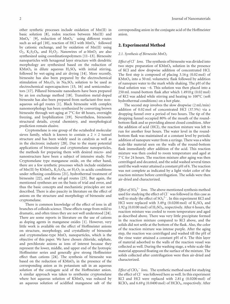
There is common knowledge of the effect of ions in allbranches of colloids science. These effects range from mild todramatic, and often times they are not well understood [24].There are some reports in literature on the use of cationsas doping agents in manganese oxides [1]. However, verylittle work is available on the effect of Hoffmeister anionson structure, morphology, and crystallinity of birnessiteand cryptomelane-type MnO2 nanoparticles, which is theobjective of this paper. We have chosen chloride, sulphate,and perchlorate anions as ions of interest because theyrepresent the lower, middle, and upper end of the lyotropicHoffmeister series and generally give strong Hoffmeistereffect than cations [24]. The synthesis of birnessite wasbased on the reduction of KMnO4 in the presence of thecorresponding anion as its potassium salt in an aqueoussolution of the conjugate acid of the Hoffmeister anion.A similar approach was taken to synthesize cryptomelanewhere hot aqueous solution of KMnO4 was reduced byan aqueous solution of acidified manganese salt of the
corresponding anion in the conjugate acid of the Hoffmeisteranion.
2. Experimental Method
2.1. Synthesis of Birnessite MnO2
Effect of Cl− Ions. The synthesis of birnessite was divided intotwo steps: preparation of KMnO4 solution in the presenceof KCl and slow dropwise addition of concentrated HCl.The first step is composed of placing 3.16 g (0.02 mol) ofKMnO4 into a 50 mL volumetric flask followed by additionof nanopure water to the mark while shaking. The pH of thefinal solution was ∼6. This solution was then placed into a250 mL round-bottom flask after which 1.4910 g (0.02 mol)of KCl was added while stirring and heating to 100oC (softhydrothermal conditions) on a hot plate.
The second step involves the slow dropwise (2 mL/min)addition of 0.02 mol of concentrated HCl (37.5%) via adropping funnel over a period of two hours. The tip of thedropping funnel occupied 80% of the mouth of the round-bottom flask and so providing almost closed condition. Afterthe addition of acid (HCl), the reaction mixture was left torun for another four hours. The water level in the round-bottom flask was maintained at a constant level by periodicaddition of nanopure water from a syringe. There was a whitescale-like material seen on the walls of the round-bottomflask immediately after addition of the acid. This reactionmixture was then cooled to room temperature and aged at7◦C for 24 hours. The reaction mixture after aging was thencentrifuged and decanted, and the solid washed several timesuntil the wash water attained a constant pH of 6. The reactionwas not complete as indicated by a light violet color of thereaction mixture before centrifugation. The solids were thenair-dried and characterized.
Effect of SO42− Ions. The above mentioned synthesis method
used for studying the effect of Cl− was followed in this case aswell to study the effect of SO4
2−. In this experiment KCl andHCl were replaced with 3.49 g (0.0200 mol) of K2SO4 and1.92 g (0.0100 mol) of H2SO4, respectively. After 6 hours, thereaction mixture was cooled to room temperature and agedas described above. There was very little precipitate formedin the reaction mixture compared to KCl above, and thesolids did not settle at the bottom of the flask. Also, the colorof the reaction mixture was intense purple. After the agingstep, the reaction was centrifuged and washed till the pH ofthe rinse water attained a constant pH of 6. The thin layerof material adsorbed to the walls of the reaction vessel wascollected as well. During the washing stage, a white scale-likematerial appeared floating on the surface of the mixture. Thesolids collected after centrifugation were then air-dried andcharacterized.
Effect of ClO4− Ions. The synthetic method used for studying
the effect of Cl− was followed here as well. In this experimentKCl and HCl were replaced with 2.81 g (0.0200 mol) ofKClO4 and 6.69 g (0.0400 mol) of HClO4, respectively. After

Journal of Nanomaterials 3
6 hours of reaction under soft hydrothermal conditions,the reaction mixture was cooled to room temperature andaged as described above. There was much more of the blackprecipitate formed compared to the synthesis in the presenceof SO4
2− anion, and the solids formed completely settled atthe bottom of the flask. The supernatant liquid was colorlessindicating that the reaction was completed. The solids werethen centrifuged, washed until the wash water attained aconstant pH of 5, then air-dried and characterized.
2.2. Synthesis of Cryptomelane MnO2
Effect of Cl−. The synthesis of cryptomelane was dividedinto two steps: preparation of KMnO4 solution followed byslow dropwise addition of acidified (HCl) MnCl2solution.The first step is composed of placing 2.76 g (0.0175 mol) ofKMnO4 into a 50 mL volumetric flask followed by additionof nanopure water to the mark while shaking. The pH ofthe final solution was ∼6. This solution was then placedinto a 250 mL round-bottom flask and heated to 65◦C (softhydrothermal conditions) on a hot plate under stirring.
The second step involves the slow dropwise (2 mL/min)addition of a solution of 0.025 mol (4.95 g) of MnCl2 •4H2O in HCl (2 M, 9.72 g in 50 mL water), while stirring,via a separatory funnel over a period of two hours. Thetip of the separatory funnel occupied 80% of the mouthof the round-bottom flask and so providing almost closedcondition. Solids formed immediately upon addition ofacidified MnCl2. The temperature of the reaction mixturewas increased to 85◦C and maintained for 20 minutes. Thewater level in the round-bottom flask was maintained at aconstant level by periodic addition of nanopure water froma syringe. The reaction mixture was then cooled to roomtemperature and aged at 7◦C for 48 hours. The mixture afteraging was then centrifuged, decanted and the solid washedseveral times until the wash water attained a constant pH of4. The pH of the first rinse water was ∼1, and the washingcontinued till the rinse water attained a constant pH of 4.The solids after the rinse step showed a clay-like consistency.The reaction was complete as indicated by a colorless of thereaction mixture before centrifugation. The solids were thenair-dried and characterized.
Effect of SO42− Ions. The procedure used above for studying
the effect of Cl− was used here. Here MnCl2 and HClwere replaced by 4.23 g (0.0250 mol) of MnSO4 and 10.15 g(2.00 M) of H2SO4, respectively. The reaction was completeas indicated by the colorless supernatant liquid at the endof the reaction. After the aging step, the mixture wascentrifuged; the solids rinsed as described above, air-dried,and characterized.
Effect of ClO4−. The procedure used for studying the effect
of Cl− was used here. Here MnCl2 and HCl were replacedby 6.35 g (0.0250 mol) of MnClO4 and 16.74 g (2.00 M) ofHClO4 (60.0%), respectively. The reaction rate was not asfast compared to the chloride or sulfate reaction as indicatedby the slow disappearance of the permanganate purple
color. However, the reaction was complete as indicated bythe colorless transparent liquid observed at the end of thereaction. After the aging step, the mixture was centrifuged,and the solids rinsed as described above. During the rinsestep, there was some scale-like white material floating onthe surface of the wash liquid. The consistency of theisolated material was more like birnessite than the clay-likeconsistency of cryptomelane. Isolated product was then air-dried and characterized.
2.3. Characterization Techniques. The manganese oxideswere characterized by X-ray powder diffraction (XRD) usinga PANalytical X’Pert PRO X-ray diffractometer with a CuKα
radiation (40 KV, 40 mA) and an X’Celerator solid statedetector. The samples were prepared on silicon sample holderby suspending the sample in ethanol to form slurry. Thepatterns were recorded at room temperature with stepsizesof 0.008◦, 2θ, and 50 seconds per step.
Transmission electron microscopy (TEM) and high-resolution transmission electron microscopy (HRTEM)images were obtained with a Tecnai G2 F30 S-Twin TEMinstrument. The TEM operates at 300 KV using a fieldemission gun in Schottky mode as an electron source. Thesamples for TEM analysis were prepared by placing 3 mg ofthe air-dried solid manganese oxide in 10 mL of 2-propanol,and sonication for 5 minutes for homogeneity. One drop ofthe slurry was deposited on a holey-carbon-coated coppergrid for analysis.
3. Results and Discussion
In the birnessite synthesis, the effect of Hoffmeister series wasevident in the extent of reaction/product yield. Sulfate whichis at the left end of the lyotropic Hoffmeister series was lesseffective compared to chloride which is in the middle of theseries which was less effective than perchlorate which is in theright end of the series in reducing potassium permanganate.In the cryptomelane synthesis it was noted that the rate ofreaction is affected by anions as sulfate was faster reducingthan chloride which was faster than perchlorate. Thoughthe reduction was complete in the cases of all three anions,nature of the product varied from a clay-like consistencyto granular form suggesting a structure and morphologychange.
3.1. X-ray Powder Diffraction
3.1.1. Birnessite Structure. Figure 1 shows the XRD patternsfor birnessite material synthesized in the presence of chloride(a), perchlorate (b), and sulfate (c) ions. The observeddiffraction patterns were then compared with the JCPDS-ICDD File Card [25, 26] to confirm the proper phaseformation of the birnessite structure. The peaks of thediffraction pattern are identified to originate from (001),(002), (111), and (005) reflections. This pattern closelyreflects the monoclinic crystal structure with C2/m spacegroup [25]. It has been observed that the peak intensityand sharpness is the maximum for the samples synthesized

4 Journal of Nanomaterials
1.41
Å (
005)3.
6 Å
(00
2)
7.2
Å (
001)
Birnessite sample: chloride ion treated
2.44
Å (
−11
1)
908070605040302010
Inte
nsi
ty(a
.u.)
(a)
1.41
Å (
005)
2.44
Å (
−11
1)
3.62
Å (
002)
7.19
Å (
001)
Birnessite sample: perchlorate ion treated
908070605040302010
Inte
nsi
ty(a
.u.)
(b)
1.41
Å (
005)
2.44
Å (
−11
1)
3.64
Å (
002)7.
26 Å
(00
1)
Birnessite sample: sulphate ion treated
908070605040302010
2θ (deg)
Inte
nsi
ty(a
.u.)
(c)
Figure 1: XRD spectra of (a) Cl− - treated birnessite nanomaterial,(b) ClO4
− - treated birnessite nanomaterial, and (c) SO4−2 - treated
birnessite nanomaterial.
6543210
−4
−2
0
2
4
βco
sθ/λ
Birnessite-chloridecrystallite size = 12.38 nmstrain = 4.74 ×10⁻³
(a)
6543210
−4
−2
0
2
4
βco
sθ/λ
Birnessite-perchloratecrystallite size = 7.81 nmstrain = 8.58 ×10⁻³
(b)
6543210
sin θ/λ
−2
−1
0
1
2
βco
sθ/λ
Birnessite-sulphatecrystallite size = 6.71 nmstrain = 8.02 ×10⁻³
(c)
Figure 2: Size-Strain analyses of birnessite samples treated under(a) Cl−, (b) ClO4
−, and (c) SO4−2 ions. The size-strain data are
obtained from the corresponding XRD data furnished in Figure 1.
Journal of Nanomaterials 5
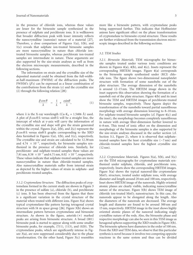
in the presence of chloride ions, whereas these valuesare lesser for the birnessite sample synthesized in thepresence of sulphate and perchlorate ions. It is wellknownthat broader diffraction peak with lesser intensity reflectsthe nanocrystalline (nanosize) nature of a material [27].Therefore, a close comparison of Figures 1(a), 1(b), and1(c) reveals that sulphate ion-treated birnessite samplesare more nanocrystalline in nature than chloride ion-treated birnessite samples, whereas perchlorate ion-treatedsamples are intermediate in nature. These arguments arealso supported by the size-strain analyses as well as fromthe electron microscopic measurements, described in thefollowing sections.
The information on strain and the crystallite size of thedeposited material could be obtained from the full-width-at-half-maximum (FWHM) of the diffraction peaks. TheFWHM’s (β’s) can be expressed as a linear combination ofthe contributions from the strain (ε) and the crystallite size(L) through the following relation [28]:
β cos θλ
= 1L
+εsinθλ
, (1)
where λ is the X-ray wavelength (Cu-Kα = 1.5406 A) used.A plot of β cos θ/λ versus sinθ/λ will be a straight line, theintercept of which at y-axis will carry the information ofthe crystallite size and slope will give the strain generatedwithin the crystal. Figures 2(a), 2(b), and 2(c) represent theβ cos θ/λ versus sinθ/λ graphs corresponding to the XRDdata furnished in Figures 1(a), 1(b), and 1(c), respectively.The crystallite size and strain are calculated to be 12.38 nmand 4.74 × 10⁻³, respectively, for birnessite samples syn-thesized in the presence of chloride ions. Similarly, forperchlorate- and sulphate-treated samples these values are7.81 nm, 8.58 ×10⁻³ and 6.71 nm, 8.02 ×10⁻³, respectively.These values indicate that sulphate-treated samples are morenanocrystalline in nature than chloride-treated samples.Also nanocrystalline materials suffer from internal strainas depicted by the higher values of strain in sulphate- andperchlorate-treated samples.
3.1.2. Cryptomelane Structure. The diffraction peaks of cryp-tomelane formed in the current study are shown in Figure 3in the presence of sulfate (a), chloride (b), and perchlorate(c) ions. It has been observed from the figures that thereis a significant variation in the crystal structure of thematerial when treated with different ions. Figure 3(a) showstypical cryptomelane-like pattern having tetragonal crystalstructure with I4 m space group [26]. Figure 3(b) shows anintermediate pattern between cryptomelane and birnessitestructure. As shown in the figure, asterisk-(∗) markedpeaks are arising from birnessite structure. A broad (001)birnessite peak is started to appear along with some smallerbirnessite peaks, for example, (111), (113), and (020). Thecryptomelane peaks, which are significantly intense in Fig-ure 3(a), are now suppressed considerably due to the phasetransformation. On the other hand, Figure 3(c) resembles
more like a birnessite pattern, with cryptomelane peaksbeing suppressed further. This indicates that Hoffmeisteranions have significant effect on the phase transformationof cryptomelane to birnessite crystal structure. These resultsare further evidenced from the transmission electron micro-scopic images described in the following sections.
3.2. TEM Studies
3.2.1. Birnessite Materials. TEM micrographs for birnes-site samples treated under various ionic conditions areshown in Figures 4(a), 4(b), and 4(c). Insets represent thecorresponding HRTEM images. Figure 4(a) correspondsto the birnessite sample synthesized under (KCl) chlo-ride ions. The figure shows two-dimensional nanoplateletstructure with formation of some nanobelts out of theplate structure. The average dimension of the nanobeltsis around 12–15 nm. The HRTEM image shown in theinset supports this observation showing the formation of ananobelt out of the plate structure. Figure 4(b) and its insetshow the TEM and HRTEM images of perchlorate-treatedbirnessite samples, respectively. These figures depict thetransformation of the nanobelts toward partial nanofibrousmorphology with average dimension lowering to 8–10 nm.For sulphate-treated birnessite samples (cf. Figure 4(c) andthe inset), the morphology becomes completely nanofibrousin nature with average fiber-dimension further lowered to6–8 nm. This trend of ionic effect on the dimension andmorphology of the birnessite samples is also supported bythe size-strain analyses discussed in the earlier section (cf.Section 3.1, Figure 2), where it is observed that sulphate-treated samples have the least crystallite size (∼7 nm) andchloride-treated samples have the highest crystallite size(12 nm).
3.2.2. Cryptomelane Materials. Figures 5(a), 5(b), and 5(c)are the TEM micrographs for cryptomelane materials syn-thesized under sulphate, chloride, and perchlorate ions,respectively. Insets show the corresponding HRTEM images.Figure 5(a) shows the typical nanorod-like cryptomelaneMnO2 structure, treated under sulphate ions, with averagediameter and length around 20 nm and 100 nm, respectively.Inset shows HRTEM image of the nanorods. Highly orientedatomic planes are clearly visible, indicating nanocrystallinenature of the structure. Figure 5(b) shows TEM image ofchloride ion-treated MnO2 nanostructure. In this case, thenanorods appear to be elongated along the length. Alsothe diameters of the nanorods are decreased. The averagelength and diameter are found to be around 300 nm and15 nm, respectively. HRTEM image in the inset shows highlyoriented atomic planes of the nanorod indicating singlycrystalline nature of the rods. Also, the birnessite phase andrespective morphology can also be seen in this TEM image ashexagonal spheres supporting the XRD results in Figure 3(b).The birnessite spheres have diameters in the order of 100 nm.From the XRD and TEM data, we observe that this particularsynthesis is novel because it involves two competing oppositereactions in the same system and thus can be divided

6 Journal of Nanomaterials
1.16
Å (
660)
1.22
Å (
721)
1.29
Å (
312)
1.34
Å (
451)
1.42
Å (
002)
1.53
Å (
521)
1.64
Å (
600)
1.83
Å (
411)
2.15
Å (
301)
2.39
Å (
121)
3.10
Å (
310)
4.91
Å (
200)
6.94
Å (
110)
908070605040302010
MnO2 structure: SO−24 treated
dominant phase-cryptomelaneIn
ten
sity
(a.u
.)
(a)
1.83
Å (
411)
2.15
Å (
301)
2.4
Å (
121)
3.1
Å (
310)
4.91
Å (
200)
908070605040302010
Inte
nsi
ty(a
.u.)
∗arising from birnessite phase
MnO2 structure: Cl−1 treatedphase-mixture ofcryptomelane and birnessite
7.05
A(0
01)∗
2.41
A(-
111)∗
1.63
A(1
13)∗
1.42
A(0
20)∗
(b)
908070605040302010
2θ (deg)
Inte
nsi
ty(a
.u.)
∗arising from birnessite phase
MnO2 structure: ClO−14 treated
dominant phase-birnessite
7.05 A (001)∗
4.91
A(2
00)
3.55
A(0
02)∗
3.47
A(2
20)
2.41
A(-
111)∗
2.13
A(1
12)∗
1.64
A(1
13)∗
1.42
A(0
20)∗
(c)
Figure 3: XRD patterns of MnO2 nanostructures showing (a) a dominant cryptomelane phase under SO4−2 treatment, (b) a mixed phase
of cryptomelane and birnessite under Cl− treatment, and (c) a dominant birnessite phase under ClO4− treatment.
5 nm
50 nm
(a)
5 nm
50 nm
(b)
5 nm
50 nm
(c)
Figure 4: TEM micrographs of (a) Cl−, treated birnessite MnO2 nanostructure, (b) ClO4−, treated birnessite MnO2 nanostructure, and (c)
SO4−2 -treated birnessite MnO2 nanostructure. Insets show the corresponding HRTEM images.

Journal of Nanomaterials 7
5 nm
50 nm
(a)
5 nm
50 nm
(b)
5 nm
50 nm
(c)
Figure 5: TEM micrographs of (a) SO4−2-treated cryptomelane MnO2 nanostructure, (b) Cl−, treated mixed phase cryptomelane and
birnessite MnO2 nanostructure, and (c) ClO4− -treated dominant birnessite MnO2 nanostructure. Insets show the corresponding HRTEM
images.
into three steps: formation of cryptomelane by oxidation,formation of birnessite by reduction, and interconversion ofcryptomelane to birnessite. The formation of cryptomelaneis by the oxidation of Mn(II) to Mn(IV) by permanganateat pH 6. The formation of birnessite is via reduction ofMn(VII) (from permanganate) to Mn(IV) by HCl at pH3. Note that the Cl− ions present at pH 6, due to theuse of MnCl2, do not participate in the reaction until thepH is lowered, and thus the formation of the needles ofcryptomelane is entirely due to the oxidation of Mn(II).The transformation of cryptomelane to birnessite is both,simultaneously, a chemical process and a physical process:the first guided by the redox reaction between MnO4
−,Mn(II) and Cl−, and the second guided by flexibility inthe developmental process and the osmotic pressure. Adistortion in the development of the subunits due to theanions present during synthesis promotes either needle-likeor curved sheet morphologies both of which can be seenin TEM images. Figure 5(c) represents the TEM image ofperchlorate-treated MnO2 nanomaterials. From the image, itappears that the morphology of the material has transformedto nanofibrous structure which more resembles to birnessiteMnO2, as shown in Figure 4(c). The average dimension of thenanofibers is calculated to be around 8 nm. Inset shows theHRTEM image of it. These images clearly supports the find-ings obtained in XRD data indicating that the Hoffmeisteranions have profound effect on the phase transformation ofMnO2 nanostructures from cryptomelane phase to birnessitephase for sulphate to perchlorate treatments. The chemistryand detailed mechanism behind this phase transformationat low pH is under investigation. However, we speculate thatthe formation of these intriguing morphologies may be dueto structural control of crystal growth guided by osmoticpressure. The methods reported here are highly reproducibleif the synthetic parameters such as temperature, stirring rateand speed of addition of one reactant into another are strictlycontrolled.
4. Conclusion
Birnessite structural details, crystal chemistry, and morpho-logical prediction remain an enigma. This is due, in part,to the fact that the foundation of most of the synthesismentioned in literature was based on trial and error and thuslack-basic concepts and mechanistic principles that wouldallow preparation of nanomaterials with desired size, shape,and morphology in a control way.
A new understanding of the influence of the counterions on the morphology of the produced nanomaterialis provided by studying the effect of Hoffmeister anionsCl−, SO4
−2, and ClO4− on the structure and morphology
of birnessite and cryptomelane-type manganese dioxidenanostructures. The Hoffmeister anion effect is first seen inthe production of birnessite with different morphologies andcrystallite sizes. The same effect was observed in the pro-duction of cryptomelane where a mixture of cryptomelaneand birnessite was obtained under MnCl2 and HCl involvingtwo competing opposite redox reactions. The conversion ofcryptomelane to birnessite is accomplished by controlling theredox reaction between MnO4
−, Mn(II), and HCl.
Acknowledgment
This work is supported, in part, by a visiting fundingto SQ from the key laboratory of the Three GorgesReservoir Region’s Eco-Environment, Ministry of Education,Chongqing University, China.
References
[1] I. Djerdj, D. Arcon, Z. Jaglicic, and M. Niederberger, “Non-aqueous synthesis of metal oxide nanoparticles: short reviewand doped titanium dioxide as case study for the preparationof transition metal-doped oxide nanoparticles,” Journal ofSolid State Chemistry, vol. 181, no. 7, pp. 1571–1581, 2008.

8 Journal of Nanomaterials
[2] A. R. Armstrong and P. G. Bruce, “Synthesis of layeredLiMnO2 as an electrode for rechargeable lithium batteries,”Nature, vol. 381, no. 6582, pp. 499–500, 1996.
[3] S. L. Brock, N. Duan, Z. R. Tian, O. Giraldo, H. Zhou, andS. L. Suib, “A review of porous manganese oxide materials,”Chemistry of Materials, vol. 10, no. 10, pp. 2619–2628, 1998.
[4] Q. Feng, H. Kanoh, and K. Ooi, “Manganese oxide porouscrystals,” Journal of Materials Chemistry, vol. 9, no. 2, pp. 319–333, 1999.
[5] R. Ma, Y. Bando, L. Zhang, and T. Sasaki, “Layered MnO2
nanobelts: hydrothermal synthesis and electrochemical mea-surements,” Advanced Materials, vol. 16, no. 11, pp. 918–922,2004.
[6] C. Calvert, R. Joesten, K. Ngala, et al., “Synthesis, characteri-zation, and rietveld refinement of tungsten-framework-dopedporous manganese oxide (K-OMS-2) material,” Chemistry ofMaterials, vol. 20, no. 20, pp. 6382–6388, 2008.
[7] R. M. McKenzie, “The synthesis of birnessite, cryptomelane,and some other oxides and hydroxides of manganese,” Miner-alogical Magazine, vol. 38, pp. 493–502, 1971.
[8] D. C. Golden, C. C. Chen, and J. B. Dixon, “Synthesis oftodorokite,” Science, vol. 231, no. 4739, pp. 717–719, 1986.
[9] J. Luo and S. L. Suib, “Formation and transformation of meso-porous and layered manganese oxides in the presence of long-chain ammonium hydroxides,” Chemical Communications, no.11, pp. 1031–1032, 1997.
[10] J. Cai, J. Liu, and S. L. Suib, “Preparative parameters andframework dopant effects in the synthesis of layer-structurebirnessite by air oxidation,” Chemistry of Materials, vol. 14, no.5, pp. 2071–2077, 2002.
[11] T. D. Xiao, E. R. Strutt, M. Benaissa, H. Chen, and B. H.Kear, “Synthesis of high active-site density nanofibrous MnO2-base materials with enhanced permeabilities,” NanostructuredMaterials, vol. 10, no. 6, pp. 1051–1061, 1998.
[12] Y. Xiong, Y. Xie, Z. Li, and C. Wu, “Growth of well-alignedγ-MnO2 monocrystalline nanowires through a coordination-polymer-precursor route,” Chemistry: A European Journal, vol.9, no. 7, pp. 1645–1651, 2003.
[13] X. Wang and Y. Li, “Selected-control hydrothermal synthesisof α- and β-MnO2 single crystal nanowires,” Journal of theAmerican Chemical Society, vol. 124, no. 12, pp. 2880–2881,2002.
[14] M. A. Cheney, P. K. Bhowmik, S. Moriuchi, M. Villalobos, S.Qian, and S. W. Joo, “The effect of stirring on the morphologyof birnessite nanoparticles,” Journal of Nanomaterials, vol.2008, Article ID 168716, 9 pages, 2008.
[15] S. Komaba, A. Ogata, and T. Tsuchikawa, “Enhanced superca-pacitive behaviors of birnessite,” Electrochemistry Communica-tions, vol. 10, no. 10, pp. 1435–1437, 2008.
[16] L. Athouel, F. Moser, R. Dugas, O. Crosnier, D. Belanger,and T. Brousse, “Variation of the MnO2 birnessite structureupon charge/discharge in an electrochemical supercapacitorelectrode in aqueous Na2SO4 electrolyte,” The Journal ofPhysical Chemistry C, vol. 112, no. 18, pp. 7270–7277, 2008.
[17] N. Larabi-Gruet, S. Peulon, A. Lacroix, and A. Chausse, “Stud-ies of electrodeposition from Mn(II) species of thin layersof birnessite onto transparent semiconductor,” ElectrochimicaActa, vol. 53, no. 24, pp. 7281–7287, 2008.
[18] B. Ma, W. Hou, Y. Han, R. Sun, and Z.-H. Liu, “Exfoliationreaction of birnessite-type manganese oxide by a host-guest electrostatic repulsion in aqueous solution,” Solid StateSciences, vol. 10, no. 2, pp. 141–147, 2008.
[19] M. A. Cheney, P. K. Bhowmik, S. Qian, S. W. Joo, W.Hou, and J. M. Okoh, “A new method of synthesizing black
birnessite nanoparticles: from brown to black birnessite withnanostructures,” Journal of Nanomaterials, vol. 2008, ArticleID 763706, 8 pages, 2008.
[20] S. L. Suib, “Microporous manganese oxides,” Current Opinionin Solid State and Materials Science, vol. 3, no. 1, pp. 63–70,1998.
[21] J. C. Villegas, L. J. Garces, S. Gomez, J. P. Durand, and S. L.Suib, “Particle size control of cryptomelane nanomaterials byuse of H2O2 in acidic conditions,” Chemistry of Materials, vol.17, no. 7, pp. 1910–1918, 2005.
[22] L. D. Conde and S. L. Suib, “Catalyst nature and frequencyeffects on the oligomerization of methane via microwaveheating,” The Journal of Physical Chemistry B, vol. 107, no. 15,pp. 3663–3670, 2003.
[23] S. Ching, J. L. Roark, N. Duan, and S. L. Suib, “Sol-gel routeto the tunneled manganese oxide cryptomelane,” Chemistry ofMaterials, vol. 9, no. 3, pp. 750–754, 1997.
[24] E. Leontidiz, “Hoffmeister anion effects on surfactant self-assembly and the formation of mesoporous solids,” CurrentOpinion in Colloid & Interface Science, vol. 7, no. 1-2, pp. 81–91, 2002.
[25] Joint Committee on Powder Diffraction standards/Intern-ational Centre for Diffraction Data (JCPDS-ICDD), File Cardno. 80–1098.
[26] Joint Committee on Powder Diffraction standards/Intern-ational Centre for Diffraction Data (JCPDS-ICDD), File Cardno. 72–1982.
[27] B. D. Cullity, Elements of X-Ray Diffraction, Addison-Wesley,Reading, Mass, USA, 1978.
[28] S. B. Qadri, E. F. Skelton, D. Hsu, et al., “Size-inducedtransition-temperature reduction in nanoparticles of ZnS,”Physical Review B, vol. 60, no. 13, pp. 9191–9193, 1999.