Embed Size (px)
Citation preview
HAL Id: tel-01684234https://tel.archives-ouvertes.fr/tel-01684234
Submitted on 15 Jan 2018
HAL is a multi-disciplinary open accessarchive for the deposit and dissemination of sci-entific research documents, whether they are pub-lished or not. The documents may come fromteaching and research institutions in France orabroad, or from public or private research centers.
L’archive ouverte pluridisciplinaire HAL, estdestinée au dépôt et à la diffusion de documentsscientifiques de niveau recherche, publiés ou non,émanant des établissements d’enseignement et derecherche français ou étrangers, des laboratoirespublics ou privés.
Synthesis and biological evaluation of novel imidazolinic,thiazolinic derivatives and aminosugars
Vinicius Barros Ribeiro Da Silva
To cite this version:Vinicius Barros Ribeiro Da Silva. Synthesis and biological evaluation of novel imidazolinic, thiazolinicderivatives and aminosugars. Agricultural sciences. Universidade federal de Pernambuco (Récife,Brésil), 2017. Portuguese. �NNT : 2017GREAV022�. �tel-01684234�
THÈSE
Pour obtenir le grade de
DOCTEUR DE LA COMMUNAUTÉ UNIVERSITÉ GRENOBLE
ALPES ET UNIVERSIDADE FEDERAL DE PERNAMBUCO
préparée dans le cadre d’une cotutelle entre la Communauté Université Grenoble Alpes et Universidade Federal de Pernambuco
Spécialité Sciences du Médicament (Grenoble) et Planejamento e Síntese de Fármacos (Recife)
Arrêté ministériel : 25 mai 2016 Présentée par
« Vinícius / BARROS RIBEIRO DA SILVA » Thèse dirigée par «Maria do Carmo/ALVES DE LIMA» et «Jean-Luc/DECOUT» préparée au sein des Laboratoires « Laboratorio Pesquisa e Inovação Terapêutica (Recife)» et « Departement de Pharmacochimie Moléculaire (Grenoble)» dans les Écoles Doctorales de Chimie et Sciences du Vivant et Programa de Pós-Graduação em Ciências Farmacêuticas
Synthèse et Evaluation Biologique de Nouveaux Dérivés Imidazoliniques, Thiazoliniques et d'Aminosucres Thèse soutenue publiquement le «20 février de 2017», devant le jury composé de :
Pr., Maria do Carmo, ALVES DE LIMA (Directeur de thèse, Membre)
Pr. Vania, BERNARDES-GENISSON (Rapporteur)
Pr, Jean-Luc, DECOUT (Directeur de thèse, Membre)
Dr., Alice, KANAZAWA DELAIR (Membre)
Pr, Ricardo, OLIMPIO de MOURA (Président, Rapporteur)
Dr., Anekécia Lauro, da SILVA (Membre)
0
AGRADECIMENTOS
À Profª. Drª. Maria do Carmo Alves de Lima, minha eterna gratidão pela orientação, paciência,
confiança, conhecimento e oportunidade a mim oferecida neste percurso.
Ao Prof. Dr. Jean-Luc Decout pela oportunidade de trabalhar no Departement de Pharmacochimie
Moléculaire.
A todos do Programa de Pós-Graduação em Ciências Farmacêuticas e da Ecole Doctorale Chimie et
Sciences du Vivant, em especial aos professores e funcionários que contribuíram para enriquecer meu
conhecimento.
A todos os amigos e colegas do Laboratório de Planejamento e Síntese de Fármacos (LPSF).
Aos meus queridos colegas do DPM Antoine, Ben, Isa, Marie-Ange, Marie-Carmen, Marine, Philippe,
Romain, Serge et Yung. Em especial gostaria de agradecer a Dr. Martine Demeunynck pela ajuda e
amizade tão fundamentais no decorrer deste doutorado.
Um muito obrigado aos amigos estagiarios, doutorandos e pos-doutores que fiz no decorrer desta tese,
Daniela, David, Doudou, Emile, Emma, Florine, Jamie, Julien, Kim, Laurent, Louise, Matt, Momo,
Yasmina. Obrigado por todas a ajuda, e todas as coxinhas que fizemos juntos. Um muito obrigado
especial a Flavia, por toda a ajuda nos primeiros momentos da minha instalação a Grenoble.
Aos meus amigos de Grenoble Colin, Milena, Rodolphe, Hoi, Manon, German, Hortanse, Pedro,
Mayra, Rodrigo, Tiago, Adele, Elisa, e tantos outros que com certeza esqueci de mencionar. Obrigado
por todos os momentos que passamos juntos durante estes 3 anos.
Um agradecimento especial ao meu companheiro de vida e aventuras, François Almeras. Você foi
parte indispensavel nesta minha etapa. Obrigado pelo apoio e confiança.
Aos meus pais, Manoel e Verônica, ao meu irmão, Gustavo, à minha avó Priscila, à minha prima
Luzia pelo amor, apoio e confiança a mim proporcionados. À minha família carioca, Rosimery,
Gilmar, Maycon, Tia Celina. Amo vocês!
Aos meus bons amigos Bruno Magalhães, Clarissa Nunes, Vinícius Gouveia, Bruno Menezes,
Leonardo Evedove, Lincoln Ribeiro, Luigi Rocha, Amanda Bezerra e Elisa Bezerra; aos meus amados
primos Ricardo Braga, Salomão Barros, Rafael Barros e Kassandra Sá; às minhas amigas da
graduação Larissa Morgana, Sarah Palácio e Giovanna Medeiros. Obrigado pelo companheirismo e
por todos os momentos de apoio nas minhas inseguranças. Amo vocês!
Obrigado a todos que acreditam e acreditarem em mim.
1
LISTA DE ABREVIATURAS E SIGLAS
2-DOS 2-deoxistreptamina
2-NM 2-metilnaftil
2-PyM 2-metilpiridinil
2-QM 2-metilquinolinil
AAG Aminoglicosideos anfifilicos
Ac Acetyl
ACC Canal de Cloreto-Acetilcolina
ACh Acetilcolina
ACT artemisinin-based combination therapy
ADMET Acyclic diene metathesis polymerization
ANVISA Agência Nacional de Viigilância Sanitaria
Bn Benzyle
Boc t-Butyloxycarbonyle
Bu Butil
CBz Carboxibenzil
CCD Cromatografia em camada delgada
CEUA Comissão de Ética no Uso de Animais
CM Cross metathesis
CMI Concentração inibitoria minima
CCM Chromatographie sur Couche Mince
CMSP Células Mononucleares do Sangue Periférico
COSY Espectroscopia de correlação
DCM Diclorometano
DCPT 3-(3,5-diclorofenil)-tiazolidina-2,4-diona
DCE Dicloroetano
DL50 Dose letal mediana
DMF Dimetilformamida
DMSO-d6 Dimetilsulfoxido deuterado
DNA Ácido desoxirribonucleico
DNDi Drug for Neglected Diseases Iniciative
DPM Département de Pharmacochimie Moléculaire
DQF Departamento de Química Fundamental
DTN Doenças Tropicais Negligenciadas
EPA Environmental Protection Agency
FDA Food and Drug Administration
FIOCRUZ Fundação Oswaldo Cruz
FLAP Fingerprints for Ligands and Proteins
Fmoc 9-fluorenil-metiloxicarbonila
GABA Acido -aminobutírico
GPIT Grupo de Pesquisa em Inovação Terapêutica
GSH Glutationa
HMBC Heteronuclear multiple-bond correlation spectroscopy
-I Efeito indutivo negativo
IR Infrarouge
IV Infravermelho
LGIC superfamília de receptores Cys-loop
LPS Lipopolissacarideos bacterianos
LPSF-UFPE Laboratório de Planejamento e Síntese de Fármacos
mCPBA Acido metacloroperbenzoico
MEV Microscopia eletrônica de varredura
MIF Molecular Interaction Fields
MRSA Staphylococcus aureus resistentes à vancomicina
MS Ministério da Saude
MTT Brometo de 3-(4,5-dimetiltiazol-2-il)-2,5-difeniltetrazolium
nAChRs receptores nicotínicos pós-sinápticos de acetilcolina
Nea Neamina
NECT terapia combinada de nifurtimox e eflornitina
Nn Nonil
NOESY Nuclear Overhauser effect spectroscopy
ODM Objetivos de Desenvolvimento do Milênio
OM Membrana bacteriana externa
OMS Organização Mundial da Saude
ONU Organização das Nações Unidas
OXA Oxamniquina
PAPS 3'fosfoadenosina 5'fosfosulfato
Par Paramina
PBS Phosphate Buffered Saline
PC Componente principal
PCA Analise de Componentes Principais
P,D&I Pesquisa, Desenvolvimento e Inovação
PDKL leishmaniose cutânea pós-calazar
PEG Polietilenoglicol
PIF campos de interação farmacofórica
PGZ Pioglitazona
PPAR receptor gama proliferador ativado do peroxisoma
PZQ Praziquantel
QSAR Relação Quantitativa Estrutura Atividade
RCM Ring-closing metathesis
RGZ Rosiglitazona
RMN Ressonância Magnética Nuclear
RNA Ácido ribonucleico
RNAi RNA inibitorio
ROM Ring-opening metathesis
ROMP Ring-opening metathesis polymerization
RPM Rotação por Minuto
RPMI Roswell Park Memorial Institute Medium
SAR Relação Estrutura Atividade
SDS Sodium Dodecyl Sulfate
SFB Soro fetal bovino
SIQUIM Sistema de Informação sobre a Indústria Química
SmACC nAChR anion-selectivos
SN2 Substituição Nucleofilica de Segunda Ordem
SUS Sistema Unico de Saude
TBAOH Hidroxido de tetrabutilamônio
TBDMS t-Butildimetilsilil
t-Bu t-Butila
TFA Acido trifluoroacético
TDR Special Programme for Research & Training in Tropical Diseases

TGZ Troglitazona
TZD Tiazolidinas
UFPE Universidade Federal de Pernambuco
UGA Université Grenoble Alpes
UV Ultravioleta
VRE Enterococos resistentes à vancomicina
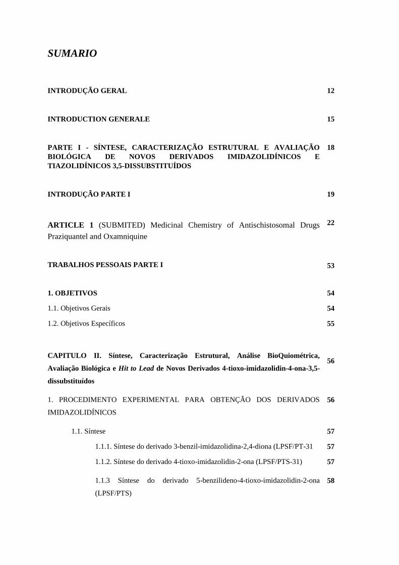
SUMARIO
INTRODUÇÃO GERAL
12
INTRODUCTION GENERALE
15
PARTE I - SÍNTESE, CARACTERIZAÇÃO ESTRUTURAL E AVALIAÇÃO
BIOLÓGICA DE NOVOS DERIVADOS IMIDAZOLIDÍNICOS E
TIAZOLIDÍNICOS 3,5-DISSUBSTITUÍDOS
18
INTRODUÇÃO PARTE I
19
ARTICLE 1 (SUBMITED) Medicinal Chemistry of Antischistosomal Drugs
Praziquantel and Oxamniquine
22
TRABALHOS PESSOAIS PARTE I
53
1. OBJETIVOS 54
1.1. Objetivos Gerais 54
1.2. Objetivos Específicos 55
CAPITULO II. Síntese, Caracterização Estrutural, Análise BioQuiométrica,
Avaliação Biológica e Hit to Lead de Novos Derivados 4-tioxo-imidazolidin-4-ona-3,5-
dissubstituídos
56
1. PROCEDIMENTO EXPERIMENTAL PARA OBTENÇÃO DOS DERIVADOS
IMIDAZOLIDÍNICOS
56
1.1. Síntese 57
1.1.1. Síntese do derivado 3-benzil-imidazolidina-2,4-diona (LPSF/PT-31 57
1.1.2. Síntese do derivado 4-tioxo-imidazolidin-2-ona (LPSF/PTS-31) 57
1.1.3 Síntese do derivado 5-benzilideno-4-tioxo-imidazolidin-2-ona
(LPSF/PTS)
58
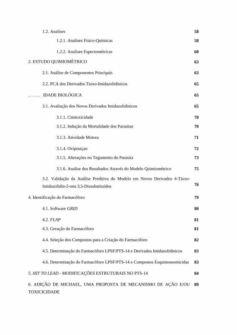
1.2. Analises 58
1.2.1. Analises Fisico-Químicas 58
1.2.2. Analises Espectométricas
2. ESTUDO QUIMIOMÉTRICO
60
63
2.1. Análise de Componentes Principais 63
2.2. PCA dos Derivados Tioxo-Imidazolidinicos 65
3. ATIVIDADE BIOLÓGICA 65
3.1. Avaliação dos Novos Derivados Imidazolidinicos 65
3.1.1. Citotoxicidade 70
3.1.2. Indução da Mortalidade dos Parasitas 70
3.1.3. Atividade Motora 71
3.1.4. Oviposiçao 72
3.1.5. Alterações no Tegumento do Parasita 73
3.1.6. Analise dos Resultados Através do Modelo Quimiométrico
3.2. Validação da Análise Preditiva do Modelo em Novos Derivados 4-Tioxo-
Imidazolidin-2-ona 3,5-Dissubstituídos
75
76
4. Identificação do Farmacóforo 79
4.1. Software GRID 80
4.2. FLAP 81
4.3. Geração do Farmacóforo 81
4.4. Seleção dos Compostos para a Criação do Farmacóforo 82
4.5. Determinação do Farmacóforo LPSF/PTS-14 e Derivados Imidazolidinicos 83
4.6. Determinação do Farmacóforo LPSF/PTS-14 e Compostos Esquistossomicidas 83
5. HIT TO LEAD - MODIFICAÇÕES ESTRUTURAIS NO PTS-14 84
6. ADIÇÃO DE MICHAEL, UMA PROPOSTA DE MECANISMO DE AÇÃO E/OU
TOXICICIDADE
89
6.1. Adição de Michael 89
6.2. Adição de Michael em Química Medicinal 91
6.2.1. Oxamniquina 91
6.3. Proposiçao do Mecanismo de Ação 92
6.4. Adição de Michael em Compostos Imidazolidinicos 93
6.5. Importância da Ligação C-3 e da Tionila em C-4 para a Adição de Michael 95
7. RECEPTORES COLINERGICOS
96
CONCLUSÕES E PERSPECTIVAS PARTE I
99
RESUME PARTIE I
103
1. INTRODUCTION 104
2. OBJECTIFS 105
2.1. Objectifs Géneraux 106
2.2. Objetifs Spécifiques 106
3. RESULTATS ET DISCUSSION 107
3.1. Synthèse des Dérivés Imidazolidiniques 107
3.2. Etude Chimiometrique 108
3.3. Evaluation de l’Activité Schistosomicide 111
3.4. Evaluation des Propriétés Prédictives du Modele PCA 111
3.5. Identification du Pharmacophore 115
3.6. Hit to Lead – Modifications Structurales sur le LPSF / PTS-14 116
3.7. Addition de Michael ; Une Proposition de Mécanisme d’Action et / ou Toxicité 117
4. CONCLUSIONS ET PERSPECTIVES 118
PARTIE II – SYNTHESE d’AMINOGLYCOSIDES AMPHIPHILES A BASE DE
NEOSAMINE 119
INTRODUCTION – PARTIE III 120
CHAPITRE I - Des Aminoglycosides Classiques et Amphiphiles et le Combat aux
Bactéries Resistants 125
1. INTRODUCTION 125
2. RELATIONS STRUCTURE-ACTIVITE (REA) ET RELATIONS STRUCTURE-
TOXICITE (RET) 126
3. AMINOGLYCOSIDES AMPHIPHILES 129
3.1. Nouveaux Aminoglycosides Amphiphiles Synthétisés au Département de
Pharmacochimie Moléculaire
130
TRAVAUX PERSONNALS – PARTIE II 136
1. CONTEXTE ET OBJECTIFS 137
1.1. Objetif Général 139
1.2. Objetifs Spécifiques 139
CHAPITRE II - Synthèse de Nouveaux Dérivés Amphiphiles des Aminoglycosides par
la Réaction de Métathèse 140
1. OBTENTION DU PRINCIPAL INTERMÉDIAIRE DE SYNTHÈSE 140
2. REACTIONS DE METATHESE ET UTILISATION DE GROUPEMENTS
PROTECTEURS 140
2.1. Groupe Protecteur Acétyle (Ac) 145
2.1.1. Synthèse des intermediaires protégés 145
2.1.2. Série 3,4-diAc - Intermediaire (25) 146
2.1.2.1. Réaction de Métathèse Croisée 2146
2.1.2.2. Homométathèse 2149
2.1.3. Série 3,4-diNn - Intermediaire (28) 150
2.1.3.1. Réaction de Métathèse Croisée 150
2.1.3.2. Homométathèse 151
2.2. Groupe Protecteur Boc 152
2.2.1. Synthèse des intermediaires 3,4-diNn 152
2.2.2. Métathèse 154
2.2.2.1. Homométathèse 154
2.2.2.2. Réaction de Métathèse Croisée 154
2.3. Groupe Protecteur Sels d’Ammonium (NH4+X-) 155
2.3.1. Synthèse des intermediaires 3,4-diNn 156
2.3.2. Métathèse 157
2.4. Groupe Protecteur Azoture (N3) 157
2.4.1. Synthèse des Intermediaires Protégés 158
2.4.2. Métathèse de la Série 3,4-diAc - Intermediaire (85) 163
2.4.2. Métathèse de la Série 3,4-diNn - Intermediaire (86) 163
2.5. Groupes Protecteurs dans l’Allylamine 165
3. REACTIONS D4OXIDATION 166
3.1. Produits Non Alkylés Série Néamine (38) 166
3.1. Produits Alkylés Série Néamine (40) 168
4. HYDROGENATION CATALITIQUE 168
4.1. Produit de homométathèse 3,4-diAc (39) 168
CONCLUSIONS ET PERSPECTIVES – PARTIE III 170
PARTIE EXPERIMENTALE - PARTIE III 177
1. MATERIAL AND METHODS 178
2. SYNTHESIS PROTOCOLS 179
CONCLUSÃO GERAL 207
CONCLUSION GENERALE 209
REFERENCIAS 211
ANEXOS 228
10
INTRODUÇÃO GERAL
11
INTRODUÇÃO GERAL
Doenças negligenciadas são doenças que não só prevalecem em condições de pobreza, mas também
contribuem para a manutenção do quadro de desigualdade, já que representam um forte entrave ao
desenvolvimento dos países.1,2 Dentre as 17 doenças classificadas como negligenciadas pela
Organização Mundial da Saude (OMS) à esquistossomose é considerada a segunda doença parasitária
mais devastadora socioeconomicamente, atrás apenas da malária, infectando mais de 200 milhões de
pessoas no mundo. 2
Compostos derivados da imidazolidina são um grupo de substâncias heterocíclicas pentagonais
possuidoras de diversas atividades biológicas.3 Estudos mostraram que os derivados imidazolidínicos
apresentam atividade leishmanicida.4 antifúngica, antimicrobiana e tripanocida. 5. As imidazolidinas
tiveram no passado um representante na clínica médica como um fármaco de ação comprovada frente
a vermes adultos de S. mansoni, o niridazol.6 A atividade esquistossomicida in vitro frente a vermes
adultos de S. mansoni de derivados imidazolidínicos vem sendo relatado em alguns estudos
publicados. 7–11
Tendo em vista os projetos desenvolvidos no Grupo de Pesquisa em Inovação Terapêutica (Gpit) com
imidazolidinas o primeiro objetivo do presente trabalho é a identificação de uma molecula promissora,
hit, através da síntese e atividade atividade esquistossomicida de 24 novos derivados imidazolidinicos.
Técnicas de analises quimiométricas serão aplicadas para auxiliar a identificação deste derivado. O
modelo quimiométrico terá sua previsibilidade testada através da síntese de novos derivados
imidazolidinicos da mesma série, que terão suas atividades previstas e em seguida confirmadas através
dos resultados biologicos encontrados. Serão realizadas novas reações químicas variando-se o
esqueleto molecular do hit para uma maior compreensão da relação estrutura química e atividade
biológica. Um modelo de farmacóforo será proposto utilizando-se o software FLAP e o modo de ação
do derivado sera avaliado através da reação de adição de Michael.
Um segundo objetivo deste trabalho é a revisão do estado da arte dos dois principais medicamentos
utilizados no tratamento da esquistossomose no Brasil a Oxaminiquina (OXA) (Mansil® – Pfizer S.A)
e o Praziquantel (PZQ) (Farmanguinhos - Fundação Oswaldo Cruz/ FIOCRUZ). O PZQ é utilizado
para tratar a esquistossomose humana, uma vez que pode eliminar os vermes adultos, durante a fase
crônica da infecção.12 A OXA é outro fármaco disponível, mas a sua utilização é limitada à infecções
por S. mansoni, não sendo eficaz contra todas as formas do parasito. 13
Serão explorados os diferentes pontos de cada um deste dois medicamentos utilizados na clinica
brasileira contra a esquistossomose, como os aspectos farmacêuticos, farmacocinéticos e
farmacodinâmicos; efeitos colaterais, casos de resistência e um estudo de relação estrutura química e
atividade biológica (SAR). No caso do PZQ todos os derivados desenvolvidos ao longo de 30 anos
estão listados neste estudo, permitindo a elaboração de um modelo preciso de relação estrutura
quimica atividade biologica (SAR).
Apesar de não ser conhecido o alvo biologico e o mecanismo de ação do PZQ, através desta revisão
será possível de se desenvolver um modelo de farmacóforo e de previsão da atividade de seus
derivados. Estes estudos ainda estão em desenvolvimentos, porém serão apresentados os primeiros
resultados em forma de um segundo artigo científico.
O terceiro objetivo desta tese é a síntese de novos aminoglicosideos anfifilicos com ação antibiotica
contra bactérias resistentes. Em um trabalho publicado em 2016 pela equipe de JL Decout du
Département de Pharmacochimie Moléculaire da Université Grenoble Alpes,14 um novo grupo de
derivados com ação antibiótica frente a bactéricas Gram (+) e Gram (-) resistentes foi identificado pelo
grupo de Pharmacochimie Moléculaire da Université Joseph Fourier, Grenoble. Estes compostos são
ampicilinas derivadas da Neamine, preparadas através do antibiótico natural Neomicina B. Tais
moléculas possuem aplicação como fármacos antibióticos e desinfectantes. O grupo obteve diversos
derivados da neamine 3’,4’-dialquil e derivados 3,4-dialquil-6-aminoglicosídeos. Os derivados
possuem na posição 1 um grupamento alila que pode ser modificado assemelhando-se ao anel I da
Neamine. As substituições podem acarretar em uma maior flexibilidade dos derivados em comparação
aos derivados da Neamine, tornando mais eficiente a interação entre os derivados e os componentes da
membrana bacteriana, como por exemplo os lipossacarídeos. Os novos derivados aminoglicosideos
serão sintetizados a partir da reação de metatese para a formação dos compostos finais. Como este
trabalho foi desenvolvido na França, em colaboração com uma equipe francesa, optou-se pela redação
destes resultados em francês. O que facilitaria a correção dos envolvidos no trabalho.
Assim, esta tese está organizada em duas partes, uma para cada objetivo,
Parte I: abordando a síntese dos derivados imidazolidinicos desenvolvidos como agentes
esquistossomicidas, assim como os estudos posteriores, como a proposição do modelo de farmacóforo.
Como anexos nesta primeira parte serão apresentados os artigos, publicados e submetidos, referentes a
esta pesquisa. Nestes artigos estão presentes as metodologias de sintese empregadas para a obtenção
dos derivados assim como informações adicionais não discutidas na presente tese.
Parte II: dedicada aos aminoglicosideos, e sua síntese através da reação de metatese. A bibliografia de
cada parte sera apresentada diretamente apos a mesma, assim como os anexos correspondentes.
Com este trabalho, espera-se contribuir para o processo de conscientização da comunidade científica
sobre a importância da pesquisa no desenvolvimento de novas imidazolidinas e aminoglicosideos
como alternativas no tratamento da esquistossomose e de infecções bacterianas, respectivamente.
Assim como colaborar para um maior entendimento do PZQ e seu mecanismo de ação a nivel
farmoquimico..
13
INTRODUCTION GENERALE
14
INTRODUCTION GENERALE
Les maladies négligées sont des maladies qui prévalent dans les pays en voie de développement et
contribuent également au maintien de l’inégalité, car elles représentent une forte barrière au
développement de pays.1,2 Parmi les 17 maladies classées comme négligées par l'Organisation
mondiale de la Santé (OMS), la schistosomiase est considérée comme la deuxième maladie parasitaire
la plus dévastatrice socio-économiquement, derrière le paludisme, infectant plus de 200 millions de
personnes dans le monde entier.2
Des composés dérivés d’imidazolidine représentent un groupe de substances hétérocycliques à cinq
chaînons possédant des activités biologiques variées; des études avec des imidazolidines ont montré
des activités sur les leishmanioses.4 antifongiques, antimicrobiennes et trypanocides.5 Les
imidazolidines ont déjà été utilisées dans la pratique médicale comme médicaments efficaces contre
les vers adultes de S. mansoni.6 L'activité in vitro du niridazol, dérivé d’imidazolidine schistosomicide
in vitro contre les vers adultes de S. mansoni, a été décrit dans la littérature. 7–11
Compte tenu des projets développés dans le Groupe de Recherche sur l'Innovation Thérapeutique
(Gpit) sur les imidazolidines, le premier objectif de cette thèse est l'identification d'une molécule
prometteuse (hit), par la synthèse et l’étude de l'activité de 24 nouveaux dérivés d’imidazolidine. Des
techniques d'analyse chimiométriques seront appliquées pour aider à l'identification de ce dérivé. Le
modèle chimiométrique aura sa prévisibilité testée grâce à la synthèse de nouveaux dérivés de la
même série, et à leurs résultats biologiques. De nouvelles réactions de modulation du squelette
moléculaire amèneront une meilleure compréhension de la relation structure chimique-activité
biologique. Un modèle de pharmacophore sera proposé à l'aide du logiciel de FLAP et un modèle du
mode d'action possible, impliquant une addition de Michael, sera évalué.
Un deuxième objectif de ce travail est une revue concise de l'état de l'art des deux principaux
médicaments utilisés dans le traitement de la schistosomiase au Brésil, l’Oxaminiquina (OXA)
(Mansil® - Pfizer SA) et le Praziquantel (PZQ) (Farmanguinhos - Oswaldo Cruz Foundation /
FIOCRUZ). Le PZQ est utilisé pour traiter la schistosomiase humaine, car il peut éliminer les vers
adultes, pendant la phase chronique de l'infection,12 l’OXA est un autre médicament disponible, mais
leur utilisation est limitée aux infections à S. mansoni, car ils ne sont pas efficaces contre toutes les
formes du parasite.13
Les différents aspects de ces deux médicaments tels que les aspects pharmaceutiques, les propriétés
pharmacocinétiques et pharmacodynamiques, les effets secondaires, les cas de résistance, ou encore la
relation entre la structure chimique et l'activité biologique (SAR). Dans le cas de PZQ, tous les dérivés
développés depuis plus de 30 ans sont répertoriés dans cette étude, permettant le développement d'un
modèle précis de SAR.
15
Bien que la cible biologique du PZQ ne soit pas connue, à travers de cette analyse de la littérature sera
possible de développer un modèle de pharmacophore et de prévoir l'activité de ses dérivés. Ces études
sont encore en développement, mais les premiers résultats sont présentés sous la forme d'un deuxième
article scientifique.
Le troisième objectif de cette thèse est la synthèse de nouveaux aminoglycosides amphiphiles avec une
activité antibiotique contre les bactéries résistantes. Dans un article publié récemment en 2016 par
l’équipe du Professeur JL Decout du Département de Pharmacochimie Moléculaire de l'Université
Grenoble-Alpes,14 de nouveaux dérivés avec une activité antibiotique contre les bactéries Gram (+) et
Gram (-) résistantes a été identifié. Ces composés amphiphiles sont issus de néamine, préparée à partir
de la néomycine B, antibiotique naturel. Ces molécules ont une application comme médicaments
antibiotiques et désinfectants. L’équipe a préparé divers dérivés de néamine 3 ', 4'-dialkyle et de 3,4-
dialkyl-6-aminoglycoside. Les dérivés portent en position 1 un groupement allyle peut être modifié.
Les substitutions peuvent se traduire par une plus grande flexibilité des dérivés par rapport à la
néamine pour une interaction plus efficace entre le dérivé et les composants des membranes
bactériennes, telles que les liposaccharides (LPS). De nouveaux dérivés aminoglycosides seront
synthétisés à partir de la réaction de métathèse. Comme ce travail a été développé en France dans le
cadre d’une co-tutelle de thèse, ces résultats seront décrits en français.
Ainsi, ce manuscrit est organisé en deux parties.
Partie I : consacrée aux imidazolidines, avec la synthèse de dérivés développés comme agents
schistosomicides, ainsi que d'autres études associées, telle que la proposition du modèle de
pharmacophore. Comme pièces jointes à cette première partie seront présentés les articles publiés et
soumis relatifs à cette recherche. Ces articles contiennent des informations supplémentaires non
discutées dans ce manuscrit.
Partie II : La dernière partie sera consacrée aux aminoglycosides, et leur synthèse par réaction de
métathèse.
Ce travail devrait contribuer à la prise de conscience de la communauté scientifique sur l'importance
de la recherche dans le développement de nouveaux dérivés imidazolidines et aminoglycosides comme
alternatives dans le traitement de la schistosomiase et des infections bactériennes respectivement, et en
plus de contribuer à une meilleure compréhension mécanisme d'action du Praziquantel (PZQ).
16
PARTE I SÍNTESE, CARACTERIZAÇÃO ESTRUTURAL E
AVALIAÇÃO BIOLÓGICA DE NOVOS DERIVADOS
IMIDAZOLIDÍNICOS E TIAZOLIDÍNICOS 3,5-
DISSUBSTITUÍDOS
INTRODUÇÃO
PARTE I
18
INTRODUÇÃO
As doenças parasitárias possuem características comuns em relação ao contexto em que ocorrem
como, as condições precárias ou inexistentes de higiene, falta de saneamento básico,
subdesenvolvimento de países, ou ainda, devido a presença de vetores que contribuem diretamente na
transmissão, como por exemplo, a esquistossomose e a leishmaniose. Os métodos de tratamento e
diagnóstico dessas doenças são antigos e inadequados e demandam investimento em pesquisa e
desenvolvimento para se tornarem mais simples e efetivos.15
Também chamadas de doenças negligenciadas, ou da pobreza, estas parasitoses são importantes
problemas de saúde pública em vários países subdesenvolvidos e em desenvolvimento, afetando mais
de dois bilhões de pessoas no mundo sendo causas substanciais de morbidade e mortalidade.16 São
assim chamadas pela falta de atenção dada pela industria farmacêutica ao seu tratamento. Atualmente,
a Organização Mundial da Saúde classifica 17 enfermidades como doenças negligenciadas: doença de
chagas, esquistossomose, leishmanioses, malária, tuberculose, dentre outras.17 Estas doenças
apresentam áreas endêmicas no Brasil e possuem uma importante taxa de mortalidade e morbidade no
país. A esquistossomose mansoni (EM CID 10 B 659), causada pelo trematódeo Schistosoma mansoni,
originalmente chamada de bilharziose, ocupa o segundo lugar das doenças de importância na Saúde
Pública, ficando atrás apenas da malária. Registros recentes da Organização Mundial de Saúde (OMS)
demonstram uma incidência de cerca de 243 milhões de pessoas infectadas pelo S. mansoni. A
esquistossomose está associada a vinte mil mortes anuais causadas pelas consequências graves da
infecção da doença, incluindo fibrose hepática e hipertensão portal em todo o mundo. 2
Os dois principais medicamentos utilizados no tratamento da esquistossomose são a Praziquantel
(PZQ) (Farmanguinhos - Fundação Oswaldo Cruz/ FIOCRUZ) e a Oxaminiquina (OXA) (Mansil® –
Pfizer S.A). 18
O PZQ é utilizado para tratar a esquistossomose humana, uma vez que pode eliminar os vermes
adultos, durante a fase crônica da infecção,19 OXA é outro fármaco disponível, mas a sua utilização é
limitada, não sendo eficaz contra todas as formas do parasito.20
Atualmente, o PZQ tem sido o fármaco de escolha para o tratamento medicamentoso no Brasil, mas
seu uso extensivo inevitavelmente culminou em baixas taxas de cura, aparecimento de isolados de S.
mansoni refratários, além de terem sido observados efeitos colaterais causados por esse
medicamento.21 Portanto, a busca de novos fármacos esquistossomicidas mais eficazes se tornou
necessária, nesse contexto, pesquisas com compostos sintéticos consistem numa alternativa viável, que
poderiam servir de moldes para o planejamento de novos fármacos.
19
Políticas de incentivo à pesquisa de novos medicamentos vêm sendo estimuladas pelo Programa
Especial para Pesquisa e Treinamento em Doenças Tropicais (Special Programme for Research and
Training in Tropical Diseases) (TDR) da Organização Mundial da Saúde e o DNDi (Drugs for
Neglected Diseases).22 O emprego de novos medicamentos na farmacoterapia é necessário para o
tratamento de doenças já existentes ou recém-identificadas ou, ainda, para a implementação de
tratamentos mais seguros e eficazes.23
Compostos derivados da imidazolidina são representados por um grupo de substâncias heterocíclicas
pentagonais possuidoras de diversas atividades biológicas.3 As imidazolidinas tiveram no passado um
representante na clínica médica como um fármaco de ação comprovada frente a vermes adultos de S.
mansoni, o niridazol.24. A busca por imidazolidinas com ação esquistossomicida in vitro frente a
vermes adultos de S. mansoni vem sendo um interesse de nosso grupo em diversos estudos
publicados.7,8,10,11,25
Tendo em vista a necessidade da busca de novos compostos esquistossomicidas e os resultados da
atividade in vitro sobre o S. Mansoni dos derivados imidazolidínicos, este trabalho tem como objetivo
a síntese de novos derivados imidazolidínicos 3,5-dissubstituídos tionados na posição 4, a avaliação da
atividade biológica frente a vermes adultos de S. mansoni, assim como a tentativa de elucidação de um
possível modelo de farmacóforo e q utilização de modelos quimiométricos para previsão da atividade.
20
ARTICLE 1 (ACCEPTED)
MEDICINAL CHEMISTRY OF ANTISCHISTOSOMAl DRUGS PRAZIQUANTEL AND
OXAMNIQUINE
Vinícius Barros Ribeiro da Silva,1,2 Bruna R. K. L. Campos,3 Jamerson Ferreira de Oliveira, 1 Jean-Luc Decout,2
and Maria do Carmo Alves de Lima1
Bioorganic & Medicinal Chemistry
CAPITULO I
MEDICINAL CHEMISTRY OF ANTISCHISTOSOMAL DRUGS: PRAZIQUANTEL
AND OXAMNIQUINE
1. INTRODUCTION
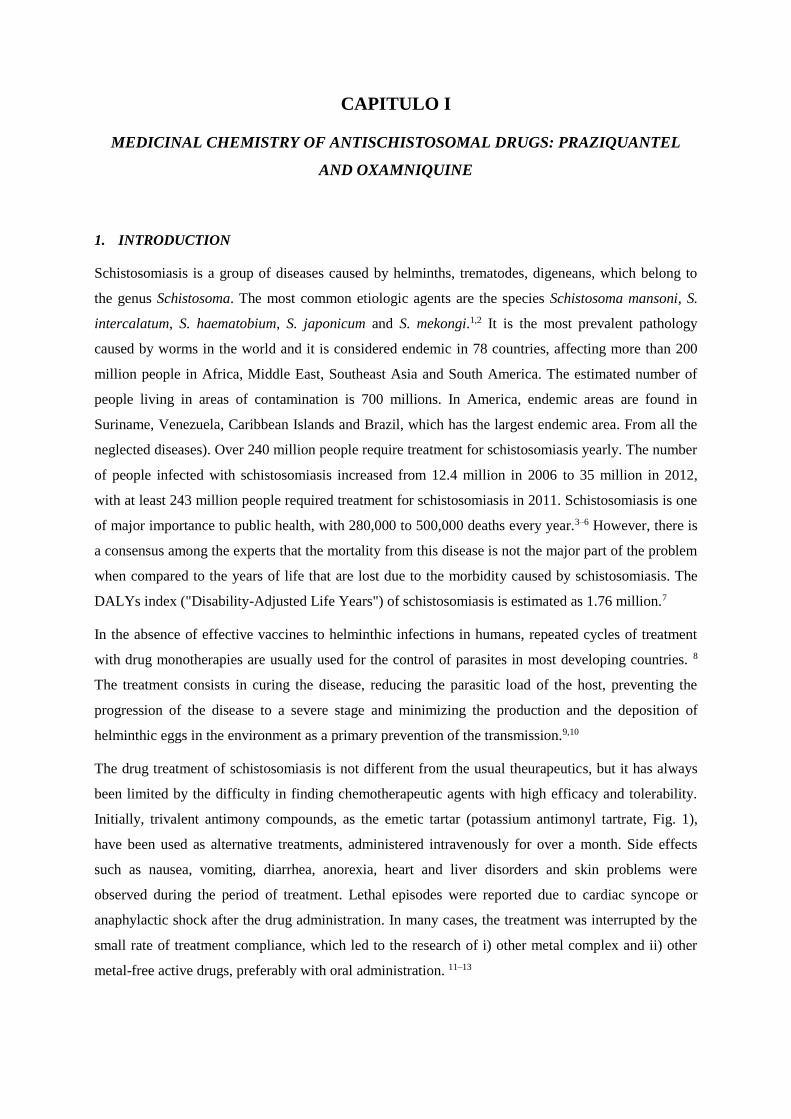
Schistosomiasis is a group of diseases caused by helminths, trematodes, digeneans, which belong to
the genus Schistosoma. The most common etiologic agents are the species Schistosoma mansoni, S.
intercalatum, S. haematobium, S. japonicum and S. mekongi.1,2 It is the most prevalent pathology
caused by worms in the world and it is considered endemic in 78 countries, affecting more than 200
million people in Africa, Middle East, Southeast Asia and South America. The estimated number of
people living in areas of contamination is 700 millions. In America, endemic areas are found in
Suriname, Venezuela, Caribbean Islands and Brazil, which has the largest endemic area. From all the
neglected diseases). Over 240 million people require treatment for schistosomiasis yearly. The number
of people infected with schistosomiasis increased from 12.4 million in 2006 to 35 million in 2012,
with at least 243 million people required treatment for schistosomiasis in 2011. Schistosomiasis is one
of major importance to public health, with 280,000 to 500,000 deaths every year.3–6 However, there is
a consensus among the experts that the mortality from this disease is not the major part of the problem
when compared to the years of life that are lost due to the morbidity caused by schistosomiasis. The
DALYs index ("Disability-Adjusted Life Years") of schistosomiasis is estimated as 1.76 million.7
In the absence of effective vaccines to helminthic infections in humans, repeated cycles of treatment
with drug monotherapies are usually used for the control of parasites in most developing countries. 8
The treatment consists in curing the disease, reducing the parasitic load of the host, preventing the
progression of the disease to a severe stage and minimizing the production and the deposition of
helminthic eggs in the environment as a primary prevention of the transmission.9,10
The drug treatment of schistosomiasis is not different from the usual theurapeutics, but it has always
been limited by the difficulty in finding chemotherapeutic agents with high efficacy and tolerability.
Initially, trivalent antimony compounds, as the emetic tartar (potassium antimonyl tartrate, Fig. 1),
have been used as alternative treatments, administered intravenously for over a month. Side effects
such as nausea, vomiting, diarrhea, anorexia, heart and liver disorders and skin problems were
observed during the period of treatment. Lethal episodes were reported due to cardiac syncope or
anaphylactic shock after the drug administration. In many cases, the treatment was interrupted by the
small rate of treatment compliance, which led to the research of i) other metal complex and ii) other
metal-free active drugs, preferably with oral administration. 11–13
The research resulted in new antimony compounds that were still administered intravenously or
intramuscularly, such as sodium antimony tartrate, sodium and antimony bis-pyrocathecol disulfonate
(or Reprodal® Stibofen®, Fig. 1), antimony and sodium thiomalate (Anthiomaline®), antimony and
sodium gluconate (Tiostam®) and sodium antimony dimercaptan (Astiban®). However, despite acting
effectively against the three main species of the genus Schistosoma that parasitize humans, the toxicity
and the side effects caused by these drugs were still intense and serious, causing the death of some
patients.11,13
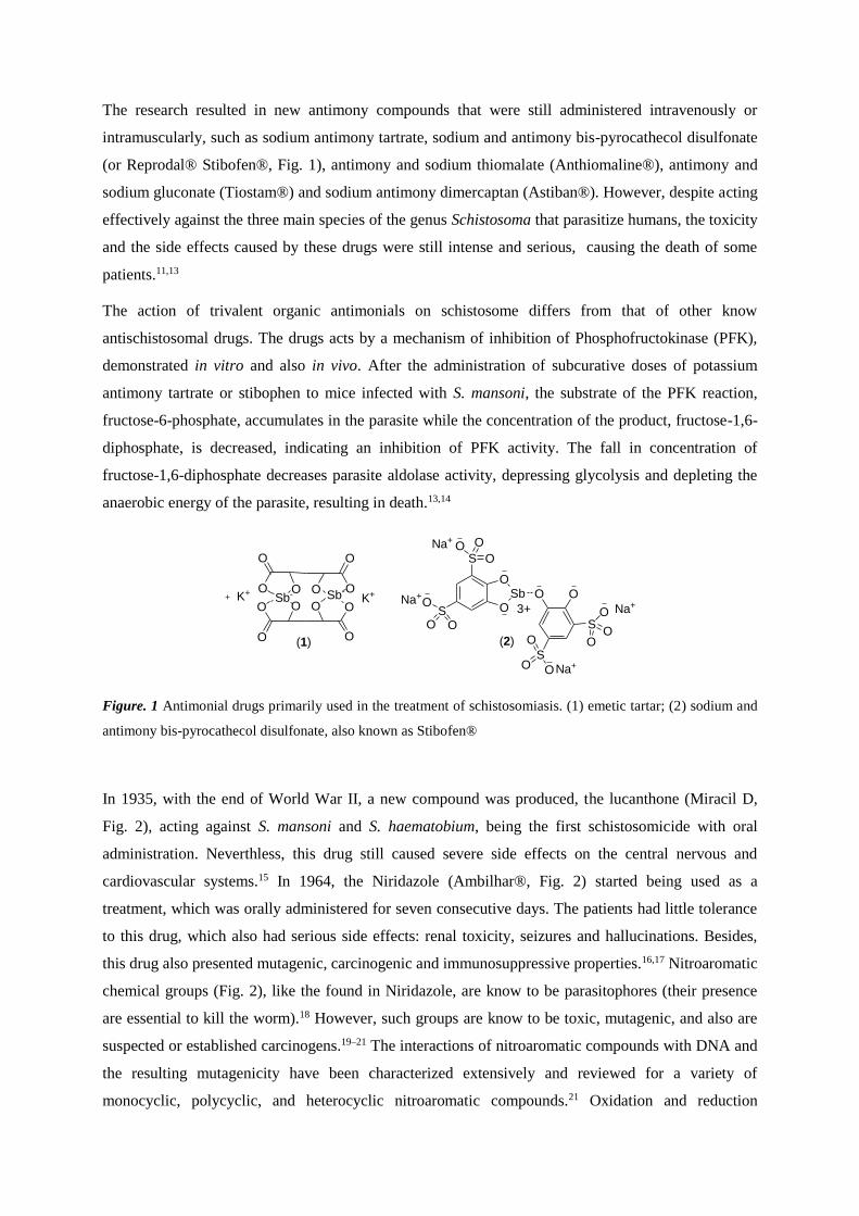
The action of trivalent organic antimonials on schistosome differs from that of other know
antischistosomal drugs. The drugs acts by a mechanism of inhibition of Phosphofructokinase (PFK),
demonstrated in vitro and also in vivo. After the administration of subcurative doses of potassium
antimony tartrate or stibophen to mice infected with S. mansoni, the substrate of the PFK reaction,
fructose-6-phosphate, accumulates in the parasite while the concentration of the product, fructose-1,6-
diphosphate, is decreased, indicating an inhibition of PFK activity. The fall in concentration of
fructose-1,6-diphosphate decreases parasite aldolase activity, depressing glycolysis and depleting the
anaerobic energy of the parasite, resulting in death.13,14
Figure. 1 Antimonial drugs primarily used in the treatment of schistosomiasis. (1) emetic tartar; (2) sodium and
antimony bis-pyrocathecol disulfonate, also known as Stibofen®
In 1935, with the end of World War II, a new compound was produced, the lucanthone (Miracil D,
Fig. 2), acting against S. mansoni and S. haematobium, being the first schistosomicide with oral
administration. Neverthless, this drug still caused severe side effects on the central nervous and
cardiovascular systems.15 In 1964, the Niridazole (Ambilhar®, Fig. 2) started being used as a
treatment, which was orally administered for seven consecutive days. The patients had little tolerance
to this drug, which also had serious side effects: renal toxicity, seizures and hallucinations. Besides,
this drug also presented mutagenic, carcinogenic and immunosuppressive properties.16,17 Nitroaromatic
chemical groups (Fig. 2), like the found in Niridazole, are know to be parasitophores (their presence
are essential to kill the worm).18 However, such groups are know to be toxic, mutagenic, and also are
suspected or established carcinogens.19–21 The interactions of nitroaromatic compounds with DNA and
the resulting mutagenicity have been characterized extensively and reviewed for a variety of
monocyclic, polycyclic, and heterocyclic nitroaromatic compounds.21 Oxidation and reduction
(1)
O
O O
O
O O
O
OO
O
OOSb- Sb-
K+K+
S
S OSb
O
O O
S
S
OO O
O
OO
O
O
O
OO
O
3+ Na+
Na+
Na+
Na+
(2)
23
products of nitroaromatic group can lead to damage in DNA and to the formation of adducts that
induce mutagenesis by misincorporation of nucleotides during DNA synthesis. 21,22
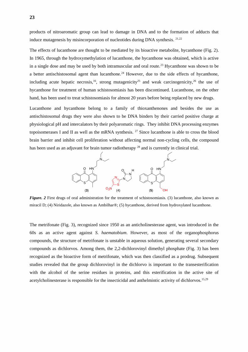
The effects of lucanthone are thought to be mediated by its bioactive metabolite, hycanthone (Fig. 2).
In 1965, through the hydroxymethylation of lucanthone, the hycanthone was obtained, which is active
in a single dose and may be used by both intramuscular and oral route.23 Hycanthone was shown to be
a better antischistosomal agent than lucanthone.24 However, due to the side effects of hycanthone,
including acute hepatic necrosis,16, strong mutagenicity25 and weak carcinogenicity,26 the use of
hycanthone for treatment of human schistosomiasis has been discontinued. Lucanthone, on the other
hand, has been used to treat schistosomiasis for almost 20 years before being replaced by new drugs.
Lucanthone and hycanthone belong to a family of thioxanthenones and besides the use as
antischistosomal drugs they were also shown to be DNA binders by their carried positive charge at
physiological pH and intercalators by their polyaromatic rings. They inhibit DNA processing enzymes
topoisomerases I and II as well as the mRNA synthesis. 27 Since lucanthone is able to cross the blood
brain barrier and inhibit cell proliferation without affecting normal non-cycling cells, the compound
has been used as an adjuvant for brain tumor radiotherapy 28 and is currently in clinical trial.
Figure. 2 First drugs of oral administration for the treatment of schistosomiasis. (3) lucanthone, also known as
miracil D; (4) Niridazole, also known as Ambilhar®; (5) hycanthone, derived from hydroxylated lucanthone.
The metrifonate (Fig. 3), recognized since 1950 as an anticholinesterase agent, was introduced in the
60s as an active agent against S. haematobium. However, as most of the organophosphorus
compounds, the structure of metrifonate is unstable in aqueous solution, generating several secondary
compounds as dichlorvos. Among them, the 2,2-dichlorovinyl dimethyl phosphate (Fig. 3) has been
recognized as the bioactive form of metrifonate, which was then classified as a prodrug. Subsequent
studies revealed that the group dichlorovinyl in the dichlorvo is important to the transesterification
with the alcohol of the serine residues in proteins, and this esterification in the active site of
acetylcholinesterase is responsible for the insecticidal and anthelmintic activity of dichlorvos.15,29
N
N
O
N
SO2N
H
S
O HN
N
S
O HN
N
OH(3) (5)(4)
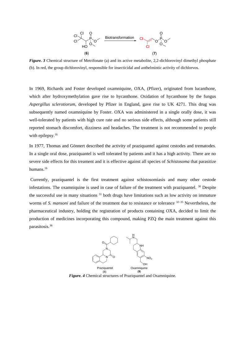
Figure. 3 Chemical structure of Metrifonate (a) and its active metabolite, 2,2-dichlorovinyl dimethyl phosphate
(b). In red, the group dichlorovinyl, responsible for insecticidal and anthelmintic activity of dichlorvos.
In 1969, Richards and Foster developed oxamniquine, OXA, (Pfizer), originated from lucanthone,
which after hydroxymethylation gave rise to hycanthone. Oxidation of hycanthone by the fungus
Aspergillus sclerotiorum, developed by Pfizer in England, gave rise to UK 4271. This drug was
subsequently named oxamniquine by Foster. OXA was administered in a single orally dose, it was
well-tolerated by patients with high cure rate and no serious side effects, although some patients still
reported stomach discomfort, dizziness and headaches. The treatment is not recommended to people
with epilepsy.16
In 1977, Thomas and Gönnert described the activity of praziquantel against cestodes and trematodes.
In a single oral dose, praziquantel is well tolerated by patients and it has a high activity. There are no
severe side effects for this treament and it is effective against all species of Schistosoma that parasitize
humans.16
Currently, praziquantel is the first treatment against schistosomiasis and many other cestode
infestations. The oxamniquine is used in case of failure of the treatment with praziquantel. 30 Despite
the successful use in many situations 31 both drugs have limitations such as low activity on immature
worms of S. mansoni and failure of the treatment due to resistance or tolerance 32–35 Nevertheless, the
pharmaceutical industry, holding the registration of products containing OXA, decided to limit the
production of medicines incorporating this compound, making PZQ the main treatment against this
parasitosis.36
Figure. 4 Chemical structures of Praziquantel and Oxamniquine.
N
N
O
O
NH
HN
OH
NO2
Praziquantel Oxamniquine
(9)(8)
Cl
Cl
OP
O
(6) (7)
BiotransformationCl
P
HO
ClCl
O
O
O O
O

25
2. PRAZIQUANTEL
In 1972, a research conducted by the industries E. Merck and Bayer A.G. selected praziquantel (Fig.
5), among more than 400 analogues tested, as the drug to be comercialized for the treatment against
schistosomiasis due to its characteristics of low toxicity, high efficacy and tolerability. This is
considered the most important development for decades in the treatment of schistosomiasis. 31,37 First
approved for veterinary treatment of schistosomiasis, its use has been expanded to treat cestode
infestations in humans in the 80s. 38–40 In 1983, new methods of synthesis have been developed to
ensure high efficiency and low cost treatment for the disease. 41
Figure. 5 Chemical structure including atom numbering of PZQ. In red, the geometrical center of the molecule.
Structurally known by the name of (±)-2-cyclohexanecarbonyl-1H,2H,3H,4H,6H,7H,11bH-
piperazino[2,1-a]isoquinolin-4-one, PZQ is a tetrahydrogenated isoquinoline pyrazine derivative with
an asymmetric center. It has a pharmacological activity much higher than the oxamniquine, which acts
only against S. mansoni, mainly against adult males. Praziquantel is effective against adults of all
species of schistosoma, but not against the young forms, and the reason is still unknown.42 Besides
being effective against all forms of Schistosoma, praziquantel shows highly effectiveness against
infestations of other species of trematodes and/or cestodes, especially Taenia solium, Taenia saginata,
Hymenolepis nana, Hymenolepis diminuta e Diphyllobothrium latum, Cysticercus bovis and
Cysticercus cellulosae, Echinochasmus fujianensis and Opisthorchis viverrini, acting against the larval
stage, immature and mature cestodes, 31,43 but not against nematodes.40
Today, PZQ is the main drug used in the treatment of all kinds of schistosomes, being also indicated
for the treatment of taeniasis, cysticercosis and infestations of Diphyllobothrium spp and Hymenolepis
nana, indicated in the list of essential drugs of WHO. 30,44,45 It is usually administered at dosages
between 40-60 mg.kg-1 and its drug efficacy, in the case of schistosomiasis, is evaluated by counting
the eggs of S. mansoni or S. japonicum in the feces by Kato-Katz smears or by filtration. In the case S.
haematobium, the drug efficacy is evaluated by the egg count in the urine. 41 All therapeutic doses
have a cure rate against S. mansoni ranging from 60 to 90%, causing a reduction of 90 to 95% in the
number of eggs, depending on the level of infection.53,66,67
87a
11a11
10
9
7
6
N 5
11b 4
3N2
1
O
O*
(8)
A. Pharmaceutical aspects of Praziquantel
Praziquantel is a white powder with bitter taste, stable under normal storage conditions. It is
practically insoluble in water but soluble in ethanol and in other organic solvents. Currently,
Praziquantel is on the market as a pill, in the dosages of 150, 500 and 600 mg, and the posology is
made according to the patient body weight.46 PZQ is classified as a Group II BCS member
(Biopharmaceutics Classification System), a drug with low solubility and high permeability in the
gastrointestinal tract (GIT), so the dissolution rate is the limiting factor of its absorption and one of the
main reasons for the high doses. The optimization of the dissolution rate of this class of drugs is,
therefore, an important and challenging aspect in the development of new formulations.47 Further
research is needed in order to improve the drug solubility and hence reduce the high doses used in the
treatment of parasitic diseases. 48
B. Pharmacokinetics of Praziquantel
According to Gonzalez-Esquivel et al. (2005),49 the apparent permeability coefficient of PZQ is 4.4 x
10-5 cm.s-1 (across Caco-2 cells), which is an indicative of its high permeability, suggesting that the
transcellular transport may be the main absorption process. In humans, peak plasma levels of 1 g/mL
of the drug in its unaltered form are obtained between 1-2h after the administration, which reflects
between 5-7% of the ingested drug. 50–54 The elimination of the drug is primarily by the kidneys
through urine, reaching a rate of 80% within 4 days, with 90% of that achieved in the first 24 hours.
The effective half-life is 1.5 hours.40,55 The absorption occurs in 75-100% of the oral dose in rats, dogs,
monkeys and humans. Tmax (the time after administration of a drug when the maximum plasma
concentration is reached) is reached after 30 to 120 minutes in animals and within 3-4 hours in
humans.56 The binding of praziquantel to plasma proteins varies from 79 to 80.5% in the range from
0.19 to 3.0 mg/mL, being the albumin binding percentage 78 to 81%. At higher plasma concentrations
(10-100 mg/mL), it was observed a reduction of 80.5 to 50% of this binding (Fig. 6).49
The major metabolite of PZQ formed by the first pass metabolism is the trans-cyclohexanol derivative
(Fig. 7), reported as 4 to 10 times less effective than PZQ against S. mansoni.40,43,57
27
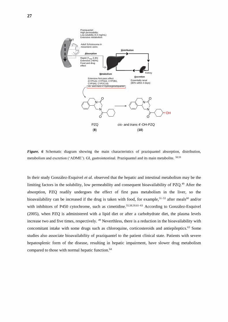
Figure. 6 Schematic diagram showing the main characteristics of praziquantel absorption, distribution,
metabolism and excretion (‘ADME’). GI, gastrointestinal. Praziquantel and its main metabolite. 58,59
In their study González-Esquivel et al. observed that the hepatic and intestinal metabolism may be the
limiting factors in the solubility, low permeability and consequent bioavailability of PZQ.49 After the
absorption, PZQ readily undergoes the effect of first pass metabolism in the liver, so the
bioavailability can be increased if the drug is taken with food, for example,51–53 after meals60 and/or
with inhibitors of P450 cytochrome, such as cimetidine.55,58,59,61–63 According to González-Esquivel
(2005), when PZQ is administered with a lipid diet or after a carbohydrate diet, the plasma levels
increase two and five times, respectively. 49 Neverthless, there is a reduction in the bioavailability with
concomitant intake with some drugs such as chloroquine, corticosteroids and antiepileptics.63 Some
studies also associate bioavailability of praziquantel to the patient clinical state. Patients with severe
hepatosplenic form of the disease, resulting in hepatic impairment, have slower drug metabolism
compared to those with normal hepatic function.64
N
N
O
ON
N
O
O
OH
GIT
Praziquantel:High permeability;Low solubility (0.4 mg/mL)Extensive metabolism
Absorption
Distribution
Metabolism
Excretion
Adult Schistosoma inmesenteric veins
Rapid (Tmax 3-4h)Extensive (>80%)Food and drug effect
Liver Kidney
Extensive first-pass effect(CYP1A2, CYP3A4, CYP2B1,CYP3A5, CYP2C19)cis- and trans-4'-hydroxypraziquantel
Essentially renal(80% within 4 days)
PZQ cis- and trans 4'-OH-PZQ
(8) (10)
C. Pharmacological Effects and Mechanism of Action
1. Adult Worms
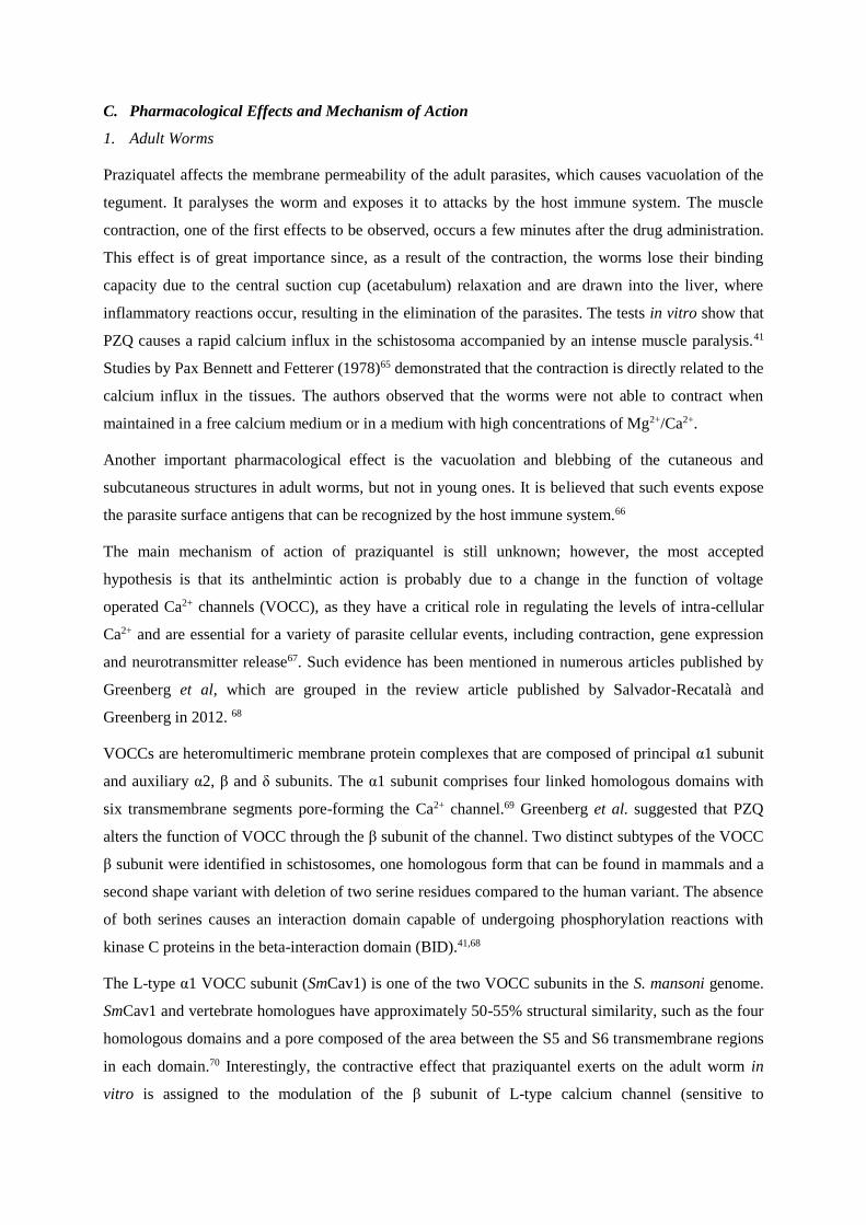
Praziquatel affects the membrane permeability of the adult parasites, which causes vacuolation of the
tegument. It paralyses the worm and exposes it to attacks by the host immune system. The muscle
contraction, one of the first effects to be observed, occurs a few minutes after the drug administration.
This effect is of great importance since, as a result of the contraction, the worms lose their binding
capacity due to the central suction cup (acetabulum) relaxation and are drawn into the liver, where
inflammatory reactions occur, resulting in the elimination of the parasites. The tests in vitro show that
PZQ causes a rapid calcium influx in the schistosoma accompanied by an intense muscle paralysis.41
Studies by Pax Bennett and Fetterer (1978)65 demonstrated that the contraction is directly related to the
calcium influx in the tissues. The authors observed that the worms were not able to contract when
maintained in a free calcium medium or in a medium with high concentrations of Mg2+/Ca2+.
Another important pharmacological effect is the vacuolation and blebbing of the cutaneous and
subcutaneous structures in adult worms, but not in young ones. It is believed that such events expose
the parasite surface antigens that can be recognized by the host immune system.66
The main mechanism of action of praziquantel is still unknown; however, the most accepted
hypothesis is that its anthelmintic action is probably due to a change in the function of voltage
operated Ca2+ channels (VOCC), as they have a critical role in regulating the levels of intra-cellular
Ca2+ and are essential for a variety of parasite cellular events, including contraction, gene expression
and neurotransmitter release67. Such evidence has been mentioned in numerous articles published by
Greenberg et al, which are grouped in the review article published by Salvador-Recatalà and
Greenberg in 2012. 68
VOCCs are heteromultimeric membrane protein complexes that are composed of principal α1 subunit
and auxiliary α2, β and δ subunits. The α1 subunit comprises four linked homologous domains with
six transmembrane segments pore-forming the Ca2+ channel.69 Greenberg et al. suggested that PZQ
alters the function of VOCC through the β subunit of the channel. Two distinct subtypes of the VOCC
β subunit were identified in schistosomes, one homologous form that can be found in mammals and a
second shape variant with deletion of two serine residues compared to the human variant. The absence
of both serines causes an interaction domain capable of undergoing phosphorylation reactions with
kinase C proteins in the beta-interaction domain (BID).41,68
The L-type α1 VOCC subunit (SmCav1) is one of the two VOCC subunits in the S. mansoni genome.
SmCav1 and vertebrate homologues have approximately 50-55% structural similarity, such as the four
homologous domains and a pore composed of the area between the S5 and S6 transmembrane regions
in each domain.70 Interestingly, the contractive effect that praziquantel exerts on the adult worm in
vitro is assigned to the modulation of the β subunit of L-type calcium channel (sensitive to
29
dihydropyridines), causing its opening.71,72 Thus, the L-type calcium channel would be the
pharmacological receptor of PZQ, at least with regard to their effect on the worm muscles. Studies by
Da Silva et Noël in 1995,73 as well as those performed by Pax Bennett et Fetterer in 1978,65 proved the
inhibition of the contractive action of praziquantel in the absence of Ca2+ in the external medium or in
the presence of verapamil.
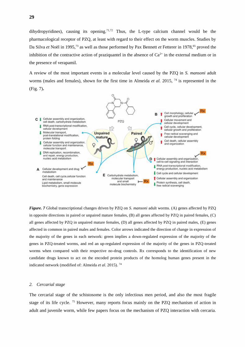
A review of the most important events in a molecular level caused by the PZQ in S. mansoni adult
worms (males and females), shown for the first time in Almeida et al. 2015, 74 is represented in the
(Fig. 7).
Figure. 7 Global transcriptional changes driven by PZQ on S. mansoni adult worms. (A) genes affected by PZQ
in opposite directions in paired or unpaired mature females, (B) all genes affected by PZQ in paired females, (C)
all genes affected by PZQ in unpaired mature females, (D) all genes affected by PZQ in paired males, (E) genes
affected in common in paired males and females. Color arrows indicated the direction of change in expression of
the majority of the genes in each network: green implies a down-regulated expression of the majority of the
genes in PZQ-treated worms, and red an up-regulated expression of the majority of the genes in PZQ-treated
worms when compared with their respective no-drug controls. Rx corresponds to the identification of new
candidate drugs known to act on the encoded protein products of the homolog human genes present in the
indicated network (modified of: Almeida et al. 2015). 74
2. Cercarial stage
The cercarial stage of the schistosome is the only infectious men period, and also the most fragile
stage of its life cycle. 75 However, many reports focus mainly on the PZQ mechanism of action in
adult and juvenile worm, while few papers focus on the mechanism of PZQ interaction with cercaria.
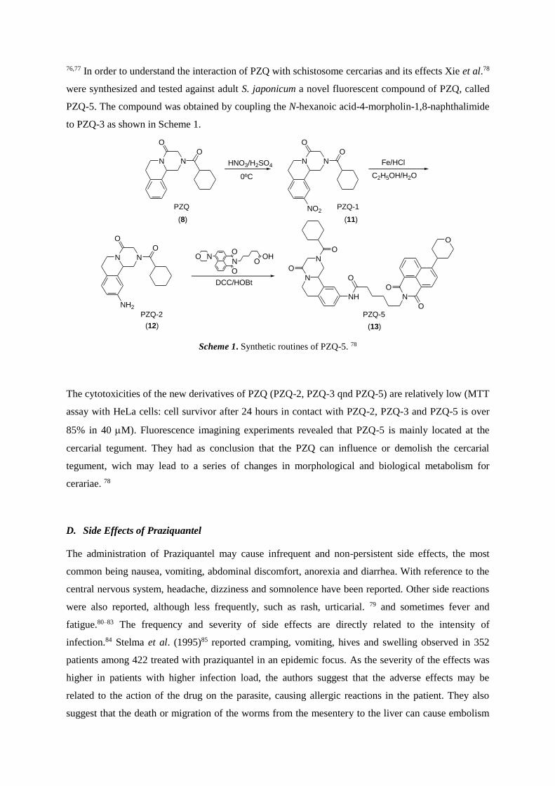
76,77 In order to understand the interaction of PZQ with schistosome cercarias and its effects Xie et al.78
were synthesized and tested against adult S. japonicum a novel fluorescent compound of PZQ, called
PZQ-5. The compound was obtained by coupling the N-hexanoic acid-4-morpholin-1,8-naphthalimide
to PZQ-3 as shown in Scheme 1.
Scheme 1. Synthetic routines of PZQ-5. 78
The cytotoxicities of the new derivatives of PZQ (PZQ-2, PZQ-3 qnd PZQ-5) are relatively low (MTT
assay with HeLa cells: cell survivor after 24 hours in contact with PZQ-2, PZQ-3 and PZQ-5 is over
85% in 40 M). Fluorescence imagining experiments revealed that PZQ-5 is mainly located at the
cercarial tegument. They had as conclusion that the PZQ can influence or demolish the cercarial
tegument, wich may lead to a series of changes in morphological and biological metabolism for
cerariae. 78
D. Side Effects of Praziquantel
The administration of Praziquantel may cause infrequent and non-persistent side effects, the most
common being nausea, vomiting, abdominal discomfort, anorexia and diarrhea. With reference to the
central nervous system, headache, dizziness and somnolence have been reported. Other side reactions
were also reported, although less frequently, such as rash, urticarial. 79 and sometimes fever and
fatigue.80–83 The frequency and severity of side effects are directly related to the intensity of
infection.84 Stelma et al. (1995)85 reported cramping, vomiting, hives and swelling observed in 352
patients among 422 treated with praziquantel in an epidemic focus. As the severity of the effects was
higher in patients with higher infection load, the authors suggest that the adverse effects may be
related to the action of the drug on the parasite, causing allergic reactions in the patient. They also
suggest that the death or migration of the worms from the mesentery to the liver can cause embolism
N N
O
O
N N
O
O
NO2
N N
O
O
NH2
N
N
O
O
NH
O
N
O
O
O
O NN O
OHO
O
HNO3/H2SO4
0ºC
Fe/HCl
C2H5OH/H2O
PZQ PZQ-1
PZQ-2 PZQ-5
DCC/HOBt
(8) (11)
(12) (13)
31
and intestinal colic. Praziquantel has a low toxicity86 and has no mutagenic, carcinogenic or
teratogenic risk. However, the contraindications for the use of any drug of the schistosomiasis
therapeutic arsenal are hepatic insufficiency and renal failure or other serious situations of clinical
decompensation, such as hepatointestinal decompensation.86,87
E. Resistance to Praziquantel
In 1971, Rogers and Bueding demonstrated for the first time the existence of S. mansoni strains that
were resistant to a schistosomiasis drug, hycanthone. Mice and hamsters infected with strains of S.
mansoni from Puerto Rico and treated with hycanthone, started to eliminate viable eggs again after
one year of treatment. The cercariae from these eggs were used to infect other mouse, in which the
adult worms were resistant to the treatment with hicantone.88
The first evidence of resistance to PZQ was registered in 1994, with Fallon and Doenhoff
experimental studies.89,90 Mice were infected using a "pool" of cercariae from four distinct geographic
regions and the animals were treated with subdoses of PZQ. After seven cycles of treatment with
increasing doses of PZQ, it was observed one strain of which 93% of adult worms survived three
doses of 300 mg/kg. This strain was resistant to PZQ, but it was sensitive to oxaminiquine, being the
case of a strain that was resistant to a specific drug.16
In 1995, it was recorded the first case of acquired resistance to praziquantel (PZQ) evidenced in
Senegal, where there was an outbreak of intestinal schistosomiasis and the treatment of patients with
40 mg.kg-1 PZQ did not achieve satisfactory results, with cure rates of 18-39%.85 Such percentage is
considered low, since the treatment with this drug has a cure rate of approximately 80-90%.79 The
dose was increased to 60 mg.kg-1 in order to obtain a therapeutic success, but the results remained
unsatisfactory.91
Melman et al. (2009) questioned the resistance, since the reinfection is very likely in endemic areas
and it can lead to misinterpretation of the cure rates, as it is not possible to know if the patient had
another exposure to the disease or not. However, advanced studies were conducted and it was actually
found a reduced susceptibility of the worms to PZQ, suggesting an important resistance to the drug.92
The mechanism of resistance to Praziquantel is not yet elucidated. An evidence for the resistance of
young worms can be found by observing the ABC transport proteins (ATP-binding cassette), involved
in the transport of toxins and xenobiotics. Kasinathan and Greenberg demonstrated that young worms
express approximately 2.5 times more transcription genes of ABC transport proteins (SmMRP1and
SMDR2) than adult worms.93 Hines-Kay et al. demonstrated a significant increase in the transcription
of genes that encode ABC transport protein genes (SMDR1, SmMRP1, SmMRP2 and SMDR3) in
young worms exposed to PZQ in vitro.94 Higher levels of parasite SMDR2 are found in females, while
SmMRP1 is mostly found in males.95 Couto at al. shown that resistant adult worms (produced from S.
mansoni-infected B. glabrata snails subjected to PZQ drug pressure) exhibit less tegumental damage
and less inhibition of the excretory system than normal parasites. 96
Until the present days, there is no clear evidence of a clinical resistance in wide range to the treatment
with PZQ. Kasinathan, Morgan and Greenberg (2010) clearly indicate that the resistance to PZQ can
be selected. It was verified that a homolog of schistosomal P-glycoprotein is upregulated in resistant
worms treated with PZQ and in young worms.93 However, it can be argued that resistence is
inevitable.
F. Structure-Activity Relationships (SAR)
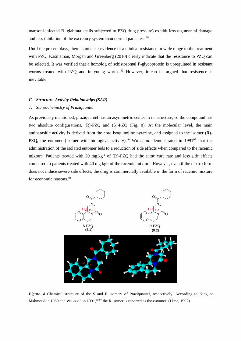
1. Stereochemistry of Praziquantel
As previously mentioned, praziquantel has an asymmetric center in its structure, so the compound has
two absolute configurations, (R)-PZQ and (S)-PZQ (Fig. 8). At the molecular level, the main
antiparasitic activity is derived from the core isoquinoline pyrazine, and assigned to the isomer (R)-
PZQ, the eutomer (isomer with biological activity).40 Wu et al. demonstrated in 199197 that the
administration of the isolated eutomer leds to a reduction of side effects when compared to the racemic
mixture. Patients treated with 20 mg.kg-1 of (R)-PZQ had the same cure rate and less side effects
compared to patients treated with 40 mg kg-1 of the racemic mixture. However, even if the dextro form
does not induce severe side effects, the drug is commercially available in the form of racemic mixture
for economic reasons.98
Figure. 8 Chemical structure of the S and R isomers of Praziquantel, respectively. According to King et
Mahmoud in 1989 and Wu et al. in 1991,40,97 the R isomer is reported as the eutomer (Lima, 1997)
N
N
11b 11b
O
O
H
N
N
O
O
H
S-PZQ R-PZQ
* *
(8.1) (8.2)
33
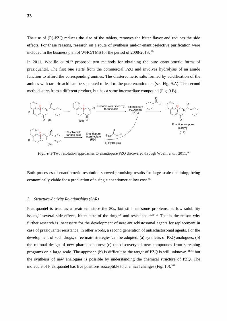
The use of (R)-PZQ reduces the size of the tablets, removes the bitter flavor and reduces the side
effects. For these reasons, research on a route of synthesis and/or enantioselective purification were
included in the business plan of WHO/TMS for the period of 2008-2013. 99
In 2011, Woelfle et al.46 proposed two methods for obtaining the pure enantiomeric forms of
praziquantel. The first one starts from the commercial PZQ and involves hydrolysis of an amide
function to afford the corresponding amines. The diastereomeric salts formed by acidification of the
amines with tartaric acid can be separated to lead to the pure enantiomers (see Fig. 9.A). The second
method starts from a different product, but has a same intermediate compound (Fig. 9.B).
Figure. 9 Two resolution approaches to enantiopure PZQ discovered through Woelfl et al., 2011.46
Both processes of enantiomeric resolution showed promising results for large scale obtaining, being
economically viable for a production of a single enantiomer at low cost.46
2. Structure-Activity Relationships (SAR)
Praziquantel is used as a treatment since the 80s, but still has some problems, as low solubility
issues,47 several side effects, bitter taste of the drug100 and resistance.16,89–91 That is the reason why
further research is necessary for the development of new antischistosomal agents for replacement in
case of praziquantel resistance, in other words, a second generation of antischistosomal agents. For the
development of such drugs, three main strategies can be adopted: (a) synthesis of PZQ analogues; (b)
the rational design of new pharmacophores; (c) the discovery of new compounds from screaning
programs on a large scale. The approach (b) is difficult as the target of PZQ is still unknown,41,44 but
the synthesis of new analogues is possible by understanding the chemical structure of PZQ. The
molecule of Praziquantel has five positions susceptible to chemical changes (Fig. 10).101
N
N
O
OH
N
N
O
HH Resolve with dibenzoyl
tartaric acidEnantiopurePZQamine
(R)-2
Cl
O
NH
NH
OH Resolve with
tartaric acid Enqntiopureintermediate
(R)-3
i) ClCl
O
ii) Hydrolysis
A
B
N
N
O
HO
R-PZQ
Enantiomere pure
(8)
(14)
(15)
(8.2)
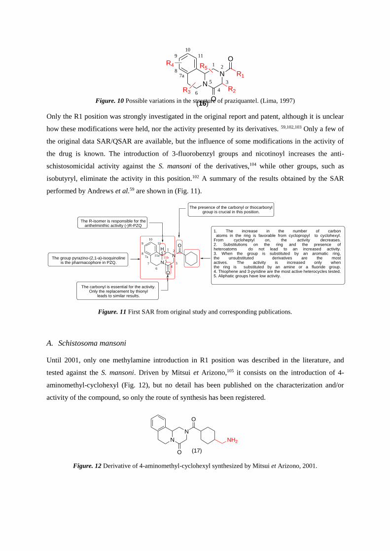
Figure. 10 Possible variations in the structure of praziquantel. (Lima, 1997)
Only the R1 position was strongly investigated in the original report and patent, although it is unclear
how these modifications were held, nor the activity presented by its derivatives. 59,102,103 Only a few of
the original data SAR/QSAR are available, but the influence of some modifications in the activity of
the drug is known. The introduction of 3-fluorobenzyl groups and nicotinoyl increases the anti-
schistosomicidal activity against the S. mansoni of the derivatives,104 while other groups, such as
isobutyryl, eliminate the activity in this position.102 A summary of the results obtained by the SAR
performed by Andrews et al.59 are shown in (Fig. 11).
Figure. 11 First SAR from original study and corresponding publications.
A. Schistosoma mansoni
Until 2001, only one methylamine introduction in R1 position was described in the literature, and
tested against the S. mansoni. Driven by Mitsui et Arizono,105 it consists on the introduction of 4-
aminomethyl-cyclohexyl (Fig. 12), but no detail has been published on the characterization and/or
activity of the compound, so only the route of synthesis has been registered.
Figure. 12 Derivative of 4-aminomethyl-cyclohexyl synthesized by Mitsui et Arizono, 2001.
N
N
O
O
NH2
(17)
The presence of the carbonyl or thiocarbonyl group is crucial in this position.
The R-isomer is responsible for the anthelminthic activity (-)R-PZQ
The group pyrazino-(2,1-a)-isoquinolineis the pharmacophore in PZQ.
The carbonyl is essential for the activity. Only the replacement by thionyl
leads to similar results.
1. The increase in the number of carbonatoms in the ring is favorable from cyclopropyl to cyclohexyl.
From cycloheptyl on, the activity decreases.2. Substitutions on the ring and the presence ofheteroatoms do not lead to an increased activity.3. When the group is substituted by an aromatic ring,the unsubstituted derivatives are the mostactives. The activity is increased only whenthe ring is substituted by an amine or a fluoride group.4. Thiophene and 3-pyridine are the most active heterocycles tested.5. Aliphatic groups have low activity.
87a 11a
11
10
9
7
6
N 511b
43
N21
O
O
*H
87a
11
10
9
6
N 5
4
3
N21
R1
O
O
R2R3
R4 R5
(16)
35
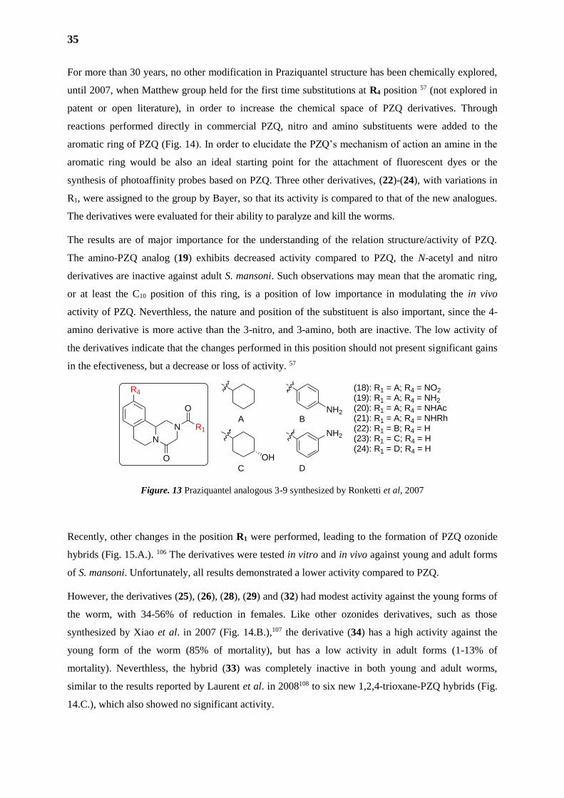
For more than 30 years, no other modification in Praziquantel structure has been chemically explored,
until 2007, when Matthew group held for the first time substitutions at R4 position 57 (not explored in
patent or open literature), in order to increase the chemical space of PZQ derivatives. Through
reactions performed directly in commercial PZQ, nitro and amino substituents were added to the
aromatic ring of PZQ (Fig. 14). In order to elucidate the PZQ’s mechanism of action an amine in the
aromatic ring would be also an ideal starting point for the attachment of fluorescent dyes or the
synthesis of photoaffinity probes based on PZQ. Three other derivatives, (22)-(24), with variations in
R1, were assigned to the group by Bayer, so that its activity is compared to that of the new analogues.
The derivatives were evaluated for their ability to paralyze and kill the worms.
The results are of major importance for the understanding of the relation structure/activity of PZQ.
The amino-PZQ analog (19) exhibits decreased activity compared to PZQ, the N-acetyl and nitro
derivatives are inactive against adult S. mansoni. Such observations may mean that the aromatic ring,
or at least the C10 position of this ring, is a position of low importance in modulating the in vivo
activity of PZQ. Neverthless, the nature and position of the substituent is also important, since the 4-
amino derivative is more active than the 3-nitro, and 3-amino, both are inactive. The low activity of
the derivatives indicate that the changes performed in this position should not present significant gains
in the efectiveness, but a decrease or loss of activity. 57
Figure. 13 Praziquantel analogous 3-9 synthesized by Ronketti et al, 2007
Recently, other changes in the position R1 were performed, leading to the formation of PZQ ozonide
hybrids (Fig. 15.A.). 106 The derivatives were tested in vitro and in vivo against young and adult forms
of S. mansoni. Unfortunately, all results demonstrated a lower activity compared to PZQ.
However, the derivatives (25), (26), (28), (29) and (32) had modest activity against the young forms of
the worm, with 34-56% of reduction in females. Like other ozonides derivatives, such as those
synthesized by Xiao et al. in 2007 (Fig. 14.B.),107 the derivative (34) has a high activity against the
young form of the worm (85% of mortality), but has a low activity in adult forms (1-13% of
mortality). Neverthless, the hybrid (33) was completely inactive in both young and adult worms,
similar to the results reported by Laurent et al. in 2008108 to six new 1,2,4-trioxane-PZQ hybrids (Fig.
14.C.), which also showed no significant activity.
N
N
R4
R1
O
O
NH2
NH2
OH
A B
C D
(18): R1 = A; R4 = NO2
(19): R1 = A; R4 = NH2
(20): R1 = A; R4 = NHAc(21): R1 = A; R4 = NHRh(22): R1 = B; R4 = H(23): R1 = C; R4 = H(24): R1 = D; R4 = H
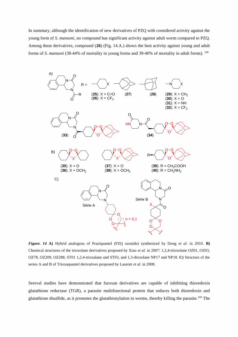
In summary, although the identification of new derivatives of PZQ with considered activity against the
young form of S. mansoni, no compound has significant activity against adult worm compared to PZQ.
Among these derivatives, compound (26) (Fig. 14.A.) shows the best activity against young and adult
forms of S. mansoni (38-44% of mortality in young forms and 39-40% of mortality in adult forms). 106
Figure. 14 A) Hybrid analogous of Praziquantel (PZQ ozonide) synthesized by Dong et al. in 2010. B)
Chemical structures of the trioxolane derivatives proposed by Xiao et al. in 2007: 1,2,4-trioxolane OZ01, OZ03,
OZ78, OZ209, OZ288, ST01 1,2,4-trioxalane and ST03, and 1,3-dioxolane NP17 and NP18. C) Structure of the
series A and B of Trioxaquantel derivatives proposed by Laurent et al. in 2008.
Sereval studies have demonstrated that furoxan derivatives are capable of inhibiting thioredoxin
glutathione reductase (TGR), a parasite multifunctional protein that reduces both thioredoxin and
glutathione disulfide, as it promotes the glutathionylation in worms, thereby killing the parasite.109 The
N
N
R
O
O
R = X N X
(25): X = C=O(26): X = CF2
(29): X = CH2
(30): X = O(31): X = NH(32): X = CF2
(27) (28)
N
N
O
O
O
OO
O
OON
O
HN
O
(33) (34)
N
N
ON
N
OX
O
O
O
O
O
O
O
n = 0,1
Série A
Série B
X
OO
O
OO
RX
OO
(35): X = O(36): X = OCH2
(37): X = O(38): X = OCH2
(39): R = CH2COOH(40): R = CH2NH2
B)
C)
A)
37
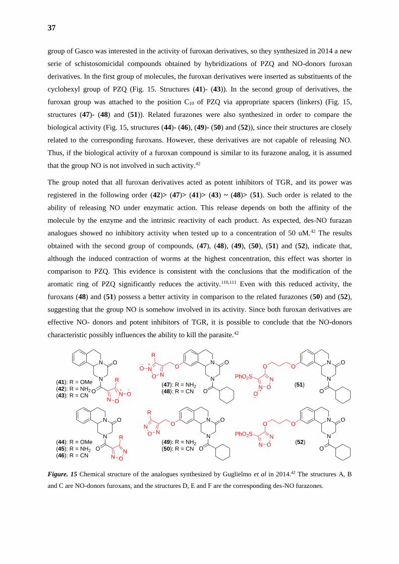
group of Gasco was interested in the activity of furoxan derivatives, so they synthesized in 2014 a new
serie of schistosomicidal compounds obtained by hybridizations of PZQ and NO-donors furoxan
derivatives. In the first group of molecules, the furoxan derivatives were inserted as substituents of the
cyclohexyl group of PZQ (Fig. 15. Structures (41)- (43)). In the second group of derivatives, the
furoxan group was attached to the position C10 of PZQ via appropriate spacers (linkers) (Fig. 15,
structures (47)- (48) and (51)). Related furazones were also synthesized in order to compare the
biological activity (Fig. 15, structures (44)- (46), (49)- (50) and (52)), since their structures are closely
related to the corresponding furoxans. However, these derivatives are not capable of releasing NO.
Thus, if the biological activity of a furoxan compound is similar to its furazone analog, it is assumed
that the group NO is not involved in such activity.42
The group noted that all furoxan derivatives acted as potent inhibitors of TGR, and its power was
registered in the following order (42)> (47)> (41)> (43) ~ (48)> (51). Such order is related to the
ability of releasing NO under enzymatic action. This release depends on both the affinity of the
molecule by the enzyme and the intrinsic reactivity of each product. As expected, des-NO furazan
analogues showed no inhibitory activity when tested up to a concentration of 50 uM.42 The results
obtained with the second group of compounds, (47), (48), (49), (50), (51) and (52), indicate that,
although the induced contraction of worms at the highest concentration, this effect was shorter in
comparison to PZQ. This evidence is consistent with the conclusions that the modification of the
aromatic ring of PZQ significantly reduces the activity.110,111 Even with this reduced activity, the
furoxans (48) and (51) possess a better activity in comparison to the related furazones (50) and (52),
suggesting that the group NO is somehow involved in its activity. Since both furoxan derivatives are
effective NO- donors and potent inhibitors of TGR, it is possible to conclude that the NO-donors
characteristic possibly influences the ability to kill the parasite.42
Figure. 15 Chemical structure of the analogues synthesized by Guglielmo et al in 2014.42 The structures A, B
and C are NO-donors furoxans, and the structures D, E and F are the corresponding des-NO furazones.
N
N
O
ON
N
O
O
N O
N O
R
N
N
O
O
N O
N
R
ON
O N
R
O
N
N
O
OO
N
O N
R
N
N
O
O
(51)
OO
N O
NPhO2S
O
N
N
O
O
(52)
OO
N O
NPhO2S
(41): R = OMe(42): R = NH2
(43): R = CN
(47): R = NH2
(48): R = CN
(44): R = OMe(45): R = NH2
(46): R = CN
(49): R = NH2
(50): R = CN
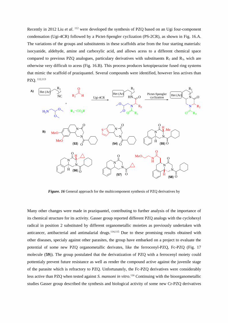
Recently in 2012 Liu et al. 112 were developed the synthesis of PZQ based on an Ugi four-component
condensation (Ugi-4CR) followed by a Pictet-Spengler cyclization (PS-2CR), as shown in Fig. 16.A.
The variations of the groups and substitutents in these scaffolds arise from the four starting materials:
isocyanide, aldehyde, amine and carboxylic acid, and allows acess to a different chemical space
compared to previous PZQ analogues, particulary derivatives with substituents R2 and R3, wich are
otherwise very difficult to acess (Fig. 16.B). This process produces ketopiperazine fused ring systems
that mimic the scaffold of praziquantel. Several compounds were identified, however less actives than
PZQ. 112,113
Figure. 16 General approach for the multicomponent synthesis of PZQ derivatives by
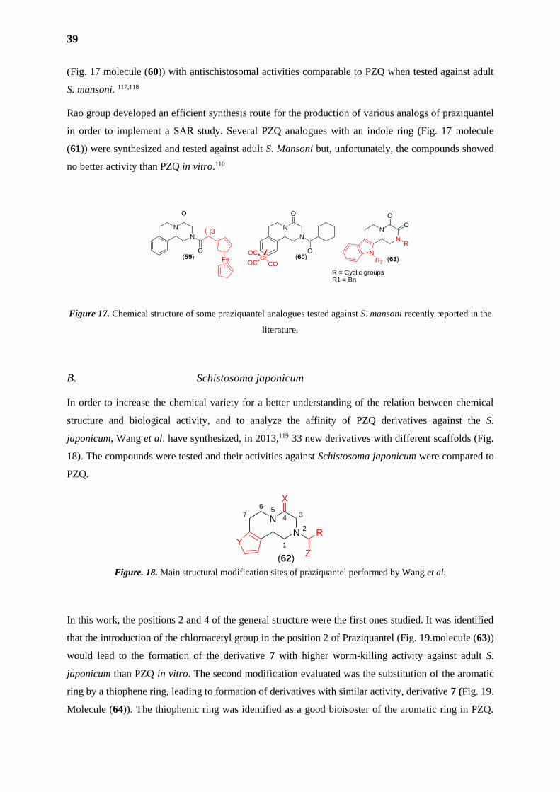
Many other changes were made in praziquantel, contributing to further analysis of the importance of
its chemical structure for its activity. Gasser group reported different PZQ analogs with the cyclohexyl
radical in position 2 substituted by different organometallic moieties as previously undertaken with
anticancer, antibacterial and antimalarial drugs.114,115 Due to these promising results obtained with
other diseases, specialy against other parasites, the group have embarked on a project to evaluate the
potential of some new PZQ organometallic derivates, like the ferrocenyl-PZQ, Fc-PZQ (Fig. 17
molecule (59)). The group postulated that the derivatization of PZQ with a ferrocenyl moiety could
pottentialy prevent future resistance as well as render the compound active against the juvenile stage
of the parasite which is refractory to PZQ. Unfortunately, the Fc-PZQ derivatives were considerably
less active than PZQ when tested against S. mansoni in vitro.116 Continuing with the bioorganometallic
studies Gasser group described the synthesis and biological activity of some new Cr-PZQ derivatives
N
N
O
O
MeO
MeO
N
N
O
O
SN
N
OH
HMe
O
N
N
OH
HO
N
N
O
O
N
N
O
O
H
O
NH
MeO
(53) (54) (55)
(56)
(57)(58)
R2
O
H
H2NO
O
NC
R1
R3 CO2H
+
HN
R1
O
N R2
O R3
O
O
N
N
R3O
O
R2
Het (Ar)
R1
Het (Ar)Het (Ar)
Ugi-4CRPictet-Spengler
cyclization
A)
B)
39
(Fig. 17 molecule (60)) with antischistosomal activities comparable to PZQ when tested against adult
S. mansoni. 117,118
Rao group developed an efficient synthesis route for the production of various analogs of praziquantel
in order to implement a SAR study. Several PZQ analogues with an indole ring (Fig. 17 molecule
(61)) were synthesized and tested against adult S. Mansoni but, unfortunately, the compounds showed
no better activity than PZQ in vitro.110
Figure 17. Chemical structure of some praziquantel analogues tested against S. mansoni recently reported in the
literature.
B. Schistosoma japonicum
In order to increase the chemical variety for a better understanding of the relation between chemical
structure and biological activity, and to analyze the affinity of PZQ derivatives against the S.
japonicum, Wang et al. have synthesized, in 2013,119 33 new derivatives with different scaffolds (Fig.
18). The compounds were tested and their activities against Schistosoma japonicum were compared to
PZQ.
Figure. 18. Main structural modification sites of praziquantel performed by Wang et al.
In this work, the positions 2 and 4 of the general structure were the first ones studied. It was identified
that the introduction of the chloroacetyl group in the position 2 of Praziquantel (Fig. 19.molecule (63))
would lead to the formation of the derivative 7 with higher worm-killing activity against adult S.
japonicum than PZQ in vitro. The second modification evaluated was the substitution of the aromatic
ring by a thiophene ring, leading to formation of derivatives with similar activity, derivative 7 (Fig. 19.
Molecule (64)). The thiophenic ring was identified as a good bioisoster of the aromatic ring in PZQ.
7N56
1
N2
34
X
R
Z
Y
(62)
N
N
O
OCr
OC
OC CO
N
N
O
R
NR2
N
N
O
O
Fe
3
(59) (60) (61)
R1 = BnR = Cyclic groups
O
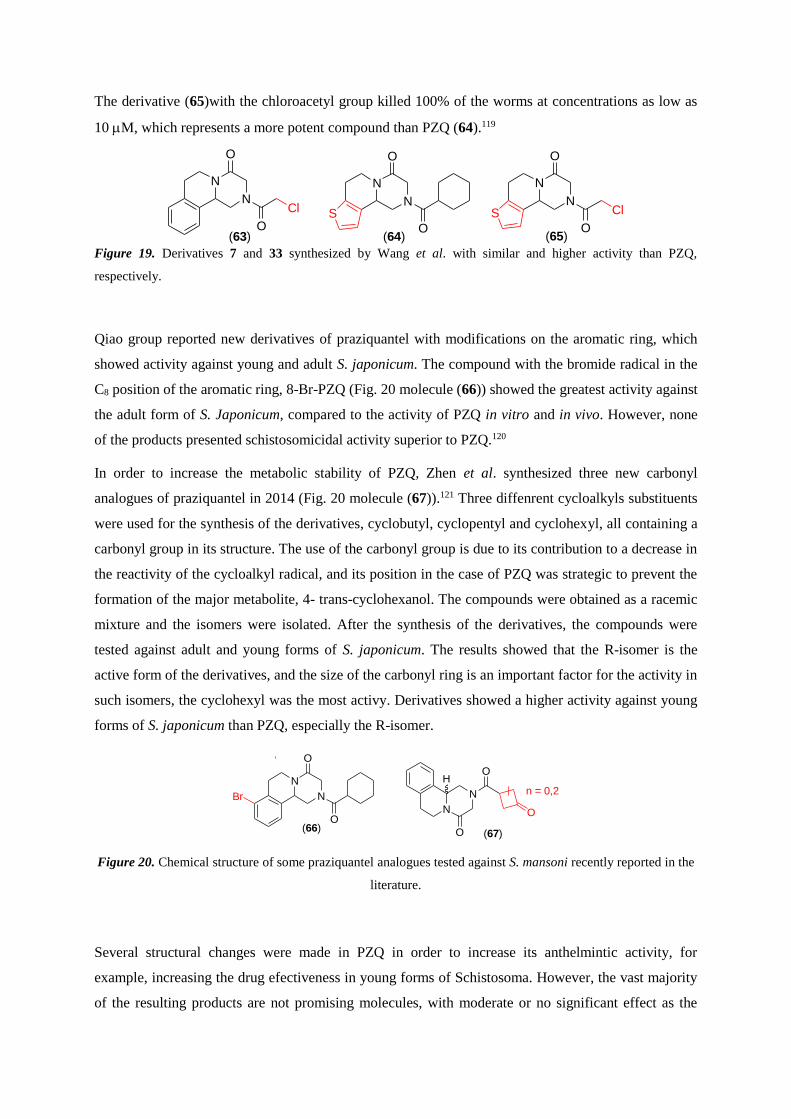
The derivative (65)with the chloroacetyl group killed 100% of the worms at concentrations as low as
10 M, which represents a more potent compound than PZQ (64).119
Figure 19. Derivatives 7 and 33 synthesized by Wang et al. with similar and higher activity than PZQ,
respectively.
Qiao group reported new derivatives of praziquantel with modifications on the aromatic ring, which
showed activity against young and adult S. japonicum. The compound with the bromide radical in the
C8 position of the aromatic ring, 8-Br-PZQ (Fig. 20 molecule (66)) showed the greatest activity against
the adult form of S. Japonicum, compared to the activity of PZQ in vitro and in vivo. However, none
of the products presented schistosomicidal activity superior to PZQ.120
In order to increase the metabolic stability of PZQ, Zhen et al. synthesized three new carbonyl
analogues of praziquantel in 2014 (Fig. 20 molecule (67)).121 Three diffenrent cycloalkyls substituents
were used for the synthesis of the derivatives, cyclobutyl, cyclopentyl and cyclohexyl, all containing a
carbonyl group in its structure. The use of the carbonyl group is due to its contribution to a decrease in
the reactivity of the cycloalkyl radical, and its position in the case of PZQ was strategic to prevent the
formation of the major metabolite, 4- trans-cyclohexanol. The compounds were obtained as a racemic
mixture and the isomers were isolated. After the synthesis of the derivatives, the compounds were
tested against adult and young forms of S. japonicum. The results showed that the R-isomer is the
active form of the derivatives, and the size of the carbonyl ring is an important factor for the activity in
such isomers, the cyclohexyl was the most activy. Derivatives showed a higher activity against young
forms of S. japonicum than PZQ, especially the R-isomer.
Figure 20. Chemical structure of some praziquantel analogues tested against S. mansoni recently reported in the
literature.
Several structural changes were made in PZQ in order to increase its anthelmintic activity, for
example, increasing the drug efectiveness in young forms of Schistosoma. However, the vast majority
of the resulting products are not promising molecules, with moderate or no significant effect as the
N
N
O
O
S
N
N
O
O
Cl
(63) (64)
N
N
O
O
S Cl
(65)
n = 0,2
N
N
O
OH
O
N
N
O
O
Br
(66)(67)
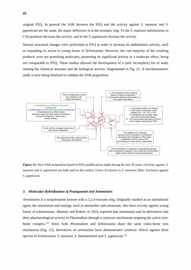
41
original PZQ. In general the SAR between the PZQ and the activity against S. mansoni and S.
japonicam are the same, the major difference is in the aromatic ring. To the S. mansoni substitutions in
C10 positions decrease the activity, and to the S. japonicum increase the activity.
Several structural changes were performed in PZQ in order to increase its anthelmintic activity, such
as expanding its action to young forms of Schistosoma. However, the vast majority of the resulting
products were not promising molecules, presenting no significant activity or a moderate effect, being
not comparable to PZQ. These studies allowed the development of a (still incomplete) list of study
relating the chemical structure and the biological activity, diagrammed in Fig. 21. A biochemometric
study is now being finalized to validate the SAR proposition.
Figure 21. New SAR proposition based in PZQ modifications made during the last 30 years. (Activity against S.
mansoni and S. japonicam are both used in this study). Green: Exclusive to S. mansoni; Blue: Exclusive against
S. japonicum
3. Molecular Hybridization of Praziquantel and Artemisinin
Artemisinin is a sesquiterpene lactone with a 1,2,4-trioxane ring. Originally studied as an antimalarial
agent, the artemisinin and analogs, such as artemether and artesunate, also have activity against young
forms of schistosomes. Meunier and Robert, in 2010, reported that artemisinin and its derivatives had
their pharmacological activity in Plasmodium through a cytotoxic mechanism targeting the active iron-
heme complex.122 Since both Plasmodium and Schistosoma share the same redox-heme iron
mechanism (Fig. 22), derivatives of artemisinin have demonstrated cytotoxic effects against three
species of Schistosoma: S. mansoni, S. haematobium and S. japonicum.123
N
N
O
OH
H
H
H
HH H
The addition of a carbonyl group at the C4 position in cyclohexyl decreases the formation of the major
metabolite of PZQ, trans-4-OH, and leading to more active compounds.
1. The reduction of the carbonyl leads to inactive molecules.
2. Increased carbonic chain leads to less active derivatives.
The reduction of the carbonyl form less active derivatives.
The R-configuration forms the eutomer in alltested derivatives.
1. Increased number of carbons in the cycle contributes to the activity (C6 > C5 > C4 > C3).
2. Substitutes by aromatic rings form less active derivatives.
3. The chloroacetyl derivative (CH2Cl) has a higher activity than PZQ.
1. Substitutions in C10 form less activecompounds.
2. Substituitions in C10 form most active compounds3.Substitutions by other isoster
aromatic rings form less active derivatives, except for thiophene, which has a
similar activity.
The introduction of carbonyl or hydrophobic groups, with consequent changes in the molecule conformation,
leads to less active or inactive derivatives.
So far, all the changes have led to less active derivatives.
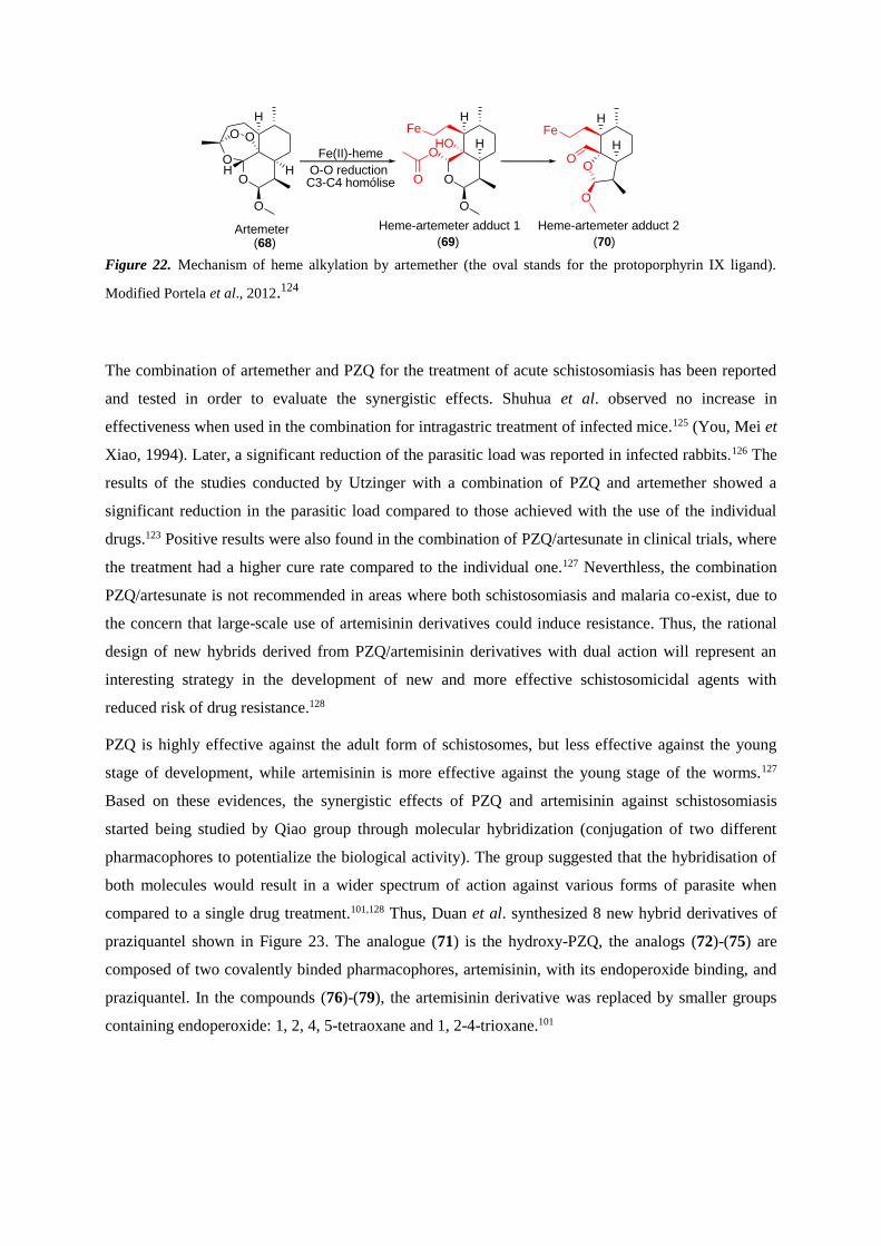
Figure 22. Mechanism of heme alkylation by artemether (the oval stands for the protoporphyrin IX ligand).
Modified Portela et al., 2012.124
The combination of artemether and PZQ for the treatment of acute schistosomiasis has been reported
and tested in order to evaluate the synergistic effects. Shuhua et al. observed no increase in
effectiveness when used in the combination for intragastric treatment of infected mice.125 (You, Mei et
Xiao, 1994). Later, a significant reduction of the parasitic load was reported in infected rabbits.126 The
results of the studies conducted by Utzinger with a combination of PZQ and artemether showed a
significant reduction in the parasitic load compared to those achieved with the use of the individual
drugs.123 Positive results were also found in the combination of PZQ/artesunate in clinical trials, where
the treatment had a higher cure rate compared to the individual one.127 Neverthless, the combination
PZQ/artesunate is not recommended in areas where both schistosomiasis and malaria co-exist, due to
the concern that large-scale use of artemisinin derivatives could induce resistance. Thus, the rational
design of new hybrids derived from PZQ/artemisinin derivatives with dual action will represent an
interesting strategy in the development of new and more effective schistosomicidal agents with
reduced risk of drug resistance.128
PZQ is highly effective against the adult form of schistosomes, but less effective against the young
stage of development, while artemisinin is more effective against the young stage of the worms.127
Based on these evidences, the synergistic effects of PZQ and artemisinin against schistosomiasis
started being studied by Qiao group through molecular hybridization (conjugation of two different
pharmacophores to potentialize the biological activity). The group suggested that the hybridisation of
both molecules would result in a wider spectrum of action against various forms of parasite when
compared to a single drug treatment.101,128 Thus, Duan et al. synthesized 8 new hybrid derivatives of
praziquantel shown in Figure 23. The analogue (71) is the hydroxy-PZQ, the analogs (72)-(75) are
composed of two covalently binded pharmacophores, artemisinin, with its endoperoxide binding, and
praziquantel. In the compounds (76)-(79), the artemisinin derivative was replaced by smaller groups
containing endoperoxide: 1, 2, 4, 5-tetraoxane and 1, 2-4-trioxane.101
O
O
H
H
O O
H
O
Fe(II)-heme
O-O reductionC3-C4 homólise O
H
O
OH
O
Fe
HO
H
H
Fe
OO
O
Artemeter Heme-artemeter adduct 1 Heme-artemeter adduct 2
(68) (69) (70)
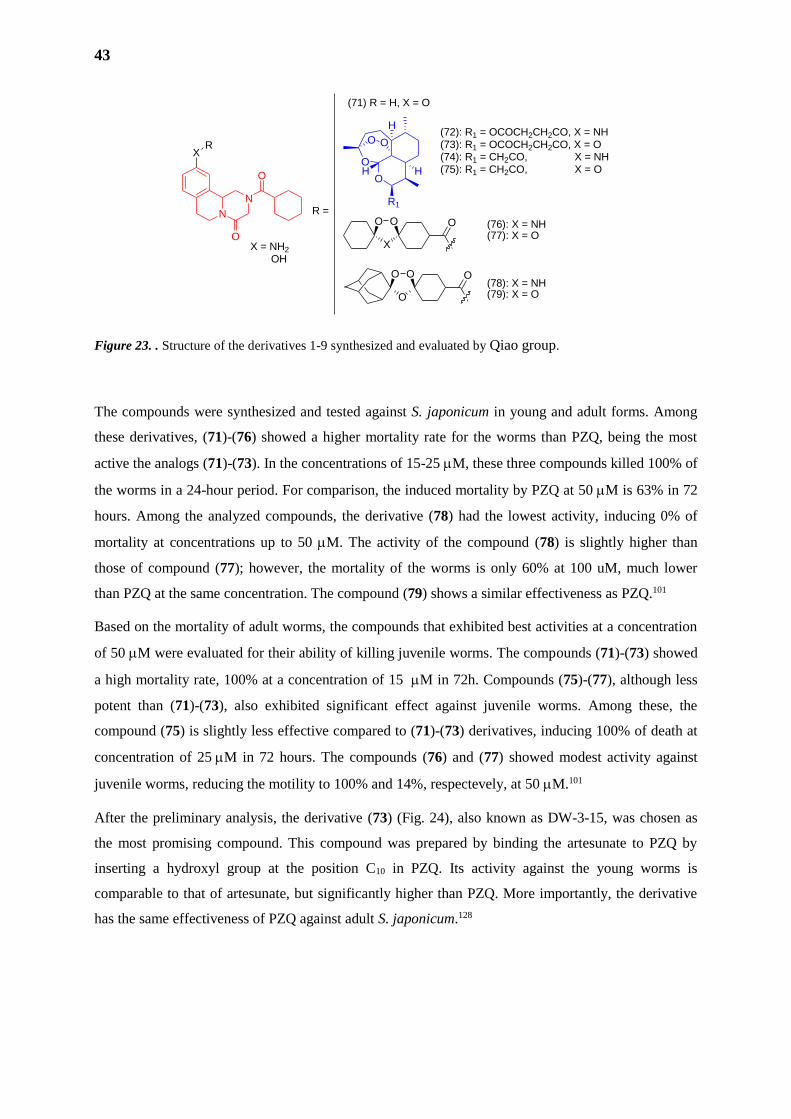
43
Figure 23. . Structure of the derivatives 1-9 synthesized and evaluated by Qiao group.
The compounds were synthesized and tested against S. japonicum in young and adult forms. Among
these derivatives, (71)-(76) showed a higher mortality rate for the worms than PZQ, being the most
active the analogs (71)-(73). In the concentrations of 15-25 M, these three compounds killed 100% of
the worms in a 24-hour period. For comparison, the induced mortality by PZQ at 50M is 63% in 72
hours. Among the analyzed compounds, the derivative (78) had the lowest activity, inducing 0% of
mortality at concentrations up to 50 M. The activity of the compound (78) is slightly higher than
those of compound (77); however, the mortality of the worms is only 60% at 100 uM, much lower
than PZQ at the same concentration. The compound (79) shows a similar effectiveness as PZQ.101
Based on the mortality of adult worms, the compounds that exhibited best activities at a concentration
of 50 M were evaluated for their ability of killing juvenile worms. The compounds (71)-(73) showed
a high mortality rate, 100% at a concentration of 15 M in 72h. Compounds (75)-(77), although less
potent than (71)-(73), also exhibited significant effect against juvenile worms. Among these, the
compound (75) is slightly less effective compared to (71)-(73) derivatives, inducing 100% of death at
concentration of 25M in 72 hours. The compounds (76) and (77) showed modest activity against
juvenile worms, reducing the motility to 100% and 14%, respectevely, at 50 M.101
After the preliminary analysis, the derivative (73) (Fig. 24), also known as DW-3-15, was chosen as
the most promising compound. This compound was prepared by binding the artesunate to PZQ by
inserting a hydroxyl group at the position C10 in PZQ. Its activity against the young worms is
comparable to that of artesunate, but significantly higher than PZQ. More importantly, the derivative
has the same effectiveness of PZQ against adult S. japonicum.128
O
O O
X
OO
O
O
O
O
H
H
O O
H
R1
(71) R = H, X = O
(72): R1 = OCOCH2CH2CO, X = NH(73): R1 = OCOCH2CH2CO, X = O(74): R1 = CH2CO, X = NH(75): R1 = CH2CO, X = O
(76): X = NH(77): X = O
(78): X = NH(79): X = O
N
N
XR
O
O
R =
X = NH2
OH
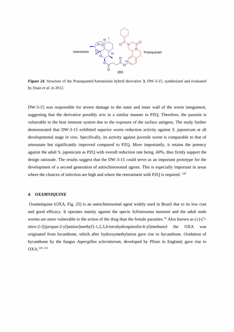
Figure 24. Structure of the Praziquantel/Artemisinin hybrid derivative 3, DW-3-15, synthesized and evaluated
by Duan et al. in 2012.
DW-3-15 was responsible for severe damage to the outer and inner wall of the worm integument,
suggesting that the derivative possibly acts in a similar manner to PZQ. Therefore, the parasite is
vulnerable to the host immune system due to the exposure of the surface antigens. The study further
demonstrated that DW-3-15 exhibited superior worm reduction activity against S. japonicum at all
developmental stage in vivo. Specifically, its activity against juvenile worm is comparable to that of
artesunate but significantly improved compared to PZQ. More importantly, it retains the potency
against the adult S. japonicum as PZQ with overall reduction rate being .60%, thus firmly support the
design rationale. The results suggest that the DW-3-15 could serve as an important prototype for the
development of a second generation of antischistosomal agents. This is especially important in areas
where the chances of infection are high and where the retreatment with PZQ is required. 128
4. OXAMNIQUINE
Oxamniquine (OXA, Fig. 25) is an antischistosomal agent widely used in Brazil due to its low cost
and good efficacy. It operates mainly against the specie Schistosoma mansoni and the adult male
worms are more vulnerable to the action of the drug than the female parasites.16 Also known as (±)-(7-
nitro-2-{[(propan-2-yl)amino]methyl}-1,2,3,4-tetrahydroquinolin-6-yl)methanol the OXA was
originated from lucanthone, which after hydroxymethylation gave rise to hycanthone. Oxidation of
hycanthone by the fungus Aspergillus sclerotiorum, developed by Pfizer in England, gave rise to
OXA.129–131
O
O
H
H
O O
H
O
O
O
O10
N
NOO
PraziquantelArtemisinin
(80)
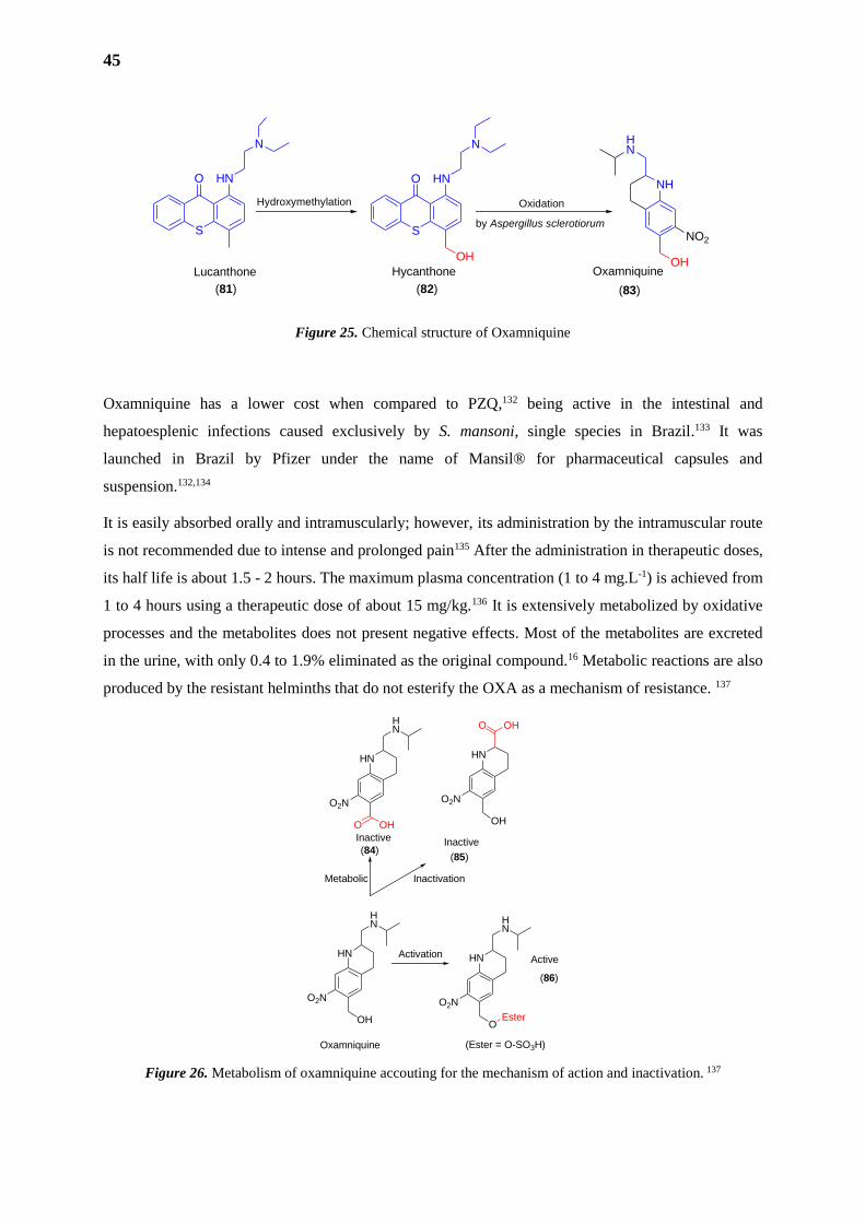
45
Figure 25. Chemical structure of Oxamniquine
Oxamniquine has a lower cost when compared to PZQ,132 being active in the intestinal and
hepatoesplenic infections caused exclusively by S. mansoni, single species in Brazil.133 It was
launched in Brazil by Pfizer under the name of Mansil® for pharmaceutical capsules and
suspension.132,134
It is easily absorbed orally and intramuscularly; however, its administration by the intramuscular route
is not recommended due to intense and prolonged pain135 After the administration in therapeutic doses,
its half life is about 1.5 - 2 hours. The maximum plasma concentration (1 to 4 mg.L-1) is achieved from
1 to 4 hours using a therapeutic dose of about 15 mg/kg.136 It is extensively metabolized by oxidative
processes and the metabolites does not present negative effects. Most of the metabolites are excreted
in the urine, with only 0.4 to 1.9% eliminated as the original compound.16 Metabolic reactions are also
produced by the resistant helminths that do not esterify the OXA as a mechanism of resistance. 137
Figure 26. Metabolism of oxamniquine accouting for the mechanism of action and inactivation. 137
S
O HN
N
S
O HN
N
OH
NH
HN
OH
NO2
Lucanthone Hycanthone Oxamniquine
Hydroxymethylation Oxidation
by Aspergillus sclerotiorum
(81) (82) (83)
HN
HN
OH
O2N
Oxamniquine
Activation HN
HN
O
O2N
Ester
HN
HN
OH
O2N
HN
OH
O2N
O
OHO
Metabolic Inactivation
Inactive Inactive
(Ester = O-SO3H)
Active
(84)(85)
(86)
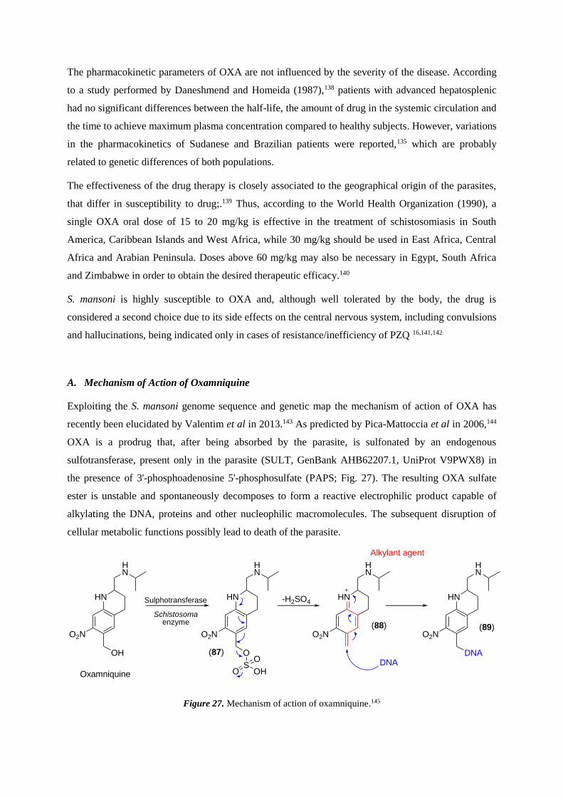
The pharmacokinetic parameters of OXA are not influenced by the severity of the disease. According
to a study performed by Daneshmend and Homeida (1987),138 patients with advanced hepatosplenic
had no significant differences between the half-life, the amount of drug in the systemic circulation and
the time to achieve maximum plasma concentration compared to healthy subjects. However, variations
in the pharmacokinetics of Sudanese and Brazilian patients were reported,135 which are probably
related to genetic differences of both populations.
The effectiveness of the drug therapy is closely associated to the geographical origin of the parasites,
that differ in susceptibility to drug;.139 Thus, according to the World Health Organization (1990), a
single OXA oral dose of 15 to 20 mg/kg is effective in the treatment of schistosomiasis in South
America, Caribbean Islands and West Africa, while 30 mg/kg should be used in East Africa, Central
Africa and Arabian Peninsula. Doses above 60 mg/kg may also be necessary in Egypt, South Africa
and Zimbabwe in order to obtain the desired therapeutic efficacy.140
S. mansoni is highly susceptible to OXA and, although well tolerated by the body, the drug is
considered a second choice due to its side effects on the central nervous system, including convulsions
and hallucinations, being indicated only in cases of resistance/inefficiency of PZQ 16,141,142
A. Mechanism of Action of Oxamniquine
Exploiting the S. mansoni genome sequence and genetic map the mechanism of action of OXA has
recently been elucidated by Valentim et al in 2013.143 As predicted by Pica-Mattoccia et al in 2006,144
OXA is a prodrug that, after being absorbed by the parasite, is sulfonated by an endogenous
sulfotransferase, present only in the parasite (SULT, GenBank AHB62207.1, UniProt V9PWX8) in
the presence of 3'-phosphoadenosine 5'-phosphosulfate (PAPS; Fig. 27). The resulting OXA sulfate
ester is unstable and spontaneously decomposes to form a reactive electrophilic product capable of
alkylating the DNA, proteins and other nucleophilic macromolecules. The subsequent disruption of
cellular metabolic functions possibly lead to death of the parasite.
Figure 27. Mechanism of action of oxamniquine.145
HN
HN
OH
O2N
HN
HN
O
O2N
HN
HN
O2N
HN
HN
O2N
DNA
SOHO
O
-H2SO4Sulphotransferase
Schistosomaenzyme
Oxamniquine
DNA
Alkylant agent
(87)
(88) (89)
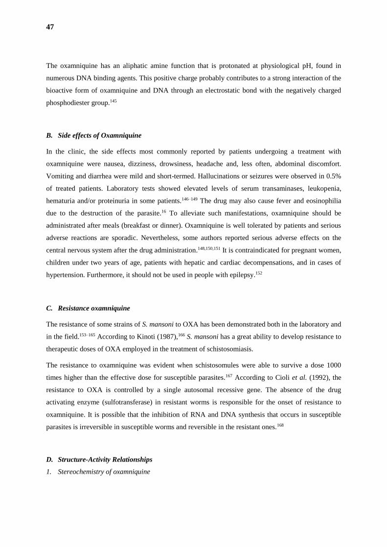
47
The oxamniquine has an aliphatic amine function that is protonated at physiological pH, found in
numerous DNA binding agents. This positive charge probably contributes to a strong interaction of the
bioactive form of oxamniquine and DNA through an electrostatic bond with the negatively charged
phosphodiester group.145
B. Side effects of Oxamniquine
In the clinic, the side effects most commonly reported by patients undergoing a treatment with
oxamniquine were nausea, dizziness, drowsiness, headache and, less often, abdominal discomfort.
Vomiting and diarrhea were mild and short-termed. Hallucinations or seizures were observed in 0.5%
of treated patients. Laboratory tests showed elevated levels of serum transaminases, leukopenia,
hematuria and/or proteinuria in some patients.146–149 The drug may also cause fever and eosinophilia
due to the destruction of the parasite.16 To alleviate such manifestations, oxamniquine should be
administrated after meals (breakfast or dinner). Oxamniquine is well tolerated by patients and serious
adverse reactions are sporadic. Nevertheless, some authors reported serious adverse effects on the
central nervous system after the drug administration.148,150,151 It is contraindicated for pregnant women,
children under two years of age, patients with hepatic and cardiac decompensations, and in cases of
hypertension. Furthermore, it should not be used in people with epilepsy.152
C. Resistance oxamniquine
The resistance of some strains of S. mansoni to OXA has been demonstrated both in the laboratory and
in the field.153–165 According to Kinoti (1987),166 S. mansoni has a great ability to develop resistance to
therapeutic doses of OXA employed in the treatment of schistosomiasis.
The resistance to oxamniquine was evident when schistosomules were able to survive a dose 1000
times higher than the effective dose for susceptible parasites.167 According to Cioli et al. (1992), the
resistance to OXA is controlled by a single autosomal recessive gene. The absence of the drug
activating enzyme (sulfotransferase) in resistant worms is responsible for the onset of resistance to
oxamniquine. It is possible that the inhibition of RNA and DNA synthesis that occurs in susceptible
parasites is irreversible in susceptible worms and reversible in the resistant ones.168
D. Structure-Activity Relationships
1. Stereochemistry of oxamniquine
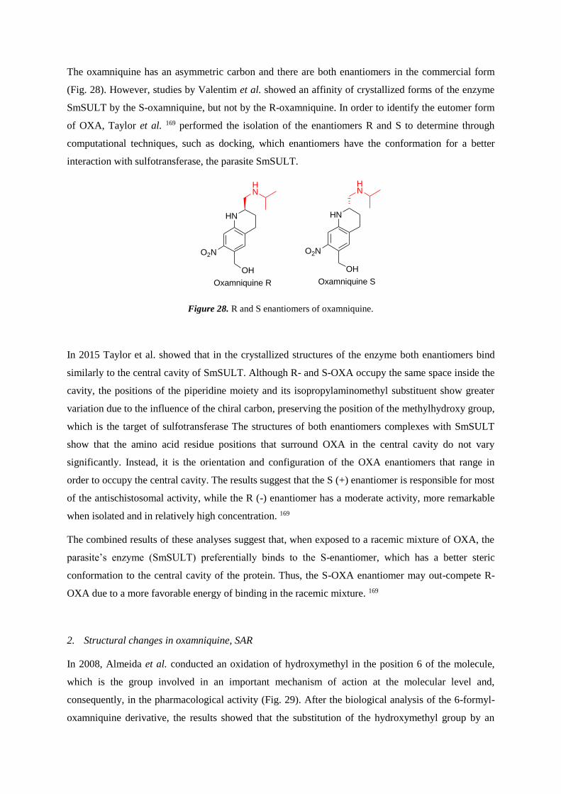
The oxamniquine has an asymmetric carbon and there are both enantiomers in the commercial form
(Fig. 28). However, studies by Valentim et al. showed an affinity of crystallized forms of the enzyme
SmSULT by the S-oxamniquine, but not by the R-oxamniquine. In order to identify the eutomer form
of OXA, Taylor et al. 169 performed the isolation of the enantiomers R and S to determine through
computational techniques, such as docking, which enantiomers have the conformation for a better
interaction with sulfotransferase, the parasite SmSULT.
Figure 28. R and S enantiomers of oxamniquine.
In 2015 Taylor et al. showed that in the crystallized structures of the enzyme both enantiomers bind
similarly to the central cavity of SmSULT. Although R- and S-OXA occupy the same space inside the
cavity, the positions of the piperidine moiety and its isopropylaminomethyl substituent show greater
variation due to the influence of the chiral carbon, preserving the position of the methylhydroxy group,
which is the target of sulfotransferase The structures of both enantiomers complexes with SmSULT
show that the amino acid residue positions that surround OXA in the central cavity do not vary
significantly. Instead, it is the orientation and configuration of the OXA enantiomers that range in
order to occupy the central cavity. The results suggest that the S (+) enantiomer is responsible for most
of the antischistosomal activity, while the R (-) enantiomer has a moderate activity, more remarkable
when isolated and in relatively high concentration. 169
The combined results of these analyses suggest that, when exposed to a racemic mixture of OXA, the
parasite’s enzyme (SmSULT) preferentially binds to the S-enantiomer, which has a better steric
conformation to the central cavity of the protein. Thus, the S-OXA enantiomer may out-compete R-
OXA due to a more favorable energy of binding in the racemic mixture. 169
2. Structural changes in oxamniquine, SAR
In 2008, Almeida et al. conducted an oxidation of hydroxymethyl in the position 6 of the molecule,
which is the group involved in an important mechanism of action at the molecular level and,
consequently, in the pharmacological activity (Fig. 29). After the biological analysis of the 6-formyl-
oxamniquine derivative, the results showed that the substitution of the hydroxymethyl group by an
HN
HN
OH
O2N
Oxamniquine R
HN
HN
OH
O2N
Oxamniquine S
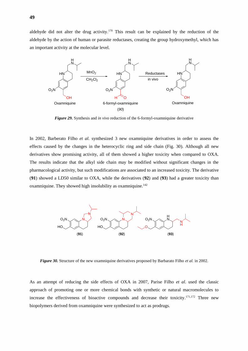
49
aldehyde did not alter the drug activity.170 This result can be explained by the reduction of the
aldehyde by the action of human or parasite reductases, creating the group hydroxymethyl, which has
an important activity at the molecular level.
Figure 29. Synthesis and in vivo reduction of the 6-formyl-oxamniquine derivative
In 2002, Barberato Filho et al. synthesized 3 new oxamniquine derivatives in order to assess the
effects caused by the changes in the heterocyclic ring and side chain (Fig. 30). Although all new
derivatives show promising activity, all of them showed a higher toxicity when compared to OXA.
The results indicate that the alkyl side chain may be modified without significant changes in the
pharmacological activity, but such modifications are associated to an increased toxicity. The derivative
(91) showed a LD50 similar to OXA, while the derivatives (92) and (93) had a greater toxicity than
oxamniquine. They showed high insolubility as oxamniquine.142
Figure 30. Structure of the new oxamniquine derivatives proposed by Barbarato Filho et al. in 2002.
As an attempt of reducing the side effects of OXA in 2007, Parise Filho et al. used the classic
approach of promoting one or more chemical bonds with synthetic or natural macromolecules to
increase the effectiveness of bioactive compounds and decrease their toxicity.171,172 Three new
biopolymers derived from oxamniquine were synthesized to act as prodrugs.
N
N
O2N
HO
NO2N
HO
NHNO2N
O
NH
(91) (92) (93)
HN
HN
OH
O2N
Oxamniquine
HN
HN
O
O2N
6-formyl-oxamniquine
H
MnO2
CH2Cl2
Reductases
in vivo
HN
HN
OH
O2N
Oxamniquine
(90)
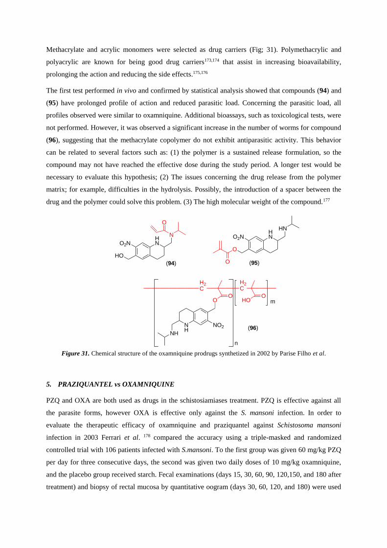
Methacrylate and acrylic monomers were selected as drug carriers (Fig; 31). Polymethacrylic and
polyacrylic are known for being good drug carriers173,174 that assist in increasing bioavailability,
prolonging the action and reducing the side effects.175,176
The first test performed in vivo and confirmed by statistical analysis showed that compounds (94) and
(95) have prolonged profile of action and reduced parasitic load. Concerning the parasitic load, all
profiles observed were similar to oxamniquine. Additional bioassays, such as toxicological tests, were
not performed. However, it was observed a significant increase in the number of worms for compound
(96), suggesting that the methacrylate copolymer do not exhibit antiparasitic activity. This behavior
can be related to several factors such as: (1) the polymer is a sustained release formulation, so the
compound may not have reached the effective dose during the study period. A longer test would be
necessary to evaluate this hypothesis; (2) The issues concerning the drug release from the polymer
matrix; for example, difficulties in the hydrolysis. Possibly, the introduction of a spacer between the
drug and the polymer could solve this problem. (3) The high molecular weight of the compound.177
Figure 31. Chemical structure of the oxamniquine prodrugs synthetized in 2002 by Parise Filho et al.
5. PRAZIQUANTEL vs OXAMNIQUINE
PZQ and OXA are both used as drugs in the schistosiamiases treatment. PZQ is effective against all
the parasite forms, however OXA is effective only against the S. mansoni infection. In order to
evaluate the therapeutic efficacy of oxamniquine and praziquantel against Schistosoma mansoni
infection in 2003 Ferrari et al. 178 compared the accuracy using a triple-masked and randomized
controlled trial with 106 patients infected with S.mansoni. To the first group was given 60 mg/kg PZQ
per day for three consecutive days, the second was given two daily doses of 10 mg/kg oxamniquine,
and the placebo group received starch. Fecal examinations (days 15, 30, 60, 90, 120,150, and 180 after
treatment) and biopsy of rectal mucosa by quantitative oogram (days 30, 60, 120, and 180) were used
HNO2N
HO
(94)
HNO2N
O
(95)
N
OHN
O
NH
NH
NO2
OO
HOO
n
m
H2C
H2C
(96)
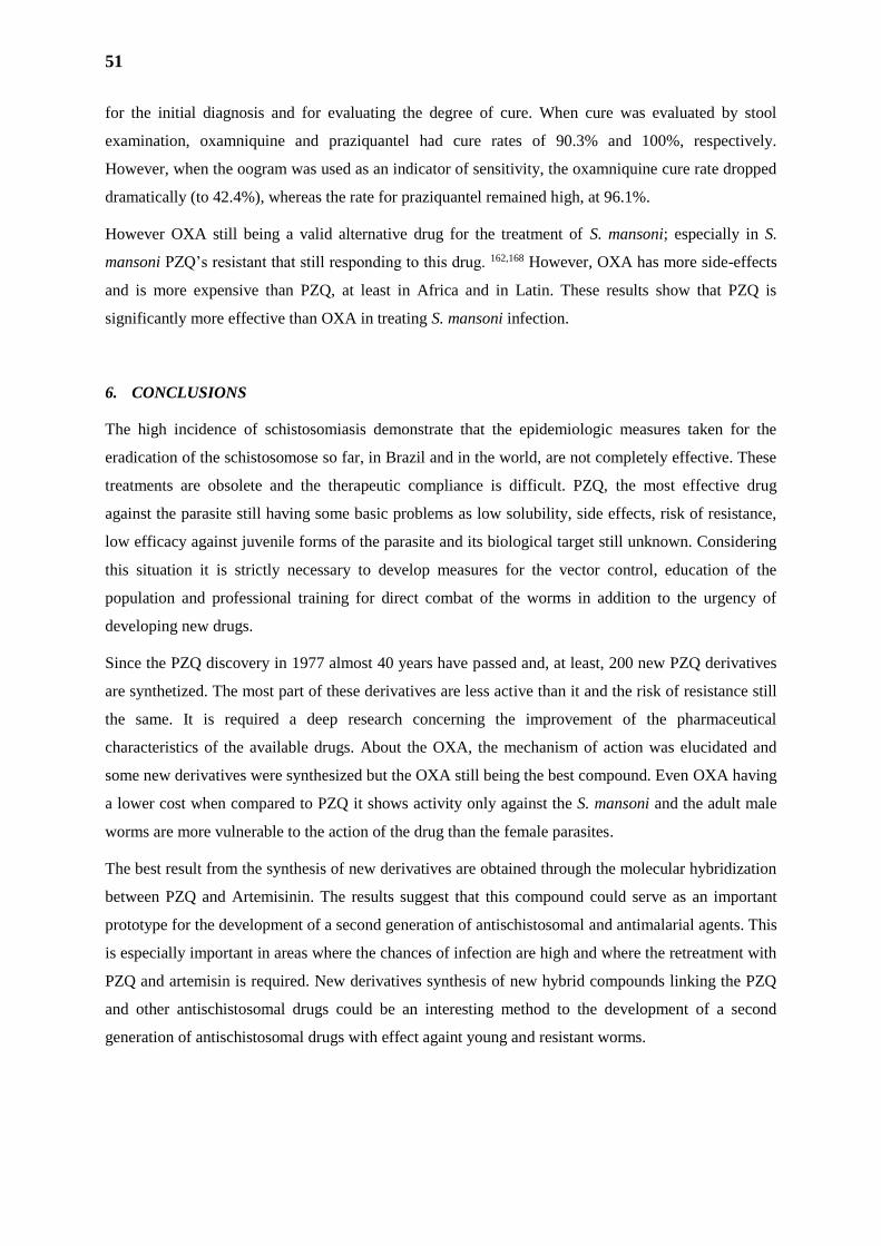
51
for the initial diagnosis and for evaluating the degree of cure. When cure was evaluated by stool
examination, oxamniquine and praziquantel had cure rates of 90.3% and 100%, respectively.
However, when the oogram was used as an indicator of sensitivity, the oxamniquine cure rate dropped
dramatically (to 42.4%), whereas the rate for praziquantel remained high, at 96.1%.
However OXA still being a valid alternative drug for the treatment of S. mansoni; especially in S.
mansoni PZQ’s resistant that still responding to this drug. 162,168 However, OXA has more side-effects
and is more expensive than PZQ, at least in Africa and in Latin. These results show that PZQ is
significantly more effective than OXA in treating S. mansoni infection.
6. CONCLUSIONS
The high incidence of schistosomiasis demonstrate that the epidemiologic measures taken for the
eradication of the schistosomose so far, in Brazil and in the world, are not completely effective. These
treatments are obsolete and the therapeutic compliance is difficult. PZQ, the most effective drug
against the parasite still having some basic problems as low solubility, side effects, risk of resistance,
low efficacy against juvenile forms of the parasite and its biological target still unknown. Considering
this situation it is strictly necessary to develop measures for the vector control, education of the
population and professional training for direct combat of the worms in addition to the urgency of
developing new drugs.
Since the PZQ discovery in 1977 almost 40 years have passed and, at least, 200 new PZQ derivatives
are synthetized. The most part of these derivatives are less active than it and the risk of resistance still
the same. It is required a deep research concerning the improvement of the pharmaceutical
characteristics of the available drugs. About the OXA, the mechanism of action was elucidated and
some new derivatives were synthesized but the OXA still being the best compound. Even OXA having
a lower cost when compared to PZQ it shows activity only against the S. mansoni and the adult male
worms are more vulnerable to the action of the drug than the female parasites.
The best result from the synthesis of new derivatives are obtained through the molecular hybridization
between PZQ and Artemisinin. The results suggest that this compound could serve as an important
prototype for the development of a second generation of antischistosomal and antimalarial agents. This
is especially important in areas where the chances of infection are high and where the retreatment with
PZQ and artemisin is required. New derivatives synthesis of new hybrid compounds linking the PZQ
and other antischistosomal drugs could be an interesting method to the development of a second
generation of antischistosomal drugs with effect againt young and resistant worms.
TRABALHOS PESSOAIS
PARTE I
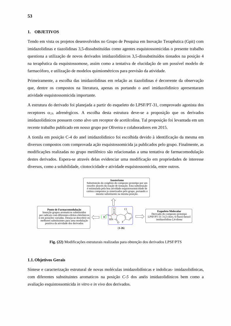
53
1. OBJETIVOS
Tendo em vista os projetos desenvolvidos no Grupo de Pesquisa em Inovação Terapêutica (Gpit) com
imidazolidinas e tiazolidinas 3,5-dissubstituídas como agentes esquistossomicidas o presente trabalho
questiona a utilização de novos derivados imidazolidínicos 3,5-dissubstituídos tionados na posição 4
na terapêutica da esquistossomose, assim como a tentativa de elucidação de um possível modelo de
farmacóforo, e utilização de modelos quimiométricos para previsão da atividade.
Primeiramente, a escolha das imidazolidinas em relação as tiazolidinas é decorrente da observação
que, dentre os compostos na literatura, apenas os portando o anel imidazolidinico apresentaram
atividade esquistossomicida importante.
A estrutura do derivado foi planejada a partir do esqueleto do LPSF/PT-31, comprovado agonista dos
receptores 2A adrenérgicos. A escolha desta estrutura deve-se a proposição que os derivados
imidazolidinicos possuem como alvo um receptor de acetilcolina. Tal proposição foi levantada em um
recente trabalho publicado em nosso grupo por Oliveira e colaboradores em 2015.
A tionila em posição C-4 do anel imidazolidinico foi escolhida devido à identificação da mesma em
diversos compostos com comprovada ação esquistossomicida ja publicados pelo grupo. Finalmente, as
modificações realizadas no grupo metilênico são relacionadas a uma tentativa de farmacomodulação
destes derivados. Espera-se através delas evidenciar uma modificação em propriedades de interesse
diversos, como a solubilidade, citotocicidade e atividade esquistossomicida, entre outros.
Fig. (22) Modificações estruturais realizadas para obtenção dos derivados LPSF/PTS
1.1. Objetivos Gerais
Síntese e caracterização estrutural de novas moléculas imidazolidínicas e indolicas- imidazolidínicas,
com diferentes substituintes aromaticos na posição C-5 dos anéis imidazolidínicos bem como a
avaliação esquistossomicida in vitro e in vivo dos derivados.
HN
S
O
R
Cl
F
Esqueleto MolecularDerivado do composto prototipo
LPSF/PT-31 3-(2-cloro, 6-fluor)-benzil imidazolidina-2,4-diona
IsosterismoSubstituição do oxigênio do composto prototipo por umenxofre através da reação de tionação. Esta substituição é estimulada pela boa atividade esquistossomicidade de certtos compostos ja sintetizados pelo grupo, portando o
mesmo substituinte na mesma posição.
Ponto de FarmacomodulaçãoInserção grupos aromaticos substituidos
por radicais com diferentes efeitos eletrônicos e em posições variadas. Deseja-se descobrir os
melhores substituintes para uma modulação positiva da atividade dos derivados
(1-26)
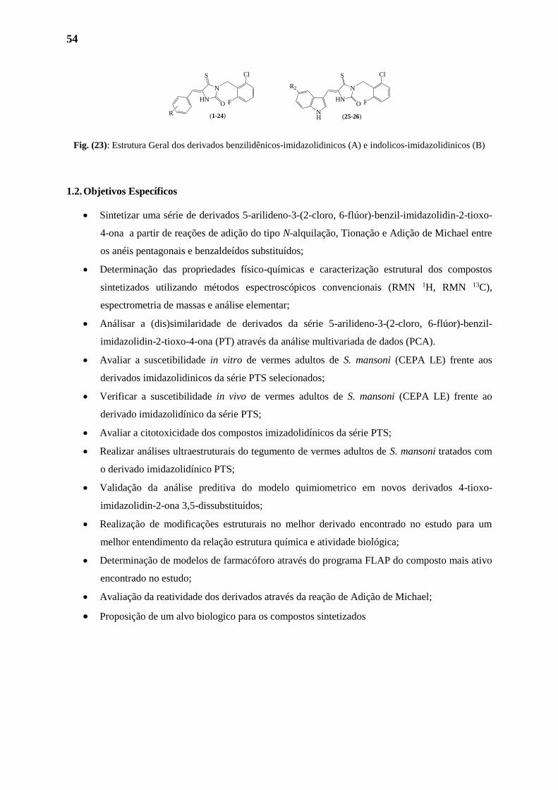
54
Fig. (23): Estrutura Geral dos derivados benzilidênicos-imidazolidinicos (A) e indolicos-imidazolidinicos (B)
1.2. Objetivos Específicos
Sintetizar uma série de derivados 5-arilideno-3-(2-cloro, 6-flúor)-benzil-imidazolidin-2-tioxo-
4-ona a partir de reações de adição do tipo N-alquilação, Tionação e Adição de Michael entre
os anéis pentagonais e benzaldeídos substituídos;
Determinação das propriedades físico-químicas e caracterização estrutural dos compostos
sintetizados utilizando métodos espectroscópicos convencionais (RMN 1H, RMN 13C),
espectrometria de massas e análise elementar;
Análisar a (dis)similaridade de derivados da série 5-arilideno-3-(2-cloro, 6-flúor)-benzil-
imidazolidin-2-tioxo-4-ona (PT) através da análise multivariada de dados (PCA).
Avaliar a suscetibilidade in vitro de vermes adultos de S. mansoni (CEPA LE) frente aos
derivados imidazolidinicos da série PTS selecionados;
Verificar a suscetibilidade in vivo de vermes adultos de S. mansoni (CEPA LE) frente ao
derivado imidazolidínico da série PTS;
Avaliar a citotoxicidade dos compostos imizadolidínicos da série PTS;
Realizar análises ultraestruturais do tegumento de vermes adultos de S. mansoni tratados com
o derivado imidazolidínico PTS;
Validação da análise preditiva do modelo quimiometrico em novos derivados 4-tioxo-
imidazolidin-2-ona 3,5-dissubstituídos;
Realização de modificações estruturais no melhor derivado encontrado no estudo para um
melhor entendimento da relação estrutura química e atividade biológica;
Determinação de modelos de farmacóforo através do programa FLAP do composto mais ativo
encontrado no estudo;
Avaliação da reatividade dos derivados através da reação de Adição de Michael;
Proposição de um alvo biologico para os compostos sintetizados
O
S
HN
N
R
F
Cl
O
S
HN
N
F
Cl
NH
R2
(1-24) (25-26)
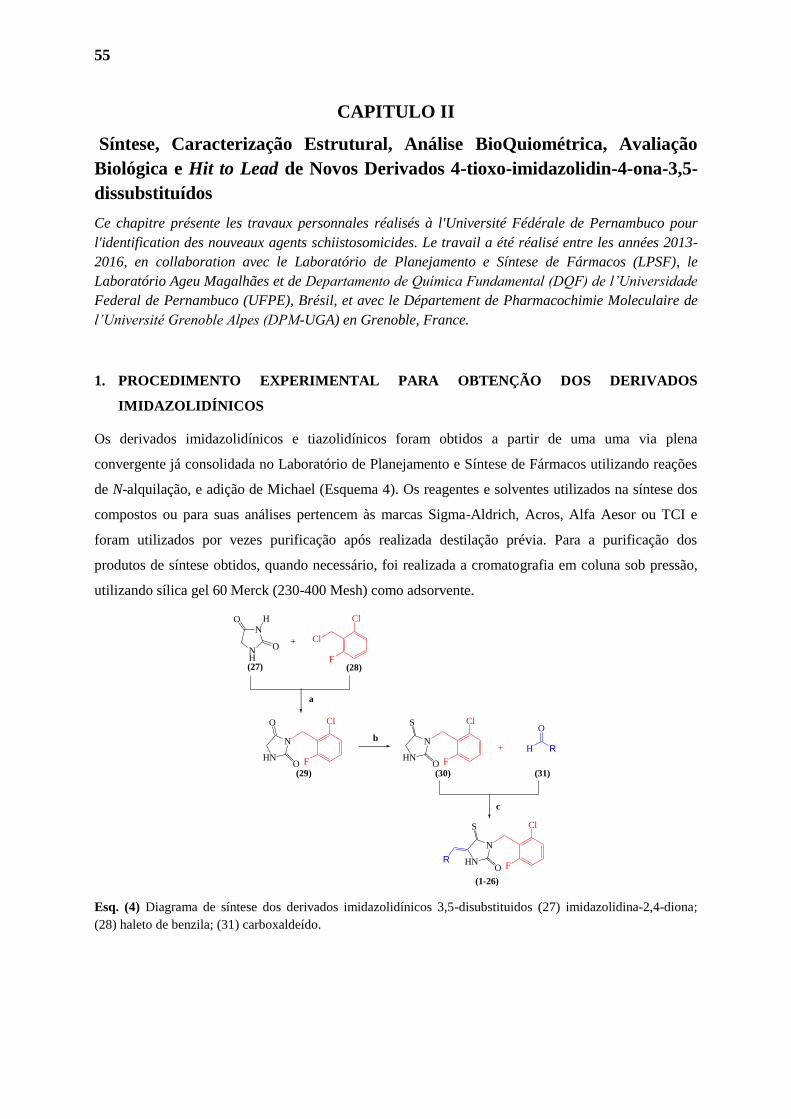
55
CAPITULO II
Síntese, Caracterização Estrutural, Análise BioQuiométrica, Avaliação
Biológica e Hit to Lead de Novos Derivados 4-tioxo-imidazolidin-4-ona-3,5-
dissubstituídos
Ce chapitre présente les travaux personnales réalisés à l'Université Fédérale de Pernambuco pour
l'identification des nouveaux agents schiistosomicides. Le travail a été réalisé entre les années 2013-
2016, en collaboration avec le Laboratório de Planejamento e Síntese de Fármacos (LPSF), le
Laboratório Ageu Magalhães et de Departamento de Química Fundamental (DQF) de l’Universidade
Federal de Pernambuco (UFPE), Brésil, et avec le Département de Pharmacochimie Moleculaire de
l’Université Grenoble Alpes (DPM-UGA) en Grenoble, France.
1. PROCEDIMENTO EXPERIMENTAL PARA OBTENÇÃO DOS DERIVADOS
IMIDAZOLIDÍNICOS
Os derivados imidazolidínicos e tiazolidínicos foram obtidos a partir de uma uma via plena
convergente já consolidada no Laboratório de Planejamento e Síntese de Fármacos utilizando reações
de N-alquilação, e adição de Michael (Esquema 4). Os reagentes e solventes utilizados na síntese dos
compostos ou para suas análises pertencem às marcas Sigma-Aldrich, Acros, Alfa Aesor ou TCI e
foram utilizados por vezes purificação após realizada destilação prévia. Para a purificação dos
produtos de síntese obtidos, quando necessário, foi realizada a cromatografia em coluna sob pressão,
utilizando sílica gel 60 Merck (230-400 Mesh) como adsorvente.
Esq. (4) Diagrama de síntese dos derivados imidazolidínicos 3,5-disubstituidos (27) imidazolidina-2,4-diona;
(28) haleto de benzila; (31) carboxaldeído.
RO
S
HN
N
O
ON
NH
Cl
O
O
HN
N
O
S
HN
NR
O
H+
+
(27) (28)
(29) (30) (31)
(1-26)
H
F
Cl
Cl
F
Cl
F
Cl
F
a
b
c
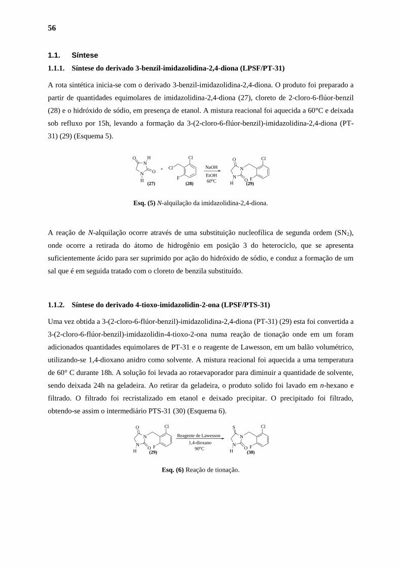
56
1.1. Síntese
1.1.1. Síntese do derivado 3-benzil-imidazolidina-2,4-diona (LPSF/PT-31)
A rota sintética inicia-se com o derivado 3-benzil-imidazolidina-2,4-diona. O produto foi preparado a
partir de quantidades equimolares de imidazolidina-2,4-diona (27), cloreto de 2-cloro-6-flúor-benzil
(28) e o hidróxido de sódio, em presença de etanol. A mistura reacional foi aquecida a 60°C e deixada
sob refluxo por 15h, levando a formação da 3-(2-cloro-6-flúor-benzil)-imidazolidina-2,4-diona (PT-
31) (29) (Esquema 5).
Esq. (5) N-alquilação da imidazolidina-2,4-diona.
A reação de N-alquilação ocorre através de uma substituição nucleofílica de segunda ordem (SN2),
onde ocorre a retirada do átomo de hidrogênio em posição 3 do heterociclo, que se apresenta
suficientemente ácido para ser suprimido por ação do hidróxido de sódio, e conduz a formação de um
sal que é em seguida tratado com o cloreto de benzila substituído.
1.1.2. Síntese do derivado 4-tioxo-imidazolidin-2-ona (LPSF/PTS-31)
Uma vez obtida a 3-(2-cloro-6-flúor-benzil)-imidazolidina-2,4-diona (PT-31) (29) esta foi convertida a
3-(2-cloro-6-flúor-benzil)-imidazolidin-4-tioxo-2-ona numa reação de tionação onde em um foram
adicionados quantidades equimolares de PT-31 e o reagente de Lawesson, em um balão volumétrico,
utilizando-se 1,4-dioxano anidro como solvente. A mistura reacional foi aquecida a uma temperatura
de 60° C durante 18h. A solução foi levada ao rotaevaporador para diminuir a quantidade de solvente,
sendo deixada 24h na geladeira. Ao retirar da geladeira, o produto solido foi lavado em n-hexano e
filtrado. O filtrado foi recristalizado em etanol e deixado precipitar. O precipitado foi filtrado,
obtendo-se assim o intermediário PTS-31 (30) (Esquema 6).
Esq. (6) Reação de tionação.
Cl
FO
O
N
N
Cl
FO
S
N
N
(29) (30)
Reagente de Lawesson
1,4-dioxano
90oCH H
O
ON
N
Cl
F
Cl Cl
FO
O
N
N+
(27) (28) (29)
H
NaOH
EtOH
60oCHH
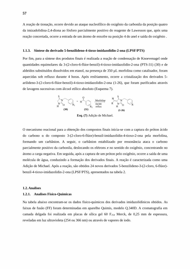
57
A reação de tionação, ocorre devido ao ataque nucleofílico do oxigênio da carbonila da posição quatro
da imizadolidina-2,4-diona ao fósforo parcialmente positivo do reagente de Lawesson que, após uma
reação concertada, ocorre a entrada de um átomo de enxofre na posição 4 do anel e saída do oxigênio .
1.1.3. Síntese do derivado 5-benzilideno-4-tioxo-imidazolidin-2-ona (LPSF/PTS)
Por fim, para a síntese dos produtos finais é realizada a reação de condensação de Knoevenagel onde
quantidades equimolares da 3-(2-cloro-6-flúor-benzil)-4-tioxo-imidazolidin-2-ona (PTS-31) (30) e de
aldeídos substituídos dissolvidos em etanol, na presença de 350 µL morfolina como catalisador, foram
aquecidas sob refluxo durante 4 horas. Após resfriamento, ocorre a cristalização dos derivados 5-
arilideno-3-(2-cloro-6-flúor-benzil)-4-tioxo-imidazolidin-2-ona (1-26), que foram purificados através
de lavagens sucessivas com álcool etílico absoluto (Esquema 7).
Esq. (7) Adição de Michael.
O mecanismo reacional para a obtenção dos compostos finais inicia-se com a captura do próton ácido
do carbono α do composto 3-(2-cloro-6-flúor)-benzil-imidazolidin-4-tioxo-2-ona pela morfolina,
formando um carbânion. A seguir, o carbânion estabilizado por ressonância ataca o carbono
parcialmente positivo da carbonila, deslocando os elétrons π no sentido do oxigênio, concentrando no
átomo a carga negativa. Em seguida, após a captura de um próton pelo oxigênio, ocorre a saída de uma
molécula de água, conduzindo a formação dos derivados finais. A reação é caracterizada como uma
Adição de Michael. Após a reação, são obtidos 24 novos derivados 5-benzilideno-3-(2-cloro, 6-flúor)-
benzil-4-tioxo-imidazolidin-2-ona (LPSF/PTS), apresentados na tabela 2.
1.2. Analises
1.2.1. Analises Fisico-Químicas
Na tabela abaixo encontram-se os dados físico-químicos dos derivados imidazolidínicos obtidos. As
faixas de fusão (FF) foram determinadas em aparelho Quimis, modelo Q.340D. A cromatografia em
camada delgada foi realizada em placas de sílica gel 60 F254 Merck, de 0,25 mm de espessura,
reveladas em luz ultravioleta (254 ou 366 nm) ou através de vapores de iodo.
MorfolineR
O
S
N
N
O
S
N
NO
H+
(30) (31) (1-26)
R2
R1 RH H
EtOHreflux
H

58
Tab (2) Dados físico-químicos dos derivados imidazolidínicos obtidos
Derivados R Rendimento Rf Faixa de Fusão Massa Molar
PTS-1
36% 0.42 (n-Hex/AcOEt, 6:4) 268oC 415.92
PTS-2
20,5% 0,56 (n-Hex/AcOEt, 1:1) 260oC 429.94
PTS-3
49% 0.48 (n-Hex/AcOEt, 7:3) 232oC 431.92
PTS-4 33% 0,6 (n-Hex/AcOEt, 7:3) 264oC 452.93
PTS-40
76% 0.59 (n-Hex/AcOEt, 1:1) 197oC 350.77
PTS-6 59% 0.57 (n-Hex/AcOEt,
7.5:0.5)
224oC 423.90
PTS-7
48% 0,4% (n-Hex/AcOEt, 7:3) 216oC 376.84
PTS-8
70,5% 0,57 (n-Hex/AcOEt, 7:3) 230oC 360.84
PTS-9
26% 0,48 (n-Hex/AcOEt, 7:3) 215oC 346.81
PTS-10
27% 0,55 (n-Hex/AcOEt, 6:4) 269oC 389.88
PTS-11
66% 0,4 (n-Hex/AcOEt, 6:4) 210oC 406.86
PTS-12 69% 0,44 (n-Hex/AcOEt,7:3) 236oC 436.89
PTS-13
76% 0.52 (n-Hex/AcOEt, 1:1) 197oC 374.86
N
O
N
N
N
O
F
N
O
O
O
O
O
O
NH 1
23
N
45
O
S
R
F
Cl
(1-24)
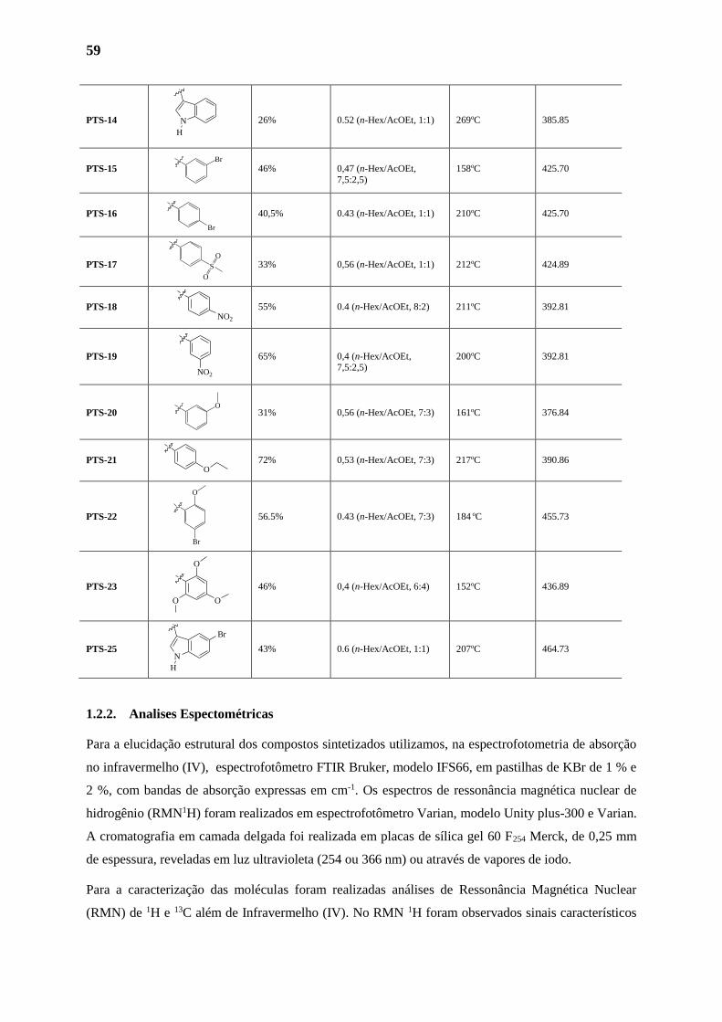
59
PTS-14 26% 0.52 (n-Hex/AcOEt, 1:1) 269oC 385.85
PTS-15 46% 0,47 (n-Hex/AcOEt,
7,5:2,5)
158oC 425.70
PTS-16 40,5% 0.43 (n-Hex/AcOEt, 1:1) 210oC 425.70
PTS-17 33% 0,56 (n-Hex/AcOEt, 1:1) 212oC 424.89
PTS-18 55% 0.4 (n-Hex/AcOEt, 8:2) 211oC 392.81
PTS-19 65% 0,4 (n-Hex/AcOEt, 7,5:2,5)
200oC 392.81
PTS-20 31% 0,56 (n-Hex/AcOEt, 7:3) 161oC 376.84
PTS-21 72% 0,53 (n-Hex/AcOEt, 7:3) 217oC 390.86
PTS-22 56.5% 0.43 (n-Hex/AcOEt, 7:3) 184 oC 455.73
PTS-23 46% 0,4 (n-Hex/AcOEt, 6:4) 152oC 436.89
PTS-25 43% 0.6 (n-Hex/AcOEt, 1:1) 207oC 464.73
1.2.2. Analises Espectométricas
Para a elucidação estrutural dos compostos sintetizados utilizamos, na espectrofotometria de absorção
no infravermelho (IV), espectrofotômetro FTIR Bruker, modelo IFS66, em pastilhas de KBr de 1 % e
2 %, com bandas de absorção expressas em cm-1. Os espectros de ressonância magnética nuclear de
hidrogênio (RMN1H) foram realizados em espectrofotômetro Varian, modelo Unity plus-300 e Varian.
A cromatografia em camada delgada foi realizada em placas de sílica gel 60 F254 Merck, de 0,25 mm
de espessura, reveladas em luz ultravioleta (254 ou 366 nm) ou através de vapores de iodo.
Para a caracterização das moléculas foram realizadas análises de Ressonância Magnética Nuclear
(RMN) de 1H e 13C além de Infravermelho (IV). No RMN 1H foram observados sinais característicos
N
H
Br
O
O
O
O
NO2
NO2
N
H
S
O
O
O
Br
O
Br
Br
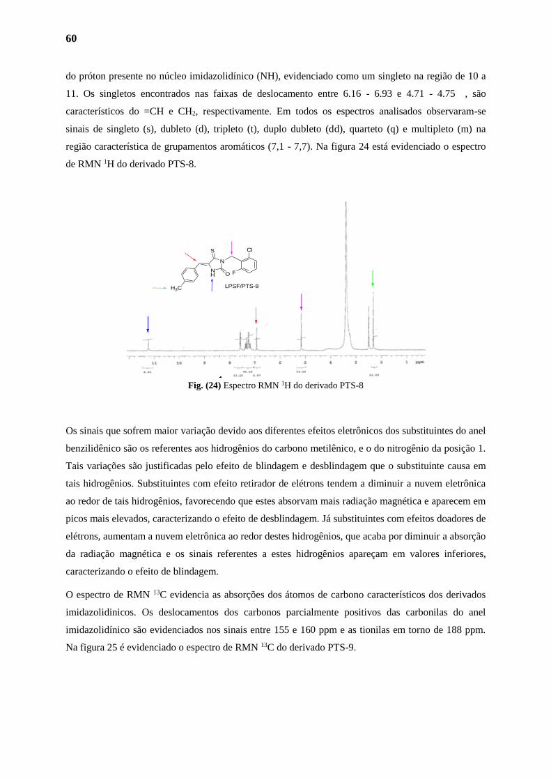
60
do próton presente no núcleo imidazolidínico (NH), evidenciado como um singleto na região de 10 a
11. Os singletos encontrados nas faixas de deslocamento entre 6.16 - 6.93 e 4.71 - 4.75 , são
característicos do =CH e CH2, respectivamente. Em todos os espectros analisados observaram-se
sinais de singleto (s), dubleto (d), tripleto (t), duplo dubleto (dd), quarteto (q) e multipleto (m) na
região característica de grupamentos aromáticos (7,1 - 7,7). Na figura 24 está evidenciado o espectro
de RMN 1H do derivado PTS-8.
Fig. (24) Espectro RMN 1H do derivado PTS-8
Os sinais que sofrem maior variação devido aos diferentes efeitos eletrônicos dos substituintes do anel
benzilidênico são os referentes aos hidrogênios do carbono metilênico, e o do nitrogênio da posição 1.
Tais variações são justificadas pelo efeito de blindagem e desblindagem que o substituinte causa em
tais hidrogênios. Substituintes com efeito retirador de elétrons tendem a diminuir a nuvem eletrônica
ao redor de tais hidrogênios, favorecendo que estes absorvam mais radiação magnética e aparecem em
picos mais elevados, caracterizando o efeito de desblindagem. Já substituintes com efeitos doadores de
elétrons, aumentam a nuvem eletrônica ao redor destes hidrogênios, que acaba por diminuir a absorção
da radiação magnética e os sinais referentes a estes hidrogênios apareçam em valores inferiores,
caracterizando o efeito de blindagem.
O espectro de RMN 13C evidencia as absorções dos átomos de carbono característicos dos derivados
imidazolidinicos. Os deslocamentos dos carbonos parcialmente positivos das carbonilas do anel
imidazolidínico são evidenciados nos sinais entre 155 e 160 ppm e as tionilas em torno de 188 ppm.
Na figura 25 é evidenciado o espectro de RMN 13C do derivado PTS-9.
Cl
FO
S
N
N
H
H3C LPSF/PTS-8

61
Fig. (25) Espectro RMN 13C do derivado PTS-9
Outro ponto de interesse nas análises é observado devido à absorção do carbono benzílico ligado ao
flúor, que encontra-se bastante desblindado, e por vezes aparece como sinais de altos valores, como no
caso do derivado PTS-1, que o referente a este carbono apresenta valor de 160 ppm.
A espectroscopia na região do infravermelho foi utilizada como ferramenta adjunta na caracterização
dos compostos sintetizados, por meio de grupos funcionais. Esses grupos originam bandas que
ocorrem sempre na mesma região do espectro, independente da molécula em questão.130 Os derivados
imidazolidinicos apresentam bandas vibracionais características dos grupos funcionais N-H e C=O
além do sinal característico a funcionalidade C=C. A figura 26 evidencia o espectro de IV do derivado
PTS-4.
Fig. (26) Espectro de infra-vermelho do derivado PTS-4
Cl
FO
S
N
N
H
O
H
Cl
FO
S
N
N
H
(LPSF/PTS-9)
62
2. ESTUDO QUIMIOMÉTRICO
A análise de dados multivariados tem se tornado, de modo crescente, uma importante área da química
e de outras ciências. Isto sugere que a tendência atual do pensamento científico envolve um raciocínio
multivariado, principalmente devido ao desenvolvimento tecnológico atingido neste último século.
Historicamente, a análise de dados multivariados em química tem utilizado ferramentas desenvolvidas
na área de ciências sociais como economia (econometria) e psicologia (psicometria). Apesar de serem
interpretadas para objetivos específicos em cada área, a maioria destas ferramentas se baseia em
sofisticados métodos matemáticos, estatísticos, computacionais e de lógica formal. Em química, as
análises multivariadas são úteis para (1) planejar ou selecionar procedimentos experimentais ótimos;
(2) extrair o máximo da informação química relevante pela análise dos dados; e (3) obter
conhecimentos sobre os sistemas químicos estudados.131–133
2.1. Análise de Componentes Principais
Uma das técnicas quimiométricas mais empregadas para analisar conjuntos de dados contendo muitas
variáveis é a análise de componentes principais (PCA). Uma das principais razões para o sucesso da
PCA é o fato que ela oferece, em geral, representações gráficas de baixa dimensão, com razoável
precisão, para problemas multivariados.131
A PCA é provavelmente o método quimiométrico mais amplamente difundido, e devido à importância
das medidas multivariadas em química, ela é considerada por muitos como o método que mais
significativamente muda o ponto de vista dos químicos para a análise multivariada dos dados.134
A aplicação mais frequente do método PCA ocorre nos casos em que as colunas em X são altamente
colineares, i.e., as variáveis são correlacionadas e apresentam redundâncias. Tais colinearidades
indicam que a matriz X apresenta algum tipo de variabilidade dominante que carrega a maioria da
informação disponível. Estas redundâncias e pequenas variabilidades devem então ser removidas. A
proposta da PCA é expressar as informações mais significativas contidas nas variáveis originais em
um pequeno número de novas variáveis, as então chamadas componentes principais de X.
Naturalmente, pode-se estender esta descrição para outros métodos multivariados, pois muitos diferem
apenas no critério utilizado para determinar como as componentes são construídas. Assim, a PCA
constitui, em muitas maneiras, a base para a análise multivariada dos dados.132
PCA é simplesmente uma rotação do sistema de eixos originais que tenta simultaneamente: i)
encontrar no espaço de dimensão x uma direção ao longo da qual os pontos se encontrem espalhados
de forma máxima e no qual espera-se encontrar algum tipo de padrão e ii) resguardar ao máximo a
informação de partida. De um ponto de vista prático, isto é feito através da diagonalização da matriz
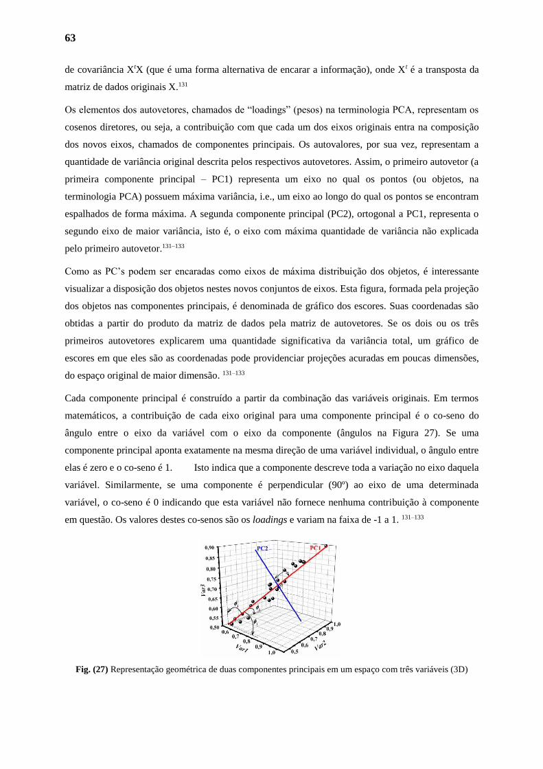
63
de covariância XtX (que é uma forma alternativa de encarar a informação), onde Xt é a transposta da
matriz de dados originais X.131
Os elementos dos autovetores, chamados de “loadings” (pesos) na terminologia PCA, representam os
cosenos diretores, ou seja, a contribuição com que cada um dos eixos originais entra na composição
dos novos eixos, chamados de componentes principais. Os autovalores, por sua vez, representam a
quantidade de variância original descrita pelos respectivos autovetores. Assim, o primeiro autovetor (a
primeira componente principal – PC1) representa um eixo no qual os pontos (ou objetos, na
terminologia PCA) possuem máxima variância, i.e., um eixo ao longo do qual os pontos se encontram
espalhados de forma máxima. A segunda componente principal (PC2), ortogonal a PC1, representa o
segundo eixo de maior variância, isto é, o eixo com máxima quantidade de variância não explicada
pelo primeiro autovetor.131–133
Como as PC’s podem ser encaradas como eixos de máxima distribuição dos objetos, é interessante
visualizar a disposição dos objetos nestes novos conjuntos de eixos. Esta figura, formada pela projeção
dos objetos nas componentes principais, é denominada de gráfico dos escores. Suas coordenadas são
obtidas a partir do produto da matriz de dados pela matriz de autovetores. Se os dois ou os três
primeiros autovetores explicarem uma quantidade significativa da variância total, um gráfico de
escores em que eles são as coordenadas pode providenciar projeções acuradas em poucas dimensões,
do espaço original de maior dimensão. 131–133
Cada componente principal é construído a partir da combinação das variáveis originais. Em termos
matemáticos, a contribuição de cada eixo original para uma componente principal é o co-seno do
ângulo entre o eixo da variável com o eixo da componente (ângulos na Figura 27). Se uma
componente principal aponta exatamente na mesma direção de uma variável individual, o ângulo entre
elas é zero e o co-seno é 1. Isto indica que a componente descreve toda a variação no eixo daquela
variável. Similarmente, se uma componente é perpendicular (90º) ao eixo de uma determinada
variável, o co-seno é 0 indicando que esta variável não fornece nenhuma contribuição à componente
em questão. Os valores destes co-senos são os loadings e variam na faixa de -1 a 1. 131–133
Fig. (27) Representação geométrica de duas componentes principais em um espaço com três variáveis (3D)
64
A direção da primeira componente principal descreve o espalhamento máximo das amostras (linhas)
i.e., a quantidade máxima de variação possível dos dados e pode ser definida como o melhor ajuste da
reta no espaço multidimensional. A próxima componente, perpendicular à primeira (Figura 27),
captura a variância comum em sua direção e em menor quantidade que a primeira e assim
sucessivamente. As componentes principais são, portanto, calculadas em ordem decrescente de
significância e a percentagem da variação total nos dados descrita por qualquer componente pode ser
calculada com precisão usando, por exemplo, informações da diagonal da matriz D. Porém, como as
componentes são perpendiculares entre si, elas contêm informações diferentes e complementares em
ordem decrescente de quantidade de variância. 131–133
Devido à reorientação das coordenadas do sistema de maneira a obter a máxima variância comum, a
maioria da informação dos dados pode ser capturada por um pequeno número de componentes, que
compacta as informações, eliminando as redundâncias e correlações nos dados originais. 131–133
2.2. PCA dos Derivados Tioxo-Imidazolidinicos
A primeira etapa nos estudos de correlação envolve a seleção de um conjunto relevante de parêmetros
ou propriedades moleculares para obter-se a melhor correlação possível. As variáveis escolhidas para
um estudo preliminar de PCA devem abranger, principalmente, dados eletrônicos, de lipofilia e
volume, além de serem facilmente mensurados. Os parâmetros escolhidos para a avaliação na PCA no
presente trabalho foram: (1) Deslocamento do próton imidazolidínico no espectro de RMN 1H, δN-H;
(2) Deslocamento do próton do carbono metilênico no espectro de RMN 1H, δ=CH; (3) Deslocamento
do carbono carbonílico no espectro de RMN 13C, δC=O; (4) Deslocamento do carbono tionílico no
espectro de RMN 13C, δC=S; (5) Log P teórico dos 24 derivados calculado através da plataforma EPA,
Environmental Protection Agency, através do site (http://www.epa.gov/opptintr/exposure/index.htm);
(6) Volume molecular calculado através da programa MarvinSketch; (7) Área de Superfície Polar
calculado através da programa MarvinSketch.
Para PCA os dados da tabela 3 podem ser representados por uma matriz X composta de p = 7 variáveis
(deslocamentos químicos, volume molecular, área de superfície polar e LogP) e n = 24 objetos (os
compostos sintetizados). As analises estatisticas foram realizadas utilizando-se o pacote FactoMineR,
dedicado às analyses multivariadas, disponivel no software R.135
Observe que neste trabalho os valores de deslocamentos químicos podem ser visualizados como as
coordenadas de pontos num espaço de dimensão-4, onde cada eixo estaria associado a um
deslocamento químico. A PCA foi realizada usando dados centrados na média, i.e., o valor do
deslocamento químico dos hidrogênios foi subtraído pela média dos demais valores na mesma coluna.
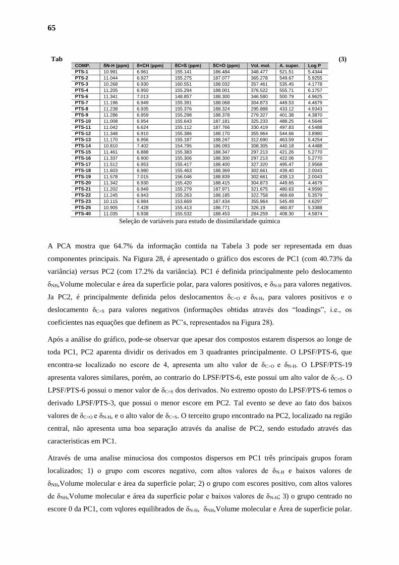
65
Tab (3)
Seleção de variáveis para estudo de dissimilaridade química
A PCA mostra que 64.7% da informação contida na Tabela 3 pode ser representada em duas
componentes principais. Na Figura 28, é apresentado o gráfico dos escores de PC1 (com 40.73% da
variância) versus PC2 (com 17.2% da variância). PC1 é definida principalmente pelo deslocamento
δNH,Volume molecular e área da superficie polar, para valores positivos, e δN-H para valores negativos.
Ja PC2, é principalmente definida pelos deslocamentos δC=O e δN-H, para valores positivos e o
deslocamento δC=S para valores negativos (informações obtidas através dos “loadings”, i.e., os
coeficientes nas equações que definem as PC’s, representados na Figura 28).
Após a análise do gráfico, pode-se observar que apesar dos compostos estarem dispersos ao longe de
toda PC1, PC2 aparenta dividir os derivados em 3 quadrantes principalmente. O LPSF/PTS-6, que
encontra-se localizado no escore de 4, apresenta um alto valor de δC=O e δN-H. O LPSF/PTS-19
apresenta valores similares, porém, ao contrario do LPSF/PTS-6, este possui um alto valor de δC=S. O
LPSF/PTS-6 possui o menor valor de δC=S dos derivados. No extremo oposto do LPSF/PTS-6 temos o
derivado LPSF/PTS-3, que possui o menor escore em PC2. Tal evento se deve ao fato dos baixos
valores de δC=O e δN-H, e o alto valor de δC=S. O terceito grupo encontrado na PC2, localizado na região
central, não apresenta uma boa separação através da analise de PC2, sendo estudado através das
caracteristicas em PC1.
Através de uma analise minuciosa dos compostos dispersos em PC1 três principais grupos foram
localizados; 1) o grupo com escores negativo, com altos valores de δN-H e baixos valores de
δNH,Volume molecular e área da superficie polar; 2) o grupo com escores positivo, com altos valores
de δNH,Volume molecular e área da superficie polar e baixos valores de δN-H; 3) o grupo centrado no
escore 0 da PC1, com vqlores equilibrados de δN-H, δNH,Volume molecular e Área de superficie polar.
COMP. δN-H (ppm) δ=CH (ppm) δC=S (ppm) δC=O (ppm) Vol. mol. A. super. Log P
PTS-1 10.991 6.961 155.141 186.484 348.477 521.51 5.4344
PTS-2 11.044 6.927 155.275 187.077 365.278 549.67 5.9255
PTS-3 10.268 6.930 160.551 188.032 357.461 535.45 4.1778
PTS-4 11.205 6.950 155.294 188.001 376.522 555.71 6.1757
PTS-6 11.341 7.013 148.857 188.300 346.580 500.79 4.9625
PTS-7 11.196 6.949 155.391 188.068 304.873 449.53 4.4679
PTS-8 11.238 6.935 155.376 188.324 295.888 433.12 4.9343
PTS-9 11.286 6.959 155.298 188.378 279.327 401.38 4.3870
PTS-10 11.008 6.954 155.643 187.181 325.233 488.25 4.5646
PTS-11 11.042 6.624 155.112 187.766 330.419 497.83 4.5488
PTS-12 11.348 6.910 155.386 188.170 355.964 544.66 3.8980
PTS-13 11.170 6.956 155.187 188.247 312.690 463.59 5.4254
PTS-14 10.810 7.402 154.795 186.093 308.305 440.18 4.4488
PTS-15 11.461 6.888 155.383 188.347 297.213 421.26 5.2770
PTS-16 11.337 6.900 155.306 188.300 297.213 422.06 5.2770
PTS-17 11.512 6.953 155.417 188.400 327.320 495.47 2.9568
PTS-18 11.603 6.980 155.463 188.369 302.661 439.40 2.0043
PTS-19 11.578 7.015 156.046 188.839 302.661 439.13 2.0043
PTS-20 11.342 6.930 155.420 188.415 304.873 449.65 4.4679
PTS-21 11.202 6.949 155.279 187.971 321.675 480.63 4.9590
PTS-22 11.245 6.943 155.263 188.185 322.758 469.69 5.3579
PTS-23 10.115 6.984 153.669 187.434 355.964 545.49 4.6297
PTS-25 10.905 7.428 155.413 186.771 326.19 460.87 5.3388
PTS-40 11.035 6.938 155.532 188.453 284.259 408.30 4.5874
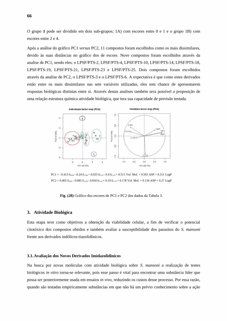
66
O grupo 1 pode ser dividido em dois sub-grupos; 1A) com escores entre 0 e 1 e o grupo 1B) com
escores entre 2 e 4.
Após a análise do gráfico PC1 versus PC2, 11 compostos foram escolhidos como os mais dissimilares,
devido às suas distâncias no gráfico dos de escore. Nove compostos foram escolhidos através da
analise de PC1, sendo eles: o LPSF/PTS-2, LPSF/PTS-4, LPSF/PTS-10, LPSF/PTS-14, LPSF/PTS-18,
LPSF/PTS-19, LPSF/PTS-21, LPSF/PTS-23 e LPSF/PTS-25. Dois compostos foram escolhidos
através da analise de PC2, o LPSF/PTS-3 e o LPSF/PTS-6. A expectativa é que como estes derivados
estão entre os mais dissimilares nas sete variáveis utilizadas, eles tem chance de apresentarem
respostas biológicas distintas entre si. Através destas analises também sera possível a proposição de
uma relação estrutura química atividade biológica, que tera sua capacidade de previsão testada.
PC1 = - 0.413 N-H - 0.24 =CH + 0.025 C=S - 0.4 C=O + 0.511 Vol. Mol. + 0.503 ASP + 0.311 LogP
PC2 = 0.405 N-H - 0.085 =CH - 0.818 C=S + 0.19 C=O + 0.178 Vol. Mol. + 0.134 ASP + 0.27 LogP
Fig. (28) Gráfico dos escores de PC1 e PC2 dos dados da Tabela 3.
3. Atividade Biológica
Esta etapa teve como objetivos a obtenção da viabilidade celular, a fim de verificar o potencial
citotóxico dos compostos obtidos e também avaliar a susceptibilidade dos parasitos do S. mansoni
frente aos derivados indólicos-tiazolidínicos.
3.1. Avaliação dos Novos Derivados Imidazolidinicos
Na busca por novas moléculas com atividade biológica sobre S. mansoni a realização de testes
biológicos in vitro torna-se relevante, pois esse passo é vital para encontrar uma substância líder que
possa ser posteriormente usada em ensaios in vivo, reduzindo os custos desse processo. Por essa razão,
quando são testadas empiricamente substâncias em que não há um prévio conhecimento sobre a ação
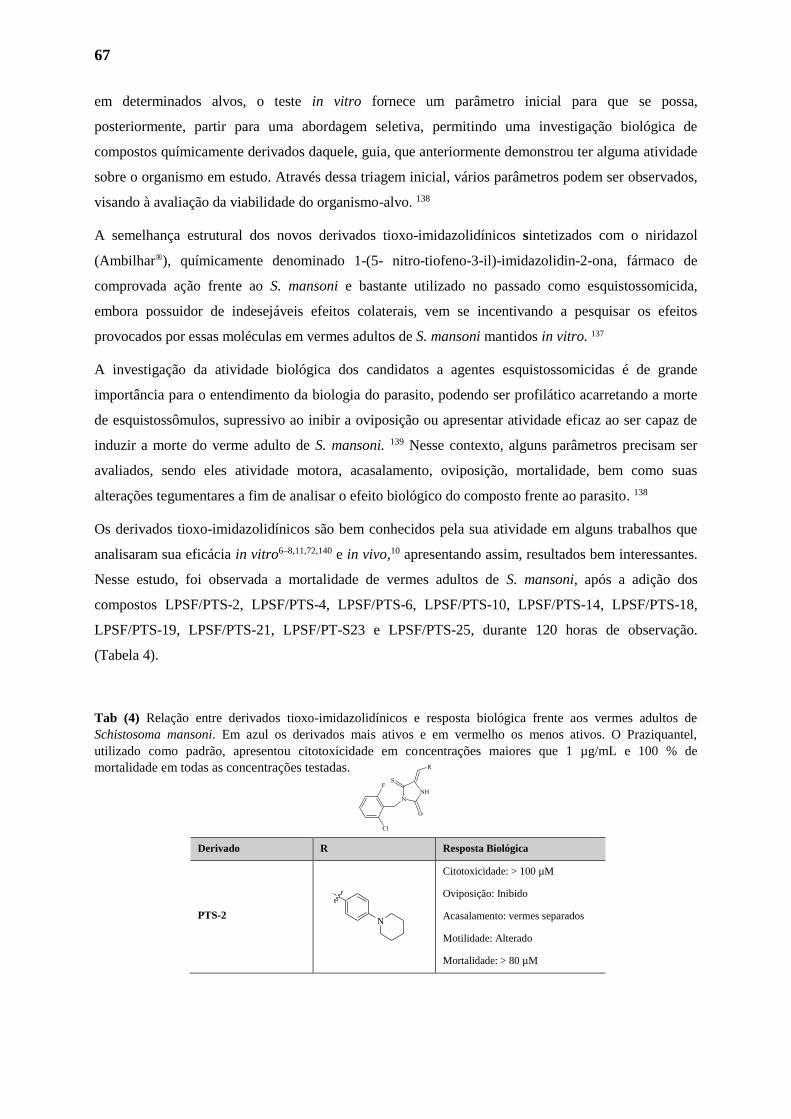
67
em determinados alvos, o teste in vitro fornece um parâmetro inicial para que se possa,
posteriormente, partir para uma abordagem seletiva, permitindo uma investigação biológica de
compostos químicamente derivados daquele, guia, que anteriormente demonstrou ter alguma atividade
sobre o organismo em estudo. Através dessa triagem inicial, vários parâmetros podem ser observados,
visando à avaliação da viabilidade do organismo-alvo. 138
A semelhança estrutural dos novos derivados tioxo-imidazolidínicos sintetizados com o niridazol
(Ambilhar®), químicamente denominado 1-(5- nitro-tiofeno-3-il)-imidazolidin-2-ona, fármaco de
comprovada ação frente ao S. mansoni e bastante utilizado no passado como esquistossomicida,
embora possuidor de indesejáveis efeitos colaterais, vem se incentivando a pesquisar os efeitos
provocados por essas moléculas em vermes adultos de S. mansoni mantidos in vitro. 137
A investigação da atividade biológica dos candidatos a agentes esquistossomicidas é de grande
importância para o entendimento da biologia do parasito, podendo ser profilático acarretando a morte
de esquistossômulos, supressivo ao inibir a oviposição ou apresentar atividade eficaz ao ser capaz de
induzir a morte do verme adulto de S. mansoni. 139 Nesse contexto, alguns parâmetros precisam ser
avaliados, sendo eles atividade motora, acasalamento, oviposição, mortalidade, bem como suas
alterações tegumentares a fim de analisar o efeito biológico do composto frente ao parasito. 138
Os derivados tioxo-imidazolidínicos são bem conhecidos pela sua atividade em alguns trabalhos que
analisaram sua eficácia in vitro6–8,11,72,140 e in vivo,10 apresentando assim, resultados bem interessantes.
Nesse estudo, foi observada a mortalidade de vermes adultos de S. mansoni, após a adição dos
compostos LPSF/PTS-2, LPSF/PTS-4, LPSF/PTS-6, LPSF/PTS-10, LPSF/PTS-14, LPSF/PTS-18,
LPSF/PTS-19, LPSF/PTS-21, LPSF/PT-S23 e LPSF/PTS-25, durante 120 horas de observação.
(Tabela 4).
Tab (4) Relação entre derivados tioxo-imidazolidínicos e resposta biológica frente aos vermes adultos de
Schistosoma mansoni. Em azul os derivados mais ativos e em vermelho os menos ativos. O Praziquantel,
utilizado como padrão, apresentou citotoxicidade em concentrações maiores que 1 µg/mL e 100 % de
mortalidade em todas as concentrações testadas.
Derivado R Resposta Biológica
PTS-2
Citotoxicidade: > 100 µM
Oviposição: Inibido
Acasalamento: vermes separados
Motilidade: Alterado
Mortalidade: > 80 µM
N
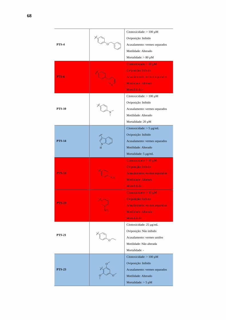
68
PTS-4
Citotoxicidade: > 100 µM
Oviposição: Inibido
Acasalamento: vermes separados
Motilidade: Alterado
Mortalidade: > 80 µM
PTS-6
Citotoxicidade: > 10 µM
Oviposição: Inibido
Acasalamento: vermes separados
Motilidade: Alterado
Mortalidade:-
PTS-10
Citotoxicidade: > 100 µM
Oviposição: Inibido
Acasalamento: vermes separados
Motilidade: Alterado
Mortalidade: 20 µM
PTS-14
Citotoxicidade: > 5 µg/mL
Oviposição: Inibido
Acasalamento: vermes separados
Motilidade: Alterado
Mortalidade: 5 µg/mL
PTS-18
Citotoxicidade: > 10 µM
Oviposição: Inibido
Acasalamento: vermes separados
Motilidade: Alterado
Mortalidade: -
PTS-19
Citotoxicidade: > 10 µM
Oviposição: Inibido
Acasalamento: vermes separados
Motilidade: Alterada
Mortalidade: -
PTS-21
Citotoxicidade: 25 µg/mL
Oviposição: Não inibido
Acasalamento: vermes unidos
Motilidade: Não alterada
Mortalidade: -
PTS-23
Citotoxicidade: > 100 µM
Oviposição: Inibido
Acasalamento: vermes separados
Motilidade: Alterado
Mortalidade: > 5 µM
O
N
N
N
H
NO2
O
NO2
O
O
O
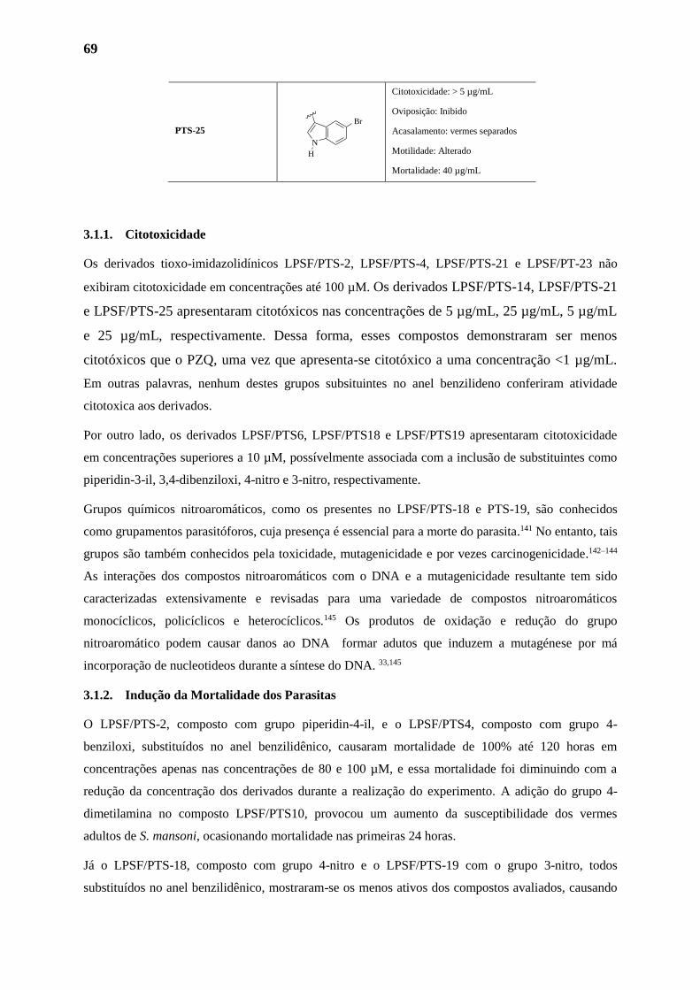
69
PTS-25
Citotoxicidade: > 5 µg/mL
Oviposição: Inibido
Acasalamento: vermes separados
Motilidade: Alterado
Mortalidade: 40 µg/mL
3.1.1. Citotoxicidade
Os derivados tioxo-imidazolidínicos LPSF/PTS-2, LPSF/PTS-4, LPSF/PTS-21 e LPSF/PT-23 não
exibiram citotoxicidade em concentrações até 100 µM. Os derivados LPSF/PTS-14, LPSF/PTS-21
e LPSF/PTS-25 apresentaram citotóxicos nas concentrações de 5 µg/mL, 25 µg/mL, 5 µg/mL
e 25 µg/mL, respectivamente. Dessa forma, esses compostos demonstraram ser menos
citotóxicos que o PZQ, uma vez que apresenta-se citotóxico a uma concentração <1 µg/mL.
Em outras palavras, nenhum destes grupos subsituintes no anel benzilideno conferiram atividade
citotoxica aos derivados.
Por outro lado, os derivados LPSF/PTS6, LPSF/PTS18 e LPSF/PTS19 apresentaram citotoxicidade
em concentrações superiores a 10 µM, possívelmente associada com a inclusão de substituintes como
piperidin-3-il, 3,4-dibenziloxi, 4-nitro e 3-nitro, respectivamente.
Grupos químicos nitroaromáticos, como os presentes no LPSF/PTS-18 e PTS-19, são conhecidos
como grupamentos parasitóforos, cuja presença é essencial para a morte do parasita.141 No entanto, tais
grupos são também conhecidos pela toxicidade, mutagenicidade e por vezes carcinogenicidade.142–144
As interações dos compostos nitroaromáticos com o DNA e a mutagenicidade resultante tem sido
caracterizadas extensivamente e revisadas para uma variedade de compostos nitroaromáticos
monocíclicos, policíclicos e heterocíclicos.145 Os produtos de oxidação e redução do grupo
nitroaromático podem causar danos ao DNA formar adutos que induzem a mutagénese por má
incorporação de nucleotideos durante a síntese do DNA. 33,145
3.1.2. Indução da Mortalidade dos Parasitas
O LPSF/PTS-2, composto com grupo piperidin-4-il, e o LPSF/PTS4, composto com grupo 4-
benziloxi, substituídos no anel benzilidênico, causaram mortalidade de 100% até 120 horas em
concentrações apenas nas concentrações de 80 e 100 µM, e essa mortalidade foi diminuindo com a
redução da concentração dos derivados durante a realização do experimento. A adição do grupo 4-
dimetilamina no composto LPSF/PTS10, provocou um aumento da susceptibilidade dos vermes
adultos de S. mansoni, ocasionando mortalidade nas primeiras 24 horas.
Já o LPSF/PTS-18, composto com grupo 4-nitro e o LPSF/PTS-19 com o grupo 3-nitro, todos
substituídos no anel benzilidênico, mostraram-se os menos ativos dos compostos avaliados, causando
N
H
Br
70
mortalidade de 50% ao final de 120 horas na concentração de 10 µM. Estes compostos foram
avaliados apenas na concentração de 10 µM, uma vez que a adição desses grupos ao anel
benzilidênico tornou estes mais citotóxicos.
O LPSF/PTS-23, composto com grupo metoxila nas posições 2,4,6 do anel benzilidênico, causou a
morte de 100% dos vermes até 120 horas em concentrações variando de 5 a 100 µM. Achados da
literatura também mostraram que os compostos heterocíclicos contendo um grupo metoxila exibiram
maior atividade esquistossomicida do que compostos sem este grupo.146
O composto LPSF/PTS-25 apresentou uma taxa de mortalidade de 40% dos parasitos na concentração
de 100 µg/mL como pode ser visto na tabela 3. Dentre todos os compostos imidazolidínicos testados e
observados o composto LPSF/PTS-14 apresentou o resultado mais expressivo com uma taxa de
mortalidade de 100% dos parasitos nas primeiras 24 horas de observação em todas as doses testadas.
O mesmo resultado pôde ser visto com o composto padrão do experimento, o PZQ. No entanto, o
derivados LPSF/PTS-21 e não foi capaz de causar mortalidade em nenhuma das doses testadas.
O LPSF/PTS-14, além de ser o melhor composto esquistossomicida desta série, representa o primeiro
derivado indolico-imidazolidinico com ação esquistossomicida desenvolvido pelo Gpit. O radical
indol é de grande importância na estrutura do LPSF/PTS-14, uma vez que possui uma vasta gama de
propriedades associadas, tais como anti-fúngica,147 anti-inflamatória 148 e anti-tuberculose.149 O radical
indol é também um bioisóstere de quinolina, encontrada em trioxaquinas e mefloquina, com alto nível
de mortalidade de vermes adultos de S. mansoni.150–152
3.1.3. Atividade Motora
Os vermes adultos de S. mansoni, principalmente os vermes machos, apresentam uma variedade de
movimentos entre os quais, rápidos encurtamentos e prolongamento do corpo, movimentos
ondulatórios e ondas peristálticas, parcial ou de uma extremidade a outra.153 A motilidade do S.
mansoni, e o efeitos de compostos esquistossomicida sobre essa atividade, baseiam-se, principalmente,
na observação visual. A análise macroscópica da atividade motora, apresenta a vantagem de ser um
método simples, direto, de baixo custo e facilidade operacional.154,155
Nesse estudo, a atividade motora dos vermes adultos de S. mansoni, baseada na observação
microscópica dos vermes, foi diariamente monitorada qualitativamente definida como “normal”, “com
menor perda dos movimentos” e “sem movimentos”. Apesar de subjetivo, esse critério qualitativo é
comumente empregado nos ensaios in vitro.139,155–157 Nos experimentos realizados no estudo,
comparativamente aos vermes adultos incubados apenas em meio de cultura, a motilidade dos
parasitos foi alterada quando incubados na presença dos derivados tioxo-imidazolidínicos. Foi descrito
também, que os compostos tioxo-imidazolidínicos que foram letais para os vermes adultos,
71
independentemente do tempo de incubação. Nesse mesmo estudo, observou-se que, nenhum dos
derivados induziu a contração muscular nos parasitos. Por outro lado, PZQ induziu, nos instantes
iniciais, contração dos helmintos e redução na motilidade sem a separação dos pares de vermes adultos
de S. mansoni, achados esses também observados no estudo realizado por Moraes (2011).155
Em S. mansoni o controle da atividade motora ocorre por diversos neurotransmissores ou
neuromoduladores, tais como: os pertencentes ao grupo de mediadores do tipo amina (serotonina,
dopamina, noradrenalina, adrenalina, acetilcolina); aos do tipo aminoácidos (glutamato, γ-ácido
aminobutírico-GABA) e aos neuropeptídeos (peptídeos da família FMRF-amidas - FaRPs).170,171
Cada um desses neurotransmissores/neuromoduladores atua de formas diferentes na motilidade
aumentando-a (serotonina), diminuindo-a (acetilcolina) ou mantendo-a constante (dopamina,
noradrenalina, adrenalina, glutamato e GABA). Assim, a partir da movimentação que o verme adquire
após a adição de uma amostra a ser testada, ocorrem evidências farmacológicas da presença desses
neurotransmissores no meio de incubação.154
Desta forma, os neurotransmissores são possíveis alvos de agentes esquistossomicidas, sobretudo
depois de ter sido mostrado que agonistas/antagonistas desses sistemas produziram alterações no
padrão de motilidade dos vermes.154,158 Então, levando em consideração os dados já publicados, é
possível que as alterações na atividade motora em vermes adultos de S. mansoni em presença de
compostos esquistossomicida estejam associados à neurotransmissores ou neuromoduladores.155
3.1.4. Oviposiçao
As manifestações clínicas da esquistossomose estão associadas, principalmente, à presença dos ovos
de S. mansoni, no tecido do hospedeiro.159 Dessa forma, na busca de compostos para terapêutica
experimental deve-se considerar drogas supressoras, que acabem com a oviposição da fêmea e,
portanto, eliminem o principal agente patogênico, o ovo, além de interromper a transmissão dessa
parasitose. 138 O objetivo dessa fase da pesquisa foi de avaliar a capacidade reprodutiva de vermes
adultos de S. mansoni quando em presença de compostos imidazolidínicos. A capacidade reprodutiva
de S. mansoni foi avaliada pela observação aos efeitos da exposição no acasalamento e oviposição das
fêmeas. Nos experimentos, foram observadas inibição da oviposição durante todo período do
experimento quando os vermes foram incubados com todos os derivados tioxo-imidazolidínicos
empregados nesse estudo, exceto o LPSF/PTS-21 que, assim como no grupo controle (vermes não
tratados), foi observada a oviposição e se os vermes permaneceram viáveis durante o período de
observação. Alguns estudos relatam através de experimentos in vitro a redução da oviposição de S.
mansoni por compostos imidazolidínicos.6,8,11,140,146
A redução da oviposição pode ser causada por alterações no sistema reprodutivo de machos e/ou
fêmeas.155 Por exemplo, um estudo sobre o efeito esquistossomicida da lovastatina realizado por
72
Araújo et al. (2002),160 mostraram que esse medicamento foi capaz de causar redução da oviposição de
vermes adultos de S. mansoni. Os estudos morfológicos em vermes adultos expostos a lovastatina
mostraram alterações degenerativas, sendo as principais no aparelho reprodutor, com redução dos
folículos vitelínicos e do ovário das fêmeas e modificações nos testículos dos machos. No estudo não
foi verificado se os derivados tioxo-imidazolidínicos causam alterações morfológicas nos órgãos
reprodutivos. No entanto, baseado na ausência de ovos observada nos experimentos desse estudo, é
provável que os órgãos reprodutores dos vermes adultos de S. mansoni tenha afetado a oogênese e/ou
espermatogênese.
3.1.5. Alterações no Tegumento do Parasita
Não obstante a análise subjetiva para avaliar o efeito de compostos esquistossomicida, sobre o
tegumento do S. mansoni, esse critério é qualitativo e comumente empregado em ensaios in
vitro.25,140,161
No que diz respeito as alterações ultraestruturais, a superfície tegumentar tem sido o alvo biológico
bastante importante no combate ao parasito, uma vez que essa estrutura se caracteriza por estar
envolvida com a resposta imune do parasito frente ao hospedeiro definitivo, com atividade sensorial e
com a capacidade de absorção de nutrientes.162 Os resultados baseados em análises ultraestruturais por
MEV demonstraram que vermes adultos após serem incubados com o controle, observou-se o
tegumento parasitário com ventosas orais e ventraies, tubérculos e espinhas em estado normal apos
120 h de incubação, respectivamente representados nas figuras 30A e 30B. Danos graves do
tegumento foram observados na incubação de vermes com o PZQ, contração e ruptura de bolhas.
Observou-se a apararição de vários orifícios no local onde as bolhas foram localizadas e perda de
espinhas (Figura 30C).
As alterações ultraestruturais em esquistossomas adultos causadas pelo LPSF/PTS-23 foram
caracterizadas por extensa erosão em todo o corpo dos vermes (Figura 30D); Após 48 h, várias bolhas
e projeções (setas sólidas) emergindo do interior de tubérculos danificados (Figura 30E); Após 72 h, a
erosão do tegumento pode ser visualizada com maior ampliação (Figura 30F); Após 96 h, alguns
tubérculos não têm espinhas (seta tracejada) (Figura 30G). Tubérculos e bolhas danificados presentes
dentro da lesão, como observado após 120 h de exposição ao derivado (Figura 30H).
O mesmo resultado foi observado em trabalhos anteriores com compostos tioxo-
imidazolidínicos.25,71,140,161 Sendo assim, suspeita-se que, de alguma forma, LPSF/PTS23 atue no
sistema tegumentar, interferindo nas células do tegumento, porém, esse mecanismo também ainda não
foi totalmente elucidado no estudo.
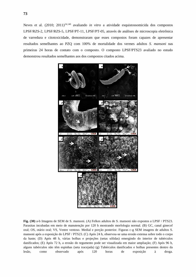
73
Neves et al. (2010; 2011)24,140 avaliando in vitro a atividade esquistossomicida dos compostos
LPSF/RZS-2, LPSF/RZS-5, LPSF/PT-11, LPSF/PT-05, através de análises de microscopia eletrônica
de varredura e citotoxicidade, demonstraram que esses compostos foram capazes de apresentar
resultados semelhantes ao PZQ com 100% de mortalidade dos vermes adultos S. mansoni nas
primeiras 24 horas de contato com o composto. O composto LPSF/PTS23 avaliado no estudo
demonstrou resultados semelhantes aos dos compostos citados acima.
Fig. (30) a-b Imagens de SEM de S. mansoni. (A) Felkes adultos de S. mansoni não expostos a LPSF / PTS23.
Parasitas incubadas em meio de manutenção por 120 h mostrando morfologia normal. (B) GC, canal ginecof
oral; OS, otário oral; VS, Ventre ventoso. Medial e porção posterior. Figuras c-g SEM imagens de adultos S.
mansoni após a exposição do LPSF / PTS23. (C) Após 24 h, observou-se uma erosão extensa sobre todo o corpo
da haste; (D) Após 48 h, várias bolhas e projeções (setas sólidas) emergindo do interior de tubérculos
danificados; (E) Após 72 h, a erosão do tegumento pode ser visualizada em maior ampliação; (F) Após 96 h,
alguns tubérculos não têm espinhas (seta tracejada) (g) Tubérculos danificados e bolhas presentes dentro da
lesão, como observado após 120 horas de exposição à droga.
E F
C D
A B
G
Cg O
v Ov
Vv
Vv
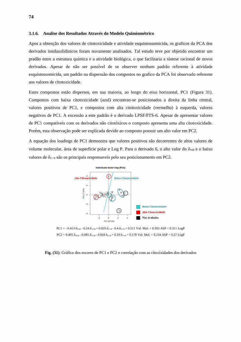
74
3.1.6. Analise dos Resultados Através do Modelo Quimiométrico
Apos a obtenção dos valores de citotoxicidade e atividade esquistossomicida, os graficos da PCA dos
derivados imidazolidinicos foram novamente analisados. Tal estudo teve por objetido encontrar um
pradão entre a estrutura química e a atividade biológica, o que facilitaria a síntese racional de novos
derivados. Apesar de não ser possível de se observer nenhum padrão referente à atividade
esquistossomicida, um padrão na disperssão dos compostos no grafico da PCA foi observado referente
aos valores de citotoxicidade.
Estes compostos estão dispersos, em sua maioria, ao longo do eixo horizontal, PC1 (Figura 31).
Compostos com baixa citotoxicidade (azul) encontran-se posicionados a direita da linha central,
valores positivos de PC1, e compostos com alta citotoxicidade (vermelho) à esquerda, valores
negativos de PC1. A excessão a este padrão é o derivado LPSF/PTS-6. Apesar de apresentar valores
de PC1 compativeis com os derivados não citotóxicos o composto apresenta uma alta citotoxicidade.
Porém, esta observação pode ser explicada devido ao composto possuir um alto valor em PC2.
A equação dos loadings de PC1 demonstra que valores positivos são decorrentes de altos valores de
volume molecular, área de superficie polar e Log P. Para o derivado 6, o alto valor do e o baixo
valores de C=S são os principais responsaveis pelo seu posicionamento em PC2.
PC1 = - 0.413 N-H - 0.24 =CH + 0.025 C=S - 0.4 C=O + 0.511 Vol. Mol. + 0.503 ASP + 0.311 LogP
PC2 = 0.405 N-H - 0.085 =CH - 0.818 C=S + 0.19 C=O + 0.178 Vol. Mol. + 0.134 ASP + 0.27 LogP
Fig. (31): Gráfico dos escores de PC1 e PC2 e correlação com as citoxixidades dos derivados
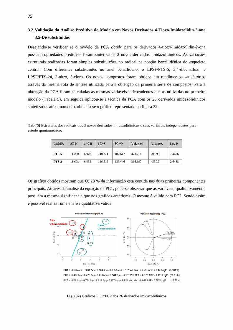
75
3.2. Validação da Análise Preditiva do Modelo em Novos Derivados 4-Tioxo-Imidazolidin-2-ona
3,5-Dissubstituídos
Desejando-se verificar se o modelo de PCA obtido para os derivados 4-tioxo-imidazolidin-2-ona
possui propriedades preditivas foram sintetizados 2 novos derivados imidazolidínicos. As variações
estruturais realizadas foram simples substituições no radical na porção benzilidênica do esqueleto
central. Com diferentes substituintes no anel benzilideno, o LPSF/PTS-5, 3,4-dibenziloxi, e
LPSF/PTS-24, 2-nitro, 5-cloro. Os novos compostos foram obtidos em rendimentos satisfatórios
através da mesma rota de síntese utilizada para a obtenção da primeira série de compostos. Para a
obtenção da PCA foram calculadas as mesmas variáveis independentes que as utilizadas no primeiro
modelo (Tabela 5), em seguida aplicou-se a técnica da PCA com os 26 derivados imidazolidínicos
sintetizados até o momento, obtendo-se o gráfico representado na figura 32.
Tab (5) Estruturas dos radicais dos 3 novos derivados imidazolidínicos e suas variáveis independentes para
estudo qumiométrico.
Os grafico obtidos mostram que 66,28 % da informação esta contida nas duas primeiras componentes
principais. Através da analise da equação de PC1, pode-se observar que as variaveis, qualitativamente,
possuem a mesma significancia que nos graficos anteriores. O mesmo é valido para PC2. Sendo assim
é possível realizar uma analise qualitativa valida.
Fig. (32) Graficos PC1xPC2 dos 26 derivados imidazolidinicos
COMP. δN-H δ=CH δC=S δC=O Vol. mol. A. super. Log P
PTS-5 11.230 6.923 148.274 187.617 473.718 709.93 7.4476
PTS-24 11.698 6.952 146.512 188.446 316.197 455.32 2.6488
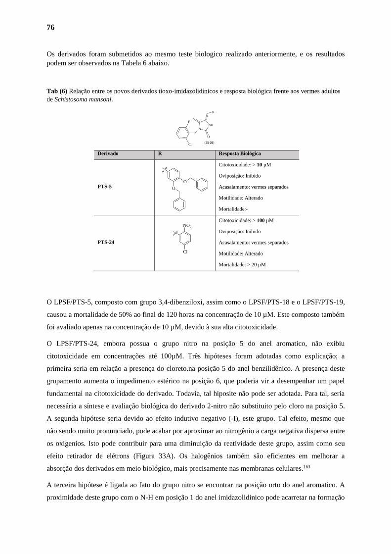
76
Os derivados foram submetidos ao mesmo teste biologico realizado anteriormente, e os resultados
podem ser observados na Tabela 6 abaixo.
Tab (6) Relação entre os novos derivados tioxo-imidazolidínicos e resposta biológica frente aos vermes adultos
de Schistosoma mansoni.
Derivado R Resposta Biológica
PTS-5
Citotoxicidade: > 10 µM
Oviposição: Inibido
Acasalamento: vermes separados
Motilidade: Alterado
Mortalidade:-
PTS-24
Citotoxicidade: > 100 µM
Oviposição: Inibido
Acasalamento: vermes separados
Motilidade: Alterado
Mortalidade: > 20 µM
O LPSF/PTS-5, composto com grupo 3,4-dibenziloxi, assim como o LPSF/PTS-18 e o LPSF/PTS-19,
causou a mortalidade de 50% ao final de 120 horas na concentração de 10 µM. Este composto também
foi avaliado apenas na concentração de 10 µM, devido à sua alta citotoxicidade.
O LPSF/PTS-24, embora possua o grupo nitro na posição 5 do anel aromatico, não exibiu
citotoxicidade em concentrações até 100µM. Três hipóteses foram adotadas como explicação; a
primeira seria em relação a presença do cloreto.na posição 5 do anel benzilidênico. A presença deste
grupamento aumenta o impedimento estérico na posição 6, que poderia vir a desempenhar um papel
fundamental na citotoxicidade do derivado. Todavia, tal hiposite não pode ser adotada. Para tal, seria
necessária a síntese e avaliação biológica do derivado 2-nitro não substituito pelo cloro na posição 5.
A segunda hipótese seria devido ao efeito indutivo negativo (-I), este grupo. Tal efeito, mesmo que
não sendo muito pronunciado, pode acabar por aproximar ao nitrogênio a carga negativa dispersa entre
os oxigenios. Isto pode contribuir para uma diminuição da reatividade deste grupo, assim como seu
efeito retirador de elétrons (Figura 33A). Os halogênios também são eficientes em melhorar a
absorção dos derivados em meio biológico, mais precisamente nas membranas celulares.163
A terceira hipótese é ligada ao fato do grupo nitro se encontrar na posição orto do anel aromatico. A
proximidade deste grupo com o N-H em posição 1 do anel imidazolidinico pode acarretar na formação
O
O
Cl
NO2
NH
N
O
S
R
F
Cl(25-26)
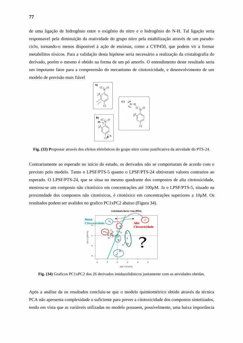
77
de uma ligação de hidrogênio entre o oxigênio do nitro e o hidrogênio do N-H. Tal ligação seria
responsavel pela diminuição da reatividade do grupo nitro pela estabilização através de um pseudo-
ciclo, tornando-o menos disponivel à ação de enzimas, como a CYP450, que podem vir a formar
metabólitos tóxicos. Para a validação desta hipótese seria necessário a realização da cristalografia do
derivado, porém o mesmo é obtido na forma de um pó amorfo. O entendimento deste resultado seria
um impotante fator para a compreensão do mecanismo de citotoxicidade, e desenvolvimento de um
modelo de previsão mais fiável
Fig. (33) Propostar através dos efeitos eletrônicos do grupo nitro como justificativa da atividade do PTS-24.
Contrariamente ao esperado no início do estudo, os derivados não se comportaram de acordo com o
previsto pelo modelo. Tanto o LPSF/PTS-5 quanto o LPSF/PTS-24 obtiveram valores contrarios ao
esperado. O LPSF/PTS-24, que se situa no mesmo quadrante dos compostos de alta citotoxicidade,
mostrou-se um composto não citotóxico em concentrações até 100µM. Ja o LPSF/PTS-5, situado na
proximidade dos compostos não citotóxicos, é citotóxico em concentrações superiores a 10µM. Os
resultados podem ser avalidos no grafico PC1xPC2 abaixo (Figura 34).
Fig. (34) Graficos PC1xPC2 dos 26 derivados imidazolidinicos juntamente com as atividades obtidas.
Após a análise da os resultados concluiu-se que o modelo quimiométrico obtido através da técnica
PCA não apresenta complexidade o suficiente para prever a citotoxicidade dos compostos sintetizados,
tendo em vista que as variáveis utilizadas no modelo possuem, possívelmente, uma baixa importância
Cl
NO O
A)
Cl
NO O
B)
Cl
NO O
I-
M-
HNC)
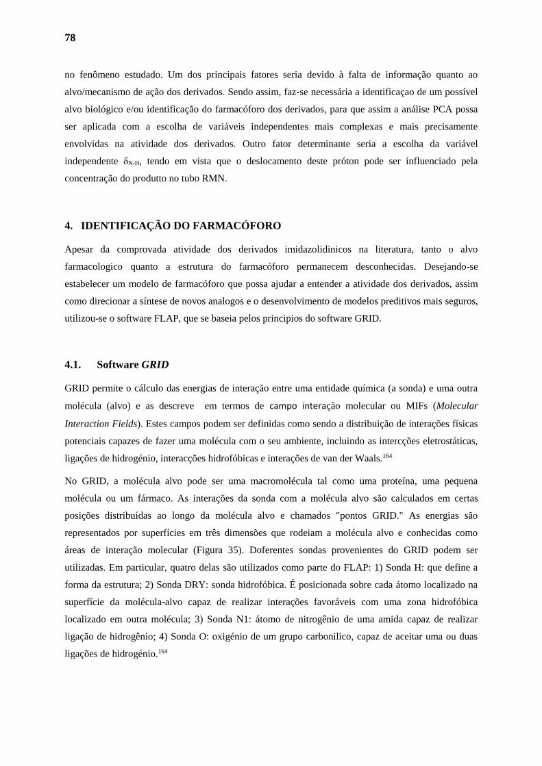
78
no fenômeno estudado. Um dos principais fatores seria devido à falta de informação quanto ao
alvo/mecanismo de ação dos derivados. Sendo assim, faz-se necessária a identificaçao de um possível
alvo biológico e/ou identificação do farmacóforo dos derivados, para que assim a análise PCA possa
ser aplicada com a escolha de variáveis independentes mais complexas e mais precisamente
envolvidas na atividade dos derivados. Outro fator determinante seria a escolha da variável
independente δN-H, tendo em vista que o deslocamento deste próton pode ser influenciado pela
concentração do produtto no tubo RMN.
4. IDENTIFICAÇÃO DO FARMACÓFORO
Apesar da comprovada atividade dos derivados imidazolidinicos na literatura, tanto o alvo
farmacologico quanto a estrutura do farmacóforo permanecem desconhecidas. Desejando-se
estabelecer um modelo de farmacóforo que possa ajudar a entender a atividade dos derivados, assim
como direcionar a síntese de novos analogos e o desenvolvimento de modelos preditivos mais seguros,
utilizou-se o software FLAP, que se baseia pelos principios do software GRID.
4.1. Software GRID
GRID permite o cálculo das energias de interação entre uma entidade química (a sonda) e uma outra
molécula (alvo) e as descreve em termos de campo interação molecular ou MIFs (Molecular
Interaction Fields). Estes campos podem ser definidas como sendo a distribuição de interações físicas
potenciais capazes de fazer uma molécula com o seu ambiente, incluindo as intercções eletrostáticas,
ligações de hidrogénio, interacções hidrofóbicas e interações de van der Waals.164
No GRID, a molécula alvo pode ser uma macromolécula tal como uma proteína, uma pequena
molécula ou um fármaco. As interações da sonda com a molécula alvo são calculados em certas
posições distribuídas ao longo da molécula alvo e chamados "pontos GRID." As energias são
representados por superfícies em três dimensões que rodeiam a molécula alvo e conhecidas como
áreas de interação molecular (Figura 35). Doferentes sondas provenientes do GRID podem ser
utilizadas. Em particular, quatro delas são utilizados como parte do FLAP: 1) Sonda H: que define a
forma da estrutura; 2) Sonda DRY: sonda hidrofóbica. É posicionada sobre cada átomo localizado na
superfície da molécula-alvo capaz de realizar interações favoráveis com uma zona hidrofóbica
localizado em outra molécula; 3) Sonda N1: átomo de nitrogênio de uma amida capaz de realizar
ligação de hidrogênio; 4) Sonda O: oxigénio de um grupo carbonilico, capaz de aceitar uma ou duas
ligações de hidrogénio.164
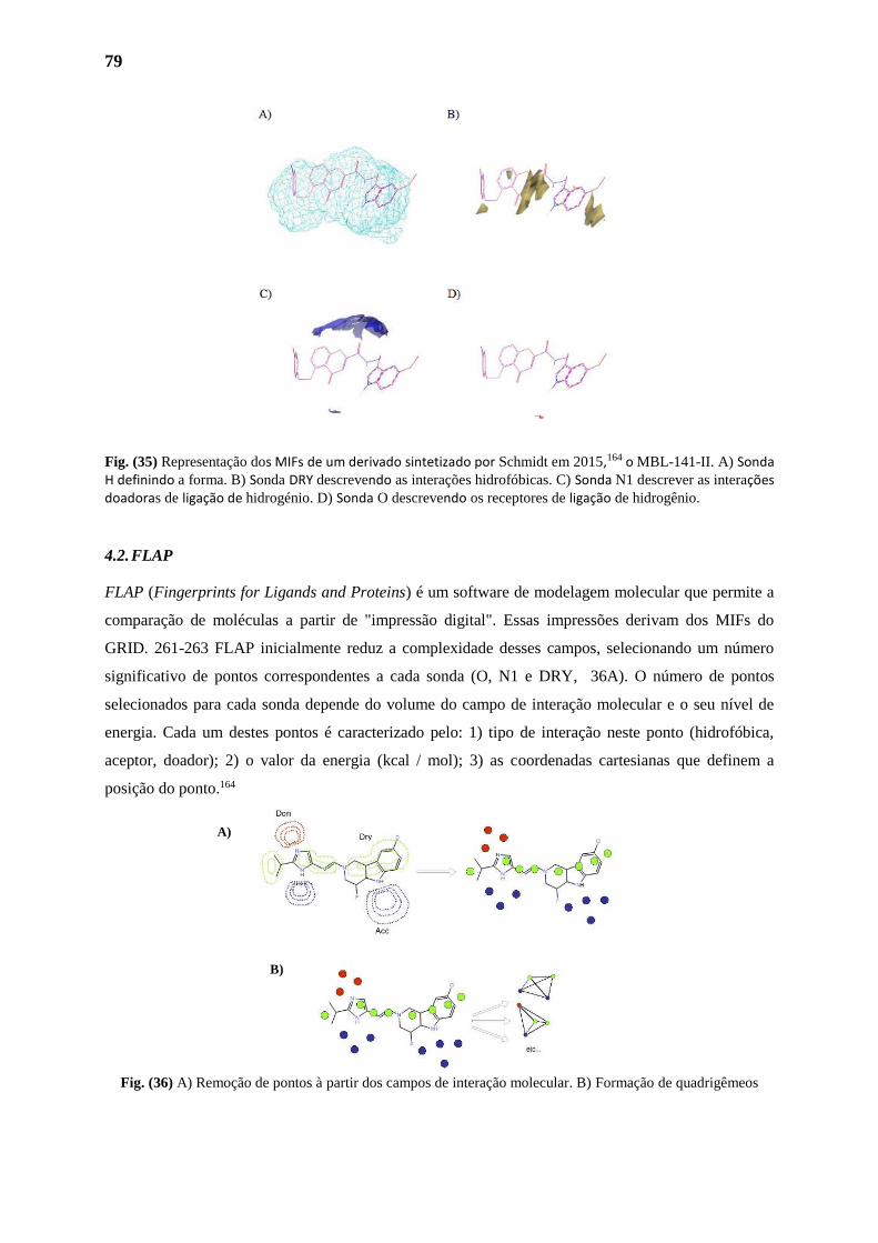
79
Fig. (35) Representação dos MIFs de um derivado sintetizado por Schmidt em 2015,164 o MBL-141-II. A) Sonda H definindo a forma. B) Sonda DRY descrevendo as interações hidrofóbicas. C) Sonda N1 descrever as interações doadoras de ligação de hidrogénio. D) Sonda O descrevendo os receptores de ligação de hidrogênio.
4.2. FLAP
FLAP (Fingerprints for Ligands and Proteins) é um software de modelagem molecular que permite a
comparação de moléculas a partir de "impressão digital". Essas impressões derivam dos MIFs do
GRID. 261-263 FLAP inicialmente reduz a complexidade desses campos, selecionando um número
significativo de pontos correspondentes a cada sonda (O, N1 e DRY, 36A). O número de pontos
selecionados para cada sonda depende do volume do campo de interação molecular e o seu nível de
energia. Cada um destes pontos é caracterizado pelo: 1) tipo de interação neste ponto (hidrofóbica,
aceptor, doador); 2) o valor da energia (kcal / mol); 3) as coordenadas cartesianas que definem a
posição do ponto.164
Fig. (36) A) Remoção de pontos à partir dos campos de interação molecular. B) Formação de quadrigêmeos
A)
B)
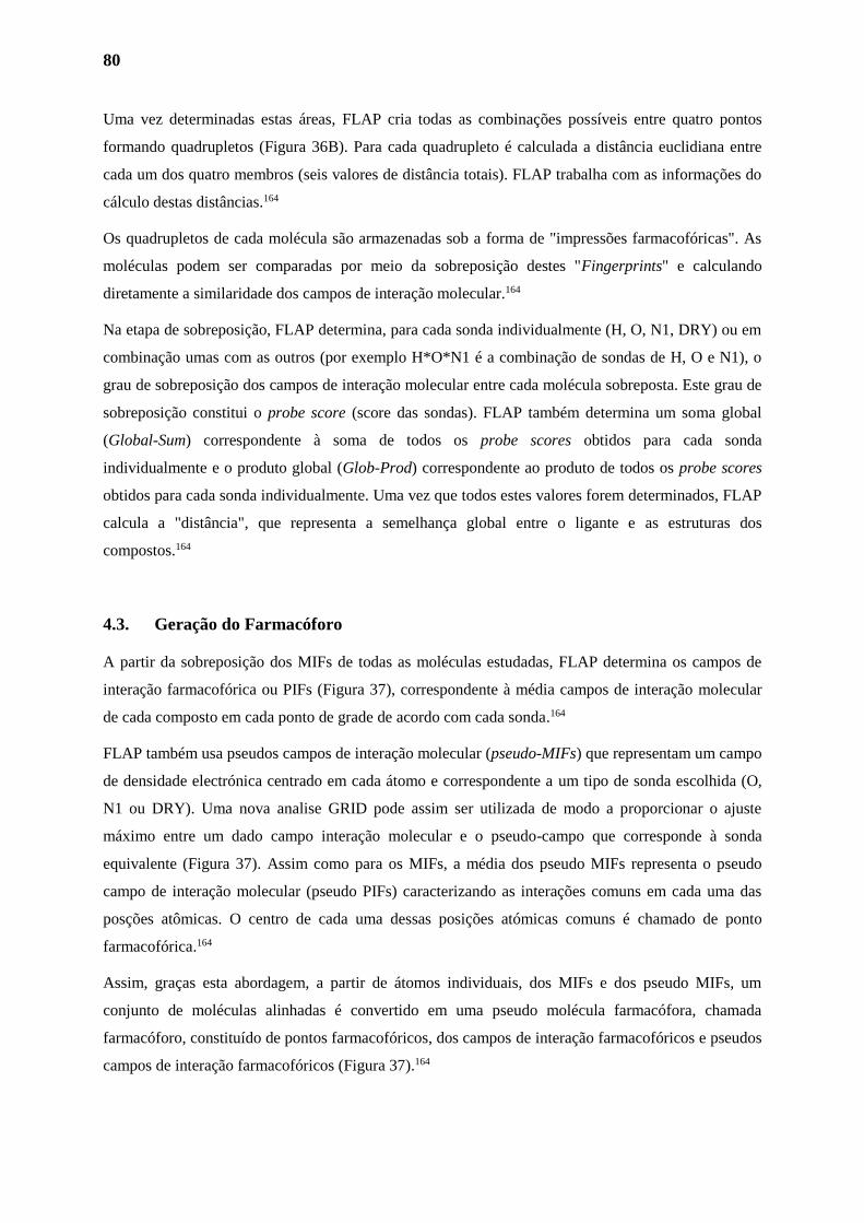
80
Uma vez determinadas estas áreas, FLAP cria todas as combinações possíveis entre quatro pontos
formando quadrupletos (Figura 36B). Para cada quadrupleto é calculada a distância euclidiana entre
cada um dos quatro membros (seis valores de distância totais). FLAP trabalha com as informações do
cálculo destas distâncias.164
Os quadrupletos de cada molécula são armazenadas sob a forma de "impressões farmacofóricas". As
moléculas podem ser comparadas por meio da sobreposição destes "Fingerprints" e calculando
diretamente a similaridade dos campos de interação molecular.164
Na etapa de sobreposição, FLAP determina, para cada sonda individualmente (H, O, N1, DRY) ou em
combinação umas com as outros (por exemplo H*O*N1 é a combinação de sondas de H, O e N1), o
grau de sobreposição dos campos de interação molecular entre cada molécula sobreposta. Este grau de
sobreposição constitui o probe score (score das sondas). FLAP também determina um soma global
(Global-Sum) correspondente à soma de todos os probe scores obtidos para cada sonda
individualmente e o produto global (Glob-Prod) correspondente ao produto de todos os probe scores
obtidos para cada sonda individualmente. Uma vez que todos estes valores forem determinados, FLAP
calcula a "distância", que representa a semelhança global entre o ligante e as estruturas dos
compostos.164
4.3. Geração do Farmacóforo
A partir da sobreposição dos MIFs de todas as moléculas estudadas, FLAP determina os campos de
interação farmacofórica ou PIFs (Figura 37), correspondente à média campos de interação molecular
de cada composto em cada ponto de grade de acordo com cada sonda.164
FLAP também usa pseudos campos de interação molecular (pseudo-MIFs) que representam um campo
de densidade electrónica centrado em cada átomo e correspondente a um tipo de sonda escolhida (O,
N1 ou DRY). Uma nova analise GRID pode assim ser utilizada de modo a proporcionar o ajuste
máximo entre um dado campo interação molecular e o pseudo-campo que corresponde à sonda
equivalente (Figura 37). Assim como para os MIFs, a média dos pseudo MIFs representa o pseudo
campo de interação molecular (pseudo PIFs) caracterizando as interações comuns em cada uma das
posções atômicas. O centro de cada uma dessas posições atómicas comuns é chamado de ponto
farmacofórica.164
Assim, graças esta abordagem, a partir de átomos individuais, dos MIFs e dos pseudo MIFs, um
conjunto de moléculas alinhadas é convertido em uma pseudo molécula farmacófora, chamada
farmacóforo, constituído de pontos farmacofóricos, dos campos de interação farmacofóricos e pseudos
campos de interação farmacofóricos (Figura 37).164
81
Fig. (37) Representação dos MIFs (esquerda) e pseudo MIFs (direita) correspondente a três compostos
selecionados (A, B e C) por Schmidt em 2015.164 No centro estão representados o farmacóforo (meio) gerado
pelo FLAP a partir destes três compostos e as representações do PIF (superior) e os pseudo-PIF (parte inferior).
Em amarelo são representadas as interações hidrofóbicas, em vermelho as interações de aceptores de ligação de
hidrogénio e em azul as interações doadores de hidrogênio
4.4. Seleção dos Compostos para a Criação do Farmacóforo
FLAP precisa apenas de 3 a 5 compostos para gerar um modelo farmacóforo confiàvel .165,166 Como
primeiro passo desta etapa foram escolhidos os compostos a partir de bases de dados disponíveis. Para
executar esta tarefa, foi realizada uma seleção "manual” com base nos melhores derivados ja
produzidos em nosso grupo, juntamento com os dois melhores derivados obtidos neste traabalho, o
LPSF/PTS-14 e o LPSF/PTS-23. Também foi realizada uma tentativa de farmacóforo através do
composto LPSF/PTS-14 e derivados classicos no tratamento da esquistossomose.
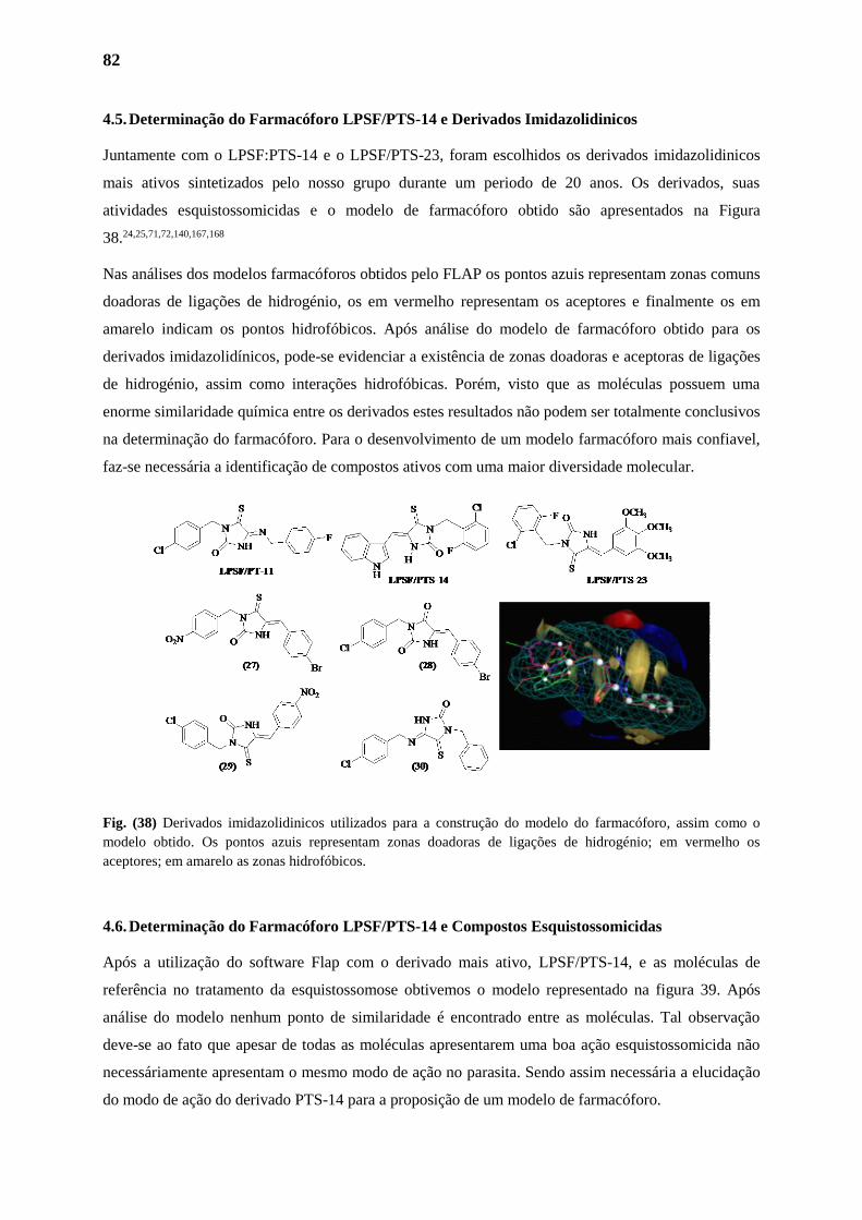
82
4.5. Determinação do Farmacóforo LPSF/PTS-14 e Derivados Imidazolidinicos
Juntamente com o LPSF:PTS-14 e o LPSF/PTS-23, foram escolhidos os derivados imidazolidinicos
mais ativos sintetizados pelo nosso grupo durante um periodo de 20 anos. Os derivados, suas
atividades esquistossomicidas e o modelo de farmacóforo obtido são apresentados na Figura
38.24,25,71,72,140,167,168
Nas análises dos modelos farmacóforos obtidos pelo FLAP os pontos azuis representam zonas comuns
doadoras de ligações de hidrogénio, os em vermelho representam os aceptores e finalmente os em
amarelo indicam os pontos hidrofóbicos. Após análise do modelo de farmacóforo obtido para os
derivados imidazolidínicos, pode-se evidenciar a existência de zonas doadoras e aceptoras de ligações
de hidrogénio, assim como interações hidrofóbicas. Porém, visto que as moléculas possuem uma
enorme similaridade química entre os derivados estes resultados não podem ser totalmente conclusivos
na determinação do farmacóforo. Para o desenvolvimento de um modelo farmacóforo mais confiavel,
faz-se necessária a identificação de compostos ativos com uma maior diversidade molecular.
Fig. (38) Derivados imidazolidinicos utilizados para a construção do modelo do farmacóforo, assim como o
modelo obtido. Os pontos azuis representam zonas doadoras de ligações de hidrogénio; em vermelho os
aceptores; em amarelo as zonas hidrofóbicos.
4.6. Determinação do Farmacóforo LPSF/PTS-14 e Compostos Esquistossomicidas
Após a utilização do software Flap com o derivado mais ativo, LPSF/PTS-14, e as moléculas de
referência no tratamento da esquistossomose obtivemos o modelo representado na figura 39. Após
análise do modelo nenhum ponto de similaridade é encontrado entre as moléculas. Tal observação
deve-se ao fato que apesar de todas as moléculas apresentarem uma boa ação esquistossomicida não
necessáriamente apresentam o mesmo modo de ação no parasita. Sendo assim necessária a elucidação
do modo de ação do derivado PTS-14 para a proposição de um modelo de farmacóforo.
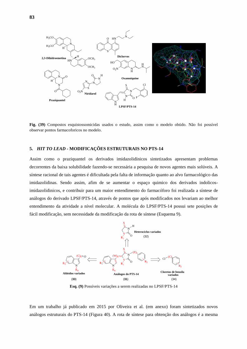
83
Fig. (39) Compostos esquistossomicidas usados o estudo, assim como o modelo obtido. Não foi possível
observar pontos farmacoforicos no modelo.
5. HIT TO LEAD - MODIFICAÇÕES ESTRUTURAIS NO PTS-14
Assim como o praziquantel os derivados imidazolidínicos sintetizados apresentam problemas
decorrentes da baixa solubilidade fazendo-se necessária a pesquisa de novos agentes mais solúveis. A
síntese racional de tais agentes é dificultada pela falta de informação quanto ao alvo farmacológico das
imidazolidinas. Sendo assim, afim de se aumentar o espaço quimico dos derivados indolicos-
imidazolidinicos, e contribuir para um maior entendimento do farmacóforo foi realizada a síntese de
análogos do derivado LPSF/PTS-14, através de pontos que após modificados nos levariam ao melhor
entendimento da atividade a nível molecular. A molécula do LPSF/PTS-14 possui sete posições de
fácil modificação, sem necessidade da modificação da rota de síntese (Esquema 9).
Esq. (9) Possíveis variações a serem realizadas no LPSF/PTS-14
Em um trabalho já publicado em 2015 por Oliveira et al. (em anexo) foram sintetizados novos
análogos estruturais do PTS-14 (Figura 40). A rota de síntese para obtenção dos análogos é a mesma
(W)n
(Z)n
O
X
Y
N
NR1
R2R3
R4
N
R4
R3
(C)n O (C)nCl
R2
H
O
X
Y
N
R1
Aldeídos variadosCloretos de benzila variados
Heterociclos variados
Análogos do PTS-14
(32)
(31)(33) (34)
N
N
O
H
HN
H
N
H3CO
H3CO
OCH3
OCH3
H
O
N
N
O
N
S
O2N
H
S
O HNN
NH
HO
O2N
HN
Praziquantel
2,3-Dihidroemetina
Niridarol
Diclorvos
Oxamniquine
LPSF/PTS-14
Cl
FO
S
N
N
NH
H
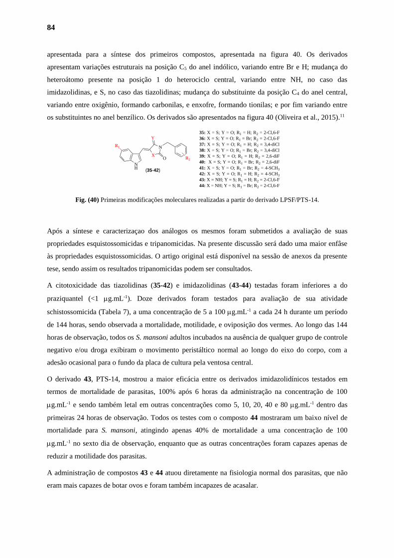
84
apresentada para a síntese dos primeiros compostos, apresentada na figura 40. Os derivados
apresentam variações estruturais na posição C5 do anel indólico, variando entre Br e H; mudança do
heteroátomo presente na posição 1 do heterociclo central, variando entre NH, no caso das
imidazolidinas, e S, no caso das tiazolidinas; mudança do substituinte da posição C4 do anel central,
variando entre oxigênio, formando carbonilas, e enxofre, formando tionilas; e por fim variando entre
os substituintes no anel benzílico. Os derivados são apresentados na figura 40 (Oliveira et al., 2015).11
Fig. (40) Primeiras modificações moleculares realizadas a partir do derivado LPSF/PTS-14.
Após a síntese e caracterizaçao dos análogos os mesmos foram submetidos a avaliação de suas
propriedades esquistossomicidas e tripanomicidas. Na presente discussão será dado uma maior enfâse
às propriedades esquistossomicidas. O artigo original está disponível na sessão de anexos da presente
tese, sendo assim os resultados tripanomicidas podem ser consultados.
A citotoxicidade das tiazolidinas (35-42) e imidazolidinas (43-44) testadas foram inferiores a do
praziquantel (<1 g.mL-1). Doze derivados foram testados para avaliação de sua atividade
schistossomicida (Tabela 7), a uma concentração de 5 a 100 g.mL-1 a cada 24 h durante um período
de 144 horas, sendo observada a mortalidade, motilidade, e oviposição dos vermes. Ao longo das 144
horas de observação, todos os S. mansoni adultos incubados na ausência de qualquer grupo de controle
negativo e/ou droga exibiram o movimento peristáltico normal ao longo do eixo do corpo, com a
adesão ocasional para o fundo da placa de cultura pela ventosa central.
O derivado 43, PTS-14, mostrou a maior eficácia entre os derivados imidazolidínicos testados em
termos de mortalidade de parasitas, 100% após 6 horas da administração na concentração de 100
g.mL-1 e sendo também letal em outras concentrações como 5, 10, 20, 40 e 80 g.mL-1 dentro das
primeiras 24 horas de observação. Todos os testes com o composto 44 mostraram um baixo nível de
mortalidade para S. mansoni, atingindo apenas 40% de mortalidade a uma concentração de 100
g.mL-1 no sexto dia de observação, enquanto que as outras concentrações foram capazes apenas de
reduzir a motilidade dos parasitas.
A administração de compostos 43 e 44 atuou diretamente na fisiologia normal dos parasitas, que não
eram mais capazes de botar ovos e foram também incapazes de acasalar.
O
Y
X
N
NH
R1
R2
35: X = S; Y = O; R1 = H; R2 = 2-Cl,6-F
36: X = S; Y = O; R1 = Br; R2 = 2-Cl,6-F
37: X = S; Y = O; R1 = H; R2 = 3,4-diCl
38: X = S; Y = O; R1 = Br; R2 = 3,4-diCl
39: X = S; Y = O; R1 = H; R2 = 2,6-diF
40: X = S; Y = O; R1 = Br; R2 = 2,6-diF
41: X = S; Y = O; R1 = Br; R2 = 4-SCH3
42: X = S; Y = O; R1 = H; R2 = 4-SCH3
43: X = NH; Y = S; R1 = H; R2 = 2-Cl,6-F
44: X = NH; Y = S; R1 = Br; R2 = 2-Cl,6-F
(35-42)
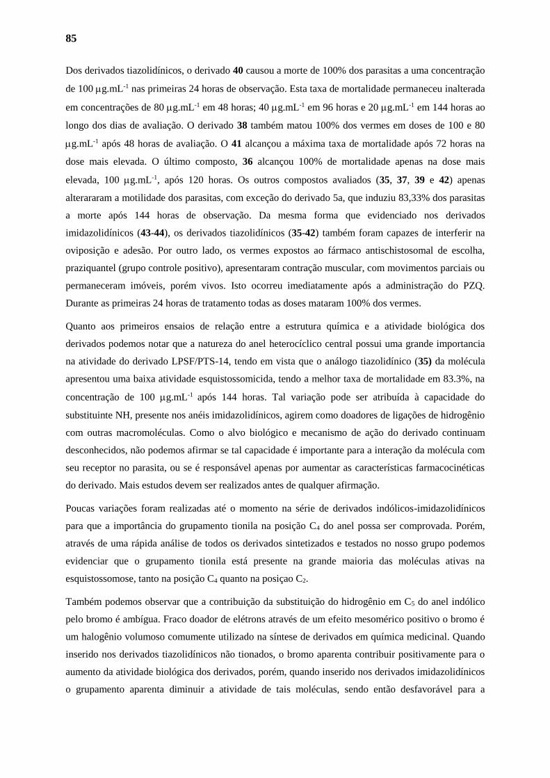
85
Dos derivados tiazolidínicos, o derivado 40 causou a morte de 100% dos parasitas a uma concentração
de 100 g.mL-1 nas primeiras 24 horas de observação. Esta taxa de mortalidade permaneceu inalterada
em concentrações de 80 g.mL-1 em 48 horas; 40 g.mL-1 em 96 horas e 20 g.mL-1 em 144 horas ao
longo dos dias de avaliação. O derivado 38 também matou 100% dos vermes em doses de 100 e 80
g.mL-1 após 48 horas de avaliação. O 41 alcançou a máxima taxa de mortalidade após 72 horas na
dose mais elevada. O último composto, 36 alcançou 100% de mortalidade apenas na dose mais
elevada, 100 g.mL-1, após 120 horas. Os outros compostos avaliados (35, 37, 39 e 42) apenas
alterararam a motilidade dos parasitas, com exceção do derivado 5a, que induziu 83,33% dos parasitas
a morte após 144 horas de observação. Da mesma forma que evidenciado nos derivados
imidazolidínicos (43-44), os derivados tiazolidínicos (35-42) também foram capazes de interferir na
oviposição e adesão. Por outro lado, os vermes expostos ao fármaco antischistosomal de escolha,
praziquantel (grupo controle positivo), apresentaram contração muscular, com movimentos parciais ou
permaneceram imóveis, porém vivos. Isto ocorreu imediatamente após a administração do PZQ.
Durante as primeiras 24 horas de tratamento todas as doses mataram 100% dos vermes.
Quanto aos primeiros ensaios de relação entre a estrutura química e a atividade biológica dos
derivados podemos notar que a natureza do anel heterocíclico central possui uma grande importancia
na atividade do derivado LPSF/PTS-14, tendo em vista que o análogo tiazolidínico (35) da molécula
apresentou uma baixa atividade esquistossomicida, tendo a melhor taxa de mortalidade em 83.3%, na
concentração de 100 g.mL-1 após 144 horas. Tal variação pode ser atribuída à capacidade do
substituinte NH, presente nos anéis imidazolidínicos, agirem como doadores de ligações de hidrogênio
com outras macromoléculas. Como o alvo biológico e mecanismo de ação do derivado continuam
desconhecidos, não podemos afirmar se tal capacidade é importante para a interação da molécula com
seu receptor no parasita, ou se é responsável apenas por aumentar as características farmacocinéticas
do derivado. Mais estudos devem ser realizados antes de qualquer afirmação.
Poucas variações foram realizadas até o momento na série de derivados indólicos-imidazolidínicos
para que a importância do grupamento tionila na posição C4 do anel possa ser comprovada. Porém,
através de uma rápida análise de todos os derivados sintetizados e testados no nosso grupo podemos
evidenciar que o grupamento tionila está presente na grande maioria das moléculas ativas na
esquistossomose, tanto na posição C4 quanto na posiçao C2.
Também podemos observar que a contribuição da substituição do hidrogênio em C5 do anel indólico
pelo bromo é ambígua. Fraco doador de elétrons através de um efeito mesomérico positivo o bromo é
um halogênio volumoso comumente utilizado na síntese de derivados em química medicinal. Quando
inserido nos derivados tiazolidínicos não tionados, o bromo aparenta contribuir positivamente para o
aumento da atividade biológica dos derivados, porém, quando inserido nos derivados imidazolidínicos
o grupamento aparenta diminuir a atividade de tais moléculas, sendo então desfavorável para a
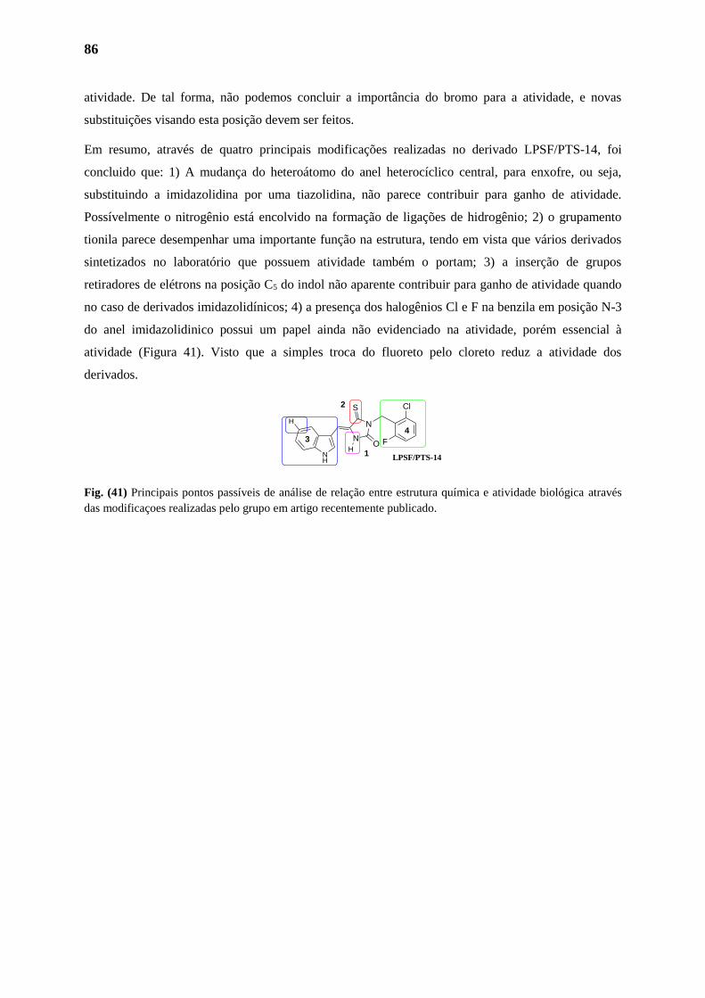
86
atividade. De tal forma, não podemos concluir a importância do bromo para a atividade, e novas
substituições visando esta posição devem ser feitos.
Em resumo, através de quatro principais modificações realizadas no derivado LPSF/PTS-14, foi
concluido que: 1) A mudança do heteroátomo do anel heterocíclico central, para enxofre, ou seja,
substituindo a imidazolidina por uma tiazolidina, não parece contribuir para ganho de atividade.
Possívelmente o nitrogênio está encolvido na formação de ligações de hidrogênio; 2) o grupamento
tionila parece desempenhar uma importante função na estrutura, tendo em vista que vários derivados
sintetizados no laboratório que possuem atividade também o portam; 3) a inserção de grupos
retiradores de elétrons na posição C5 do indol não aparente contribuir para ganho de atividade quando
no caso de derivados imidazolidínicos; 4) a presença dos halogênios Cl e F na benzila em posição N-3
do anel imidazolidinico possui um papel ainda não evidenciado na atividade, porém essencial à
atividade (Figura 41). Visto que a simples troca do fluoreto pelo cloreto reduz a atividade dos
derivados.
Fig. (41) Principais pontos passíveis de análise de relação entre estrutura química e atividade biológica através
das modificaçoes realizadas pelo grupo em artigo recentemente publicado.
Cl
FO
S
N
N
NH
H
H
1
2
34
LPSF/PTS-14
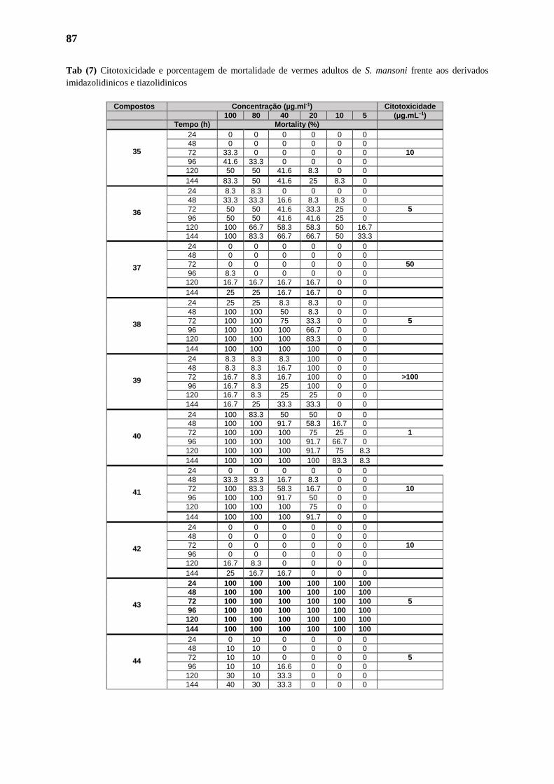
87
Tab (7) Citotoxicidade e porcentagem de mortalidade de vermes adultos de S. mansoni frente aos derivados
imidazolidinicos e tiazolidinicos
Compostos Concentração (µg.ml-1) Citotoxicidade
100 80 40 20 10 5 (μg.mL–1)
Tempo (h) Mortality (%)
35
24 0 0 0 0 0 0
48 0 0 0 0 0 0
72 33.3 0 0 0 0 0 10
96 41.6 33.3 0 0 0 0
120 50 50 41.6 8.3 0 0
144 83.3 50 41.6 25 8.3 0
36
24 8.3 8.3 0 0 0 0
48 33.3 33.3 16.6 8.3 8.3 0
72 50 50 41.6 33.3 25 0 5
96 50 50 41.6 41.6 25 0
120 100 66.7 58.3 58.3 50 16.7
144 100 83.3 66.7 66.7 50 33.3
37
24 0 0 0 0 0 0
48 0 0 0 0 0 0
72 0 0 0 0 0 0 50
96 8.3 0 0 0 0 0
120 16.7 16.7 16.7 16.7 0 0
144 25 25 16.7 16.7 0 0
38
24 25 25 8.3 8.3 0 0
48 100 100 50 8.3 0 0
72 100 100 75 33.3 0 0 5
96 100 100 100 66.7 0 0
120 100 100 100 83.3 0 0
144 100 100 100 100 0 0
39
24 8.3 8.3 8.3 100 0 0
48 8.3 8.3 16.7 100 0 0
72 16.7 8.3 16.7 100 0 0 >100
96 16.7 8.3 25 100 0 0
120 16.7 8.3 25 25 0 0
144 16.7 25 33.3 33.3 0 0
40
24 100 83.3 50 50 0 0
48 100 100 91.7 58.3 16.7 0
72 100 100 100 75 25 0 1
96 100 100 100 91.7 66.7 0
120 100 100 100 91.7 75 8.3
144 100 100 100 100 83.3 8.3
41
24 0 0 0 0 0 0
48 33.3 33.3 16.7 8.3 0 0
72 100 83.3 58.3 16.7 0 0 10
96 100 100 91.7 50 0 0
120 100 100 100 75 0 0
144 100 100 100 91.7 0 0
42
24 0 0 0 0 0 0
48 0 0 0 0 0 0
72 0 0 0 0 0 0 10
96 0 0 0 0 0 0
120 16.7 8.3 0 0 0 0
144 25 16.7 16.7 0 0 0
43
24 100 100 100 100 100 100
48 100 100 100 100 100 100
72 100 100 100 100 100 100 5
96 100 100 100 100 100 100
120 100 100 100 100 100 100
144 100 100 100 100 100 100
44
24 0 10 0 0 0 0
48 10 10 0 0 0 0
72 10 10 0 0 0 0 5
96 10 10 16.6 0 0 0
120 30 10 33.3 0 0 0
144 40 30 33.3 0 0 0
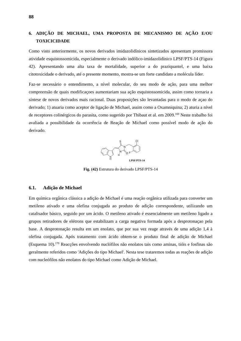
88
6. ADIÇÃO DE MICHAEL, UMA PROPOSTA DE MECANISMO DE AÇÃO E/OU
TOXICICIDADE
Como visto anteriormente, os novos derivados imidazolidínicos sintetizados apresentam promissora
atividade esquistossomicida, especialmente o derivado indólico-imidazolidínico LPSF/PTS-14 (Figura
42). Apresentando uma alta taxa de mortalidade, superior a do praziquantel, e uma baixa
citotoxicidade o derivado, até o presente momento, mostra-se um forte candidato a molécula líder.
Faz-se necessário o entendimento, a nível molecular, do seu modo de ação, para uma melhor
compreensão de quais modificaçoes aumentariam sua ação esquistossomicida, assim como tornaria a
síntese de novos derivados mais racional. Duas proposições são levantadas para o modo de açao do
derivado; 1) atuaria como aceptor de ligação de Michael, assim como a Oxamniquina; 2) aturia a nível
de receptores colinérgicos do parasita, como sugerido por Thibaut et al. em 2009.169 Neste trabalho foi
avaliada a possibilidade da ocorrência de Reação de Michael como possível modo de ação do
derivado.
Fig. (42) Estrutura do derivado LPSF/PTS-14
6.1. Adição de Michael
Em química orgânica clássica a adição de Michael é uma reação orgânica utilizada para converter um
metileno ativado e uma olefina conjugada ao produto de adição correspondente, utilizando um
catalisador básico, seguido por um ácido. O metileno ativado é essencialmente um metileno ligado a
grupos retiradores de elétrons que estabilizam a carga negativa formada após a desprotonaçao pela
base. A desprotonação resulta em um enolato, que por sua vez reage através de uma adição 1,4 à
olefina conjugada. Após tratamento com ácido obtem-se o produto final de adição de Michael
(Esquema 10).170 Reacções envolvendo nuclófilos não enolatos tais como aminas, tióis e fosfinas são
geralmente referidos como 'Adições do tipo Michael'. Nesta tese trataremos todas as reações de adição
com nucleófilos não enolatos do tipo Michael como Adição de Michael.
Cl
FO
S
N
N
NH
H
LPSF/PTS-14
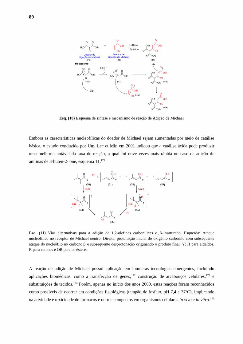
89
Esq. (10) Esquema de síntese e mecanismo de reação de Adição de Michael
Embora as características nucleofílicas do doador de Michael sejam aumentadas por meio de catálise
básica, o estudo conduzido por Um, Lee et Min em 2001 indicou que a catálise ácida pode produzir
uma melhoria notável da taxa de reação, a qual foi nove vezes mais rápida no caso da adição de
anilinas de 3-buten-2- one, esquema 11.171
Esq. (11) Vias alternativas para a adição de 1,2-olefinas carbonílicas -insaturado. Esquerda: Ataque
nucleofílico no receptor de Michael neutro. Direita: protonação inicial do oxigénio carbonilo com subsequente
ataque do nucleófilo no carbono e subsequente desprotonação originando o produto final. Y: H para aldeídos,
R para cetonas e OR para os ésteres.
A reação de adição de Michael possui aplicação em inúmeras tecnologias emergentes, incluindo
aplicações biomédicas, como a transfecção de genes,172 construção de arcabouços celulares,173 e
substituições de tecidos.174 Porém, apenas no início dos anos 2000, estas reações foram reconhecidos
como possíveis de ocorrer em condições fisiológicas (tampão de fosfato, pH 7,4 e 37oC), implicando
na atividade e toxicidade de fármacos e outros compostos em organismos celulares in vivo e in vitro.175
EtO OEt
OO
H
OEt
O
Ph O Ph
OEt OEt
O
O OEtDoador de
Ligação de MichaelAceptor de
Ligação de Michael
+ 1) Base
2) Ácido
EtO OEt
OO
HEtO OEt
OO
OEt
O
Ph
O Ph
OEt OEt
O
O OEt
O Ph
OEt OEt
O
O OEt
H+
OEt
EtOH
Mecanismo
(45) (46) (49)
(45) (47) (48)
(49)
(46)
Y
O
Y
O
NuH
NuH
Y
O
Nu
Y
OH
Y
OH
Y
OH
Y
OH
NuH
NuH
H+
-H+
(50) (51) (52) (53)
(54) (55)
(56)
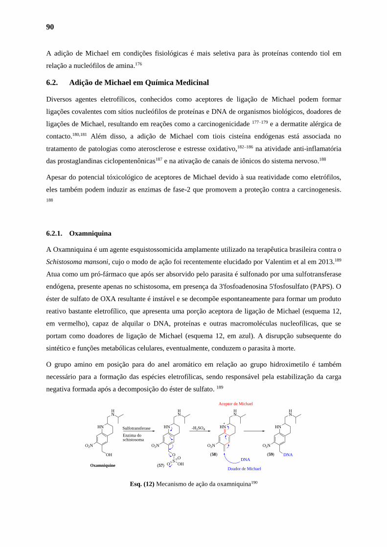
90
A adição de Michael em condições fisiológicas é mais seletiva para às proteínas contendo tiol em
relação a nucleófilos de amina.176
6.2. Adição de Michael em Química Medicinal
Diversos agentes eletrofílicos, conhecidos como aceptores de ligação de Michael podem formar
ligações covalentes com sítios nucleófilos de proteínas e DNA de organismos biológicos, doadores de
ligações de Michael, resultando em reações como a carcinogenicidade 177–179 e a dermatite alérgica de
contacto.180,181 Além disso, a adição de Michael com tiois cisteína endógenas está associada no
tratamento de patologias como aterosclerose e estresse oxidativo,182–186 na atividade anti-inflamatória
das prostaglandinas ciclopentenônicas187 e na ativação de canais de iônicos do sistema nervoso.188
Apesar do potencial tóxicológico de aceptores de Michael devido à sua reatividade como eletrófilos,
eles também podem induzir as enzimas de fase-2 que promovem a proteção contra a carcinogenesis.
188
6.2.1. Oxamniquina
A Oxamniquina é um agente esquistossomicida amplamente utilizado na terapêutica brasileira contra o
Schistosoma mansoni, cujo o modo de ação foi recentemente elucidado por Valentim et al em 2013.189
Atua como um pró-fármaco que após ser absorvido pelo parasita é sulfonado por uma sulfotransferase
endógena, presente apenas no schistosoma, em presença da 3'fosfoadenosina 5'fosfosulfato (PAPS). O
éster de sulfato de OXA resultante é instável e se decompõe espontaneamente para formar um produto
reativo bastante eletrofílico, que apresenta uma porção aceptora de ligação de Michael (esquema 12,
em vermelho), capaz de alquilar o DNA, proteínas e outras macromoléculas nucleofílicas, que se
portam como doadores de ligação de Michael (esquema 12, em azul). A disrupção subsequente do
sintético e funções metabólicas celulares, eventualmente, conduzem o parasita à morte.
O grupo amino em posição para do anel aromático em relação ao grupo hidroximetilo é também
necessário para a formação das espécies eletrofílicas, sendo responsável pela estabilização da carga
negativa formada após a decomposição do éster de sulfato. 189
Esq. (12) Mecanismo de ação da oxamniquina190
HN
HN
OH
O2N
HN
HN
O
O2N
HN
HN
O2N
HN
HN
O2N
DNA
SOHO
O
-H2SO4Sulfotransferase
Enzima do schistosoma
Oxamniquine
DNA
Aceptor de Michael
Doador de Michael
(58)
(57)
(59)
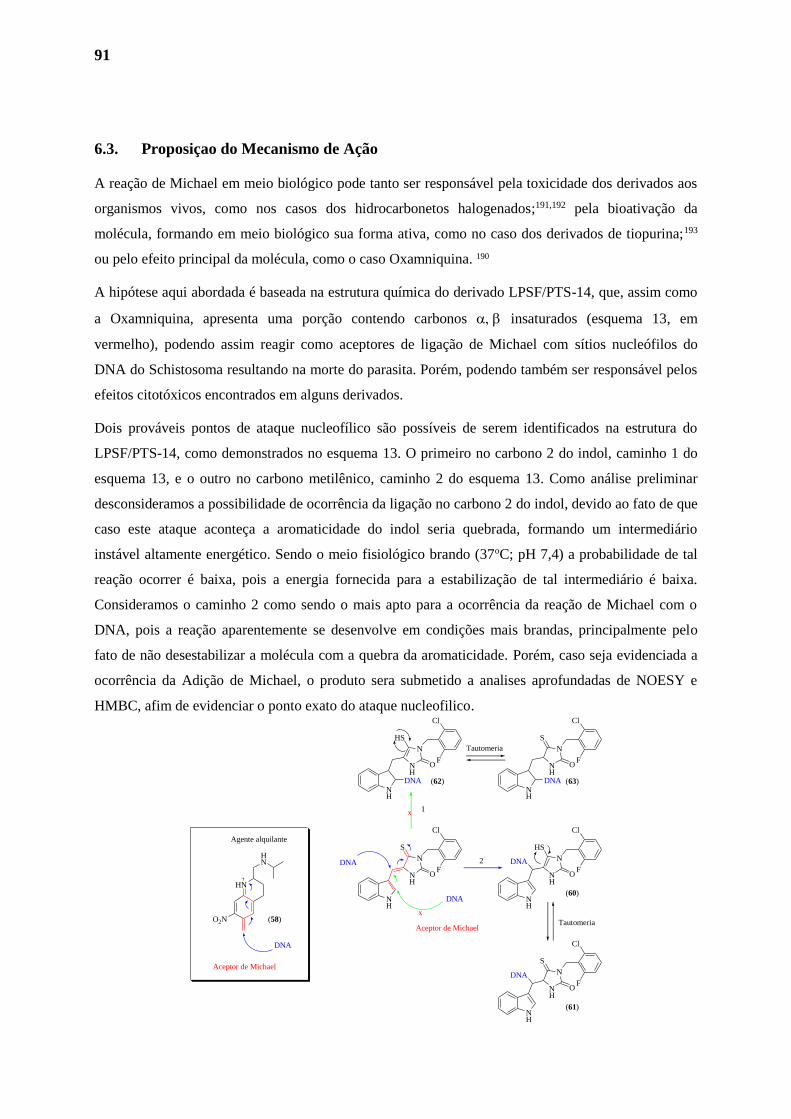
91
6.3. Proposiçao do Mecanismo de Ação
A reação de Michael em meio biológico pode tanto ser responsável pela toxicidade dos derivados aos
organismos vivos, como nos casos dos hidrocarbonetos halogenados;191,192 pela bioativação da
molécula, formando em meio biológico sua forma ativa, como no caso dos derivados de tiopurina;193
ou pelo efeito principal da molécula, como o caso Oxamniquina. 190
A hipótese aqui abordada é baseada na estrutura química do derivado LPSF/PTS-14, que, assim como
a Oxamniquina, apresenta uma porção contendo carbonos insaturados (esquema 13, em
vermelho), podendo assim reagir como aceptores de ligação de Michael com sítios nucleófilos do
DNA do Schistosoma resultando na morte do parasita. Porém, podendo também ser responsável pelos
efeitos citotóxicos encontrados em alguns derivados.
Dois prováveis pontos de ataque nucleofílico são possíveis de serem identificados na estrutura do
LPSF/PTS-14, como demonstrados no esquema 13. O primeiro no carbono 2 do indol, caminho 1 do
esquema 13, e o outro no carbono metilênico, caminho 2 do esquema 13. Como análise preliminar
desconsideramos a possibilidade de ocorrência da ligação no carbono 2 do indol, devido ao fato de que
caso este ataque aconteça a aromaticidade do indol seria quebrada, formando um intermediário
instável altamente energético. Sendo o meio fisiológico brando (37oC; pH 7,4) a probabilidade de tal
reação ocorrer é baixa, pois a energia fornecida para a estabilização de tal intermediário é baixa.
Consideramos o caminho 2 como sendo o mais apto para a ocorrência da reação de Michael com o
DNA, pois a reação aparentemente se desenvolve em condições mais brandas, principalmente pelo
fato de não desestabilizar a molécula com a quebra da aromaticidade. Porém, caso seja evidenciada a
ocorrência da Adição de Michael, o produto sera submetido a analises aprofundadas de NOESY e
HMBC, afim de evidenciar o ponto exato do ataque nucleofilico.
HN
HN
O2N
Agente alquilante
DNA
NH
NH
N
S
O
Cl
F
NH
NH
N
HS
O
Cl
F
NH
NH
N
S
O
Cl
F
DNA
DNA
DNA
Aceptor de Michael
Aceptor de MichaelTautomeria
DNA
x
NH
NH
N
HS
O
Cl
F
DNA
NH
NH
N
S
O
Cl
F
DNA
Tautomeria
x 1
2
(58)
(60)
(62) (63)
(61)
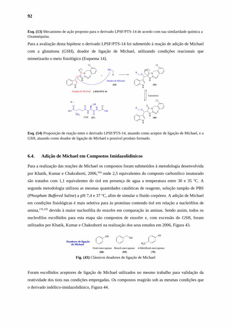
92
Esq. (13) Mecanismo de ação proposto para o derivado LPSF/PTS-14 de acordo com sua similaridade química a
Oxamniquina.
Para a avaliação desta hipótese o derivado LPSF/PTS-14 foi submetido à reação de adição de Michael
com a glutationa (GSH), doador de ligação de Michael, utilizando condições reacionais que
mimetizarão o meio fisiológico (Esquema 14).
Esq. (14) Proposição de reação entre o derivado LPSF/PTS-14, atuando como aceptor de ligação de Michael, e a
GSH, atuando como doador de ligação de Michael e possível produto formado.
6.4. Adição de Michael em Compostos Imidazolidinicos
Para a realização das reações de Michael os compostos foram submetidos à metodologia desenvolvida
por Khatik, Kumar e Chakraborti, 2006,194 onde 2,5 equivalentes do composto carbonilico insaturado
são tratados com 1,1 equivalentes do tiol em presença de agua a temperatura entre 30 e 35 oC. A
segunda metodologia utilizou as mesmas quantidades cataliticas de reagente, solução tampão de PBS
(Phosphate Buffered Saline) a pH 7,4 e 37 oC, afim de simular o fluido corpóreo. A adição de Michael
em condições fisiológicas é mais seletiva para às proteínas contendo tiol em relação a nucleófilos de
amina,176,195 devido à maior nucleofilia do enxofre em comparação às aminas. Sendo assim, todos os
nucleofilos escolhidos para esta etapa são compostos de enxofre e, com excessão do GSH, foram
utilizados por Khatik, Kumar e Chakraborti na realização dos seus estudos em 2006, Figura 43.
Fig. (43) Clássicos doadores de ligação de Michael
Foram escolhidos aceptores de ligação de Michael utilizados no mesmo trabalho para validação da
reatividade dos tiois nas condições empregadas. Os compostos reagirão sob as mesmas condições que
o derivado indólico-imidazolidínico, Figura 44.
NH
OHN
O
O
HO
NH2
GSH
SHO
OH
NH
NH
N
S
O
Cl
F
Aceptor de Michael
R
HS
+
NH
NH
N
HS
O
Cl
FS
R
NH
NH
N
S
O
Cl
FS
R
Tautomeria
R =
Doador de Michael
LPSF/PTS-14
(64) (65)
(66)(67)
SHSH
SH
H3C
Benzil-mercaptano 4-Metilfenil-mercaptanoFenil-mercaptano
Doadores de ligação de Michael
(68) (69) (70)
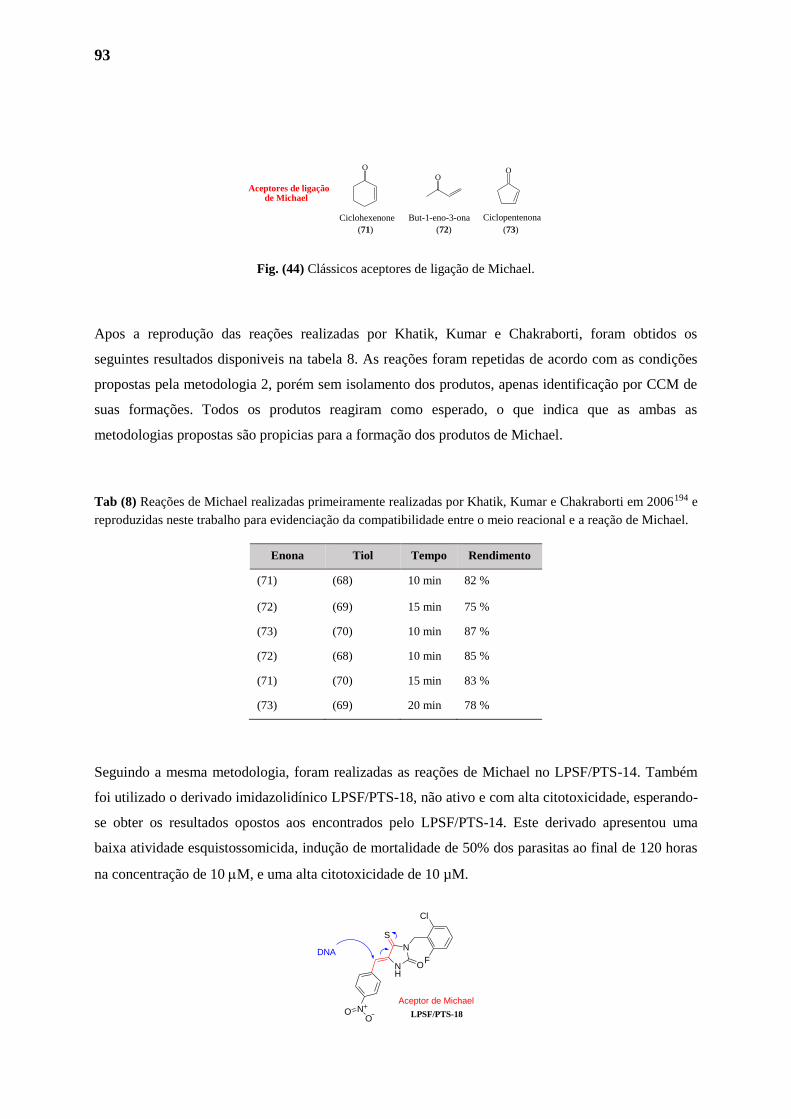
93
Fig. (44) Clássicos aceptores de ligação de Michael.
Apos a reprodução das reações realizadas por Khatik, Kumar e Chakraborti, foram obtidos os
seguintes resultados disponiveis na tabela 8. As reações foram repetidas de acordo com as condições
propostas pela metodologia 2, porém sem isolamento dos produtos, apenas identificação por CCM de
suas formações. Todos os produtos reagiram como esperado, o que indica que as ambas as
metodologias propostas são propicias para a formação dos produtos de Michael.
Tab (8) Reações de Michael realizadas primeiramente realizadas por Khatik, Kumar e Chakraborti em 2006194 e
reproduzidas neste trabalho para evidenciação da compatibilidade entre o meio reacional e a reação de Michael.
Seguindo a mesma metodologia, foram realizadas as reações de Michael no LPSF/PTS-14. Também
foi utilizado o derivado imidazolidínico LPSF/PTS-18, não ativo e com alta citotoxicidade, esperando-
se obter os resultados opostos aos encontrados pelo LPSF/PTS-14. Este derivado apresentou uma
baixa atividade esquistossomicida, indução de mortalidade de 50% dos parasitas ao final de 120 horas
na concentração de 10 M, e uma alta citotoxicidade de 10 µM.
Enona Tiol Tempo Rendimento
(71) (68) 10 min 82 %
(72) (69) 15 min 75 %
(73) (70) 10 min 87 %
(72) (68) 10 min 85 %
(71) (70) 15 min 83 %
(73) (69) 20 min 78 %
NH
N
S
O
Cl
FDNA
Aceptor de MichaelNO
OLPSF/PTS-18
Aceptores de ligação de Michael
O
OO
Ciclohexenone CiclopentenonaBut-1-eno-3-ona
(71) (72) (73)
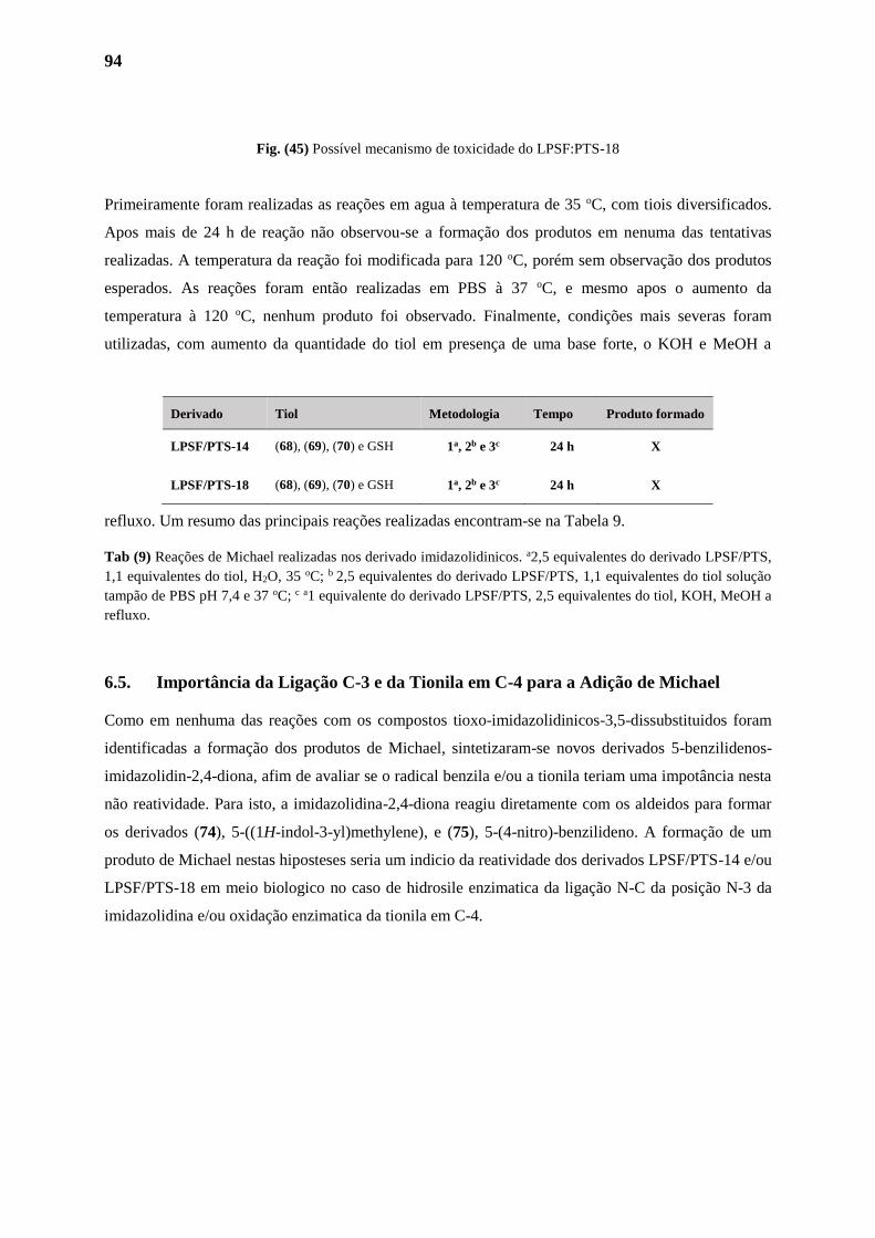
94
Fig. (45) Possível mecanismo de toxicidade do LPSF:PTS-18
Primeiramente foram realizadas as reações em agua à temperatura de 35 oC, com tiois diversificados.
Apos mais de 24 h de reação não observou-se a formação dos produtos em nenuma das tentativas
realizadas. A temperatura da reação foi modificada para 120 oC, porém sem observação dos produtos
esperados. As reações foram então realizadas em PBS à 37 oC, e mesmo apos o aumento da
temperatura à 120 oC, nenhum produto foi observado. Finalmente, condições mais severas foram
utilizadas, com aumento da quantidade do tiol em presença de uma base forte, o KOH e MeOH a
refluxo. Um resumo das principais reações realizadas encontram-se na Tabela 9.
Tab (9) Reações de Michael realizadas nos derivado imidazolidinicos. a2,5 equivalentes do derivado LPSF/PTS,
1,1 equivalentes do tiol, H2O, 35 oC; b 2,5 equivalentes do derivado LPSF/PTS, 1,1 equivalentes do tiol solução
tampão de PBS pH 7,4 e 37 oC; c a1 equivalente do derivado LPSF/PTS, 2,5 equivalentes do tiol, KOH, MeOH a
refluxo.
6.5. Importância da Ligação C-3 e da Tionila em C-4 para a Adição de Michael
Como em nenhuma das reações com os compostos tioxo-imidazolidinicos-3,5-dissubstituidos foram
identificadas a formação dos produtos de Michael, sintetizaram-se novos derivados 5-benzilidenos-
imidazolidin-2,4-diona, afim de avaliar se o radical benzila e/ou a tionila teriam uma impotância nesta
não reatividade. Para isto, a imidazolidina-2,4-diona reagiu diretamente com os aldeidos para formar
os derivados (74), 5-((1H-indol-3-yl)methylene), e (75), 5-(4-nitro)-benzilideno. A formação de um
produto de Michael nestas hiposteses seria um indicio da reatividade dos derivados LPSF/PTS-14 e/ou
LPSF/PTS-18 em meio biologico no caso de hidrosile enzimatica da ligação N-C da posição N-3 da
imidazolidina e/ou oxidação enzimatica da tionila em C-4.
Derivado Tiol Metodologia Tempo Produto formado
LPSF/PTS-14 (68), (69), (70) e GSH 1a, 2b e 3c 24 h X
LPSF/PTS-18 (68), (69), (70) e GSH 1a, 2b e 3c 24 h X
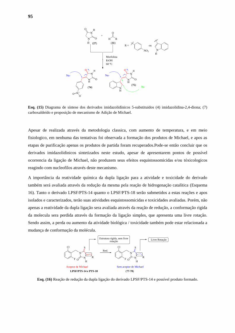
95
Esq. (15) Diagrama de síntese dos derivados imidazolidínicos 5-substituidos (4) imidazolidina-2,4-diona; (7)
carboxaldeído e proposição de mecanismo de Adição de Michael.
Apesar de realizada através da metodologia classica, com aumento de temperatura, e em meio
fisiologico, em nenhuma das tentativas foi observada a formação dos produtos de Michael, e apos as
etapas de purificação apenas os produtos de partida foram recuperados.Pode-se então concluir que os
derivados imidazolidinicos sintetizados neste estudo, apesar de apresentarem pontos de possível
ocorrencia da ligação de Michael, não produzem seus efeitos esquistossomicidas e/ou tóxicologicos
reagindo com nucleofilos através deste mecanismo.
A importância da reatividade química da dupla ligação para a atividade e toxicidade do derivado
também será avaliada através da redução da mesma pela reação de hidrogenação catalítica (Esquema
16). Tanto o derivado LPSF/PTS-14 quanto o LPSF/PTS-18 serão submetidos a estas reações e apos
isolados e caracterizados, terão suas atividades esquistossomicidas e toxicidades avaliadas. Porém, não
apenas a reatividade da dupla ligação sera avaliada através da reação de redução, a conformação rigida
da molecula sera perdida através da formação da ligação simples, que apresenta uma livre rotação.
Sendo assim, a perda ou aumento da atividade biológica / toxicidade também pode estar relacionada a
mudança de conformação da molécula.
Esq. (16) Reação de redução da dupla ligação do derivado LPSF/PTS-14 e possível produto formado.
Cl
F O
S
N
N R
Cl
F
Red.
Aceptor de Michael Sem aceptor de Michael
O
S
N
N R
H H
Estrutura rigida, sem livrerotação
Livre Rotação
LPSF/PTS-14 e PTS-18 (77-78)
NH
N
O
O
NOO
H
NH
NH
N
O H
O
Nu-
Nu-
Nu-
N
N
O
O
H
H
H R
O+
R =
NO2
ou
N
HMorfolina
EtOH
60 oC
(31)(27)
(74)(75)
96
Até o presente momento, a hipótese mais plausivel para o modo de ação destes derivados seria através
de interações com receptores biologicos, como sugerido por Thibaut et al. em 2009.
7. RECEPTORES COLINERGICOS
A acetilcolina (ACh) é um importante neurotransmissor em vertebrados e invertebrados. Os efeitos
neuromusculares da ACh são tipicamente mediados pelos receptores nicotínicos pós-sinápticos de
acetilcolina (nAChRs). Estruturalmente, os nAChRs são membros da superfamília de receptores Cys-
loop (LGIC). Eles formam estruturas homo e heteropentaméricas, que são organizadas em forma de
barril em torno de um poro iónico-seletivo central.196 Os nAChRs dos vertebrados são invariavelmente
cation-seletivos (Na+, Ca2+, K+) e mediam respostas excitatórias. Os invertebrados, por outro lado,
possuem canais de ACh cation e ânion seletivos (Cl-). Os canais de ACh ânion seletivos mediam a
hiperpolarização da membrana e, portanto, acredita-se que desempenham um papel nas respostas
inibitórias à ACh.
Um exemplo destes receptores em invertebrados é o Canal de Cloreto-Acetilcolina (ACC). Nematodos
possuem um tipo incomum de ACC, mais estreitamente relacionado com outros canais de cloreto,
como os receptores GABA, do que com os nAChRs.197,198 Os ACCs não tinham sido identificados em
nenhum espécie de parasita, no entanto, evidências experimentais suportam um papel inibitório da
ACh nos parasitas, que poderia ser mediado por este tipo de receptor. Estudos iniciais nos anos 60
observaram que a adição de agonistas colinérgicos exógenos a culturas de parasitas causou paralisia
flácida de trematodos e cestodos adultos.199,200
A paralisia flácida indica relaxamento muscular e está em contradição direta com a resposta excitatória
de contração tônica esperada pela estimulação colinérgica. Recentemente, a publicação do genoma do
S. mansoni 201 foi o estimulo necessário para uma revisão da atividade inibitória incomum da ACh no
parasita. MacDonald et al em 2014 descreveramo uma nova classe de nAChR anion-seletivos
(SmACCs) invertebrados específicos. Os canais iónicos formados por estas subunidades
desempenham um papel inibitório na atividade neuromuscular do parasita, tal como sugerido pelos
resultados de RNA inibitorio (RNAi), de sua distribuição nos tecidos e propriedades farmacológicas
frente a diversos agonistas colinergico.202
No grafico presente na figura 46 é possível de observar a mobilidade relativado dos vermes quando
submetidos a agonistas (nicotina e arecolina) e antagonistas (mecamilamina e D-tubocurarina) dos
receptores colinérgicos. Os padroões encontrados são inversos à atividade encontrada nos vertebrados.
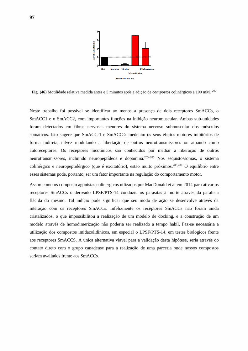
97
Fig. (46) Motilidade relativa medida antes e 5 minutos após a adição de compostos colinérgicos a 100 mM. 202
Neste trabalho foi possível se identificar ao menos a presença de dois receptores SmACCs, o
SmACC1 e o SmACC2, com importantes funções na inibição neuromuscular. Ambas sub-unidades
foram detectados em fibras nervosas menores do sistema nervoso submuscular dos músculos
somáticos. Isto sugere que SmACC-1 e SmACC-2 medeiam os seus efeitos motores inibitórios de
forma indireta, talvez modulando a libertação de outros neurotransmissores ou atuando como
autoreceptores. Os receptores nicotínicos são conhecidos por mediar a liberação de outros
neurotransmissores, incluindo neuropeptídeos e dopamina.203–205 Nos esquistossomas, o sistema
colinérgico e neuropeptidérgico (que é excitatório), estão muito próximos.206,207 O equilíbrio entre
esses sistemas pode, portanto, ser um fator importante na regulação do comportamento motor.
Assim como os composto agonistas colinergicos utlizados por MacDonald et al em 2014 para ativar os
receptores SmACCs o derivado LPSF/PTS-14 conduziu os parasitas à morte através da paralisia
flácida do mesmo. Tal indício pode significar que seu modo de ação se desenvolve através da
interação com os receptores SmACCs. Infelizmente os receptores SmACCs não foram ainda
cristalizados, o que impossibilitou a realização de um modelo de docking, e a construção de um
modelo através de homodimerização não poderia ser realizado a tempo habil. Faz-se necessária a
utilização dos compostos imidazolidinicos, em especial o LPSF/PTS-14, em testes biologicos frente
aos receptores SmACCS. A unica alternativa viavel para a validação desta hipótese, seria através do
contato direto com o grupo canadense para a realização de uma parceria onde nossos compostos
seriam avaliados frente aos SmACCs.
CONCLUSÕES E PERSPECTIVAS
PARTE I
99
CONCLUSÕES E PERSPECTIVAS
Primeiramente foram obtidos 24 derivados imidazolidínicos-3,5-dissubstituídos. Os compostos obtidos
foram caracterizados através de técnicas espectroscópicas tendo suas estruturas confirmadas. Após a
caracterização os derivados foram submetidos a análises quimiométricas, como a avaliação dos componentes
principais (PCA), para a determinação dos compostos mais (dis)similares, com objetivo de submeter a menor
quantidade a menor quantidade de compostos aos testes de atividade esquistossomicida.
Através da análise da PCA foram escolhidos 11 compostos, considerados os mais (dis)similares nos
parâmetros escolhidos para o estudo, sendo eles o LPSF/PTS-2, LPSF/PTS-3, LPSF/PTS-4, LPSF/PTS-6,
LPSF/PTS-10, LPSF/PTS-14, LPSF/PTS-18, LPSF/PTS-19, LPSF/PTS-21, LPSF/PTS-23 e LPSF/PTS-25.
Os derivados tioxo-imidazolidínicos LPSF/PTS2, LPSF/PTS4, LPSF/PTS10, LPSF/PTS-14, LPSF/PTS-21 e
LPSF/PTS-23 e LPSF/PTS25 não foram citotóxicos nas concentrações avaliadas. Os derivados tioxo-
imidazolidínicos LPSF/PTS2, LPSF/PTS4, LPSF/PTS10 e LPSF/PTS23 foram os bastante ativos nos ensaios
de viabilidade in vitro em vermes adultos de S. mansoni.
Os derivados LPSF/PTS6, LPSF/PTS18, LPSF/PTS19 e LPSF/PTS-21, embora não tenham causado
mortalidade, foram capazes de causar alteração de motilidade, inibição de oviposição e acasalamento dos
vermes.
Com relação às alterações tegumentares, foi possível observar alterações como destruição de tubérculos com
perda de espinhos e formação de vesículas nos vermes machos, e, erosão do tegumento nos vermes fêmeas
em vermes adultos de S. mansoni após incubação com LPSF/PTS23.
Porém, o melhor derivado sintetizado neste trabalho foi o composto indolico-imidazolidinico LPSF/PTS-14.
A alta taxa de mortalidade em baixas concentrações, aliadas a uma baixa citotoxicidade, alterações no
acasalamento e oviposição, colocam a LPSF/PTS-14 como um promissor agente esquistossomicida.
Com os resultados da atividade biológica foi possível notar que apesar de não ser possível de se observar
nenhum padrão referente à atividade esquistossomicida, um padrão na disperssão dos compostos no grafico
da PCA foi observado referente aos valores de citotoxicidade.
Desejando-se verificar se o modelo de PCA obtido para os derivados 4-tioxo-imidazolidin-2-ona possui
propriedades preditivas frente à citotoxicidade foram sintetizados 2 novos derivados imidazolidínicos, o
LPSF/PTS-5 e LPSF/PTS-24. Contrariamente ao esperado no início do estudo, os derivados não se
comportaram de acordo com o previsto pelo modelo. Tanto o LPSF/PTS-5 quanto o LPSF/PTS-24 obtiveram
valores contrarios ao esperado. Concluiu-se que o modelo quimiométrico obtido através da técnica PCA não
apresenta complexidade para prever a citotoxicidade dos compostos sintetizados, tendo em vista que as
variáveis utilizadas no modelo possuem, possívelmente, uma baixa importância no fenômeno estudado.
100
Desejando-se obter informações mais precisas para a síntese de novos analogos e o desenvolvimento de
modelos preditivos mais seguros, utilizou-se o software FLAP para a proposição dois modelos de
farmacóforos dos derivados. O primeiro através dos dois melhores derivados sintetizados e identificados na
etapa da atividade biológica, o LPSF/PTS-14 e o LPSF/PTS-23, juntamente com os derivados
imidazolidinicos mais ativos sintetizados pelo nosso grupo durante um periodo de 20 anos. Pode-se
evidenciar através das analises a existência de zonas doadoras e aceptoras de ligações de hidrogénio, assim
como interações hidrofóbicas. Porém, visto que as moléculas possuem uma enorme similaridade química
entre os derivados estes resultados não podem ser totalmente conclusivos na determinação do farmacóforo.
Para o desenvolvimento de um modelo farmacóforo mais confiavel, faz-se necessária a identificação de
compostos ativos com uma maior diversidade molecular.
O segundo modelo modelo foi realizado utilizando-se o derivado LPSF/PTS-14, e as moléculas de referência
no tratamento da esquistossomose. modelo nenhum ponto de similaridade é encontrado entre as moléculas.
Tal observação deve-se ao fato que apesar de todas as moléculas apresentarem uma boa ação
esquistossomicida não necessáriamente apresentam o mesmo modo de ação no parasita. Sendo necessária a
elucidação do modo de ação do derivado PTS-14 para a proposição de um modelo de farmacóforo.
Afim de se aumentar o espaço quimico dos derivados indolicos-imidazolidinicos, e contribuir para um maior
entendimento do farmacóforo foi realizada a síntese 10 novos análogos indolicos-imidazolidinicos e
indolicos-imidazolidinicos do derivado LPSF/PTS-14, através de pontos que após modificados nos levariam
ao melhor entendimento da atividade a nível molecular. Em resumo, através de quatro principais
modificações realizadas no derivado LPSF/PTS-14, foi concluido que: 1) A mudança do heteroátomo do anel
heterocíclico central, para enxofre, ou seja, substituindo a imidazolidina por uma tiazolidina, não parece
contribuir para ganho de atividade. Possívelmente o nitrogênio está encolvido na formação de ligações de
hidrogênio; 2) o grupamento tionila parece desempenhar uma importante função na estrutura, tendo em vista
que vários derivados sintetizados no laboratório que possuem atividade também o portam; 3) a inserção de
grupos retiradores de elétrons na posição C5 do indol não aparente contribuir para ganho de atividade quando
no caso de derivados imidazolidínicos; 4) a presença dos halogênios Cl e F na benzila em posição N-3 do
anel imidazolidinico possui um papel ainda não evidenciado na atividade, porém essencial à atividade. Visto
que a simples troca do fluoreto pelo cloreto reduz a atividade dos derivados.
Também foi avaliada a possibilidade da ocorrência de reação de Michael como possível mecanismo de ação
ou toxicidade do derivado. Para tanto, foram escolhidos dois derivados o LPSF/PTS-14, devido à sua alta
alta atividade e baixa citotoxicidade, e o LPSF/PTS-18, devido à sua alta alta citotoxicidade e baixa
atividade. Apesar de realizada através da metodologia classica, com aumento de temperatura, e em meio
fisiologico, em nenhuma das tentativas foi observada a formação dos produtos de Michael, e apos as etapas
de purificação apenas os produtos de partida foram recuperados.Pode-se então concluir que os derivados
imidazolidinicos sintetizados neste estudo, apesar de apresentarem pontos de possível ocorrencia da ligação
de Michael, não produzem seus efeitos esquistossomicidas e/ou tóxicologicos reagindo com nucleofilos
através deste mecanismo.
101
As principais perspectivas decorrentes deste trabalhos seriam:
1) A avaliação da importância da reatividade química da dupla ligação para a atividade e toxicidade dos
derivados através da redução da mesma pela reação de hidrogenação catalítica. Tanto o derivado LPSF/PTS-
14 quanto o LPSF/PTS-18 serão submetidos a estas reações e suas atividades esquistossomicidas e
toxicidades serão avaliadas.
2) Um modelo de farmacóforo mais confiavel, através da identificação de novos compostos ativos obtidos
pela modificação molecular nas sete posições susceptiveis do LPSF/PTS-14.
3) Os receptores de acetilconlina SmAACs, possiveis alvos biologicos dos compostos imidazolidinicos,
devem ser avaliados para o entendimento de um possível mecanismo de ação. Caso possível, em seguida, a
realização de um modelos de docking, desde que a proteina seja cristalizada. Através destas informações sera
possível o desenvolvimento de moléculas mais ativas através da síntese dirigida ao alvo
102
RESUME PARTIE I
SYNTHESE, CARACTERISATION STRUCTURALE ET
EVALUATION BIOLOGIQUE DE NOUVEAUX
DERIVES IMIDAZOLIDINIQUES 3,5-DISUBSTITUES
103
INTRODUCTION
Les maladies parasitaires ont des caractéristiques communes en relation avec le contexte dans lequel
elles se produisent comme des conditions d'hygiène précaire ou inexistante, le manque d'installations
sanitaires de base, le sous développement des pays, ou encore en raison de la présence de vecteurs qui
contribuent directement à la transmission, comme par exemple, la schistosomiase et la leishmaniose.
Les méthodes de traitement et de diagnostic de ces maladies sont vieilles et inappropriées et
nécessitent des investissements dans la recherche et le développement pour devenir plus simple et
efficace.15
Aussi appelé maladies négligées ou de pauvreté, ces maladies parasitaires sont d'importants problèmes
de santé publique dans de nombreux pays sous-développés et en développement. En affectant plus de
deux milliards de personnes dans le monde, elles sont des causes importantes de morbidité et de
mortalité.16 Elles sont appelées ainsi du fait du manque d'attention accordée par l'industrie
pharmaceutique pour leur traitement. À l'heure actuelle, l'Organisation Mondiale de la Santé classe 17
affections comme maladies négligées, entre autres la maladie de Chagas, la schistosomiase, la
leishmaniose, le paludisme et la tuberculose.17
Ces maladies présentent des zones d'endémie aux Brésil et ont une taux important de mortalité et de
morbidité dans le pays. La schistosomiase (EM CID 10 B 659), causée par le trématode Schistosoma
mansoni, à l'origine appelée bilharziose, occupe le deuxième rang em importance des maladies de
santé publique, après la malaria. Les études récentes de l'OMS ont montré une incidence d'environ 243
millions de personnes infectées par S. mansoni. La schistosomiase est associée à vingt mille décès par
an causés par les conséquences graves de l'infection, y compris la fibrose hépatique et l'hypertension
portale chez les malades.2
Les deux principaux médicaments utilisés dans le traitement de la schistosomiase sont l'Oxaminiquina
(OXA) (Mansil® - Pfizer S.A.) et le Praziquantel (PZQ) (Farmanguinhos - Oswaldo Cruz Foundation
/ FIOCRUZ).2,18
Le PZQ est utilisé pour traiter toutes les schistosomiases humaines, car il peut éliminer les vers
adultes, au cours de la phase chronique de l'infection,19 l'OXA est l'autre médicament disponible, mais
son utilisation est limitée et n'est pas efficace contre toutes les formes du parasite.13
À l'heure actuelle, le PZQ a été le médicament de choix pour le traitement médicamenteux au Brésil et
au monde, mais son utilisation extensive conduit inévitablement à baisse du taux de guérison, par
l'apparition d'isolats de S. mansoni réfractaires. Des effets secondaires importants sont par ailleurs
causés par ce médicament.21 Par conséquent, la recherche de nouveaux médicaments schistosomicides
plus efficaces est devenu nécessaire. Dans ce contexte, la recherche d'autres composés de synthèse
constitue une alternative viable, qui pourrait servir de modèle pour la conception de nouveaux
médicaments.
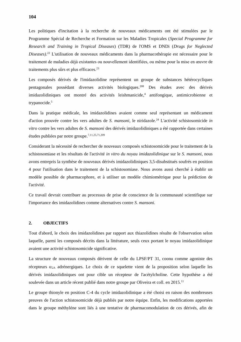
104
Les politiques d'incitation à la recherche de nouveaux médicaments ont été stimulées par le
Programme Spécial de Recherche et Formation sur les Maladies Tropicales (Special Programme for
Research and Training in Tropical Diseases) (TDR) de l'OMS et DNDi (Drugs for Neglected
Diseases).22 L'utilisation de nouveaux médicaments dans la pharmacothérapie est nécessaire pour le
traitement de maladies déjà existantes ou nouvellement identifiées, ou même pour la mise en œuvre de
traitements plus sûrs et plus efficaces.23
Les composés dérivés de l'imidazolidine représentent un groupe de substances hétérocycliques
pentagonales possédant diverses activités biologiques.208 Des études avec des dérivés
imidazolidiniques ont montré des activités leishmanicide,4 antifongique, antimicrobienne et
trypanocide.5
Dans la pratique médicale, les imidazolidines avaient comme seul représentant un médicament
d'action prouvée contre les vers adultes de S. mansoni, le niridazole.24 L'activité schistosomicide in
vitro contre les vers adultes de S. mansoni des dérivés imidazolidiniques a été rapportée dans certaines
études publiées par notre groupe.7,11,25,71,209
Considerant la nécessité de rechercher de nouveaux composés schistosomicide pour le traitement de la
schistosomiase et les résultats de l'activité in vitro du noyau imidazolidinique sur le S. mansoni, nous
avons entrepris la synthèse de nouveaux dérivés imidazolidiniques 3,5-disubstitués soufrés en position
4 pour l'utilisation dans le traitement de la schistosomiase. Nous avons aussi cherché à établir un
modèle possible de pharmacophore, et à utiliser un modèle chimiométrique pour la prédiction de
l'activité.
Ce travail devrait contribuer au processus de prise de conscience de la communauté scientifique sur
l'importance des imidazolidines comme alternatives contre S. mansoni.
2. OBJECTIFS
Tout d'abord, le choix des imidazolidines par rapport aux thiazolidines résulte de l'observation selon
laquelle, parmi les composés décrits dans la littérature, seuls ceux portant le noyau imidazolidinique
avaient une activité schistosomicide significative.
La structure de nouveaux composés dérivent de celle du LPSF/PT 31, coonu comme agoniste des
récepteurs 2A adrénergiques. Le choix de ce squelette vient de la proposition selon laquelle les
dérivés imidazolidiniques ont pour cible un récepteur de l'acétylcholine. Cette hypothèse a été
soulevée dans un article récent publié dans notre groupe par Oliveira et coll. en 2015.11
Le groupe thionyle en position C-4 du cycle imidazolidinique a été choisi en raison des nombreuses
preuves de l'action schistosomicide déjà publiés par notre équipe. Enfin, les modifications apportées
dans le groupe méthyléne sont liés à une tentative de pharmacomodulation de ces dérivés, afin de
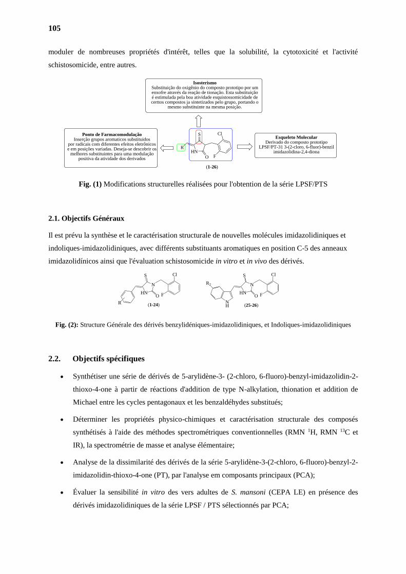
105
moduler de nombreuses propriétés d'intérêt, telles que la solubilité, la cytotoxicité et l'activité
schistosomicide, entre autres.
Fig. (1) Modifications structurelles réalisées pour l'obtention de la série LPSF/PTS
2.1. Objectifs Généraux
Il est prévu la synthèse et le caractérisation structurale de nouvelles molécules imidazolidiniques et
indoliques-imidazolidiniques, avec différents substituants aromatiques en position C-5 des anneaux
imidazolidínicos ainsi que l'évaluation schistosomicide in vitro et in vivo des dérivés.
Fig. (2): Structure Générale des dérivés benzylidéniques-imidazolidiniques, et Indoliques-imidazolidiniques
2.2. Objectifs spécifiques
Synthétiser une série de dérivés de 5-arylidène-3- (2-chloro, 6-fluoro)-benzyl-imidazolidin-2-
thioxo-4-one à partir de réactions d'addition de type N-alkylation, thionation et addition de
Michael entre les cycles pentagonaux et les benzaldéhydes substitués;
Déterminer les propriétés physico-chimiques et caractérisation structurale des composés
synthétisés à l'aide des méthodes spectrométriques conventionnelles (RMN 1H, RMN 13C et
IR), la spectrométrie de masse et analyse élémentaire;
Analyse de la dissimilarité des dérivés de la série 5-arylidène-3-(2-chloro, 6-fluoro)-benzyl-2-
imidazolidin-thioxo-4-one (PT), par l'analyse em composants principaux (PCA);
Évaluer la sensibilité in vitro des vers adultes de S. mansoni (CEPA LE) en présence des
dérivés imidazolidiniques de la série LPSF / PTS sélectionnés par PCA;
O
S
HN
N
R
F
Cl
O
S
HN
N
F
Cl
NH
R2
(1-24) (25-26)
HN
S
O
R
Cl
F
Esqueleto MolecularDerivado do composto prototipo
LPSF/PT-31 3-(2-cloro, 6-fluor)-benzil imidazolidina-2,4-diona
IsosterismoSubstituição do oxigênio do composto prototipo por umenxofre através da reação de tionação. Esta substituição é estimulada pela boa atividade esquistossomicidade de certtos compostos ja sintetizados pelo grupo, portando o
mesmo substituinte na mesma posição.
Ponto de FarmacomodulaçãoInserção grupos aromaticos substituidos
por radicais com diferentes efeitos eletrônicos e em posições variadas. Deseja-se descobrir os
melhores substituintes para uma modulação positiva da atividade dos derivados
(1-26)
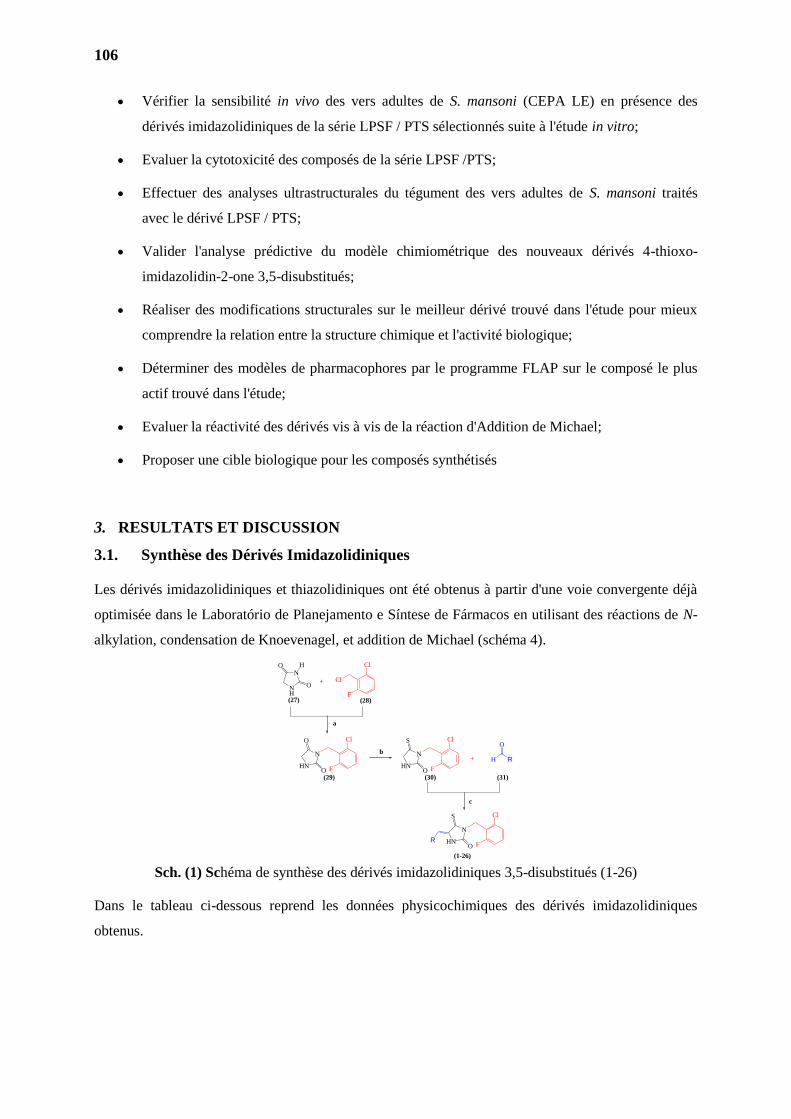
106
Vérifier la sensibilité in vivo des vers adultes de S. mansoni (CEPA LE) en présence des
dérivés imidazolidiniques de la série LPSF / PTS sélectionnés suite à l'étude in vitro;
Evaluer la cytotoxicité des composés de la série LPSF /PTS;
Effectuer des analyses ultrastructurales du tégument des vers adultes de S. mansoni traités
avec le dérivé LPSF / PTS;
Valider l'analyse prédictive du modèle chimiométrique des nouveaux dérivés 4-thioxo-
imidazolidin-2-one 3,5-disubstitués;
Réaliser des modifications structurales sur le meilleur dérivé trouvé dans l'étude pour mieux
comprendre la relation entre la structure chimique et l'activité biologique;
Déterminer des modèles de pharmacophores par le programme FLAP sur le composé le plus
actif trouvé dans l'étude;
Evaluer la réactivité des dérivés vis à vis de la réaction d'Addition de Michael;
Proposer une cible biologique pour les composés synthétisés
3. RESULTATS ET DISCUSSION
3.1. Synthèse des Dérivés Imidazolidiniques
Les dérivés imidazolidiniques et thiazolidiniques ont été obtenus à partir d'une voie convergente déjà
optimisée dans le Laboratório de Planejamento e Síntese de Fármacos en utilisant des réactions de N-
alkylation, condensation de Knoevenagel, et addition de Michael (schéma 4).
Sch. (1) Schéma de synthèse des dérivés imidazolidiniques 3,5-disubstitués (1-26)
Dans le tableau ci-dessous reprend les données physicochimiques des dérivés imidazolidiniques
obtenus.
RO
S
HN
N
O
ON
NH
Cl
O
O
HN
N
O
S
HN
NR
O
H+
+
(27) (28)
(29) (30) (31)
(1-26)
H
F
Cl
Cl
F
Cl
F
Cl
F
a
b
c
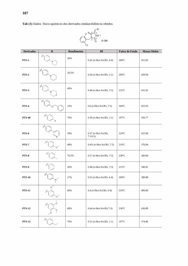
107
Tab (1) Dados físico-químicos dos derivados imidazolidínicos obtidos
Derivados R Rendimento Rf Faixa de Fusão Massa Molar
PTS-1
36% 0.42 (n-Hex/AcOEt, 6:4) 268oC 415.92
PTS-2
20,5% 0,56 (n-Hex/AcOEt, 1:1) 260oC 429.94
PTS-3
49% 0.48 (n-Hex/AcOEt, 7:3) 232oC 431.92
PTS-4 33% 0,6 (n-Hex/AcOEt, 7:3) 264oC 452.93
PTS-40
76% 0.59 (n-Hex/AcOEt, 1:1) 197oC 350.77
PTS-6 59% 0.57 (n-Hex/AcOEt,
7.5:0.5)
224oC 423.90
PTS-7
48% 0,4% (n-Hex/AcOEt, 7:3) 216oC 376.84
PTS-8
70,5% 0,57 (n-Hex/AcOEt, 7:3) 230oC 360.84
PTS-9
26% 0,48 (n-Hex/AcOEt, 7:3) 215oC 346.81
PTS-10
27% 0,55 (n-Hex/AcOEt, 6:4) 269oC 389.88
PTS-11
66% 0,4 (n-Hex/AcOEt, 6:4) 210oC 406.86
PTS-12 69% 0,44 (n-Hex/AcOEt,7:3) 236oC 436.89
PTS-13
76% 0.52 (n-Hex/AcOEt, 1:1) 197oC 374.86
NH 1
23
N
45
O
S
R
F
Cl
(1-24)
N
O
N
N
N
O
F
N
O
O
O
O
O
O
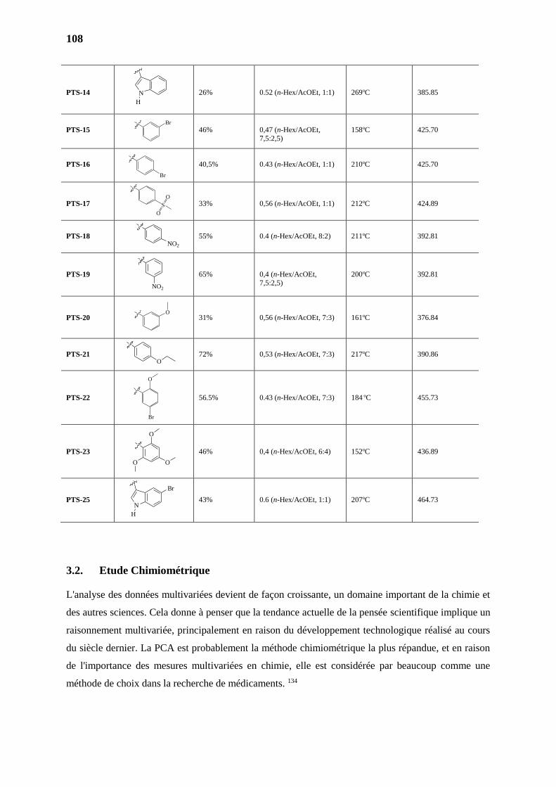
108
PTS-14 26% 0.52 (n-Hex/AcOEt, 1:1) 269oC 385.85
PTS-15 46% 0,47 (n-Hex/AcOEt,
7,5:2,5)
158oC 425.70
PTS-16 40,5% 0.43 (n-Hex/AcOEt, 1:1) 210oC 425.70
PTS-17 33% 0,56 (n-Hex/AcOEt, 1:1) 212oC 424.89
PTS-18 55% 0.4 (n-Hex/AcOEt, 8:2) 211oC 392.81
PTS-19 65% 0,4 (n-Hex/AcOEt,
7,5:2,5)
200oC 392.81
PTS-20 31% 0,56 (n-Hex/AcOEt, 7:3) 161oC 376.84
PTS-21 72% 0,53 (n-Hex/AcOEt, 7:3) 217oC 390.86
PTS-22 56.5% 0.43 (n-Hex/AcOEt, 7:3) 184 oC 455.73
PTS-23 46% 0,4 (n-Hex/AcOEt, 6:4) 152oC 436.89
PTS-25 43% 0.6 (n-Hex/AcOEt, 1:1) 207oC 464.73
3.2. Etude Chimiométrique
L'analyse des données multivariées devient de façon croissante, un domaine important de la chimie et
des autres sciences. Cela donne à penser que la tendance actuelle de la pensée scientifique implique un
raisonnement multivariée, principalement en raison du développement technologique réalisé au cours
du siècle dernier. La PCA est probablement la méthode chimiométrique la plus répandue, et en raison
de l'importance des mesures multivariées en chimie, elle est considérée par beaucoup comme une
méthode de choix dans la recherche de médicaments. 134
N
H
Br
O
O
O
O
NO2
NO2
N
H
S
O
O
O
Br
O
Br
Br
109
La première étape dans les études de corrélation implique la sélection d'un ensemble pertinent de
paramêtres ou propriétés moléculaires pour obtenir la meilleure corrélation possible. Les variables
choisies pour une étude préliminaire de l'PCA devraient couvrir principalement des données
électroniques, de lipophilie et volume, faciles à mesurer. Les paramètres retenus pour l'évaluation de la
PCA dans cette étude étaient les suivants: (1) le déplacement du proton N-H du cycle
imidazolidinique, δN-H; (2) le déplacement du proton du carbone méthylénique, δ=CH; (3) le
déplacement du carbone carbonyle, δC=O; (4) le déplacement du carbone thionyle, δC=S; (5) Log P
théorique des 24 dérivés calculées en utilisant la plate-forme EPA, (Environmental Protection Agency,
http://www.epa.gov/opptintr/exposure/index.htm); (6) le volume moléculaire calculé par le
programme MarvinSketch; (7) Aire de Surface Polaire calculée par le programme MarvinSketch.
La PCA de ces données peut être représentée par une matrice X composée de p = 7 variables
(déplacements chimiques, volume moléculaire, l'aire de superficie polaire et LogP) et n = 24 objets
(les composés synthétisés). Les analyses statistiques ont été effectuées en utilisant le pack FactoMineR
dédié aux analyses multivariées, disponible dans le logiciel R. 135
La PCA montre que 64.7 % des informations contenues dans le tableau 5 peuvent être représentés en
deux composantes principales (PC). Dans la figure 28 présente le graphique des scores de PC1 (40.73
% de la variance) versus PC2 (17.2 % de variance). PC1 est principalement définie par le volume
moléculaire, et l'aire de superficie polaire pour des valeurs positives, et δN-H pour des valeurs
négatives. La PC2 est principalement définie par le déplacement δC=O et δN-H, pour les valeurs positives
et déplacement δC=S pour des valeurs négatives (informations obtenues par les "loadings", i.e., les
coefficients dans les équations qui définissent le PC's, représentés dans la figure 3).
Grâce à l'analyse en PCA, 11 composés, considérés comme les plus (dis)similaires aux paramètres
choisis pour l'étude, ont été sélectionnés. Ceux sont LPSF/PTS-2, LPSF/PTS-3, LPSF/PTS-4,
LPSF/PTS-6, LPSF/PTS-10, LPSF/PTS-14, LPSF/PTS-18, LPSF/PTS-19, LPSF/PTS-21, LPSF/PTS-
23 et LPSF/PTS-25.
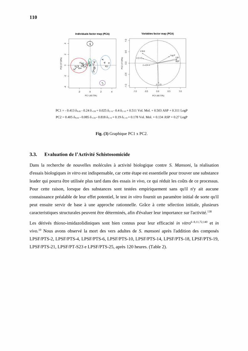
110
PC1 = - 0.413 N-H - 0.24 =CH + 0.025 C=S - 0.4 C=O + 0.511 Vol. Mol. + 0.503 ASP + 0.311 LogP
PC2 = 0.405 N-H - 0.085 =CH - 0.818 C=S + 0.19 C=O + 0.178 Vol. Mol. + 0.134 ASP + 0.27 LogP
Fig. (3) Graphique PC1 x PC2.
3.3. Evaluation de l’Activité Schistosomicide
Dans la recherche de nouvelles molécules à activité biologique contre S. Mansoni, la réalisation
d'essais biologiques in vitro est indispensable, car cette étape est essentielle pour trouver une substance
leader qui pourra être utilisée plus tard dans des essais in vivo, ce qui réduit les coûts de ce processus.
Pour cette raison, lorsque des substances sont testées empiriquement sans qu'il n'y ait aucune
connaissance préalable de leur effet potentiel, le test in vitro fournit un paramètre initial de sorte qu'il
peut ensuite servir de base à une approche rationnelle. Grâce à cette sélection initiale, plusieurs
caracteristiques structurales peuvent être déterminés, afin d'évaluer leur importance sur l'activité.138
Les dérivés thioxo-imidazolidiniques sont bien connus pour leur efficacité in vitro6–8,11,72,140 et in
vivo.10 Nous avons observé la mort des vers adultes de S. mansoni après l'addition des composés
LPSF/PTS-2, LPSF/PTS-4, LPSF/PTS-6, LPSF/PTS-10, LPSF/PTS-14, LPSF/PTS-18, LPSF/PTS-19,
LPSF/PTS-21, LPSF/PT-S23 e LPSF/PTS-25, après 120 heures. (Table 2).
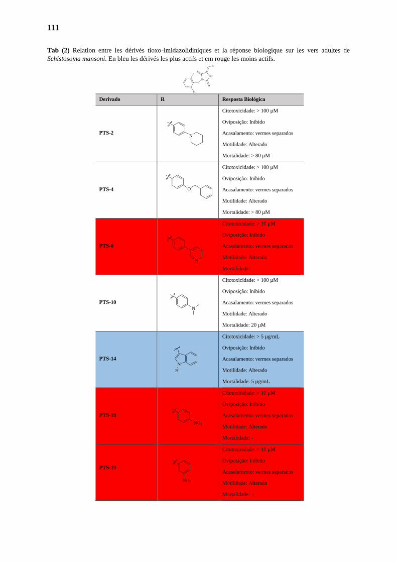
111
Tab (2) Relation entre les dérivés tioxo-imidazolidiniques et la réponse biologique sur les vers adultes de
Schistosoma mansoni. En bleu les dérivés les plus actifs et em rouge les moins actifs.
Derivado R Resposta Biológica
PTS-2
Citotoxicidade: > 100 µM
Oviposição: Inibido
Acasalamento: vermes separados
Motilidade: Alterado
Mortalidade: > 80 µM
PTS-4
Citotoxicidade: > 100 µM
Oviposição: Inibido
Acasalamento: vermes separados
Motilidade: Alterado
Mortalidade: > 80 µM
PTS-6
Citotoxicidade: > 10 µM
Oviposição: Inibido
Acasalamento: vermes separados
Motilidade: Alterado
Mortalidade:-
PTS-10
Citotoxicidade: > 100 µM
Oviposição: Inibido
Acasalamento: vermes separados
Motilidade: Alterado
Mortalidade: 20 µM
PTS-14
Citotoxicidade: > 5 µg/mL
Oviposição: Inibido
Acasalamento: vermes separados
Motilidade: Alterado
Mortalidade: 5 µg/mL
PTS-18
Citotoxicidade: > 10 µM
Oviposição: Inibido
Acasalamento: vermes separados
Motilidade: Alterado
Mortalidade: -
PTS-19
Citotoxicidade: > 10 µM
Oviposição: Inibido
Acasalamento: vermes separados
Motilidade: Alterada
Mortalidade: -
N
O
N
N
N
H
NO2
NO2
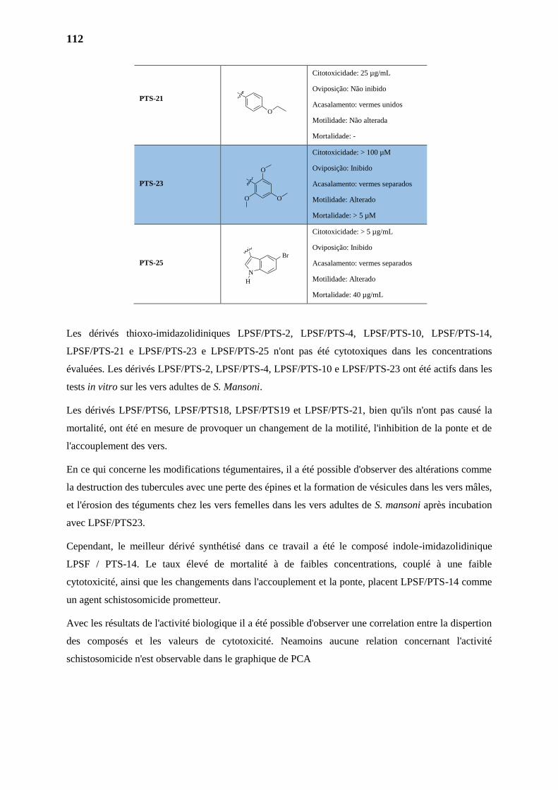
112
PTS-21
Citotoxicidade: 25 µg/mL
Oviposição: Não inibido
Acasalamento: vermes unidos
Motilidade: Não alterada
Mortalidade: -
PTS-23
Citotoxicidade: > 100 µM
Oviposição: Inibido
Acasalamento: vermes separados
Motilidade: Alterado
Mortalidade: > 5 µM
PTS-25
Citotoxicidade: > 5 µg/mL
Oviposição: Inibido
Acasalamento: vermes separados
Motilidade: Alterado
Mortalidade: 40 µg/mL
Les dérivés thioxo-imidazolidiniques LPSF/PTS-2, LPSF/PTS-4, LPSF/PTS-10, LPSF/PTS-14,
LPSF/PTS-21 e LPSF/PTS-23 e LPSF/PTS-25 n'ont pas été cytotoxiques dans les concentrations
évaluées. Les dérivés LPSF/PTS-2, LPSF/PTS-4, LPSF/PTS-10 e LPSF/PTS-23 ont été actifs dans les
tests in vitro sur les vers adultes de S. Mansoni.
Les dérivés LPSF/PTS6, LPSF/PTS18, LPSF/PTS19 et LPSF/PTS-21, bien qu'ils n'ont pas causé la
mortalité, ont été en mesure de provoquer un changement de la motilité, l'inhibition de la ponte et de
l'accouplement des vers.
En ce qui concerne les modifications tégumentaires, il a été possible d'observer des altérations comme
la destruction des tubercules avec une perte des épines et la formation de vésicules dans les vers mâles,
et l'érosion des téguments chez les vers femelles dans les vers adultes de S. mansoni après incubation
avec LPSF/PTS23.
Cependant, le meilleur dérivé synthétisé dans ce travail a été le composé indole-imidazolidinique
LPSF / PTS-14. Le taux élevé de mortalité à de faibles concentrations, couplé à une faible
cytotoxicité, ainsi que les changements dans l'accouplement et la ponte, placent LPSF/PTS-14 comme
un agent schistosomicide prometteur.
Avec les résultats de l'activité biologique il a été possible d'observer une correlation entre la dispertion
des composés et les valeurs de cytotoxicité. Neamoins aucune relation concernant l'activité
schistosomicide n'est observable dans le graphique de PCA
N
H
Br
O
O
O
O
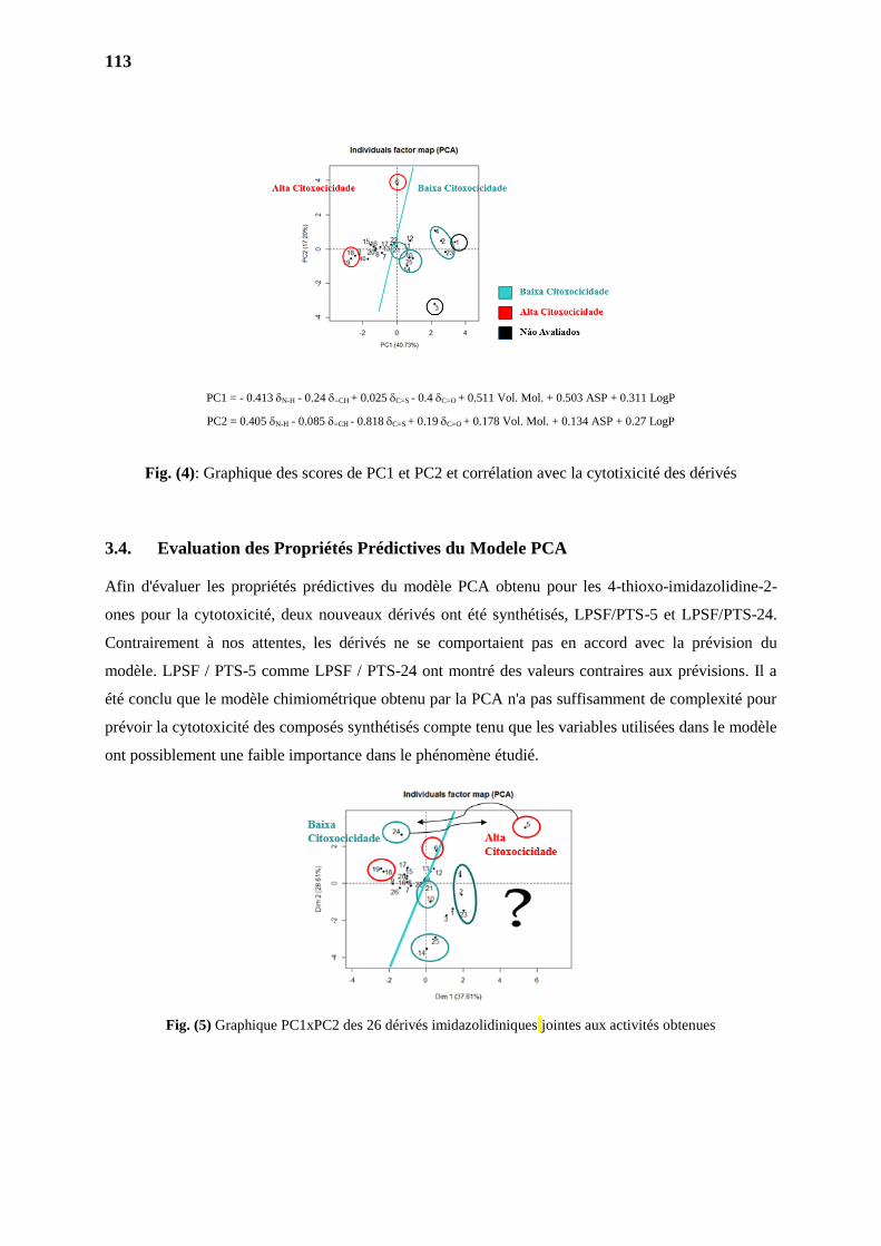
113
PC1 = - 0.413 N-H - 0.24 =CH + 0.025 C=S - 0.4 C=O + 0.511 Vol. Mol. + 0.503 ASP + 0.311 LogP
PC2 = 0.405 N-H - 0.085 =CH - 0.818 C=S + 0.19 C=O + 0.178 Vol. Mol. + 0.134 ASP + 0.27 LogP
Fig. (4): Graphique des scores de PC1 et PC2 et corrélation avec la cytotixicité des dérivés
3.4. Evaluation des Propriétés Prédictives du Modele PCA
Afin d'évaluer les propriétés prédictives du modèle PCA obtenu pour les 4-thioxo-imidazolidine-2-
ones pour la cytotoxicité, deux nouveaux dérivés ont été synthétisés, LPSF/PTS-5 et LPSF/PTS-24.
Contrairement à nos attentes, les dérivés ne se comportaient pas en accord avec la prévision du
modèle. LPSF / PTS-5 comme LPSF / PTS-24 ont montré des valeurs contraires aux prévisions. Il a
été conclu que le modèle chimiométrique obtenu par la PCA n'a pas suffisamment de complexité pour
prévoir la cytotoxicité des composés synthétisés compte tenu que les variables utilisées dans le modèle
ont possiblement une faible importance dans le phénomène étudié.
Fig. (5) Graphique PC1xPC2 des 26 dérivés imidazolidiniques jointes aux activités obtenues
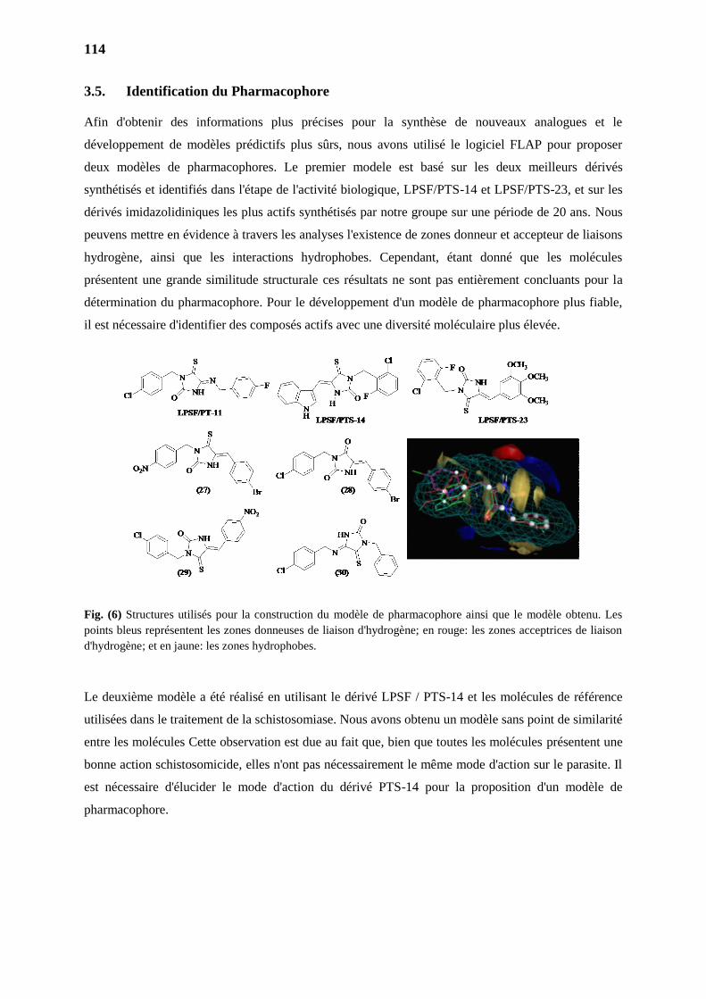
114
3.5. Identification du Pharmacophore
Afin d'obtenir des informations plus précises pour la synthèse de nouveaux analogues et le
développement de modèles prédictifs plus sûrs, nous avons utilisé le logiciel FLAP pour proposer
deux modèles de pharmacophores. Le premier modele est basé sur les deux meilleurs dérivés
synthétisés et identifiés dans l'étape de l'activité biologique, LPSF/PTS-14 et LPSF/PTS-23, et sur les
dérivés imidazolidiniques les plus actifs synthétisés par notre groupe sur une période de 20 ans. Nous
peuvens mettre en évidence à travers les analyses l'existence de zones donneur et accepteur de liaisons
hydrogène, ainsi que les interactions hydrophobes. Cependant, étant donné que les molécules
présentent une grande similitude structurale ces résultats ne sont pas entièrement concluants pour la
détermination du pharmacophore. Pour le développement d'un modèle de pharmacophore plus fiable,
il est nécessaire d'identifier des composés actifs avec une diversité moléculaire plus élevée.
Fig. (6) Structures utilisés pour la construction du modèle de pharmacophore ainsi que le modèle obtenu. Les
points bleus représentent les zones donneuses de liaison d'hydrogène; en rouge: les zones acceptrices de liaison
d'hydrogène; et en jaune: les zones hydrophobes.
Le deuxième modèle a été réalisé en utilisant le dérivé LPSF / PTS-14 et les molécules de référence
utilisées dans le traitement de la schistosomiase. Nous avons obtenu un modèle sans point de similarité
entre les molécules Cette observation est due au fait que, bien que toutes les molécules présentent une
bonne action schistosomicide, elles n'ont pas nécessairement le même mode d'action sur le parasite. Il
est nécessaire d'élucider le mode d'action du dérivé PTS-14 pour la proposition d'un modèle de
pharmacophore.
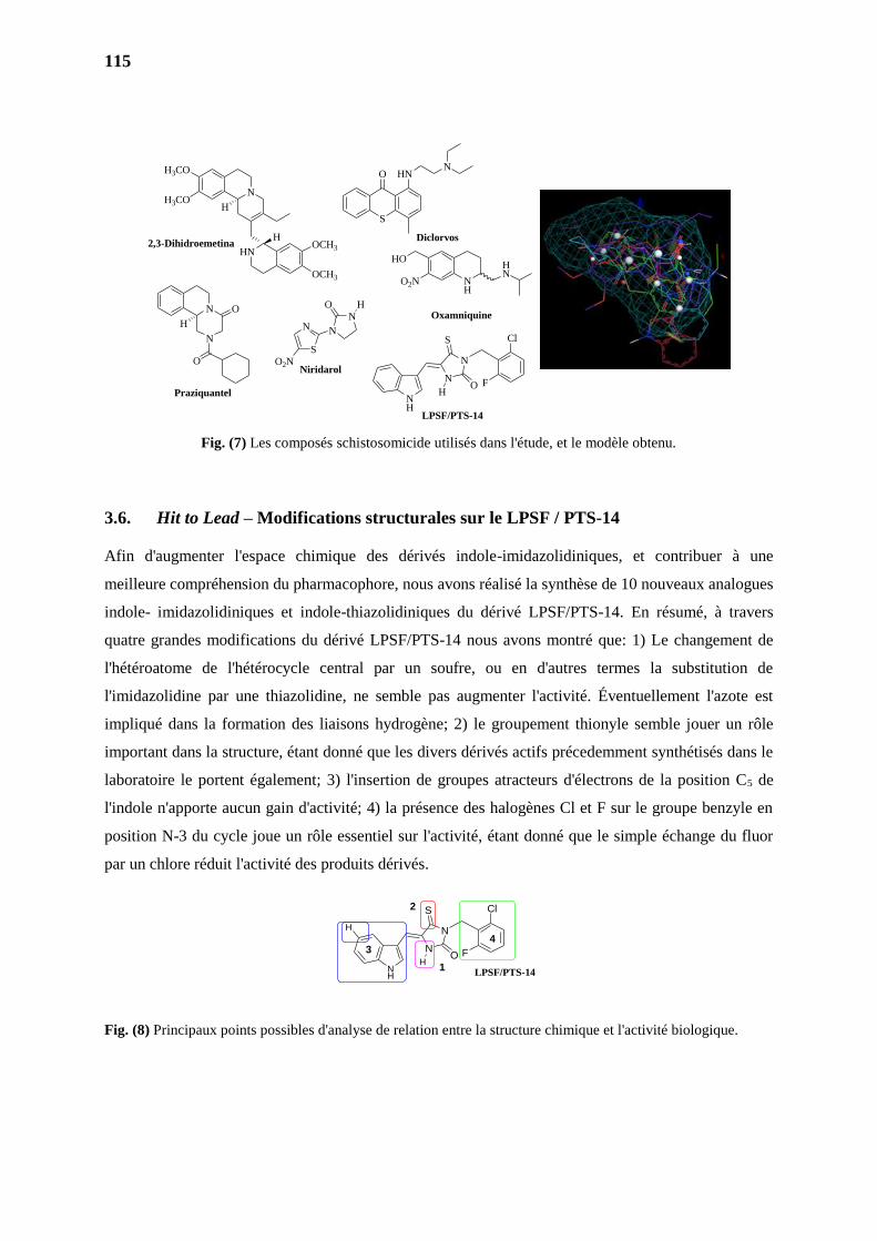
115
Fig. (7) Les composés schistosomicide utilisés dans l'étude, et le modèle obtenu.
3.6. Hit to Lead – Modifications structurales sur le LPSF / PTS-14
Afin d'augmenter l'espace chimique des dérivés indole-imidazolidiniques, et contribuer à une
meilleure compréhension du pharmacophore, nous avons réalisé la synthèse de 10 nouveaux analogues
indole- imidazolidiniques et indole-thiazolidiniques du dérivé LPSF/PTS-14. En résumé, à travers
quatre grandes modifications du dérivé LPSF/PTS-14 nous avons montré que: 1) Le changement de
l'hétéroatome de l'hétérocycle central par un soufre, ou en d'autres termes la substitution de
l'imidazolidine par une thiazolidine, ne semble pas augmenter l'activité. Éventuellement l'azote est
impliqué dans la formation des liaisons hydrogène; 2) le groupement thionyle semble jouer un rôle
important dans la structure, étant donné que les divers dérivés actifs précedemment synthétisés dans le
laboratoire le portent également; 3) l'insertion de groupes atracteurs d'électrons de la position C5 de
l'indole n'apporte aucun gain d'activité; 4) la présence des halogènes Cl et F sur le groupe benzyle en
position N-3 du cycle joue un rôle essentiel sur l'activité, étant donné que le simple échange du fluor
par un chlore réduit l'activité des produits dérivés.
Fig. (8) Principaux points possibles d'analyse de relation entre la structure chimique et l'activité biologique.
N
N
O
H
HN
H
N
H3CO
H3CO
OCH3
OCH3
H
O
N
N
O
N
S
O2N
H
S
O HNN
NH
HO
O2N
HN
Praziquantel
2,3-Dihidroemetina
Niridarol
Diclorvos
Oxamniquine
LPSF/PTS-14
Cl
FO
S
N
N
NH
H
Cl
FO
S
N
N
NH
H
H
1
2
34
LPSF/PTS-14
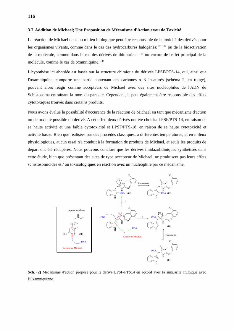
116
3.7. Addition de Michael; Une Proposition de Mécanisme d'Action et/ou de Toxicité
La réaction de Michael dans un milieu biologique peut être responsable de la toxicité des dérivés pour
les organismes vivants, comme dans le cas des hydrocarbures halogénés;191,192 ou de la bioactivation
de la molécule, comme dans le cas des dérivés de thiopurine; 193 ou encore de l'effet principal de la
molécule, comme le cas de oxamniquine.190
L'hypothèse ici abordée est basée sur la structure chimique du dérivée LPSF/PTS-14, qui, ainsi que
l'oxamniquine, comporte une partie contenant des carbones insaturés (schéma 2, en rouge),
pouvant alors réagir comme accepteurs de Michael avec des sites nucléophiles de l'ADN de
Schistosoma entraînant la mort du parasite. Cependant, il peut également être responsable des effets
cytotoxiques trouvés dans certains produits.
Nous avons évalué la possibilité d'occurence de la réaction de Michael en tant que mécanisme d'action
ou de toxicité possible du dérivé. A cet effet, deux dérivés ont été choisis: LPSF/PTS-14, en raison de
sa haute activité et une faible cytotoxicité et LPSF/PTS-18, en raison de sa haute cytotoxicité et
activité basse. Bien que réalisées par des procédés classiques, à differentes temperatures, et en mileux
physiologiques, aucun essai n'a conduit à la formation de produits de Michael, et seuls les produits de
départ ont été récupérés. Nous pouvons conclure que les dérivés imidazolidiniques synthétisés dans
cette étude, bien que présentant des sites de type accepteur de Michael, ne produisent pas leurs effets
schistosomicides et / ou toxicologiques en réaction avec un nucléophile par ce mécanisme.
Sch. (2) Mécanisme d'action proposé pour le dérivé LPSF/PTS14 en accord avec la similarité chimique avec
l'Oxamniquinne.
HN
HN
O2N
Agente alquilante
DNA
NH
NH
N
S
O
Cl
F
NH
NH
N
HS
O
Cl
F
NH
NH
N
S
O
Cl
F
DNA
DNA
DNA
Aceptor de Michael
Aceptor de MichaelTautomeria
DNA
x
NH
NH
N
HS
O
Cl
F
DNA
NH
NH
N
S
O
Cl
F
DNA
Tautomeria
x 1
2
(58)
(60)
(62) (63)
(61)
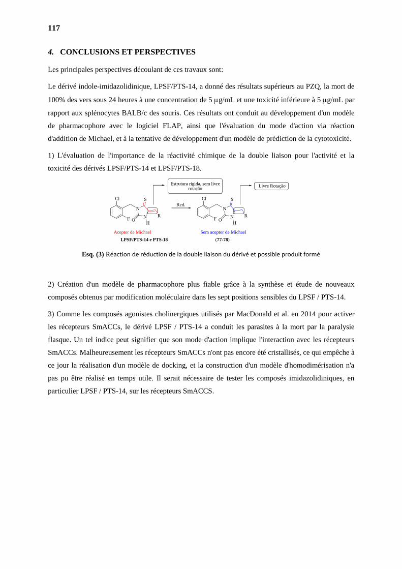
117
4. CONCLUSIONS ET PERSPECTIVES
Les principales perspectives découlant de ces travaux sont:
Le dérivé indole-imidazolidinique, LPSF/PTS-14, a donné des résultats supérieurs au PZQ, la mort de
100% des vers sous 24 heures à une concentration de 5 g/mL et une toxicité inférieure à 5 g/mL par
rapport aux splénocytes BALB/c des souris. Ces résultats ont conduit au développement d'un modèle
de pharmacophore avec le logiciel FLAP, ainsi que l'évaluation du mode d'action via réaction
d'addition de Michael, et à la tentative de développement d'un modèle de prédiction de la cytotoxicité.
1) L'évaluation de l'importance de la réactivité chimique de la double liaison pour l'activité et la
toxicité des dérivés LPSF/PTS-14 et LPSF/PTS-18.
Esq. (3) Réaction de réduction de la double liaison du dérivé et possible produit formé
2) Création d'un modèle de pharmacophore plus fiable grâce à la synthèse et étude de nouveaux
composés obtenus par modification moléculaire dans les sept positions sensibles du LPSF / PTS-14.
3) Comme les composés agonistes cholinergiques utilisés par MacDonald et al. en 2014 pour activer
les récepteurs SmACCs, le dérivé LPSF / PTS-14 a conduit les parasites à la mort par la paralysie
flasque. Un tel indice peut signifier que son mode d'action implique l'interaction avec les récepteurs
SmACCs. Malheureusement les récepteurs SmACCs n'ont pas encore été cristallisés, ce qui empêche à
ce jour la réalisation d'un modèle de docking, et la construction d'un modèle d'homodimérisation n'a
pas pu être réalisé en temps utile. Il serait nécessaire de tester les composés imidazolidiniques, en
particulier LPSF / PTS-14, sur les récepteurs SmACCS.
Cl
F O
S
N
N R
Cl
F
Red.
Aceptor de Michael Sem aceptor de Michael
O
S
N
N R
H H
Estrutura rigida, sem livrerotação
Livre Rotação
LPSF/PTS-14 e PTS-18 (77-78)
PARTIE II
SYNTHESE D’AMINOGLYCOSIDES AMPHIPHILES A
BASE DE NEOSAMINE

119
119
INTRODUCTION
PARTIE II
120
120
INTRODUCTION GENERALE
Tout au long de l'histoire, l'humanité a été victime de nombreuses pandémies extrêmement mortelles
telles que celles dues au choléra, à la peste, la grippe, la fièvre typhoïde, la tuberculose ou encore à
d'autres maladies infectieuses. D’autres maladies telles que les infections de l'oreille, de la peau et de
la gorge, peuvent également être responsables de nombreuses complications telles que la surdité.
Jusqu'au milieu du XIXème siècle, l'espérance de vie en Europe et en Amérique du Nord était en
moyenne de 50 ans, avec de grandes chances de décès prématurés dus aux maladies infectieuses.1
La découverte et l'innovation des antibiotiques sont considérées comme l'une des principales
réalisations de l'humanité. Sans ces derniers, les naissances prématurées seraient difficiles, la plupart
des interventions chirurgicales et des greffes seraient impossibles, les thérapies cytotoxiques pour le
cancer conduiraient à des infections mortelles et les hôpitaux deviendraient des nids à bactéries.
Depuis la découverte des antibiotiques, l’espérance de vie a été augmentée de plus de 20 ans et des
millions d'infections, auparavant considérées comme fatales, ont pues être traitées.2–4
Les aminoglycosides sont une partie importante de l'arsenal thérapeutique antibactérien depuis leur
découverte dans les années 40 et constituent la première classe d'antibiotiques découverts par criblage
de produits naturels avec une activité antibactérienne. En 1944, le laboratoire Waksman est le premier
à identifier et isoler la streptomycine, un aminoglycoside antibiotique synthétisé par des bactéries du
sol. Depuis, d’autres aminoglycosides ont été isolés et sont généralement administrés par voie topique
ou intraveineuse. La néomycine (néomycine) a été isolée à partir de la bactérie Streptomyces fradiae et
possède une meilleure activité contre les bactéries Gram aérobie (+) que de la streptomycine. La
gentamicine a été isolée à partir de Micromonospora en 1963 et constitue une percée majeure dans le
traitement des infections à bacilles à Gram négatif, y compris celles provoquées par Pseudomonas
aeruginosa.5
Généralement, le nom associé à une substance est lié à son origine. Par exemple, les noms se terminant
par « mycine » correspondent à des molécules qui sont directement ou indirectement dérivés de
Streptomyces. A l’inverse, ceux se terminant par « micine » correspondent à des molécules provenant
directement ou indirectement de Micromonospora.5,6
Les aminoglycosides sont des antibiotiques bactéricides, naturels ou semi-synthétiques dont le spectre
d'action implique les bactéries à Gram (+) et Gram (-). De tels dérivés possèdent un effet de synergie
avec d'autres antibiotiques, comme les ß-lactamines.7–10 Néanmoins, l’activité de cette classe
d’antibiotiques est dépendante de la concentration in vivo de dioxygène et son effet est donc très faible
sur les bactéries anaérobies et les mycoplasmes. De plus, certains effets toxiques tels que la
néphrotoxicité et l'ototoxicité ont été observés. Ces effets, en plus de l’apparition de mécanismes de
résistance bactérienne constituent des limites pour cette classe de médicaments.11
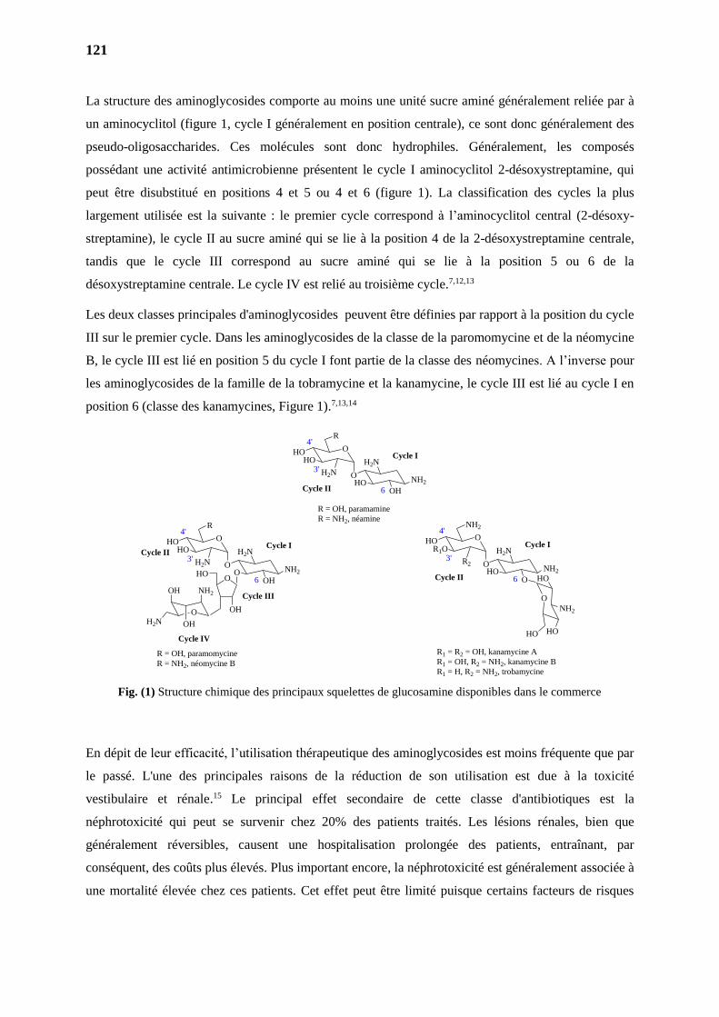
121
La structure des aminoglycosides comporte au moins une unité sucre aminé généralement reliée par à
un aminocyclitol (figure 1, cycle I généralement en position centrale), ce sont donc généralement des
pseudo-oligosaccharides. Ces molécules sont donc hydrophiles. Généralement, les composés
possédant une activité antimicrobienne présentent le cycle I aminocyclitol 2-désoxystreptamine, qui
peut être disubstitué en positions 4 et 5 ou 4 et 6 (figure 1). La classification des cycles la plus
largement utilisée est la suivante : le premier cycle correspond à l’aminocyclitol central (2-désoxy-
streptamine), le cycle II au sucre aminé qui se lie à la position 4 de la 2-désoxystreptamine centrale,
tandis que le cycle III correspond au sucre aminé qui se lie à la position 5 ou 6 de la
désoxystreptamine centrale. Le cycle IV est relié au troisième cycle.7,12,13
Les deux classes principales d'aminoglycosides peuvent être définies par rapport à la position du cycle
III sur le premier cycle. Dans les aminoglycosides de la classe de la paromomycine et de la néomycine
B, le cycle III est lié en position 5 du cycle I font partie de la classe des néomycines. A l’inverse pour
les aminoglycosides de la famille de la tobramycine et la kanamycine, le cycle III est lié au cycle I en
position 6 (classe des kanamycines, Figure 1).7,13,14
Fig. (1) Structure chimique des principaux squelettes de glucosamine disponibles dans le commerce
En dépit de leur efficacité, l’utilisation thérapeutique des aminoglycosides est moins fréquente que par
le passé. L'une des principales raisons de la réduction de son utilisation est due à la toxicité
vestibulaire et rénale.15 Le principal effet secondaire de cette classe d'antibiotiques est la
néphrotoxicité qui peut se survenir chez 20% des patients traités. Les lésions rénales, bien que
généralement réversibles, causent une hospitalisation prolongée des patients, entraînant, par
conséquent, des coûts plus élevés. Plus important encore, la néphrotoxicité est généralement associée à
une mortalité élevée chez ces patients. Cet effet peut être limité puisque certains facteurs de risques
O
O
HO
R
HO
H2N
OOH
NH2
H2N3'
4'
6
Cycle I
O
OHO
OH
H2N OH
NH2
HO
Cycle II
Cycle III
Cycle IV
R = OH, paramomycine
R = NH2, néomycine B
O
O
HO
R
HO
H2N
HOOH
NH2
H2N3'
4'
6
Cycle I
Cycle II
R = OH, paramamine
R = NH2, néamine
O
O
HO
NH2
R1O
R2
HOO
NH2
H2N3'
4'
6
Cycle I
Cycle II
O
NH2
HOHO
HO
R1 = R2 = OH, kanamycine A
R1 = OH, R2 = NH2, kanamycine B
R1 = H, R2 = NH2, trobamycine
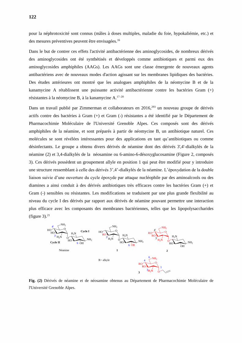
122
pour la néphrotoxicité sont connus (mâles à doses multiples, maladie du foie, hypokaliémie, etc.) et
des mesures préventives peuvent être envisagées.16
Dans le but de contrer ces effets l'activité antibactérienne des aminoglycosides, de nombreux dérivés
des aminoglycosides ont été synthétisés et développés comme antibiotiques et parmi eux des
aminoglycosides amphiphiles (AAGs). Les AAGs sont une classe émergente de nouveaux agents
antibactériens avec de nouveaux modes d'action agissant sur les membranes lipidiques des bactéries.
Des études antérieures ont montré que les analogues amphiphiles de la néomycine B et de la
kanamycine A rétablissent une puissante activité antibactérienne contre les bactéries Gram (+)
résistantes à la néomycine B, à la kanamycine A.17–20
Dans un travail publié par Zimmerman et collaborateurs en 2016,293 un nouveau groupe de dérivés
actifs contre des bactéries à Gram (+) et Gram (-) résistantes a été identifié par le Département de
Pharmacochimie Moléculaire de l'Université Grenoble Alpes. Ces composés sont des dérivés
amphiphiles de la néamine, et sont préparés à partir de néomycine B, un antibiotique naturel. Ces
molécules se sont révélées intéressantes pour des applications en tant qu’antibiotiques ou comme
désinfectants. Le groupe a obtenu divers dérivés de néamine dont des dérivés 3',4'-dialkylés de la
néamine (2) et 3,4-dialkylés de la néosamine ou 6-amino-6-désoxyglucosamine (Figure 2, composés
3). Ces dérivés possèdent un groupement allyle en position 1 qui peut être modifié pour y introduire
une structure ressemblant à celle des dérivés 3’,4’-dialkylés de la néamine. L’époxydation de la double
liaison suivie d’une ouverture du cycle époxyde par attaque nucléophile par des aminoalcools ou des
diamines a ainsi conduit à des dérivés antibiotiques très efficaces contre les bactéries Gram (+) et
Gram (-) sensibles ou résistantes. Les modifications se traduisent par une plus grande flexibilité au
niveau du cycle I des dérivés par rapport aux dérivés de néamine pouvant permettre une interaction
plus efficace avec les composants des membranes bactériennes, telles que les lipopolysaccharides
(figure 3).21
Fig. (2) Dérivés de néamine et de néosamine obtenus au Département de Pharmacochimie Moléculaire de
l'Université Grenoble Alpes.
O
O
RO
NH2
RO
H2N
HOOH
NH2
H2N
O
O
HO
NH2
HO
H2N
HOOH
NH2
H2N
O
O
RO
NH2
RO
H2N
O
O
HO
NH2
RO
H2N
HOOR
NH2
H2N3'
4'
Néamine
3'
4'
6
Cycle I
3
2
3
4
Cycle II
1
6
3'
1
6
R= alkyle

123
Dans le but d’agrandir la famille des dérivés de la néosamine amphiphiles antibiotiques à spectre large
d’action, le but de ce travail de thèse était donc de synthétiser de nouveaux dérivés de la néosamine
antibactériens en utilisant des réactions de métathèse.
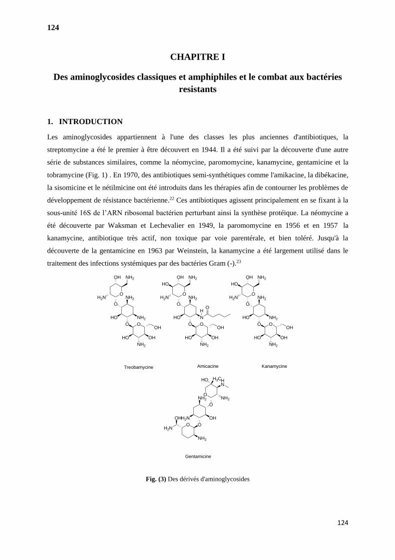
124
124
CHAPITRE I
Des aminoglycosides classiques et amphiphiles et le combat aux bactéries
resistants
1. INTRODUCTION
Les aminoglycosides appartiennent à l'une des classes les plus anciennes d'antibiotiques, la
streptomycine a été le premier à être découvert en 1944. Il a été suivi par la découverte d'une autre
série de substances similaires, comme la néomycine, paromomycine, kanamycine, gentamicine et la
tobramycine (Fig. 1) . En 1970, des antibiotiques semi-synthétiques comme l'amikacine, la dibékacine,
la sisomicine et le nétilmicine ont été introduits dans les thérapies afin de contourner les problèmes de
développement de résistance bactérienne.22 Ces antibiotiques agissent principalement en se fixant à la
sous-unité 16S de l’ARN ribosomal bactérien perturbant ainsi la synthèse protéique. La néomycine a
été découverte par Waksman et Lechevalier en 1949, la paromomycine en 1956 et en 1957 la
kanamycine, antibiotique très actif, non toxique par voie parentérale, et bien toléré. Jusqu'à la
découverte de la gentamicine en 1963 par Weinstein, la kanamycine a été largement utilisé dans le
traitement des infections systémiques par des bactéries Gram (-).23
Fig. (3) Des dérivés d'aminoglycosides
O
O
O O
NH2
HO
HO OH
OH
NH2OH
H2N
NH2
NH2
O
O
O O
NH2
HO
HO OH
OH
NH2OH
H2N
NH2
N
HO
OH
O
O
O O
NH2
HO
HO OH
OH
NH2OH
H2N
NH2
NH2
HO
O
O
O
NH2
OH
NH2
H2N
NH2
H2N
HN
H3CHO
O
OH
Treobamycine Amicacine Kanamycine
Gentamicine
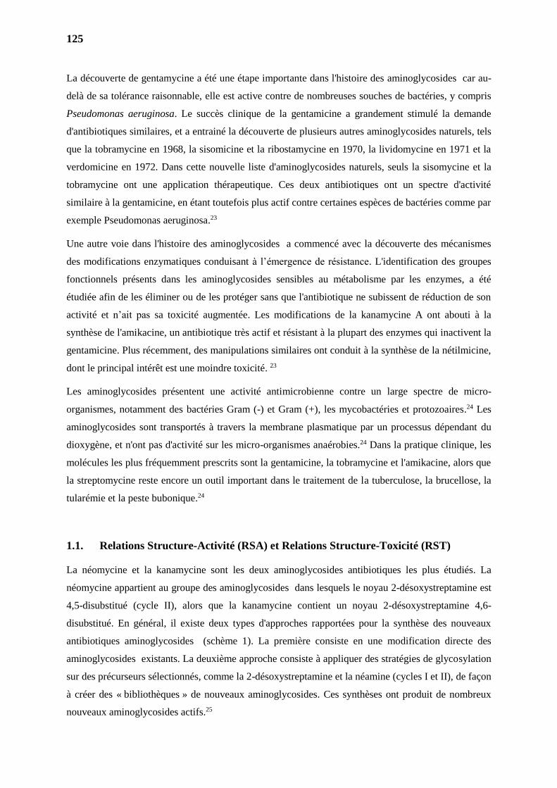
125
La découverte de gentamycine a été une étape importante dans l'histoire des aminoglycosides car au-
delà de sa tolérance raisonnable, elle est active contre de nombreuses souches de bactéries, y compris
Pseudomonas aeruginosa. Le succès clinique de la gentamicine a grandement stimulé la demande
d'antibiotiques similaires, et a entrainé la découverte de plusieurs autres aminoglycosides naturels, tels
que la tobramycine en 1968, la sisomicine et la ribostamycine en 1970, la lividomycine en 1971 et la
verdomicine en 1972. Dans cette nouvelle liste d'aminoglycosides naturels, seuls la sisomycine et la
tobramycine ont une application thérapeutique. Ces deux antibiotiques ont un spectre d'activité
similaire à la gentamicine, en étant toutefois plus actif contre certaines espèces de bactéries comme par
exemple Pseudomonas aeruginosa.23
Une autre voie dans l'histoire des aminoglycosides a commencé avec la découverte des mécanismes
des modifications enzymatiques conduisant à l’émergence de résistance. L'identification des groupes
fonctionnels présents dans les aminoglycosides sensibles au métabolisme par les enzymes, a été
étudiée afin de les éliminer ou de les protéger sans que l'antibiotique ne subissent de réduction de son
activité et n’ait pas sa toxicité augmentée. Les modifications de la kanamycine A ont abouti à la
synthèse de l'amikacine, un antibiotique très actif et résistant à la plupart des enzymes qui inactivent la
gentamicine. Plus récemment, des manipulations similaires ont conduit à la synthèse de la nétilmicine,
dont le principal intérêt est une moindre toxicité. 23
Les aminoglycosides présentent une activité antimicrobienne contre un large spectre de micro-
organismes, notamment des bactéries Gram (-) et Gram (+), les mycobactéries et protozoaires.24 Les
aminoglycosides sont transportés à travers la membrane plasmatique par un processus dépendant du
dioxygène, et n'ont pas d'activité sur les micro-organismes anaérobies.24 Dans la pratique clinique, les
molécules les plus fréquemment prescrits sont la gentamicine, la tobramycine et l'amikacine, alors que
la streptomycine reste encore un outil important dans le traitement de la tuberculose, la brucellose, la
tularémie et la peste bubonique.24
1.1. Relations Structure-Activité (RSA) et Relations Structure-Toxicité (RST)
La néomycine et la kanamycine sont les deux aminoglycosides antibiotiques les plus étudiés. La
néomycine appartient au groupe des aminoglycosides dans lesquels le noyau 2-désoxystreptamine est
4,5-disubstitué (cycle II), alors que la kanamycine contient un noyau 2-désoxystreptamine 4,6-
disubstitué. En général, il existe deux types d'approches rapportées pour la synthèse des nouveaux
antibiotiques aminoglycosides (schème 1). La première consiste en une modification directe des
aminoglycosides existants. La deuxième approche consiste à appliquer des stratégies de glycosylation
sur des précurseurs sélectionnés, comme la 2-désoxystreptamine et la néamine (cycles I et II), de façon
à créer des « bibliothèques » de nouveaux aminoglycosides. Ces synthèses ont produit de nombreux
nouveaux aminoglycosides actifs.25
126
Sch. (1) Stratégies générales pour la synthèse d'aminoglycosides de la classe de la néomycine et de la classe de
la kanamycine.
Par les modifications chimiques directes des aminoglycosides existants, plusieurs critères doivent tout
d’abord être considérés. Ceux-ci comprennent la source disponible d'aminoglycosides, les groupes
protecteurs appropriés pour les groupes amines et la différenciation régiosélective des groupes
hydroxyles à fonctionnaliser. La néomycine B et la kanamycine A sont deux aminoglycosides
disponibles dans le marché qui peuvent être utilisés comme matériaux de départ pour des
modifications chimiques. Bien que la néomycine B disponible dans le commerce soit habituellement
mélangée à 10-20% de néomycine A et C et que la kanamycine A soit habituellement mélangée à 10-
20% de kanamycine B, ces composants mineurs ne constituent pas une difficulté significative dans la
synthèse chimique et la caractérisation des composés synthétisés. 25
Les groupes protecteurs les plus utilisés pendant les synthèses pour les fonctions amine sont les
carbamates et leur transformation en azoture a également beaucoup été utilisée. La première stratégie
est la plus facile à mettre en oeuvre. Cependant, la faible solubilité des composés contenant des
polycarbamates et des azotures rend la purification et la caractérisation de ces composés très difficiles.
L'utilisation du groupe azoture pour masquer les groupes amine d'aminoglycosides présente l'avantage
de générer des composés ayant une plus grande solubilité dans des milieux organiques, facilitant leur
extraction à partir de milieux aqueux qui reste néanmoins difficile. 25
Le problème le plus difficile dans la synthèse des dérivés aminoglycosides est la différenciation
régiosélective des groupes amine qui a été étudiée en profondeur, en particulier dans la néomycine, la
kanamycine et la néamine. En général, ces procédés utilisent des ions de chélation ou de cyclisation à
médiation métallique. 25
O
R1
HOHO
H2NOO
H2N
NH2
OHOHO
OHO
OHO
H2N
HONH2
I
II
IIIO
NH2
HOHO
R OHO
H2N
NH2
OH
I
II
O
NH2
HOHO
R OHO
H2N
NH2
O
I
II
OOH
HO
H2NOH
III
Kanamycine A
Derivés de la Néomycine Derivés de la Kanamycine
Derivés de la Néamine
Néomycine B
B, C
A A
B
C C
A: Hydrolyse AcideB: Modification DirectC: Glycosylation (glycodiversification)
R = NH2
ou OH
1'
2'3'
4' 5'
6'
1
23
4
5 6
1'
2'3'
4' 5'
6'
1
2
4
5 6
1'
2'3'
4' 5'
6'
1
2
4
5 6
127
La stéréochimie de la liaison glycosidique à laquelle le composant glucidique est fixé au noyau de la
néamine est essentielle pour l'activité antibactérienne. 26
Certaines conclusions ont été tirées des études des relations Structure-activité (RSA) à travers de la
comparaison des structures naturelles de aminoglycosides et ceux qui ont été modifiés par des
réactions semi-synthétiques.26
Le cycle I est d'une importance fondamentale pour le large spectre d'activité et est également la cible
principale des enzymes bactériennes d’inactivation. Les fonctions amine sur 6' et 2' sont
particulièrement importantes, par exemple la kanamycine B (2’,6'-diamino), est plus actif que la
kanamycine A (6'-amino-2'-hydroxyl), qui à son tour est plus actif que la kanamycine C (2'-amino-6'-
hydroxyl). La méthylation en position 2' ou 6' ne réduit pas considérablement l'activité antibactérienne
et confére une résistance à l'acétylation enzymatique du groupe amine en C-6’. L'élimination du
groupe hydroxyle en 3' ou 4' ou dans les deux positions dans les kanamycines, ne réduit pas le pouvoir
antibactérien. Les gentamicines n’ont également pas de fonction hydroxyle dans ces positions ainsi
que la sisomicine et la nétilmicine, qui ont aussi une double liaison entre les positions 4 'et 5'. Aucun
de ces dérivés n’est inactivé par l'enzyme d’inactivation phosphotransférase qui phosphoryle
hydroxyle de la position 3'. Les dérivés phosphorylés dans cette position ont beaucoup moins d'affinité
pour le site actif sur le ribosome bactérien. 26
Peu de modifications sont possibles sur le cycle I car ce cycle est principalement impliqué dans la
liaison de l'antibiotique à la cible. Cependant, le groupe amine en position 1 de kanamycine A peut
être acylé (amikacine par exemple), sans perdre l'activité. La nétilmicine conserve son pouvoir
antibactérien de la sisomicine car elle est résistante à différentes enzymes bactériennes.26
Les groupes fonctionnels du cycle III semblent être moins sensibles aux changements structurels que
ceux portés par les cycles I et II bien que les gentamicines 2"-désoxy soient nettement moins actives
que leurs analogues 2’’-aminés ou -hydroxylés soient plus actifs. Le groupe amine en C-3' des
gentamicines peut être primaire ou secondaire avec dans les deux cas une grande efficacité
antibactérienne. En outre, l'hydroxyle en 4 peut être en position axiale ou équatoriale avec peu de
changement dans l’activité antibiotique (Figure 3).26
Une étude de relations structure-toxicité relative des dérivés pseudo-disaccharides similaires à la
néamine (NB23 et gentamicine C1A), contenant le cycle 2-désoxystreptamine (2-DOS), a proposé que
la basicité du groupe 2'-NH2 soit liée à la toxicité aigüe des aminoglycosides. Basé sur les résultats
obtenues, a été synthétisé un nouveau pseudo trisaccharide (NB45), qui a montré une activité
antibactérienne significative et une faible toxicité aigu, la figure 3.27
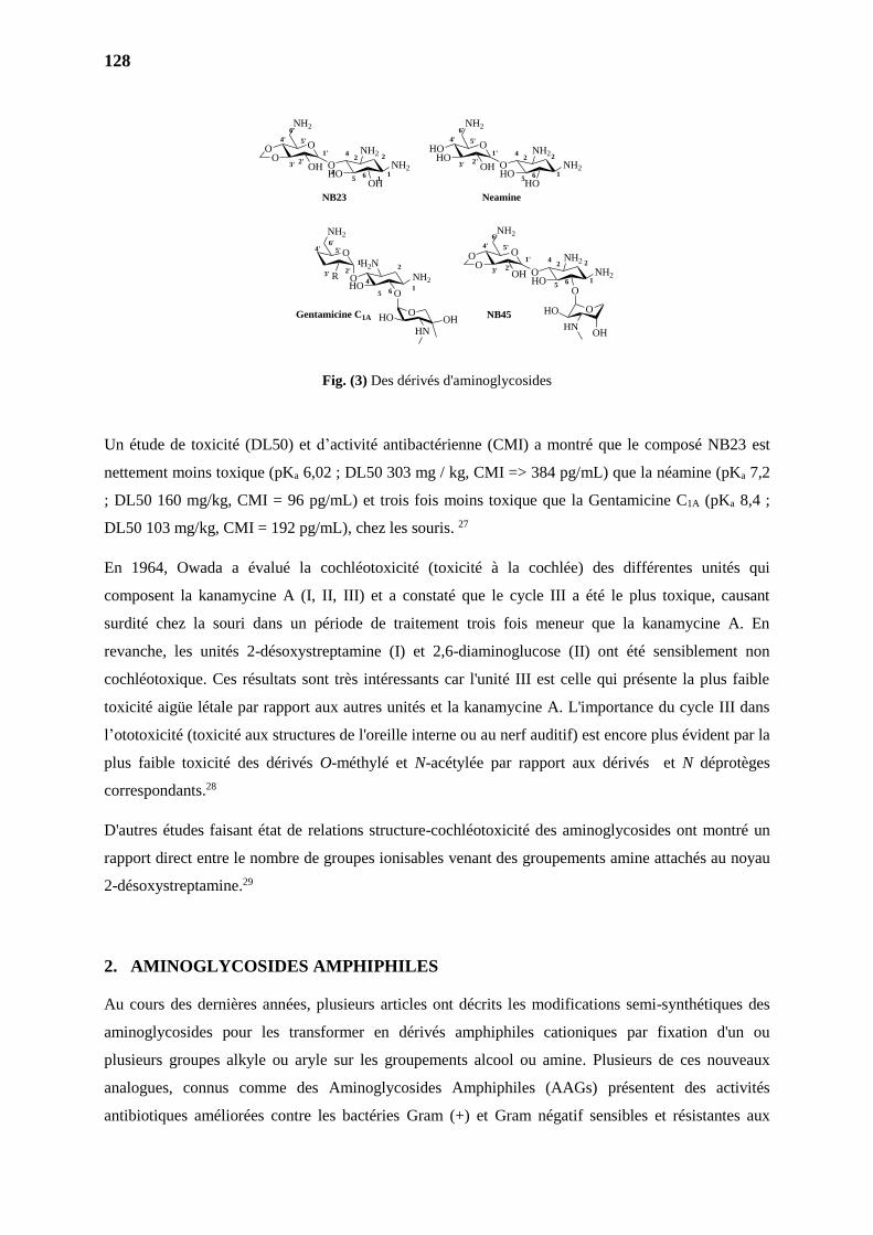
128
Fig. (3) Des dérivés d'aminoglycosides
Un étude de toxicité (DL50) et d’activité antibactérienne (CMI) a montré que le composé NB23 est
nettement moins toxique (pKa 6,02 ; DL50 303 mg / kg, CMI => 384 pg/mL) que la néamine (pKa 7,2
; DL50 160 mg/kg, CMI = 96 pg/mL) et trois fois moins toxique que la Gentamicine C1A (pKa 8,4 ;
DL50 103 mg/kg, CMI = 192 pg/mL), chez les souris. 27
En 1964, Owada a évalué la cochléotoxicité (toxicité à la cochlée) des différentes unités qui
composent la kanamycine A (I, II, III) et a constaté que le cycle III a été le plus toxique, causant
surdité chez la souri dans un période de traitement trois fois meneur que la kanamycine A. En
revanche, les unités 2-désoxystreptamine (I) et 2,6-diaminoglucose (II) ont été sensiblement non
cochléotoxique. Ces résultats sont très intéressants car l'unité III est celle qui présente la plus faible
toxicité aigüe létale par rapport aux autres unités et la kanamycine A. L'importance du cycle III dans
l’ototoxicité (toxicité aux structures de l'oreille interne ou au nerf auditif) est encore plus évident par la
plus faible toxicité des dérivés O-méthylé et N-acétylée par rapport aux dérivés et N déprotèges
correspondants.28
D'autres études faisant état de relations structure-cochléotoxicité des aminoglycosides ont montré un
rapport direct entre le nombre de groupes ionisables venant des groupements amine attachés au noyau
2-désoxystreptamine.29
2. AMINOGLYCOSIDES AMPHIPHILES
Au cours des dernières années, plusieurs articles ont décrits les modifications semi-synthétiques des
aminoglycosides pour les transformer en dérivés amphiphiles cationiques par fixation d'un ou
plusieurs groupes alkyle ou aryle sur les groupements alcool ou amine. Plusieurs de ces nouveaux
analogues, connus comme des Aminoglycosides Amphiphiles (AAGs) présentent des activités
antibiotiques améliorées contre les bactéries Gram (+) et Gram négatif sensibles et résistantes aux
O
NH2
R OHO
H2N
NH2
O
1'
2'3'
4' 5'6'
1
2
4
5 6
OHO
HNOH
HO
NH2
NH2
O
O
O
NH2
OO
OH
NB23
HO
NH2
NH2
HO
O
O
NH2
HOHO
OH
Neamine
Gentamicine C1AOHO
HNOH
HO
NH2
NH2
OH
O
O
NH2
OO
OH
NB45
1'
2'3'
4' 5'
6'
1
2
45
6
4
1
21'
2'3'
4' 5'
6'
2
56
4
1
2
1'
2'3'
4' 5'
6'
2
56
4
1
2
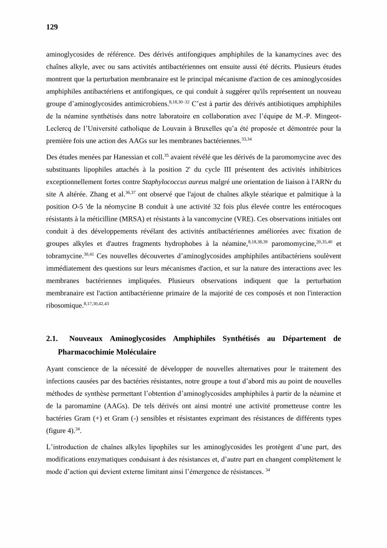
129
aminoglycosides de référence. Des dérivés antifongiques amphiphiles de la kanamycines avec des
chaînes alkyle, avec ou sans activités antibactériennes ont ensuite aussi été décrits. Plusieurs études
montrent que la perturbation membranaire est le principal mécanisme d'action de ces aminoglycosides
amphiphiles antibactériens et antifongiques, ce qui conduit à suggérer qu'ils représentent un nouveau
groupe d’aminoglycosides antimicrobiens.8,18,30–32 C’est à partir des dérivés antibiotiques amphiphiles
de la néamine synthétisés dans notre laboratoire en collaboration avec l’équipe de M.-P. Mingeot-
Leclercq de l’Université catholique de Louvain à Bruxelles qu’a été proposée et démontrée pour la
première fois une action des AAGs sur les membranes bactériennes.33,34
Des études menées par Hanessian et coll.35 avaient révélé que les dérivés de la paromomycine avec des
substituants lipophiles attachés à la position 2' du cycle III présentent des activités inhibitrices
exceptionnellement fortes contre Staphylococcus aureus malgré une orientation de liaison à l'ARNr du
site A altérée. Zhang et al.36,37 ont observé que l'ajout de chaînes alkyle stéarique et palmitique à la
position O-5 'de la néomycine B conduit à une activité 32 fois plus élevée contre les entérocoques
résistants à la méticilline (MRSA) et résistants à la vancomycine (VRE). Ces observations initiales ont
conduit à des développements révélant des activités antibactériennes améliorées avec fixation de
groupes alkyles et d'autres fragments hydrophobes à la néamine,8,18,38,39 paromomycine,20,35,40 et
tobramycine.30,41 Ces nouvelles découvertes d’aminoglycosides amphiphiles antibactériens soulèvent
immédiatement des questions sur leurs mécanismes d'action, et sur la nature des interactions avec les
membranes bactériennes impliquées. Plusieurs observations indiquent que la perturbation
membranaire est l'action antibactérienne primaire de la majorité de ces composés et non l'interaction
ribosomique.8,17,30,42,43
2.1. Nouveaux Aminoglycosides Amphiphiles Synthétisés au Département de
Pharmacochimie Moléculaire
Ayant conscience de la nécessité de développer de nouvelles alternatives pour le traitement des
infections causées par des bactéries résistantes, notre groupe a tout d’abord mis au point de nouvelles
méthodes de synthèse permettant l’obtention d’aminoglycosides amphiphiles à partir de la néamine et
de la paromamine (AAGs). De tels dérivés ont ainsi montré une activité prometteuse contre les
bactéries Gram (+) et Gram (-) sensibles et résistantes exprimant des résistances de différents types
(figure 4).34.
L’introduction de chaînes alkyles lipophiles sur les aminoglycosides les protègent d’une part, des
modifications enzymatiques conduisant à des résistances et, d’autre part en changent complètement le
mode d’action qui devient externe limitant ainsi l’émergence de résistances. 34
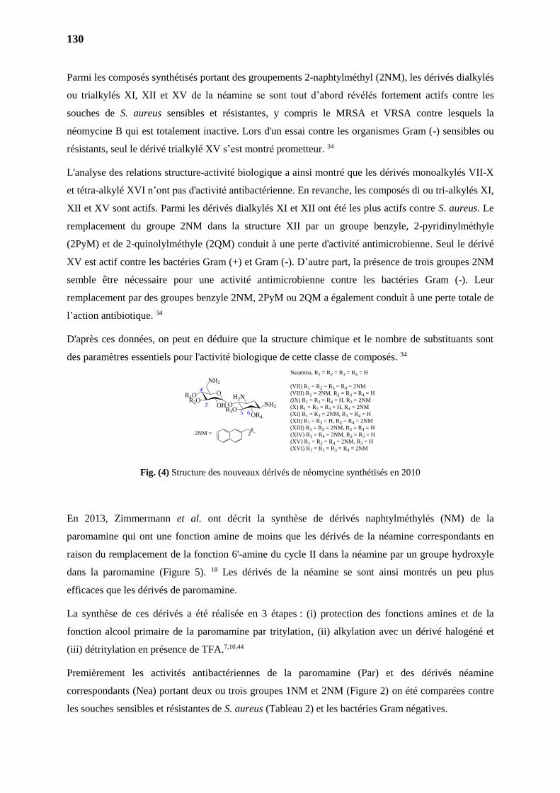
130
Parmi les composés synthétisés portant des groupements 2-naphtylméthyl (2NM), les dérivés dialkylés
ou trialkylés XI, XII et XV de la néamine se sont tout d’abord révélés fortement actifs contre les
souches de S. aureus sensibles et résistantes, y compris le MRSA et VRSA contre lesquels la
néomycine B qui est totalement inactive. Lors d'un essai contre les organismes Gram (-) sensibles ou
résistants, seul le dérivé trialkylé XV s’est montré prometteur. 34
L'analyse des relations structure-activité biologique a ainsi montré que les dérivés monoalkylés VII-X
et tétra-alkylé XVI n’ont pas d'activité antibactérienne. En revanche, les composés di ou tri-alkylés XI,
XII et XV sont actifs. Parmi les dérivés dialkylés XI et XII ont été les plus actifs contre S. aureus. Le
remplacement du groupe 2NM dans la structure XII par un groupe benzyle, 2-pyridinylméthyle
(2PyM) et de 2-quinolylméthyle (2QM) conduit à une perte d'activité antimicrobienne. Seul le dérivé
XV est actif contre les bactéries Gram (+) et Gram (-). D’autre part, la présence de trois groupes 2NM
semble être nécessaire pour une activité antimicrobienne contre les bactéries Gram (-). Leur
remplacement par des groupes benzyle 2NM, 2PyM ou 2QM a également conduit à une perte totale de
l’action antibiotique. 34
D'après ces données, on peut en déduire que la structure chimique et le nombre de substituants sont
des paramètres essentiels pour l'activité biologique de cette classe de composés. 34
Fig. (4) Structure des nouveaux dérivés de néomycine synthétisés en 2010
En 2013, Zimmermann et al. ont décrit la synthèse de dérivés naphtylméthylés (NM) de la
paromamine qui ont une fonction amine de moins que les dérivés de la néamine correspondants en
raison du remplacement de la fonction 6'-amine du cycle II dans la néamine par un groupe hydroxyle
dans la paromamine (Figure 5). 18 Les dérivés de la néamine se sont ainsi montrés un peu plus
efficaces que les dérivés de paromamine.
La synthèse de ces dérivés a été réalisée en 3 étapes : (i) protection des fonctions amines et de la
fonction alcool primaire de la paromamine par tritylation, (ii) alkylation avec un dérivé halogéné et
(iii) détritylation en présence de TFA.7,10,44
Premièrement les activités antibactériennes de la paromamine (Par) et des dérivés néamine
correspondants (Nea) portant deux ou trois groupes 1NM et 2NM (Figure 2) on été comparées contre
les souches sensibles et résistantes de S. aureus (Tableau 2) et les bactéries Gram négatives.
R3O
H2NNH2
OR4
O
O
NH2
R1OR2O
OH
2NM =
Neamina, R1 = R2 = R3 = R4 = H
(VII) R1 = R2 = R3 = R4 = 2NM
(VIII) R1 = 2NM, R2 = R3 = R4 = H
(IX) R1 = R2 = R4 = H, R3 = 2NM
(X) R1 = R2 = R3 = H, R4 = 2NM
(XI) R1 = R2 = 2NM, R3 = R4 = H
(XII) R1 = R3 = H, R2 = R4 = 2NM
(XIII) R1 = R3 = 2NM, R2 = R4 = H
(XIV) R1 = R4 = 2NM, R2 = R3 = H
(XV) R1 = R2 = R4 = 2NM, R3 = H
(XVI) R1 = R2 = R3 = R4 = 2NM
3'
4'
65
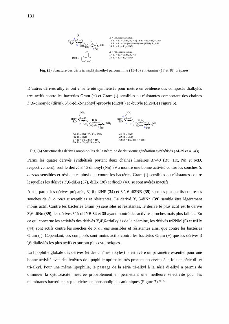
131
Fig. (5) Structure des dérivés naphtylméthyl paromamine (13-16) et néamine (17 et 18) préparés.
D’autres dérivés alkylés ont ensuite été synthétisés pour mettre en évidence des composés dialkylés
très actifs contre les bactéries Gram (+) et Gram (-) sensibles ou résistantes comportant des chaînes
3’,6-dinonyle (diNn), 3’,6-(di-2-naphtyl)-propyle (di2NP) et -butyle (di2NB) (Figure 6).
Fig. (6) Structure des dérivés amphiphiles de la néamine de deuxième génération synthétisés (34-39 et 41-43)
Parmi les quatre dérivés synthétisés portant deux chaînes linéaires 37-40 (Bu, Hx, Nn et ocD,
respectivement), seul le dérivé 3 ',6-dinonyl (Nn) 39 a montré une bonne activité contre les souches S.
aureus sensibles et résistantes ainsi que contre les bactéries Gram (-) sensibles ou résistantes contre
lesquelles les dérivés 3',6-diBu (37), diHx (38) et diocD (40) se sont avérés inactifs.
Ainsi, parmi les dérivés préparés, 3', 6-di2NP (34) et 3 ', 6-di2NB (35) sont les plus actifs contre les
souches de S. aureus susceptibles et résistantes. Le dérivé 3', 6-diNn (39) semble être légèrement
moins actif. Contre les bactéries Gram (-) sensibles et résistantes, le dérivé le plus actif est le dérivé
3',6-diNn (39), les dérivés 3′,6-di2NB 34 et 35 ayant montré des activités proches mais plus faibles. En
ce qui concerne les activités des dérivés 3',4',6-trialkylés de la néamine, les dérivés tri2NM (5) et triHx
(44) sont actifs contre les souches de S. aureus sensibles et résistantes ainsi que contre les bactéries
Gram (-). Cependant, ces composés sont moins actifs contre les bactéries Gram (+) que les dérivés 3
',6-dialkylés les plus actifs et surtout plus cytotoxiques.
La lipophilie globale des dérivés (et des chaînes alkyles) s’est avéré un paramètre essentiel pour une
bonne activité avec des fenêtres de lipophilie optimales très proches observées à la fois en série di- et
tri-alkyl. Pour une même lipophilie, le passage de la série tri-alkyl à la sérié di-alkyl a permis de
diminuer la cytotoxicité mesurée probablement en permettant une meilleure sélectivité pour les
membranes bactériennes plus riches en phospholipides anioniques (Figure 7).45–47
HO
H2N
NH2
OR
O
O
NH2
HORO
NH2
34: R = 2NP, 35: R = 2NB36: R = 2NH37: R = Bu, 38: R = Hx39: R = Nn, 40: R = ocD
3'
6HO
H2N
NH2
OR
O
O
NH2
RORO
NH2
41: R = 2NP42: R = 2NB43: R = Bu, 44: R = Hx
3'
6
4'
HO
H2NNH2
OR1
O
O
X
R3OR2O
NH2
1NM =
X = OH, série paroamine
13: R1 = R2 = 2NM, R3 = H; 14: R1 = R2 = R3 = 2NM
15: R1 = R2 = 1-naphthylmethylene (1NM), R3 = H
16: R1 = R2 = R3 = 1NM
X = NH2, série neamine
17: R1 = R2 = 1NM, R3 = H
18: R1 = R2 = R3 = 1NM
4'
6
3'
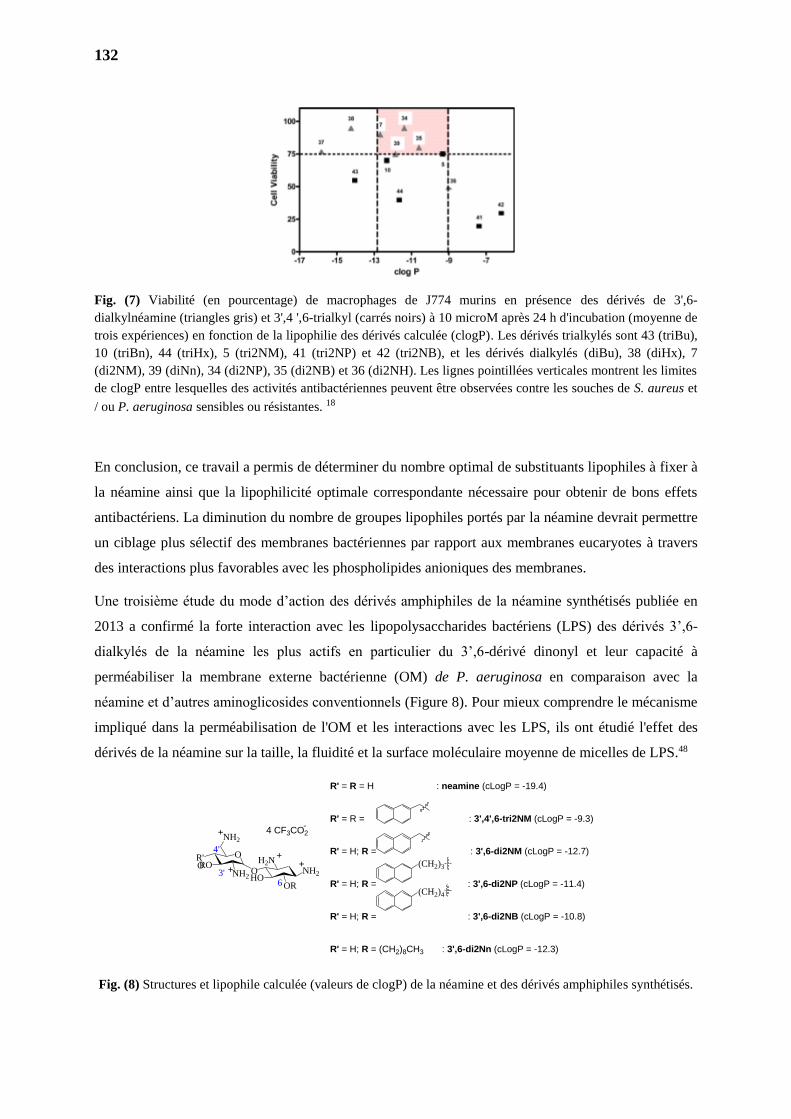
132
Fig. (7) Viabilité (en pourcentage) de macrophages de J774 murins en présence des dérivés de 3',6-
dialkylnéamine (triangles gris) et 3',4 ',6-trialkyl (carrés noirs) à 10 microM après 24 h d'incubation (moyenne de
trois expériences) en fonction de la lipophilie des dérivés calculée (clogP). Les dérivés trialkylés sont 43 (triBu),
10 (triBn), 44 (triHx), 5 (tri2NM), 41 (tri2NP) et 42 (tri2NB), et les dérivés dialkylés (diBu), 38 (diHx), 7
(di2NM), 39 (diNn), 34 (di2NP), 35 (di2NB) et 36 (di2NH). Les lignes pointillées verticales montrent les limites
de clogP entre lesquelles des activités antibactériennes peuvent être observées contre les souches de S. aureus et
/ ou P. aeruginosa sensibles ou résistantes. 18
En conclusion, ce travail a permis de déterminer du nombre optimal de substituants lipophiles à fixer à
la néamine ainsi que la lipophilicité optimale correspondante nécessaire pour obtenir de bons effets
antibactériens. La diminution du nombre de groupes lipophiles portés par la néamine devrait permettre
un ciblage plus sélectif des membranes bactériennes par rapport aux membranes eucaryotes à travers
des interactions plus favorables avec les phospholipides anioniques des membranes.
Une troisième étude du mode d’action des dérivés amphiphiles de la néamine synthétisés publiée en
2013 a confirmé la forte interaction avec les lipopolysaccharides bactériens (LPS) des dérivés 3’,6-
dialkylés de la néamine les plus actifs en particulier du 3’,6-dérivé dinonyl et leur capacité à
perméabiliser la membrane externe bactérienne (OM) de P. aeruginosa en comparaison avec la
néamine et d’autres aminoglicosides conventionnels (Figure 8). Pour mieux comprendre le mécanisme
impliqué dans la perméabilisation de l'OM et les interactions avec les LPS, ils ont étudié l'effet des
dérivés de la néamine sur la taille, la fluidité et la surface moléculaire moyenne de micelles de LPS.48
Fig. (8) Structures et lipophile calculée (valeurs de clogP) de la néamine et des dérivés amphiphiles synthétisés.
HO
H2NNH2
OR
O
O
NH2
R'ORO
NH23'6
+
++
+
4'
4 CF3CO2-
R' = R = H : neamine (cLogP = -19.4)
R' = R = : 3',4',6-tri2NM (cLogP = -9.3)
R' = H; R = : 3',6-di2NM (cLogP = -12.7)
R' = H; R = : 3',6-di2NP (cLogP = -11.4)
R' = H; R = : 3',6-di2NB (cLogP = -10.8)
R' = H; R = (CH2)8CH3 : 3',6-di2Nn (cLogP = -12.3)
(CH2)3
(CH2)4
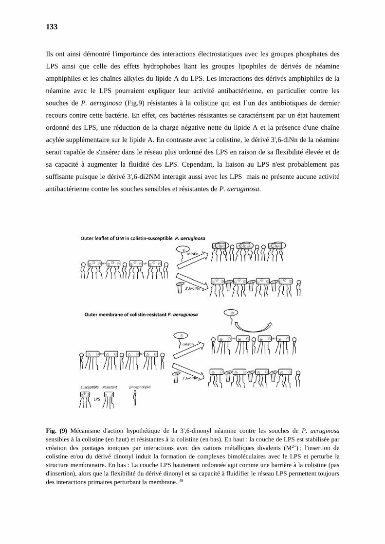
133
Ils ont ainsi démontré l'importance des interactions électrostatiques avec les groupes phosphates des
LPS ainsi que celle des effets hydrophobes liant les groupes lipophiles de dérivés de néamine
amphiphiles et les chaînes alkyles du lipide A du LPS. Les interactions des dérivés amphiphiles de la
néamine avec le LPS pourraient expliquer leur activité antibactérienne, en particulier contre les
souches de P. aeruginosa (Fig.9) résistantes à la colistine qui est l’un des antibiotiques de dernier
recours contre cette bactérie. En effet, ces bactéries résistantes se caractérisent par un état hautement
ordonné des LPS, une réduction de la charge négative nette du lipide A et la présence d'une chaîne
acylée supplémentaire sur le lipide A. En contraste avec la colistine, le dérivé 3',6-diNn de la néamine
serait capable de s'insérer dans le réseau plus ordonné des LPS en raison de sa flexibilité élevée et de
sa capacité à augmenter la fluidité des LPS. Cependant, la liaison au LPS n'est probablement pas
suffisante puisque le dérivé 3',6-di2NM interagit aussi avec les LPS mais ne présente aucune activité
antibactérienne contre les souches sensibles et résistantes de P. aeruginosa.
Fig. (9) Mécanisme d'action hypothétique de la 3',6-dinonyl néamine contre les souches de P. aeruginosa
sensibles à la colistine (en haut) et résistantes à la colistine (en bas). En haut : la couche de LPS est stabilisée par
création des pontages ioniques par interactions avec des cations métalliques divalents (M2+) ; l'insertion de
colistine et/ou du dérivé dinonyl induit la formation de complexes bimoléculaires avec le LPS et perturbe la
structure membranaire. En bas : La couche LPS hautement ordonnée agit comme une barrière à la colistine (pas
d'insertion), alors que la flexibilité du dérivé dinonyl et sa capacité à fluidifier le réseau LPS permettent toujours
des interactions primaires perturbant la membrane. 48
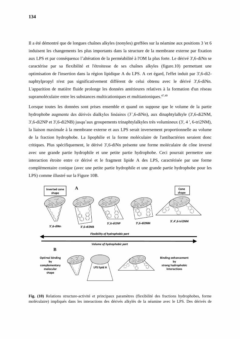
134
Il a été démontré que de longues chaînes alkyles (nonyles) greffées sur la néamine aux positions 3 'et 6
induisent les changements les plus importants dans la structure de la membrane externe par fixation
aux LPS et par conséquence l’altération de la perméabilité à l'OM la plus forte. Le dérivé 3',6-diNn se
caractérise par sa flexibilité et l'étroitesse de ses chaînes alkyles (figure.10) permettant une
optimisation de l'insertion dans la région lipidique A du LPS. A cet égard, l'effet induit par 3',6-di2-
naphtylpropyl n'est pas significativement différent de celui obtenu avec le dérivé 3',6-diNn.
L'apparition de matière fluide prolonge les données antérieures relatives à la formation d'un réseau
supramoléculaire entre les substances multicationiques et multianioniques.47,49
Lorsque toutes les données sont prises ensemble et quand on suppose que le volume de la partie
hydrophobe augmente des dérivés dialkyles linéaires (3’,6-diNn), aux dinaphtylalkyle (3',6-di2NM,
3',6-di2NP et 3',6-di2NB) jusqu’aux groupements trinaphtylalkyles très volumineux (3', 4 ', 6-tri2NM),
la liaison maximale à la membrane externe et aux LPS serait inversement proportionnelle au volume
de la fraction hydrophobe. La lipophilie et la forme moléculaire de l'antibactérien seraient donc
critiques. Plus spécifiquement, le dérivé 3',6-diNn présente une forme moléculaire de cône inversé
avec une grande partie hydrophile et une petite partie hydrophobe. Ceci pourrait permettre une
interaction étroite entre ce dérivé et le fragment lipide A des LPS, caractérisée par une forme
complémentaire conique (avec une petite partie hydrophile et une grande partie hydrophobe pour les
LPS) comme illustré sur la Figure 10B.
Fig. (10) Relations structure-activité et principaux paramètres (flexibilité des fractions hydrophobes, forme
moléculaire) impliqués dans les interactions des dérivés alkylés de la néamine avec le LPS. Des dérivés de
A
B

135
néamine amphiphiles et les LPS sont représentés par des fragments hydrophiles (gris) et hydrophobes (blancs).
La forme moléculaire complémentaire entre dérivés 3’,6-diNn et LPS est illustrée au bas du schéma. 48
Les excellents résultats obtenus par le groupe et les premières relations structure-activité établies
stimulent donc la synthèse de nouveaux aminoglycosides amphiphiles dans la recherche de nouveaux
agents antimicrobiens encore plus puissants, moins toxiques et moins sensibles au développement de
résistances bactériennes.

TRAVAUX PERSONNELS
PARTIE II
1.
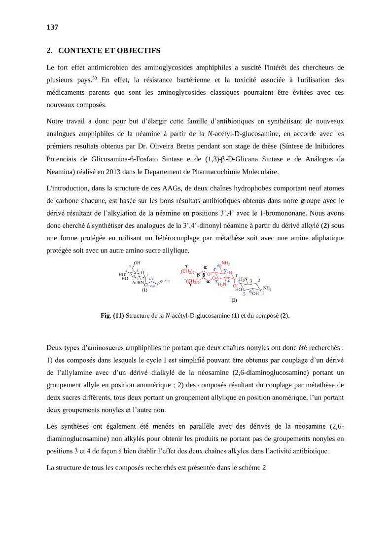
137
2. CONTEXTE ET OBJECTIFS
Le fort effet antimicrobien des aminoglycosides amphiphiles a suscité l'intérêt des chercheurs de
plusieurs pays.50 En effet, la résistance bactérienne et la toxicité associée à l'utilisation des
médicaments parents que sont les aminoglycosides classiques pourraient être évitées avec ces
nouveaux composés.
Notre travail a donc pour but d’élargir cette famille d’antibiotiques en synthétisant de nouveaux
analogues amphiphiles de la néamine à partir de la N-acétyl-D-glucosamine, en accorde avec les
prémiers resultats obtenus par Dr. Oliveira Bretas pendant son stage de thèse (Síntese de Inibidores
Potenciais de Glicosamina-6-Fosfato Sintase e de (1,3)--D-Glicana Sintase e de Análogos da
Neamina) réalisé en 2013 dans le Departement de Pharmacochimie Moleculaire.
L'introduction, dans la structure de ces AAGs, de deux chaînes hydrophobes comportant neuf atomes
de carbone chacune, est basée sur les bons résultats antibiotiques obtenus dans notre groupe avec le
dérivé résultant de l’alkylation de la néamine en positions 3’,4’ avec le 1-bromononane. Nous avons
donc cherché à synthétiser des analogues de la 3’,4’-dinonyl néamine à partir du dérivé alkylé (2) sous
une forme protégée en utilisant un hétérocouplage par métathèse soit avec une amine aliphatique
protégée soit avec un autre amino sucre allylique.
Fig. (11) Structure de la N-acétyl-D-glucosamine (1) et du composé (2).
Deux types d’aminosucres amphiphiles ne portant que deux chaînes nonyles ont donc été recherchés :
1) des composés dans lesquels le cycle I est simplifié pouvant être obtenus par couplage d’un dérivé
de l’allylamine avec d’un dérivé dialkylé de la néosamine (2,6-diaminoglucosamine) portant un
groupement allyle en position anomérique ; 2) des composés résultant du couplage par métathèse de
deux sucres différents, tous deux portant un groupement allylique en position anomérique, l’un portant
deux groupements nonyles et l’autre non.
Les synthèses ont également été menées en parallèle avec des dérivés de la néosamine (2,6-
diaminoglucosamine) non alkylés pour obtenir les produits ne portant pas de groupements nonyles en
positions 3 et 4 de façon à bien établir l’effet des deux chaînes alkyles dans l’activité antibiotique.
La structure de tous les composés recherchés est présentée dans le schème 2
O
OH
HOHO
AcHNC-a
C-b
C-c
(1)
3
4 5
6
1
2
O
5'
6'
1'2'
O
O
O
NH2
O
H2NHO
OH
NH2
H2N3'
4'
(2)
(CH2)6
(CH2)6
1
234
56
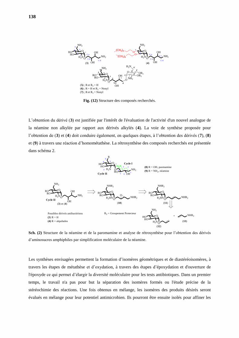
138
Fig. (12) Structure des composés recherchés.
L’obtention du dérivé (3) est justifiée par l'intérêt de l'évaluation de l'activité d'un nouvel analogue de
la néamine non alkylée par rapport aux dérivés alkylés (4). La voie de synthèse proposée pour
l’obtention de (3) et (4) doit conduire également, en quelques étapes, à l’obtention des dérivés (7), (8)
et (9) à travers une réaction d’homométathèse. La rétrosynthèse des composés recherchés est présentée
dans schéma 2.
Sch. (2) Structure de la néamine et de la paromamine et analyse de rétrosynthèse pour l’obtention des dérivés
d’aminosucres amphiphiles par simplification moléculaire de la néamine.
Les synthèses envisagées permettent la formation d’isomères géométriques et de diastéréoisomères, à
travers les étapes de métathèse et d’oxydation, à travers des étapes d’époxydation et d'ouverture de
l'époxyde ce qui permet d’élargir la diversité moléculaire pour les tests antibiotiques. Dans un premier
temps, le travail n'a pas pour but la séparation des isomères formés ou l'étude précise de la
stéréochimie des réactions. Une fois obtenus en mélange, les isomères des produits désirés seront
évalués en mélange pour leur potentiel antimicrobien. Ils pourront être ensuite isolés pour affiner les
O
NH2
OO
H2N
O
NH2
HOHO
H2N
C-d
(3) (4)
ONH2
OH
OH
ONH2
OH
OH(CH2)6
(CH2)6
C-a
C-b
C-c
C-d
3
4
a
b
c1
2
4 5
6
3O
NH2
RORO
H2NO
O
O
H2N
OR1OR1
NH2
d
1' 2'3'
4'
6'
5'
OH
OH
C-a
C-b
3
4 5
6
1
2
(5) ; R et R1 = H
(6) ; R = H et R1 = Nonyl
(7) ; R et R1 = Nonyl
O
R
HOHO
H2N OHO
H2N
NH2
OH
Possibles dérivés antibactériens
(3) R = H
(4) R = alquilados
3'
4'
6
Cycle I
Cycle II
O
NH2
RORO
H2N
O
NHR2
RORO
R2HN
O
NHR2
RORO
R2HN
O
NH2
HOHO
H2N
+ NHR2
Cycle II
(8) R = OH, paomamine
(9) R = NH2, néamine
R2 = Groupement Protecteur
5'
6'
1
23
5
4
1'
(3) et (4) (10) (11)
(12)
(13)
ONH2
OH
OH
ONHR2
O
ONHR2
O
résultats s’ils sont positifs. Toutes les substances synthétisées seront ainsi évaluées contre les bactéries
Gram (+) et Gram (-) sensibles et résistantes.
2.1. Objectif général
Developper la recherche de nouveaux agents actifs contre les bactéries résistantes aux antibiotiques, à
travers la synthèse de nouveaux analogues amphiphiles de la néamine potentiellement antibiotiques.
2.2. Objectifs spécifiques
Synthétiser des nouveaux 2,6-diaminoglycosides par réaction de métathèse, hydrolyse et
oxydation de double liaison, en accord avec les prémiers resultats obtenus par Dr. Oliveira
Bretas pendant son stage de thèse réalisé en 2013 dans le Departement de Pharmacochimie
Moleculaire ;
Elucider les structures chimiques des composés synthétisés par des méthodes spectroscopiques
telles que la résonance magnétique nucléaire du proton et du carbone 13 (RMN 1H- et 13C) et
la spectrométrie de masse ;
Evaluer l'activité biologique des dérivés contre les bactéries Gram (+) et Gram (-) résistant en
collaboration avec Marie-Paule Mingeot-Leclercq de l’Université catholoqie de Louvain à
Bruxelles.
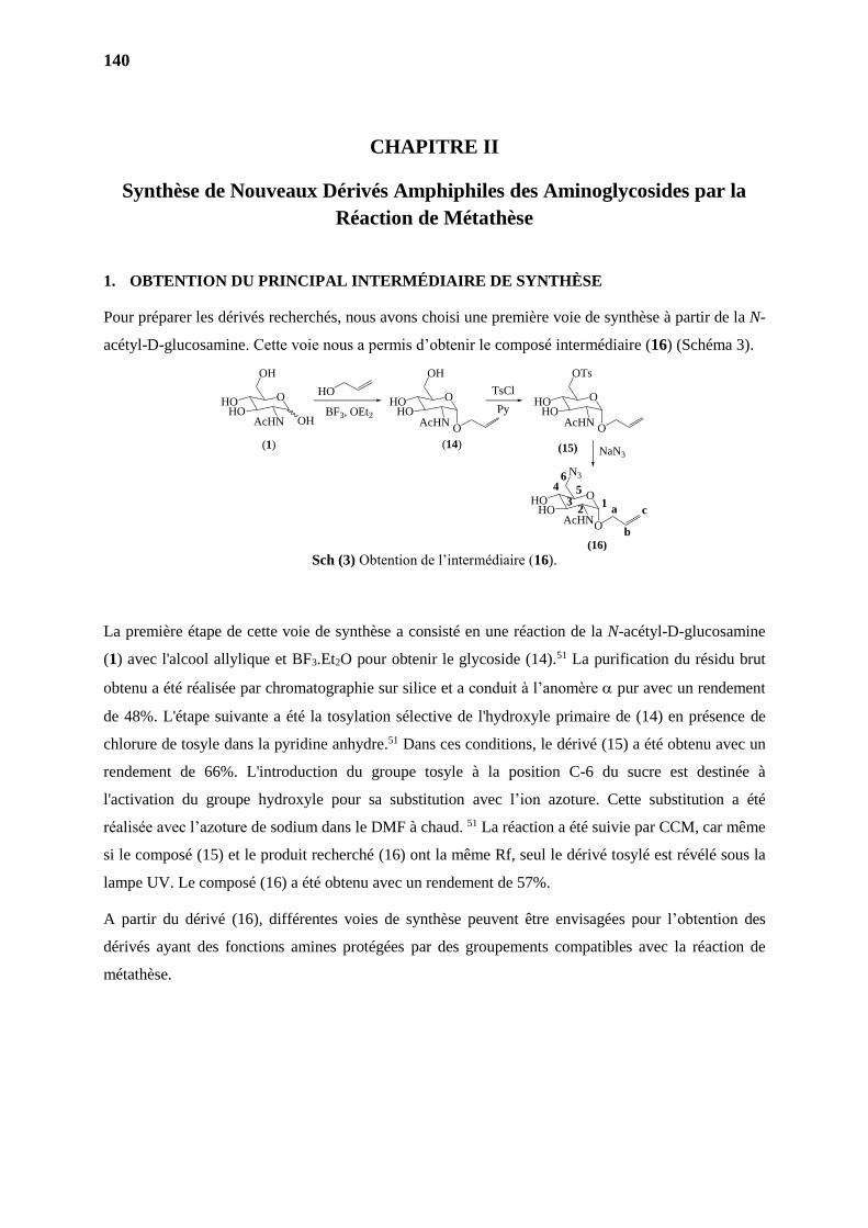
140
CHAPITRE II
Synthèse de Nouveaux Dérivés Amphiphiles des Aminoglycosides par la
Réaction de Métathèse
1. OBTENTION DU PRINCIPAL INTERMÉDIAIRE DE SYNTHÈSE
Pour préparer les dérivés recherchés, nous avons choisi une première voie de synthèse à partir de la N-
acétyl-D-glucosamine. Cette voie nous a permis d’obtenir le composé intermédiaire (16) (Schéma 3).
Sch (3) Obtention de l’intermédiaire (16).
La première étape de cette voie de synthèse a consisté en une réaction de la N-acétyl-D-glucosamine
(1) avec l'alcool allylique et BF3.Et2O pour obtenir le glycoside (14).51 La purification du résidu brut
obtenu a été réalisée par chromatographie sur silice et a conduit à l’anomère pur avec un rendement
de 48%. L'étape suivante a été la tosylation sélective de l'hydroxyle primaire de (14) en présence de
chlorure de tosyle dans la pyridine anhydre.51 Dans ces conditions, le dérivé (15) a été obtenu avec un
rendement de 66%. L'introduction du groupe tosyle à la position C-6 du sucre est destinée à
l'activation du groupe hydroxyle pour sa substitution avec l’ion azoture. Cette substitution a été
réalisée avec l’azoture de sodium dans le DMF à chaud. 51 La réaction a été suivie par CCM, car même
si le composé (15) et le produit recherché (16) ont la même Rf, seul le dérivé tosylé est révélé sous la
lampe UV. Le composé (16) a été obtenu avec un rendement de 57%.
A partir du dérivé (16), différentes voies de synthèse peuvent être envisagées pour l’obtention des
dérivés ayant des fonctions amines protégées par des groupements compatibles avec la réaction de
métathèse.
O
N3
HOHO
AcHN
(1) (15)
(16)
O
OH
HOHO
AcHN
O
OH
HOHO
AcHNO
HO
(14)
O
OTs
HOHO
AcHNO
O
TsCl
NaN3
a
b
c1
2
4 5
6
3
OHBF3, OEt2 Py
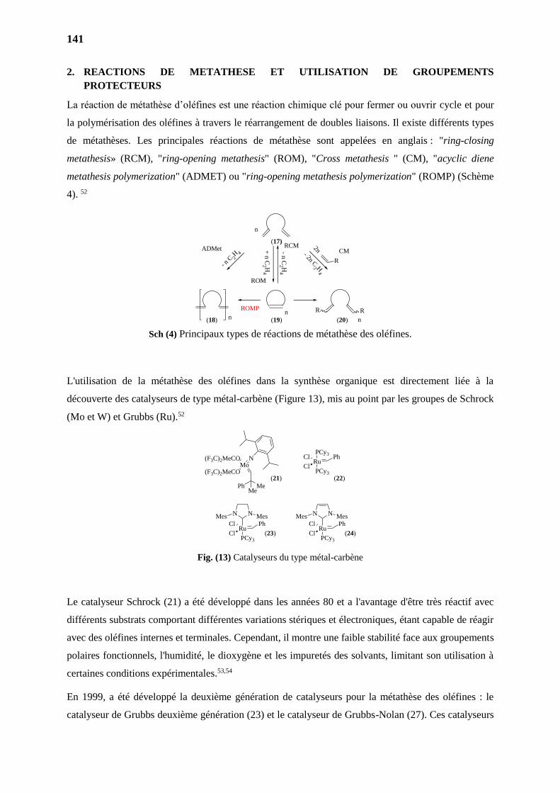
141
2. REACTIONS DE METATHESE ET UTILISATION DE GROUPEMENTS
PROTECTEURS
La réaction de métathèse d’oléfines est une réaction chimique clé pour fermer ou ouvrir cycle et pour
la polymérisation des oléfines à travers le réarrangement de doubles liaisons. Il existe différents types
de métathèses. Les principales réactions de métathèse sont appelées en anglais : "ring-closing
metathesis» (RCM), "ring-opening metathesis" (ROM), "Cross metathesis " (CM), "acyclic diene
metathesis polymerization" (ADMET) ou "ring-opening metathesis polymerization" (ROMP) (Schème
4). 52
Sch (4) Principaux types de réactions de métathèse des oléfines.
L'utilisation de la métathèse des oléfines dans la synthèse organique est directement liée à la
découverte des catalyseurs de type métal-carbène (Figure 13), mis au point par les groupes de Schrock
(Mo et W) et Grubbs (Ru).52
Fig. (13) Catalyseurs du type métal-carbène
Le catalyseur Schrock (21) a été développé dans les années 80 et a l'avantage d'être très réactif avec
différents substrats comportant différentes variations stériques et électroniques, étant capable de réagir
avec des oléfines internes et terminales. Cependant, il montre une faible stabilité face aux groupements
polaires fonctionnels, l'humidité, le dioxygène et les impuretés des solvants, limitant son utilisation à
certaines conditions expérimentales.53,54
En 1999, a été développé la deuxième génération de catalyseurs pour la métathèse des oléfines : le
catalyseur de Grubbs deuxième génération (23) et le catalyseur de Grubbs-Nolan (27). Ces catalyseurs
n
ROMPn RR
n
n
R
CM2n
- 2n C2 H
4
- n C
2 H4
+ n
C2 H
4
RCM
ROM
- n C 2
H 4ADMet
(17)
(18) (19) (20)
N N
Ru
MesMesPh
PCy3
Cl
Cl
N N
Ru
MesMesPh
PCy3
Cl
Cl
PCy3
RuPh
PCy3
Cl
Cl
MeMePh
Mo(F3C)2MeCO N
(F3C)2MeCO(21) (22)
(23) (24)
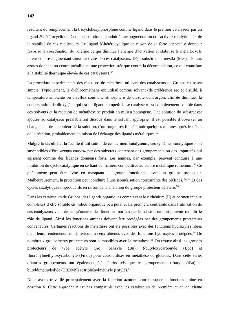
142
résultent du remplacement la tricyclohexylphosphine comme ligand dans le premier catalyseur par un
ligand N-hétérocyclique. Cette substitution a conduit à une augmentation de l'activité catalytique et de
la stabilité de ces catalyseurs. Le ligand N-hétérocyclique en raison de sa forte capacité σ donneur
favorise la coordination de l'oléfine ce qui diminue l’énergie d'activation et stabilise le métallocycle
intermédiaire augmentant ainsi l'activité de ces catalyseurs. Déjà substituants mesila (Mes) liés aux
azotes donnent au centre métallique, une protection stérique contre la décomposition, ce qui contribue
à la stabilité thermique élevée de ces catalyseurs.55
La procédure expérimentale des réactions de métathèse utilisant des catalyseurs de Grubbs est assez
simple. Typiquement, le dichlorométhane est utilisé comme solvant (de préférence sec et distillé) à
température ambiante ou à reflux sous une atmosphère de diazote ou d'argon, afin de diminuer la
concentration de dioxygène qui est un ligand compétitif. Le catalyseur est complètement soluble dans
ces solvants et la réaction de métathèse se produit en milieu homogène. Une solution du substrat est
ajoutée au catalyseur préalablemnt dissous dans le solvant approprié. Il est possible d’observer un
changement de la couleur de la solution, d'un rouge très foncé à noir quelques minutes après le début
de la réaction, probablement en raison de l'échange des ligands métalliques.52
Malgré la stabilité et la facilité d’utilisation de ces derniers catalyseurs, ces systèmes catalytiques sont
susceptibles d'être «empoisonnés» par des substrats contenant des groupements ou des impuretés qui
agissent comme des ligands donneurs forts. Les amines, par exemple, peuvent conduire à une
inhibition du cycle catalytique en se liant de manière compétitive au centre métallique ruthénium.55 Ce
phénomène peut être évité en masquant le groupe fonctionnel avec un groupe protecteur.
Malheureusement, la protection peut conduire à une isomérisation concurrente des oléfines. 56,57 Et des
cycles catalytiques improductifs en raison de la chélation du groupe protecteur délétère.58
Dans les catalyseurs de Grubbs, des ligands organiques complexent le ruthénium (II) et permettent aux
complexes d’être soluble en milieu organique peu polaire. La première contrainte dans l’utilisation de
ces catalyseurs vient de ce qu’aucune des fonctions portées par le substrat ne doit pouvoir remplir le
rôle de ligand. Ainsi les fonctions amines doivent être protégées par des groupements protecteurs
convenables. Certaines réactions de métathèse ont été possibles avec des fonctions hydroxyles libres
mais leurs rendements sont inférieurs à ceux obtenus avec des fonctions hydroxyles protégées.59 De
nombreux groupements protecteurs sont compatibles avec la métathèse.60 On trouve ainsi les groupes
protecteurs de type acétyle (Ac), benzyle (Bn), t-butyloxycarbonyle (Boc) et
fluorénylméthyloxycarbonyle (Fmoc) pour ceux utilisés en métathèse de glucides. Dans cette série,
d’autres groupements ont également été décrits tels que les groupements t-butyle (tBu), t-
butyldiméthylsilyle (TBDMS) et triphénylméthyle (trityle).61
Nous avons travaillé principalement avec la fonction azoture pour masquer la fonction amine en
position 6. Cette approche n’est pas compatible avec les catalyseurs de première et de deuxième
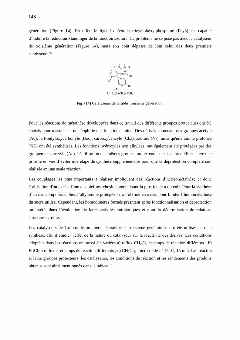
143
génération (Figure 14). En effet, le ligand qu’est la tricyclohexylphosphine (Pcy3) est capable
d’induire la réduction Staudinger de la fonction azoture. Ce problème ne se pose pas avec le catalyseur
de troisième génération (Figure 14), mais son coût dépasse de loin celui des deux premiers
catalyseurs.62
Fig. (14) Catalyseurs de Grubbs troisième génération.
Pour les réactions de métathèse développées dans ce travail des différents groupes protecteurs ont été
choisis pour masquer la nucléophilie des fonctions amine. Des dérivés contenant des groupes acétyle
(Ac), le t-butyloxycarbonyle (Boc), carboxybenzyle (Cbz), azoture (N3), ainsi qu'une amine protonée
+NH3 ont été synthétisés. Les fonctions hydroxyles non alkylées, ont également été protégées par des
groupements acétyle (Ac). L’utilisation des mêmes groupes protecteurs sur les deux oléfines a été une
priorité en vue d’éviter une étape de synthèse supplémentaire pour que la déprotection complète soit
réalisée en une seule réaction.
Les couplages les plus importants à réaliser impliquent des réactions d’hétérométathèse et donc
l'utilisation d'un excès d'une des oléfines chosie comme étant la plus facile à obtenir. Pour la synthèse
d’un des composés cibles, l’allylamine protégée sera l’oléfine en excès pour limiter l’homométathèse
du sucre utilisé. Cependant, les homodimères formés présntent après fonctionnalisation et déprotection
un intérêt dans l’évaluation de leurs activités antibiotiques et pour la détermination de relations
structure-activité.
Les catalyseurs de Grubbs de première, deuxième et troisième générations ont été utilisés dans la
synthèse, afin d’étudier l'effet de la nature du catalyseur sur la réactivité des dérivés. Les conditions
adoptées dans les réactions ont aussi été variées a) reflux CH2Cl2 et temps de réaction différents ; b)
Et2Cl2 à reflux et et temps de réaction différents ; c) CH2Cl2, micro-ondes, 115 oC, 15 min. Les réactifs
et leurs groupes protecteurs, les catalyseurs, les conditions de réaction et les rendements des produits
obtenus sont ainsi mentionnés dans le tableau 1.
Br
RuPhCl
Cl
NN RR
Br
R = 2,4,6-(CH3)3-C6H2
(25)

144
Tab (1) Conditions: 1) CH2Cl2, reflux; 24 h ; 2) ClCH2CH2Cl, reflux, 24 h ; 3) CH2Cl2, OC, 15 min ; 4) HC3CH2Cl2, OC, 15 min.
Réaction R R1 R2 R3 Catalyseur Conditions
Réactionnels
Rendement du
Produit Obtenu
1 NHAc
NHAc NHAc Ac Grubbs I
1, 2, 3, 4
30% (melange
isomère)
2 NHAc NHAc Nonyl Grubbs I X
3
NHBoc
NHAc NHAc Ac Grubbs I X
4 NHAc NHAc Nonyl Grubbs I X
5 NHBoc NHBoc Nonyl Grubbs I X
6 NHBoc NHBoc Nonyl Grubbs II X
7 NHBoc NHBoc Nonyl Grubbs III X
8 NHBoc
N3
Nonyl Grubbs III
9
N3
Ac Grubbs I ;
Grubbs II X
12 Ac Grubbs III 2 traits
13 Ac Grubbs III 3 X
14 Nonyl
Grubbs I ;
Grubbs III 1, 2, 3, 4 X
15 Nonyl Grubbs II 1 traits
16
NH3+Cl- NH3
+Cl- Nonyl
Grubbs I ;
Grubbs II ;
Grubbs III
1, 2, 3 X
17 NHAc OAc Ac Grubbs I 1 X
18 NHCBz N3 N3
Ac; Nonyl Grubbs III 1, 2, 3, 4 X
O
R2
R3OR3O
R1
R
Grubbs Catalyst
O
O
R2
R3OR3O
R1OR
a
b
c1
2
4 5
6
3
d
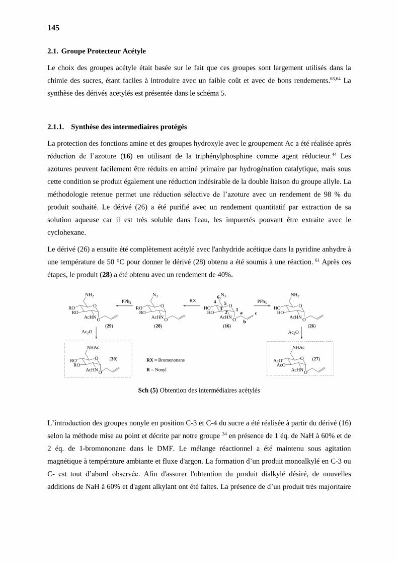
145
2.1. Groupe Protecteur Acétyle
Le choix des groupes acétyle était basée sur le fait que ces groupes sont largement utilisés dans la
chimie des sucres, étant faciles à introduire avec un faible coût et avec de bons rendements.63,64 La
synthèse des dérivés acetylés est présentée dans le schéma 5.
2.1.1. Synthèse des intermediaires protégés
La protection des fonctions amine et des groupes hydroxyle avec le groupement Ac a été réalisée après
réduction de l’azoture (16) en utilisant de la triphénylphosphine comme agent réducteur.44 Les
azotures peuvent facilement être réduits en aminé primaire par hydrogénation catalytique, mais sous
cette condition se produit également une réduction indésirable de la double liaison du groupe allyle. La
méthodologie retenue permet une réduction sélective de l’azoture avec un rendement de 98 % du
produit souhaité. Le dérivé (26) a été purifié avec un rendement quantitatif par extraction de sa
solution aqueuse car il est très soluble dans l'eau, les impuretés pouvant être extraite avec le
cyclohexane.
Le dérivé (26) a ensuite été complètement acétylé avec l'anhydride acétique dans la pyridine anhydre à
une température de 50 °C pour donner le dérivé (28) obtenu a été soumis à une réaction. 61 Après ces
étapes, le produit (28) a été obtenu avec un rendement de 40%.
Sch (5) Obtention des intermédiaires acétylés
L’introduction des groupes nonyle en position C-3 et C-4 du sucre a été réalisée à partir du dérivé (16)
selon la méthode mise au point et décrite par notre groupe 34 en présence de 1 éq. de NaH à 60% et de
2 éq. de 1-bromononane dans le DMF. Le mélange réactionnel a été maintenu sous agitation
magnétique à température ambiante et fluxe d'argon. La formation d’un produit monoalkylé en C-3 ou
C- est tout d’abord observée. Afin d'assurer l'obtention du produit dialkylé désiré, de nouvelles
additions de NaH à 60% et d'agent alkylant ont été faites. La présence de d’un produit très majoritaire
O
NH2
HOHO
AcHN
O
N3
HOHO
AcHN
O
N3
RORO
AcHN
O
NHAc
AcOAcO
AcHN
O
NH2
RORO
AcHN
O
NHAc
RORO
AcHN
(16) (26)
(27)
(28)(29)
(30)
O
O
O O O
O
a
b
c1
2
4 5
6
3
RXPPh3 PPh3
Ac2O Ac2O
RX = Bromononane
R = Nonyl
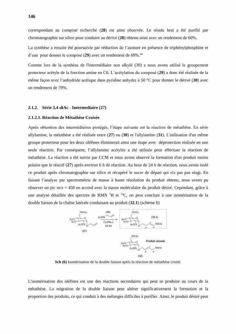
146
correspondant au composé recherché (28) est ainsi observée. Le résidu brut a été purifié par
chromatographie sur silice pour conduire au dérivé (28) obtenu ainsi avec un rendement de 60%.
La synthèse a ensuite été poursuivie par réduction de l’azoture en présence de triphénylphosphine et
d’eau pour donner le composé (29) avec un rendement de 69%.44
Comme lors de la synthèse de l'intermédiaire non alkylé (30) a nous avons utilisé le groupement
protecteur acétyle de la fonction amine en C6. L’acétylation du composé (29) a donc été réalisée de la
même façon avec l’anhydride acétique dans pyridine anhydre à 50 °C pour donner le dérivé (30) avec
un rendement de 70%.
2.1.2. Série 3,4-diAc - Intermediaire (27)
2.1.2.1. Réaction de Métathèse Croisée
Après obtention des intermédiaires protégés, l’étape suivante est la réaction de métathèse. En série
allylamine, la métathèse a été réalisée entre (27) ou (30) et l'allylamine (31). L'utilisation d'un même
groupe protecteur pour les deux oléfines éliminerait ainsi une étape avec déprotection réalisée en une
seule réaction. Par conséquent, l’allylamine acétylée a été utilisée pour effectuer la réaction de
métathèse. La réaction a été suivie par CCM et nous avons observé la formation d'un produit moins
polaire que le réactif (27) après environ 6 h de réaction. Au bout de 24 h de réaction, nous avons isolé
ce produit après chromatographie sur silice et récupéré le sucre de départ qui n'a pas pas réagi. En
faisant l’analyse par spectrométrie de masse à haute résolution du produit obtenu, nous avons pu
observer un pic m/z = 458 en accord avec la masse moléculaire du produit désiré. Cependant, grâce à
une analyse détaillée des spectres de RMN 1H et 13C, on peut conclure à une isomérisation de la
double liaison de la chaîne latérale conduisant au produit (32.1) (schème 6)
Sch (6) Isomérisation de la double liaison après la réaction de métathèse croisé.
L'isomérisation des oléfines est une des réactions secondaires qui peut se produire au cours de la
métathèse. La migration de la double liaison peut altérer significativement la formation et la
proportion des produits, ce qui conduit à des mélanges difficiles à purifier. Ainsi, le produit désiré peut
AcHN
Grubbs I
DCM
(27)
(31)
O
NHAc
AcOAcO
AcHNO
b
cNHAc
d
(32.1)
O
NHAc
AcOAcO
AcHNO
b
cNHAc
(32)
Produit attendu
d
O
NHAc
AcOAcO
AcHNO
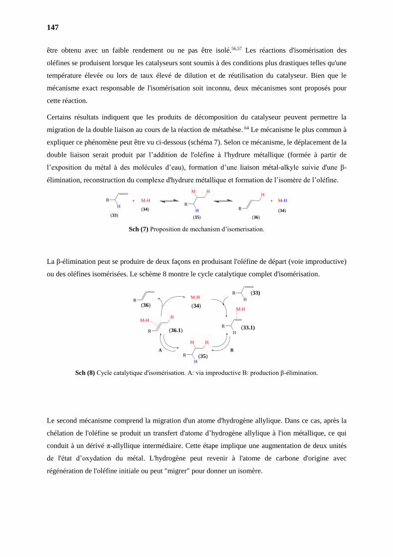
147
être obtenu avec un faible rendement ou ne pas être isolé.56,57 Les réactions d'isomérisation des
oléfines se produisent lorsque les catalyseurs sont soumis à des conditions plus drastiques telles qu'une
température élevée ou lors de taux élevé de dilution et de réutilisation du catalyseur. Bien que le
mécanisme exact responsable de l'isomérisation soit inconnu, deux mécanismes sont proposés pour
cette réaction.
Certains résultats indiquent que les produits de décomposition du catalyseur peuvent permettre la
migration de la double liaison au cours de la réaction de métathèse. 64 Le mécanisme le plus commun à
expliquer ce phénomène peut être vu ci-dessous (schéma 7). Selon ce mécanisme, le déplacement de la
double liaison serait produit par l’addition de l'oléfine à l'hydrure métallique (formée à partir de
l’exposition du métal à des molécules d’eau), formation d’une liaison métal-alkyle suivie d'une β-
élimination, reconstruction du complexe d'hydrure métallique et formation de l’isomère de l’oléfine.
Sch (7) Proposition de mechanism d’isomerisation.
La β-élimination peut se produire de deux façons en produisant l'oléfine de départ (voie improductive)
ou des oléfines isomérisées. Le schème 8 montre le cycle catalytique complet d'isomérisation.
Sch (8) Cycle catalytique d'isomérisation. A: via improductive B: production β-élimination.
Le second mécanisme comprend la migration d'un atome d'hydrogène allylique. Dans ce cas, après la
chélation de l'oléfine se produit un transfert d'atome d’hydrogène allylique à l'ion métallique, ce qui
conduit à un dérivé π-allyllique intermédiaire. Cette étape implique une augmentation de deux unités
de l'état d’oxydation du métal. L'hydrogène peut revenir à l'atome de carbone d'origine avec
régénération de l'oléfine initiale ou peut "migrer" pour donner un isomère.
M-HRR
M H
R
H
HH
M-H+ +
(33)
(34)
(35) (36)
(34)
R
M H
H
M-H
R
H
R
H
M-H
R
HM-H
R
A B
(34)
(35)
(36)
(33)
(33.1)(36.1)
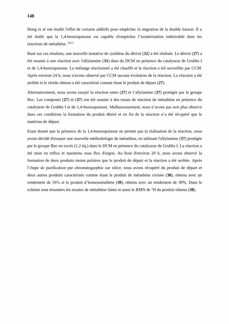
148
Hong et al ont étudié l'effet de certains additifs pour empêcher la migration de la double liaison. Il a
été établi que la 1,4-benzoquinone est capable d'empêcher l’isomérisation indésirable dans les
réactions de métathèse. 56,57
Basé sur ces résultats, une nouvelle tentative de synthèse du dérivé (32) a été réalisée. Le dérivé (27) a
été soumis à une réaction avec l'allylamine (31) dans du DCM en présence du catalyseur de Grubbs I
et de 1,4-benzoquinone. Le mélange réactionnel a été chauffé et la réaction a été surveillée par CCM.
Après environ 24 h, nous n'avons observé par CCM aucune évolution de la réaction. La réaction a été
arrêtée et le résidu obtenu a été caractérisé comme étant le produit de départ (27).
Alternativement, nous avons essayé la réaction entre (27) et l’allylamine (37) protégée par le groupe
Boc. Les composés (27) et (37) ont été soumis à des essais de réaction de métathèse en présence du
catalyseur de Grubbs I et de 1,4-benzoquinone. Malheureusement, nous n’avons pas non plus observé
dans ces conditions la formation du produit désiré et en fin de la réaction n’a été récupéré que le
matériau de départ.
Etant donné que la présence de la 1,4-benzoquinone ne permet pas la réalisation de la réaction, nous
avons décidé d'essayer une nouvelle méthodologie de métathèse, en utilisant l'allylamine (37) protégée
par le groupe Boc en excès (1.2 éq.) dans le DCM en présence du catalyseur de Grubbs I. La réaction a
été mise en reflux et maintenu sous flux d'argon. Au bout d'environ 20 h, nous avons observé la
formation de deux produits moins polaires que le produit de départ et la réaction a été arrêtée. Après
l’étape de purification par chromatogarphie sur silice, nous avons récupéré du produit de départ et
deux autres produits caractérisés comme étant le produit de métathèse croisée (38), obtenu avec un
rendement de 36% et le produit d’homométathèse (39), obtenu avec un rendement de 30%. Dans le
schème sont résumées les essaies de métathèse faites et aussi le RMN de 1H du produit obtenu (38).
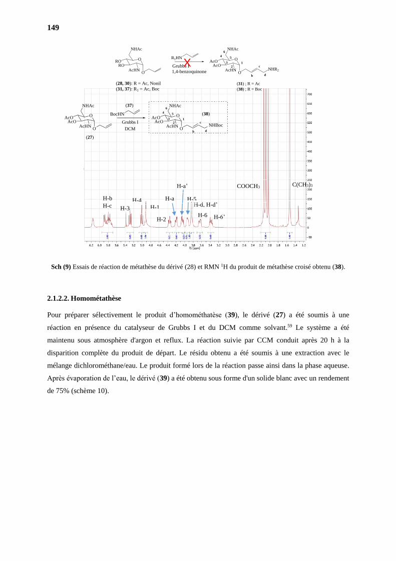
149
Sch (9) Essais de réaction de métathèse du dérivé (28) et RMN 1H du produit de métathèse croisé obtenu (38).
2.1.2.2. Homométathèse
Pour préparer sélectivement le produit d’homométhatèse (39), le dérivé (27) a été soumis à une
réaction en présence du catalyseur de Grubbs I et du DCM comme solvant.59 Le système a été
maintenu sous atmosphère d'argon et reflux. La réaction suivie par CCM conduit après 20 h à la
disparition complète du produit de départ. Le résidu obtenu a été soumis à une extraction avec le
mélange dichlorométhane/eau. Le produit formé lors de la réaction passe ainsi dans la phase aqueuse.
Après évaporation de l’eau, le dérivé (39) a été obtenu sous forme d'un solide blanc avec un rendement
de 75% (schème 10).
H-b
H-c
C(CH3)3 COOCH3
H-6’ H-6
H-1
H-4
H-3
H-2
H-d, H-d’ H-a
H-a’
H-5
R1HN
Grubbs I
O
NHAc
AcOAcO
AcHNO
b
cNHR2
d
O
NHAc
RORO
AcHNO
4 5
6
3 12
(28, 30): R = Ac, Nonil
(31, 37): R1 = Ac, Boc
1,4-benzoquinone
X
(31) ; R = Ac
(38) ; R = Boc
BocHN
Grubbs I
DCM
(27)
(37)
O
NHAc
AcOAcO
AcHNO
b
cNHBoc
d
(38)O
NHAc
AcOAcO
AcHNO
4 5
6
3 12
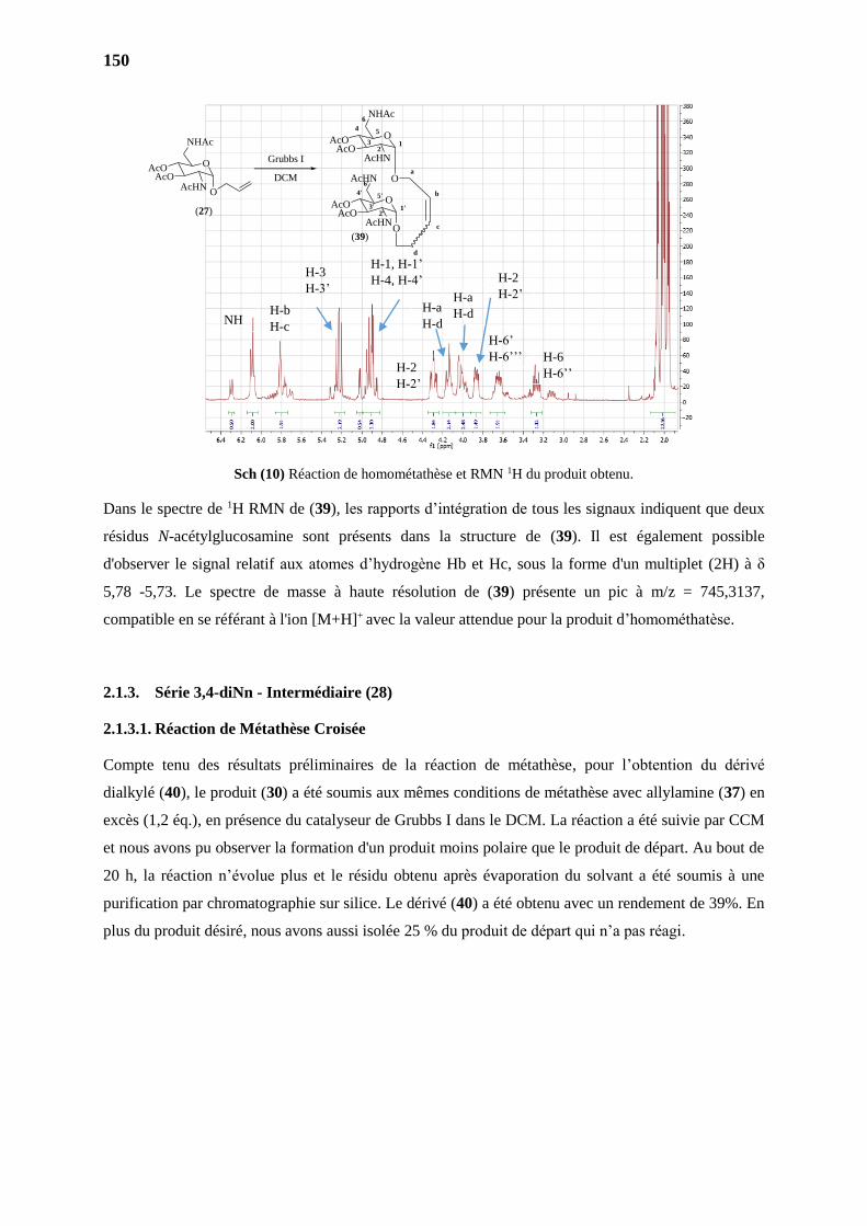
150
Sch (10) Réaction de homométathèse et RMN 1H du produit obtenu.
Dans le spectre de 1H RMN de (39), les rapports d’intégration de tous les signaux indiquent que deux
résidus N-acétylglucosamine sont présents dans la structure de (39). Il est également possible
d'observer le signal relatif aux atomes d’hydrogène Hb et Hc, sous la forme d'un multiplet (2H) à δ
5,78 -5,73. Le spectre de masse à haute résolution de (39) présente un pic à m/z = 745,3137,
compatible en se référant à l'ion [M+H]+ avec la valeur attendue pour la produit d’homométhatèse.
2.1.3. Série 3,4-diNn - Intermédiaire (28)
2.1.3.1. Réaction de Métathèse Croisée
Compte tenu des résultats préliminaires de la réaction de métathèse, pour l’obtention du dérivé
dialkylé (40), le produit (30) a été soumis aux mêmes conditions de métathèse avec allylamine (37) en
excès (1,2 éq.), en présence du catalyseur de Grubbs I dans le DCM. La réaction a été suivie par CCM
et nous avons pu observer la formation d'un produit moins polaire que le produit de départ. Au bout de
20 h, la réaction n’évolue plus et le résidu obtenu après évaporation du solvant a été soumis à une
purification par chromatographie sur silice. Le dérivé (40) a été obtenu avec un rendement de 39%. En
plus du produit désiré, nous avons aussi isolée 25 % du produit de départ qui n’a pas réagi.
NH H-b
H-c
H-3
H-3’
H-1, H-1’
H-4, H-4’
H-6
H-6’’
H-6’
H-6’’’ H-2
H-2’
H-2
H-2’ H-a
H-d
H-a
H-d
Grubbs I
DCM
(27)
O
NHAc
AcOAcO
AcHNO
(39)
O
NHAc
AcOAcO
AcHN
O
12
4 5
6
3
O
AcHN
AcOAcO
AcHNO
1'2'
4' 5'
6'
3'
a
b
c
d
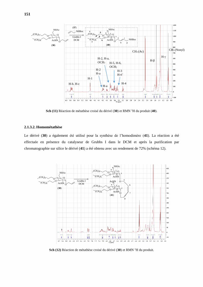
151
Sch (11) Réaction de métathèse croisé du dérivé (30) et RMN 1H du produit (40).
2.1.3.2. Homométathèse
Le dérivé (30) a également été utilisé pour la synthèse de l’homodimère (41). La réaction a été
effectuée en présence du catalyseur de Grubbs I dans le DCM et après la purification par
chromatographie sur silice le dérivé (41) a été obtenu avec un rendement de 72% (schéma 12).
Sch (15) Réaction de homométathèse du dérivé (28)
Sch (12) Réaction de métathèse croisé du dérivé (30) et RMN 1H du produit.
H-1
H-4 H-b, H-c
H-3
H-6’
H-2
H-a
H-a
H-2, H-a,
OCH2
H-
H-
CH3 (Ac) CH3 (Nonyl)
H-5, H-6,
OCH2
(30)
O
NHAc
OO
AcHNO
(CH2)6
(CH2)6
O
NHAc
OO
AcHNO
a
b
c1
2
4 5
6
3
(CH2)6
(CH2)6
(40)
NHBoc
d
Grubbs I
DCM
NHBoc
(37)
Grubbs I
DCM
(41)
O
NHAc
OO
AcHN
O
12
4 5
6
3
O
AcHN
OO
AcHNO
1'2'
4' 5'
6'
3'
a
b
c
d
(CH2)6
(CH2)6
(CH2)6
(CH2)6
(30)
O
NHAc
OO
AcHNO
(CH2)6
(CH2)6
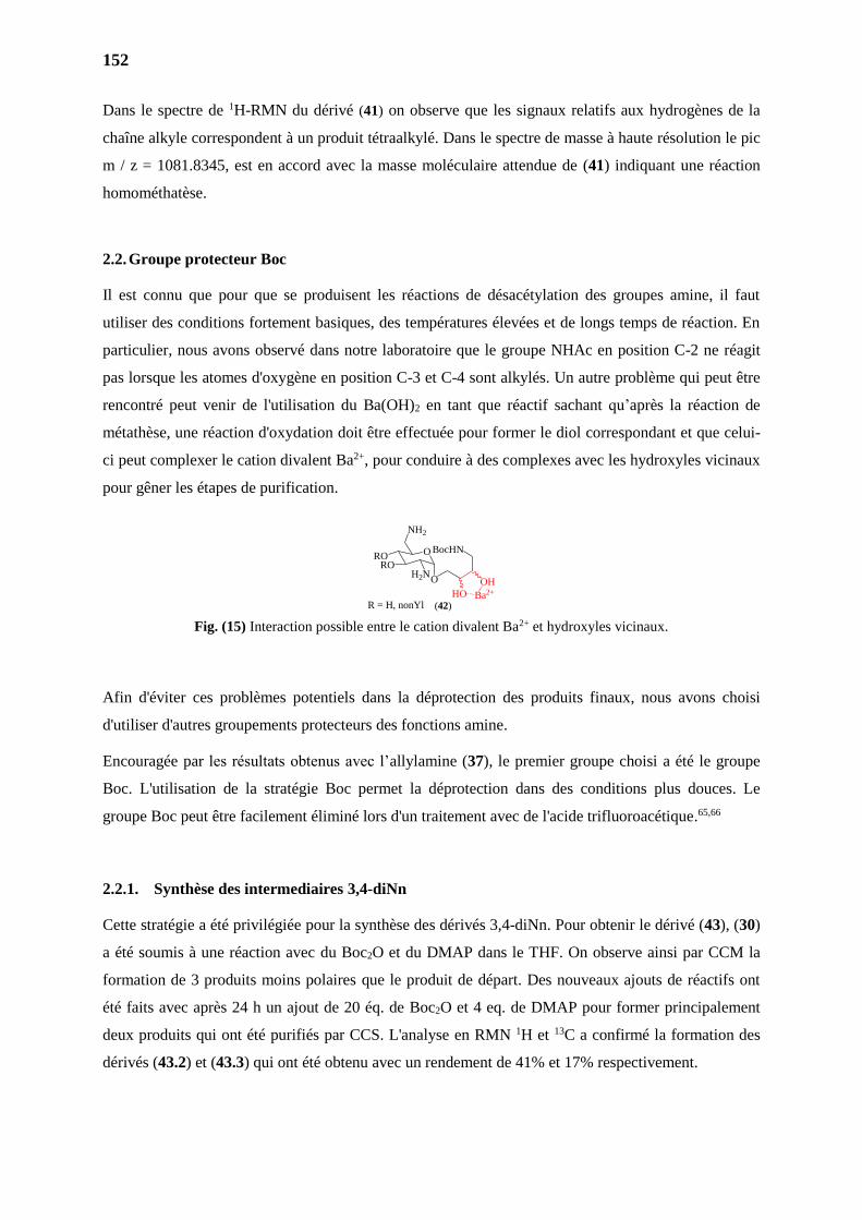
152
Dans le spectre de 1H-RMN du dérivé (41) on observe que les signaux relatifs aux hydrogènes de la
chaîne alkyle correspondent à un produit tétraalkylé. Dans le spectre de masse à haute résolution le pic
m / z = 1081.8345, est en accord avec la masse moléculaire attendue de (41) indiquant une réaction
homométhatèse.
2.2. Groupe protecteur Boc
Il est connu que pour que se produisent les réactions de désacétylation des groupes amine, il faut
utiliser des conditions fortement basiques, des températures élevées et de longs temps de réaction. En
particulier, nous avons observé dans notre laboratoire que le groupe NHAc en position C-2 ne réagit
pas lorsque les atomes d'oxygène en position C-3 et C-4 sont alkylés. Un autre problème qui peut être
rencontré peut venir de l'utilisation du Ba(OH)2 en tant que réactif sachant qu’après la réaction de
métathèse, une réaction d'oxydation doit être effectuée pour former le diol correspondant et que celui-
ci peut complexer le cation divalent Ba2+, pour conduire à des complexes avec les hydroxyles vicinaux
pour gêner les étapes de purification.
Fig. (15) Interaction possible entre le cation divalent Ba2+ et hydroxyles vicinaux.
Afin d'éviter ces problèmes potentiels dans la déprotection des produits finaux, nous avons choisi
d'utiliser d'autres groupements protecteurs des fonctions amine.
Encouragée par les résultats obtenus avec l’allylamine (37), le premier groupe choisi a été le groupe
Boc. L'utilisation de la stratégie Boc permet la déprotection dans des conditions plus douces. Le
groupe Boc peut être facilement éliminé lors d'un traitement avec de l'acide trifluoroacétique.65,66
2.2.1. Synthèse des intermediaires 3,4-diNn
Cette stratégie a été privilégiée pour la synthèse des dérivés 3,4-diNn. Pour obtenir le dérivé (43), (30)
a été soumis à une réaction avec du Boc2O et du DMAP dans le THF. On observe ainsi par CCM la
formation de 3 produits moins polaires que le produit de départ. Des nouveaux ajouts de réactifs ont
été faits avec après 24 h un ajout de 20 éq. de Boc2O et 4 eq. de DMAP pour former principalement
deux produits qui ont été purifiés par CCS. L'analyse en RMN 1H et 13C a confirmé la formation des
dérivés (43.2) et (43.3) qui ont été obtenu avec un rendement de 41% et 17% respectivement.
O
NH2
RORO
H2N
R = H, nonYl (42)
O
BocHN
HO Ba2+
OH
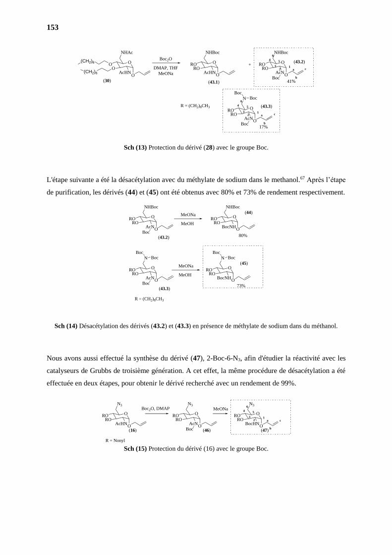
153
Sch (13) Protection du dérivé (28) avec le groupe Boc.
L'étape suivante a été la désacétylation avec du méthylate de sodium dans le methanol.67 Après l’étape
de purification, les dérivés (44) et (45) ont été obtenus avec 80% et 73% de rendement respectivement.
Sch (14) Désacétylation des dérivés (43.2) et (43.3) en présence de méthylate de sodium dans du méthanol.
Nous avons aussi effectué la synthèse du dérivé (47), 2-Boc-6-N3, afin d'étudier la réactivité avec les
catalyseurs de Grubbs de troisième génération. A cet effet, la même procédure de désacétylation a été
effectuée en deux étapes, pour obtenir le dérivé recherché avec un rendement de 99%.
Sch (15) Protection du dérivé (16) avec le groupe Boc.
O
NHBoc
RORO
AcN
O
N
RORO
AcN
Boc
BocBoc
Boc
MeONa
R = (CH2)8CH3
O
NHBoc
RORO
BocNH
80%
MeOH
MeONa
MeOH
O
N
RORO
BocNH
BocBoc
73%
(43.2)
(43.3)
(45)
(44)
O O
O O
O
NHAc
OO
AcHNO
(CH2)6
(CH2)6
DMAP, THF
O
NHBoc
RORO
AcN
O
NHBoc
RORO
AcHN
O
N
RORO
AcN
Boc
BocBoc
Boc
Boc2O
+
41%
17%
R = (CH2)8CH3
(30) (43.1)
(43.2)
(43.3)
O O
O
a
b
c1
2
4 5
6
3
a
b
c1
2
4 5
6
3
MeONa
O
N3
RORO
BocHN
O
N3
RORO
AcN
O
N3
RORO
AcHN
Boc(16) (46) (47)O O O
a
b
c1
2
4 5
6
3
R = Nonyl
Boc2O, DMAP MeONa
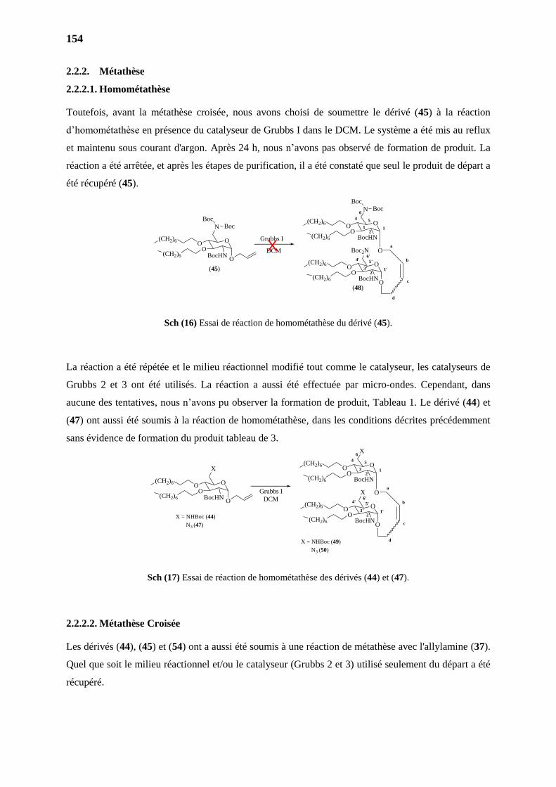
154
2.2.2. Métathèse
2.2.2.1. Homométathèse
Toutefois, avant la métathèse croisée, nous avons choisi de soumettre le dérivé (45) à la réaction
d’homométathèse en présence du catalyseur de Grubbs I dans le DCM. Le système a été mis au reflux
et maintenu sous courant d'argon. Après 24 h, nous n’avons pas observé de formation de produit. La
réaction a été arrêtée, et après les étapes de purification, il a été constaté que seul le produit de départ a
été récupéré (45).
Sch (16) Essai de réaction de homométathèse du dérivé (45).
La réaction a été répétée et le milieu réactionnel modifié tout comme le catalyseur, les catalyseurs de
Grubbs 2 et 3 ont été utilisés. La réaction a aussi été effectuée par micro-ondes. Cependant, dans
aucune des tentatives, nous n’avons pu observer la formation de produit, Tableau 1. Le dérivé (44) et
(47) ont aussi été soumis à la réaction de homométathèse, dans les conditions décrites précédemment
sans évidence de formation du produit tableau de 3.
Sch (17) Essai de réaction de homométathèse des dérivés (44) et (47).
2.2.2.2. Métathèse Croisée
Les dérivés (44), (45) et (54) ont a aussi été soumis à une réaction de métathèse avec l'allylamine (37).
Quel que soit le milieu réactionnel et/ou le catalyseur (Grubbs 2 et 3) utilisé seulement du départ a été
récupéré.
Grubbs I
DCM
(48)
O
N
OO
BocHN
O
12
4 5
6
3
O
Boc2N
OO
BocHNO
1'2'
4' 5'
6'
3'
a
b
c
d
(CH2)6
(CH2)6
(CH2)6
(CH2)6
(45)
O
N
OO
BocHNO
(CH2)6
(CH2)6
BocBoc
BocBoc
X
Grubbs I
DCM
O
X
OO
BocHN
O
12
4 5
6
3
O
X
OO
BocHNO
1'2'
4' 5'
6'
3'
a
b
c
d
(CH2)6
(CH2)6
(CH2)6
(CH2)6
O
X
OO
BocHNO
(CH2)6
(CH2)6
X = NHBoc (44)
N3 (47)
X = NHBoc (49)
N3 (50)
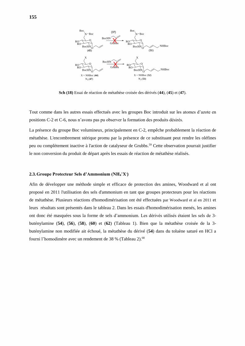
155
Sch (18) Essai de réaction de métathèse croisée des dérivés (44), (45) et (47).
Tout comme dans les autres essais effectués avec les groupes Boc introduit sur les atomes d’azote en
positions C-2 et C-6, nous n’avons pas pu observer la formation des produits désirés.
La présence du groupe Boc volumineux, principalement en C-2, empêche probablement la réaction de
métathèse. L'encombrement stérique promu par la présence de ce substituant peut rendre les oléfines
peu ou complètement inactive à l'action de catalyseur de Grubbs.59 Cette observation pourrait justifier
le non conversion du produit de départ après les essais de réaction de métathèse réalisés.
2.3. Groupe Protecteur Sels d’Ammonium (NH4+X-)
Afin de développer une méthode simple et efficace de protection des amines, Woodward et al ont
proposé en 2011 l'utilisation des sels d'ammonium en tant que groupes protecteurs pour les réactions
de métathèse. Plusieurs réactions d'homodimérisation ont été effectuées par Woodward et al en 2011 et
leurs résultats sont présentés dans le tableau 2. Dans les essais d'homodimérisation menés, les amines
ont donc été masquées sous la forme de sels d’ammonium. Les dérivés utilisés étaient les sels de 3-
buténylamine (54), (56), (58), (60) et (62) (Tableau 1). Bien que la métathèse croisée de la 3-
buténylamine non modifiée ait échoué, la métathèse du dérivé (54) dans du toluène saturé en HCl a
fourni l’homodimère avec un rendement de 38 % (Tableau 2).68
X = NHBoc (44)
N3 (47)
X = NHBoc (52)
N3 (53)
O
N
RORO
BocHN
BocHNO
N
RORO
BocHN
GrubbsX
O
X
RORO
BocHN
BocHNO
X
RORO
BocHNGrubbs
Boc
Boc
Boc
Boc
X
(45) (51)O O
NHBoc
O ONHBoc
(37)
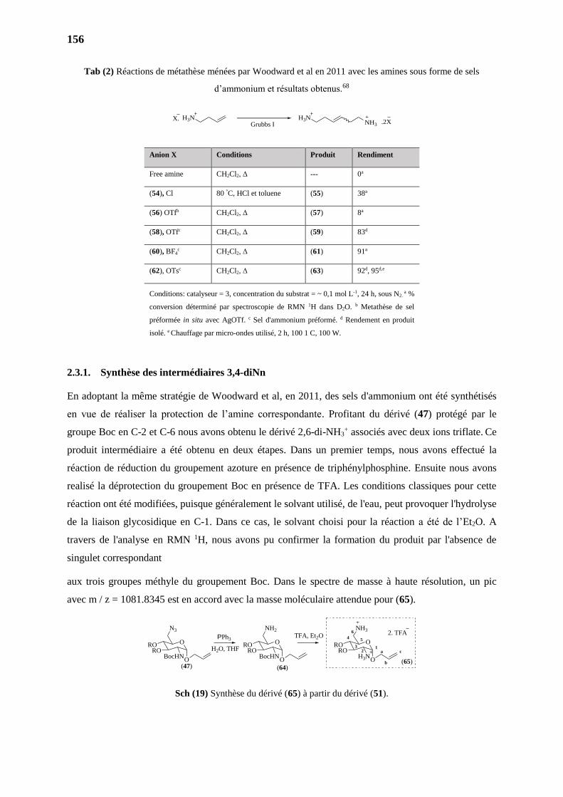
156
Tab (2) Réactions de métathèse ménées par Woodward et al en 2011 avec les amines sous forme de sels
d’ammonium et résultats obtenus.68
2.3.1. Synthèse des intermédiaires 3,4-diNn
En adoptant la même stratégie de Woodward et al, en 2011, des sels d'ammonium ont été synthétisés
en vue de réaliser la protection de l’amine correspondante. Profitant du dérivé (47) protégé par le
groupe Boc en C-2 et C-6 nous avons obtenu le dérivé 2,6-di-NH3+ associés avec deux ions triflate. Ce
produit intermédiaire a été obtenu en deux étapes. Dans un premier temps, nous avons effectué la
réaction de réduction du groupement azoture en présence de triphénylphosphine. Ensuite nous avons
realisé la déprotection du groupement Boc en présence de TFA. Les conditions classiques pour cette
réaction ont été modifiées, puisque généralement le solvant utilisé, de l'eau, peut provoquer l'hydrolyse
de la liaison glycosidique en C-1. Dans ce cas, le solvant choisi pour la réaction a été de l’Et2O. A
travers de l'analyse en RMN 1H, nous avons pu confirmer la formation du produit par l'absence de
singulet correspondant
aux trois groupes méthyle du groupement Boc. Dans le spectre de masse à haute résolution, un pic
avec m / z = 1081.8345 est en accord avec la masse moléculaire attendue pour (65).
Sch (19) Synthèse du dérivé (65) à partir du dérivé (51).
Anion X Conditions Produit Rendiment
Free amine CH2Cl2, Δ --- 0a
(54), Cl 80 °C, HCl et toluene (55) 38ª
(56) OTfb CH2Cl2, Δ (57) 8ª
(58), OTfc CH2Cl2, Δ (59) 83d
(60), BF4c CH2Cl2, Δ (61) 91ª
(62), OTsc CH2Cl2, Δ (63) 92d, 95d,e
Conditions: catalyseur = 3, concentration du substrat = ~ 0,1 mol L-1, 24 h, sous N2. a %
conversion déterminé par spectroscopie de RMN 1H dans D2O. b Metathèse de sel
préformée in situ avec AgOTf. c Sel d'ammonium préformé. d Rendement en produit
isolé. e Chauffage par micro-ondes utilisé, 2 h, 100 1 C, 100 W.
H3N H3NNH3
X.Grubbs I .2X
O
N3
RORO
BocHN
O
NH2
RORO
BocHN
O
NH3
RORO
H3N
2. TFA
(47) (64)(65)O O O
a
b
c1
2
4 5
6
3
PPh3TFA, Et2O
H2O, THF
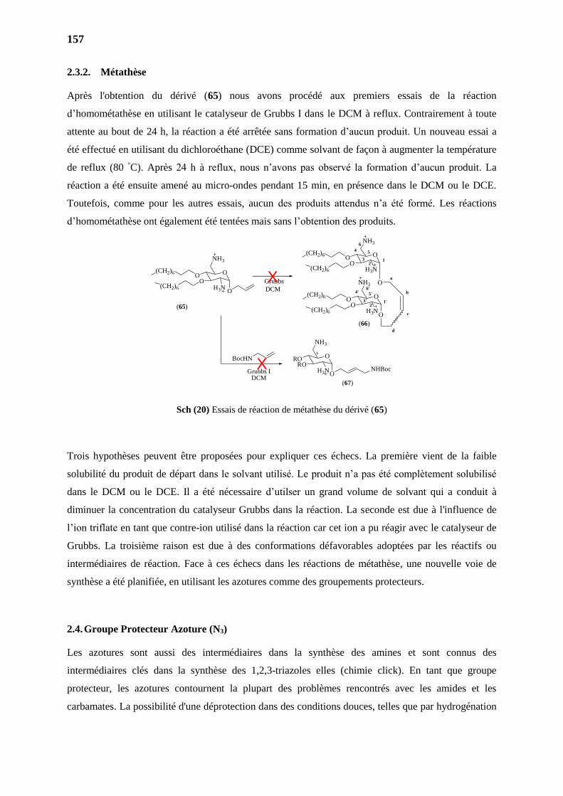
157
2.3.2. Métathèse
Après l'obtention du dérivé (65) nous avons procédé aux premiers essais de la réaction
d’homométathèse en utilisant le catalyseur de Grubbs I dans le DCM à reflux. Contrairement à toute
attente au bout de 24 h, la réaction a été arrêtée sans formation d’aucun produit. Un nouveau essai a
été effectué en utilisant du dichloroéthane (DCE) comme solvant de façon à augmenter la température
de reflux (80 °C). Après 24 h à reflux, nous n’avons pas observé la formation d’aucun produit. La
réaction a été ensuite amené au micro-ondes pendant 15 min, en présence dans le DCM ou le DCE.
Toutefois, comme pour les autres essais, aucun des produits attendus n’a été formé. Les réactions
d’homométathèse ont également été tentées mais sans l’obtention des produits.
Sch (20) Essais de réaction de métathèse du dérivé (65)
Trois hypothèses peuvent être proposées pour expliquer ces échecs. La première vient de la faible
solubilité du produit de départ dans le solvant utilisé. Le produit n’a pas été complètement solubilisé
dans le DCM ou le DCE. Il a été nécessaire d’utilser un grand volume de solvant qui a conduit à
diminuer la concentration du catalyseur Grubbs dans la réaction. La seconde est due à l'influence de
l’ion triflate en tant que contre-ion utilisé dans la réaction car cet ion a pu réagir avec le catalyseur de
Grubbs. La troisième raison est due à des conformations défavorables adoptées par les réactifs ou
intermédiaires de réaction. Face à ces échecs dans les réactions de métathèse, une nouvelle voie de
synthèse a été planifiée, en utilisant les azotures comme des groupements protecteurs.
2.4. Groupe Protecteur Azoture (N3)
Les azotures sont aussi des intermédiaires dans la synthèse des amines et sont connus des
intermédiaires clés dans la synthèse des 1,2,3-triazoles elles (chimie click). En tant que groupe
protecteur, les azotures contournent la plupart des problèmes rencontrés avec les amides et les
carbamates. La possibilité d'une déprotection dans des conditions douces, telles que par hydrogénation
Grubbs
DCM
O
NH3
OO
H3N
O
12
4 5
6
3
O
NH3
OO
H3NO
1'2'
4' 5'
6'
3'
a
b
c
d
(CH2)6
(CH2)6
(CH2)6
(CH2)6
O
NH3
OO
H3NO
(CH2)6
(CH2)6
(65)
X
O
NH3
RORO
H3NO
NHBoc
BocHN
Grubbs IDCM
X(67)
(66)
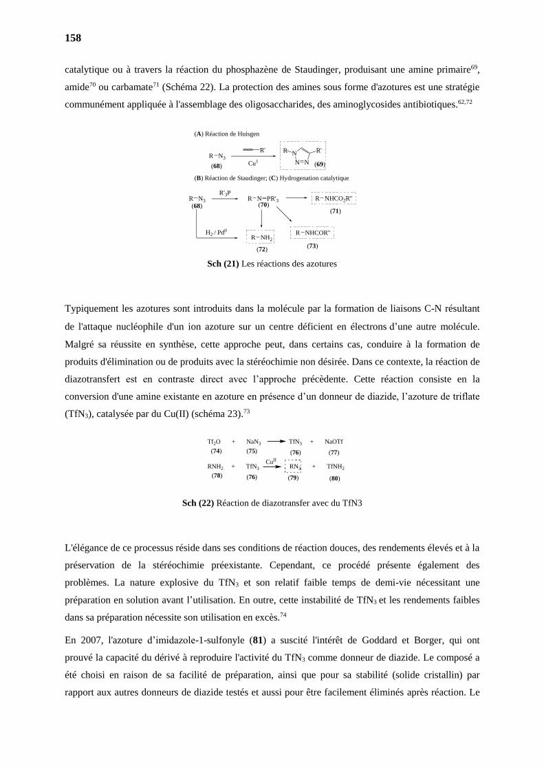
158
catalytique ou à travers la réaction du phosphazène de Staudinger, produisant une amine primaire69,
amide70 ou carbamate71 (Schéma 22). La protection des amines sous forme d'azotures est une stratégie
communément appliquée à l'assemblage des oligosaccharides, des aminoglycosides antibiotiques.62,72
Sch (21) Les réactions des azotures
Typiquement les azotures sont introduits dans la molécule par la formation de liaisons C-N résultant
de l'attaque nucléophile d'un ion azoture sur un centre déficient en électronsd’une autre molécule.
Malgré sa réussite en synthèse, cette approche peut, dans certains cas, conduire à la formation de
produits d'élimination ou de produits avec la stéréochimie non désirée. Dans ce contexte, la réaction de
diazotransfert est en contraste direct avec l’approche précèdente. Cette réaction consiste en la
conversion d'une amine existante en azoture en présence d’un donneur de diazide, l’azoture de triflate
(TfN3), catalysée par du Cu(II) (schéma 23).73
Sch (22) Réaction de diazotransfer avec du TfN3
L'élégance de ce processus réside dans ses conditions de réaction douces, des rendements élevés et à la
préservation de la stéréochimie préexistante. Cependant, ce procédé présente également des
problèmes. La nature explosive du TfN3 et son relatif faible temps de demi-vie nécessitant une
préparation en solution avant l’utilisation. En outre, cette instabilité de TfN3 et les rendements faibles
dans sa préparation nécessite son utilisation en excès.74
En 2007, l'azoture d’imidazole-1-sulfonyle (81) a suscité l'intérêt de Goddard et Borger, qui ont
prouvé la capacité du dérivé à reproduire l'activité du TfN3 comme donneur de diazide. Le composé a
été choisi en raison de sa facilité de préparation, ainsi que pour sa stabilité (solide cristallin) par
rapport aux autres donneurs de diazide testés et aussi pour être facilement éliminés après réaction. Le
Tf2O + NaN3 TfN3 + NaOTf
RNH2 + TfN3 RN3 + TfNH2
CuII
(74) (75) (76) (77)
(78) (76) (79) (80)
R N3
Cu1
R'
N N
N R'R
R N3
R'3PR N PR'3
R NH2
R NHCOR''
R NHCO2R''
H2 / Pd0
(A) Réaction de Huisgen
(B) Réaction de Staudinger; (C) Hydrogenation catalytique
(68) (69)
(68) (70)(71)
(73)(72)
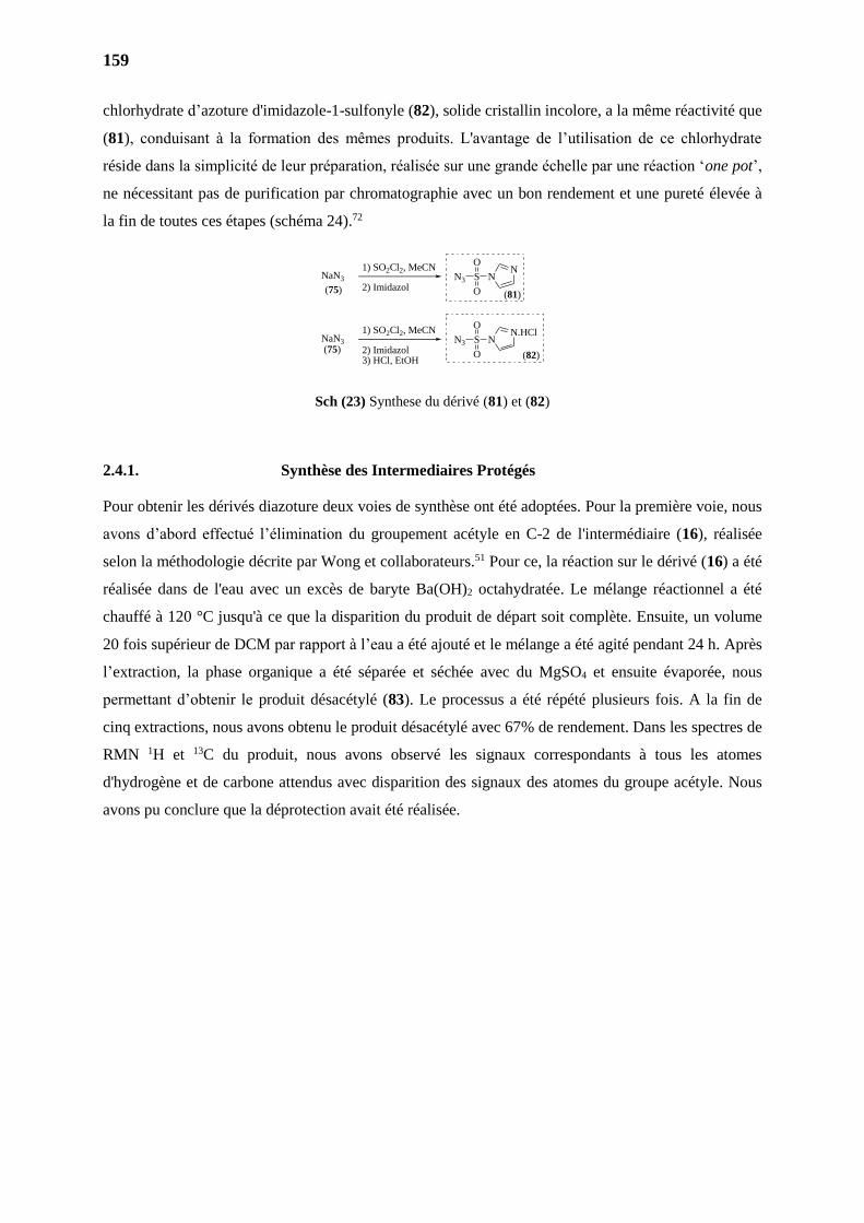
159
chlorhydrate d’azoture d'imidazole-1-sulfonyle (82), solide cristallin incolore, a la même réactivité que
(81), conduisant à la formation des mêmes produits. L'avantage de l’utilisation de ce chlorhydrate
réside dans la simplicité de leur préparation, réalisée sur une grande échelle par une réaction ‘one pot’,
ne nécessitant pas de purification par chromatographie avec un bon rendement et une pureté élevée à
la fin de toutes ces étapes (schéma 24).72
Sch (23) Synthese du dérivé (81) et (82)
2.4.1. Synthèse des Intermediaires Protégés
Pour obtenir les dérivés diazoture deux voies de synthèse ont été adoptées. Pour la première voie, nous
avons d’abord effectué l’élimination du groupement acétyle en C-2 de l'intermédiaire (16), réalisée
selon la méthodologie décrite par Wong et collaborateurs.51 Pour ce, la réaction sur le dérivé (16) a été
réalisée dans de l'eau avec un excès de baryte Ba(OH)2 octahydratée. Le mélange réactionnel a été
chauffé à 120 °C jusqu'à ce que la disparition du produit de départ soit complète. Ensuite, un volume
20 fois supérieur de DCM par rapport à l’eau a été ajouté et le mélange a été agité pendant 24 h. Après
l’extraction, la phase organique a été séparée et séchée avec du MgSO4 et ensuite évaporée, nous
permettant d’obtenir le produit désacétylé (83). Le processus a été répété plusieurs fois. A la fin de
cinq extractions, nous avons obtenu le produit désacétylé avec 67% de rendement. Dans les spectres de
RMN 1H et 13C du produit, nous avons observé les signaux correspondants à tous les atomes
d'hydrogène et de carbone attendus avec disparition des signaux des atomes du groupe acétyle. Nous
avons pu conclure que la déprotection avait été réalisée.
NaN3
2) Imidazol
1) SO2Cl2, MeCN NNS
O
O
N3
NaN3
2) Imidazol3) HCl, EtOH
1) SO2Cl2, MeCN N.HClNS
O
O
N3
(75)
(75)
(81)
(82)
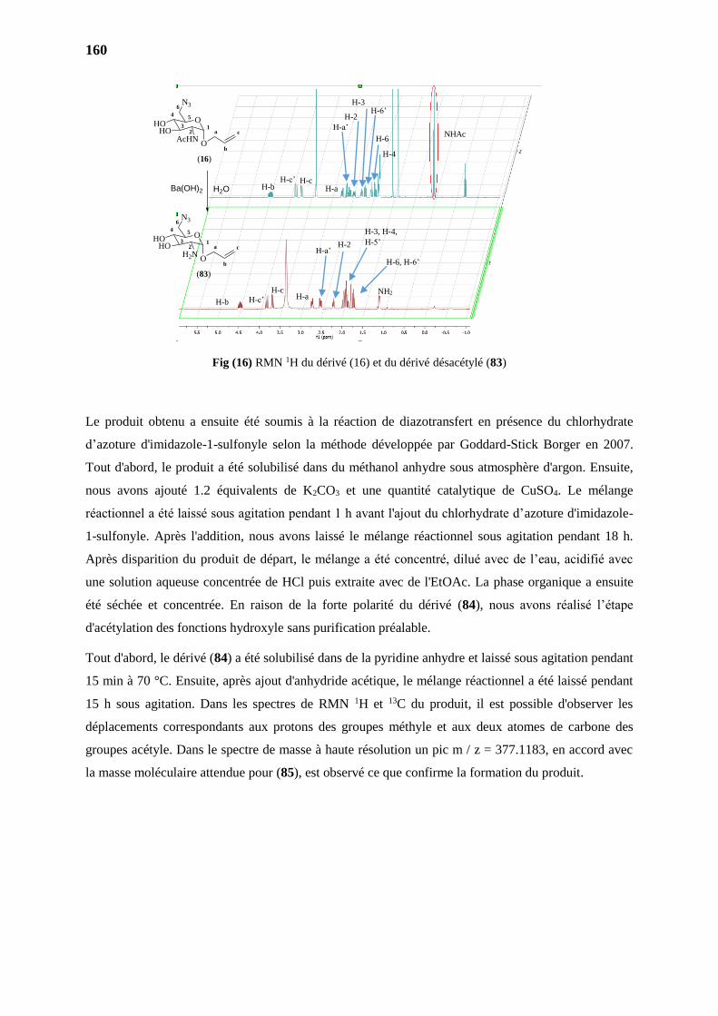
160
Fig (16) RMN 1H du dérivé (16) et du dérivé désacétylé (83)
Le produit obtenu a ensuite été soumis à la réaction de diazotransfert en présence du chlorhydrate
d’azoture d'imidazole-1-sulfonyle selon la méthode développée par Goddard-Stick Borger en 2007.
Tout d'abord, le produit a été solubilisé dans du méthanol anhydre sous atmosphère d'argon. Ensuite,
nous avons ajouté 1.2 équivalents de K2CO3 et une quantité catalytique de CuSO4. Le mélange
réactionnel a été laissé sous agitation pendant 1 h avant l'ajout du chlorhydrate d’azoture d'imidazole-
1-sulfonyle. Après l'addition, nous avons laissé le mélange réactionnel sous agitation pendant 18 h.
Après disparition du produit de départ, le mélange a été concentré, dilué avec de l’eau, acidifié avec
une solution aqueuse concentrée de HCl puis extraite avec de l'EtOAc. La phase organique a ensuite
été séchée et concentrée. En raison de la forte polarité du dérivé (84), nous avons réalisé l’étape
d'acétylation des fonctions hydroxyle sans purification préalable.
Tout d'abord, le dérivé (84) a été solubilisé dans de la pyridine anhydre et laissé sous agitation pendant
15 min à 70 °C. Ensuite, après ajout d'anhydride acétique, le mélange réactionnel a été laissé pendant
15 h sous agitation. Dans les spectres de RMN 1H et 13C du produit, il est possible d'observer les
déplacements correspondants aux protons des groupes méthyle et aux deux atomes de carbone des
groupes acétyle. Dans le spectre de masse à haute résolution un pic m / z = 377.1183, en accord avec
la masse moléculaire attendue pour (85), est observé ce que confirme la formation du produit.
NHAc
H-b H-c’ H-c
H-a
H-4
H-6
H-b H-c’
H-c NH2 H-a
H-a’
H-a’
H-2
H-6, H-6’
H-3, H-4,
H-5’
H-2
H-3 H-6’
O
N3
HOHO
H2NO
a
b
c1
2
4 5
6
3
(83)
Ba(OH)2
O
N3
HOHO
AcHNO
a
b
c1
2
4 5
6
3
(16)
H2O
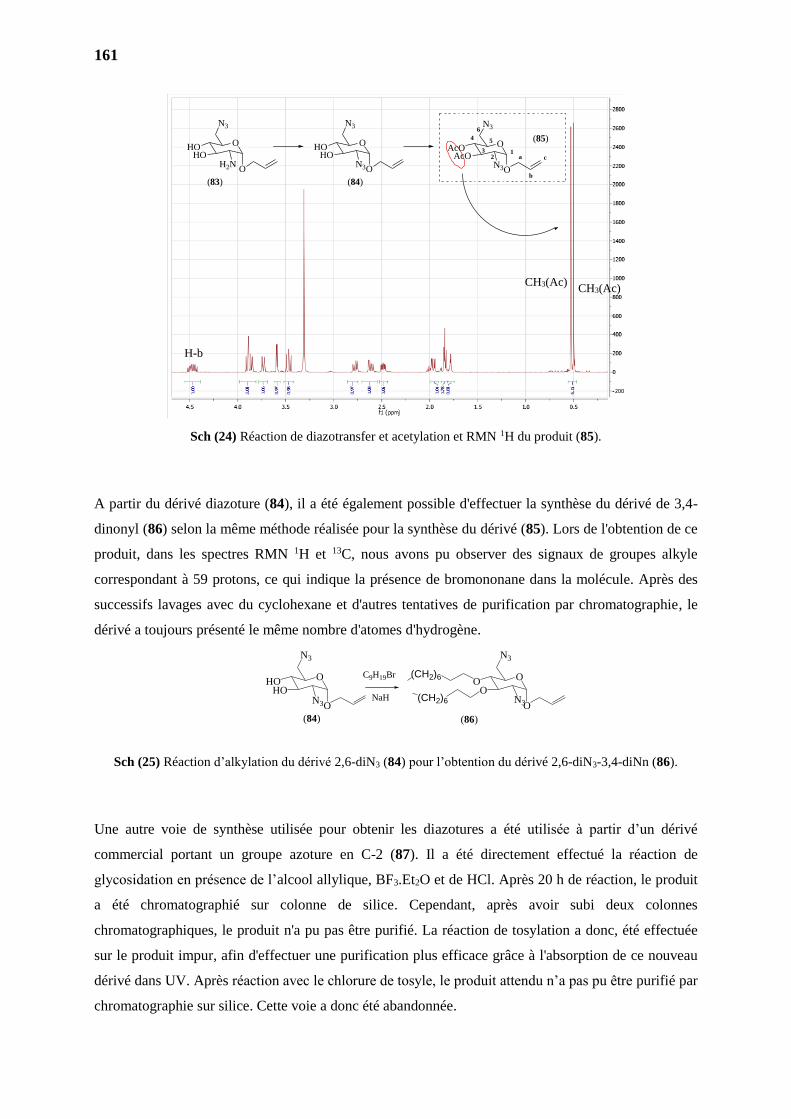
161
Sch (24) Réaction de diazotransfer et acetylation et RMN 1H du produit (85).
A partir du dérivé diazoture (84), il a été également possible d'effectuer la synthèse du dérivé de 3,4-
dinonyl (86) selon la même méthode réalisée pour la synthèse du dérivé (85). Lors de l'obtention de ce
produit, dans les spectres RMN 1H et 13C, nous avons pu observer des signaux de groupes alkyle
correspondant à 59 protons, ce qui indique la présence de bromononane dans la molécule. Après des
successifs lavages avec du cyclohexane et d'autres tentatives de purification par chromatographie, le
dérivé a toujours présenté le même nombre d'atomes d'hydrogène.
Sch (25) Réaction d’alkylation du dérivé 2,6-diN3 (84) pour l’obtention du dérivé 2,6-diN3-3,4-diNn (86).
Une autre voie de synthèse utilisée pour obtenir les diazotures a été utilisée à partir d’un dérivé
commercial portant un groupe azoture en C-2 (87). Il a été directement effectué la réaction de
glycosidation en présence de l’alcool allylique, BF3.Et2O et de HCl. Après 20 h de réaction, le produit
a été chromatographié sur colonne de silice. Cependant, après avoir subi deux colonnes
chromatographiques, le produit n'a pu pas être purifié. La réaction de tosylation a donc, été effectuée
sur le produit impur, afin d'effectuer une purification plus efficace grâce à l'absorption de ce nouveau
dérivé dans UV. Après réaction avec le chlorure de tosyle, le produit attendu n’a pas pu être purifié par
chromatographie sur silice. Cette voie a donc été abandonnée.
(83) (84)
(85)O
N3
AcOAcO
N3O
a
b
c1
2
4 5
6
3O
N3
HOHO
N3O
O
N3
HOHO
H2NO
H-b
CH3(Ac) CH3(Ac)
(84)
O
N3
HOHO
N3O
O
N3
OO
N3O
(CH2)6
(CH2)6
(86)
C9H19Br
NaH
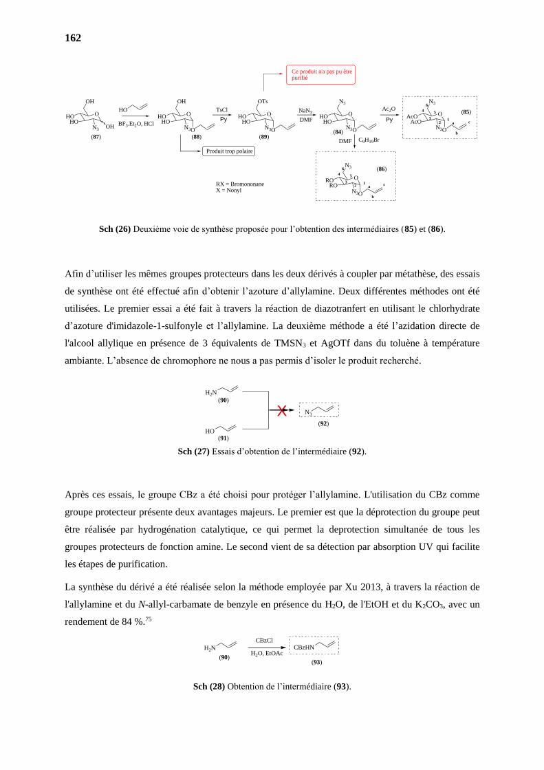
162
Sch (26) Deuxième voie de synthèse proposée pour l’obtention des intermédiaires (85) et (86).
Afin d’utiliser les mêmes groupes protecteurs dans les deux dérivés à coupler par métathèse, des essais
de synthèse ont été effectué afin d’obtenir l’azoture d’allylamine. Deux différentes méthodes ont été
utilisées. Le premier essai a été fait à travers la réaction de diazotranfert en utilisant le chlorhydrate
d’azoture d'imidazole-1-sulfonyle et l’allylamine. La deuxième méthode a été l’azidation directe de
l'alcool allylique en présence de 3 équivalents de TMSN3 et AgOTf dans du toluène à température
ambiante. L’absence de chromophore ne nous a pas permis d’isoler le produit recherché.
Sch (27) Essais d’obtention de l’intermédiaire (92).
Après ces essais, le groupe CBz a été choisi pour protéger l’allylamine. L'utilisation du CBz comme
groupe protecteur présente deux avantages majeurs. Le premier est que la déprotection du groupe peut
être réalisée par hydrogénation catalytique, ce qui permet la deprotection simultanée de tous les
groupes protecteurs de fonction amine. Le second vient de sa détection par absorption UV qui facilite
les étapes de purification.
La synthèse du dérivé a été réalisée selon la méthode employée par Xu 2013, à travers la réaction de
l'allylamine et du N-allyl-carbamate de benzyle en présence du H2O, de l'EtOH et du K2CO3, avec un
rendement de 84 %.75
Sch (28) Obtention de l’intermédiaire (93).
O
OH
HOHO
N3
Ce produit n'a pas pu êtrepurifié
Produit trop polaire
(87) (88) (89)(84)
(86)
(85)O
OH
HOHO
N3O
O
OTs
HOHO
N3O
O
N3
HOHO
N3O
O
N3
AcOAcO
N3O
O
N3
RORO
N3O
TsCl NaN3Ac2O
RX = BromononaneX = Nonyl
HO
a
b
c12
4 5
6
3
a
b
c1
2
4 5
6
3
OH
Py DMF Py
C9H19BrDMF
BF3.Et2O, HCl
N3
H2N
HO
X(90)
(91)
(92)
H2N CBzHN
(90)(93)
CBzCl
H2O, EtOAc
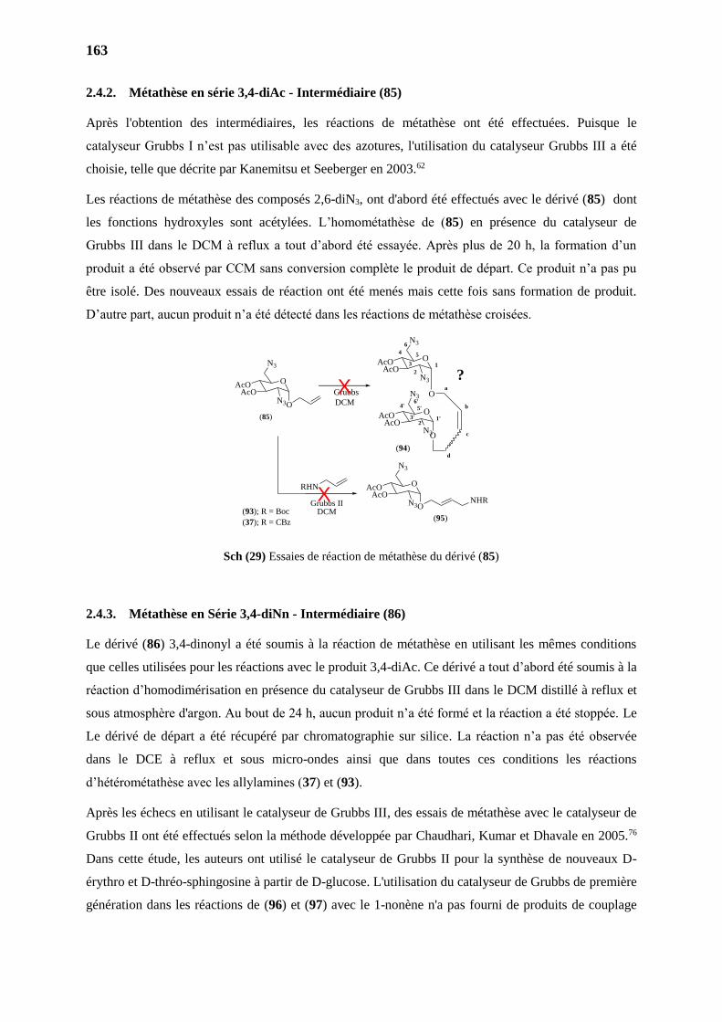
163
2.4.2. Métathèse en série 3,4-diAc - Intermédiaire (85)
Après l'obtention des intermédiaires, les réactions de métathèse ont été effectuées. Puisque le
catalyseur Grubbs I n’est pas utilisable avec des azotures, l'utilisation du catalyseur Grubbs III a été
choisie, telle que décrite par Kanemitsu et Seeberger en 2003.62
Les réactions de métathèse des composés 2,6-diN3, ont d'abord été effectués avec le dérivé (85) dont
les fonctions hydroxyles sont acétylées. L’homométathèse de (85) en présence du catalyseur de
Grubbs III dans le DCM à reflux a tout d’abord été essayée. Après plus de 20 h, la formation d’un
produit a été observé par CCM sans conversion complète le produit de départ. Ce produit n’a pas pu
être isolé. Des nouveaux essais de réaction ont été menés mais cette fois sans formation de produit.
D’autre part, aucun produit n’a été détecté dans les réactions de métathèse croisées.
Sch (29) Essaies de réaction de métathèse du dérivé (85)
2.4.3. Métathèse en Série 3,4-diNn - Intermédiaire (86)
Le dérivé (86) 3,4-dinonyl a été soumis à la réaction de métathèse en utilisant les mêmes conditions
que celles utilisées pour les réactions avec le produit 3,4-diAc. Ce dérivé a tout d’abord été soumis à la
réaction d’homodimérisation en présence du catalyseur de Grubbs III dans le DCM distillé à reflux et
sous atmosphère d'argon. Au bout de 24 h, aucun produit n’a été formé et la réaction a été stoppée. Le
Le dérivé de départ a été récupéré par chromatographie sur silice. La réaction n’a pas été observée
dans le DCE à reflux et sous micro-ondes ainsi que dans toutes ces conditions les réactions
d’hétérométathèse avec les allylamines (37) et (93).
Après les échecs en utilisant le catalyseur de Grubbs III, des essais de métathèse avec le catalyseur de
Grubbs II ont été effectués selon la méthode développée par Chaudhari, Kumar et Dhavale en 2005.76
Dans cette étude, les auteurs ont utilisé le catalyseur de Grubbs II pour la synthèse de nouveaux D-
érythro et D-thréo-sphingosine à partir de D-glucose. L'utilisation du catalyseur de Grubbs de première
génération dans les réactions de (96) et (97) avec le 1-nonène n'a pas fourni de produits de couplage
Grubbs
DCM
O
N3
AcOAcO
O
1
2
4 5
6
3
O
N3
AcOAcO
N3O
1'2'
4' 5'
6'
3'
a
b
c
d
O
N3
AcOAcO
N3O
(85)
X
O
N3
AcOAcO
N3ONHR
RHN
Grubbs IIDCM
(95)
X
N3
(94)
(93); R = Boc
(37); R = CBz
?
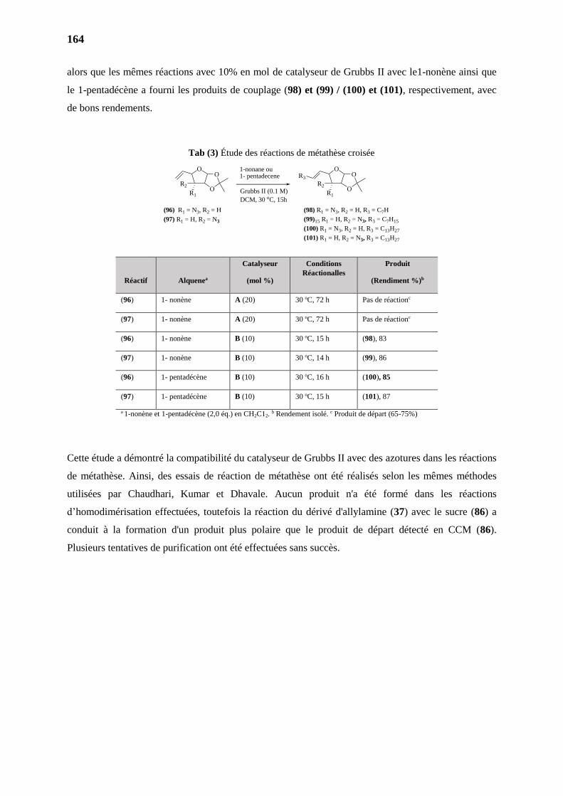
164
alors que les mêmes réactions avec 10% en mol de catalyseur de Grubbs II avec le1-nonène ainsi que
le 1-pentadécène a fourni les produits de couplage (98) et (99) / (100) et (101), respectivement, avec
de bons rendements.
Tab (3) Étude des réactions de métathèse croisée
Réactif
Alquenea
Catalyseur
(mol %)
Conditions
Réactionalles
Produit
(Rendiment %)b
(96) 1- nonène A (20) 30 oC, 72 h Pas de réactionc
(97) 1- nonène A (20) 30 oC, 72 h Pas de réactionc
(96) 1- nonène B (10) 30 oC, 15 h (98), 83
(97) 1- nonène B (10) 30 oC, 14 h (99), 86
(96) 1- pentadécène B (10) 30 oC, 16 h (100), 85
(97) 1- pentadécène B (10) 30 oC, 15 h (101), 87
a 1-nonène et 1-pentadécène (2,0 éq.) en CH2C12. b Rendement isolé. c Produit de départ (65-75%)
Cette étude a démontré la compatibilité du catalyseur de Grubbs II avec des azotures dans les réactions
de métathèse. Ainsi, des essais de réaction de métathèse ont été réalisés selon les mêmes méthodes
utilisées par Chaudhari, Kumar et Dhavale. Aucun produit n'a été formé dans les réactions
d’homodimérisation effectuées, toutefois la réaction du dérivé d'allylamine (37) avec le sucre (86) a
conduit à la formation d'un produit plus polaire que le produit de départ détecté en CCM (86).
Plusieurs tentatives de purification ont été effectuées sans succès.
O
R2
R1
(96) R1 = N3, R2 = H
(97) R1 = H, R2 = N3
1-nonane ou1- pentadecene
Grubbs II (0.1 M)
DCM, 30 oC, 15h
O
OO
R2
R1O
OR3
(98) R1 = N3, R2 = H, R3 = C7H
(99)15 R1 = H, R2 = N3, R3 = C7H15
(100) R1 = N3, R2 = H, R3 = C13H27
(101) R1 = H, R2 = N3, R3 = C13H27
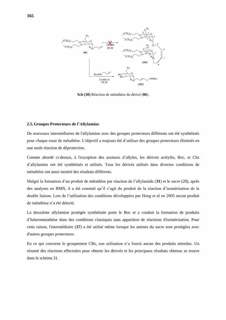
165
Sch (30) Réaction de métathèse du dérivé (86).
2.5. Groupes Protecteurs de l’Allylamine
De nouveaux intermédiaires de l'allylamine avec des groupes protecteurs différents ont été synthétisés
pour chaque essai de métathèse. L'objectif a toujours été d’utiliser des groupes protecteurs éliminés en
une seule réaction de déprotection.
Comme abordé ci-dessus, à l'exception des azotures d’allyles, les dérivés acétylés, Boc, et Cbz
d’allylamine ont été synthétisés et utilisés. Tous les dérivés utilisés dans diverses conditions de
métathèse ont aussi montré des résultats différents.
Malgré la formation d’un produit de métathèse par réaction de l’allylamide (31) et le sucre (28), après
des analyses en RMN, il a été constaté qu’il s’agit du produit de la réaction d’isomérisation de la
double liaison. Lors de l’utilisation des conditions développées par Hong et al en 2005 aucun produit
de métathèse n’a été détecté.
La deuxième allylamine protégée synthétisée porte le Boc et a conduit la formation de produits
d’héterometathèse dans des conditions classiques sans apparition de réactions d'isomérisation. Pour
cette raison, l'intermédiaire (37) a été utilisé même lorsque les amines du sucre sont protégées avec
d'autres groupes protecteurs.
En ce qui concerne le groupement CBz, son utilisation n’a fourni aucun des produits attendus. Un
résumé des réactions effectuées pour obtenir les dérivés et les principaux résultats obtenus se trouve
dans le schéma 31.
Grubbs
DCM
O
N3
OO
O
1
2
4 5
6
3
O
N3
OO
N3O
1'2'
4' 5'
6'
3'
a
b
c
d
(CH2)6
(CH2)6
(CH2)6
(CH2)6
O
N3
OO
N3O
(CH2)6
(CH2)6
(86)
X
O
N3
RORO
N3ONHBoc
BocHN
Grubbs IIDCM
(103)
N3
(102)
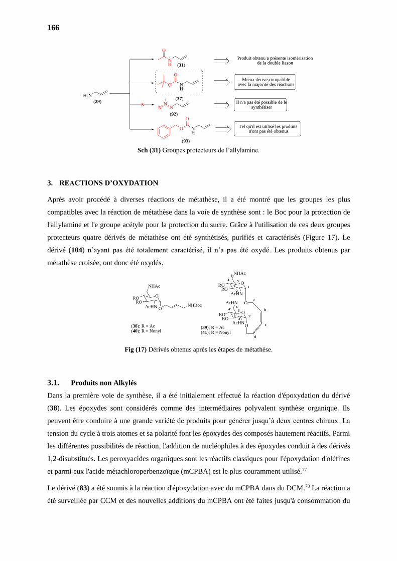
166
Sch (31) Groupes protecteurs de l’allylamine.
3. REACTIONS D’OXYDATION
Après avoir procédé à diverses réactions de métathèse, il a été montré que les groupes les plus
compatibles avec la réaction de métathèse dans la voie de synthèse sont : le Boc pour la protection de
l'allylamine et l'e groupe acétyle pour la protection du sucre. Grâce à l'utilisation de ces deux groupes
protecteurs quatre dérivés de métathèse ont été synthétisés, purifiés et caractérisés (Figure 17). Le
dérivé (104) n’ayant pas été totalement caractérisé, il n’a pas été oxydé. Les produits obtenus par
métathèse croisée, ont donc été oxydés.
Fig (17) Dérivés obtenus après les étapes de métathèse.
3.1. Produits non Alkylés
Dans la première voie de synthèse, il a été initialement effectué la réaction d'époxydation du dérivé
(38). Les époxydes sont considérés comme des intermédiaires polyvalent synthèse organique. Ils
peuvent être conduire à une grande variété de produits pour générer jusqu’à deux centres chiraux. La
tension du cycle à trois atomes et sa polarité font les époxydes des composés hautement réactifs. Parmi
les différentes possibilités de réaction, l'addition de nucléophiles à des époxydes conduit à des dérivés
1,2-disubstitués. Les peroxyacides organiques sont les réactifs classiques pour l'époxydation d'oléfines
et parmi eux l'acide métachloroperbenzoïque (mCPBA) est le plus couramment utilisé.77
Le dérivé (83) a été soumis à la réaction d'époxydation avec du mCPBA dans du DCM.78 La réaction a
été surveillée par CCM et des nouvelles additions du mCPBA ont été faites jusqu'à consommation du
H2N
NH
NH
NH
NX
Mieux dérivé,compatible avec la majorité des réactions
Il n'a pas été possible de le synthétiser
Tel qu'il est utilisé les produits n'ont pas été obtenus
Produit obtenu a présente isomérisation de la double liason
O
O
O
NN
O
O
(29)
(92)
(93)
(37)
(31)
O
NHAc
RORO
O
1
2
4 5
6
3
O
AcHN
RORO
AcHNO
1'2'
4' 5'
6'
3'
a
b
c
d
O
NHAc
RORO
AcHN ONHBoc
AcHN
(38); R = Ac(40); R = Nonyl
(39); R = Ac(41); R = Nonyl
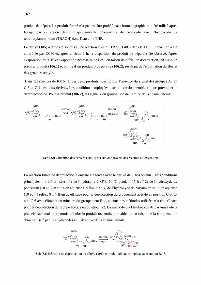
167
produit de départ. Le produit formé n’a pas pu être purifié par chromatographie et a été utilisé après
lavage par extraction dans l’étape suivante d’ouverture de l'époxyde avec l'hydroxyde de
tétrabutylammonium (TBAOH) dans l'eau et le THF.
Le dérivé (105) a donc été soumis à une réaction avec du TBAOH 40% dans le THF. La réaction a été
contrôlée par CCM et, après environ 1 h, la disparition du produit de départ a été observé. Après
évaporation du THF et évaporation nécessaire de l’eau en raison de difficulté d’extraction, 20 mg d’un
premier produit (106.1) et 60 mg d’un produit plus polaire (106.2), résultant de l'élimination du Boc et
des groupes acétyle.
Dans les spectres de RMN 1H des deux produits nous notons l’absence du signal des groupes Ac en
C-3 et C-4 des deux dérivés. Les conditions employées dans la réaction semblent donc provoquer la
déprotection de. Pour le produit (106.2), les signaux du groupe Boc de l’amine de la chaîne latérale
Sch (32) Obtention des dérivés (106.1) et (106.2) à travers des réactions d’oxydation
La réaction finale de déprotection a ensuite été tentée avec le dérivé de (106) obtenu. Trois conditions
principales ont été utilisées : 1) de l’hydrazine à 85%, 70 oC pendant 15 h ;79 2) de l’hydroxyde de
potassium (10 éq.) en solution aqueuse à reflux 6 h ; 3) de l’hydroxyde de baryum en solution aqueuse
(10 éq.) à reflux 6 h.51 Bien qu'efficace pour la déprotection du groupement acétyle en position C-3, C-
4 et C-6 avec élimination mineure du groupement Boc, aucune des méthodes utilisées n’a été efficace
pour la déprotection du groupe acétyle en position C-2. La méthode 3 à l’hydroxyde de baryum a été la
plus efficace mais n’a permis d’isoler le produit recherché probablemnt en raison de la complexation
d’un ion Ba+2 par les hydroxyles en C-b et C-c de la chaîne latérale.
Sch (33) Réaction de deprotection du dérivé (106) et produit obtenu complexé avec un ion Ba+2.
O
NHAc
AcOAcO
AcHN
O
NHAc
AcOAcO
AcHN
O
NHAc
HOHO
AcHN
(38) (104) (105)
mCPBA TBAOH
O
NHAc
HOHO
AcHN
(106)
O ONHBoc NHBoc
O
ONHBoc
ONH2
OH
OH
OH
OH
a
b
c1
4 5
6
3
d
a
b
c1
4 5
6
3
d
2
2
a
b
c1
4 5
6
3
d
2DCM THF; H2O
O
NHAc
HOHO
AcHN
(106)
O
NH2
HOHO
AcHNOO
NHBoc
OH
OHa
b
c1
4 5
6
3d
2
BocHN
HO Ba2+
OH
Ba(OH)2
(107)
H2O
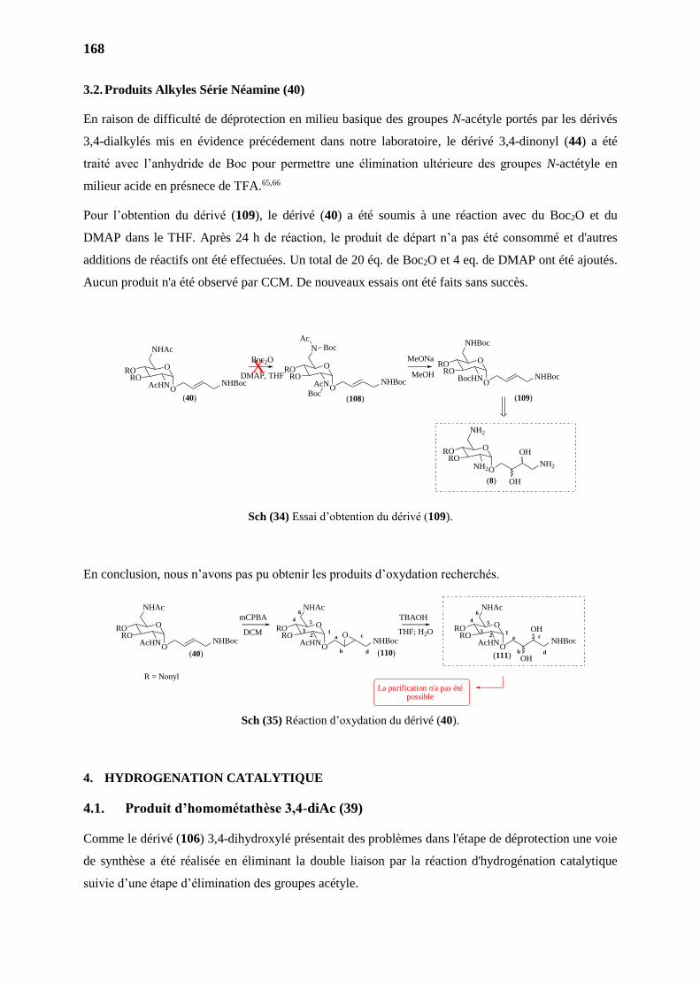
168
3.2. Produits Alkyles Série Néamine (40)
En raison de difficulté de déprotection en milieu basique des groupes N-acétyle portés par les dérivés
3,4-dialkylés mis en évidence précédement dans notre laboratoire, le dérivé 3,4-dinonyl (44) a été
traité avec l’anhydride de Boc pour permettre une élimination ultérieure des groupes N-actétyle en
milieur acide en présnece de TFA.65,66
Pour l’obtention du dérivé (109), le dérivé (40) a été soumis à une réaction avec du Boc2O et du
DMAP dans le THF. Après 24 h de réaction, le produit de départ n’a pas été consommé et d'autres
additions de réactifs ont été effectuées. Un total de 20 éq. de Boc2O et 4 eq. de DMAP ont été ajoutés.
Aucun produit n'a été observé par CCM. De nouveaux essais ont été faits sans succès.
Sch (34) Essai d’obtention du dérivé (109).
En conclusion, nous n’avons pas pu obtenir les produits d’oxydation recherchés.
Sch (35) Réaction d’oxydation du dérivé (40).
4. HYDROGENATION CATALYTIQUE
4.1. Produit d’homométathèse 3,4-diAc (39)
Comme le dérivé (106) 3,4-dihydroxylé présentait des problèmes dans l'étape de déprotection une voie
de synthèse a été réalisée en éliminant la double liaison par la réaction d'hydrogénation catalytique
suivie d’une étape d’élimination des groupes acétyle.
O
NHAc
RORO
AcHN
O
N
RORO
AcN
O
NHBoc
RORO
BocHN
O
NH2
RORO
NH2
Boc
AcBoc
X
(40) (108) (109)
(8)
MeOH
ONHBoc
ONHBoc O
NHBoc
ONH2
OH
OH
DMAP, THF
Boc2O MeONa
DCM THF; H2OO
NHAc
RORO
AcHN
O
NHAc
RORO
AcHN
O
NHAc
RORO
AcHN
(40) (110) (111)
mCPBA TBAOH
O ONHBoc NHBoc
O
ONHBoc
OH
OHa
b
c1
4 5
6
3
d
2a
b
c1
4 5
6
3
d
2
La purification n'a pas étépossible
R = Nonyl
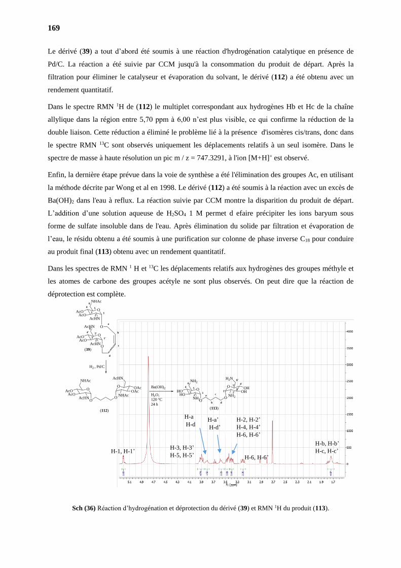
169
Le dérivé (39) a tout d’abord été soumis à une réaction d'hydrogénation catalytique en présence de
Pd/C. La réaction a été suivie par CCM jusqu'à la consommation du produit de départ. Après la
filtration pour éliminer le catalyseur et évaporation du solvant, le dérivé (112) a été obtenu avec un
rendement quantitatif.
Dans le spectre RMN 1H de (112) le multiplet correspondant aux hydrogènes Hb et Hc de la chaîne
allylique dans la région entre 5,70 ppm à 6,00 n’est plus visible, ce qui confirme la réduction de la
double liaison. Cette réduction a éliminé le problème lié à la présence d'isomères cis/trans, donc dans
le spectre RMN 13C sont observés uniquement les déplacements relatifs à un seul isomère. Dans le
spectre de masse à haute résolution un pic m / z = 747.3291, à l'ion [M+H]+ est observé.
Enfin, la dernière étape prévue dans la voie de synthèse a été l'élimination des groupes Ac, en utilisant
la méthode décrite par Wong et al en 1998. Le dérivé (112) a été soumis à la réaction avec un excès de
Ba(OH)2 dans l'eau à reflux. La réaction suivie par CCM montre la disparition du produit de départ.
L’addition d’une solution aqueuse de H2SO4 1 M permet d efaire précipiter les ions baryum sous
forme de sulfate insoluble dans de l'eau. Après élimination du solide par filtration et évaporation de
l’eau, le résidu obtenu a été soumis à une purification sur colonne de phase inverse C18 pour conduire
au produit final (113) obtenu avec un rendement quantitatif.
Dans les spectres de RMN 1 H et 13C les déplacements relatifs aux hydrogènes des groupes méthyle et
les atomes de carbone des groupes acétyle ne sont plus observés. On peut dire que la réaction de
déprotection est complète.
Sch (36) Réaction d’hydrogénation et déprotection du dérivé (39) et RMN 1H du produit (113).
H-1, H-1’
H-b, H-b’
H-c, H-c’
H-3, H-3’
H-5, H-5’
H-6, H-6’
H-a’
H-d’
H-a
H-d H-2, H-2’
H-4, H-4’
H-6, H-6’
H2 , Pd/C
(112)
O
NHAc
AcOAcO
AcHNO
O
O
AcHN
OAcOAc
NHAc a
b
c12
4 5
6
3O
NH2
HOHO
NH2O
(113)
O
O
H2N
OHOH
NH2
d
1' 2'3'
4'
6'
5'Ba(OH)2
H2O,
120 °C
24 h
(39)
O
NHAc
AcOAcO
AcHN
O
12
4 5
6
3
O
AcHN
AcOAcO
AcHNO
1'2'
4' 5'
6'
3'
a
b
c
d
CONCLUSION ET PERSPECTIVES
PARTIE II
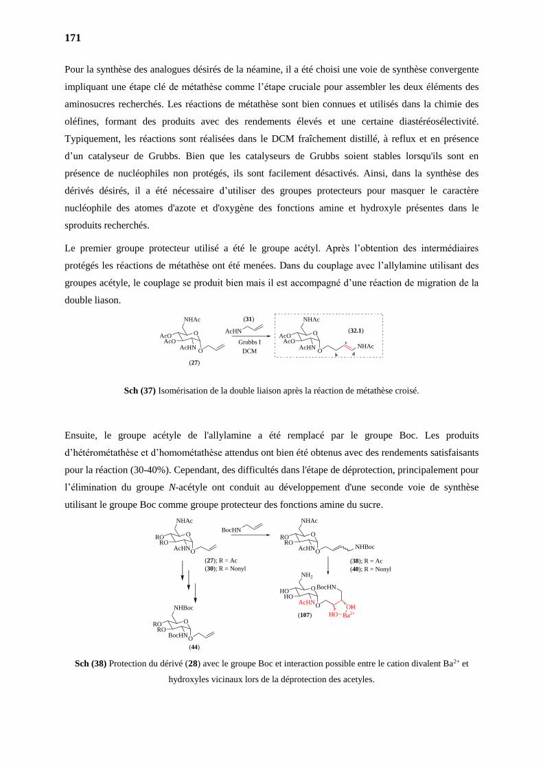
171
Pour la synthèse des analogues désirés de la néamine, il a été choisi une voie de synthèse convergente
impliquant une étape clé de métathèse comme l’étape cruciale pour assembler les deux éléments des
aminosucres recherchés. Les réactions de métathèse sont bien connues et utilisés dans la chimie des
oléfines, formant des produits avec des rendements élevés et une certaine diastéréosélectivité.
Typiquement, les réactions sont réalisées dans le DCM fraîchement distillé, à reflux et en présence
d’un catalyseur de Grubbs. Bien que les catalyseurs de Grubbs soient stables lorsqu'ils sont en
présence de nucléophiles non protégés, ils sont facilement désactivés. Ainsi, dans la synthèse des
dérivés désirés, il a été nécessaire d’utiliser des groupes protecteurs pour masquer le caractère
nucléophile des atomes d'azote et d'oxygène des fonctions amine et hydroxyle présentes dans le
sproduits recherchés.
Le premier groupe protecteur utilisé a été le groupe acétyl. Après l’obtention des intermédiaires
protégés les réactions de métathèse ont été menées. Dans du couplage avec l’allylamine utilisant des
groupes acétyle, le couplage se produit bien mais il est accompagné d’une réaction de migration de la
double liason.
Sch (37) Isomérisation de la double liaison après la réaction de métathèse croisé.
Ensuite, le groupe acétyle de l'allylamine a été remplacé par le groupe Boc. Les produits
d’hétérométathèse et d’homométathèse attendus ont bien été obtenus avec des rendements satisfaisants
pour la réaction (30-40%). Cependant, des difficultés dans l'étape de déprotection, principalement pour
l’élimination du groupe N-acétyle ont conduit au développement d'une seconde voie de synthèse
utilisant le groupe Boc comme groupe protecteur des fonctions amine du sucre.
Sch (38) Protection du dérivé (28) avec le groupe Boc et interaction possible entre le cation divalent Ba2+ et
hydroxyles vicinaux lors de la déprotection des acetyles.
AcHN
Grubbs I
DCM
(27)
(31)
O
NHAc
AcOAcO
AcHNO
b
cNHAc
d
(32.1)O
NHAc
AcOAcO
AcHNO
O
NHAc
RORO
AcHN
BocHNO
NHAc
RORO
AcHN
O
NH2
HOHO
AcHN
O
NHBoc
RORO
BocHN
O O
O
BocHN
HO Ba2+
OH
O
NHBoc
(27); R = Ac
(30); R = Nonyl
(38); R = Ac
(40); R = Nonyl
(107)
(44)
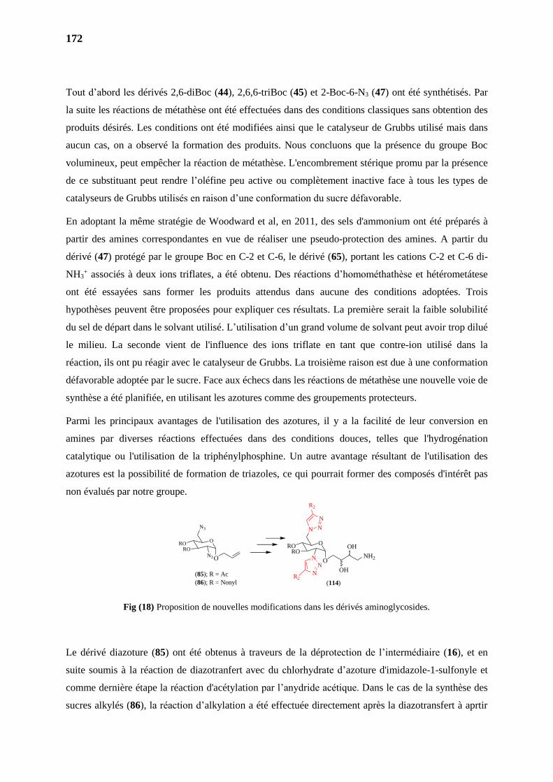
172
Tout d’abord les dérivés 2,6-diBoc (44), 2,6,6-triBoc (45) et 2-Boc-6-N3 (47) ont été synthétisés. Par
la suite les réactions de métathèse ont été effectuées dans des conditions classiques sans obtention des
produits désirés. Les conditions ont été modifiées ainsi que le catalyseur de Grubbs utilisé mais dans
aucun cas, on a observé la formation des produits. Nous concluons que la présence du groupe Boc
volumineux, peut empêcher la réaction de métathèse. L'encombrement stérique promu par la présence
de ce substituant peut rendre l’oléfine peu active ou complètement inactive face à tous les types de
catalyseurs de Grubbs utilisés en raison d’une conformation du sucre défavorable.
En adoptant la même stratégie de Woodward et al, en 2011, des sels d'ammonium ont été préparés à
partir des amines correspondantes en vue de réaliser une pseudo-protection des amines. A partir du
dérivé (47) protégé par le groupe Boc en C-2 et C-6, le dérivé (65), portant les cations C-2 et C-6 di-
NH3+ associés à deux ions triflates, a été obtenu. Des réactions d’homométhathèse et hétérometátese
ont été essayées sans former les produits attendus dans aucune des conditions adoptées. Trois
hypothèses peuvent être proposées pour expliquer ces résultats. La première serait la faible solubilité
du sel de départ dans le solvant utilisé. L’utilisation d’un grand volume de solvant peut avoir trop dilué
le milieu. La seconde vient de l'influence des ions triflate en tant que contre-ion utilisé dans la
réaction, ils ont pu réagir avec le catalyseur de Grubbs. La troisième raison est due à une conformation
défavorable adoptée par le sucre. Face aux échecs dans les réactions de métathèse une nouvelle voie de
synthèse a été planifiée, en utilisant les azotures comme des groupements protecteurs.
Parmi les principaux avantages de l'utilisation des azotures, il y a la facilité de leur conversion en
amines par diverses réactions effectuées dans des conditions douces, telles que l'hydrogénation
catalytique ou l'utilisation de la triphénylphosphine. Un autre avantage résultant de l'utilisation des
azotures est la possibilité de formation de triazoles, ce qui pourrait former des composés d'intérêt pas
non évalués par notre groupe.
Fig (18) Proposition de nouvelles modifications dans les dérivés aminoglycosides.
Le dérivé diazoture (85) ont été obtenus à traveurs de la déprotection de l’intermédiaire (16), et en
suite soumis à la réaction de diazotranfert avec du chlorhydrate d’azoture d'imidazole-1-sulfonyle et
comme dernière étape la réaction d'acétylation par l’anydride acétique. Dans le cas de la synthèse des
sucres alkylés (86), la réaction d’alkylation a été effectuée directement après la diazotransfert à aprtir
(114)
O
N3
RORO
N3
O
N
RORO
N
N
N
N
N
R2
R2
O ONH2
OH
OH
(85); R = Ac
(86); R = Nonyl
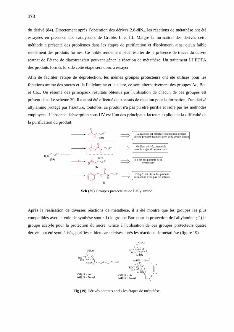
173
du dérivé (84). Directement après l’obtention des dérivés 2,6-diN3, les réactions de métathèse ont été
essayées en présence des catalyseurs de Grubbs II et III. Malgré la formation des dérivés cette
méthode a présenté des problèmes dans les étapes de purification et d'isolement, ainsi qu'un faible
rendement des produits formés. Ce faible rendement peut résulter de la présence de traces du cuivre
reatnat de l’étape de diazotransfert pouvant gêner la réaction de métathèse. Un traitement à l’EDTA
des produits formés lors de cette étape sera donc à essayer.
Afin de faciliter l'étape de déprotection, les mêmes groupes protecteurs ont été utilisés pour les
fonctions amine des sucres et de l’allylamine et le sucre, ce sont alternativement des groupes Ac, Boc
et Cbz. Un résumé des principaux résultats obtenus par l'utilisation de chacun de ces groupes est
présent dans Le schème 39. Il a aussi été effectué deux essais de réaction pour la formation d’un dérivé
allylamine protégé par l’azoture, toutefois, ce produit n'a pas pu être purifié et isolé par les méthodes
employées. L’absence d'absorption sous UV est l’un des principaux facteurs expliquant la difficulté de
la purification du produit.
Sch (39) Groupes protecteurs de l’allylamine.
Après la réalisation de diverses réactions de métathèse, il a été montré que les groupes les plus
compatibles avec la voie de synthèse sont : 1) le groupe Boc pour la protection de l'allylamine ; 2) le
groupe acétyle pour la protection du sucre. Grâce à l'utilisation de ces groupes protecteurs quatre
dérivés ont été synthétisés, purifiés et bien caractérisés après les réactions de métathèse (figure 19).
Fig (19) Dérivés obtenus après les étapes de métathèse.
H2N
NH
NH
NH
NX
Meilleur dérivé,compatible avec la majorité des réactions
Il a été pas possible de le synthétiser
Tel qu'il est utilisé les produits de réaction n'ont pas été obtenus
La réaction est effectué cependent,le produit obtenu présente isomérisation de la double liason
O
O
O
NN
O
O
(29)
(92)
(93)
(37)
(30)
O
NHAc
RORO
O
1
2
4 5
6
3
O
AcHN
RORO
AcHNO
1'2'
4' 5'
6'
3'
a
b
c
d
O
NHAc
RORO
AcHN ONHBoc
AcHN
(38); R = Ac(40); R = Nonyl
(39); R = Ac(41); R = Nonyl
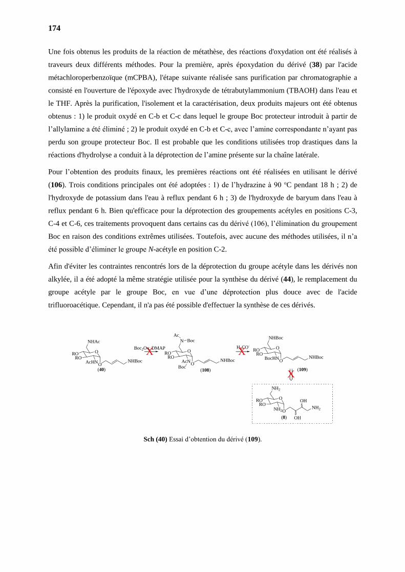
174
Une fois obtenus les produits de la réaction de métathèse, des réactions d'oxydation ont été réalisés à
traveurs deux différents méthodes. Pour la première, après époxydation du dérivé (38) par l'acide
métachloroperbenzoïque (mCPBA), l'étape suivante réalisée sans purification par chromatographie a
consisté en l'ouverture de l'époxyde avec l'hydroxyde de tétrabutylammonium (TBAOH) dans l'eau et
le THF. Après la purification, l'isolement et la caractérisation, deux produits majeurs ont été obtenus
obtenus : 1) le produit oxydé en C-b et C-c dans lequel le groupe Boc protecteur introduit à partir de
l’allylamine a été éliminé ; 2) le produit oxydé en C-b et C-c, avec l’amine correspondante n’ayant pas
perdu son groupe protecteur Boc. Il est probable que les conditions utilisées trop drastiques dans la
réactions d'hydrolyse a conduit à la déprotection de l’amine présente sur la chaîne latérale.
Pour l’obtention des produits finaux, les premières réactions ont été réalisées en utilisant le dérivé
(106). Trois conditions principales ont été adoptées : 1) de l’hydrazine à 90 oC pendant 18 h ; 2) de
l'hydroxyde de potassium dans l'eau à reflux pendant 6 h ; 3) de l'hydroxyde de baryum dans l'eau à
reflux pendant 6 h. Bien qu'efficace pour la déprotection des groupements acétyles en positions C-3,
C-4 et C-6, ces traitements provoquent dans certains cas du dérivé (106), l’élimination du groupement
Boc en raison des conditions extrêmes utilisées. Toutefois, avec aucune des méthodes utilisées, il n’a
été possible d’éliminer le groupe N-acétyle en position C-2.
Afin d'éviter les contraintes rencontrés lors de la déprotection du groupe acétyle dans les dérivés non
alkylée, il a été adopté la même stratégie utilisée pour la synthèse du dérivé (44), le remplacement du
groupe acétyle par le groupe Boc, en vue d’une déprotection plus douce avec de l'acide
trifluoroacétique. Cependant, il n'a pas été possible d'effectuer la synthèse de ces dérivés.
Sch (40) Essai d’obtention du dérivé (109).
O
NHAc
RORO
AcHN
O
N
RORO
AcN
O
NHBoc
RORO
BocHN
O
NH2
RORO
NH2
Boc
AcBoc
X
(40) (108) (109)
(8)
Boc2Ov, DMAP H3CO-
ONHBoc
ONHBoc O
NHBoc
ONH2
OH
OH
X
X
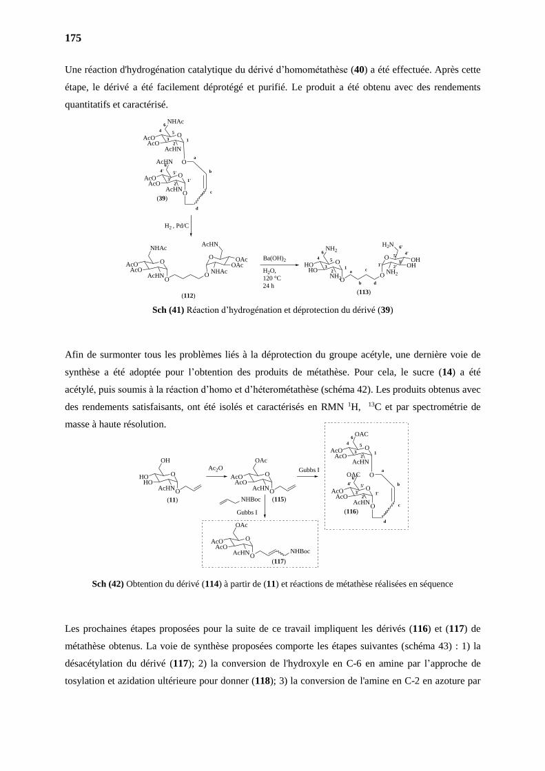
175
Une réaction d'hydrogénation catalytique du dérivé d’homométathèse (40) a été effectuée. Après cette
étape, le dérivé a été facilement déprotégé et purifié. Le produit a été obtenu avec des rendements
quantitatifs et caractérisé.
Sch (41) Réaction d’hydrogénation et déprotection du dérivé (39)
Afin de surmonter tous les problèmes liés à la déprotection du groupe acétyle, une dernière voie de
synthèse a été adoptée pour l’obtention des produits de métathèse. Pour cela, le sucre (14) a été
acétylé, puis soumis à la réaction d’homo et d’héterométathèse (schéma 42). Les produits obtenus avec
des rendements satisfaisants, ont été isolés et caractérisés en RMN 1H, 13C et par spectrométrie de
masse à haute résolution.
Sch (42) Obtention du dérivé (114) à partir de (11) et réactions de métathèse réalisées en séquence
Les prochaines étapes proposées pour la suite de ce travail impliquent les dérivés (116) et (117) de
métathèse obtenus. La voie de synthèse proposées comporte les étapes suivantes (schéma 43) : 1) la
désacétylation du dérivé (117); 2) la conversion de l'hydroxyle en C-6 en amine par l’approche de
tosylation et azidation ultérieure pour donner (118); 3) la conversion de l'amine en C-2 en azoture par
H2 , Pd/C
(112)
O
NHAc
AcOAcO
AcHNO
O
O
AcHN
OAcOAc
NHAc a
b
c12
4 5
6
3O
NH2
HOHO
NH2O
(113)
O
O
H2N
OHOH
NH2
d
1' 2'3'
4'
6'
5'Ba(OH)2
H2O,
120 °C
24 h
(39)
O
NHAc
AcOAcO
AcHN
O
12
4 5
6
3
O
AcHN
AcOAcO
AcHNO
1'2'
4' 5'
6'
3'
a
b
c
d
(116)
O
OAC
AcOAcO
AcHN
O
12
4 5
6
3
O
OAC
AcOAcO
AcHNO
1'2'
4' 5'
6'
3'
a
b
c
d
O
OAc
AcOAcO
AcHN
(117)O
NHBoc
O
OH
HOHO
AcHN
O
OAc
AcOAcO
AcHN
(11) (115)
Ac2O Gubbs I
Gubbs I
NHBoc
O O
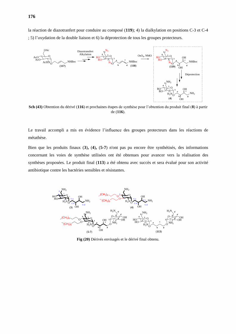
176
la réaction de diazotranfert pour conduire au composé (119); 4) la dialkylation en positions C-3 et C-4
; 5) l’oxydation de la double liaison et 6) la déprotection de tous les groupes protecteurs.
Sch (43) Obtention du dérivé (116) et prochaines étapes de synthèse pour l’obtention du produit final (8) à partir
de (116).
Le travail accompli a mis en évidence l’influence des groupes protecteurs dans les réactions de
métathèse.
Bien que les produits finaux (3), (4), (5-7) n'ont pas pu encore être synthétisés, des informations
concernant les voies de synthèse utilisées ont été obtenues pour avancer vers la réalisation des
synthèses proposées. Le produit final (113) a été obtenu avec succès et sera évalué pour son activité
antibiotique contre les bactéries sensibles et résistantes.
Fig (20) Dérivés envisagés et le dérivé final obtenu.
O
NH2
OO
H2N
O
NH2
HOHO
H2N
C-d
(3) (4)
ONH2
OH
OH
ONH2
OH
OH(CH2)6
(CH2)6
C-a
C-b
C-c
C-d
3
4
a
b
c1
2
4 5
6
3O
NH2
OO
H2NO
(5-7)
O
O
H2N
OHOH
NH2
d
1' 2'3'
4'
6'
5'
OH
OH
C-a
C-b
3
4 5
6
1
2
(CH2)6
(CH2)6
a
b
c12
4 5
6
3O
NH2
HOHO
H2NO
(113)
O
O
H2N
OHOH
NH2
d
1' 2'3'
4'
6'
5'
O
OAc
AcOAcO
AcHN
O
N3
RORO
N3
O
N3
RORO
N3
(117) (118) (119)
O
NH2
RORO
H2N
(4)
O ONHBoc NHBoc
ONHBoc
ONH2
OH
OH
OH
OH
a
b
c1
4 5
6
3
d
a
b
c1
4 5
6
3
d
2
2
a
b
c1
4 5
6
3
d
2
OsO4, NMO
DiazotransfertAlkylation
Déprotection
177
PARTIE EXPERIMENTALE
PARTIE II
PARTIE 1. MATERIAL AND METHODS
Thin-layer chromatography (TLC) was performed on aluminum backed silica plates (Alugram Sil
G/UV254 from Macherey–Nagel with fluorescent indicator UV254). Purifications were perfomed by
flash chromatography in silica gel 60, 200–400 mesh from Merck or in Reveleris® Flash
Chromatography System with GraceResolv® Flash cartidges or Reveleris® cartridges. Data for 1H,
13C NMR spectra were recorded on Brüker AC-400 instrument (400 MHz for 1H and 100 MHz for
13C) or Brüker 500 instrument (500 MHz for 1H and 126 MHz for 13C) and are internally referenced to
residual solvent signals. Data for 1H NMR are reported as follows: chemical shift ( ppm), integration,
multiplicity (s = singlet, d = doublet, t = triplet, q = quartet, m = multiplet, bs = broad singlet),
coupling constant and assignment. Interpretation of 1H and 13C NMR spectra often needed the support
of 2D experiments COSY, HMQC and HMBC records. Otherwise stated, protons and carbons of
purine and derivatives were numbered as below when associated with 1H and 13C NMR signals.
Electrospray ionization ESI mass was acquired by the analytical service of Institut de Chimie
Organique et Analytique at Orleans, France, using a HRMS Q-Tof MaXis spectrometer.
Unless otherwise specified, chemicals were purchased and used without further purification.
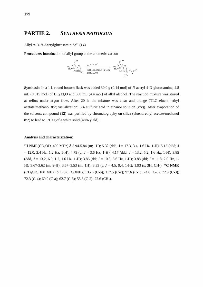
179
PARTIE 2. SYNTHESIS PROTOCOLS
Allyl -D-N-Acetylglucosaminide51 (14)
Procedure: Introduction of allyl group at the anomeric carbon
Synthesis: In a 1 L round bottom flask was added 30.0 g (0.14 mol) of N-acetyl-4-D-glucosamine, 4.8
mL (0.015 mol) of BF3.Et2O and 300 mL (4.4 mol) of allyl alcohol. The reaction mixture was stirred
at reflux under argon flow. After 20 h, the mixture was clear and orange (TLC eluent: ethyl
acetate/methanol 8:2; visualization: 5% sulfuric acid in ethanol solution (v/v)). After evaporation of
the solvent, compound (12) was purified by chromatography on silica (eluent: ethyl acetate/methanol
8:2) to lead to 19.0 g of a white solid (48% yield).
Analysis and characterization:
1H NMR(CD3OD, 400 MHz) 5.94-5.84 (m; 1H); 5.32 (ddd; J = 17.3, 3.4, 1.6 Hz, 1-H); 5.15 (ddd; J
= 12.0, 3.4 Hz; 1.2 Hz, 1-H); 4.79 (d, J = 3.6 Hz; 1-H); 4.17 (ddd, J = 13.2, 5.2, 1.6 Hz; 1-H); 3.85
(ddd, J = 13.2, 6.0, 1.2, 1.6 Hz; 1-H); 3.86 (dd; J = 10.8, 3.6 Hz, 1-H); 3.88 (dd; J = 11.8, 2.0 Hz, 1-
H); 3.67-3.62 (m; 2-H); 3.57–3.53 (m; 1H); 3.33 (t; J = 4.5, 9.4, 1-H); 1.93 (s; 3H, CH3). 13C NMR
(CD3OD, 100 MHz) 173.6 (CONH); 135.6 (C-b); 117.5 (C-c); 97.6 (C-1); 74.0 (C-5); 72.9 (C-3);
72.3 (C-4); 69.9 (C-a); 62.7 (C-6); 55.3 (C-2); 22.6 (CH3).
O
OH
HOHO
AcHNOH
O
OH
HOHO
AcHNO
HO
1) BF3Et2O (0.3 eq.), 2h
2) HCl, 20h
a
b
c1
2
4 5
6
3
(12)
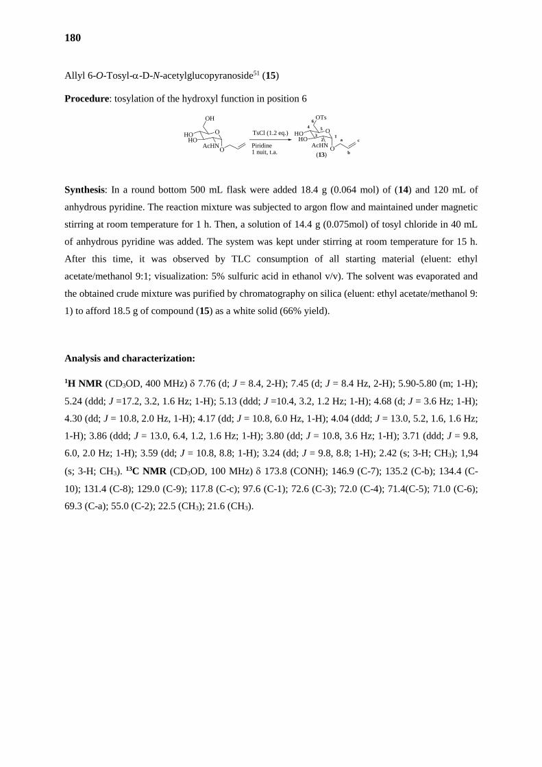
180
Allyl 6-O-Tosyl--D-N-acetylglucopyranoside51 (15)
Procedure: tosylation of the hydroxyl function in position 6
Synthesis: In a round bottom 500 mL flask were added 18.4 g (0.064 mol) of (14) and 120 mL of
anhydrous pyridine. The reaction mixture was subjected to argon flow and maintained under magnetic
stirring at room temperature for 1 h. Then, a solution of 14.4 g (0.075mol) of tosyl chloride in 40 mL
of anhydrous pyridine was added. The system was kept under stirring at room temperature for 15 h.
After this time, it was observed by TLC consumption of all starting material (eluent: ethyl
acetate/methanol 9:1; visualization: 5% sulfuric acid in ethanol v/v). The solvent was evaporated and
the obtained crude mixture was purified by chromatography on silica (eluent: ethyl acetate/methanol 9:
1) to afford 18.5 g of compound (15) as a white solid (66% yield).
Analysis and characterization:
1H NMR (CD3OD, 400 MHz) 7.76 (d; J = 8.4, 2-H); 7.45 (d; J = 8.4 Hz, 2-H); 5.90-5.80 (m; 1-H);
5.24 (ddd; J =17.2, 3.2, 1.6 Hz; 1-H); 5.13 (ddd; J =10.4, 3.2, 1.2 Hz; 1-H); 4.68 (d; J = 3.6 Hz; 1-H);
4.30 (dd; J = 10.8, 2.0 Hz, 1-H); 4.17 (dd; J = 10.8, 6.0 Hz, 1-H); 4.04 (ddd; J = 13.0, 5.2, 1.6, 1.6 Hz;
1-H); 3.86 (ddd; J = 13.0, 6.4, 1.2, 1.6 Hz; 1-H); 3.80 (dd; J = 10.8, 3.6 Hz; 1-H); 3.71 (ddd; J = 9.8,
6.0, 2.0 Hz; 1-H); 3.59 (dd; J = 10.8, 8.8; 1-H); 3.24 (dd; J = 9.8, 8.8; 1-H); 2.42 (s; 3-H; CH3); 1,94
(s; 3-H; CH3). 13C NMR (CD3OD, 100 MHz) 173.8 (CONH); 146.9 (C-7); 135.2 (C-b); 134.4 (C-
10); 131.4 (C-8); 129.0 (C-9); 117.8 (C-c); 97.6 (C-1); 72.6 (C-3); 72.0 (C-4); 71.4(C-5); 71.0 (C-6);
69.3 (C-a); 55.0 (C-2); 22.5 (CH3); 21.6 (CH3).
O
OH
HOHO
AcHNO
Piridine1 nuit, t.a.
O
OTs
HOHO
AcHNO
a
b
c1
2
4 5
6
3TsCl (1.2 eq.)
(13)
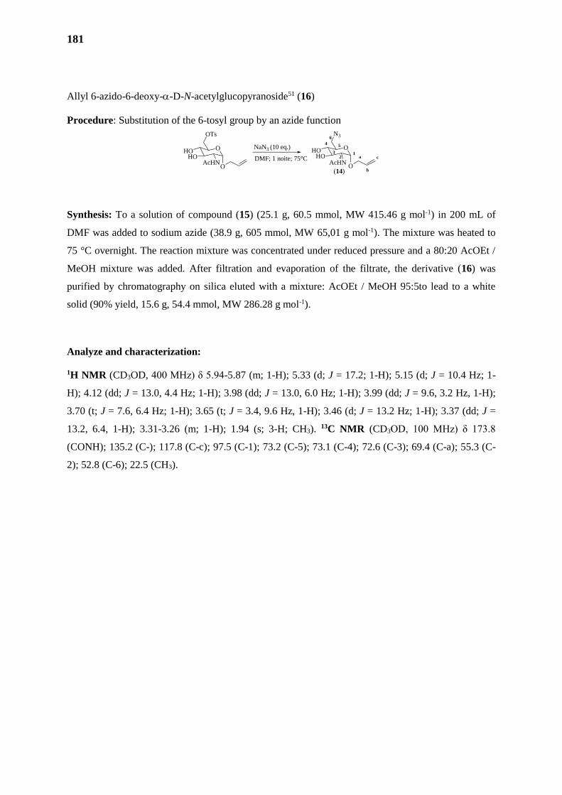
181
Allyl 6-azido-6-deoxy--D-N-acetylglucopyranoside51 (16)
Procedure: Substitution of the 6-tosyl group by an azide function
Synthesis: To a solution of compound (15) (25.1 g, 60.5 mmol, MW 415.46 g mol-1) in 200 mL of
DMF was added to sodium azide (38.9 g, 605 mmol, MW 65,01 g mol-1). The mixture was heated to
75 °C overnight. The reaction mixture was concentrated under reduced pressure and a 80:20 AcOEt /
MeOH mixture was added. After filtration and evaporation of the filtrate, the derivative (16) was
purified by chromatography on silica eluted with a mixture: AcOEt / MeOH 95:5to lead to a white
solid (90% yield, 15.6 g, 54.4 mmol, MW 286.28 g mol-1).
Analyze and characterization:
1H NMR (CD3OD, 400 MHz) δ 5.94-5.87 (m; 1-H); 5.33 (d; J = 17.2; 1-H); 5.15 (d; J = 10.4 Hz; 1-
H); 4.12 (dd; J = 13.0, 4.4 Hz; 1-H); 3.98 (dd; J = 13.0, 6.0 Hz; 1-H); 3.99 (dd; J = 9.6, 3.2 Hz, 1-H);
3.70 (t; J = 7.6, 6.4 Hz; 1-H); 3.65 (t; J = 3.4, 9.6 Hz, 1-H); 3.46 (d; J = 13.2 Hz; 1-H); 3.37 (dd; J =
13.2, 6.4, 1-H); 3.31-3.26 (m; 1-H); 1.94 (s; 3-H; CH3). 13C NMR (CD3OD, 100 MHz) δ 173.8
(CONH); 135.2 (C-); 117.8 (C-c); 97.5 (C-1); 73.2 (C-5); 73.1 (C-4); 72.6 (C-3); 69.4 (C-a); 55.3 (C-
2); 52.8 (C-6); 22.5 (CH3).
O
OTs
HOHO
AcHN
NaN3 (10 eq.)
DMF; 1 noite; 75°C
O
O
N3
HOHO
AcHNO
a
b
c1
2
4 5
6
3
(14)
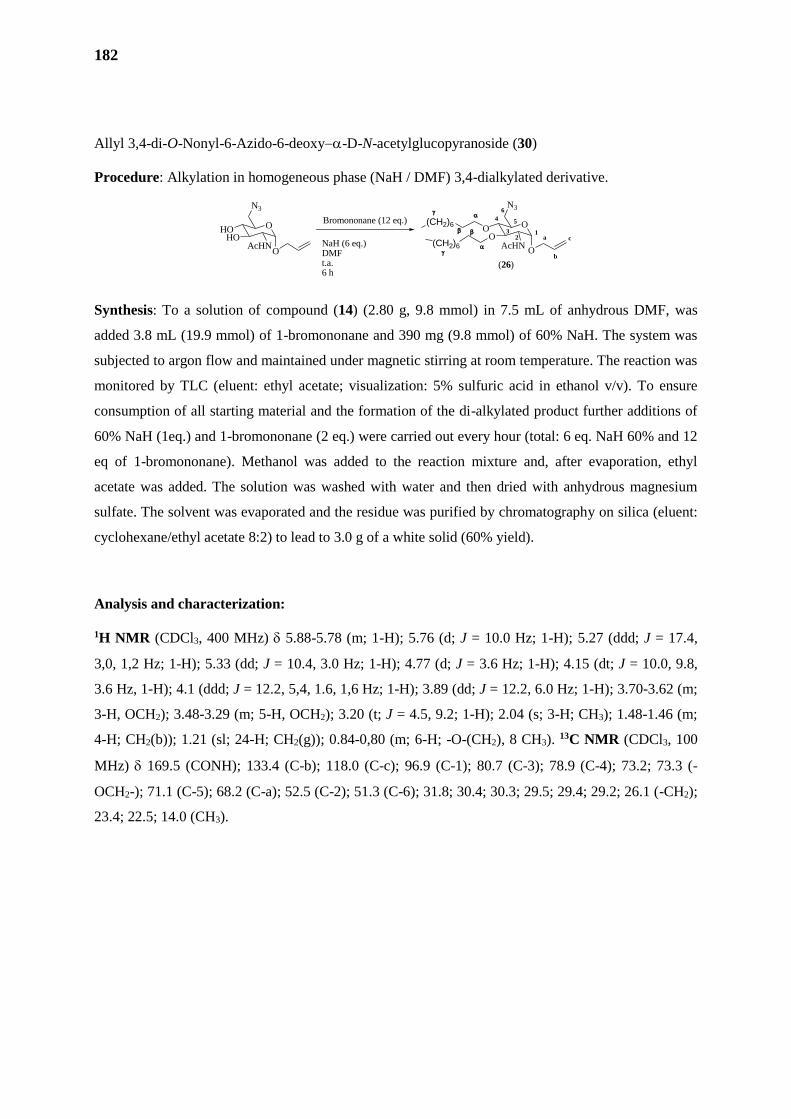
182
Allyl 3,4-di-O-Nonyl-6-Azido-6-deoxy-D-N-acetylglucopyranoside (30)
Procedure: Alkylation in homogeneous phase (NaH / DMF) 3,4-dialkylated derivative.
Synthesis: To a solution of compound (14) (2.80 g, 9.8 mmol) in 7.5 mL of anhydrous DMF, was
added 3.8 mL (19.9 mmol) of 1-bromononane and 390 mg (9.8 mmol) of 60% NaH. The system was
subjected to argon flow and maintained under magnetic stirring at room temperature. The reaction was
monitored by TLC (eluent: ethyl acetate; visualization: 5% sulfuric acid in ethanol v/v). To ensure
consumption of all starting material and the formation of the di-alkylated product further additions of
60% NaH (1eq.) and 1-bromononane (2 eq.) were carried out every hour (total: 6 eq. NaH 60% and 12
eq of 1-bromononane). Methanol was added to the reaction mixture and, after evaporation, ethyl
acetate was added. The solution was washed with water and then dried with anhydrous magnesium
sulfate. The solvent was evaporated and the residue was purified by chromatography on silica (eluent:
cyclohexane/ethyl acetate 8:2) to lead to 3.0 g of a white solid (60% yield).
Analysis and characterization:
1H NMR (CDCl3, 400 MHz) 5.88-5.78 (m; 1-H); 5.76 (d; J = 10.0 Hz; 1-H); 5.27 (ddd; J = 17.4,
3,0, 1,2 Hz; 1-H); 5.33 (dd; J = 10.4, 3.0 Hz; 1-H); 4.77 (d; J = 3.6 Hz; 1-H); 4.15 (dt; J = 10.0, 9.8,
3.6 Hz, 1-H); 4.1 (ddd; J = 12.2, 5,4, 1.6, 1,6 Hz; 1-H); 3.89 (dd; J = 12.2, 6.0 Hz; 1-H); 3.70-3.62 (m;
3-H, OCH2); 3.48-3.29 (m; 5-H, OCH2); 3.20 (t; J = 4.5, 9.2; 1-H); 2.04 (s; 3-H; CH3); 1.48-1.46 (m;
4-H; CH2(b)); 1.21 (sl; 24-H; CH2(g)); 0.84-0,80 (m; 6-H; -O-(CH2), 8 CH3). 13C NMR (CDCl3, 100
MHz) 169.5 (CONH); 133.4 (C-b); 118.0 (C-c); 96.9 (C-1); 80.7 (C-3); 78.9 (C-4); 73.2; 73.3 (-
OCH2-); 71.1 (C-5); 68.2 (C-a); 52.5 (C-2); 51.3 (C-6); 31.8; 30.4; 30.3; 29.5; 29.4; 29.2; 26.1 (-CH2);
23.4; 22.5; 14.0 (CH3).
O
N3
OO
AcHNO
a
b
c1
2
4 5
6
3
(CH2)6
(CH2)6
Bromononane (12 eq.)
NaH (6 eq.)DMFt.a.6 h
O
N3
HOHO
AcHNO
(26)
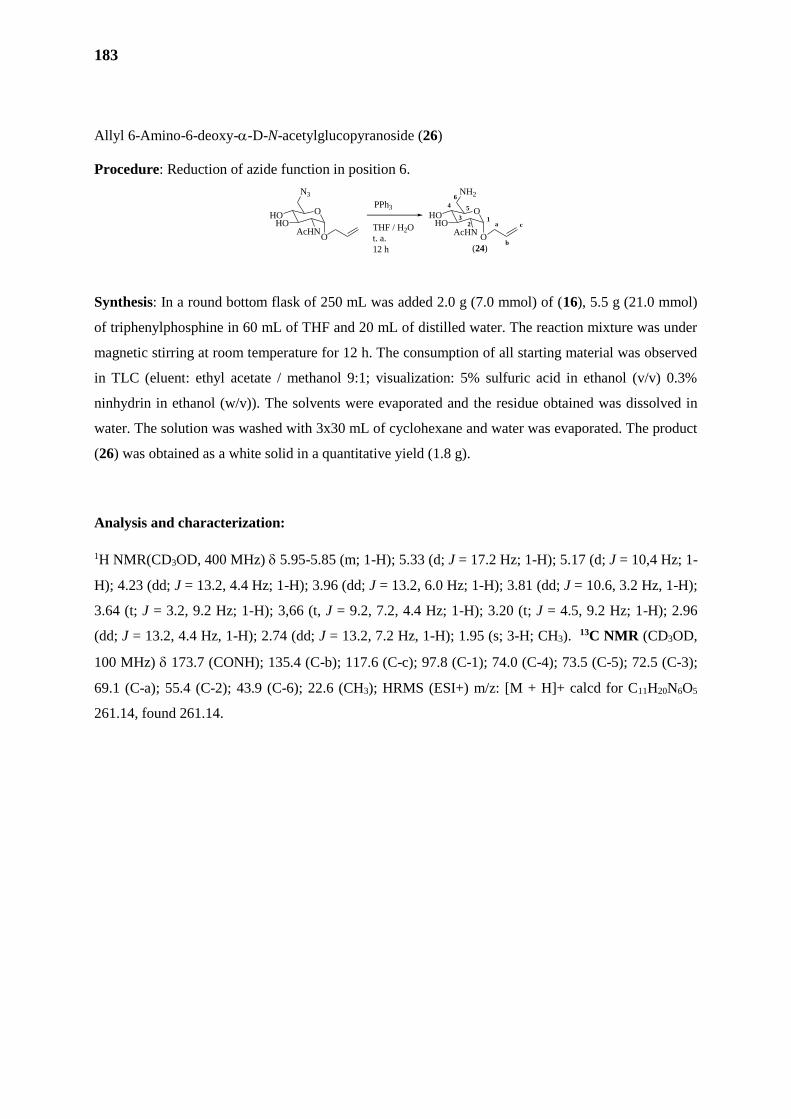
183
Allyl 6-Amino-6-deoxy--D-N-acetylglucopyranoside (26)
Procedure: Reduction of azide function in position 6.
Synthesis: In a round bottom flask of 250 mL was added 2.0 g (7.0 mmol) of (16), 5.5 g (21.0 mmol)
of triphenylphosphine in 60 mL of THF and 20 mL of distilled water. The reaction mixture was under
magnetic stirring at room temperature for 12 h. The consumption of all starting material was observed
in TLC (eluent: ethyl acetate / methanol 9:1; visualization: 5% sulfuric acid in ethanol (v/v) 0.3%
ninhydrin in ethanol (w/v)). The solvents were evaporated and the residue obtained was dissolved in
water. The solution was washed with 3x30 mL of cyclohexane and water was evaporated. The product
(26) was obtained as a white solid in a quantitative yield (1.8 g).
Analysis and characterization:
1H NMR(CD3OD, 400 MHz) 5.95-5.85 (m; 1-H); 5.33 (d; J = 17.2 Hz; 1-H); 5.17 (d; J = 10,4 Hz; 1-
H); 4.23 (dd; J = 13.2, 4.4 Hz; 1-H); 3.96 (dd; J = 13.2, 6.0 Hz; 1-H); 3.81 (dd; J = 10.6, 3.2 Hz, 1-H);
3.64 (t; J = 3.2, 9.2 Hz; 1-H); 3,66 (t, J = 9.2, 7.2, 4.4 Hz; 1-H); 3.20 (t; J = 4.5, 9.2 Hz; 1-H); 2.96
(dd; J = 13.2, 4.4 Hz, 1-H); 2.74 (dd; J = 13.2, 7.2 Hz, 1-H); 1.95 (s; 3-H; CH3). 13C NMR (CD3OD,
100 MHz) 173.7 (CONH); 135.4 (C-b); 117.6 (C-c); 97.8 (C-1); 74.0 (C-4); 73.5 (C-5); 72.5 (C-3);
69.1 (C-a); 55.4 (C-2); 43.9 (C-6); 22.6 (CH3); HRMS (ESI+) m/z: [M + H]+ calcd for C11H20N6O5
261.14, found 261.14.
O
N3
HOHO
AcHNO
PPh3
THF / H2O
t. a.
12 h
O
NH2
HOHO
AcHNO
a
b
c1
2
4 5
6
3
(24)
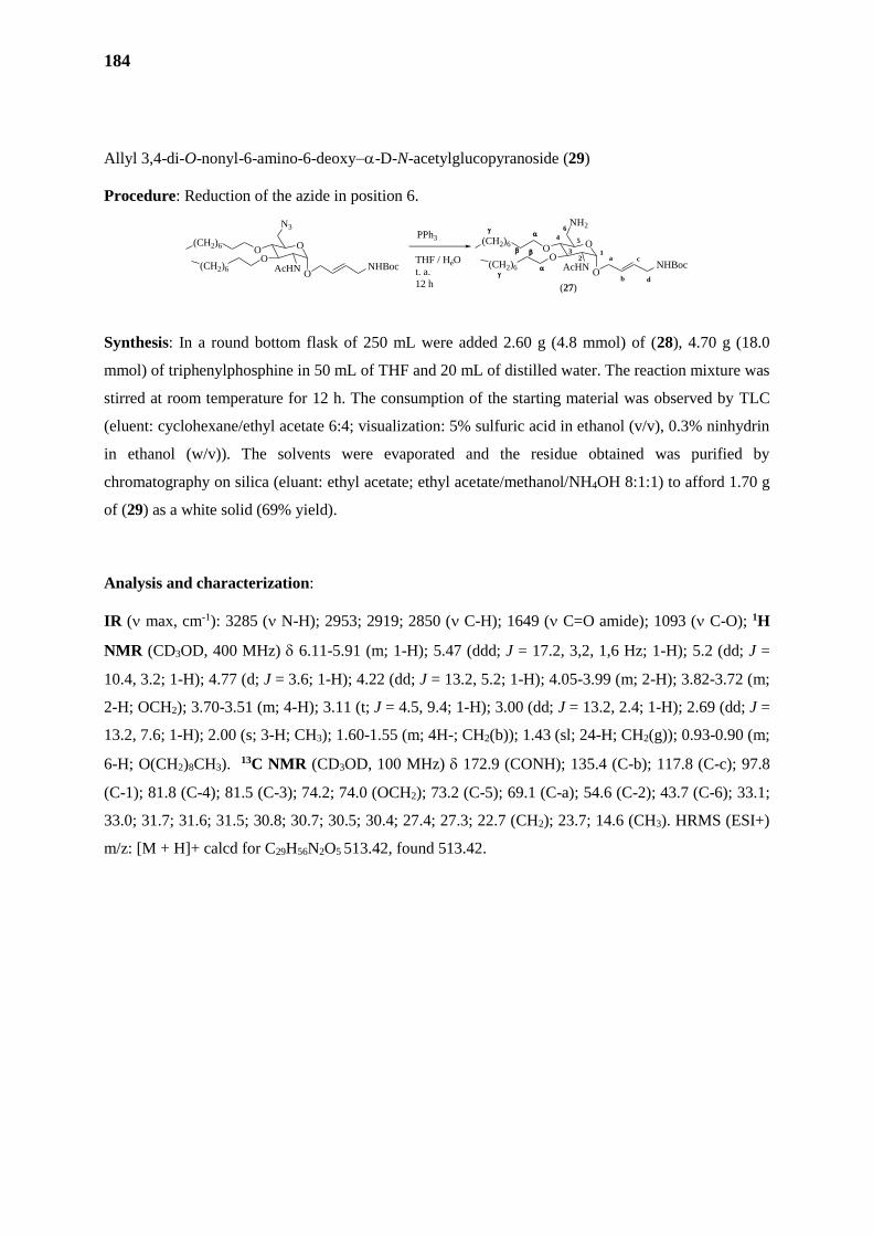
184
Allyl 3,4-di-O-nonyl-6-amino-6-deoxy-D-N-acetylglucopyranoside (29)
Procedure: Reduction of the azide in position 6.
Synthesis: In a round bottom flask of 250 mL were added 2.60 g (4.8 mmol) of (28), 4.70 g (18.0
mmol) of triphenylphosphine in 50 mL of THF and 20 mL of distilled water. The reaction mixture was
stirred at room temperature for 12 h. The consumption of the starting material was observed by TLC
(eluent: cyclohexane/ethyl acetate 6:4; visualization: 5% sulfuric acid in ethanol (v/v), 0.3% ninhydrin
in ethanol (w/v)). The solvents were evaporated and the residue obtained was purified by
chromatography on silica (eluant: ethyl acetate; ethyl acetate/methanol/NH4OH 8:1:1) to afford 1.70 g
of (29) as a white solid (69% yield).
Analysis and characterization:
IR ( max, cm-1): 3285 ( N-H); 2953; 2919; 2850 ( C-H); 1649 ( C=O amide); 1093 ( C-O); 1H
NMR (CD3OD, 400 MHz) 6.11-5.91 (m; 1-H); 5.47 (ddd; J = 17.2, 3,2, 1,6 Hz; 1-H); 5.2 (dd; J =
10.4, 3.2; 1-H); 4.77 (d; J = 3.6; 1-H); 4.22 (dd; J = 13.2, 5.2; 1-H); 4.05-3.99 (m; 2-H); 3.82-3.72 (m;
2-H; OCH2); 3.70-3.51 (m; 4-H); 3.11 (t; J = 4.5, 9.4; 1-H); 3.00 (dd; J = 13.2, 2.4; 1-H); 2.69 (dd; J =
13.2, 7.6; 1-H); 2.00 (s; 3-H; CH3); 1.60-1.55 (m; 4H-; CH2(b)); 1.43 (sl; 24-H; CH2(g)); 0.93-0.90 (m;
6-H; O(CH2)8CH3). 13C NMR (CD3OD, 100 MHz) 172.9 (CONH); 135.4 (C-b); 117.8 (C-c); 97.8
(C-1); 81.8 (C-4); 81.5 (C-3); 74.2; 74.0 (OCH2); 73.2 (C-5); 69.1 (C-a); 54.6 (C-2); 43.7 (C-6); 33.1;
33.0; 31.7; 31.6; 31.5; 30.8; 30.7; 30.5; 30.4; 27.4; 27.3; 22.7 (CH2); 23.7; 14.6 (CH3). HRMS (ESI+)
m/z: [M + H]+ calcd for C29H56N2O5 513.42, found 513.42.
PPh3
THF / HéO
t. a.
12 h
O
N3
OO
AcHNO
(CH2)6
(CH2)6 NHBoc
O
NH2
OO
AcHNO
a
b
c1
2
4 5
6
3
(CH2)6
(CH2)6
(27)
NHBoc
d
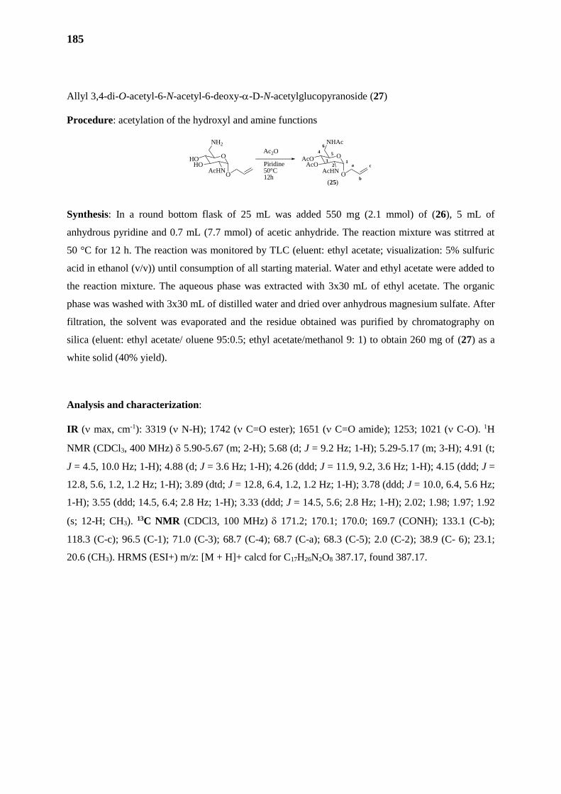
185
Allyl 3,4-di-O-acetyl-6-N-acetyl-6-deoxy--D-N-acetylglucopyranoside (27)
Procedure: acetylation of the hydroxyl and amine functions
Synthesis: In a round bottom flask of 25 mL was added 550 mg (2.1 mmol) of (26), 5 mL of
anhydrous pyridine and 0.7 mL (7.7 mmol) of acetic anhydride. The reaction mixture was stitrred at
50 °C for 12 h. The reaction was monitored by TLC (eluent: ethyl acetate; visualization: 5% sulfuric
acid in ethanol (v/v)) until consumption of all starting material. Water and ethyl acetate were added to
the reaction mixture. The aqueous phase was extracted with 3x30 mL of ethyl acetate. The organic
phase was washed with 3x30 mL of distilled water and dried over anhydrous magnesium sulfate. After
filtration, the solvent was evaporated and the residue obtained was purified by chromatography on
silica (eluent: ethyl acetate/ oluene 95:0.5; ethyl acetate/methanol 9: 1) to obtain 260 mg of (27) as a
white solid (40% yield).
Analysis and characterization:
IR ( max, cm-1): 3319 ( N-H); 1742 ( C=O ester); 1651 ( C=O amide); 1253; 1021 ( C-O). 1H
NMR (CDCl3, 400 MHz) 5.90-5.67 (m; 2-H); 5.68 (d; J = 9.2 Hz; 1-H); 5.29-5.17 (m; 3-H); 4.91 (t;
J = 4.5, 10.0 Hz; 1-H); 4.88 (d; J = 3.6 Hz; 1-H); 4.26 (ddd; J = 11.9, 9.2, 3.6 Hz; 1-H); 4.15 (ddd; J =
12.8, 5.6, 1.2, 1.2 Hz; 1-H); 3.89 (dtd; J = 12.8, 6.4, 1.2, 1.2 Hz; 1-H); 3.78 (ddd; J = 10.0, 6.4, 5.6 Hz;
1-H); 3.55 (ddd; 14.5, 6.4; 2.8 Hz; 1-H); 3.33 (ddd; J = 14.5, 5.6; 2.8 Hz; 1-H); 2.02; 1.98; 1.97; 1.92
(s; 12-H; CH3). 13C NMR (CDCl3, 100 MHz) 171.2; 170.1; 170.0; 169.7 (CONH); 133.1 (C-b);
118.3 (C-c); 96.5 (C-1); 71.0 (C-3); 68.7 (C-4); 68.7 (C-a); 68.3 (C-5); 2.0 (C-2); 38.9 (C- 6); 23.1;
20.6 (CH3). HRMS (ESI+) m/z: [M + H]+ calcd for C17H26N2O8 387.17, found 387.17.
O
NH2
HOHO
AcHNO
Ac2O
Piridine50°C12h
O
NHAc
AcOAcO
AcHNO
a
b
c1
2
4 5
6
3
(25)
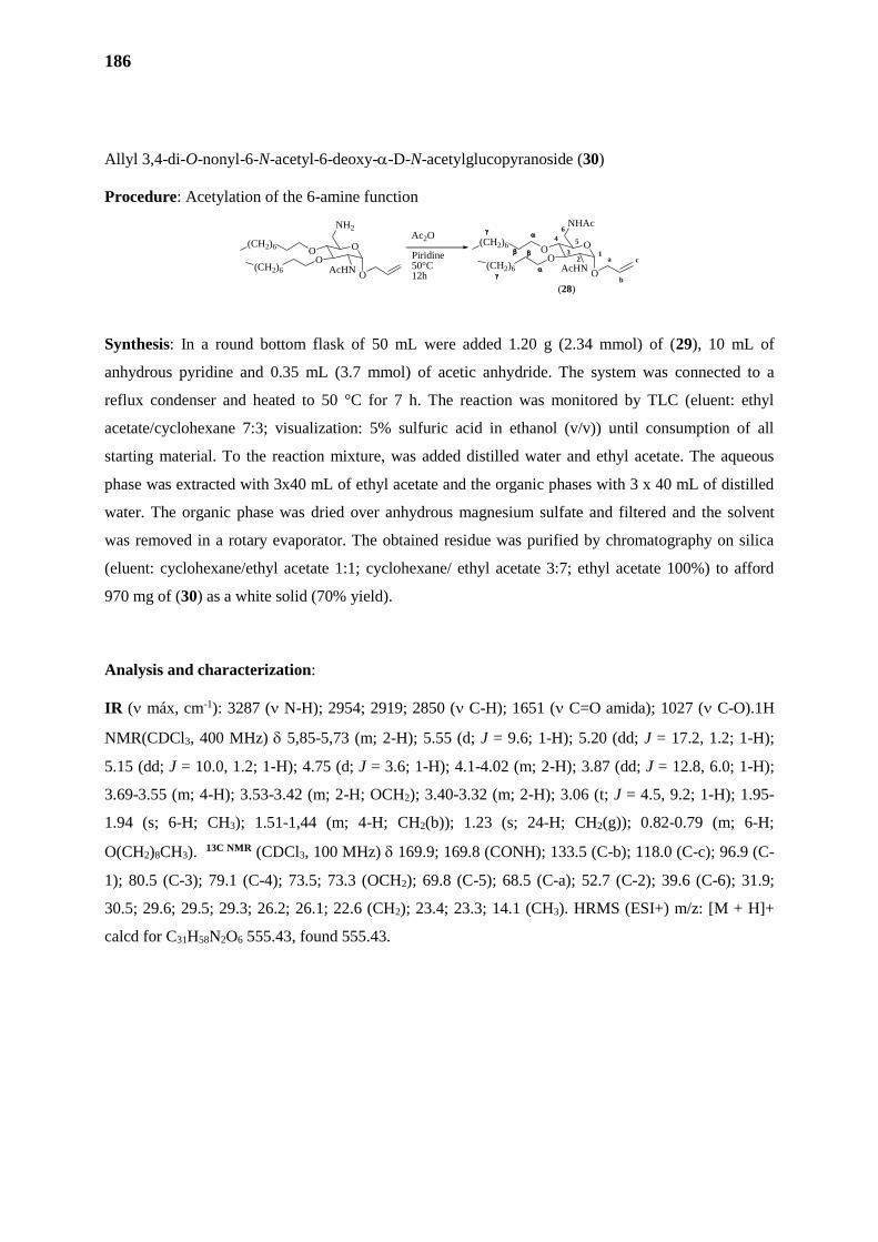
186
Allyl 3,4-di-O-nonyl-6-N-acetyl-6-deoxy--D-N-acetylglucopyranoside (30)
Procedure: Acetylation of the 6-amine function
Synthesis: In a round bottom flask of 50 mL were added 1.20 g (2.34 mmol) of (29), 10 mL of
anhydrous pyridine and 0.35 mL (3.7 mmol) of acetic anhydride. The system was connected to a
reflux condenser and heated to 50 °C for 7 h. The reaction was monitored by TLC (eluent: ethyl
acetate/cyclohexane 7:3; visualization: 5% sulfuric acid in ethanol (v/v)) until consumption of all
starting material. To the reaction mixture, was added distilled water and ethyl acetate. The aqueous
phase was extracted with 3x40 mL of ethyl acetate and the organic phases with 3 x 40 mL of distilled
water. The organic phase was dried over anhydrous magnesium sulfate and filtered and the solvent
was removed in a rotary evaporator. The obtained residue was purified by chromatography on silica
(eluent: cyclohexane/ethyl acetate 1:1; cyclohexane/ ethyl acetate 3:7; ethyl acetate 100%) to afford
970 mg of (30) as a white solid (70% yield).
Analysis and characterization:
IR ( máx, cm-1): 3287 ( N-H); 2954; 2919; 2850 ( C-H); 1651 ( C=O amida); 1027 ( C-O).1H
NMR(CDCl3, 400 MHz) 5,85-5,73 (m; 2-H); 5.55 (d; J = 9.6; 1-H); 5.20 (dd; J = 17.2, 1.2; 1-H);
5.15 (dd; J = 10.0, 1.2; 1-H); 4.75 (d; J = 3.6; 1-H); 4.1-4.02 (m; 2-H); 3.87 (dd; J = 12.8, 6.0; 1-H);
3.69-3.55 (m; 4-H); 3.53-3.42 (m; 2-H; OCH2); 3.40-3.32 (m; 2-H); 3.06 (t; J = 4.5, 9.2; 1-H); 1.95-
1.94 (s; 6-H; CH3); 1.51-1,44 (m; 4-H; CH2(b)); 1.23 (s; 24-H; CH2(g)); 0.82-0.79 (m; 6-H;
O(CH2)8CH3). 13C NMR (CDCl3, 100 MHz) 169.9; 169.8 (CONH); 133.5 (C-b); 118.0 (C-c); 96.9 (C-
1); 80.5 (C-3); 79.1 (C-4); 73.5; 73.3 (OCH2); 69.8 (C-5); 68.5 (C-a); 52.7 (C-2); 39.6 (C-6); 31.9;
30.5; 29.6; 29.5; 29.3; 26.2; 26.1; 22.6 (CH2); 23.4; 23.3; 14.1 (CH3). HRMS (ESI+) m/z: [M + H]+
calcd for C31H58N2O6 555.43, found 555.43.
O
NH2
OO
AcHNO
(CH2)6
(CH2)6
O
NHAc
OO
AcHNO
a
b
c1
2
4 5
6
3
(CH2)6
(CH2)6
(28)
Ac2O
Piridine50°C12h

187
1-[But-4’-amino-(tert-butyl-carbonate)-2’-E-enyl]-2,6-di-N-acetyl-2,6-dideoxy-3,4-di-O-acetyl--D-
N-acetylglucopyranoside (38)
Procedure: Procedure: Heterometathesis in the presence of the Grubbs I catalyst
Synthesis: In a 25 mL round bottom flask, 300 mg (0.78 mmol) of (27) were solubilized in 5 mL of
distilled DCM. Then 250 mg (1.6 mmol) of the protected allylamine (38) was added. The system was
fitted to a reflux condenser, and subjected to argon flow under magnetic stirring for 20 min. 160 mg
(0.20 mmol) of Grubbs I catalyst were added to the reaction mixture and the system was heated at
reflux. The reaction was followed by TLC (eluent: ethyl acetate/methanol 9:1; visualization: 5%
sulfuric acid in ethanol (v/v)) and after 20 h, it was observed that the reaction was no longer evolving.
The solvent was evaporated and the crude residue obtained was purified by chromatography on silica
(eluent: 100% ethyl acetate, 9.5: 0.5 ethyl acetate / methanol). 42 mg of starting material (27) was
recovered at the end of the purification step and two products A and B were isolated. Product A was
characterized as derivative (38) and B was characterized as the homometathesis product (39). 145 mg
of (38) (36% yield) and 87 mg of (39) (30% yield).
Analysis and characterization (39):
1H NMR (CDCl3, 400 MHz) 5.71-5.56 (m; 2-H); 5.17 (dd; J = 9.6, 10.8 Hz; 1-H); 4.87 (t; J = 4.5,
9.6 Hz; 1-H); 4.62 (d; J = 3.6 Hz; 1-H); 4.23 (dd; J = 10.8, 3.6 Hz; 1-H); 4.06 (dd; J = 12.8, 4.8 Hz; 1-
H); 3.91 (dd; J = 12.8, 6.0 Hz; 1-H); 3.75 (ddd; J = 10.0, 5.6, 3.2 Hz; 1-H); 3.69 (d; J = 4.4; 2-H); 3.45
(dd; J = 14.6, 3.2 Hz, 1-H); 3.27 (dd; J = 14.6, 5.6, 1-H); 2.02; 1.98; 1.97; 1.92 (s; 12-H; CH3); 1.39 (s;
9-H; C(CH3)3). 13C NMR (CDCl3, 100 MHz) 171.3; 170.2; 170.0; 169.9 (CO); 155.8 (COC(CH3)3);
133.5 (C-b); 126.2 (C-c); 96.3 (C-1); 79.6 (C(CH3)3; 71.0 (C-3); 69.1 (C-4); 68.2 (C-5); 68.0 (C-a);
52.0 (C-2); 41.7 (C-d); 38.8 (C-6); 28.3 (C(CH3)3; 23.1; 23.0; 20.7, 20.6 (CH3). HRMS (ESI+) m/z:
[M + H]+ calcd for C23H37N3O10 516.25, found 516.25.
O
NHAc
AcOAcO
AcHNO
Grubbs I
CH2Cl2,
40 °C
20 h
O
NHAc
AcOAcO
AcHNO
a
b
c1
2
4 5
6
3
NHBocd
(38)
NHBoc
(37)
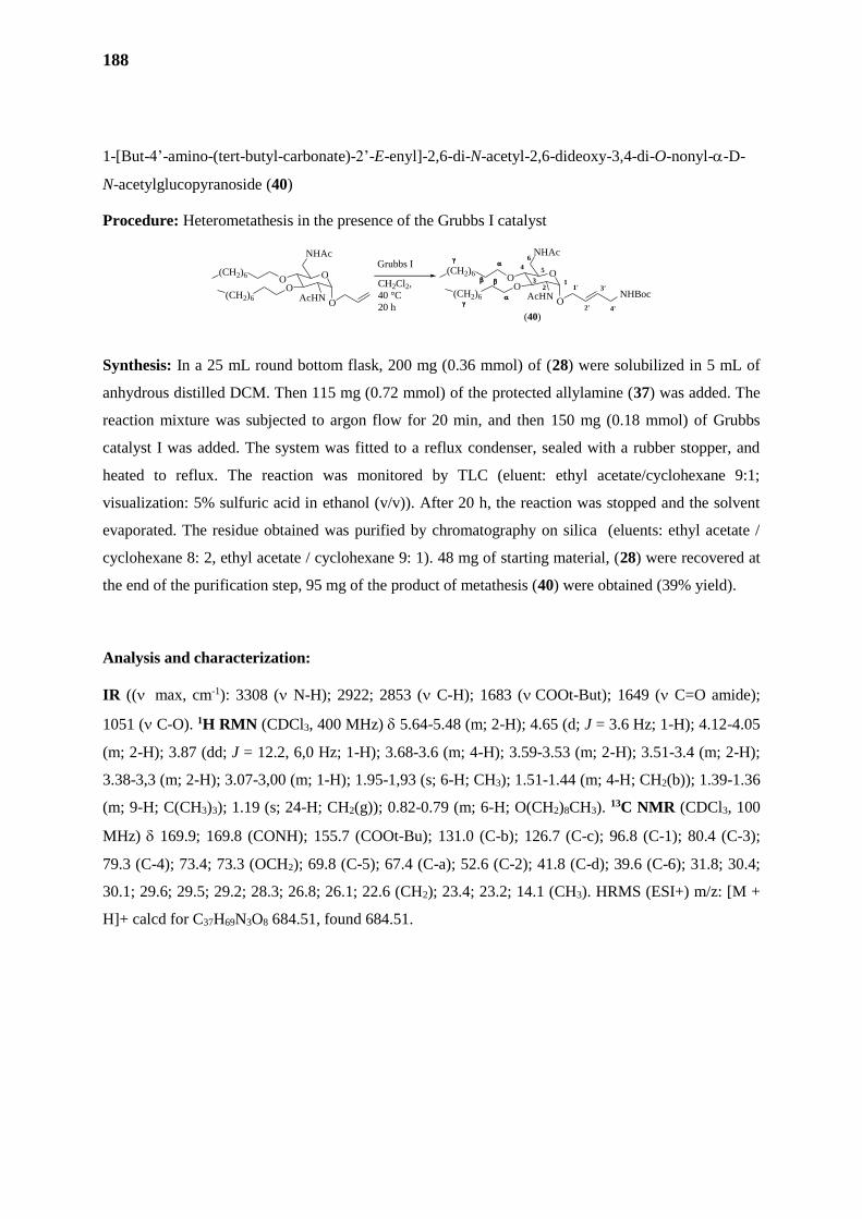
188
1-[But-4’-amino-(tert-butyl-carbonate)-2’-E-enyl]-2,6-di-N-acetyl-2,6-dideoxy-3,4-di-O-nonyl--D-
N-acetylglucopyranoside (40)
Procedure: Heterometathesis in the presence of the Grubbs I catalyst
Synthesis: In a 25 mL round bottom flask, 200 mg (0.36 mmol) of (28) were solubilized in 5 mL of
anhydrous distilled DCM. Then 115 mg (0.72 mmol) of the protected allylamine (37) was added. The
reaction mixture was subjected to argon flow for 20 min, and then 150 mg (0.18 mmol) of Grubbs
catalyst I was added. The system was fitted to a reflux condenser, sealed with a rubber stopper, and
heated to reflux. The reaction was monitored by TLC (eluent: ethyl acetate/cyclohexane 9:1;
visualization: 5% sulfuric acid in ethanol (v/v)). After 20 h, the reaction was stopped and the solvent
evaporated. The residue obtained was purified by chromatography on silica (eluents: ethyl acetate /
cyclohexane 8: 2, ethyl acetate / cyclohexane 9: 1). 48 mg of starting material, (28) were recovered at
the end of the purification step, 95 mg of the product of metathesis (40) were obtained (39% yield).
Analysis and characterization:
IR (( max, cm-1): 3308 ( N-H); 2922; 2853 ( C-H); 1683 (COOt-But); 1649 ( C=O amide);
1051 ( C-O). 1H RMN (CDCl3, 400 MHz) 5.64-5.48 (m; 2-H); 4.65 (d; J = 3.6 Hz; 1-H); 4.12-4.05
(m; 2-H); 3.87 (dd; J = 12.2, 6,0 Hz; 1-H); 3.68-3.6 (m; 4-H); 3.59-3.53 (m; 2-H); 3.51-3.4 (m; 2-H);
3.38-3,3 (m; 2-H); 3.07-3,00 (m; 1-H); 1.95-1,93 (s; 6-H; CH3); 1.51-1.44 (m; 4-H; CH2(b)); 1.39-1.36
(m; 9-H; C(CH3)3); 1.19 (s; 24-H; CH2(g)); 0.82-0.79 (m; 6-H; O(CH2)8CH3). 13C NMR (CDCl3, 100
MHz) 169.9; 169.8 (CONH); 155.7 (COOt-Bu); 131.0 (C-b); 126.7 (C-c); 96.8 (C-1); 80.4 (C-3);
79.3 (C-4); 73.4; 73.3 (OCH2); 69.8 (C-5); 67.4 (C-a); 52.6 (C-2); 41.8 (C-d); 39.6 (C-6); 31.8; 30.4;
30.1; 29.6; 29.5; 29.2; 28.3; 26.8; 26.1; 22.6 (CH2); 23.4; 23.2; 14.1 (CH3). HRMS (ESI+) m/z: [M +
H]+ calcd for C37H69N3O8 684.51, found 684.51.
O
NHAc
OO
AcHNO
(CH2)6
(CH2)6
O
NHAc
OO
AcHNO
1'
2'
3'1
2
4 5
6
3
(CH2)6
(CH2)6
(40)
NHBoc
4'
Grubbs I
CH2Cl2,
40 °C
20 h
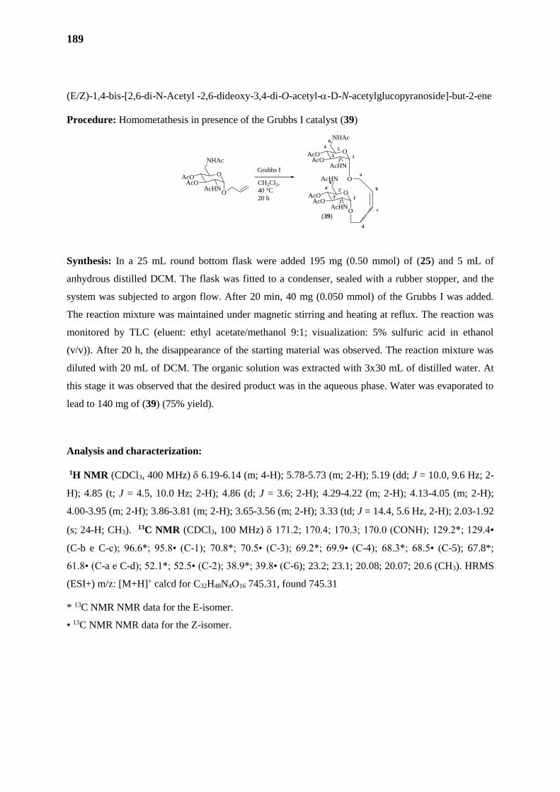
189
(E/Z)-1,4-bis-[2,6-di-N-Acetyl -2,6-dideoxy-3,4-di-O-acetyl--D-N-acetylglucopyranoside]-but-2-ene
Procedure: Homometathesis in presence of the Grubbs I catalyst (39)
Synthesis: In a 25 mL round bottom flask were added 195 mg (0.50 mmol) of (25) and 5 mL of
anhydrous distilled DCM. The flask was fitted to a condenser, sealed with a rubber stopper, and the
system was subjected to argon flow. After 20 min, 40 mg (0.050 mmol) of the Grubbs I was added.
The reaction mixture was maintained under magnetic stirring and heating at reflux. The reaction was
monitored by TLC (eluent: ethyl acetate/methanol 9:1; visualization: 5% sulfuric acid in ethanol
(v/v)). After 20 h, the disappearance of the starting material was observed. The reaction mixture was
diluted with 20 mL of DCM. The organic solution was extracted with 3x30 mL of distilled water. At
this stage it was observed that the desired product was in the aqueous phase. Water was evaporated to
lead to 140 mg of (39) (75% yield).
Analysis and characterization:
1H NMR (CDCl3, 400 MHz) 6.19-6.14 (m; 4-H); 5.78-5.73 (m; 2-H); 5.19 (dd; J = 10.0, 9.6 Hz; 2-
H); 4.85 (t; J = 4.5, 10.0 Hz; 2-H); 4.86 (d; J = 3.6; 2-H); 4.29-4.22 (m; 2-H); 4.13-4.05 (m; 2-H);
4.00-3.95 (m; 2-H); 3.86-3.81 (m; 2-H); 3.65-3.56 (m; 2-H); 3.33 (td; J = 14.4, 5.6 Hz, 2-H); 2.03-1.92
(s; 24-H; CH3). 13C NMR (CDCl3, 100 MHz) 171.2; 170.4; 170.3; 170.0 (CONH); 129.2*; 129.4•
(C-b e C-c); 96.6*; 95.8• (C-1); 70.8*; 70.5• (C-3); 69.2*; 69.9• (C-4); 68.3*; 68.5• (C-5); 67.8*;
61.8• (C-a e C-d); 52.1*; 52.5• (C-2); 38.9*; 39.8• (C-6); 23.2; 23.1; 20.08; 20.07; 20.6 (CH3). HRMS
(ESI+) m/z: [M+H]+ calcd for C32H48N4O16 745.31, found 745.31
* 13C NMR NMR data for the E-isomer.
• 13C NMR NMR data for the Z-isomer.
(39)
O
NHAc
AcOAcO
AcHN
O
12
4 5
6
3
O
AcHN
AcOAcO
AcHNO
1'2'
4' 5'
6'
3'
a
b
c
d
O
NHAc
AcOAcO
AcHNO
Grubbs I
CH2Cl2,
40 °C
20 h

190
(E/Z)-1,4-bis-[2,6-di-N-Acetyl-2,6-dideoxy-3,4-di-O-nonyl--D-N-acetylglucopyranoside]-but-2-ene
(41)
Procedure: Homometathesis in presence of the Grubbs I catalyst
Synthesis: Into a 10 mL round bottom flask were added 110 mg (0.20 mmol) of (30) and 3 mL of
anhydrous distilled DCM. The reaction mixture was subjected to argon flow and kept under magnetic
stirring at room temperature for 20 min. Thereafter, 30 mg (0.036 mmol) of the Grubbs I catalyst was
added. The system was fitted to a condenser, sealed with a rubber stopper, and heated at reflux. The
reaction was monitored by TLC (eluent: DCM / methanol 96: 4: visualization: 5% sulfuric acid in
ethanol (v/v)). After 12 h the reaction was stopped and the solvent was evaporated. The residue
obtained was purified by chromatography on silica (eluents: 100% ethyl acetate, 9:1 ethyl
acetate/methanol) to afford 78 mg of derivative (41) (72% yield).
Analysis and characterization:
IR ( max, cm-1): 3294 ( N-H); 2921; 2852 ( C-H); 1650 ( C=O amide); 1124 ( C-O). 1H RMN
CDCl3, 400 MHz) 5.70-5.67 (m; 2-H); 4.73 (d; J = 4.0; 1-H); 4.73 (d; J = 3.6 Hz; 1-H)*; 4.10-4.82
(m; 6-H); 3.70-3.47 (m; 12-H); 3.55-3.47 (m; 3-H); 3.95 (dd; J = 12.8, 6.0 Hz; 1-H); 3.06 (t; J = 4.5,
9.2 Hz; 1-H); 2,91 (t; J = 4.5, 9.2 Hz; 1-H)*; 1.97-1.94 (s; 12-H; CH3); 1.49-1.47 (m; 8-H; CH2(b));
1.22 (s; 48-H; CH2(g)); 0.82-0.79 (m; 12-H; O(CH2)8CH3). 13C NMR (CDCl3, 100 MHz) 170.3;
170.1; 169.9 (CONH); 129.4; 129.0 (C-b e C-c)*; 97.3; 96.8 (C-1)*; 80.4; 80.2 (C-3)*; 79.8; 79.5 (C-
4)*; 73.5; 73.0 (OCH2)*; 70.3; 69.8 (C-5)*; 66.9; 62.5 (C-a e C-d)*; 52.9; 52.7 (C-2)*; 40.2; 39.6 (C-
6)*; 35.6; 34.9; 31.9; 30.5; 30.4; 29.7; 29.6; 29.5; 29.3; 26.9; 26.8; 26.3 (CH2); 23.2; 22.6; 14.1 (CH3).
HRMS (ESI+) m/z: [M + H]+ calcd for C60H113N4O12 1081.83, found 1081.83.
*Z/E isomeric mixture
O
NHAc
OO
AcHNO
(CH2)6
(CH2)6
Grubbs I
CH2Cl2,
40 °C
20 h
(41)
O
NHAc
OO
AcHN
O
12
4 5
6
3
O
AcHN
OO
AcHNO
1'2'
4' 5'
6'
3'
a
b
c
d
(CH2)6
(CH2)6
(CH2)6
(CH2)6
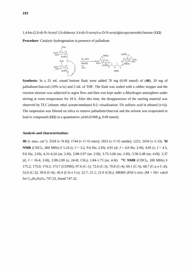
191
1,4-bis-[2,6-di-N-Acetyl 2,6-dideoxy-3,4-di-O-acetyl--D-N-acetylglucopyranoside]-butane (112)
Procedure: Catalytic hydrogenation in presence of palladium
Synthesis: In a 25 mL round bottom flask were added 70 mg (0.09 mmol) of (40), 20 mg of
palladium/charcoal (10% w/w) and 5 mL of THF. The flask was sealed with a rubber stopper and the
reaction mixture was subjected to argon flow and then was kept under a dihydrogen atmosphere under
stirring at room temperature for 18 h. After this time, the disappearence of the starting material was
observed by TLC (eluent: ethyl acetate/methanol 8:2; visualization: 5% sulfuric acid in ethanol (v/v)).
The suspension was filtered on silica to remove palladium/charcoal and the solvent was evaporated to
lead to compound (112) in a quantitative yield (0.068 g, 0.09 mmol).
Analysis and characterization:
IR ( max, cm-1): 3318 ( N-H); 1744 ( C=O ester); 1653 ( C=O amide); 1221; 1034 ( C-O). 1H
NMR (CDCl3, 400 MHz) 5.24 (t; J = 3.2, 9.6 Hz; 2-H); 4.91 (d; J = 4.0 Hz; 2-H); 4.85 (t; J = 4.5,
9.6 Hz; 2-H); 4.31-4.24 (m; 2-H); 3.98-3.97 (m; 2-H); 3.75-3.66 (m; 2-H); 3.58-3.48 (m; 4-H); 3.37
(d; J = 16.4; 2-H); 2.08-2.00 (s; 24-H; CH3); 1.84-1.73 (m; 4-H). 13C NMR (CDCl3, 100 MHz)
175.2; 175.0; 174.3; 173.7 (CONH); 97.6 (C-1); 72.6 (C-3); 70.8 (C-4); 69.1 (C-5); 68.7 (C-a e C-d);
52.6 (C-2); 39.8 (C-6); 26.4 (C-b e Cc); 22.7; 21.1; 21.0 (CH3). HRMS (ESI+) m/z: [M + H]+ calcd
for C32H50N4O16 747.33, found 747.32.
O
OAc
AcOAcO
AcHN
O
O
AcO
AcOAcO
AcHNO
H2, Pd/C
MeOH, r.t.10 h
a
b
c12
4 5
6
3O
NHAc
AcOAcO
AcHNO
(112)
O
O
AcHN
OAcOAc
NHAc
d
1' 2'3'
4'
6'
5'
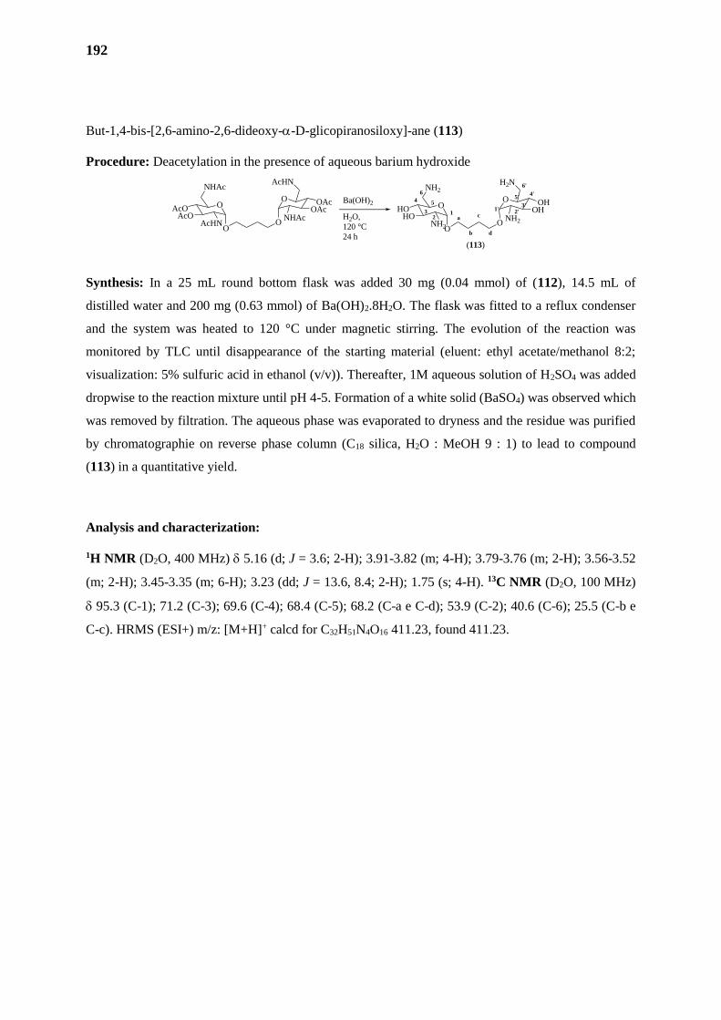
192
But-1,4-bis-[2,6-amino-2,6-dideoxy--D-glicopiranosiloxy]-ane (113)
Procedure: Deacetylation in the presence of aqueous barium hydroxide
Synthesis: In a 25 mL round bottom flask was added 30 mg (0.04 mmol) of (112), 14.5 mL of
distilled water and 200 mg (0.63 mmol) of Ba(OH)2.8H2O. The flask was fitted to a reflux condenser
and the system was heated to 120 °C under magnetic stirring. The evolution of the reaction was
monitored by TLC until disappearance of the starting material (eluent: ethyl acetate/methanol 8:2;
visualization: 5% sulfuric acid in ethanol (v/v)). Thereafter, 1M aqueous solution of H2SO4 was added
dropwise to the reaction mixture until pH 4-5. Formation of a white solid (BaSO4) was observed which
was removed by filtration. The aqueous phase was evaporated to dryness and the residue was purified
by chromatographie on reverse phase column (C18 silica, H2O : MeOH 9 : 1) to lead to compound
(113) in a quantitative yield.
Analysis and characterization:
1H NMR (D2O, 400 MHz) 5.16 (d; J = 3.6; 2-H); 3.91-3.82 (m; 4-H); 3.79-3.76 (m; 2-H); 3.56-3.52
(m; 2-H); 3.45-3.35 (m; 6-H); 3.23 (dd; J = 13.6, 8.4; 2-H); 1.75 (s; 4-H). 13C NMR (D2O, 100 MHz)
95.3 (C-1); 71.2 (C-3); 69.6 (C-4); 68.4 (C-5); 68.2 (C-a e C-d); 53.9 (C-2); 40.6 (C-6); 25.5 (C-b e
C-c). HRMS (ESI+) m/z: [M+H]+ calcd for C32H51N4O16 411.23, found 411.23.
O
NHAc
AcOAcO
AcHNO
O
O
AcHN
OAcOAc
NHAc a
b
c12
4 5
6
3O
NH2
HOHO
NH2O
(113)
O
O
H2N
OHOH
NH2
d
1' 2'3'
4'
6'
5'Ba(OH)2
H2O,
120 °C
24 h
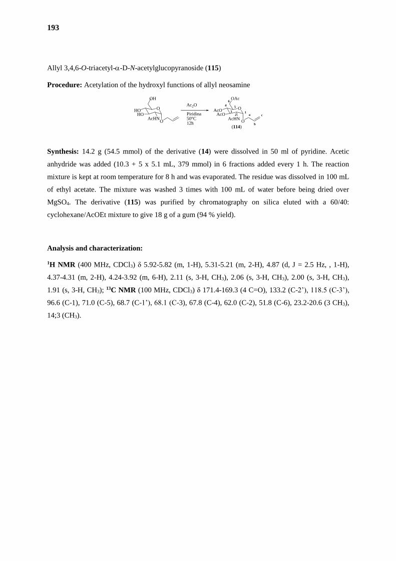
193
Allyl 3,4,6-O-triacetyl--D-N-acetylglucopyranoside (115)
Procedure: Acetylation of the hydroxyl functions of allyl neosamine
Synthesis: 14.2 g (54.5 mmol) of the derivative (14) were dissolved in 50 ml of pyridine. Acetic
anhydride was added (10.3 + 5 x 5.1 mL, 379 mmol) in 6 fractions added every 1 h. The reaction
mixture is kept at room temperature for 8 h and was evaporated. The residue was dissolved in 100 mL
of ethyl acetate. The mixture was washed 3 times with 100 mL of water before being dried over
MgSO4. The derivative (115) was purified by chromatography on silica eluted with a 60/40:
cyclohexane/AcOEt mixture to give 18 g of a gum (94 % yield).
Analysis and characterization:
1H NMR (400 MHz, CDCl3) δ 5.92-5.82 (m, 1-H), 5.31-5.21 (m, 2-H), 4.87 (d, J = 2.5 Hz, , 1-H),
4.37-4.31 (m, 2-H), 4.24-3.92 (m, 6-H), 2.11 (s, 3-H, CH3), 2.06 (s, 3-H, CH3), 2.00 (s, 3-H, CH3),
1.91 (s, 3-H, CH3); 13C NMR (100 MHz, CDCl3) δ 171.4-169.3 (4 C=O), 133.2 (C-2’), 118.5 (C-3’),
96.6 (C-1), 71.0 (C-5), 68.7 (C-1’), 68.1 (C-3), 67.8 (C-4), 62.0 (C-2), 51.8 (C-6), 23.2-20.6 (3 CH3),
14;3 (CH3).
O
OH
HOHO
AcHNO
Ac2O
Piridina50°C12h
O
OAc
AcOAcO
AcHNO
a
b
c1
2
4 5
6
3
(114)
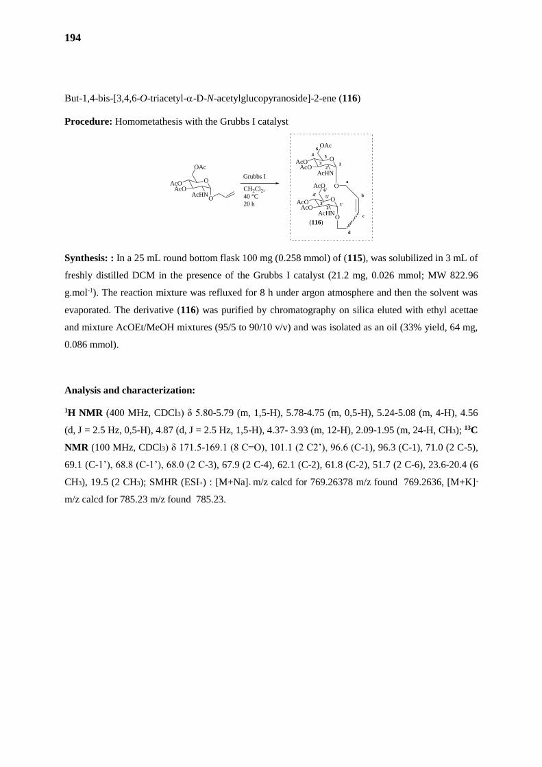
194
But-1,4-bis-[3,4,6-O-triacetyl--D-N-acetylglucopyranoside]-2-ene (116)
Procedure: Homometathesis with the Grubbs I catalyst
Synthesis: : In a 25 mL round bottom flask 100 mg (0.258 mmol) of (115), was solubilized in 3 mL of
freshly distilled DCM in the presence of the Grubbs I catalyst (21.2 mg, 0.026 mmol; MW 822.96
g.mol-1). The reaction mixture was refluxed for 8 h under argon atmosphere and then the solvent was
evaporated. The derivative (116) was purified by chromatography on silica eluted with ethyl acettae
and mixture AcOEt/MeOH mixtures (95/5 to 90/10 v/v) and was isolated as an oil (33% yield, 64 mg,
0.086 mmol).
Analysis and characterization:
1H NMR (400 MHz, CDCl3) δ 5.80-5.79 (m, 1,5-H), 5.78-4.75 (m, 0,5-H), 5.24-5.08 (m, 4-H), 4.56
(d, J = 2.5 Hz, 0,5-H), 4.87 (d, J = 2.5 Hz, 1,5-H), 4.37- 3.93 (m, 12-H), 2.09-1.95 (m, 24-H, CH3); 13C
NMR (100 MHz, CDCl3) δ 171.5-169.1 (8 C=O), 101.1 (2 C2’), 96.6 (C-1), 96.3 (C-1), 71.0 (2 C-5),
69.1 (C-1’), 68.8 (C-1’), 68.0 (2 C-3), 67.9 (2 C-4), 62.1 (C-2), 61.8 (C-2), 51.7 (2 C-6), 23.6-20.4 (6
CH3), 19.5 (2 CH3); SMHR (ESI+) : [M+Na]+ m/z calcd for 769.26378 m/z found 769.2636, [M+K]+
m/z calcd for 785.23 m/z found 785.23.
(116)
O
OAc
AcOAcO
AcHN
O
12
4 5
6
3
O
AcO
AcOAcO
AcHNO
1'2'
4' 5'
6'
3'
a
b
c
d
O
OAc
AcOAcO
AcHNO
Grubbs I
CH2Cl2,
40 °C
20 h
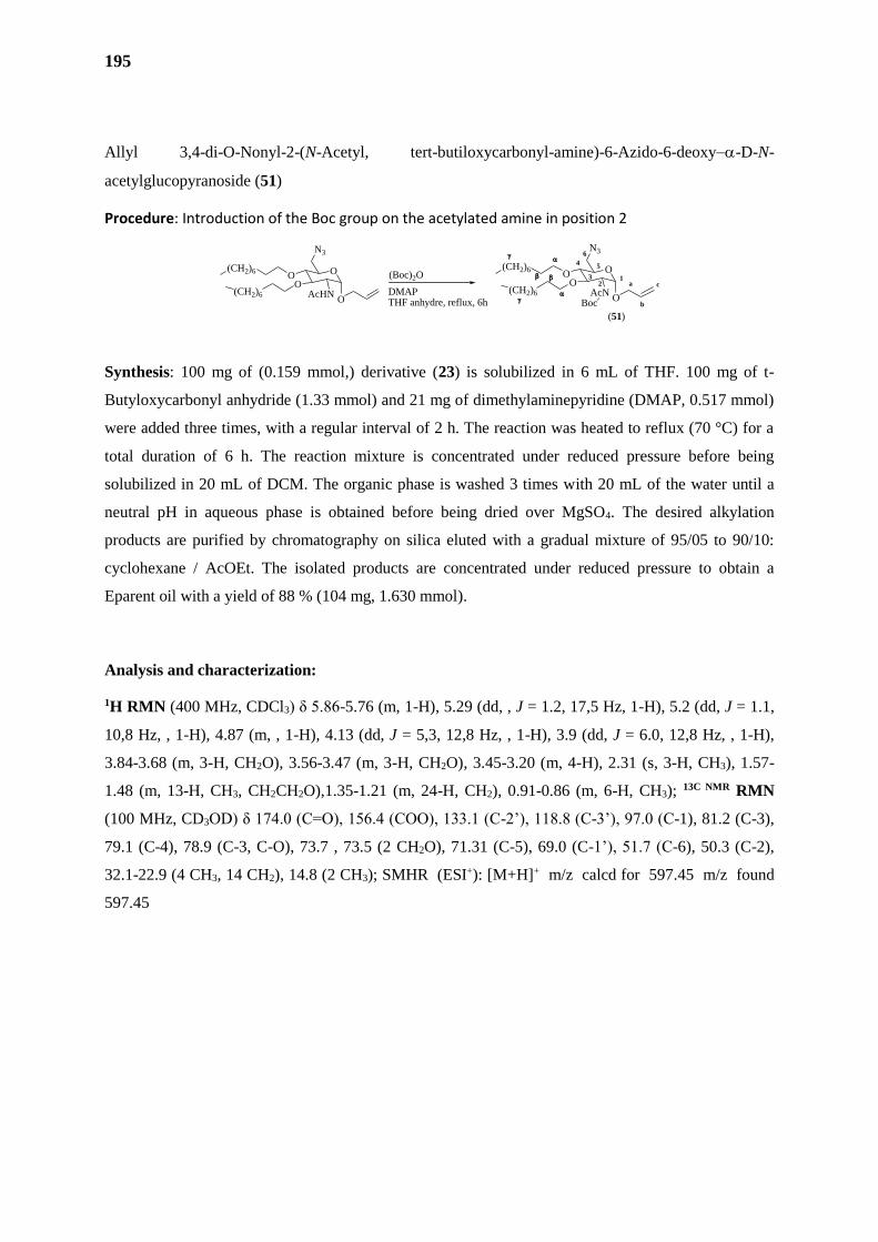
195
Allyl 3,4-di-O-Nonyl-2-(N-Acetyl, tert-butiloxycarbonyl-amine)-6-Azido-6-deoxy-D-N-
acetylglucopyranoside (51)
Procedure: Introduction of the Boc group on the acetylated amine in position 2
Synthesis: 100 mg of (0.159 mmol,) derivative (23) is solubilized in 6 mL of THF. 100 mg of t-
Butyloxycarbonyl anhydride (1.33 mmol) and 21 mg of dimethylaminepyridine (DMAP, 0.517 mmol)
were added three times, with a regular interval of 2 h. The reaction was heated to reflux (70 °C) for a
total duration of 6 h. The reaction mixture is concentrated under reduced pressure before being
solubilized in 20 mL of DCM. The organic phase is washed 3 times with 20 mL of the water until a
neutral pH in aqueous phase is obtained before being dried over MgSO4. The desired alkylation
products are purified by chromatography on silica eluted with a gradual mixture of 95/05 to 90/10:
cyclohexane / AcOEt. The isolated products are concentrated under reduced pressure to obtain a
Eparent oil with a yield of 88 % (104 mg, 1.630 mmol).
Analysis and characterization:
1H RMN (400 MHz, CDCl3) δ 5.86-5.76 (m, 1-H), 5.29 (dd, , J = 1.2, 17,5 Hz, 1-H), 5.2 (dd, J = 1.1,
10,8 Hz, , 1-H), 4.87 (m, , 1-H), 4.13 (dd, J = 5,3, 12,8 Hz, , 1-H), 3.9 (dd, J = 6.0, 12,8 Hz, , 1-H),
3.84-3.68 (m, 3-H, CH2O), 3.56-3.47 (m, 3-H, CH2O), 3.45-3.20 (m, 4-H), 2.31 (s, 3-H, CH3), 1.57-
1.48 (m, 13-H, CH3, CH2CH2O),1.35-1.21 (m, 24-H, CH2), 0.91-0.86 (m, 6-H, CH3); 13C NMR RMN
(100 MHz, CD3OD) δ 174.0 (C=O), 156.4 (COO), 133.1 (C-2’), 118.8 (C-3’), 97.0 (C-1), 81.2 (C-3),
79.1 (C-4), 78.9 (C-3, C-O), 73.7 , 73.5 (2 CH2O), 71.31 (C-5), 69.0 (C-1’), 51.7 (C-6), 50.3 (C-2),
32.1-22.9 (4 CH3, 14 CH2), 14.8 (2 CH3); SMHR (ESI+): [M+H]+ m/z calcd for 597.45 m/z found
597.45
(Boc)2O
DMAPTHF anhydre, reflux, 6h
O
N3
OO
AcHNO
(CH2)6
(CH2)6
O
N3
OO
AcNO
a
b
c1
2
4 5
6
3
(CH2)6
(CH2)6
(51)
Boc
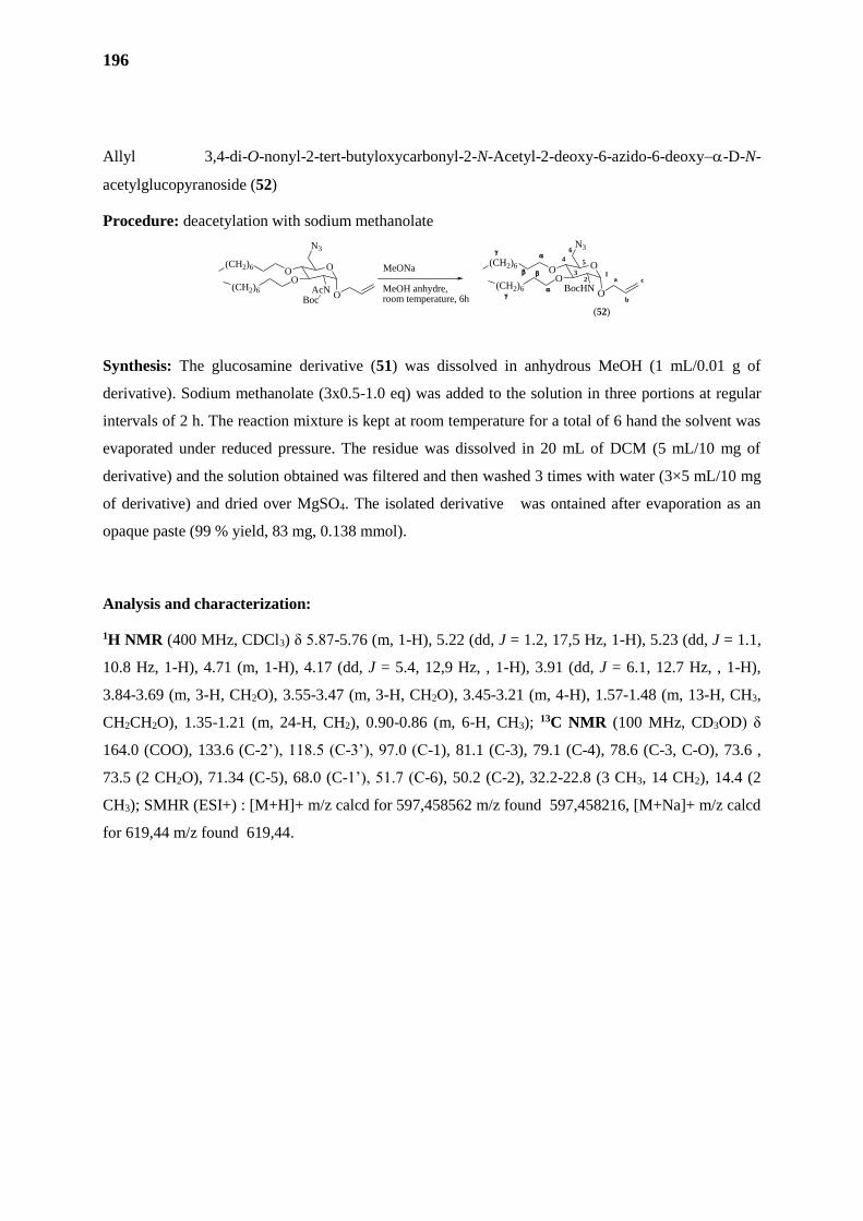
196
Allyl 3,4-di-O-nonyl-2-tert-butyloxycarbonyl-2-N-Acetyl-2-deoxy-6-azido-6-deoxy-D-N-
acetylglucopyranoside (52)
Procedure: deacetylation with sodium methanolate
Synthesis: The glucosamine derivative (51) was dissolved in anhydrous MeOH (1 mL/0.01 g of
derivative). Sodium methanolate (3x0.5-1.0 eq) was added to the solution in three portions at regular
intervals of 2 h. The reaction mixture is kept at room temperature for a total of 6 hand the solvent was
evaporated under reduced pressure. The residue was dissolved in 20 mL of DCM (5 mL/10 mg of
derivative) and the solution obtained was filtered and then washed 3 times with water (3×5 mL/10 mg
of derivative) and dried over MgSO4. The isolated derivative was ontained after evaporation as an
opaque paste (99 % yield, 83 mg, 0.138 mmol).
Analysis and characterization:
1H NMR (400 MHz, CDCl3) δ 5.87-5.76 (m, 1-H), 5.22 (dd, J = 1.2, 17,5 Hz, 1-H), 5.23 (dd, J = 1.1,
10.8 Hz, 1-H), 4.71 (m, 1-H), 4.17 (dd, J = 5.4, 12,9 Hz, , 1-H), 3.91 (dd, J = 6.1, 12.7 Hz, , 1-H),
3.84-3.69 (m, 3-H, CH2O), 3.55-3.47 (m, 3-H, CH2O), 3.45-3.21 (m, 4-H), 1.57-1.48 (m, 13-H, CH3,
CH2CH2O), 1.35-1.21 (m, 24-H, CH2), 0.90-0.86 (m, 6-H, CH3); 13C NMR (100 MHz, CD3OD) δ
164.0 (COO), 133.6 (C-2’), 118.5 (C-3’), 97.0 (C-1), 81.1 (C-3), 79.1 (C-4), 78.6 (C-3, C-O), 73.6 ,
73.5 (2 CH2O), 71.34 (C-5), 68.0 (C-1’), 51.7 (C-6), 50.2 (C-2), 32.2-22.8 (3 CH3, 14 CH2), 14.4 (2
CH3); SMHR (ESI+) : [M+H]+ m/z calcd for 597,458562 m/z found 597,458216, [M+Na]+ m/z calcd
for 619,44 m/z found 619,44.
O
N3
OO
AcNO
(CH2)6
(CH2)6
O
N3
OO
BocHNO
a
b
c1
2
4 5
6
3
(CH2)6
(CH2)6
(52)
MeONa
MeOH anhydre, room temperature, 6hBoc
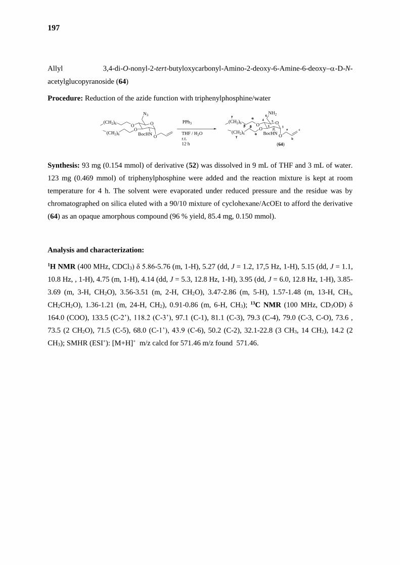
197
Allyl 3,4-di-O-nonyl-2-tert-butyloxycarbonyl-Amino-2-deoxy-6-Amine-6-deoxy-D-N-
acetylglucopyranoside (64)
Procedure: Reduction of the azide function with triphenylphosphine/water
Synthesis: 93 mg (0.154 mmol) of derivative (52) was dissolved in 9 mL of THF and 3 mL of water.
123 mg (0.469 mmol) of triphenylphosphine were added and the reaction mixture is kept at room
temperature for 4 h. The solvent were evaporated under reduced pressure and the residue was by
chromatographed on silica eluted with a 90/10 mixture of cyclohexane/AcOEt to afford the derivative
(64) as an opaque amorphous compound (96 % yield, 85.4 mg, 0.150 mmol).
Analysis and characterization:
1H NMR (400 MHz, CDCl3) δ 5.86-5.76 (m, 1-H), 5.27 (dd, J = 1.2, 17,5 Hz, 1-H), 5.15 (dd, J = 1.1,
10.8 Hz, , 1-H), 4.75 (m, 1-H), 4.14 (dd, J = 5.3, 12.8 Hz, 1-H), 3.95 (dd, J = 6.0, 12.8 Hz, 1-H), 3.85-
3.69 (m, 3-H, CH2O), 3.56-3.51 (m, 2-H, CH2O), 3.47-2.86 (m, 5-H), 1.57-1.48 (m, 13-H, CH3,
CH2CH2O), 1.36-1.21 (m, 24-H, CH2), 0.91-0.86 (m, 6-H, CH3); 13C NMR (100 MHz, CD3OD) δ
164.0 (COO), 133.5 (C-2’), 118.2 (C-3’), 97.1 (C-1), 81.1 (C-3), 79.3 (C-4), 79.0 (C-3, C-O), 73.6 ,
73.5 (2 CH2O), 71.5 (C-5), 68.0 (C-1’), 43.9 (C-6), 50.2 (C-2), 32.1-22.8 (3 CH3, 14 CH2), 14.2 (2
CH3); SMHR (ESI+): [M+H]+ m/z calcd for 571.46 m/z found 571.46.
O
N3
OO
BocHNO
(CH2)6
(CH2)6
O
NH2
OO
BocHNO
a
b
c1
2
4 5
6
3
(CH2)6
(CH2)6
(64)
PPh3
THF / H2Or.t.12 h
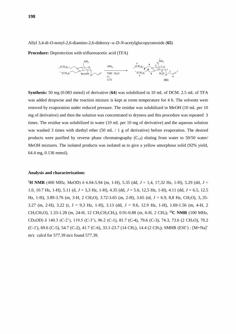
198
Allyl 3,4-di-O-nonyl-2,6-diamino-2,6-dideoxy-D-N-acetylglucopyranoside (65)
Procedure: Deprotection with trifluoroacetic acid (TFA)
Synthesis: 50 mg (0.083 mmol) of derivative (64) was solubilized in 10 mL of DCM. 2.5 mL of TFA
was added dropwise and the reaction mixture is kept at room temperature for 4 h. The solvents were
renoved by evaporation under reduced pressure. The residue was solubilized in MeOH (10 mL per 10
mg of derivative) and then the solution was concentrated to dryness and this procedure was repeated 3
times. The residue was solubilized in water (10 mL per 10 mg of derivative) and the aqueous solution
was washed 3 times with diethyl ether (50 mL / 1 g of derivative) before evaporation. The desired
products were purified by reverse phase chromatography (C18) eluting from water to 50/50 water/
MeOH mixtures. The isolated products was isolated as to give a yellow amorphous solid (92% yield,
64.4 mg, 0.136 mmol).
Analysis and characterization:
1H NMR (400 MHz, MeOD) δ 6.04-5.94 (m, 1-H), 5.35 (dd, J = 1,4, 17,32 Hz, 1-H), 5.29 (dd, J =
1.0, 10.7 Hz, 1-H), 5.11 (d, J = 3,3 Hz, 1-H), 4.35 (dd, J = 5.6, 12,5 Hz, 1-H), 4.11 (dd, J = 6.5, 12.5
Hz, 1-H), 3.89-3.76 (m, 3-H, 2 CH2O), 3.72-3.65 (m, 2-H), 3.65 (td, J = 6.9, 8,8 Hz, CH2O), 3,.35-
3.27 (m, 2-H), 3.22 (t, J = 9,3 Hz, 1-H), 3.13 (dd, J = 9.6, 12.9 Hz, 1-H), 1.69-1.56 (m, 4-H, 2
CH2CH2O), 1.33-1.28 (m, 24-H, 12 CH2CH2CH2), 0.91-0.88 (m, 6-H, 2 CH3); 13C NMR (100 MHz,
CD3OD) δ 140.3 (C-2’), 119.5 (C-3’), 96.2 (C-1), 81.7 (C-4), 79.6 (C-3), 74.3, 73.6 (2 CH2O), 70.2
(C-1'), 69.6 (C-5), 54.7 (C-2), 41.7 (C-6), 33.1-23.7 (14 CH2), 14.4 (2 CH3); SMHR (ESI+) : [M+Na]+
m/z calcd for 577.39 m/z found 577.39.
O
NH2
OO
BocHNO
(CH2)6
(CH2)6
O
NH3
OO
H3NO
a
b
c1
2
4 5
6
3
(CH2)6
(CH2)6
(65)
PPh3
THF / H2Or.t.12 h
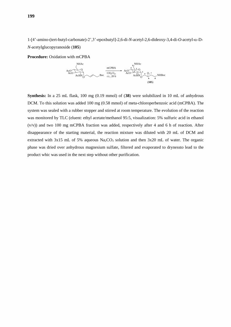
199
1-[4’-amino-(tert-butyl-carbonate)-2’,3’-epoxbutyl]-2,6-di-N-acetyl-2,6-dideoxy-3,4-di-O-acetyl--D-
N-acetylglucopyranoside (105)
Procedure: Oxidation with mCPBA
Synthesis: In a 25 mL flask, 100 mg (0.19 mmol) of (38) were solubilized in 10 mL of anhydrous
DCM. To this solution was added 100 mg (0.58 mmol) of meta-chloroperbenzoic acid (mCPBA). The
system was sealed with a rubber stopper and stirred at room temperature. The evolution of the reaction
was monitored by TLC (eluent: ethyl acetate/methanol 95:5, visualization: 5% sulfuric acid in ethanol
(v/v)) and two 100 mg mCPBA fraction was added, respectively after 4 and 6 h of reaction. After
disappearance of the starting material, the reaction mixture was diluted with 20 mL of DCM and
extracted with 3x15 mL of 5% aqueous Na2CO3 solution and then 3x20 mL of water. The organic
phase was dried over anhydrous magnesium sulfate, filtered and evaporated to drynessto lead to the
product whic was used in the next step without other purification.
a
b
c12
4 5
6
3O
NHAc
AcOAcO
AcHNO
mCPBA
CH2Cl2,
r.t., 20 h
O
NHAc
AcOAcO
AcHNO
(105)
d
NHBocO
Boc
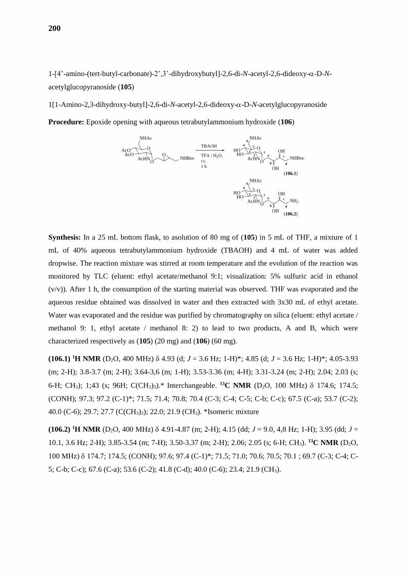
200
1-[4’-amino-(tert-butyl-carbonate)-2’,3’-dihydroxybutyl]-2,6-di-N-acetyl-2,6-dideoxy--D-N-
acetylglucopyranoside (105)
1[1-Amino-2,3-dihydroxy-butyl]-2,6-di-N-acetyl-2,6-dideoxy--D-N-acetylglucopyranoside
Procedure: Epoxide opening with aqueous tetrabutylammonium hydroxide (106)
Synthesis: In a 25 mL bottom flask, to asolution of 80 mg of (105) in 5 mL of THF, a mixture of 1
mL of 40% aqueous tetrabutylammonium hydroxide (TBAOH) and 4 mL of water was added
dropwise. The reaction mixture was stirred at room temperature and the evolution of the reaction was
monitored by TLC (eluent: ethyl acetate/methanol 9:1; visualization: 5% sulfuric acid in ethanol
(v/v)). After 1 h, the consumption of the starting material was observed. THF was evaporated and the
aqueous residue obtained was dissolved in water and then extracted with 3x30 mL of ethyl acetate.
Water was evaporated and the residue was purified by chromatography on silica (eluent: ethyl acetate /
methanol 9: 1, ethyl acetate / methanol 8: 2) to lead to two products, A and B, which were
characterized respectively as (105) (20 mg) and (106) (60 mg).
(106.1) 1H NMR (D2O, 400 MHz) 4.93 (d; J = 3.6 Hz; 1-H)*; 4.85 (d; J = 3.6 Hz; 1-H)*; 4.05-3.93
(m; 2-H); 3.8-3.7 (m; 2-H); 3.64-3,6 (m; 1-H); 3.53-3.36 (m; 4-H); 3.31-3.24 (m; 2-H); 2.04; 2.03 (s;
6-H; CH3); 1;43 (s; 96H; C(CH3)3).* Interchangeable. 13C NMR (D2O, 100 MHz) 174.6; 174.5;
(CONH); 97.3; 97.2 (C-1)*; 71.5; 71.4; 70.8; 70.4 (C-3; C-4; C-5; C-b; C-c); 67.5 (C-a); 53.7 (C-2);
40.0 (C-6); 29.7; 27.7 (C(CH3)3); 22.0; 21.9 (CH3). *Isomeric mixture
(106.2) 1H NMR (D2O, 400 MHz) 4.91-4.87 (m; 2-H); 4.15 (dd; J = 9.0, 4,8 Hz; 1-H); 3.95 (dd; J =
10.1, 3.6 Hz; 2-H); 3.85-3.54 (m; 7-H); 3.50-3.37 (m; 2-H); 2.06; 2.05 (s; 6-H; CH3). 13C NMR (D2O,
100 MHz) 174.7; 174.5; (CONH); 97.6; 97.4 (C-1)*; 71.5; 71.0; 70.6; 70.5; 70.1 ; 69.7 (C-3; C-4; C-
5; C-b; C-c); 67.6 (C-a); 53.6 (C-2); 41.8 (C-d); 40.0 (C-6); 23.4; 21.9 (CH3).
a
b
c
12
4 5
6
3O
NHAc
AcOAcO
AcHNO
O
NHAc
HOHO
AcHNO
(106.1)
d
NHBoc
OH
OH
a
b
c
12
4 5
6
3O
NHAc
HOHO
AcHNO
d
NH2
OH
OH
TBAOH
TFA / H2O,
r.t.
1 h
(106.2)
NHBocO
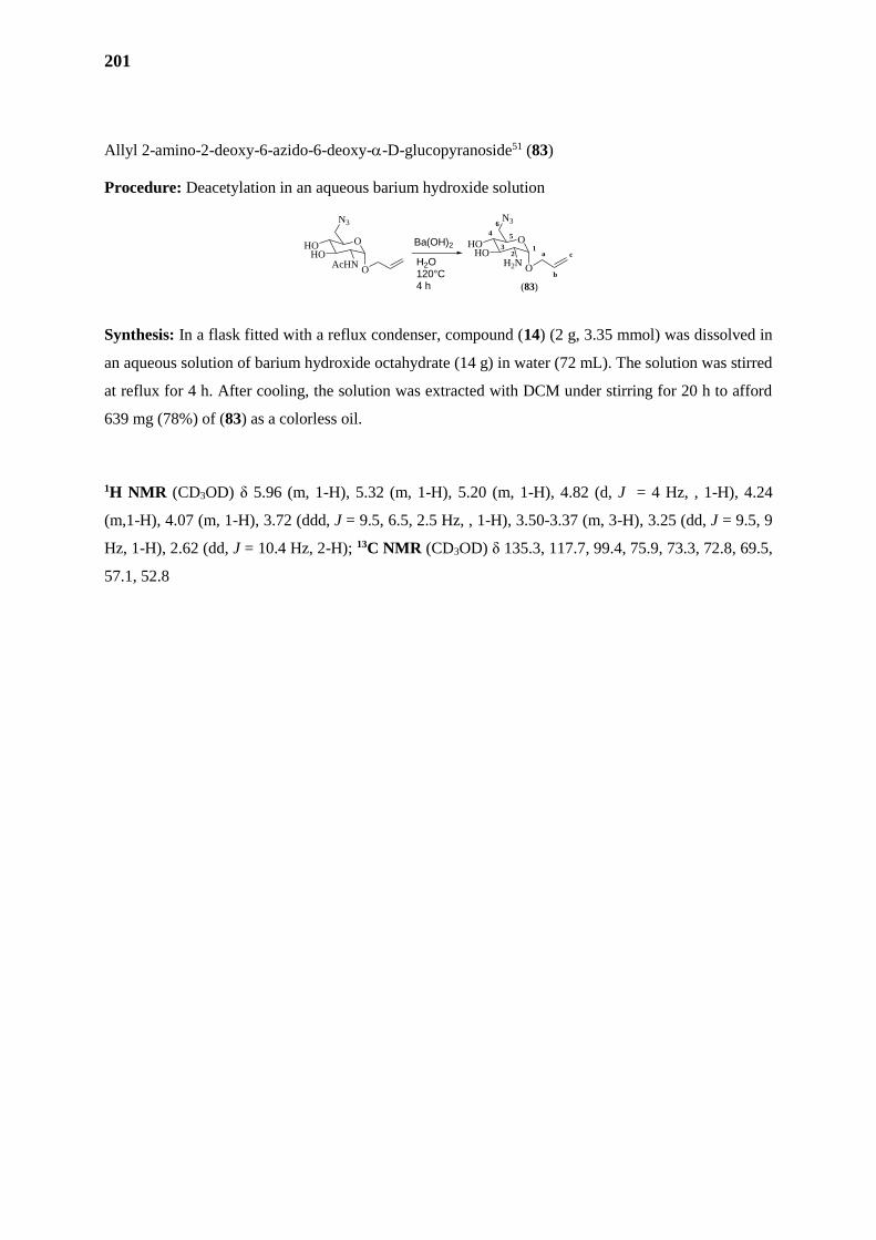
201
Allyl 2-amino-2-deoxy-6-azido-6-deoxy--D-glucopyranoside51 (83)
Procedure: Deacetylation in an aqueous barium hydroxide solution
Synthesis: In a flask fitted with a reflux condenser, compound (14) (2 g, 3.35 mmol) was dissolved in
an aqueous solution of barium hydroxide octahydrate (14 g) in water (72 mL). The solution was stirred
at reflux for 4 h. After cooling, the solution was extracted with DCM under stirring for 20 h to afford
639 mg (78%) of (83) as a colorless oil.
1H NMR (CD3OD) δ 5.96 (m, 1-H), 5.32 (m, 1-H), 5.20 (m, 1-H), 4.82 (d, J = 4 Hz, , 1-H), 4.24
(m,1-H), 4.07 (m, 1-H), 3.72 (ddd, J = 9.5, 6.5, 2.5 Hz, , 1-H), 3.50-3.37 (m, 3-H), 3.25 (dd, J = 9.5, 9
Hz, 1-H), 2.62 (dd, J = 10.4 Hz, 2-H); 13C NMR (CD3OD) δ 135.3, 117.7, 99.4, 75.9, 73.3, 72.8, 69.5,
57.1, 52.8
O
N3
HOHO
AcHNO
O
N3
HOHO
H2NO
a
b
c1
2
4 5
6
3
(83)
H2O120°C4 h
Ba(OH)2
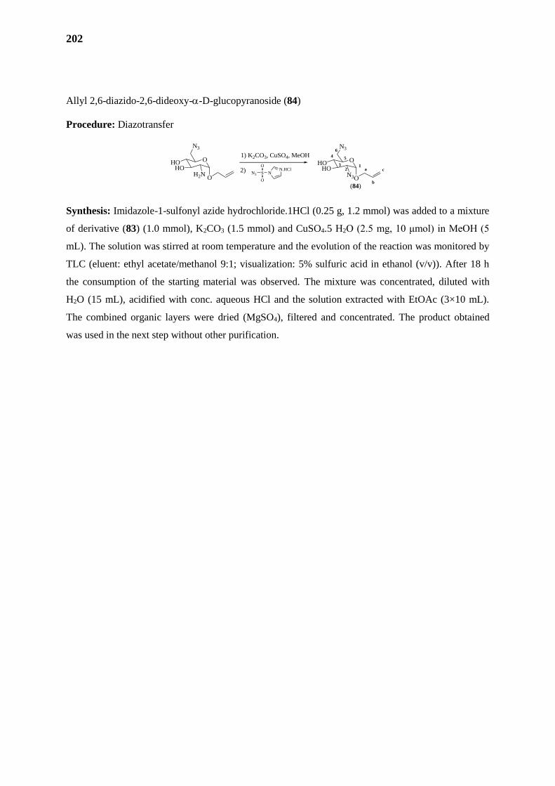
202
Allyl 2,6-diazido-2,6-dideoxy--D-glucopyranoside (84)
Procedure: Diazotransfer
Synthesis: Imidazole-1-sulfonyl azide hydrochloride.1HCl (0.25 g, 1.2 mmol) was added to a mixture
of derivative (83) (1.0 mmol), K2CO3 (1.5 mmol) and CuSO4.5 H2O (2.5 mg, 10 μmol) in MeOH (5
mL). The solution was stirred at room temperature and the evolution of the reaction was monitored by
TLC (eluent: ethyl acetate/methanol 9:1; visualization: 5% sulfuric acid in ethanol (v/v)). After 18 h
the consumption of the starting material was observed. The mixture was concentrated, diluted with
H2O (15 mL), acidified with conc. aqueous HCl and the solution extracted with EtOAc (3×10 mL).
The combined organic layers were dried (MgSO4), filtered and concentrated. The product obtained
was used in the next step without other purification.
(84)
O
N3
HOHO
H2NO
O
N3
HOHO
N3O
a
b
c1
2
4 5
6
3
1) K2CO3, CuSO4, MeOH
N.HClNS
O
O
N32)
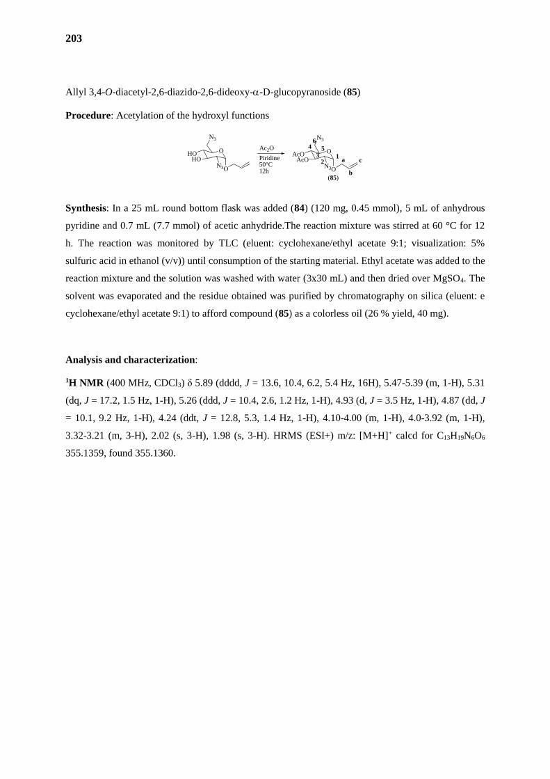
203
Allyl 3,4-O-diacetyl-2,6-diazido-2,6-dideoxy--D-glucopyranoside (85)
Procedure: Acetylation of the hydroxyl functions
Synthesis: In a 25 mL round bottom flask was added (84) (120 mg, 0.45 mmol), 5 mL of anhydrous
pyridine and 0.7 mL (7.7 mmol) of acetic anhydride.The reaction mixture was stirred at 60 °C for 12
h. The reaction was monitored by TLC (eluent: cyclohexane/ethyl acetate 9:1; visualization: 5%
sulfuric acid in ethanol (v/v)) until consumption of the starting material. Ethyl acetate was added to the
reaction mixture and the solution was washed with water (3x30 mL) and then dried over MgSO4. The
solvent was evaporated and the residue obtained was purified by chromatography on silica (eluent: e
cyclohexane/ethyl acetate 9:1) to afford compound (85) as a colorless oil (26 % yield, 40 mg).
Analysis and characterization:
1H NMR (400 MHz, CDCl3) δ 5.89 (dddd, J = 13.6, 10.4, 6.2, 5.4 Hz, 16H), 5.47-5.39 (m, 1-H), 5.31
(dq, J = 17.2, 1.5 Hz, 1-H), 5.26 (ddd, J = 10.4, 2.6, 1.2 Hz, 1-H), 4.93 (d, J = 3.5 Hz, 1-H), 4.87 (dd, J
= 10.1, 9.2 Hz, 1-H), 4.24 (ddt, J = 12.8, 5.3, 1.4 Hz, 1-H), 4.10-4.00 (m, 1-H), 4.0-3.92 (m, 1-H),
3.32-3.21 (m, 3-H), 2.02 (s, 3-H), 1.98 (s, 3-H). HRMS (ESI+) m/z: [M+H]+ calcd for C13H19N6O6
355.1359, found 355.1360.
(85)
O
N3
HOHO
N3O
Ac2O
Piridine50°C12h
O
N3
AcOAcO
N3O
a
b
c1
2
4 5
6
3
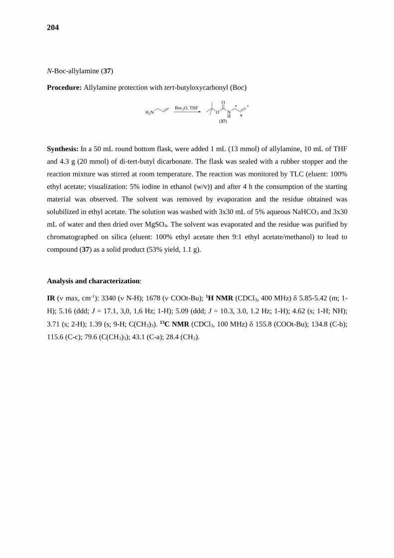
204
N-Boc-allylamine (37)
Procedure: Allylamine protection with tert-butyloxycarbonyl (Boc)
Synthesis: In a 50 mL round bottom flask, were added 1 mL (13 mmol) of allylamine, 10 mL of THF
and 4.3 g (20 mmol) of di-tert-butyl dicarbonate. The flask was sealed with a rubber stopper and the
reaction mixture was stirred at room temperature. The reaction was monitored by TLC (eluent: 100%
ethyl acetate; visualization: 5% iodine in ethanol (w/v)) and after 4 h the consumption of the starting
material was observed. The solvent was removed by evaporation and the residue obtained was
solubilized in ethyl acetate. The solution was washed with 3x30 mL of 5% aqueous NaHCO3 and 3x30
mL of water and then dried over MgSO4. The solvent was evaporated and the residue was purified by
chromatographed on silica (eluent: 100% ethyl acetate then 9:1 ethyl acetate/methanol) to lead to
compound (37) as a solid product (53% yield, 1.1 g).
Analysis and characterization:
IR ( max, cm-1): 3340 ( N-H); 1678 ( COOt-Bu); 1H NMR (CDCl3, 400 MHz) 5.85-5.42 (m; 1-
H); 5.16 (ddd; J = 17.1, 3,0, 1,6 Hz; 1-H); 5.09 (ddd; J = 10.3, 3.0, 1.2 Hz; 1-H); 4.62 (s; 1-H; NH);
3.71 (s; 2-H); 1.39 (s; 9-H; C(CH3)3). 13C NMR (CDCl3, 100 MHz) 155.8 (COOt-Bu); 134.8 (C-b);
115.6 (C-c); 79.6 (C(CH3)3); 43.1 (C-a); 28.4 (CH3).
a
b
c
H2N NH
O
O
(37)
Boc2O, THF
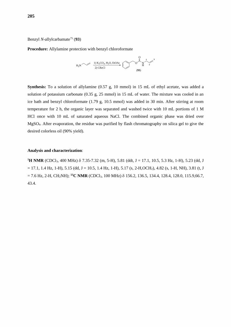
205
Benzyl N-allylcarbamate75 (93)
Procedure: Allylamine protection with benzyl chloroformate
Synthesis: To a solution of allylamine (0.57 g, 10 mmol) in 15 mL of ethyl acetate, was added a
solution of potassium carbonate (0.35 g, 25 mmol) in 15 mL of water. The mixture was cooled in an
ice bath and benzyl chloroformate (1.79 g, 10.5 mmol) was added in 30 min. After stirring at room
temperature for 2 h, the organic layer was separated and washed twice with 10 mL portions of 1 M
HCl once with 10 mL of saturated aqueous NaCl. The combined organic phase was dried over
MgSO4. After evaporation, the residue was purified by flash chromatography on silica gel to give the
desired colorless oil (90% yield).
Analysis and characterization:
1H NMR (CDCl3, 400 MHz) 7.35-7.32 (m, 5-H), 5.81 (ddt, J = 17.1, 10.5, 5.3 Hz, 1-H), 5.23 (dd, J
= 17.1, 1.4 Hz, 1-H), 5.15 (dd, J = 10.5, 1.4 Hz, 1-H), 5.17 (s, 2-H,OCH2), 4.82 (s, 1-H, NH), 3.81 (t, J
= 7.6 Hz, 2-H, CH2NH); 13C NMR (CDCl3, 100 MHz) 156.2, 136.5, 134.4, 128.4, 128.0, 115.9,66.7,
43.4.
1
2
3
H2NNH
O
O
(93)
1) K2CO3, H2O, EtOAc
2) CBzCl
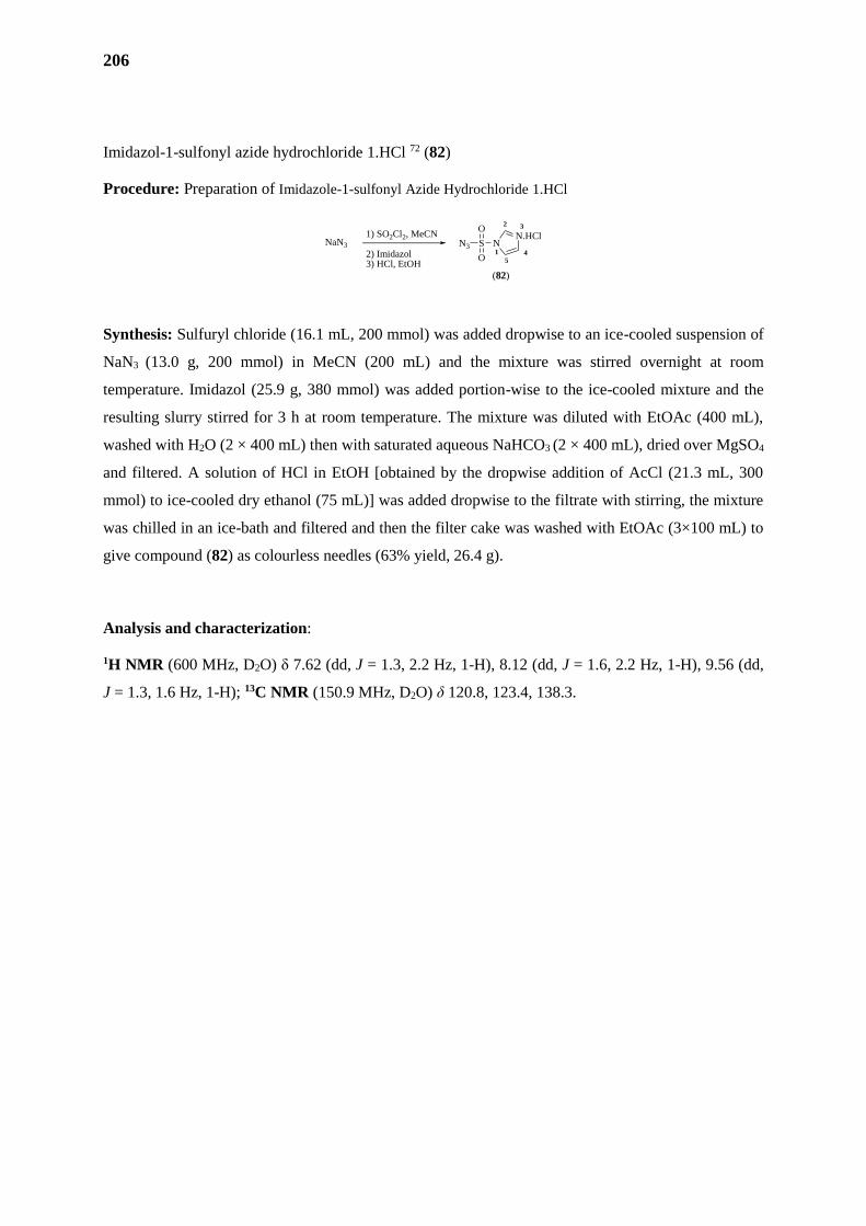
206
Imidazol-1-sulfonyl azide hydrochloride 1.HCl 72 (82)
Procedure: Preparation of Imidazole-1-sulfonyl Azide Hydrochloride 1.HCl
Synthesis: Sulfuryl chloride (16.1 mL, 200 mmol) was added dropwise to an ice-cooled suspension of
NaN3 (13.0 g, 200 mmol) in MeCN (200 mL) and the mixture was stirred overnight at room
temperature. Imidazol (25.9 g, 380 mmol) was added portion-wise to the ice-cooled mixture and the
resulting slurry stirred for 3 h at room temperature. The mixture was diluted with EtOAc (400 mL),
washed with H2O (2 × 400 mL) then with saturated aqueous NaHCO3 (2 × 400 mL), dried over MgSO4
and filtered. A solution of HCl in EtOH [obtained by the dropwise addition of AcCl (21.3 mL, 300
mmol) to ice-cooled dry ethanol (75 mL)] was added dropwise to the filtrate with stirring, the mixture
was chilled in an ice-bath and filtered and then the filter cake was washed with EtOAc (3×100 mL) to
give compound (82) as colourless needles (63% yield, 26.4 g).
Analysis and characterization:
1H NMR (600 MHz, D2O) δ 7.62 (dd, J = 1.3, 2.2 Hz, 1-H), 8.12 (dd, J = 1.6, 2.2 Hz, 1-H), 9.56 (dd,
J = 1.3, 1.6 Hz, 1-H); 13C NMR (150.9 MHz, D2O) δ 120.8, 123.4, 138.3.
(82)
1
2
45
3
NaN3
2) Imidazol3) HCl, EtOH
1) SO2Cl2, MeCN N.HClNS
O
O
N3
207
CONCLUSÃO GERAL
208
A presente tese foi dividida em três principais partes, possuindo cada uma um objetivo especifico.
A primeira parte foi dedicada à sintese de 24 novos derivados imidazolidínicos-3,5-dissubstituídos
como possiveis agentes esquistossomicidas. Após a caracterização os derivados foram submetidos a
análises quimiometricas. Através da análise da PCA foram escolhidos 11 compostos, considerados os
mais (dis)similares nos parâmetros escolhidos para o estudo. Dentre eles, o composto indolico-
imidazolidinico LPSF/PTS-14, apresentou uma alta taxa de mortalidade em baixas concentrações,
aliadas a uma baixa citotoxicidade, alterações no acasalamento e oviposição, colocando-o como um
promissor agente esquistossomicida. Um modelo de farmacoforo foi proposto para os derivados
imidazolidinicos, porém, devido à baixa diversidade estrutural entre as moléculas escolhidas o modelo
não é confiavel. Também foi avaliada a possibilidade da ocorrência de Reação de Michael como
possível mecanismo de ação ou toxicidade do derivado. Para tanto, foram escolhidos dois derivados o
LPSF/PTS-14, devido à sua alta alta atividade e baixa citotoxicidade, e o LPSF/PTS-18, devido à sua
alta alta citotoxicidade e baixa atividade. Apesar de realizada através da metodologia classica, com
aumento de temperatura, não foi possivel de se obter produtos da reação de Michael. A principal
perspectiva deste trabalho é a avaliação da atividade do LPSF/PTS-14 frente aos receptores de
acetilcolina SmAACs, possiveis alvos biologicos dos compostos imidazolidinicos. Caso evidenciada
atividade, faz-se essencial a realização de um modelos de docking desde que a proteina seja
cristalizada. Através destas informações sera possível o desenvolvimento de moléculas mais ativas
através da sintese dirigida ao alvo:
A segunda parte desta tese foi dedicada ao Praziquantel, no que vem a ser o inicio de um projeto em
colaboração entre o Brasil e à França. Primeiramente, foi realizada uma proposição de modelo SAR
para este farmaco através de uma revisão de literatura concisa de todas as publicações com novos
derivados do PZQ com ação esquistossomicida nos ultimos 35 anos. Este modelo foi capaz de provar,
mesmo que de forma qualitativa, a importância da substituição nas mais variadas posições do PZQ.
Desejando-se validar o modelo farmacoforico, assim como entender o modo de ação a nivel quimico
destes derivados, um modelo bioquimiométrico (em colaboração com o Departement de
Pharmacochimie Moleculaire, UGA, Grenoble, France) e de farmacoforo (em colaboração com a
equipe Bioinformatic et Biophysique da Université Pierre et Marie Curie).
A terceira parte desta tese foi concentrada à sintese de novos derivados amfifilicos de
aminoglicosideos através da reação de metatese, a serem avaliados como agentes antibioticos contra
bactérias resistentes. Apesar dos derivados não terem sido sintetizados em tempo habil, importantes
progressos foram realizados, como a sintese de intermediarios chaves, assim como a avaliação da
compatibilidade de diversos grupos protetores e a reação de metatese, etapa chave para a sintese dos
derivados. Através destes avanços uma nova rota de sintese mais adequada foi proposta e iniciada
neste trabalho, e sera finalizada oportunamente.
209
CONCLUSION GÉNÉRALE
210
Cette thèse a été divisée en trois parties principales, chacune ayant un but précis.
La première partie a été consacrée à la synthèse de 24 nouveaux dérivés en tant qu'agents de schistosomicide
potentiels imidazolidínicos-3,5-di-substitué. Après dérivés de caractérisation ont été soumis à une analyse
chimiométrique. Grâce à l'analyse PCA ont été choisis 11 composés considérés comme plus (dis) similaires
aux paramètres choisis pour l'étude. Parmi eux, l'indole-imidazolidinico composé LPSF / PTS-14, a montré
un taux élevé de mortalité en faibles concentrations, couplée à une faible cytotoxicité, les changements dans
l'accouplement et la ponte, le plaçant comme un agent de schistosomicide prometteur. Un modèle de
pharmacophore a été proposé pour les dérivés imidazolidiniques, cependant, en raison de la faible diversité
structurelle entre les molécules choisies le modèle n’a pas été fiable. Il a également été évalué si le
mécanisme d'action ou toxicité des dérivés se produise par la réaction de Michael. A cet effet, deux dérivés
ont été choisis pour LPSF / PTS-14, en raison de sa forte activité élevée et une faible cytotoxicité et LPSF /
PTS-18, en raison de sa forte cytotoxicité activité haute et basse. Bien que réalisée par des procédés
classiques, la température augmente, nous avons pas observé la formation du produit de la réaction de
Michael. La perspective de ce travail est d'évaluer l'activité de LPSF / PTS-14 contre les récepteurs
d’acétylcholine SmAACs, possibles cibles biologiques de composés de imidazolidiniques et la réalisation
d’un docking. Par cette information, il sera possible de développer des molécules plus actives à travers la
synthèse dirigée vers la cible:
La deuxième partie de cette thèse a été consacrée au praziquantel et c’était aussi le début d'un projet en
collaboration entre le Brésil et la France. Tout d'abord, un modèle proposition SAR a été faite pour le PZQ à
travers une revue de la littérature concise de toutes les publications avec de nouveaux dérivés PZQ avec
action schistosomicide au cours des 35 dernières années. Ce modèle a prouvé, même qualitativement,
l'importance du remplacement dans diverses positions de PZQ. Désireux de valider le modèle de
pharmacophore, ainsi que de comprendre le mode d'action au niveau chimique de ces dérivés, un modèle de
biochimiometrique (en collaboration avec le Département de Pharmacochimie Moléculaire, UGA, Grenoble,
France) et pharmacophore (en collaboration avec le Bioinformatic d'équipe et Biophysique et le Université
Pierre Marie Curie).
La troisième partie de cette thèse a été concentrée à la synthèse de nouveaux dérivés aminoglycosides
amphiphiles par la réaction de métathèse, et leur évaluation contre les bactéries résistantes aux antibiotiques.
Bien que les dérivés ne sont pas synthétisés en temps opportun, des progrès importants ont été réalisés,
comme la synthèse des intermédiaires clés, ainsi que l'évaluation de la compatibilité des divers groupes
protecteurs avec la réaction de métathèse. Grâce à ces progrès une nouvelle voie de synthèse la plus
appropriée a été proposé et initié.
211
REFERÊNCIAS
212
1. WHO | First WHO report on neglected tropical diseases. WHO (2010). Available at:
http://www.who.int/neglected_diseases/2010report/en/. (Accessed: 17th October 2016)
2. WHO IRIS: Schistosomiasis: progress report 2001 - 2011, strategic plan 2012 - 2020. WHO (2013).
Available at: http://apps.who.int/iris/handle/10665/78074. (Accessed: 17th October 2016)
3. Rossi, M. H. & Zelnik, R. Contribuição à química das imidazolidinadionas - síntese de ciclanilideno-
hidantoínas. Arq. Inst. Biológico 67, 125–130 (2000).
4. Carvalho, G. S. G., Machado, P. A., de Paula, D. T. S., Coimbra, E. S. & da Silva, A. D. Synthesis,
Cytotoxicity, and Antileishmanial Activity of N,N’-Disubstituted Ethylenediamine and Imidazolidine
Derivatives. 10, 1723–1730 (2010).
5. Caterina, M. C. et al. Imidazolidines as new anti-Trypanosoma cruzi agents: biological evaluation and
structure-activity relationships. Bioorg. Med. Chem. 16, 2226–2234 (2008).
6. Neves, J. K. de A. L. et al. Biological and immunological activity of new imidazolidines against adult
worms of Schistosoma mansoni. Parasitol. Res. 107, 531–538 (2010).
7. Albuquerque, J. F. et al. Synthesis and antimicrobial activity of substituted imidazolidinediones and
thioxoimidazolidinones. Farm. Soc. Chim. Ital. 1989 54, 77–82 (1999).
8. Pitta, M. G. R. et al. New imidazolidinic bioisosters: potential candidates for antischistosomal drugs.
Mem. Inst. Oswaldo Cruz 101 Suppl 1, 313–316 (2006).
9. Silva, A. L. da et al. Tegumental Changes in Adult Schistosoma mansoni Induced by a New
Imidazolidinic Derivative. Br. J. Pharm. Res. 4, 1988–2005 (2014).
10. da Silva, A. C. A. et al. Study of the activity of 3-benzyl-5-(4-chloro-arylazo)-4-thioxo-imidazolidin-2-
one against Schistosomiasis mansoni in mice. ScientificWorldJournal 2012, 520524 (2012).
11. Oliveira, J. F. de et al. Synthesis and Evaluation of the Schistosomicidal and Trypanocidal Properties
of Thioxo- Imidazolidines and Thiazolidin-2, 4-Diones. Br. J. Pharm. Res. 7, 386–400 (2015).
12. Watson, M. Praziquantel. J. Exot. Pet Med. 18, 229–231 (2009).
13. Taylor, A. B. et al. Structural and Functional Characterization of the Enantiomers of the
Antischistosomal Drug Oxamniquine. PLoS Negl. Trop. Dis. 9, (2015).
14. Zimmermann, L. et al. New Broad-Spectrum Antibacterial Amphiphilic Aminoglycosides Active
against Resistant Bacteria: From Neamine Derivatives to Smaller Neosamine Analogues. J. Med.
Chem. 59, 9350–9369 (2016).
15. Osorio, E. J., Robledo, S. M. & Bastida, J. Alkaloids with antiprotozoal activity. Alkaloids Chem. Biol.
66, 113–190 (2008).
16. WHO. WHO | Investing to overcome the global impact of neglected tropical diseases. WHO (2010).
Available at: http://www.who.int/neglected_diseases/9789241564861/en/. (Accessed: 11th December
2016)
17. Kappagoda, S., Singh, U. & Blackburn, B. G. Antiparasitic therapy. Mayo Clin. Proc. 86, 561–583
(2011).
18. WHO. 19th WHO Model List of Essential Medicines. WHO (2015). Available at:
http://www.who.int/medicines/publications/essentialmedicines/EML2015_8-May-15.pdf. (Accessed:
11th December 2016)
19. Watson, M. Praziquantel. J. Exot. Pet Med. 18, 229–231 (2009).
20. Sayed, A. A. et al. Identification of oxadiazoles as new drug leads for the control of schistosomiasis.
Nat. Med. 14, 407–412 (2008).
21. Abdul-Ghani, R., Loutfy, N., el-Sahn, A. & Hassan, A. Current chemotherapy arsenal for
schistosomiasis mansoni: alternatives and challenges. Parasitol. Res. 104, 955–965 (2009).
22. Ruminski, P. G. From bench to clinic: bridging the translational gap in neglected disease drug
development. Future Med. Chem. 3, 1253–1257 (2011).
23. Robertson, S. A. & Renslo, A. R. Drug discovery for neglected tropical diseases at the Sandler Center.
Future Med. Chem. 3, 1279–1288 (2011).
213
24. Neves, J. K. de A. L. et al. Biological and immunological activity of new imidazolidines against adult
worms of Schistosoma mansoni. Parasitol. Res. 107, 531–538 (2010).
25. Silva, A. L. et al. Tegumental Changes in Adult Schistosoma mansoni Induced by a New
Imidazolidinic Derivative. Br. J. Pharm. Res. 4, 1988–2005 (2014).
26. Brooker, S., Clements, A. C. & Bundy, D. A. Global epidemiology, ecology and control of soil-
transmitted helminth infections. Adv. Parasitol. 62, 221–261 (2006).
27. Gryseels, B., Polman, K., Clerinx, J. & Kestens, L. Human schistosomiasis. Lancet Lond. Engl. 368,
1106–1118 (2006).
28. Steinmann, P., Keiser, J., Bos, R., Tanner, M. & Utzinger, J. Schistosomiasis and water resources
development: systematic review, meta-analysis, and estimates of people at risk. Lancet Infect. Dis. 6,
411–425 (2006).
29. Bergquist, N. R. Schistosomiasis: from risk assessment to control. Trends Parasitol. 18, 309–314
(2002).
30. Pearce, E. J. & MacDonald, A. S. The immunobiology of schistosomiasis. Nat. Rev. Immunol. 2, 499–
511 (2002).
31. Harder, A. Chemotherapeutic approaches to schistosomes: current knowledge and outlook. Parasitol.
Res. 88, 395–397 (2002).
32. Hotez, P. J. et al. Incorporating a rapid-impact package for neglected tropical diseases with programs
for HIV/AIDS, tuberculosis, and malaria. PLoS Med. 3, e102 (2006).
33. Hotez, P. J. & Fenwick, A. Schistosomiasis in Africa: An Emerging Tragedy in Our New Global
Health Decade. PLoS Negl. Trop. Dis. 3, (2009).
34. Coura, J. R. & Conceição, M. J. Specific schistosomiasis treatment as a strategy for disease control.
Available at: http://www.scielo.br/pdf/mioc/v105n4/40.pdf. (Accessed: 6th October 2016)
35. Christopherson, J. B. THE SUCCESSFUL USE OF ANTIMONY IN BILHARZIOSIS. The Lancet
192, 325–327 (1918).
36. Bueding, E. & Mansour, J. M. The relationship between inhibition of phosphofructokinase activity and
the mode of action of trivalent organic antimonials on schistosoma mansoni. Br. J. Pharmacol.
Chemother. 12, 159–165 (1957).
37. Maddison, J. E., Page, S. W. & (BVSc.), D. C. Small Animal Clinical Pharmacology. (Elsevier Health
Sciences, 2008).
38. Hofer, W. Chemistry of Metrifonate and Dichlorvos. Acta Pharmacol. Toxicol. (Copenh.) 49, 7–14
(1981).
39. Cioli, D., Pica-Mattoccia, L. & Archer, S. Antischistosomal drugs: past, present ... and future?
Pharmacol. Ther. 68, 35–85 (1995).
40. Katz, N., Zicker, F. & Pereira, J. P. Field trials with oxamniquine in a Schistosomiasis mansoni-
endemic area. Am. J. Trop. Med. Hyg. 26, 234–237 (1977).
41. Dias, L. C. et al. Chemotherapy of Chagas’ disease: state of the art and perspectives for the
development of new drugs. Quím. Nova 32, 2444–2457 (2009).
42. Padda, R. S., Wang, C., Hughes, J. B., Kutty, R. & Bennett, G. N. Mutagenicity of nitroaromatic
degradation compounds. Environ. Toxicol. Chem. SETAC 22, 2293–2297 (2003).
43. Rieger, P.-G. & Knackmuss, H.-J. in Biodegradation of Nitroaromatic Compounds (ed. Spain, J. C.) 1–
18 (Springer US, 1995). doi:10.1007/978-1-4757-9447-2_1
44. Purohit, V. & Basu, A. K. Mutagenicity of nitroaromatic compounds. Chem. Res. Toxicol. 13, 673–692
(2000).
45. Ju, K.-S. & Parales, R. E. Nitroaromatic Compounds, from Synthesis to Biodegradation. Microbiol.
Mol. Biol. Rev. MMBR 74, 250–272 (2010).
46. Archer, S. & Yarinsky, A. Recent developments in the chemotherapy of schistosomiasis. Prog. Drug
Res. Fortschritte Arzneimittelforschung Progres Rech. Pharm. 16, 11–66 (1972).
214
47. Berberian, D. A., Freele, H., Rosi, D., Dennis, E. W. & Archer, S. A comparison of oral and parenteral
activity of hycanthone and lucanthone in experimental infections with Schistosoma mansoni. Am. J.
Trop. Med. Hyg. 16, 487–491 (1967).
48. Russell, W. L. Results of tests for possible transmitted genetic effects of hycanthone in mammals. J.
Toxicol. Environ. Health 1, 301–304 (1975).
49. Bulay, O., Urman, H., Patil, K., Clayson, D. B. & Shubik, P. Carcinogenic potential of hycanthone in
mice and hamsters. Int. J. Cancer 23, 97–104 (1979).
50. Bases, R. E. & Mendez, F. Topoisomerase inhibition by lucanthone, an adjuvant in radiation therapy.
Int. J. Radiat. Oncol. Biol. Phys. 37, 1133–1137 (1997).
51. Del Rowe, J. D. et al. Accelerated regression of brain metastases in patients receiving whole brain
radiation and the topoisomerase II inhibitor, lucanthone. Int. J. Radiat. Oncol. Biol. Phys. 43, 89–93
(1999).
52. Nordgren, I., Holmstedt, B., Bengtsson, E. & Finkel, Y. Plasma levels of metrifonate and dichlorvos
during treatment of schistosomiasis with bilarcil. Am. J. Trop. Med. Hyg. 29, 426–430 (1980).
53. WHO. WHO Model List of Essential Medicines. (2015). Available at:
http://www.who.int/medicines/publications/essentialmedicines/EML_2015_FINAL_amended_NOV20
15.pdf?ua=1. (Accessed: 6th October 2016)
54. Novaes, M. R. C. G. Síntese do anti-helmíntico praziquantel,a partir da glicina. (1999).
55. Wilkins, H. A. Reinfection after treatment of schistosome infections. Parasitol. Today Pers. Ed 5, 83–
88 (1989).
56. Frézard, F. & Melo, A. L. de. Evaluation of the schistosomicidal efficacy of liposome - Entrapped
Oxamniquine. Rev. Inst. Med. Trop. São Paulo 39, 97–100 (1997).
57. Giboda, M. & Smith, J. M. Schistosoma mansoni eggs as a target for praziquantel: efficacy of oral
application in mice. J. Trop. Med. Hyg. 97, 98–102 (1994).
58. Cioli, D. Chemotherapy of schistosomiasis: an update. Parasitol. Today Pers. Ed 14, 418–422 (1998).
59. Frezza, T. F., Madi, R. R., Banin, T. M. & Allegretti, S. Efeito do praziquantel incorporado a
lipossomas nos diferentes estágios de desenvolvimento dos ovos de Schistosoma mansoni. Rev. Cienc.
Farm. Basica E Apl. 28,
60. Reich, M. R. & Govindaraj, R. Dilemmas in drug development for tropical diseases. Experiences with
praziquantel. Health Policy Amst. Neth. 44, 1–18 (1998).
61. Sinha, S. & Sharma, B. S. Neurocysticercosis: a review of current status and management. J. Clin.
Neurosci. Off. J. Neurosurg. Soc. Australas. 16, 867–876 (2009).
62. King, C. H. & Mahmoud, A. A. Drugs five years later: praziquantel. Ann. Intern. Med. 110, 290–296
(1989).
63. Cupit, P. M. & Cunningham, C. What is the mechanism of action of praziquantel and how might
resistance strike? Future Med. Chem. 7, 701–705 (2015).
64. Guglielmo, S. et al. New Praziquantel Derivatives Containing NO-donor Furoxans and Related
Furazans as Active Agents against Schistosoma mansoni. Eur. J. Med. Chem. 84, 135–145 (2014).
65. Doenhoff, M. J., Cioli, D. & Utzinger, J. Praziquantel: mechanisms of action, resistance and new
derivatives for schistosomiasis: Curr. Opin. Infect. Dis. 21, 659–667 (2008).
66. Greenberg, R. M. Are Ca2+ channels targets of praziquantel action? Int. J. Parasitol. 35, 1–9 (2005).
67. Taylor, M. Global trends in schistosomiasis control. Bull. World Health Organ. 86, 738 (2008).
68. Woelfle, M. et al. Resolution of Praziquantel. PLoS Negl. Trop. Dis. 5, (2011).
69. Passerini, N., Albertini, B., Perissutti, B. & Rodriguez, L. Evaluation of melt granulation and ultrasonic
spray congealing as techniques to enhance the dissolution of praziquantel. Int. J. Pharm. 318, 92–102
(2006).
70. Barsoum, R. S. Schistosomiasis and the kidney. Semin. Nephrol. 23, 34–41 (2003).
215
71. González-Esquivel, D., Rivera, J., Castro, N., Yepez-Mulia, L. & Jung Cook, H. In vitro
characterization of some biopharmaceutical properties of praziquantel. Int. J. Pharm. 295, 93–99
(2005).
72. Cioli, D. & Pica-Mattoccia, L. Praziquantel. Parasitol. Res. 90 Supp 1, S3-9 (2003).
73. Giorgi, M. et al. Effects of liquid and freeze-dried grapefruit juice on the pharmacokinetics of
praziquantel and its metabolite 4’-hydroxy praziquantel in beagle dogs. Pharmacol. Res. 47, 87–92
(2003).
74. Castro, N., Medina, R., Sotelo, J. & Jung, H. Bioavailability of Praziquantel Increases with
Concomitant Administration of Food. Antimicrob. Agents Chemother. 44, 2903–2904 (2000).
75. Mandour, M. E. et al. Pharmacokinetics of praziquantel in healthy volunteers and patients with
schistosomiasis. Trans. R. Soc. Trop. Med. Hyg. 84, 389–393 (1990).
76. PEARSON, R. D. & GUERRANT, R. L. Praziquantel: A Major Advance in Anthelminthic Therapy.
Ann. Intern. Med. 99, 195–198 (1983).
77. Leopold, G. et al. Clinical pharmacology in normal volunteers of praziquantel, a new drug against
schistosomes and cestodes. An example of a complex study covering both tolerance and
pharmacokinetics. Eur. J. Clin. Pharmacol. 14, 281–291 (1978).
78. Dayan, A. D. Albendazole, mebendazole and praziquantel. Review of non-clinical toxicity and
pharmacokinetics. Acta Trop. 86, 141–159 (2003).
79. Ronketti, F. et al. Praziquantel derivatives I: Modification of the aromatic ring. Bioorg. Med. Chem.
Lett. 17, 4154–4157 (2007).
80. Lima, R. M. et al. Enantioselective analysis of praziquantel and trans-4-hydroxypraziquantel in human
plasma by chiral LC-MS/MS: Application to pharmacokinetics. J. Chromatogr. B Analyt. Technol.
Biomed. Life. Sci. 877, 3083–3088 (2009).
81. Andrews, P., Thomas, H., Pohlke, R. & Seubert, Jür. Praziquantel. Med. Res. Rev. 3, 147–200 (1983).
82. Masimirembwa, C. M. & Hasler, J. A. Characterisation of praziquantel metabolism by rat liver
microsomes using cytochrome P450 inhibitors. Biochem. Pharmacol. 48, 1779–1783 (1994).
83. Olds, G. R. et al. Double-blind placebo-controlled study of concurrent administration of albendazole
and praziquantel in schoolchildren with schistosomiasis and geohelminths. J. Infect. Dis. 179, 996–
1003 (1999).
84. Jung, H., Medina, R., Castro, N., Corona, T. & Sotelo, J. Pharmacokinetic study of praziquantel
administered alone and in combination with cimetidine in a single-day therapeutic regimen.
Antimicrob. Agents Chemother. 41, 1256–1259 (1997).
85. Sotelo, J. & Jung, H. Pharmacokinetic optimisation of the treatment of neurocysticercosis. Clin.
Pharmacokinet. 34, 503–515 (1998).
86. el Guiniady, M. A., el Touny, M. A., Abdel-Bary, M. A., Abdel-Fatah, S. A. & Metwally, A. Clinical
and pharmacokinetic study of praziquantel in Egyptian schistosomiasis patients with and without liver
cell failure. Am. J. Trop. Med. Hyg. 51, 809–818 (1994).
87. Pax, R., Bennett, J. L. & Fetterer, R. A benzodiazepine derivative and praziquantel: effects on
musculature of Schistosoma mansoni and Schistosoma japonicum. Naunyn. Schmiedebergs Arch.
Pharmacol. 304, 309–315 (1978).
88. Xiao, S.-H., Mei, J.-Y. & Jiao, P.-Y. The in vitro effect of mefloquine and praziquantel against juvenile
and adult Schistosoma japonicum. Parasitol. Res. 106, 237–246 (2009).
89. Salvador-Recatalà, V., Schneider, T. & Greenberg, R. M. Atypical properties of a conventional calcium
channel beta subunit from the platyhelminth Schistosoma mansoni. BMC Physiol. 8, 6 (2008).
90. Salvador-Recatalà, V. & Greenberg, R. M. Calcium channels of schistosomes: unresolved questions
and unexpected answers. Wiley Interdiscip. Rev. Membr. Transp. Signal. 1, 85–93 (2012).
91. Catterall, W. A. Structure and regulation of voltage-gated Ca2+ channels. Annu. Rev. Cell Dev. Biol.
16, 521–555 (2000).
216
92. Kohn, A. B., Lea, J. M., Roberts-Misterly, J. M., Anderson, P. a. V. & Greenberg, R. M. Structure of
three high voltage-activated calcium channel α1 subunits from Schistosoma mansoni. Parasitology
123, 489–497 (2001).
93. Kohn, A. B., Anderson, P. A. V., Roberts-Misterly, J. M. & Greenberg, R. M. Schistosome Calcium
Channel β Subunits UNUSUAL MODULATORY EFFECTS AND POTENTIAL ROLE IN THE
ACTION OF THE ANTISCHISTOSOMAL DRUG PRAZIQUANTEL. J. Biol. Chem. 276, 36873–
36876 (2001).
94. Kohn, A. B., Roberts-Misterly, J. M., Anderson, P. A. V., Khan, N. & Greenberg, R. M. Specific
residues in the Beta Interaction Domain of a schistosome Ca2+ channel b subunit are key to its role in
sensitivity to the antischistosomal drug praziquantel. Parasitology 127, 349–356 (2003).
95. da Silva, S. P. & Noël, F. Time course of the effect of praziquantel on Schistosoma mansoni
attachment in vitro: comparison with its effects on worm length and motility. Parasitol. Res. 81, 543–
548 (1995).
96. Almeida, G. T. et al. Synergy of Omeprazole and Praziquantel In Vitro Treatment against Schistosoma
mansoni Adult Worms. PLoS Negl. Trop. Dis. 9, (2015).
97. Cherfas, J. New weapon in the war against schistosomiasis. Science 246, 1242–1243 (1989).
98. Pica-Mattoccia, L. & Cioli, D. Sex- and stage-related sensitivity of Schistosoma mansoni to in vivo and
in vitro praziquantel treatment. Int. J. Parasitol. 34, 527–533 (2004).
99. Jiraungkoorskul, W., Sahaphong, S., Sobhon, P., Riengrojpitak, S. & Kangwanrangsan, N.
Schistosoma mekongi: the in vitro effect of praziquantel and artesunate on the adult fluke. Exp.
Parasitol. 113, 16–23 (2006).
100. Xie, Y. et al. Synthesis of fluorescent derivatives of praziquantel: cell-imaging and interaction with
Schistosoma japonicum cercariae. Org. Biomol. Chem. 11, 5989–5993 (2013).
101. Katz, N. & Coelho, P. M. Z. Clinical therapy of schistosomiasis mansoni: the Brazilian contribution.
Acta Trop. 108, 72–78 (2008).
102. Katz, N., Rocha, R. S. & Chaves, A. Preliminary trials with praziquantel in human infections due to
Schistosoma mansoni. Bull. World Health Organ. 57, 781–785 (1979).
103. Davis, A. & Wegner, D. H. Multicentre trials of praziquantel in human schistosomiasis: design and
techniques. Bull. World Health Organ. 57, 767–771 (1979).
104. Ishizaki, T., Kamo, E. & Boehme, K. Double-blind studies of tolerance to praziquantel in Japanese
patients with Schistosoma japonicum infections. Bull. World Health Organ. 57, 787–791 (1979).
105. Santos, A. T. et al. Preliminary clinical trials with praziquantel in Schistosoma japonicum infections in
the Philippines. Bull. World Health Organ. 57, 793–799 (1979).
106. Polderman, A. M., Gryseels, B., Gerold, J. L., Mpamila, K. & Manshande, J. P. Side effects of
praziquantel in the treatment of Schistosoma mansoni in Maniema, Zaire. Trans. R. Soc. Trop. Med.
Hyg. 78, 752–754 (1984).
107. Stelma, F. F. et al. Efficacy and side effects of praziquantel in an epidemic focus of Schistosoma
mansoni. Am. J. Trop. Med. Hyg. 53, 167–170 (1995).
108. Frohberg, H. The toxicological profile of praziquantel in comparison to other anthelminthic drugs. Acta
Leiden. 57, 201–215 (1989).
109. Kramers, P. G. et al. International Commission for Protection Against Environmental Mutagens and
Carcinogens. ICPEMC publication No. 18. Review of the genotoxicity and carcinogenicity of
antischistosomal drugs; is there a case for a study of mutation epidemiology? Report of a task group on
mutagenic antischistosomals. Mutat. Res. 257, 49–89 (1991).
110. Rogers, S. H. & Bueding, E. Hycanthone resistance: development in Schistosoma mansoni. Science
172, 1057–1058 (1971).
111. Fallon, P. G. Schistosome resistance to praziquantel. Drug Resist. Updat. 1, 236–241 (1998).
112. Fallon, P. G., Tao, L. F., Ismail, M. M. & Bennett, J. L. Schistosome resistance to praziquantel: Fact or
artifact? Parasitol. Today Pers. Ed 12, 316–320 (1996).

217
113. Stelma, F. F. et al. Oxamniquine cures Schistosoma mansoni infection in a focus in which cure rates
with praziquantel are unusually low. J. Infect. Dis. 176, 304–307 (1997).
114. Melman, S. D. et al. Reduced Susceptibility to Praziquantel among Naturally Occurring Kenyan
Isolates of Schistosoma mansoni. PLoS Negl. Trop. Dis. 3, (2009).
115. Kasinathan, R. S., Goronga, T., Messerli, S. M., Webb, T. R. & Greenberg, R. M. Modulation of a
Schistosoma mansoni multidrug transporter by the antischistosomal drug praziquantel. FASEB J. Off.
Publ. Fed. Am. Soc. Exp. Biol. 24, 128–135 (2010).
116. Hines-Kay, J. et al. Transcriptional analysis of Schistosoma mansoni treated with praziquantel in vitro.
Mol. Biochem. Parasitol. 186, 87–94 (2012).
117. Wang, W., Wang, L. & Liang, Y.-S. Susceptibility or resistance of praziquantel in human
schistosomiasis: a review. Parasitol. Res. 111, 1871–1877 (2012).
118. Couto, F. F. B. et al. Use of fluorescent probes as a useful tool to identify resistant Schistosoma
mansoni isolates to praziquantel. Parasitology 137, 1791–1797 (2010).
119. Wu, M. H. et al. Comparison of the therapeutic efficacy and side effects of a single dose of levo-
praziquantel with mixed isomer praziquantel in 278 cases of schistosomiasis japonica. Am. J. Trop.
Med. Hyg. 45, 345–349 (1991).
120. WHO | Drug development and evaluation for helminths and other neglected tropical diseases: Business
Plan 2008-2013. WHO Available at: http://www.who.int/tdr/publications/about-tdr/business-plans/bl6-
business-plan-2008-2013/en/. (Accessed: 7th October 2016)
121. Meyer, T. et al. Taste, a new incentive to switch to (R)-praziquantel in schistosomiasis treatment. PLoS
Negl. Trop. Dis. 3, e357 (2009).
122. Duan, W. et al. Praziquantel derivatives exhibit activity against both juvenile and adult Schistosoma
japonicum. Bioorg. Med. Chem. Lett. 22, 1587–1590 (2012).
123. Seubert, J., Thomas, H., Andrews, P. & Haftung, M. P. G. M. B. 2-acyl-4-oxo-pyrazino-isoquinoline
derivatives and process for the preparation thereof. (1977).
124. Seubert, J., Pohlke, R. & Loebich, F. Synthesis and properties of praziquantel, a novel broad spectrum
anthelmintic with excellent activity against Schistosomes and Cestodes. Experientia 33, 1036–1037
(1977).
125. Abo-Ghalia, M. H. & Soliman, A. E. Synthesis and antischistosomal activity of a new 2-nicotinoyl
dipeptide analogue of praziquantel. Arzneimittelforschung. 46, 207–209 (1996).
126. Mitsui, Y. & Arizono, K. A direct competitive enzyme-linked immunosorbent assay (ELISA) for
determination of praziquantel concentration in serum. Int. J. Parasitol. 31, 87–91 (2001).
127. Dong, Y. et al. Praziquantel analogs with activity against juvenile Schistosoma mansoni. Bioorg. Med.
Chem. Lett. 20, 2481–2484 (2010).
128. Xiao, S.-H. et al. In vitro and in vivo activities of synthetic trioxolanes against major human
schistosome species. Antimicrob. Agents Chemother. 51, 1440–1445 (2007).
129. Laurent, S. A.-L. et al. Synthesis of ‘Trioxaquantel’® Derivatives as Potential New Antischistosomal
Drugs. Eur. J. Org. Chem. 2008, 895–913 (2008).
130. Alger, H. M., Sayed, A. A., Stadecker, M. J. & Williams, D. L. Molecular and enzymatic
characterisation of Schistosoma mansoni thioredoxin. Int. J. Parasitol. 32, 1285–1292 (2002).
131. Sadhu, P. S. et al. Synthesis of new praziquantel analogues: Potential candidates for the treatment of
schistosomiasis. Bioorg. Med. Chem. Lett. 22, 1103–1106 (2012).
132. Dömling, A. & Khoury, K. Praziquantel and schistosomiasis. ChemMedChem 5, 1420–1434 (2010).
133. Liu, H., William, S., Herdtweck, E., Botros, S. & Dömling, A. MCR Synthesis of Praziquantel
Derivatives. Chem. Biol. Drug Des. 79, 470–477 (2012).
134. Orru, R. V. A. & Ruijter, E. Synthesis of Heterocycles via Multicomponent Reactions I. (Springer
Science & Business Media, 2010).
218
135. Hartinger, C. G. & Dyson, P. J. Bioorganometallic chemistry--from teaching paradigms to medicinal
applications. Chem. Soc. Rev. 38, 391–401 (2009).
136. Gasser, G., Ott, I. & Metzler-Nolte, N. Organometallic Anticancer Compounds. J. Med. Chem. 54, 3–
25 (2011).
137. Patra, M. et al. Ferrocenyl Derivatives of the Anthelmintic Praziquantel: Design, Synthesis, and
Biological Evaluation. J. Med. Chem. 55, 8790–8798 (2012).
138. Patra, M. et al. [(η(6)-Praziquantel)Cr(CO)3] derivatives with remarkable in vitro anti-schistosomal
activity. Chem. Weinh. Bergstr. Ger. 19, 2232–2235 (2013).
139. Patra, M. et al. In vitro metabolic profile and in vivo antischistosomal activity studies of (η(6)-
praziquantel)Cr(CO)3 derivatives. J. Med. Chem. 56, 9192–9198 (2013).
140. Wang, W.-L. et al. Synthesis and SAR Studies of Praziquantel Derivatives with Activity against
Schistosoma japonicum. Molecules 18, 9163–9178 (2013).
141. Wang, Z., Chen, J. & Qiao, C. Praziquantel Derivatives with Antischistosomal Activity: Aromatic Ring
Modification. Chem. Biol. Drug Des. 82, 216–225 (2013).
142. Zheng, Y. et al. Development of chiral praziquantel analogues as potential drug candidates with
activity to juvenile Schistosoma japonicum. Bioorg. Med. Chem. Lett. 24, 4223–4226 (2014).
143. Meunier, B. & Robert, A. Heme as Trigger and Target for Trioxane-Containing Antimalarial Drugs.
Acc. Chem. Res. 43, 1444–1451 (2010).
144. Utzinger, J., Xiao, S., N’Goran, E. K., Bergquist, R. & Tanner, M. The potential of artemether for the
control of schistosomiasis. Int. J. Parasitol. 31, 1549–1562 (2001).
145. Portela, J. et al. Antischistosomal Activity of Trioxaquines: In Vivo Efficacy and Mechanism of Action
on Schistosoma mansoni. PLOS Negl Trop Dis 6, e1474 (2012).
146. You, J., Mei, J. & Xiao, S. Observation on effect of praziquantel combined with artemether on mouse
schistosomiasis. Chin. J. Parasit. Dis. Control 50–51
147. Shuhua, X. et al. Effect of praziquantel together with artemether on Schistosoma japonicum parasites
of different ages in rabbits. Parasitol. Int. 49, 25–30 (2000).
148. Utzinger, J., Keiser, J., Shuhua, X., Tanner, M. & Singer, B. H. Combination chemotherapy of
schistosomiasis in laboratory studies and clinical trials. Antimicrob. Agents Chemother. 47, 1487–1495
(2003).
149. Dong, L. et al. An artemisinin derivative of praziquantel as an orally active antischistosomal agent.
PloS One 9, e112163 (2014).
150. Farmacopéia dos Estados Unidos do Brasil. (Atheneu, 1988).
151. MERCK index. (Merck & CO., INC., 1995).
152. Korolkovas, A. Essentials of Medicinal Chemistry. (Wiley Interscience, 1988).
153. Korolkovas, A. Dicionário terapêutico Guanabara. (Guanabara Koogan, 1999).
154. Tzisin, Y. & Bronshtin, A. M. Advances in the development of new anthelminthics. Pharm Chem
J Engl Trans 20, 696–713 (1986).
155. DEF. Dicionário de especialidades farmacêuticas. (EPUB, 2016).
156. Foster, R. A review of clinical experience with oxamniquine. Trans. R. Soc. Trop. Med. Hyg. 81, 55–59
(1987).
157. Kokwaro, G. O. & Taylor, G. Oxamniquine pharmacokinetics in healthy Kenyan African volunteers.
East Afr. Med. J. 68, 359–364 (1991).
158. Lemke, T. L. & Williams, D. A. Foye’s Principles of Medicinal Chemistry. (Lippincott Williams &
Wilkins, 2012).
159. Daneshmend, T. K. & Homeida, M. A. Oxamniquine pharmacokinetics in hepatosplenic
schistosomiasis in the Sudan. J. Antimicrob. Chemother. 19, 87–93 (1987).
160. Kaye, B. Clinical experiences with, and pharmacokinetics of, oxamniquine. Adv. Pharmacol. Ther. 10,
41–47 (1978).
161. WHO. WHO Drug Information. 3, (WHO, 1989).

219
162. Shekhar, K. C. Schistosomiasis drug therapy and treatment considerations. Drugs 42, 379–405 (1991).
163. Filho, S. B. et al. Synthesis and evaluation of new oxamniquine derivatives. Int. J. Pharm. 233, 35–41
(2002).
164. Valentim, C. L. L. et al. Genetic and molecular basis of drug resistance and species-specific drug
action in schistosome parasites. Science 342, 1385–1389 (2013).
165. Pica-Mattoccia, L. et al. The schistosome enzyme that activates oxamniquine has the characteristics of
a sulfotransferase. Mem. Inst. Oswaldo Cruz 101, 307–312 (2006).
166. Graham, P. An Introduction to Medicinal Chemistry. (OXFORD, 2013).
167. Silva, L. C. et al. Clinical trials with oral oxamniquine (UK 4271) for the treatment of mansoni
schistosomiasis. Rev. Inst. Med. Trop. São Paulo 16, 103–109 (1974).
168. Domingues, A. & Coutinho, A. Tratamento da esquistossomose mansônica com oxaminquine oral.
Rev. Inst. Med. Trop. São Paulo 17, 164–180 (1975).
169. Bina, J. C. & Spínola, A. Convulsão associada ao uso de Oxamniquine: relato de um caso. Rev. Soc.
Bras. Med. Trop. 10, 221–223 (1976).
170. Katz, N., Grinbaum, E., Chaves, A., Zicker, F. & Pellegrino, J. Clinical trials with oxamniquine, by
oral route, in schistosomiasis mansoni. Rev. Inst. Med. Trop. Sao Paulo 18, 371–377 (1976).
171. Krajden, S., Keystone, J. S. & Glenn, C. Safety and toxicity of oxamni quina in the treatment of
Schistosoma mansoni infections, with particular reference to electroencephalographic
abnormalities. Am. J. Trop. Med. Hyg. 32, 1344–1346 (1983).
172. Carvalho, A. S., Shikanai-Yasuda, M. A., Amato Neto, V., Shiroma, M. & Lucas, F. J. C.
Neurotoxicidade do oxamniquina no tratamento da infecção humana pelo Schistosoma mansoni.
Rev. Inst. Med. Trop. Sao Paulo 27, 132–142 (1985).
173. Vitorino, R. R. et al. Esquistossomose mansônica: diagnóstico, tratamento, epidemiologia, profilaxia
e controle. Rev. Bras. 10, 39–45 (2012).
174. Katz, N., Dias, E. P., Araújo, N. & Souza, C. P. Estudo de uma cepa humana de Schistosoma mansoni
resistente a agentes esquistossomicidas. Rev. Soc. Bras. Med. Trop. 7, 381–387 (1973).
175. Guimarães, R. X. et al. Resistência ao hycanthone e oxamniquine em doentes com esquistossomose
com a forma clínica hepatointestinal. Rev. Assoc. Médica Bras. 25, 48–50 (1979).
176. Dias, L. C. ., Pedro, R. ., Rigo, E., Goto, M. M. . & Mafra, G. . Linhagem humana de Schistosoma
mansoni resistente a esquistossomicidas.
177. Pedro, R. de J., de Souza Dias, L. C., Amato Neto, V. & de Carvalho, S. A. Observations of the
treatment of mansoni schistosomiasis with oxamniquine: efficacy in children and in persistent
salmonellosis; resistance of a strain of Schistosoma mansoni; hepatic toxicity and neurological side
effects. Rev. Inst. Med. Trop. São Paulo 22, 32–36, 148–152 (1980).
178. Araujo, N., Katz, N., Pinto Dias, E. & de Souza, C. P. Susceptibility to chemotherapeutic agents of
strains of Schistosoma mansoni isolated from treated and untreated patients. Am. J. Trop. Med. Hyg.
29, 890–894 (1980).
179. de Souza Dias, L. C., de Jesus Pedro, R. & Deberaldini, E. R. Use of praziquantel in patients with
schistosomiasis mansoni previously treated with oxamniquine and/or hycanthone: resistance of
Schistosoma mansoni to schistosomicidal agents. Trans. R. Soc. Trop. Med. Hyg. 76, 652–659 (1982).
180. Coles, G. C., Mutahi, W. T., Kinoti, G. K., Bruce, J. I. & Katz, N. Tolerance of Kenyan Schistosoma
mansoni to oxamniquine. Trans. R. Soc. Trop. Med. Hyg. 81, 782–785 (1987).
181. Dias, L. C., Bruce, J. I. & Coles, G. C. Variation in response of Schistosoma mansoni strains to
schistosomicides. Rev. Inst. Med. Trop. São Paulo 30, 81–85 (1988).
182. Drescher, K. M. et al. Response of drug resistant isolates of Schistosoma mansoni to antischistosomal
agents. Mem. Inst. Oswaldo Cruz 88, 89–95 (1993).
220
183. Fallon, P. G. & Doenhoff, M. J. Drug-resistant schistosomiasis: resistance to praziquantel and
oxamniquine induced in Schistosoma mansoni in mice is drug specific. Am. J. Trop. Med. Hyg. 51, 83–
88 (1994).
184. COELHO, P. M. Z., LIMA E SILVA, F. C. & NOGUEIRA-MACHADO, J. A. RESISTANCE TO
OXAMNIQUINE OF A Schistosoma mansoni STRAIN ISOLATED FROM PATIENT SUBMITTED
TO REPEATED TREATMENTS. Rev. Inst. Med. Trop. São Paulo 39, 101–106 (1997).
185. Conceição, M. J., Argento, C. A. & Corrêa, A. Study of Schistosoma mansoni isolates from patients
with failure of treatment with oxamniquine. Mem. Inst. Oswaldo Cruz 95, 375–380 (2000).
186. Bonesso-Sabadini, P. I. P. & de Souza Dias, L. C. Altered response of strain of Schistosoma mansoni
to oxamniquine and praziquantel. Mem. Inst. Oswaldo Cruz 97, 381–385 (2002).
187. Kinoti, G. K. The significance of variation in the susceptibility of Schistosoma mansoni to the
antischistosomal drug oxamniquine. Mem. Inst. Oswaldo Cruz 82 Suppl 4, 151–156 (1987).
188. Picamattoccia, L., Dias, L. C. D., Moroni, R. & Cioli, D. Schistosoma mansoni: Genetic
Complementation Analysis Shows That Two Independent Hycanthone/Oxamniquine-Resistant Strains
Are Mutated in the Same Gene. Exp. Parasitol. 77, 445–449 (1993).
189. Cioli, D., Pica-Mattoccia, L. & Archer, S. Drug resistance in schistosomes. Parasitol. Today Pers. Ed
9, 162–166 (1993).
190. Almeida, A. E. de et al. Síntese e atividade biológica do derivado 6-formil-oxamniquina. Rev. Bras.
Ciênc. Farm. 44, 749–754 (2008).
191. Ouchi, T. & Ohya, Y. Macromolecular prodrugs. Prog. Polym. Sci. 20, 211–257 (1995).
192. Patel, H., Raval, D. A., Madamwar, D. & Sinha, T. J. M. Polymeric prodrugs. Synthesis, release
study and antim-icrobial properties of polymer-bound acriflavine. Angew. Makromol. Chem. 245,
(1997).
193. Gallardo, A. et al. Modulated release of cyclosporine from soluble vinyl pyrrolidone–hydroxyethyl
methacrylate copolymer hydrogels: A correlation of ‘in vitro’ and ‘in vivo’ experiments. J. Controlled
Release 72, 1–11 (2001).
194. Jiang, H. L. & Zhu, K. J. Synthesis, characterization and in vitro degradation of a new family of
alternate poly(ester-anhydrides) based on aliphatic and aromatic diacids. Biomaterials 22, 211–218
(2001).
195. Davaran, S. & Entezami, A. A. Acrylic type polymers containing ibuprofen and indomethacin with
difunctional spacer group: synthesis and hydrolysis. J. Controlled Release 47, 41–49 (1997).
196. Wermuth, C. G., Aldous, D., Raboisson, P. & Rognan, D. The Practice of Medicinal Chemistry.
(Academic Press Books, 2015).
197. Filho, R. P. et al. Design, synthesis, and in vivo evaluation of oxamniquine methacrylate and
acrylamide prodrugs. Bioorg. Med. Chem. 15, 1229–1236 (2007).
198. Ferrari, M. L. A., Coelho, P. M. Z., Antunes, C. M. F., Tavares, C. A. P. & da Cunha, A. S. Efficacy of
oxamniquine and praziquantel in the treatment of Schistosoma mansoni infection: a controlled trial.
Available at: http://www.ncbi.nlm.nih.gov/pmc/articles/PMC2572425/. (Accessed: 22nd September
2016)
199. Silverstein, R. M., Webster, F. X. & Kiemle, D. J. Identificação Espectrométrica de Compostos
Orgânicos. (LTC, 2013).
200. Rencher, A. C. in Methods of Multivariate Analysis 380–407 (John Wiley & Sons, Inc., 2002).
201. Beebe, K., Pell, R. J. & Seasholtz, M. B. Chemometrics: A Practical Guide. (Wiley-Interscience,
1998).
202. Wold, S., Esbensen, K. & Geladi, P. Principal component analysis. Chemom. Intell. Lab. Syst. 2, 37–52
(1987).
203. Brereton. Chemometrics: Data Analysis for the Laboratory and Chemical Plant. (Wiley, 2003).
204. Le, S., Josse, J. & Husson, F. FactoMineR: An R Package for Multivariate Analysis. J. Stat. Softw. 25,
1–18 (2008).
221
205. Katz, N. in Schistosoma mansoni e esquistossomose uma visão multidisciplinar 823–847 (Fiocruz,
2008).
206. Silva, A. C. A. Novas imidazolidinas potencialmente esquistossomicidas: Bioatividade de 2-tioxo-
imidazolidin-4-onas frente a vermes adultos de Schistosoma mansoni (CEPA BH). (Universidade
Federal de Pernambuco, 2004).
207. Moraes, J. de. in Current Topics in Tropical Medicine 1, 333–358 (Dr. Alfonso Rodriguez-Morales
(Ed.), InTech, 2012).
208. Oliveira, S. et al. A resposta do Schistosoma mansoni mantido in vitro frente a derivados
imidazolidinônicos. Acta Farm. Bonaer. 23, no. 3, (2004).
209. Neves, J. K. A. L. et al. Antischistosomal action of thioxo-imidazolidine compounds: an ultrastructural
and cytotoxicity study. Exp. Parasitol. 128, 82–90 (2011).
210. Frezza, T. F. et al. Efeito do praziquantel incorporado a lipossomas nos diferentes estágios de
desenvolvimento dos ovos de Schistosoma mansoni. Rev. Ciênc. Farm. Básica E Apl. 28, 209–214
(2007).
211. Santiago, E. de F. et al. Evaluation of the anti-Schistosoma mansoni activity of thiosemicarbazones and
thiazoles. Antimicrob. Agents Chemother. 58, 352–363 (2014).
212. Tiwari, R. K. et al. Synthesis and antifungal activity of substituted-10-methyl-1,2,3,4-
tetrahydropyrazino[1,2-a]indoles. Bioorg. Med. Chem. 14, 2747–2752 (2006).
213. Santin, J. R. et al. Chemical synthesis, docking studies and biological effects of a pan peroxisome
proliferator-activated receptor agonist and cyclooxygenase inhibitor. Eur. J. Pharm. Sci. Off. J. Eur.
Fed. Pharm. Sci. 48, 689–697 (2013).
214. Karalı, N. et al. Synthesis and structure–antituberculosis activity relationship of 1H-indole-2,3-dione
derivatives. Bioorg. Med. Chem. 15, 5888–5904 (2007).
215. Boissier, J., Coslédan, F., Robert, A. & Meunier, B. In vitro activities of trioxaquines against
Schistosoma mansoni. Antimicrob. Agents Chemother. 53, 4903–4906 (2009).
216. Keiser, J. et al. Mefloquine—An Aminoalcohol with Promising Antischistosomal Properties in Mice.
PLoS Negl. Trop. Dis. 3, (2009).
217. Manneck, T., Haggenmüller, Y. & Keiser, J. Morphological effects and tegumental alterations induced
by mefloquine on schistosomula and adult flukes of Schistosoma mansoni. Parasitology 137, 85–98
(2010).
218. Noel, F. in Schistosoma mansoni & Esquistossomose: Uma visão interdisciplinar 207–244 (Fiocruz,
2008).
219. Moraes, J. Efeito in vitro de extratos de compostos naturais em Schistosoma mansoni. (Universidade
de São Paulo, 2011).
220. Silva-Moraes, V. et al. Antischistosomal activity of a calcium channel antagonist on schistosomula and
adult Schistosoma mansoni worms. Mem. Inst. Oswaldo Cruz 108, 600–604 (2013).
221. Matos-Rocha, T. J. et al. In vitro evaluation of schistosomicidal activity of essential oil of Mentha x
villosa and some of its chemical constituents in adult worms of Schistosoma mansoni. Planta Med. 79,
1307–1312 (2013).
222. Sangster, N. C., Song, J. & Demeler, J. Resistance as a tool for discovering and understanding targets
in parasite neuromusculature. Parasitology 131, S179–S190 (2005).
223. Araújo, N., Kohn, A., Oliveira, Á. A. de & Katz, N. Schistosoma mansoni: the action of lovastatin on
the murine model. Rev. Soc. Bras. Med. Trop. 35, 35–38 (2002).
224. Albuquerque, M. C. P. A. et al. Tegumental alterations in adult Schistosoma mansoni treated with
Imidazolidine derivatives. Lat. Am. J. Pharm. 26, 65–69 (2007).
225. Xiao, S., Shen, B., Utzinger, J., Chollet, J. & Tanner, M. Ultrastructural alterations in adult
Schistosoma mansoni caused by artemether. Mem. Inst. Oswaldo Cruz 97, 717–724 (2002).
222
226. Pitta, M. G. R. et al. New imidazolidinic bioisosters: potential candidates for antischistosomal drugs.
Mem. Inst. Oswaldo Cruz 101 Suppl 1, 313–316 (2006).
227. Thomas, G. Medicinal chemistry: an introduction. (Guanabara Koogan, 2003).
228. Schmidt, F. L. Design and development of new ligands of ABCG2 and MRP1 transporters targeting the
Multidrug Resistance (MDR). (Université Grenoble Alpes, 2015).
229. Sirci, F. et al. Ligand-, structure- and pharmacophore-based molecular fingerprints: a case study on
adenosine A(1), A (2A), A (2B), and A (3) receptor antagonists. J. Comput. Aided Mol. Des. 26, 1247–
1266 (2012).
230. Goracci, L. et al. Evaluating the risk of phospholipidosis using a new multidisciplinary pipeline
approach. Eur. J. Med. Chem. 92, 49–63 (2015).
231. da Silva, A. C. A. et al. Study of the Activity of 3-benzyl-5-(4-chloro-arylazo)-4-thioxo-imidazolidin-
2-one against Schistosomiasis Mansoni in Mice. Sci. World J. 2012, e520524 (2012).
232. Albuquerque, M. C. P. A. et al. Synthesis and schistosomicidal activity of new substituted thioxo-
imidazolidine compounds. Pharm. 60, 13–17 (2005).
233. Thibaut, J. P. B. et al. The effects of 3-methylclonazepam on Schistosoma mansoni musculature are not
mediated by benzodiazepine receptors. Eur. J. Pharmacol. 606, 9–16 (2009).
234. Michael, A. ‘Ueber die Addition von Natriumacetessig- und Natriummalonsäureäthern zu den Aethern
ungesättigter Säuren’ [On the addition of sodium acetoacetate- and sodium malonic acid esters to the
esters of unsaturated acids]. J. Für Prakt. Chem. 35, 349–356 (1887).
235. Um, I.-H., Lee, E.-J. & Min, J.-S. Remarkable catalytic effect of H+ in Michael-type additions of
anilines to 3-butyn-2-one. Tetrahedron 57, 9585–9589 (2001).
236. Richardson, S. C., Pattrick, N. G., Man, Y. K., Ferruti, P. & Duncan, R. Poly(amidoamine)s as
potential nonviral vectors: ability to form interpolyelectrolyte complexes and to mediate transfection in
vitro. Biomacromolecules 2, 1023–1028 (2001).
237. Ferruti, P., Bianchi, S., Ranucci, E., Chiellini, F. & Caruso, V. Novel Poly(amido-amine)-Based
Hydrogels as Scaffolds for Tissue Engineering. Macromol. Biosci. 5, 613–622 (2005).
238. Vernon, B., Tirelli, N., Bächi, T., Haldimann, D. & Hubbell, J. A. Water-borne, in situ crosslinked
biomaterials from phase-segregated precursors. J. Biomed. Mater. Res. A 64, 447–456 (2003).
239. Elfarra, A. A. & Krause, R. J. in Advances in Bioactivation Research (ed. Elfarra, A.) 1–12 (Springer
New York, 2008). doi:10.1007/978-0-387-77300-1_3
240. Harris, J. M. & Kozlowski, A. Polyethylene (glycol) derivatives with proximal reactive groups. (2007).
241. Benigni, R. & Bossa, C. Mechanisms of chemical carcinogenicity and mutagenicity: a review with
implications for predictive toxicology. Chem. Rev. 111, 2507–2536 (2011).
242. Coles, B. & Ketterer, B. The role of glutathione and glutathione transferases in chemical
carcinogenesis. Crit. Rev. Biochem. Mol. Biol. 25, 47–70 (1990).
243. Miller, E. C. & Miller, J. A. Mechanisms of chemical carcinogenesis. Cancer 47, 1055–1064 (1981).
244. Karlberg, A.-T., Bergström, M. A., Börje, A., Luthman, K. & Nilsson, J. L. G. Allergic contact
dermatitis--formation, structural requirements, and reactivity of skin sensitizers. Chem. Res. Toxicol.
21, 53–69 (2008).
245. Smith, C. & Hotchkiss, S. Allergic Contact Dermatitis: Chemical and Metabolic Mechanisms. (Taylor
& Francis, 2001).
246. Mendez, D., Hernaez, M. L., Diez, A., Puyet, A. & Bautista, J. M. Combined proteomic approaches for
the identification of specific amino acid residues modified by 4-hydroxy-2-nonenal under physiological
conditions. J. Proteome Res. 9, 5770–5781 (2010).
247. Szapacs, M. E., Riggins, J. N., Zimmerman, L. J. & Liebler, D. C. Covalent Adduction of Human
Serum Albumin by 4-Hydroxy-2-Nonenal: Kinetic Analysis of Competing Alkylation Reactions.
Biochemistry (Mosc.) 45, 10521–10528 (2006).
223
248. Levonen, A.-L. et al. Cellular mechanisms of redox cell signalling: role of cysteine modification in
controlling antioxidant defences in response to electrophilic lipid oxidation products. Biochem. J. 378,
373–382 (2004).
249. Butterfield, D. A., Castegna, A., Lauderback, C. M. & Drake, J. Evidence that amyloid beta-peptide-
induced lipid peroxidation and its sequelae in Alzheimer’s disease brain contribute to neuronal death.
Neurobiol. Aging 23, 655–664 (2002).
250. Uchida, K. et al. Michael addition-type 4-hydroxy-2-nonenal adducts in modified low-density
lipoproteins: markers for atherosclerosis. Biochemistry (Mosc.) 33, 12487–12494 (1994).
251. Rossi, A. et al. Anti-inflammatory cyclopentenone prostaglandins are direct inhibitors of IkappaB
kinase. Nature 403, 103–108 (2000).
252. Macpherson, L. J. et al. Noxious compounds activate TRPA1 ion channels through covalent
modification of cysteines. Nature 445, 541–545 (2007).
253. Patrick, G. L. An Introduction to Medicinal Chemistry. (OXFORD, 2013).
254. Ripp, S. L., Overby, L. H., Philpot, R. M. & Elfarra, A. A. Oxidation of cysteine S-conjugates by rabbit
liver microsomes and cDNA-expressed flavin-containing mono-oxygenases: studies with S-(1,2-
dichlorovinyl)-L-cysteine, S-(1,2,2-trichlorovinyl)-L-cysteine, S-allyl-L-cysteine, and S-benzyl-L-
cysteine. Mol. Pharmacol. 51, 507–515 (1997).
255. Krause, R. J., Lash, L. H. & Elfarra, A. A. Human kidney flavin-containing monooxygenases and their
potential roles in cysteine s-conjugate metabolism and nephrotoxicity. J. Pharmacol. Exp. Ther. 304,
185–191 (2003).
256. Gunnarsdottir, S. & Elfarra, A. A. Distinct tissue distribution of metabolites of the novel glutathione-
activated thiopurine prodrugs cis-6-(2-acetylvinylthio)purine and trans-6-(2-acetylvinylthio)guanine
and 6-thioguanine in the mouse. Drug Metab. Dispos. Biol. Fate Chem. 31, 718–726 (2003).
257. Khatik, G. L., Kumar, R. & Chakraborti, A. K. Catalyst-Free Conjugated Addition of Thiols to α,β-
Unsaturated Carbonyl Compounds in Water. Org. Lett. 8, 2433–2436 (2006).
258. Mather, B. D., Viswanathan, K., Miller, K. M. & Long, T. E. Michael addition reactions in
macromolecular design for emerging technologies. Prog. Polym. Sci. 31, 487–531 (2006).
259. Albuquerque, E. X., Pereira, E. F. R., Alkondon, M. & Rogers, S. W. Mammalian nicotinic
acetylcholine receptors: from structure to function. Physiol. Rev. 89, 73–120 (2009).
260. Putrenko, I., Zakikhani, M. & Dent, J. A. A Family of Acetylcholine-gated Chloride Channel Subunits
in Caenorhabditis elegans. J. Biol. Chem. 280, 6392–6398 (2005).
261. Beech, R. N., Callanan, M. K., Rao, V. T. S., Dawe, G. B. & Forrester, S. G. Characterization of cys-
loop receptor genes involved in inhibitory amine neurotransmission in parasitic and free living
nematodes. Parasitol. Int. 62, 599–605 (2013).
262. Barker, L. R., Bueding, E. & Timms, A. R. The possible role of acetylcholine in Schistosoma mansoni.
Br. J. Pharmacol. Chemother. 26, 656–665 (1966).
263. Wilson, C. V. L. C. & Schiller, E. L. The neuroanatomy of Hymenolepis dimimuta and H. nana. J.
Parasitol. 55, 261–270 (1969).
264. Berriman, M. et al. The genome of the blood fluke Schistosoma mansoni. Nature 460, 352–358 (2009).
265. MacDonald, K. et al. Functional characterization of a novel family of acetylcholine-gated chloride
channels in Schistosoma mansoni. PLoS Pathog. 10, e1004181 (2014).
266. Barik, J. & Wonnacott, S. Indirect Modulation by _7 Nicotinic Acetylcholine Receptors of
Noradrenaline Release in Rat Hippocampal Slices: Interaction with Glutamate and GABA Systems
and Effect of Nicotine Withdrawal. Mol. Pharmacol. 69, 618–628 (2006).
267. Akasu, T., Ohta, Y. & Koketsu, K. Neuropeptides facilitate the desensitization of nicotinic
acetylcholine-receptor in frog skeletal muscle endplate. Brain Res. 290, 342–347 (1984).
224
268. Di Angelantonio, S., Giniatullin, R., Costa, V., Sokolova, E. & Nistri, A. Modulation of neuronal
nicotinic receptor function by the neuropeptides CGRP and substance P on autonomic nerve cells. Br.
J. Pharmacol. 139, 1061–1073 (2003).
269. Halton, D. W. & Gustafsson, M. K. S. Functional morphology of the platyhelminth nervous system.
Parasitology 113, S47–S72 (1996).
270. Halton, D. W. & Maule, A. G. Flatworm nerve–muscle: structural and functional analysis. Can. J.
Zool. 82, 316–333 (2004).
271. Rossi, M. H. & Zelnik, R. Contribuição à química das imidazolidinadionas - síntese de ciclanilideno-
hidantoínas. Arq. Inst. Biológico 67, 125–130 (2000).
272. Pitta, M. G. da R. et al. Synthesis and in vitro anticancer activity of novel thiazacridine derivatives.
Med. Chem. Res. 22, 2421–2429 (2012).
273. WHO. Vencendo a resistência microbiana. World Health Report on Infectious Diseases. WHO (2000).
274. Alvan, G., Edlund, C. & Heddini, A. The global need for effective antibiotics-a summary of plenary
presentations. Drug Resist. Updat. Rev. Comment. Antimicrob. Anticancer Chemother. 14, 70–76
(2011).
275. Butler, M. S. & Cooper, M. A. Antibiotics in the clinical pipeline in 2011. J. Antibiot. (Tokyo) 64, 413–
425 (2011).
276. Carlet, J. et al. Society’s failure to protect a precious resource: antibiotics. Lancet Lond. Engl. 378,
369–371 (2011).
277. Gonzalez, L. S. & Spencer, J. P. Aminoglycosides: a practical review. Am. Fam. Physician 58, 1811–
1820 (1998).
278. Tavares, W. Antibióticos e quimioterápicos para o clínico. (Atheneu, 2009).
279. Jackowski, O. et al. Major increases of the reactivity and selectivity in aminoglycoside O-alkylation
due to the presence of fluoride ions. Tetrahedron 68, 737–746 (2012).
280. Ouberai, M. et al. The Pseudomonas aeruginosa membranes: a target for a new amphiphilic
aminoglycoside derivative? Biochim. Biophys. Acta 1808, 1716–1727 (2011).
281. Le Gall, T. et al. Synthesis and Transfection Properties of a Series of Lipidic Neamine Derivatives.
Bioconjug. Chem. 20, 2032–2046 (2009).
282. Riguet, E. et al. Neamine dimers targeting the HIV-1 TAR RNA. Bioorg. Med. Chem. Lett. 15, 4651–
4655 (2005).
283. Sucheck, S. J. et al. Design of Bifunctional Antibiotics that Target Bacterial rRNA and Inhibit
Resistance-Causing Enzymes. J. Am. Chem. Soc. 122, 5230–5231 (2000).
284. Magnet, S. & Blanchard, J. S. Molecular insights into aminoglycoside action and resistance. Chem.
Rev. 105, 477–498 (2005).
285. Decout, J. L., Baussanne, I., Desire, J. & Mingeot-Leclercq, M. P. Derivatives of 6-amino-6-
deoxyglucosamine and use of same as antibacterial agents.
286. Chittapragada, M., Roberts, S. & Ham, Y. W. Aminoglycosides: Molecular Insights on the Recognition
of RNA and Aminoglycoside Mimics. Perspect. Med. Chem. 3, 21–37 (2009).
287. Davies, J. E. Aminoglycosides: Ancient and Modern. J. Antibiot. (Tokyo) 59, 529–532 (2006).
288. Oliveira, J. F. P., Cipullo, J. P. & Burdmann, E. A. Aminoglycoside nephrotoxicity. Braz. J.
Cardiovasc. Surg. 21, 444–452 (2006).
289. Hanessian, S. et al. Structure-based design, synthesis and A-site rRNA co-crystal complexes of novel
amphiphilic aminoglycoside antibiotics with new binding modes: a synergistic hydrophobic effect
against resistant bacteria. Bioorg. Med. Chem. Lett. 20, 7097–7101 (2010).
290. Zimmermann, L. et al. Tuning the antibacterial activity of amphiphilic neamine derivatives and
comparison to paromamine homologues. J. Med. Chem. 56, 7691–7705 (2013).
291. Guchhait, G. et al. Amphiphilic tobramycins with immunomodulatory properties. Angew. Chem. Int.
Ed Engl. 54, 6278–6282 (2015).
225
292. Szychowski, J. et al. Inhibition of aminoglycoside-deactivating enzymes APH(3’)-IIIa and AAC(6’)-Ii
by amphiphilic paromomycin O2’’-ether analogues. ChemMedChem 6, 1961–1966 (2011).
293. Zimmermann, L. et al. New Broad-Spectrum Antibacterial Amphiphilic Aminoglycosides Active
against Resistant Bacteria: From Neamine Derivatives to Smaller Neosamine Analogues. J. Med.
Chem. 59, 9350–9369 (2016).
294. Sande, M. A. & Manell, G. L. in Goodman & Gilman (eds) As Bases Farmacológicas da Terapêutica
(ARTMED, 2012).
295. TrabulsI, L. R. & Soares, L. A. in Farmacologia - Penildon Silva (Guanabara Koogan, 2010).
296. Durante-Mangoni, E., Grammatikos, A., Utili, R. & Falagas, M. E. Do we still need the
aminoglycosides? Int. J. Antimicrob. Agents 33, 201–205 (2009).
297. Wang, J. & Chang, C. W. T. in Aminoglycoside Antibiotics (John Wiley & Sons, Inc., 2007).
298. Beale, J. M. & Block, J. in Wilson and Gisvold’s Textbook of Organic Medicinal and Pharmaceutical
Chemistry (Lippincott Williams and Wilkins, 2010).
299. Chen, L. et al. Structure-toxicity relationship of aminoglycosides: correlation of 2’-amine basicity with
acute toxicity in pseudo-disaccharide scaffolds. Bioorg. Med. Chem. 16, 8940–8951 (2008).
300. Owada, K. Experimental studies on the toxicity of kanamycin, its hydrolyzed products and neomycin.
Chemotherapia 5, 277–293 (1962).
301. Lima, A. L. C. M., Marseillan, R. F. & Corrado, A. P. Aminoglycoside antibiotics: structure-toxicity
relationship. Symp. Giovanni Lorenzini Found. New Trends Antibiot Re Ther 274–276 (1981).
302. Herzog, I. M. et al. 6’’-Thioether tobramycin analogues: towards selective targeting of bacterial
membranes. Angew. Chem. Int. Ed Engl. 51, 5652–5656 (2012).
303. Chang, C.-W. T. et al. Antibacterial to antifungal conversion of neamine aminoglycosides through
alkyl modification. Strategy for reviving old drugs into agrofungicides. J. Antibiot. (Tokyo) 63, 667–
672 (2010).
304. Shrestha, S., Grilley, M., Fosso, M. Y., Chang, C.-W. T. & Takemoto, J. Y. Membrane Lipid-
Modulated Mechanism of Action and Non-Cytotoxicity of Novel Fungicide Aminoglycoside FG08.
PLOS ONE 8, e73843 (2013).
305. Ouberai, M. et al. The Pseudomonas aeruginosa membranes: a target for a new amphiphilic
aminoglycoside derivative? Biochim. Biophys. Acta 1808, 1716–1727 (2011).
306. Baussanne, I. et al. Synthesis and antimicrobial evaluation of amphiphilic neamine derivatives. J. Med.
Chem. 53, 119–127 (2010).
307. Hanessian, S. et al. Structure-based design, synthesis, and A-site rRNA cocrystal complexes of
functionally novel aminoglycoside antibiotics: C2" ether analogues of paromomycin. J. Med. Chem.
50, 2352–2369 (2007).
308. Zhang, J. et al. Synthesis and Combinational Antibacterial Study of 5″-Modified Neomycin. J.
Antibiot. (Tokyo) 62, 539–544 (2009).
309. Zhang, J. et al. Surprising alteration of antibacterial activity of 5"-modified neomycin against resistant
bacteria. J. Med. Chem. 51, 7563–7573 (2008).
310. Bera, S., Zhanel, G. G. & Schweizer, F. Antibacterial Activities of Aminoglycoside Antibiotics-
Derived Cationic Amphiphiles. Polyol-Modified Neomycin B-, Kanamycin A-, Amikacin-, and
Neamine-Based Amphiphiles with Potent Broad Spectrum Antibacterial Activity. J. Med. Chem. 53,
3626–3631 (2010).
311. Zimmermann, L. et al. Tuning the antibacterial activity of amphiphilic neamine derivatives and
comparison to paromamine homologues. J. Med. Chem. 56, 7691–7705 (2013).
312. Dhondikubeer, R., Bera, S., Zhanel, G. G. & Schweizer, F. Antibacterial activity of amphiphilic
tobramycin. J. Antibiot. (Tokyo) 65, 495–498 (2012).
313. Elchert, B. et al. Application of the synthetic aminosugars for glycodiversification: synthesis and
antimicrobial studies of pyranmycin. J. Org. Chem. 69, 1513–1523 (2004).
226
314. Udumula, V. et al. Investigation of antibacterial mode of action for traditional and amphiphilic
aminoglycosides. Bioorg. Med. Chem. Lett. 23, 1671–1675 (2013).
315. Riguet, E., Désiré, J., Bailly, C. & Décout, J.-L. A route for preparing new neamine derivatives
targeting HIV-1 TAR RNA. Tetrahedron 60, 8053–8064 (2004).
316. Loh, B., Grant, C. & Hancock, R. E. Use of the fluorescent probe 1-N-phenylnaphthylamine to study
the interactions of aminoglycoside antibiotics with the outer membrane of Pseudomonas aeruginosa.
Antimicrob. Agents Chemother. 26, 546–551 (1984).
317. Hancock, R. E., Farmer, S. W., Li, Z. S. & Poole, K. Interaction of aminoglycosides with the outer
membranes and purified lipopolysaccharide and OmpF porin of Escherichia coli. Antimicrob. Agents
Chemother. 35, 1309–1314 (1991).
318. Velkov, T., Thompson, P. E., Nation, R. L. & Li, J. Structure—Activity Relationships of Polymyxin
Antibiotics. J. Med. Chem. 53, 1898–1916 (2010).
319. Sautrey, G. et al. New Amphiphilic Neamine Derivatives Active against Resistant Pseudomonas
aeruginosa: Interactions with Lipopolysaccharides. Antimicrob. Agents Chemother. AAC.02536-13
(2014). doi:10.1128/AAC.02536-13
320. Pristovsek, P. & Kidric, J. Solution structure of polymyxins B and E and effect of binding to
lipopolysaccharide: an NMR and molecular modeling study. J. Med. Chem. 42, 4604–4613 (1999).
321. Mingeot-Leclercq, M.-P. & Décout, J.-L. Bacterial lipid membranes as promising targets to fight
antimicrobial resistance, molecular foundations and illustration through the renewal of aminoglycoside
antibiotics and emergence of amphiphilic aminoglycosides. MedChemComm 7, 586–611 (2016).
322. Wong, C. H., Hendrix, M., Manning, D. D., ROSENBOHM, C. & Greenberg, W. A. ALibrary
Approach to the Discovery of Small Molecules That Recognize RNA: Use of a 1,3-Hydroxyamine
Motif as Core. J. Am. Chem. Soc. 120, 8319–8327 (1998).
323. Frederico, D., Brocksom, U. & Brocksom, T. J. The olefin metathesis reaction: reorganization and
ciclization of organic compounds. Quím. Nova 28, 692–702 (2005).
324. Schrock, R., Rocklage, S., Wengrovius, J., Rupprecht, G. & Fellmann, J. Preparation and
characterization of active niobium, tantalum and tungsten metathesis catalysts. J. Mol. Catal. 8, 73–83
(1980).
325. Schrock, R. R. & Hoveyda, A. H. Molybdenum and Tungsten Imido Alkylidene Complexes as
Efficient Olefin-Metathesis Catalysts. Angew. Chem. Int. Ed. 42, 4592–4633 (2003).
326. Scholl, M., Ding, S., Lee, C. W. & Grubbs, R. H. Synthesis and Activity of a New Generation of
Ruthenium-Based Olefin Metathesis Catalysts Coordinated with 1,3-Dimesityl-4,5-dihydroimidazol-2-
ylidene Ligands. Org. Lett. 1, 953–956 (1999).
327. Bilel, H., Hamdi, N., Zagrouba, F., Fischmeister, C. & Bruneau, C. Eugenol as a renewable feedstock
for the production of polyfunctional alkenes via olefin cross-metathesis. RSC Adv. 2, 9584–9589
(2012).
328. Hong, S. H., Sanders, D. P., Lee, C. W. & Grubbs, R. H. Prevention of Undesirable Isomerization
during Olefin Metathesis. J. Am. Chem. Soc. 127, 17160–17161 (2005).
329. Lummiss, J. A. M., Ireland, B. J., Sommers, J. M. & Fogg, D. E. Amine-Mediated Degradation in
Olefin Metathesis Reactions that Employ the Second-Generation Grubbs Catalyst. ChemCatChem 6,
459–463 (2014).
330. Dominique, R. et al. Ruthenium Carbenoids as Catalysts for Olefin Metathesis of ω-Alkenyl
Glycosides. Methods Enzymol. 362, 17–28 (2003).
331. Aljarilla, A., López, J. C. & Plumet, J. Metathesis Reactions of Carbohydrates: Recent Highlights in
Cross-Metathesis. Eur. J. Org. Chem. 2010, 6123–6143 (2010).
332. Blackwell, H. E. et al. New Approaches to Olefin Cross-Metathesis. J. Am. Chem. Soc. 122, 58–71
(2000).
333. Kanemitsu, T. & Seeberger, P. H. Use of Olefin Cross-Metathesis to Release Azide-Containing Sugars
from Solid Support. Org. Lett. 5, 4541–4544 (2003).
227
334. Ch, R., Tyagi, M., Patil, P. R. & Ravindranathan Kartha, K. P. DABCO: an efficient promoter for the
acetylation of carbohydrates and other substances under solvent-free conditions. Tetrahedron Lett. 52,
5841–5846 (2011).
335. Horton, D. The Anomeric 1,3,4,6-Tetra-O-acetyl-2-deoxy-2-(2,4-dinitroanilino)-Dglucopyranoses. J.
Org. Chem. 29, 1776–1782 (1964).
336. Meng, F.-C. et al. Total Synthesis of Polyprenyl N-Glycolyl Lipid II as a Mycobacterial
Transglycosylase Substrate. Org. Lett. 13, 5306–5309 (2011).
337. Benakli, K., Zha, C. & Kerns, R. J. Oxazolidinone Protected 2-Amino-2-deoxy-d-glucose Derivatives
as Versatile Intermediates in Stereoselective Oligosaccharide Synthesis and the Formation of α-Linked
Glycosides. J. Am. Chem. Soc. 123, 9461–9462 (2001).
338. Roy, R. et al. Transition metal catalyzed neoglycoconjugate syntheses. Pure Appl. Chem. 71, 565–571
(1999).
339. Woodward, C. P., Spiccia, N. D., Jackson, W. R. & Robinson, A. J. A simple amine protection strategy
for olefin metathesis reactions. Chem. Commun. 47, 779–781 (2010).
340. Gololobov, Y. G. & Kasukhin, L. F. Recent advances in the staudinger reaction. Tetrahedron 48,
1353–1406 (1992).
341. Bosch, I., González, A., Urpí, F. & Vilarrasa, J. On the Reaction of Acyl Chlorides and Carboxylic
Anhydrides with Phosphazenes. J. Org. Chem. 61, 5638–5643 (1996).
342. Ariza, X., Urpí, F. & Vilarrasa, J. A practical procedure for the preparation of carbamates from azides.
Tetrahedron Lett. 40, 7515–7517 (1999).
343. Goddard-Borger, E. D. & Stick, R. V. An Efficient, Inexpensive, and Shelf-Stable Diazotransfer
Reagent: Imidazole-1-sulfonyl Azide Hydrochloride. Org. Lett. 9, 3797–3800 (2007).
344. Alper, P. B., Hung, S.-C. & Wong, C.-H. Metal catalyzed diazo transfer for the synthesis of azides
from amines. Tetrahedron Lett. 37, 6029–6032 (1996).
345. Nyffeler, P. T., Liang, C.-H., Koeller, K. M. & Wong, C.-H. The chemistry of amine-azide
interconversion: catalytic diazotransfer and regioselective azide reduction. J. Am. Chem. Soc. 124,
10773–10778 (2002).
346. Kakaei, S., Chen, N. & Xu, J. Expeditious synthesis of 1-substituted taurines with diverse
functionalized side-chains. Tetrahedron 69, 302–309 (2013).
347. Chaudhari, V. D., Kumar, K. S. A. & Dhavale, D. D. An efficient synthesis of D-erythro- and D-threo-
sphingosine from D-glucose: olefin cross-metathesis approach. Org. Lett. 7, 5805–5807 (2005).
348. von Holleben, M. L. A. & Schuch, C. M. Ativação do Peróxido de Hidrogênio para a Epoxidação de
Olefinas Não- Funcionalizadas. Quimica Nova 20, 58–71 (1997).
349. Wu, B. et al. Synthesis of linked carbohydrates and evaluation of their binding for 16S RNA by mass
spectrometry. Bioorg. Med. Chem. Lett. 13, 3915–3918 (2003).
350. Keith, D. D., Tortora, J. A. & Yang, R. J. Org. Chem. 43, 3711 (1978).
228
ANEXOS
229
1. VALORIZAÇÃO DA TESE
1.1. Publicações
ROCHA, T. J. M. ; Lima, M.C.A. ; SILVA, A. L. ; GOUVEIA, A. L. A. ; Silva, V. B. R. ; ALMEIDA
JUNIOR, A. S. A. ; SANTOS, F. A. B. ; CARDOSO, P. R. G. ; PITTA-GALDINO, M. R. ; PITTA, I. R. ;
REGO, M. J. B. M. ; ALVES, L. C. ; PITTA, M. G. R. . Synthesis and biological evaluation of novel
imidazolidine derivatives as candidate schistosomicidal agents. Revista do Instituto de Medicina Tropical de
São Paulo (Impresso) (ACEITO PARA PUBLICAÇÃO)
ZINERMAN, L. ; DAS, I. ; DESIRE, J. ; SAUTREY, G. ; SILVA, VINÍCIUS ; KHOURY, M. E. ;
MINGEOT-LECLERCQ, M. ; DECOUT, J. L. . New Broad-spectrum Antibacterial Amphiphilic
Aminoglycosides Active Against Resistant Bacteria: From Neamine Derivatives to Smaller Neosamine
Analogues. Journal of Medicinal Chemistry, v. 53, p. 9350-9369, 2016.
OLIVEIRA, JAMERSON ; SILVA, ANEKÉCIA ; JUNIOR, ANTÔNIO ; SANTIAGO, EDNA ;
OLIVEIRA, SHEILLA ; SILVA, VINÍCIUS ; SILVA, ALINE ; BARROS, ANDREA ; PEREIRA,
VALÉRIA ; PITTA, IVAN ; LIMA, MARIA . Synthesis and Evaluation of the Schistosomicidal and
Trypanocidal Properties of Thioxo-Imidazolidines and Thiazolidin-2, 4-Diones. British Journal of
Pharmaceutical Research, v. 7, p. 386-400, 2015.
MAGALHÃES, LAUDELINA ; ALVES, IANE ; SANTOS, EVERALDO ; SILVA, VINÍCIUS ; SILVA,
LUIZ ; SILVA, ANEKÉCIA ; OLIVEIRA, LEIDIANE ; CRUZ, ANNE ; SILVA, BRUNO ; LEITE,
LÚCIA ; ZALDINI, MARCELO ; PITTA, IVAN ; SILVA, TERESINHA ; LIMA, MARIA . Activity Anti-
Inflammatory and in silico Study of New Thiazolidinedione Derivatives. British Journal of Pharmaceutical
Research, v. 4, p. 1739-1752, 2014.
1.2. Resumos Expandidos Publicados em Anais de Congressos
MATOS-ROCHA, T. J. ; LIMA, MARIA C. A. ; SILVA, A. L. ; ALMEIDA JUNIOR, A. S. A. ; Rêgo,
M. J. B. M. ; PITTA, M. G. R. ; PITTA, I. R. ; ALVES, L. C. ; BRAYNER, F. A. ; PITTA, M.
G. R. . In vivo Schistosomicidal activity of (Z)-1-(2-chloro-6-fluorobenzyl)-5-thioxo-4-(2,46-
trimethoxybenzylidene)imidazolidin-2-one. In: XVIII Congresso da Federación Farmacéutica Sudamericana
8º Congresso RIOPHARMA de Ciências Farmacêuticas, 2015, Rio de Janeiro - RJ. RioFarma 2015. Rio de
Janeiro - RJ: RioFarma, 2015. v. 1. p. 12-12.
SILVA, P. R.; SILVA, V. B. R. ; LIMA, M. C. A. ; PITTA, I. R. ; JACOB, I. T. T. ; CRUZ, M. F. P. .
SÍNTESE E CARACTERIZAÇÃO ESTRUTURAL DE NOVO DERIVADO 4-TIOXO-IMIDAZOLIDIN-
2-ONA. In: 65ª Reunião Anual da SBPC, 2013, Recife. Ciência para um Novo Brasil, 2012.
1.3. Outros Tipos de Comunicação
Novas perspectivas na Profissão Farmacêutica, 2014 (Mesa Redonda), Dia Internacional do Farmacêutico,
Conselho Regional de Farmacia de Pernambuco.
230
ARTIGO (PUBLISHED)
SYNTHESIS AND BIOLOGICAL EVALUATION OF NOVEL IMIDAZOLIDINE
DERIVATIVES AS CANDIDATE SCHISTOSOMICIDAL AGENTS
Thiago José MATOS-ROCHA, Maria do Carmo Alves de LIMA, Anekécia Lauro da SILVA,
Allana Lemos Andrade GOUVEIA, Vinícius Barros Ribeiro da SILVA, Antônio Sérgio
Alves de ALMEIDA JÚNIOR, Fábio André BRAYNER, Pablo Ramon Gualberto
CARDOSO, Marina da Rocha PITTA-GALDINO, Ivan da Rocha PITTA, Moacyr Jesus
Barreto de Melo RÊGO, Luiz Carlos ALVES and Maira Galdino da Rocha PITTA
JOURNAL OF THE SÃO PAULO INSTITUTE OF TROPICAL MEDICINE
231
SYNTHESIS AND BIOLOGICAL EVALUATION OF NOVEL IMIDAZOLIDINE
DERIVATIVES AS CANDIDATE SCHISTOSOMICIDAL AGENTS
Thiago José MATOS-ROCHA (1,3,4), Maria do Carmo Alves de LIMA (2), Anekécia Lauro da
SILVA (2), Jamerson Ferreira de OLIVEIRA (2), Allana Lemos Andrade GOUVEIA (2), Vinícius
Barros Ribeiro da SILVA (2), Antônio Sérgio Alves de ALMEIDA JÚNIOR (2), Fábio André
BRAYNER (1,3), Pablo Ramon Gualberto CARDOSO (4), Marina da Rocha PITTA-GALDINO (2),
Ivan da Rocha PITTA (2), Moacyr Jesus Barreto de Melo RÊGO (4), Luiz Carlos ALVES (1,3) and
Maira Galdino da Rocha PITTA (4)
(1) Laboratório de Biologia Celular e Molecular (LBCM), Centro de Pesquisas Aggeu Magalhães,
Fundação Oswaldo Cruz (FIOCRUZ/PE), Av. Prof. Moraes Rego s/n, Cidade Universitária, 50670-
420 Recife, PE, Brasil.
(2) Laboratório de Planejamento e Síntese de Fármacos (LPSF), Núcleo de pesquisas em inovação
terapêutica (NUPIT), Universidade Federal de Pernambuco (UFPE), Av. Prof. Moraes Rego, 1235 -
Cidade Universitária, Recife - PE - CEP: 50670-901, Brasil.
(3) Laboratório de Imunopatologia Keizo Asami (LIKA), Universidade Federal de Pernambuco
(UFPE). Av. Prof. Moraes Rego s/n, Cidade Universitária, 50670-901 Recife, PE, Brasil.
(4) Laboratório de Imunomodulação e Novas Abordagens Terapêuticas (LINAT), Núcleo de pesquisas
em inovação terapêutica (NUPIT), Universidade Federal de Pernambuco (UFPE) Rua Thereza Amélia
s/n, Cidade Universitária, Recife - PE - CEP: 50670-901, Brasil.
SUMMARY
Introduction: Schistosomiasis is an infectious parasitic disease caused by trematode of the genus
Schistosoma, which threatens at least 258 people worldwide and its control is dependent on a single
drug, praziquantel. The aim of this study was to evaluate the anti-Schistosoma mansoni activity in
vitro of novel imidazolidine derivatives. Material and methods: We synthesized three novel
imidazolidines derivatives: (LPSF/PTS10) (Z)-1-(2-chloro-6-fluorobenzyl)-4-(4-
dimethylaminobenzylidene)-5-thioxoimidazolidin-2-one and (LPSF/PTS23) (Z)-1-(2-chloro-6-fluoro-
benzyl)-5-thioxo-4-(2,4,6-trimethoxy-benzylidene)-imidazolidin-2-one. The structures of these
compounds were determined by spectroscopic methods. During the biological assays parameters such
as motility, oviposition, mortality and analysis by Scanning Electron Microscopy were evaluated.
Results: LPSF/PTS10 and LPSF/PTS23 were considered to be active in the separation of coupled
pairs, mortality, decrease in motor activity. In addition, LPSF/PTS23 induced ultrastructural
alterations in worms, after 24 h of contact, causing extensive erosion over the entire body of the
worms. Conclusion: The imidazolidinic derivatives which present the trimetoxy and benzylidene
halogens showed promising in vitro schistosomicidal activity.
KEYWORDS: Schistosoma mansoni; Imidazolidines; Ultrastructure.
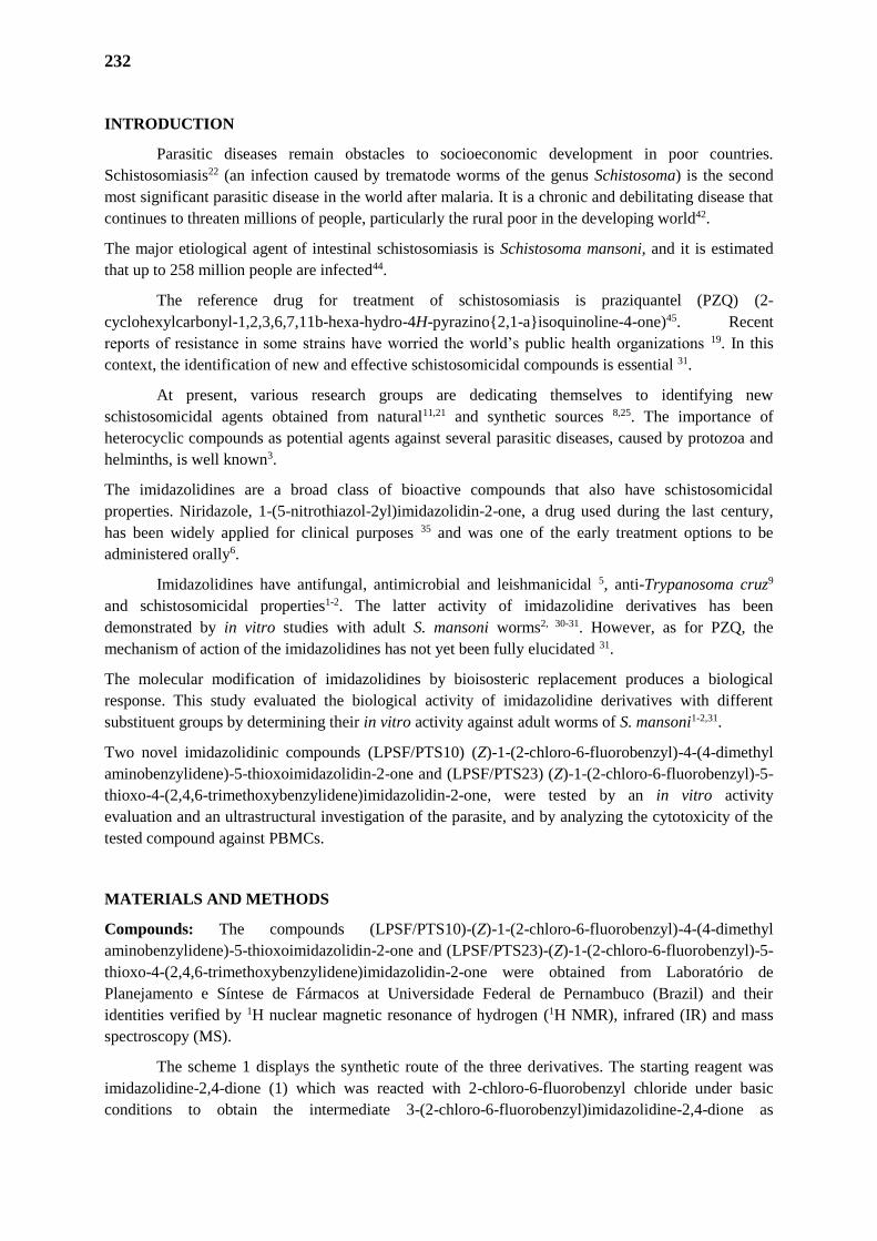
232
INTRODUCTION
Parasitic diseases remain obstacles to socioeconomic development in poor countries.
Schistosomiasis22 (an infection caused by trematode worms of the genus Schistosoma) is the second
most significant parasitic disease in the world after malaria. It is a chronic and debilitating disease that
continues to threaten millions of people, particularly the rural poor in the developing world42.
The major etiological agent of intestinal schistosomiasis is Schistosoma mansoni, and it is estimated
that up to 258 million people are infected44.
The reference drug for treatment of schistosomiasis is praziquantel (PZQ) (2-
cyclohexylcarbonyl-1,2,3,6,7,11b-hexa-hydro-4H-pyrazino{2,1-a}isoquinoline-4-one)45. Recent
reports of resistance in some strains have worried the world’s public health organizations 19. In this
context, the identification of new and effective schistosomicidal compounds is essential 31.
At present, various research groups are dedicating themselves to identifying new
schistosomicidal agents obtained from natural11,21 and synthetic sources 8,25. The importance of
heterocyclic compounds as potential agents against several parasitic diseases, caused by protozoa and
helminths, is well known3.
The imidazolidines are a broad class of bioactive compounds that also have schistosomicidal
properties. Niridazole, 1-(5-nitrothiazol-2yl)imidazolidin-2-one, a drug used during the last century,
has been widely applied for clinical purposes 35 and was one of the early treatment options to be
administered orally6.
Imidazolidines have antifungal, antimicrobial and leishmanicidal 5, anti-Trypanosoma cruz9
and schistosomicidal properties1-2. The latter activity of imidazolidine derivatives has been
demonstrated by in vitro studies with adult S. mansoni worms2, 30-31. However, as for PZQ, the
mechanism of action of the imidazolidines has not yet been fully elucidated 31.
The molecular modification of imidazolidines by bioisosteric replacement produces a biological
response. This study evaluated the biological activity of imidazolidine derivatives with different
substituent groups by determining their in vitro activity against adult worms of S. mansoni1-2,31.
Two novel imidazolidinic compounds (LPSF/PTS10) (Z)-1-(2-chloro-6-fluorobenzyl)-4-(4-dimethyl
aminobenzylidene)-5-thioxoimidazolidin-2-one and (LPSF/PTS23) (Z)-1-(2-chloro-6-fluorobenzyl)-5-
thioxo-4-(2,4,6-trimethoxybenzylidene)imidazolidin-2-one, were tested by an in vitro activity
evaluation and an ultrastructural investigation of the parasite, and by analyzing the cytotoxicity of the
tested compound against PBMCs.
MATERIALS AND METHODS
Compounds: The compounds (LPSF/PTS10)-(Z)-1-(2-chloro-6-fluorobenzyl)-4-(4-dimethyl
aminobenzylidene)-5-thioxoimidazolidin-2-one and (LPSF/PTS23)-(Z)-1-(2-chloro-6-fluorobenzyl)-5-
thioxo-4-(2,4,6-trimethoxybenzylidene)imidazolidin-2-one were obtained from Laboratório de
Planejamento e Síntese de Fármacos at Universidade Federal de Pernambuco (Brazil) and their
identities verified by 1H nuclear magnetic resonance of hydrogen (1H NMR), infrared (IR) and mass
spectroscopy (MS).
The scheme 1 displays the synthetic route of the three derivatives. The starting reagent was
imidazolidine-2,4-dione (1) which was reacted with 2-chloro-6-fluorobenzyl chloride under basic
conditions to obtain the intermediate 3-(2-chloro-6-fluorobenzyl)imidazolidine-2,4-dione as
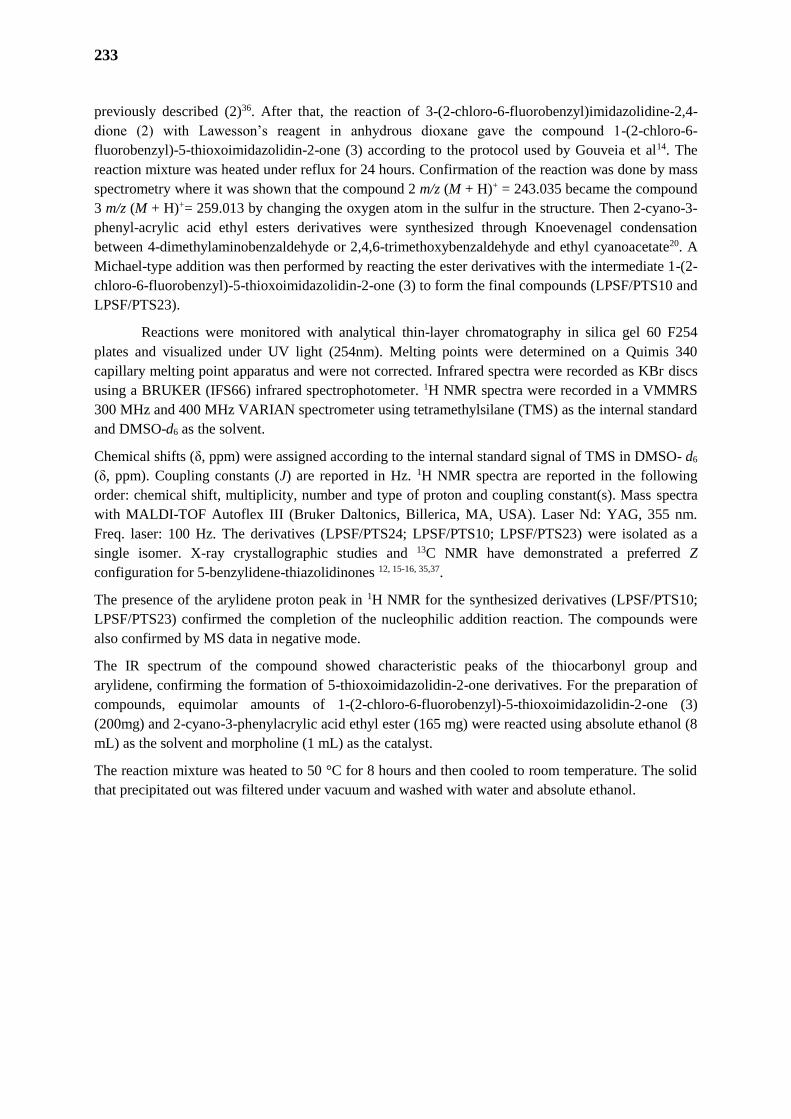
233
previously described (2)36. After that, the reaction of 3-(2-chloro-6-fluorobenzyl)imidazolidine-2,4-
dione (2) with Lawesson’s reagent in anhydrous dioxane gave the compound 1-(2-chloro-6-
fluorobenzyl)-5-thioxoimidazolidin-2-one (3) according to the protocol used by Gouveia et al14. The
reaction mixture was heated under reflux for 24 hours. Confirmation of the reaction was done by mass
spectrometry where it was shown that the compound 2 m/z (M + H)+ = 243.035 became the compound
3 m/z (M + H)+= 259.013 by changing the oxygen atom in the sulfur in the structure. Then 2-cyano-3-
phenyl-acrylic acid ethyl esters derivatives were synthesized through Knoevenagel condensation
between 4-dimethylaminobenzaldehyde or 2,4,6-trimethoxybenzaldehyde and ethyl cyanoacetate20. A
Michael-type addition was then performed by reacting the ester derivatives with the intermediate 1-(2-
chloro-6-fluorobenzyl)-5-thioxoimidazolidin-2-one (3) to form the final compounds (LPSF/PTS10 and
LPSF/PTS23).
Reactions were monitored with analytical thin-layer chromatography in silica gel 60 F254
plates and visualized under UV light (254nm). Melting points were determined on a Quimis 340
capillary melting point apparatus and were not corrected. Infrared spectra were recorded as KBr discs
using a BRUKER (IFS66) infrared spectrophotometer. 1H NMR spectra were recorded in a VMMRS
300 MHz and 400 MHz VARIAN spectrometer using tetramethylsilane (TMS) as the internal standard
and DMSO-d6 as the solvent.
Chemical shifts (δ, ppm) were assigned according to the internal standard signal of TMS in DMSO- d6
(δ, ppm). Coupling constants (J) are reported in Hz. 1H NMR spectra are reported in the following
order: chemical shift, multiplicity, number and type of proton and coupling constant(s). Mass spectra
with MALDI-TOF Autoflex III (Bruker Daltonics, Billerica, MA, USA). Laser Nd: YAG, 355 nm.
Freq. laser: 100 Hz. The derivatives (LPSF/PTS24; LPSF/PTS10; LPSF/PTS23) were isolated as a
single isomer. X-ray crystallographic studies and 13C NMR have demonstrated a preferred Z
configuration for 5-benzylidene-thiazolidinones 12, 15-16, 35,37.
The presence of the arylidene proton peak in 1H NMR for the synthesized derivatives (LPSF/PTS10;
LPSF/PTS23) confirmed the completion of the nucleophilic addition reaction. The compounds were
also confirmed by MS data in negative mode.
The IR spectrum of the compound showed characteristic peaks of the thiocarbonyl group and
arylidene, confirming the formation of 5-thioxoimidazolidin-2-one derivatives. For the preparation of
compounds, equimolar amounts of 1-(2-chloro-6-fluorobenzyl)-5-thioxoimidazolidin-2-one (3)
(200mg) and 2-cyano-3-phenylacrylic acid ethyl ester (165 mg) were reacted using absolute ethanol (8
mL) as the solvent and morpholine (1 mL) as the catalyst.
The reaction mixture was heated to 50 °C for 8 hours and then cooled to room temperature. The solid
that precipitated out was filtered under vacuum and washed with water and absolute ethanol.
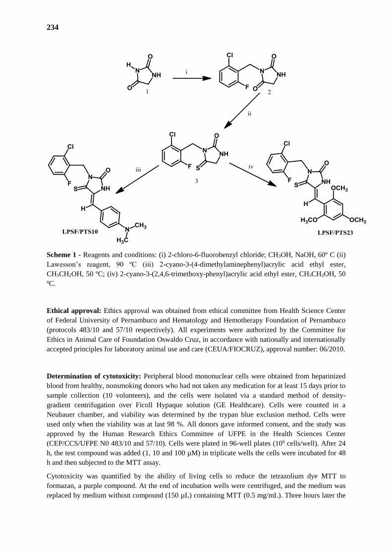
234
Scheme 1 - Reagents and conditions: (i) 2-chloro-6-fluorobenzyl chloride; CH3OH, NaOH, 60º C (ii)
Lawesson’s reagent, 90 ºC (iii) 2-cyano-3-(4-dimethylaminephenyl)acrylic acid ethyl ester,
CH3CH2OH, 50 ºC; (iv) 2-cyano-3-(2,4,6-trimethoxy-phenyl)acrylic acid ethyl ester, CH3CH2OH, 50
ºC.
Ethical approval: Ethics approval was obtained from ethical committee from Health Science Center
of Federal University of Pernambuco and Hematology and Hemotherapy Foundation of Pernambuco
(protocols 483/10 and 57/10 respectively). All experiments were authorized by the Committee for
Ethics in Animal Care of Foundation Oswaldo Cruz, in accordance with nationally and internationally
accepted principles for laboratory animal use and care (CEUA/FIOCRUZ), approval number: 06/2010.
Determination of cytotoxicity: Peripheral blood mononuclear cells were obtained from heparinized
blood from healthy, nonsmoking donors who had not taken any medication for at least 15 days prior to
sample collection (10 volunteers), and the cells were isolated via a standard method of density-
gradient centrifugation over Ficoll Hypaque solution (GE Healthcare). Cells were counted in a
Neubauer chamber, and viability was determined by the trypan blue exclusion method. Cells were
used only when the viability was at last 98 %. All donors gave informed consent, and the study was
approved by the Human Research Ethics Committee of UFPE in the Health Sciences Center
(CEP/CCS/UFPE N0 483/10 and 57/10). Cells were plated in 96-well plates (106 cells/well). After 24
h, the test compound was added (1, 10 and 100 µM) in triplicate wells the cells were incubated for 48
h and then subjected to the MTT assay.
Cytotoxicity was quantified by the ability of living cells to reduce the tetrazolium dye MTT to
formazan, a purple compound. At the end of incubation wells were centrifuged, and the medium was
replaced by medium without compound (150 µL) containing MTT (0.5 mg/mL). Three hours later the
235
MTT formazan was diluted with 100 µL of 20% SDS, and its absorbance was measured at 570 nm in a
BioTek EL808. Cytotoxic activity was quantified as the percentage reduction in absorbance relative to
a vehicle treated control. In all analyzed experiments, the vehicle (DMSO 0.1%) treated group
presented >98% viability compared to control cells without vehicle in three independent assays 32.
Antischistosomal evaluation criteria
Parasites: S. mansoni (LE strain) worms were maintained in Biomphalaria glabrata snails and Swiss
webster mice hosts at the Schistosomiasis Laboratory of the Department of Parasitology, Oswaldo
Cruz Institute, (Pernambuco, Brazil). Female mice weighing 28-30g were each exposed to around 120
cercariae.
Mice infection. Mice were infected by the tail immersion method. Briefly, mice were individually
placed in a mouse-holding chamber with their tails outside the chambers. After cleaning with
dechlorinated tap water, the tail was inserted into a vial containing 120 cercariae in 2 mL
dechlorinated tap water. Mice were left in contact with the infective cercariae for 2 h, and then
removed from the chamber, and their tails were allowed to dry. After 8 weeks, adults S. mansoni
worms were recovered from the mice by perfusion with RPMI 1640 medium supplemented with
heparin. The worms were washed in RPMI 1640 medium (Gibco) supplemented with 100 µg.mL-1 of
streptomycin, 100 UI.mL-1 of penicillin (Invitrogen), and 25 mM of Hepes. Two pairs of adult worms
(male and female) were incubated in a 24-well culture plate (Techno Plastic Products, TPP) containing
2 mL of the same medium supplemented with 10% heat-inactivated calf serum at 37 ºC in a 5% CO2
atmosphere, in three independent assays 17.
In vitro viability and motility assay with S. mansoni. For the in vitro test with S. mansoni,
LPSF/PTS10 and LPSF/PTS23 imidazolidine derivatives were the compounds rather dissolved in
DMSO and the final concentration of DMSO in the culture medium was a maximum of 1.6% dimethyl
sulphoxide (DMSO) and used in concentrations varying from 5 to 100 µM, which were added to the
medium containing the worms after a period of 2 h of adaptation to the culture medium. In the positive
control group the adult worms of S. mansoni were incubated in the presence of PZQ, triplicates were
carried out for each concentration used.
An inverted microscope and a stereomicroscope were used to evaluate the motility and survival of
worms monitored at 24, 48, 72, 96 and 120 h of incubation. Motility and survival of worms were
assessed according to the criteria scored in a viability scale of 0-3. The scoring system was as follows:
3, complete body movement; 1.5, partial body movement or immobile but alive; and 0, dead, at least
three independent assays13. Changes in the pairing and egg production, were also evaluated using an
inverted microscope.
The parasites were kept for 5 days and monitored every 24 h to evaluate their general condition: motor
activity, alterations in the tegument, and mortality rate. The control worms were treated with 1.6%
DMSO in an RPMI 1640 medium18.
Scanning Electron Microscopy (SEM): The worms were incubated for 24 h and, after their death,
they were washed with sodium cacodylate buffer (pH = 7.2), fixed with 2.5% glutaraldehyde (pH =
7.4) during 24 h, and then fixed with 1% osmium tetroxide for 1 h. The samples were dehydrated by
an increasing amount of ethanol solution, dried in a critical point dryer, and then mounted on stubs and
coated with gold using a sputter coater. The material was examined under a JEOL - 5600 LV
microscope.
236
RESULTS
Compound: (Z)-1-(2-chloro-6-fluorobenzyl)-4-(4-(dimethylaminobenzylidene)-5-thioxo
imidazolidin-2-one (LPSF/PTS10): The product was an orange solid. Formula: C19H17ClFN3OS;
M.W.: 389.8742 g/moles; Yield: 45%; Melting point: 269 – 270º C; Rf: 0.55 (n-hexane/AcOEt 6:4); 1H NMR (300MHz, DMSO- d6): δ 3.00 (s, 6H, H3C-N); 5.15 (s, 2H, CH2); 6.72 (d, 2H, H-Ar, J= 9.2
Hz); 7.57 (d, 2H, H-Ar, J= 8.8 Hz); 6.95 (s, 1H, HC=); 7.33 (m, 2H, CH-Ar, benzyl); 7.17 (t, 1H, CH-
Ar, benzyl, J= 8.8 Hz); 11.00 (s, 1H, NH). IR (KBr, cm-1): 3226.68; 1725.22; 1588.95; 1531.52. MS
[M+H]+: calculated= 390.07; found= 390.00.
(Z)-1-(2-chloro-6-fluorobenzyl)-5-thioxo-4-(2,4,6-trimethoxybenzylidene)imidazolidin-2-one
(LPSF/PTS23): The product was an orange solid. Formula: C20H18ClFN2O4S; M.W.: 436.8843
g/moles; Yield: 55%; Melting point: 152-153º C; Rf: 0.40 (n-hexane/AcOEt 6:4); 1H NMR (300MHz,
DMSO- d6): δ 3.80 9 (s, 6H, OCH3); 3.83 (s, 3H, OCH3); 5.13 (s, 2H, CH2); 6.26 (s, 2H, CH-Ar); 6.99
(s, 1H, CH=); 7.18 (t, 1H,CH-benzyl, J= 8.1 Hz); 7.34 (m, 2H, CH- benzyl); 10.08 (s, 1H, NH). IR
(KBr, cm-1): 3413.59; 1747.92; 1598.88; 1510.63. MS [M+H]+: calculated= 437.06; found= 437.00.
Schistosomicidal activity: Imidazolidine composites have previously shown action against S.
mansoni adult worms 1-2, 23-25, 34.
Initially, we performed cell viability tests with the newly synthesized imidazolidine
derivatives using peripheral blood mononuclear cells (PBMCs). Our results show that compounds
LPSF/PTS10 and LPSF/PTS23, present no toxic effects at different concentrations ranging from 5–
100 μM (Table 1).
These compounds were then evaluated for their effects on adult schistosomes at a concentration of 5 to
100 µM every 24 h for a period of 120 h, and mortality, motility, and alterations in the tegument of the
worms were observed (Table 1). PZQ was used as the reference schistosomicidal drug.
In order to evaluate the pairing and egg production by adult worms of S. mansoni, the LPSF/PTS10
and LPSF/PTS23 were tested at concentrations which cause separation of coupled adult worms and
inhibition in the egg production after 120 h of incubation.
LPSF/PTS10 and LPSF/PTS23 all demonstrated lethality against adult worms of S. mansoni,
whereas no mortality was observed for worms incubated in medium alone or in the presence of
DMSO. LPSF/PTS10 was more effective, causing 100% mortality after 72 h at 20 μM. The most
effective compound was LPSF/PTS23, which caused 100% mortality after 24 h at a concentration of
20 μM, and some mortality at 5 μM after 120 h incubation.
In these experiments PZQ induced 100% mortality of adult worms after 24 h of incubation at all
concentrations used, down to 5 μM.
Analysis of the effects of the compounds on worm motility allowed the detection of their action at
sub-lethal concentrations (Table 2). During the complete observation period (up to 120 h) the negative
control group displayed peristaltic movements and characteristic waves throughout the whole body,
with suckers in constant movement and occasionally adhering to the bottom of the culture plate (score
= 3). PZQ caused loss of motility in worms at all concentrations used and as early as 24 h after the
start of incubation leaving them shortened, this effect persisted and became stronger with the passing
of time (score=0).
After 24 h of exposure to LPSF/PTS23 and 48 h to LPSF/PTS10 at concentration of 100 µM, 100% of
the worms has lost movement completely, being then considered dead (score = 0).

237
LPSF/PTS10 at the concentration of 5µM, did not alter the motility of S. mansoni during the
observation period. However, a reduced motility was observed for 50% of worms after 24 h of
incubation at 20 μM and 75% were dead after 48 h incubation at this concentration. In the case of
LPSF/PTS23, its effects on the worms were far more radical and it was therefore not possible to detect
early effects on motility at the drug concentrations used. At 5 μM thus compound caused a reduction
in motility (score 1.5) in 50% of the worms and 25% were dead.
Observations of worm morphology using SEM showed that in the controls, the parasite tegument was
observed with oral and ventral suckers, and tubercles and spines in normal state at 120 h of incubation,
(Fig. 1A-B). Severe damage of the tegument was observed in worms incubated with PZQ,
characterized by a contraction and rupture of blisters. The emergence of several holes in the place
where the blisters had been located, and loss of spines was also seen (Fig. 1C).
The ultrastructural changes in adult schistosomes caused by LPSFPTS23 were characterized by
extensive erosion over the entire body of the worms (Fig. 1D); After 48 h, multiple bubbles and
projections (arrows) emerging from the interior of damaged tubercles (Fig. 1E); After 72 h, tegument
erosion can be visualized at higher magnification (Fig. 1F); After 96 h, some tubercles lack their
spines (arrow) (Fig. 1G).
DISCUSSION
Schistosomiasis is a neglected disease that has one drug of choice, PZQ3-5. Because of this, the Special
Program for Research and Training in Tropical Diseases promoted by WHO provides opportunities for
studies in the development of new anti-Schistosoma drugs6-8 and encourages worldwide synthesis of
new compounds for these neglected diseases10.
Imidazolidine derivatives have previously been investigated for their anti-parasitic activity1, 23-25, 34.
The main activities assigned to imidazolidines are antibacterial and anti-L. amazonensis5, anti-T. cruzi6
and anti-S. mansoni1-2, 23-25,34. Among the various schistosomicidal compounds already tested,
imidazolidine derivatives have in vitro and in vivo efficacy in several studies, giving promising results
when compared to PZQ, the only drug currently available for the treatment of schistosomiasis23-
25,29,30,34,43.
The imidazolidine derivatives used in this study, LPSF/PTS10 and LPSF/PTS23 showed no
cytotoxicity up to 100 μM on PBMCs. LPSF/PTS23 exhibited the best schistosomicidal properties in
relation to other compounds, with 100% mortality within 24 h at concentrations of 20–100 µM and
marked effects on motility and viability after 120 h at 5 μM. These results suggest that the efficacy
varies according to the substituent in the 4-position of the imidazolidine group. The compound
LPSF/PTS23 has a methoxyl group attached in the 2,4,6-position of the imidazoline ring. In a recent
study it was also shown that heterocyclic compounds (thiosemicarbazones) containing a methoxyl
group exhibited higher schistosomicidal activity than compounds without this group33.
All adult worm couples were separated into individual males and females after incubation with 20, 40
or 100 μM of compounds LPSF/PTS10 and LPSF/PTS23. Oviposition by adult worms was also not
observed at any concentration of these imidazolidines (not shown) while in the control group
(untreated worms) oviposition was observed and the worms remained viable during the entire
observation period. These same physiological alterations seen here were observed in adult worms
treated with imidazolidine derivatives in other studies realized by our group (Silva et al.34). For
example, similar results were achieved by Neves et al. 24 and Neves et al. 25 using 1-(4-chlorobenzyl)-
238
4-[(4-fluorophenyl)hydrazono]-5-thioxoimidazolidin-2-one (LPSF/PT11) and 5-(4-fluorobenzylidene-
3-(4-nitrobenzyl)-4-thioxoimidazolidin-2-one (LPSF/RSZ05).
The tegument of S. mansoni is an important structure involved in the absorption of nutrients, in
secretion of some products, variety of movements, including rapid shortening and extension of the
body, typical wavy and peristaltic movement along the body anterior and posterior axis39. Therefore,
the worm’s tegument is the critical target for imidazolidine derivates, as it has been other
schistosomicidal activity drugs studied1-2,23-25, 34.
To test this hypothesis, we first established the concentration of LPSF/PTS23 that is capable of
altering worm motility, causing death of worms in the period of a 120 h exposure in vitro. Based on
these criteria, through SEM, we observed marked changes in the surface of the tegument of S. mansoni
adult worms including extensive erosion and the emergence of bubbles and projections2. Thus, we
contribute, along with other studies, to the demonstration that imidazolidine derivatives induce surface
membrane damage to adult worms of S. mansoni27, 31, 34.
However, despite their effect on motor activity, the exact mechanisms by which LPSF/PTS10 and
LPSF/PTS23 cause mortality in adult worms remain unclear.
One possible clue is provided by the fact that, some imidazolidine derivatives show toxicity to cells 23-
25, 34. There is evidence that these compounds act on cholinergic receptors38, 41.
Silva et al.34 evaluated the schistosomicidal potential of the imidazolidinic derivative 3 (5Z)-3-(4-
bromobenzyl)-5-(4-chlorobenzylidene)-4-thioxoimidazolidin-2-one. After 24 hours of incubation at a
dose of 100µg.mL-1, adult S. mansoni worms had a significant opening of the gynecophoral canal,
contraction of the head and suckers, collapse of the tubercle with erosion of the tegument and a severe
lesion revealing the layer of subtegument tissue. In this case, there was an enormous destruction of the
subtegument surface.
Acetylcholinesterase is also found on the tegumental surface of the worm and has the function of
obtaining glucose4, but there is already evidence showing that this contraction is due to calcium influx
which also causes contraction of the muscle fibers. Since this enzyme is inhibited, the absorption of
nutrients for the survival of the parasite may be compromised26. Imidazolidinic compounds such as
(Z)-3-(4-chlorobenzyl)-5-(4-nitrobenzylidene)imidazolidine-2,4-dione, (Z)-3-(4-chloro-benzyl)-5-(4-
fluorobenzylidene)-1-methyl-2-thioxoimidazolidin-4-one and (Z)-5-(4-fluorobenzilidene)-1-methyl-3-
(4-phenylbenzyl)-2-thioxoimidazolidin-4-one induced significant changes in the tegumental surface of
the body of adult S. mansoni worms, causing damage in the tegument with contraction of the body and
of oral and ventral suckers, disorganization and total collapse of the tubercles with loss of spines.
Thus, the nitro, fluorine and phenyl radicals can justify the good activity of the imidazolidine
derivatives mentioned above 28,37.
In addition, promising results were also obtained with other imidazolidine derivatives presenting
chlorine and fluorine radicals in their structure, which were also able to cause ultrastructural changes
in the tegument of adult worms of S. mansoni, such as the derivatives 1-benzyl-4-[(4-chlorophenyl)-
hydrazono]-5-thioxoimidazolidin-2-one and 1-(4-chlorobenzyl)-4-[(4-fluorophenyl)-hydrazono]-5-
thioxoimidazolidin-2-one1-3.
Furthermore, Neves et al.25 showed disruption to the tegument, blisters, spine loss and tissue wrinkling
after contact with LPSF/PT5 and blisters and swelling of the tegument and loss of a few spines in the
tubercules after incubation with LPSF/PT11.
Thus, in these cases the presence of nitro, fluorine and phenyl radicals could explain the high activity
of the imidazolidine derivatives27 one explanation may be that halogens have the ability to enhance the
239
absorption of the derivatives by the cell membranes40. Thus, this may be the case in the tegumental
surface of the parasite treated with the above compounds as well as compounds used in the present
study which all present the halogens in their chemical structures.
Our study reinforced the use of imidazoline derivatives as drug anti-S. mansoni, and identified
LPSF/PTS23 as a lead candidate for further optimization as a potential agent against S. mansoni.
CONCLUSION
In conclusion, the imidazolidinic derivatives which presents the trimetoxy and benzylidene
halogens showed a promising in vitro schistosomicidal activity. This is the first time that the in vitro
schistosomicidal activity was reported for LPSF/PTS10 and LPSF/PTS23. Thus, it is necessary to
investigate the elucidation of the mechanism of action of this compound as well as to invest in further
studies to investigate its biological activity such as an in vivo evaluation.
ACKNOWLEDGMENTS
Fundação de Amparo à Ciência e Tecnologia do Estado de Pernambuco (FACEPE), Instituto
Nacional de Ciência e Tecnologia para Inovação Farmacêutica (INCT-if), Centro de Pesquisas Aggeu
Magalhães/Fundação Oswaldo Cruz (CPqAM/FIOCRUZ/PE) and Universidade Federal de
Pernambuco (UFPE).
COMPETING INTERESTS
The authors have declared that no competing interests exist.
CONSENT (WHERE EVER APPLICABLE)
The present study did not involve patients.
ETHICAL APPROVAL (WHERE EVER APPLICABLE)
All authors hereby declare that "Principles of laboratory animal care" (NIH publication No.
85-23, revised 1985) were followed, as well as the ethical principles of the Brazilian Society of
Laboratory Animal Science (SBCAL). This project was approved by the Animal Ethics Committee
from Centro de Pesquisa Aggeu Magalhães/Fundação Oswaldo Cruz (CPqAM/FIOCRUZ) and
authorized by the license no. 06/2010.
Síntese e avaliação biológica de novos derivados imidazolidínicos candidatos a agentes
esquistossomicidas
240
A esquistossomose é uma doença parasitária crônica causada pelos trematódeos do gênero
Schistosoma, que ameaça quase 258 milhões de pessoas no mundo e seu controle é dependente de uma
única droga, o praziquantel. O objetivo deste estudo foi avaliar a atividade anti-Schistosoma mansoni
in vitro novos derivados de imidazolidina. Nós sintetizamos dois novos derivados de imidazolidina:
(LPSF/PTS10) (Z) -1- (2-cloro-6-fluoro-benzil) -4- (4- dimetil aminobenzilideno) -5-tioxo-
imidazolidin-2-ona e (LPSF/PTS23) (Z) -1- (2-cloro-6-fluoro-benzil) -5-tioxo-4- (2,4,6-trimetoxi-
benzilideno)-imidazolidin-2-ona. As estruturas destes compostos foram determinadas por meio de
métodos espectroscópicos. Durante os ensaios, parâmetros como a motilidade, oviposição, mortalidade
e análise de microscopia eletrônica de varredura foram avaliados. LPSF/PTS10 e LPFS/PTS23 foram
considerados ativos na separação ativa de pares acoplados, mortalidade e diminuição da atividade
motora, enquanto LPSF/PTS24 só causou mortalidade de vermes após 120 h de incubação. Além
disso, LPSF/PTS23 induziu alterações ultraestruturais em vermes, após 24 h de contato, causando
erosão extensiva ao longo de todo o corpo dos vermes. Os derivados imidazolidínicos que apresentam
os halogênios trimetoxi e benzilideno mostraram uma promissora atividade esquistossomicida in vitro.
REFERENCES
1. Albuquerque MCA, Silva TG, Pitta MGR, Silva ACA, Silva PG, Malagueño, E, Santana JV,
Wanderley AG, Lima MCA, Galdino SL, Barbe J, Pitta IR. Synthesis and schistosomicidal
activity of new substituted thioxoimidazolidine compounds. Pharmazie. 2005; 60: 13-7.
2. Albuquerque MCA, Pitta MGR, Irmão JI, Malagueño, E, Santana JV, Lima MCA, Galdino SL,
Pitta IR. Tegumental alterations in adult Schistosoma mansoni treated with imidazolidine
derivatives.. Acta Farmaceutica Bonaerense. 2007; 26: 65-69.
3. Brandão SSF, Andrade AMC, Pereira DTM, Barbosa-Filho JM, Lima MCA, Galdino SL, Pitta IR,
Barbe J. A novel way of synthesis of 1,3,5-trisubstituted-2-thioxoimidazolidinones. Heterocycl
Comm. 2004; 10: 9-14.
4. Camacho M, Tarrab-Hazdai R, Espinoza B, Arnon R, Agnew A. The amount of
acetylcholinesterase on the parasite surface reflects the differential sensitivity of schistosome
species to metrifonate. Parasitology. 1994; 108:153-60.
5. Carvalho GS, Dias RM, Pavan FR, Leite CQ, Silva VL, Diniz CG, Paula DT, Coimbra ES,
Retailleau P, Silva AD. Synthesis, cytotoxicity, antibacterial and antileishmanial activities of
imidazolidine and hexahydropyrimidine derivatives. Med Chem. 2013; 9: 351-9.
6. Caterina MC, Perillo IA, Boiani L, Pezaroglo H, Cerecetto H, González M. Imidazolidines as new
anti-Trypanosoma cruzi agents: biological evaluation and structure-activity relationships. Bioorg
Med Chem. 2008; 16: 2226-34.
7. Cioli D, Pica-Mattoccia L, Archer S. “Antischistosomal drugs: past, present ... and future?”
Pharmacology and Therapeutics. 1995; 68: 35-85.
8. Cowan N, Yaremenko IA, Krylov IB, Terent'ev AO, Keiser J. Elucidation of the in vitro
and in vivo activities of bridged 1,2,4-trioxolanes, bridged 1,2,4,5-
tetraoxanes, tricyclic monoperoxides, silyl peroxides, and hydroxylamine

241
derivatives against Schistosoma mansoni. Bioorg Med Chem. 2015; 15;23(16):5175-
81.
9. Dias LC, Dessoy MA, Silva JJN, Thiemann OH, Oliva G, Andricopulo AD, Quimioterapia da
Doença de Chagas: Estado da Arte e Perspectivas no Desenvolvimento de Novos Fármacos.
Química Nova. 2009; 15: 2444-57.
10. Dubois F, Caby S, Oger F, Cosseau C, Capron M, Grunau C, Dissous C, Pierce RJ. Histone
deacetylase inhibitors induce apoptosis, histone hyperacetylation and up-regulation of gene
transcription in Schistosoma mansoni. Mol Biochem Parasitol. 2009; 168: 7-15.
11. Fabri RL, Aragão DM, Florêncio JR, Pinto Nde C, Mattos AC, Coelho PM, Castañon MC,
Vasconcelos EG, Pinto Pde F, Scio E. Chromatographic fingerprint analysis and effects of the
medicinal plant species Mitracarpus frigidus on adult Schistosoma mansoni worms. Biomed Res
Int. 2014; 2014:9413-8.
12. Handzlik J, Szymanska E, Wojcik R, Dela A, Jastrzebska-Wiesek M, Karolak-Wojciechowska J,
Fruzinski A, Siwek A, Filipek B, Kiec-Kononowicz K. Synthesis and SAR-study for novel
arylpiperazine derivatives of 5-arylidenehydantoin with a1-adrenoceptor antagonistic properties.
Bioorg Med Chem. 2012; 20: 4245-7.
13. Horiuchi A, Satou T, Akao N, Koike K, Fujita K, Nikaido T. The effect of free and polyethylene
glycol-liposome-entrapped albendazole on larval mobility and number in Toxocara canis infected
mice. Vet Parasitol. 2005; 129: 83-7.
14. Gouveia FL, Oliveira RMB, Oliveira TB, Silva IM, Nascimento SC, Sena KXFR, Albuquerque
JFC. Synthesis, antimicrobial and cytotoxic activities of some 5-arylidene-4-thioxo-thiazolidine-2-
ones. Eur J Med Chem. 2009; 44: 2038-43.
15. Guarda VLM, Pereira MA, De Simone CA, Albuquerque JC, Galdino SL, Chantegrel J, Perrissin
M, Beney C, Thomasson F, Pitta IR, Luu-Duc C. Synthesis and structural study of arylidene
thiazolidine and benzothiazine compounds. Sulfur Letters. 2003; 26: 17-27.
16. Karolak-Wojciechowska J, Szymanska E, Mrozek A, Kiec-Kononowicz K. Crystallographic and
spectroscopic studies of 5-arylidene-2-amino-imidazol-4-ones. J Mol Struct. 2009; 930: 126-34.
17. Lima CM, Freitas FI, Morais LC, Cavalcanti MG, Silva LF, Padilha RJ, Barbosa CG, Santos FA,
Alves LC, Diniz MF. Ultrastructural study on the morphological changes to male worms of
Schistosoma mansoni after in vitro exposure to allicin. Rev Soc Bras Med Trop. 2011, 44, 327.
18. Matos-Rocha TJ, Cavalcanti MGS, Barbosa-Filho JM, Lúcio AS, Veras DL, Feitosa AP, Siqueira
Júnior JP, Almeida RN, Marques MO, Alves LC, Brayner FA. In vitro evaluation of
schistosomicidal activity of essential oil of Mentha x villosa and some of its chemical constituents
in adult worms of Schistosoma mansoni. Planta Med. 2013; 79: 1307-12.
19. Melman SD, Steinauer ML, Cunningham C, Kubatko LS, Mwangi IN, Wynn NB, Mutuku MW,
Karanja DM, Colley DG, Black CL. Reduced susceptibility to praziquantel among naturally
occurring Kenyan isolates of Schistosoma mansoni. PloS Negl Trop Dis. 2009; 3: e504.
20. Mourão RH, Silva TG, Soares ALM, Vieira ES, Santos JN, Lima MCA, Lima VLM, Galdino SL,
Barbe J, Pitta IR. Eur. J. Med. Chem. 2005, 40, 1129. Synthesis and Biological Activity of Novel
Acridinylidene and Benzylidene thiazolidinediones. Eur J Med Chem. 2005; 40: 1129-33.
242
21. Moraes J, Carvalho AA, Nakano E, Almeida AA, Marques TH, Andrade LN, Freitas RM, Sousa
DP. Anthelmintic activity of carvacryl acetate against Schistosoma mansoni. Parasitol Res. 2013;
112: 603-10.
22. Nascimento GL, Oliveira MR. Severe forms of schistosomiasis mansoni: epidemiologic and
economic impact in Brazil, 2010. Trans R Soc Trop Med Hyg. 2014; 108: 29-36.
23. Neves JKAL, Sarinho S, Melo CML, Pereira VRA, Lima MCA, Pitta IR, Albuquerque MCP,
Galdino SL. Immunological studies and in vitro schistosomicide action of new imidazolidine
derivatives. J Venom Anim Toxins Incl Trop Dis. 2011; 17: 277-86.
24. Neves JK, Botelho SP, Melo CM, Pereira VR, Lima MCA, Pitta IR, Albuquerque MCP, Galdino
SL. Biological and immunological activity of new imidazolidines against adult worms of
Schistosoma mansoni. Parasitol Res. 2010; 107: 531-8.
25. Neves JK, Lima MCA, Pereira VR, Melo CM, Peixoto CA, Pitta IR, Albuquerque MC, Galdino
SL. Antischistosomal action of thioxo-imidazolidine compounds: An ultrastructural and
cytotoxicity study. Exp Parasitol. 2011; 128: 82-90.
26. Noel, F. Sistema neuromuscular e controle da motilidade do verme adulto. In: Carvalho OS,
Coelho PMZ, Lenzi HL. Schistosoma mansoni & Esquistossomose: Uma interdisciplinary vision.
Rio de Janeiro: Fiocruz. 2008; 207-44.
27. Oliveira SM, Albuquerque MCP, Pitta MGR, Malagueño E, Santana JV, Lima MCA, Pitta IR,
Galdino SL. A Resposta do Schistosoma mansoni Mantido In Vitro frente a Derivados
Imidazolidinônicos. Acta Farm Bonaerense. 2004; 3: 343-8.
28. Paula FR, Serrano SHP, Tavares LC Aspects of bioactivity and toxicity of nitrocompounds. New
Quim. 2009; 32: 1013-20.
29. Pica-Mattoccia L, Cioli D. Sex- and stage-related sensitivity of Schistosoma mansoni to in vivo
and in vitro praziquantel treatment. Int J Parasitol. 2004; 34: 527-33.
30. Pitta IR, Lima MCA, Albuquerque MCP, Galdino SL. Braz. Patent PI, PI-0305000-9, 2005; 25.
31. Pitta MGR, Pitta MGR, Rêgo MJBM, Galdino SL. The evolution of drugs on schistosoma
treatment: looking to the past to improve the future. Mini Rev Med Chem. 2013; 13: 493-508.
32. Rêgo MJBM, Galdino-Pitta MR, Pereira DTM, Silva JC, Rabello MM, Lima MCA, Hernandes
MZ, Pitta IR, Galdino SL, Pitta MGR. Synthesis, in vitro anticancer activity and in silico study of
new disubstituted thiazolidinedione derivatives. Med Chem Res. 2014; 23: 3220-6.
33. Santiago EF, Oliveira SA, Oliveira GB, Moreira DR, Gomes PA, Silva AL, Barros AF, Silva
AC, Santos TA, Pereira VR, Gonçalves GG, Brayner FA, Alves LC, Wanderley AG, Leite AC.
Evaluation of the Anti-Schistosoma mansoni Activity of Thiosemicarbazones and Thiazoles.
Antimicrob Agents Chemother. 2014, 58, 352-63.
34. Silva AL, Oliveira SA, Oliveira JF, Santiago EF, Almeida Júnior ASA, Jacobi ITT, Peixoto CA,
Rocha VPC, Soares MBP, Pitta IR, Lima MCA. Tegumental Changes in Adult Schistosoma
mansoni Induced by a New Imidazolidinic Derivative. Brit J Pharm Res. 2014; 4: 1988-2005.
35. Simone CA, Zukerman-Schpector J, Pereira MA, Luu-Duc C, Pitta IR, Galdino SL, Amorim ELC.
3-(4-Bromobenzyl)-5-(4-fluorobenzylidene)-imidazolidine-2,4-dione. Acta Crystallog. 1995;
2620-1.
243
36. Sudo RT, Calasans-Maia JA, Galdino SL, Lima MCA, Zapata-Sudo, G, Hernandes MZ, Pitta IR.
Interaction of Morphine With a New α2-Adrenoceptor Agonist in Mice. J Pain. 2010; 11: 71-78.
37. Tan F, Ang KP, Fong YF. (Z)- and (E)-5-arylmethylenehydantoins: spectroscopic properties and
configuration assignment Journal of the Chemical Society, Perkin Transactions II. 1986; 12: 1941-
4.
38. Tallima H, Ridi R. Praziquantel binds Schistosoma mansoni adult worm actin. Int J Antimicrob
Agents. 2007; 29: 570-5.
39. Taman A, Ribeiro P. Glutamate-mediated signaling in Schistosoma mansoni: A novel glutamate
receptor is expressed in neurons and the female reproductive tract. Mol Biochem Parasitol. 2011;
176:42-50.
40. Thomas G. Medicinal chemistry: an introduction. Editora Guanabara Koogan; 2003.
41. Thibaut JPB, Monteiro LM, Leite LCC, Menezes CMS, Lima LM, Noel F. The effects of 3-
methylclonazepam on Schistosoma mansoni musculature are not mediated by benzodiazepine
receptors. Eur J Pharmacol. 2009; 606: 9-16.
42. Vimieiro AC, Araujo N, Katz N, Kusel JR, Coelho PM. Schistogram changes after administration
of antischistosomal drugs in mice at the early phase of Schistosoma mansoni infection. Mem Inst
Oswaldo Cruz. 2013; 108: 881-6.
43. Werbel LM, Elslager EF, Islip PJ, Closier MD. Antischistosomal effects of 5-(2,4,5-
trichlorophenyl)hydantoin and related compounds. J Med Chem. 1977; 20:1569-72.
44. World Health Organization. Wkly. Epidemiol. Rec. 2016, 86, 73.
45. World Health Organization. WHO Geneva Fact Sheet, 2012,115.
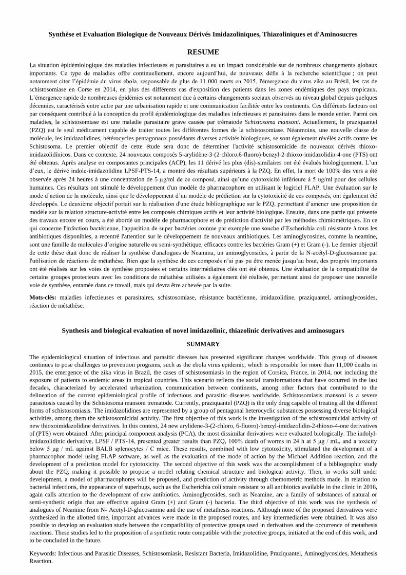
244
Synthèse et Evaluation Biologique de Nouveaux Dérivés Imidazoliniques, Thiazoliniques et d'Aminosucres
RESUME
La situation épidémiologique des maladies infectieuses et parasitaires a eu un impact considérable sur de nombreux changements globaux
importants. Ce type de maladies offre continuellement, encore aujourd’hui, de nouveaux défis à la recherche scientifique ; on peut
notamment citer l’épidémie du virus ebola, responsable de plus de 11 000 morts en 2015, l'émergence du virus zika au Brésil, les cas de
schistosomiase en Corse en 2014, en plus des différents cas d'exposition des patients dans les zones endémiques des pays tropicaux.
L’émergence rapide de nombreuses épidémies est notamment due à certains changements sociaux observés au niveau global depuis quelques
décennies, caractérisés entre autre par une urbanisation rapide et une communication facilitée entre les continents. Ces différents facteurs ont
par conséquent contribué à la conception du profil épidémiologique des maladies infectieuses et parasitaires dans le monde entier. Parmi ces
maladies, la schistosomiase est une maladie parasitaire grave causée par trématode Schistosoma mansoni. Actuellement, le praziquantel
(PZQ) est le seul médicament capable de traiter toutes les différentes formes de la schistosomiase. Néanmoins, une nouvelle classe de
molécule, les imidazolidines, hétérocycles pentagonaux possédants diverses activités biologiques, se sont également révélés actifs contre les
Schistosoma. Le premier objectif de cette étude sera donc de déterminer l'activité schistosomicide de nouveaux dérivés thioxo-
imidazolidínicos. Dans ce contexte, 24 nouveaux composés 5-arylidène-3-(2-chloro,6-fluoro)-benzyl-2-thioxo-imidazolidin-4-one (PTS) ont
été obtenus. Après analyse en composantes principales (ACP), les 11 dérivé les plus (dis)-similaires ont été évalués biologiquement. L’un
d’eux, le dérivé indole-imidazolidine LPSF-PTS-14, a montré des résultats supérieurs à la PZQ. En effet, la mort de 100% des vers a été
observée après 24 heures à une concentration de 5 g/ml de ce composé, ainsi qu’une cytotoxicité inférieure à 5 ug/ml pour des cellules
humaines. Ces résultats ont stimulé le développement d'un modèle de pharmacophore en utilisant le logiciel FLAP. Une évaluation sur le
mode d’action de la molécule, ainsi que le développement d’un modèle de prédiction sur la cytotoxicité de ces composés, ont également été
développés. Le deuxième objectif portait sur la réalisation d'une étude bibliographique sur le PZQ, permettant d’amener une proposition de
modèle sur la relation structure-activité entre les composés chimiques actifs et leur activité biologique. Ensuite, dans une partie qui présente
des travaux encore en cours, a été abordé un modèle de pharmacophore et de prédiction d'activité par les méthodes chimiométriques. En ce
qui concerne l'infection bactérienne, l'apparition de super bactéries comme par exemple une souche d’Escherichia coli résistante à tous les
antibiotiques disponibles, a recentré l'attention sur le développement de nouveaux antibiotiques. Les aminoglycosides, comme la neamine,
sont une famille de molécules d’origine naturelle ou semi-synthétique, efficaces contre les bactéries Gram (+) et Gram (-). Le dernier objectif
de cette thèse était donc de réaliser la synthèse d'analogues de Neamina, un aminoglycosides, à partir de la N-acétyl-D-glucosamine par
l'utilisation de réactions de métathèse. Bien que la synthèse de ces composés n’ai pas pu être menée jusqu’au bout, des progrès importants
ont été réalisés sur les voies de synthèse proposées et certains intermédiaires clés ont été obtenus. Une évaluation de la compatibilité de
certains groupes protecteurs avec les conditions de métathèse utilisées a également été réalisée, permettant ainsi de proposer une nouvelle
voie de synthèse, entamée dans ce travail, mais qui devra être achevée par la suite.
Mots-clés: maladies infectieuses et parasitaires, schistosomiase, résistance bactérienne, imidazolidine, praziquantel, aminoglycosides,
réaction de métathèse.
Synthesis and biological evaluation of novel imidazolinic, thiazolinic derivatives and aminosugars
SUMMARY
The epidemiological situation of infectious and parasitic diseases has presented significant changes worldwide. This group of diseases
continues to pose challenges to prevention programs, such as the ebola virus epidemic, which is responsible for more than 11,000 deaths in
2015, the emergence of the zika virus in Brazil, the cases of schistosomiasis in the region of Corsica, France, in 2014, not including the
exposure of patients to endemic areas in tropical countries. This scenario reflects the social transformations that have occurred in the last
decades, characterized by accelerated urbanization, communication between continents, among other factors that contributed to the
delineation of the current epidemiological profile of infectious and parasitic diseases worldwide. Schistosomiasis mansoni is a severe
parasitosis caused by the Schistosoma mansoni trematode. Currently, praziquantel (PZQ) is the only drug capable of treating all the different
forms of schistosomiasis. The imidazolidines are represented by a group of pentagonal heterocyclic substances possessing diverse biological
activities, among them the schistosomicidal activity. The first objective of this work is the investigation of the schistosomicidal activity of
new thioxoimidazolidine derivatives. In this context, 24 new arylidene-3-(2-chloro, 6-fluoro)-benzyl-imidazolidin-2-thioxo-4-one derivatives
of (PTS) were obtained. After principal component analysis (PCA), the most dissimilar derivatives were evaluated biologically. The indolyl-
imidazolidinic derivative, LPSF / PTS-14, presented greater results than PZQ, 100% death of worms in 24 h at 5 μg / mL, and a toxicity
below 5 μg / mL against BALB splenocytes / C mice. These results, combined with low cytotoxicity, stimulated the development of a
pharmacophor model using FLAP software, as well as the evaluation of the mode of action by the Michael Addition reaction, and the
development of a prediction model for cytotoxicity. The second objective of this work was the accomplishment of a bibliographic study
about the PZQ, making it possible to propose a model relating chemical structure and biological activity. Then, in works still under
development, a model of pharmacophores will be proposed, and prediction of activity through chemometric methods made. In relation to
bacterial infections, the appearance of superbugs, such as the Escherichia coli strain resistant to all antibiotics available in the clinic in 2016,
again calls attention to the development of new antibiotics. Aminoglycosides, such as Neamine, are a family of substances of natural or
semi-synthetic origin that are effective against Gram (+) and Gram (-) bacteria. The third objective of this work was the synthesis of
analogues of Neamine from N- Acetyl-D-glucosamine and the use of metathesis reactions. Although none of the proposed derivatives were
synthesized in the allotted time, important advances were made in the proposed routes, and key intermediaries were obtained. It was also
possible to develop an evaluation study between the compatibility of protective groups used in derivatives and the occurrence of metathesis
reactions. These studies led to the proposition of a synthetic route compatible with the protective groups, initiated at the end of this work, and
to be concluded in the future.
Keywords: Infectious and Parasitic Diseases, Schistosomiasis, Resistant Bacteria, Imidazolidine, Praziquantel, Aminoglycosides, Metathesis
Reaction.


![Synthesis, Characterization and Biological Evaluation of ... · pynthesis, Characterization and Biological evaluation of some novel myrazolo IR-a]Pyrimidine derivatives kilesh M](https://img.pdfslide.us/doc/110x75/60f4066a1c78f1609b715fe2/synthesis-characterization-and-biological-evaluation-of-pynthesis-characterization.jpg)