Embed Size (px)
Citation preview
Iranian Journal of Veterinary Medicine
IJVM (2015), 9(2): 143
Syndactyly and polydactyly in a mixed-breed dogParyani, M.R.
Department of Basic Sciences, Faculty of Veterinary Medicine, Karaj branch, Islamic Azad University, Karaj, Iran
Abstract:Polydactyly is the congenital presence of one or more extra
digits. The extra digit often does not contain a full complement of bones. Unilateral syndactyly and polydactyl mixed-breed dog was used as a case study. A three-month old unvaccinated intact female mixed breed dog weighing 8 kg, presented with a deformity of the left forepaw was used for this study. The deformity had been present since birth according to the own-er. During physical examination the dog was given a lameness score of 0/5 for the affected limb. A complete blood count, and serum biochemistry did not reveal any abnormalities. Radio-graphs of both distal forelimbs were obtained. These showed Fused 2nd and 3rd metacarpal bones, a rudimentary metacarpi and a hypoplastic digit located on the axial side of the distal end of the left forelimb. As a result of the lameness score of 0/5 and the owner’s lack of interest, a decision was made to moni-tor the condition and consider surgery once the dog had grown larger. It seems this is the first case of polydactyly associated with syndactyly and the rudimentary metacarpal bone.
Key words:polydactyly, syndatyly, dog
CorrespondenceParyani, M.R.Department of Basic Sciences, Faculty of Veterinary Medicine, Karaj branch, Islamic Azad Uni-versity, Karaj, IranTel: +98(26) 34182548Fax: +98(26) 34402251Email: mrparyani@ kiau.ac.ir
Received: 6 December 2014Accepted: 24 February 2015
Case History
Numerous congenital bone dysmorpholo-gies have been described in animals, includ-ing; Amelia, Hemimelia, Syndactyly, Ectro-dactyly, Polydactyly etc.
Ectrodactyly, split-hand or cleft-hand is characterized by a paraxial longitudinal defi-ciency of one or more of the individual ele-ments of the distal end of the limb of a de-veloping embryo (Giofre et al., 2004). It is grossly characterized by longitudinal bony and/or soft tissue cleft in the distal end of the limb (Barrand, 2004).
Polydactyly is the congenital presence of one or more extra digits. The extra digit often does not contain a full complement of bones (Towle et al., 2004).
Polydactyly has been described in veter-
inary literature, examples can be found in horse (Giofre et al., 2004), goat (Al-Ani et al., 1997), foal (Castanjen et al., 2007), bird (Cro-sta et al., 2002), cattle (Castanjen et al., 2001), arctic foxes (Gugolek et al., 2011), roe deer (Chapman et al., 2005) and cat (Lockwood et al., 2009). Ectrodactylism is also reported in veterinary literature, especially in dogs (Car-rig et al., 1981; Bingel et al., 1997; Innes et al., 2001; Barrand, 2004) and cat (Searle, 1953).
To the author’s knowledge, this is the first case of polydactyly associated with syndacty-ly and the rudimentary metacarpal bone.
Clinical Presentation
Three months old, unvaccinated intact fe-male mixed breed dog weighing 8 kg present-ed with a deformity of left forepaw was used for this study. The deformity had been present
143-147
IJVM (2015), 9(2):144
since birth according to the owner. The dog was in good general condition. Body condi-tion score was 3.5. The owner reported that the acute onset of lameness occurred at the begin-ning of exercise.
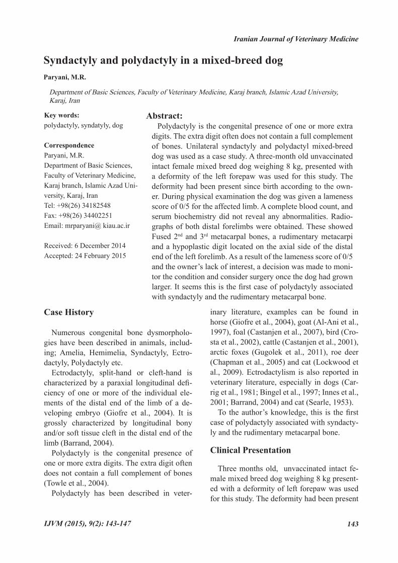
Orthopedic examination of the left forepaw revealed separated metacarpal pad and one of the digital pads had two digits (Figs. 1 and 2). Palpation of the paws, carpus and elbow of the limb did not elicit pain. No soft tissue mass was palpated and the dog was given a lame-ness score of 0/5 (None) for the affected limb.
Diagnostic Testing
A complete blood count, and serum bio-
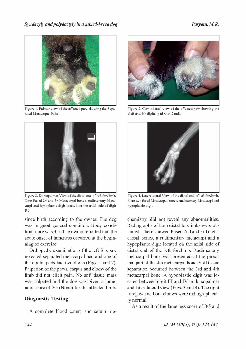
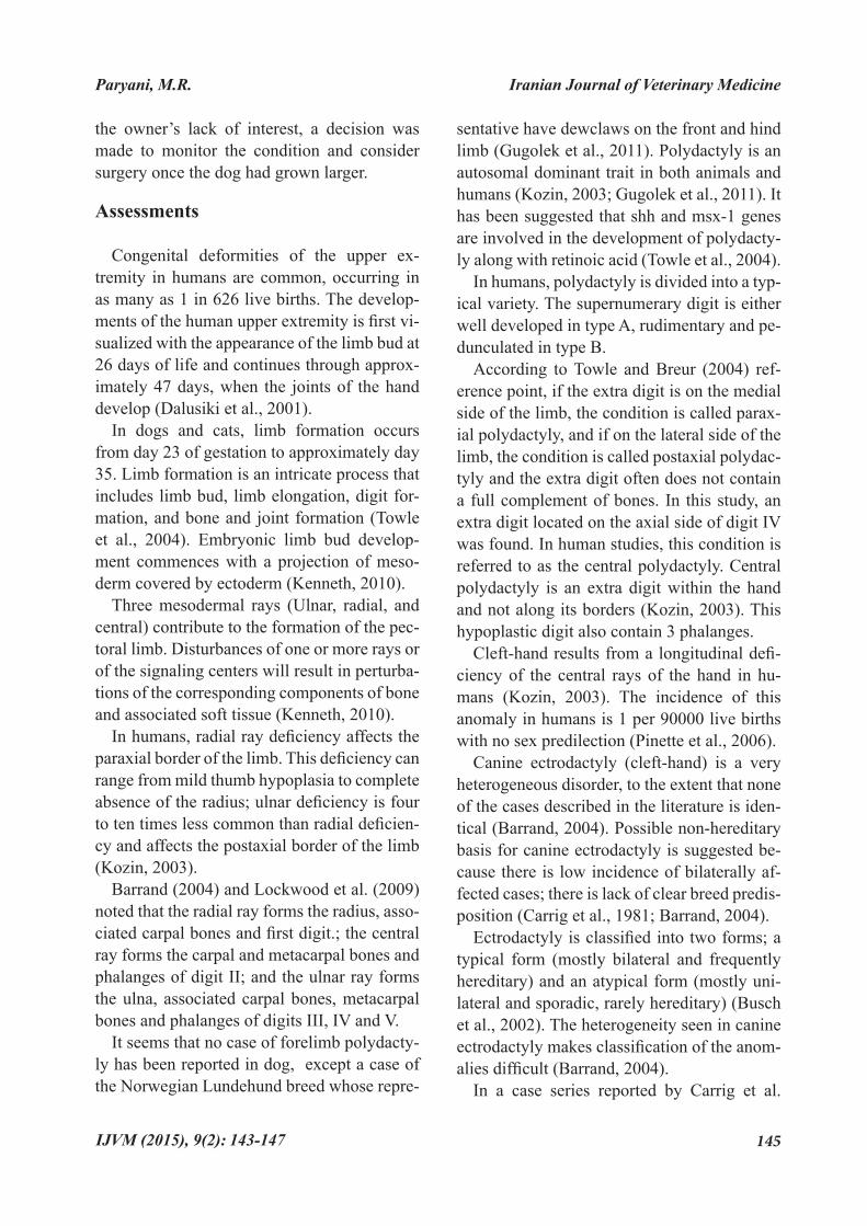
chemistry, did not reveal any abnormalities. Radiographs of both distal forelimbs were ob-tained. These showed Fused 2nd and 3rd meta-carpal bones, a rudimentary metacarpi and a hypoplastic digit located on the axial side of distal end of the left forelimb. Rudimentary metacarpal bone was presented at the proxi-mal part of the 4th metacarpal bone. Soft tissue separation occurred between the 3rd and 4th metacarpal bone. A hypoplastic digit was lo-cated between digit III and IV in dorsopalmar and laterolateral view (Figs. 3 and 4). The right forepaw and both elbows were radiographical-ly normal.
As a result of the lameness score of 0/5 and
Figure 1. Palmar view of the affected paw showing the Sepa-rated Metacarpal Pads.
Figure 3. Dorsopalmar View of the distal end of left forelimb. Note Fused 2nd and 3rd Metacarpal bones, rudimentary Meta-carpi and hypoplastic digit located on the axial side of digit IV.
Figure 2. Carniodorsal view of the affected paw showing the cleft and 4th digital pad with 2 nail.
Figure 4. Laterolateral View of the distal end of left forelimb. Note two fused Metacarpal bones, rudimentary Metacarpi and hypoplastic digit.
Syndacyly and polydactyly in a mixed-breed dog Paryani, M.R.
143-147
Iranian Journal of Veterinary Medicine
IJVM (2015), 9(2): 145
the owner’s lack of interest, a decision was made to monitor the condition and consider surgery once the dog had grown larger.
Assessments
Congenital deformities of the upper ex-tremity in humans are common, occurring in as many as 1 in 626 live births. The develop-ments of the human upper extremity is first vi-sualized with the appearance of the limb bud at 26 days of life and continues through approx-imately 47 days, when the joints of the hand develop (Dalusiki et al., 2001).
In dogs and cats, limb formation occurs from day 23 of gestation to approximately day 35. Limb formation is an intricate process that includes limb bud, limb elongation, digit for-mation, and bone and joint formation (Towle et al., 2004). Embryonic limb bud develop-ment commences with a projection of meso-derm covered by ectoderm (Kenneth, 2010).
Three mesodermal rays (Ulnar, radial, and central) contribute to the formation of the pec-toral limb. Disturbances of one or more rays or of the signaling centers will result in perturba-tions of the corresponding components of bone and associated soft tissue (Kenneth, 2010).
In humans, radial ray deficiency affects the paraxial border of the limb. This deficiency can range from mild thumb hypoplasia to complete absence of the radius; ulnar deficiency is four to ten times less common than radial deficien-cy and affects the postaxial border of the limb (Kozin, 2003).
Barrand (2004) and Lockwood et al. (2009) noted that the radial ray forms the radius, asso-ciated carpal bones and first digit.; the central ray forms the carpal and metacarpal bones and phalanges of digit II; and the ulnar ray forms the ulna, associated carpal bones, metacarpal bones and phalanges of digits III, IV and V.
It seems that no case of forelimb polydacty-ly has been reported in dog, except a case of the Norwegian Lundehund breed whose repre-
sentative have dewclaws on the front and hind limb (Gugolek et al., 2011). Polydactyly is an autosomal dominant trait in both animals and humans (Kozin, 2003; Gugolek et al., 2011). It has been suggested that shh and msx-1 genes are involved in the development of polydacty-ly along with retinoic acid (Towle et al., 2004).
In humans, polydactyly is divided into a typ-ical variety. The supernumerary digit is either well developed in type A, rudimentary and pe-dunculated in type B.
According to Towle and Breur (2004) ref-erence point, if the extra digit is on the medial side of the limb, the condition is called parax-ial polydactyly, and if on the lateral side of the limb, the condition is called postaxial polydac-tyly and the extra digit often does not contain a full complement of bones. In this study, an extra digit located on the axial side of digit IV was found. In human studies, this condition is referred to as the central polydactyly. Central polydactyly is an extra digit within the hand and not along its borders (Kozin, 2003). This hypoplastic digit also contain 3 phalanges.
Cleft-hand results from a longitudinal defi-ciency of the central rays of the hand in hu-mans (Kozin, 2003). The incidence of this anomaly in humans is 1 per 90000 live births with no sex predilection (Pinette et al., 2006).
Canine ectrodactyly (cleft-hand) is a very heterogeneous disorder, to the extent that none of the cases described in the literature is iden-tical (Barrand, 2004). Possible non-hereditary basis for canine ectrodactyly is suggested be-cause there is low incidence of bilaterally af-fected cases; there is lack of clear breed predis-position (Carrig et al., 1981; Barrand, 2004).
Ectrodactyly is classified into two forms; a typical form (mostly bilateral and frequently hereditary) and an atypical form (mostly uni-lateral and sporadic, rarely hereditary) (Busch et al., 2002). The heterogeneity seen in canine ectrodactyly makes classification of the anom-alies difficult (Barrand, 2004).
In a case series reported by Carrig et al.
Paryani, M.R.
143-147
IJVM (2015), 9(2):146
(1981) metacarpal bone hypoplasia adjacent to the plane of separation was noted in 13 limbs. It was also found in this study with separation between Digits II and III.
Barrand (2004) stated that the absence of compartments in the distal end of the forelimb occurs due to concurrent abnormality of the ul-nar and radial rays. In the case described here, the hypoplastic metacarpus and the digit adja-cent to digit IV fused with metacarpus II and III, this suggests Central and Ulnar ray abnor-mality.
In the split-hand anomaly in humans, the thumb is often absent (Kelikian, 1974). In Bar-rand’s (2004) study, this absence of digit I was also described. No abnormality was found in digit I.
On the basis of chick, mice, and human studies, mutations are suspected to play a role in canine and feline dysostoses, although none has been identified. Environmental factors have also been implicated in the development of dysostoses. Such environmental factors may include drugs, maternal disease, faulty mater-nal diet, modified-live vaccines, radiation, and trauma to the mother, embryo, or placenta. Therefore, most canine and feline dysostoses may be mutations (Towle et al., 2004).
In conclusion, Polydactyly is an autosomal dominant trait. The cause of the condition in the present case could not be ascertained. The defect is present at birth and prognosis is con-sidered excellent, it is necessary that breeding of these animals should be discouraged.
Acknowledgments
I am very grateful to Dr. S. M. Rajae and Dr. R. Sadjadi for their assistance.
Al-Ani, F.K., Hailat, N.Q., Fathalla M.A. (1997) Polydactyly in Shami breed goats in jordan. Small Rumin Res. 26: 177-179.
1. References
Syndacyly and polydactyly in a mixed-breed dog Paryani, M.R.
Barrand, K.R. (2004) Ectrodactyly in a West Highland white terrier. J Small Anim Pract. 45: 315–318.Bingel, S.A., Riser, W.H. (1997) Congenital elbow luxation in the the dog. J Small Anim Pract. 18: 445-456.Busch, R., Kjaer, K. (2002) Ectrodactyly and Germany’s eugenics law of 14 July 1933. Am J Med Genet. 110: 184-190.Carrig, C.B., Wortman, J.A., Morris, E.L., Blevins, W.E., Root, C.R., Hanlon, G.F., Sut-er, P.F. (1981) Ectrodactyly (split-hand defor-mity) in the dog. Vet Radiol Ultrasound. 22: 123-144.Carstanjen, B., Abitbol, M., Desbois, C. (2007) Bilateral polydactyly in a foal. J Vet Sci. 8: 201–203.Carstanjen, B., Pennecke, J., Boehart, S., Muller, K.E. (2001) Unilateral polydactylism in a German Holstein-Friesian calf- A case re-port. Thai J Vet Med. 40: 69-74.Chapman, N.G. (2005) Polydactyly in roe deer (Capreolus capreolus). Euro J Wildlife Res. 52: 142–144.Crosta, L., Burkle, M., Timossi, L., Kaleta E.F. (2002) Unilateral pentadactyly in a yellow shouldered Amazon (Amazona barbadensis). J Avian Med Surg. 16: 26-30.Dalusiki, A., Yi, S.E., Lyons, K.M. (2001) The Molecular control of upper extremity develop-ment: implications for congenital hand anom-alies. J hand surg. 26A: 8-22.Giofré, F., Caracciolo, V., Zanotti, M., Polli, M., De Giovanni, A.M. Polydactyly in a Murgese horse (2004) A case report. J Equine Vet Sci. 24: 248-250.Gugolek, A., Strychalski, J., Konstantynowicz, M. (2011) Polydactyly in Arctic foxes (Vulpes lagopus). Turk J Vet Anim Sci. 35: 277-280.Innes, J.F., Mckee, W.M., Mitchell, R.A.S., Johnson, K.A. (2001) Surgical reconstruction of ectrodactyly deformity in four dogs. Vet Comp Orthop Traumatol. 14: 201-209.Kenneth, A.J. (2010) Skeletal disease. In: Text-book of Veterinary Internal Medicine. Etting-
2.
3.
4.
5.
6.
7.
8.
9.
10.
11.
12.
13.
14.
143-147
Iranian Journal of Veterinary Medicine
IJVM (2015), 9(2): 147
er, S.J., Feldman, E.C. (eds.). (7th ed.) W.B. Saunders, Philadelphia, USA.Kelikian, H. (1974) Congenital Deformities of the Hand and Forearm. (1st ed.) W.B. Saun-ders, Philadelphia, USA.Kozin, H.S. (2003) Upper-extremity congeni-tal anomalies. J Bone Joint Surg. 85A: 1564-1576.Lockwood, A., Montgomery, R., McEwen, V. (2009) Bilateral radial hemimelia, polydactyly and cardiomegaly in two cats. Vet Comp Or-thop Traumatol. 6: 511-513.Pinette, M., Garcia, L., Joseph, R., Cartin, A., Blackstone, J. (2006) Familial ectrodactyly. J Ultras Med. 25: 1465–1467.Searle, A.G. (1953) Hereditary ‘split-hand’ in the domestic cat. Ann Eugen. 17: 279-282.Towle, H.A.M., Breur, G.J. (2004) Dysostoses of the canine and feline appendicular skeleton. JAVMA. 225: 1685-1692.
15.
16.
17.
18.
19.
20.
Paryani, M.R.
143-147
Abstracts in Persian Language
20
مجله طب دامی ایران، 1394، دوره 9، شماره 2، 143-147
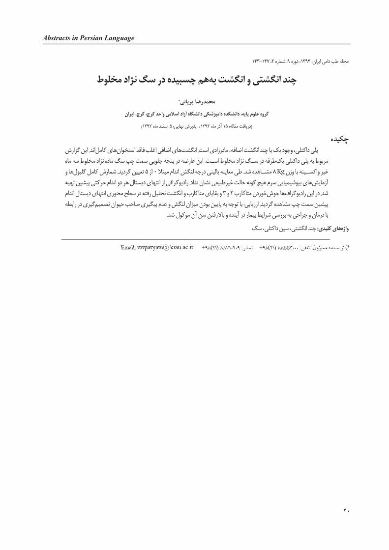
چند انگشتی و انگشت به هم چسبیده در سگ نژاد مخلوطمحمدرضا پریانی*
گروه علوم پایه، دانشکده دامپزشکی دانشگاه آزاد اسالمی واحد كرج، كرج، ایران
) دریافت مقاله: 15 آذر ماه 1393، پذیرش نهایی: 5 اسفند ماه 1393(
چكیده پلی داکتلی، وجود یک یا چند انگشت اضافه، مادرزادی است. انگشت های اضافی اغلب فاقد استخوان های کامل اند. این گزارش مربوط به پلی داکتلی یک طرفه در ســگ نژاد مخلوط اســت. این عارضه در پنجه جلویی سمت چپ سگ ماده نژاد مخلوط سه ماه غیر واکســینه با وزن Kg 8 مشــاهده شد. طی معاینه بالینی درجه لنگش اندام مبتال 0 از 5 تعیین گردید. شمارش کامل گلبول ها و آزمایش های بیوشیمیایی سرم هیچ گونه حالت غیرطبیعی نشان نداد. رادیوگرافی از انتهای دیستال هر دو اندام حرکتی پیشین تهیه شد. در این رادیوگراف ها جوش خوردن متاکارپ 2 و 3 و بقایای متاکارپ و انگشت تحلیل رفته در سطح محوری انتهای دیستال اندام پیشین سمت چپ مشاهده گردید. ارزیابی: با توجه به پایین بودن میزان لنگش و عدم پیگیری صاحب حیوان تصمیم گیری در رابطه
با درمان و جراحی به بررسی شرایط بیمار در آینده و باالرفتن سن آن موکول شد.
واژه های کلیدی: چند انگشتی، سین داکتلی، سگ________________________________________________________________________________________________
Email: mrparyani@ kiau.ac.ir +98)21( 88710409 :98+ نمابر)( نویسنده مسؤول: تلفن: 88553000 )21*