Embed Size (px)
Citation preview

SyncopeA Diagnostic and
Treatment Strategy
David G. Benditt, M.D.University of Minnesota Medical School
Minneapolis, MN USA
Richard Sutton, DScMedRoyal Brompton Hospital
London, UK

Transient Loss of Consciousness (TLOC)

Classification of Transient Loss of Consciousness (TLOC)
Syncope
• Neurally-mediated reflex syndromes
• Orthostatic hypotension
• Cardiac arrhythmias
• Structural cardiovascular disease
Disorders Mimicking Syncope
• With loss of consciousness, i.e., seizure disorders, concussion
• Without loss of consciousness, i.e., psychogenic “pseudo-syncope”
Real or Apparent TLOCReal or Apparent TLOC
Brignole M, et al. Europace, 2004;6:467-537.

Syncope – A Symptom, Not a Diagnosis
Self-limited loss of consciousness and postural tone
Relatively rapid onset
Variable warning symptoms
Spontaneous, complete, and usually prompt recovery without medical or surgical intervention
Underlying mechanism is transient global cerebral hypoperfusion.
Underlying mechanism is transient global cerebral hypoperfusion.
Brignole M, et al. Europace, 2004;6:467-537.

Presentation Overview
I. Etiology, Prevalence, Impact
II. Diagnosis
III. Specific Conditions and Treatment
IV. Special Issues

Section I:
Etiology, Prevalence, Impact

Causes of True Syncope
OrthostaticOrthostatic CardiacArrhythmia
CardiacArrhythmia
StructuralCardio-
Pulmonary
StructuralCardio-
Pulmonary
1• VVS• CSS• Situational
CoughPost- Micturition
2• Drug-Induced• ANS Failure
PrimarySecondary
3• Brady
SN Dysfunction
AV Block
• TachyVTSVT
• Long QT Syndrome
4 • Acute
Myocardial Ischemia
• Aortic Stenosis
• HCM• Pulmonary
Hypertension• Aortic
Dissection
Neurally-Mediated
Neurally-Mediated
Unexplained Causes = Approximately 1/3Unexplained Causes = Approximately 1/3
DG Benditt, MD. U of M Cardiac Arrhythmia Center

Syncope Mimics
Acute intoxication (e.g., alcohol)
Seizures
Sleep disorders
Somatization disorder (psychogenic pseudo-syncope)
Trauma/concussion
Hypoglycemia
Hyperventilation
Brignole M, et al. Europace, 2004;6:467-537.

Impact of Syncope
1Kenny RA, Kapoor WN. In: Benditt D, et al. eds. The Evaluation and Treatment of Syncope. Futura;2003:23-27.2Kapoor W. Medicine. 1990;69:160-175.
3Brignole M, et al. Europace. 2003;5:293-298.4 Blanc J-J, et al. Eur Heart J. 2002;23:815-820.5Campbell A, et al. Age and Ageing. 1981;10:264-270.
40% will experience syncope at least once in a lifetime1
1-6% of hospital admissions2
1% of emergency room visits per year3,4
10% of falls by elderly are due to syncope5
Major morbidity reported in 6%1
eg, fractures, motor vehicle accidents
Minor injury in 29%1
eg, lacerations, bruises
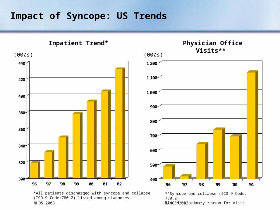
Impact of Syncope: US Trends
300
320
340
360
380
400
420
440
'96 '97 '98 '99 '00 '01 '02
*All patients discharged with syncope and collapse (ICD-9 Code:780.2) listed among diagnoses.NHDS 2003. NAMCS 2002.
Inpatient Trend* Physician Office Visits**
**Syncope and collapse (ICD-9 Code: 780.2) listed as primary reason for visit.
400
500
600
700
800
900
1,000
1,100
1,200
'96 '97 '98 '99 '00 '01
(000s) (000s)

30
40
50
60
70
80
'96 '97 '98 '99 '00 '01 '02
Impact of Syncope: US Trends
500
600
700
800
900
'96 '97 '98 '99 '00 '01 '02
EmergencyDepartment Visits*
HospitalOutpatient Visits*
(000s)
+ Not available
NHAMCS 2002.
+
*Syncope and collapse (ICD-9 Code:780.2) listed as primary reason for visit.
(000s)

Impact of Syncope: NHS Hospitals, England, 2002-2003*
74,813 hospital consults for syncope and collapse
80% required hospital admission
Average length of stay: 6.1 days
327,201 hospital bed days, second only to senility
*Hospital Episode Statistics, Dept. of Health, Eng. 2002-2003.
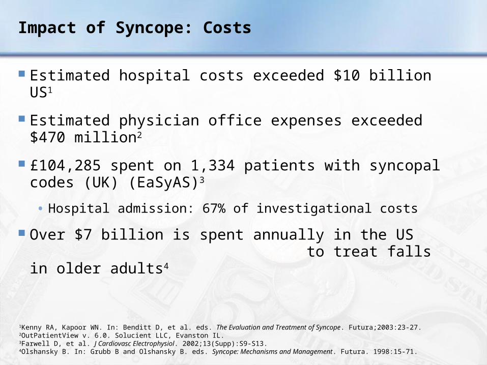
Impact of Syncope: Costs
Estimated hospital costs exceeded $10 billion US1
Estimated physician office expenses exceeded $470 million2
£104,285 spent on 1,334 patients with syncopal codes (UK) (EaSyAS)3
• Hospital admission: 67% of investigational costs
Over $7 billion is spent annually in the US to treat falls in older adults4
1Kenny RA, Kapoor WN. In: Benditt D, et al. eds. The Evaluation and Treatment of Syncope. Futura;2003:23-27.2OutPatientView v. 6.0. Solucient LLC, Evanston IL.3Farwell D, et al. J Cardiovasc Electrophysiol. 2002;13(Supp):S9-S13.4Olshansky B. In: Grubb B and Olshansky B. eds. Syncope: Mechanisms and Management. Futura. 1998:15-71.

Impact of Syncope: Quality of Life
1Linzer M. J Clin Epidemiol. 1991;44:1037.2Linzer M. J Gen Int Med. 1994;9:181.
0
20
40
60
80
100
Anxiety/Depression
Alter DailyActivities
RestrictedDriving
ChangeEmployment
73%171%2
60%2
37%2
Per
cen
t o
f P
atie
nts

Quality of Life: UK Population Norms vs. Syncope Patients
Rose M, et al. J Clin Epidemiol. 2000;53:1209-1216.
0
10
20
30
40
50 UK Population Norms
Patients with Syncope
3%
26%
4%
37%
1%
9%
36%
49%
19%
43%
Mobility Usual Activities
Self-Care Pain/Discomfort
Anxiety/Depression
% P
reva
len
ce

Syncope Mortality
Low mortality vs. high mortality
Neurally-mediated syncope vs. syncope with a cardiac cause
Soteriades ES, Evans JC, Larson MG, et al. Incidence and prognosis of syncope. N Engl J Med. 2002;347(12):878-885. [Framingham Study Population]

Implications of Syncope for Driving a Vehicle
Those who drive and have recurrent syncope risk their lives and the lives of others
Places considerable burden on the physician
Essential to know local laws and physician responsibilities
Some states – Invasion of privacy to notify motor vehicle department*
• Other states – Reporting is mandatory*
If the patient has sufficient warning of impending syncope – Driving may be permitted
Olshansky B, Grubb B. In: Syncope: Mechanisms and Management. Futura. Armonk, NY. 1998.*Medtronic, Inc. Follow-up Forum. 1995/96;1(3):8-10.

Challenges of Syncope
Diagnosis
• Complex
Quality of life implications
• Work
• Mobility (automobiles)
• Psychological
Cost
• Cost/year
• Cost/diagnosis

Section II:
Diagnosis

Diagnostic Objectives
Distinguish true syncope from syncope mimics
Determine presence of heart disease
Establish the cause of syncope with sufficient certainty to:
• Assess prognosis confidently
• Initiate effective preventive treatment

A Diagnostic Plan is Essential
Initial Examination• Detailed patient history• Physical exam• ECG• Supine and upright
blood pressure Monitoring
• Holter• Event• Insertable Loop Recorder (ILR)
Cardiac Imaging Special Investigations
• Head-up tilt test• Hemodynamics • Electrophysiology study
Brignole M, et al. Europace, 2004;6:467-537.

Diagnostic Flow Diagram for TLOC
Brignole M, et al. Europace, 2004;6:467-537.
Initial EvaluationInitial Evaluation
TreatmentTreatment
SyncopeSyncope Not SyncopeNot Syncope
Certain Diagnosis
Certain Diagnosis
Unexplained Syncope
Unexplained Syncope
Cardiac Likely
Cardiac Likely
Cardiac Tests
Cardiac Tests
Neurally-Mediated or Orthostatic Likely
Neurally-Mediated or Orthostatic Likely
Tests for Neurally-Mediated SyncopeTests for Neurally-Mediated Syncope
Frequent or Severe Episodes
Frequent or Severe Episodes
Tests for Neurally-Mediated SyncopeTests for Neurally-Mediated Syncope
Single/Rare Episodes
Single/Rare Episodes
No Further EvaluationNo Further Evaluation
Confirm with Specific Test or
Specialist Consultation
Confirm with Specific Test or
Specialist Consultation
Suspected DiagnosisSuspected Diagnosis
+ - + - + -
TreatmentTreatment TreatmentTreatment
Re-AppraisalRe-AppraisalRe-AppraisalRe-Appraisal
TreatmentTreatment

Initial Exam: Detailed Patient History
Circumstances of recent event• Eyewitness account of event• Symptoms at onset of event• Sequelae• Medications
Circumstances of more remote events
Concomitant disease, especially cardiac
Pertinent family history• Cardiac disease• Sudden death• Metabolic disorders
Past medical history• Neurological history• Syncope
Brignole M, et al. Europace, 2004;6:467-537.

Initial Exam: Thorough Physical
Vital signs
• Heart rate
• Orthostatic blood pressure change
Cardiovascular exam: Is heart disease present?
• ECG: Long QT, pre-excitation, conduction system disease
• Echo: LV function, valve status, HCM
Neurological exam
Carotid sinus massage
• Perform under clinically appropriate conditions preferably during head-up tilt test
• Monitor both ECG and BP
Brignole M, et al. Europace, 2004;6:467-537.

Carotid Sinus Massage (CSM)
Method1
• Massage, 5-10 seconds
• Don’t occlude
• Supine and upright posture (on tilt table)
Outcome
• 3 second asystole and/or 50 mmHg fall in systolic BP with reproduction of symptoms = Carotid Sinus Syndrome
Absolute contraindications2
• Carotid bruit, known significant carotid arterial disease, previous CVA, MI last 3 months
Complications
• Primarily neurological
• Less than 0.2%3
• Usually transient
1Kenny RA. Heart. 2000;83:564.2Linzer M. Ann Intern Med. 1997;126:989.3Munro N, et al. J Am Geriatr Soc. 1994;42:1248-1251.
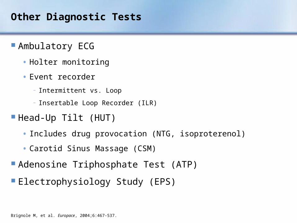
Other Diagnostic Tests
Ambulatory ECG
• Holter monitoring
• Event recorder
− Intermittent vs. Loop
− Insertable Loop Recorder (ILR)
Head-Up Tilt (HUT)
• Includes drug provocation (NTG, isoproterenol)
• Carotid Sinus Massage (CSM)
Adenosine Triphosphate Test (ATP)
Electrophysiology Study (EPS)
Brignole M, et al. Europace, 2004;6:467-537.
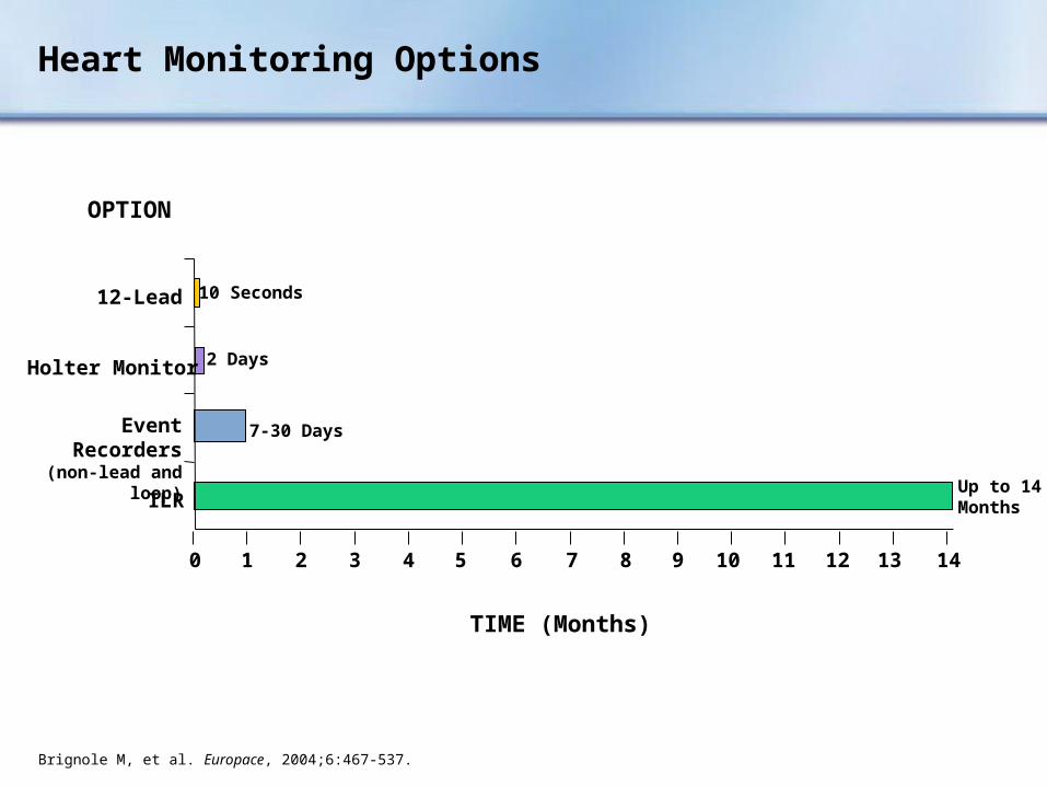
Heart Monitoring Options
ILR
Event Recorders(non-lead and loop)
Holter Monitor
12-Lead
2 Days
7-30 Days
Up to 14 Months
10 Seconds
OPTION
TIME (Months)
0 1 2 3 4 5 6 7 8 9 10 11 12 13 14
Brignole M, et al. Europace, 2004;6:467-537.

Diagnostic Assessment: Yields (N=3411 to 4332)
References Available
Yield (%)
Initial Evaluation
History, Physical Exam, ECG, Cardiac Massage38-40
Other Tests/Procedures
Head-Up Tilt 27
External Cardiac Monitoring 5-13
Insertable Loop Recorder (ILR) 43-883-5
EP Study <2-5
Exercise Test 0.5
EEG 0.3-0.5
MRINo data
available6

Neurological Tests: Rarely Diagnostic for Syncope
EEG, Head CT, Head MRI
May help diagnose seizure
Brignole M, et al. Europace. 2004;6:467-537.

Head-Up Tilt Test (HUT)
Protocols vary
Useful as diagnostic adjunct in atypical syncope cases
Useful in teaching patients to recognize prodromal symptoms
Not useful in assessing treatment
Brignole M, et al. Europace. 2004;6:467-537.
60° - 80°

Head-up Tilt Test
Carlos Morillo, MD, FRCPCProfessor, Faculty of Health SciencesMcMaster University, Hamilton Ontario
Click once on image to play video.

Head-Up Tilt Test:ECG Leads and Intra-Arterial Pressure Tracing
DG Benditt, MD. U of M Cardiac Arrhythmia Center
2
1

Adenosine Triphosphate (ATP) Test
Ongoing investigation in the US
Provokes a short and potent cardioinhibitory vasovagal response
Advantages
• Simple
• Inexpensive
• Correlation with pacing benefit
Seems to identify a unique mechanism of syncope found in patients with:
• Advanced age
• More hypertension
• More ECG abnormalities
Brignole M. Heart. 2000;83:24-28. Donateo P. J Am Coll Cardiol. 2003;41:93-98.Flammang D. Circ. 1999;99:2427-2433.

Reveal® Plus ILR
Insertable Loop Recorder (ILR)
Typical Location of theReveal® Plus ILR
Click once on black screen to play video.

Insertable Loop Recorder (ILR)
The ILR is an implantable patient – and automatically – activated monitoring system that records subcutaneous ECG and is indicated for:
Patients with clinical syndromes or situations at increased risk of cardiac arrhythmias
Patients who experience transient symptoms that may suggest a cardiac arrhythmia

Insertable Loop Recorder (ILR)
Click once on black screen to play video.

Symptom-Rhythm Correlation with the ILR
CASE: 65 year-old man with syncope accompanied by brief retrograde amnesia.
Medtronic data on file.
CASE: 56 year-old woman with refractory syncope accompanied with seizures.

Randomized Assessment of Syncope Trial (RAST)
Results: Combining primary strategy with crossover, the diagnostic yield is
43% ILR only vs. 20% conventional only1
Cost/diagnosis is 26% less than conventional testing2
1Krahn AD, et al. Circ. 2001;104:46-51. 2Krahn AD, et al. JACC. 2003;42:495-501.
Unexplained Syncope
EF > 35%
60 Patients
AECG, Tilt,EP Study
Diagnosis
ILR
++
–++
–
ILRConventional
Testing(AECG, Tilt, EPS)
30 Patients 30 Patients
PrimaryStrategy
Crossover
14 6
1 8
++ ++

Conventional EP Testing in Syncope
Greater diagnostic value in older patients or those with SHD
Less diagnostic value in healthy patients without SHD
Useful diagnostic observations:
• Inducible monomorphic VT
• SNRT > 3000 ms or CSNRT > 600 ms
• Inducible SVT with hypotension
• HV interval ≥ 100 ms (especially in absence of inducible VT)
• Pacing induced infra-nodal block
Benditt D. In: Topol E, ed. Textbook of Cardiovascular Medicine. Lippencott;2002:1529-1542.Lu F, et al. In: Benditt D, et al. The Evaluation and Treatment of Syncope. Futura. 2003;80-95.Brignole M, et al. Europace. 2004;6:467-537.

Diagnostic Limitations of EPS
Difficult to correlate spontaneous events and laboratory findings
Positive findings1
• Without SHD: 6-17%
• With SHD: 25-71%
Less effective in assessing bradyarrhythmias than tachyarrhythmias2
EPS findings must be consistent with clinical history
• Beware of false positive
1Linzer M, et al. Ann Int Med. 1997;127:76-86.2Lu F, et al. In: Benditt D, et al. The Evaluation and Treatment of Syncope. Futura. 2003;80-95.

ISSUEInternational Study of Syncope of Uncertain Etiology
Multicenter, international, prospective study
Analyzed the diagnostic contribution of an ILR in three predefined groups of patients with syncope of uncertain origin:
1) Isolated syncope: No SHD, Normal ECG1
• Negative tilt
• Positive tilt
2) Patients with heart disease and negative EP test2
3) Patients with bundle branch block and negative EP test3
1Moya A. Circulation. 2001; 104:1261-1267. 2Menozzi C, et al. Circulation. 2002;105:2741-2745.3Brignole M, et al. Circulation. 2001;104:2045-2050.

ISSUEPatients with Isolated Syncope and Tilt-Positive Syncope
Moya A. Circulation.
2001;104:1261-1267.
Follow-Up to Recurrent Spontaneous Episode
111 Patients with Syncope No SHD, Normal ECG
29: Tilt-Positive 82: Tilt-Negative “Isolated Syncope”
Tilt Test Followed byInsertable Loop Recorder

ISSUEIsolated Syncope vs. Tilt-Positive Syncope
Conclusions
Results similar in the two arms, including syncope recurrence and ECG correlation
Tilt-negative patients had as many bradycardias (18%) astilt-positive patients (21%)
Most frequent finding was asystole secondary to progressive sinus bradycardia, suggesting a neuro-mediated origin
Homogeneous findings from tilt-negative and tilt-positive infer low sensitivity of tilt-testing
Moya A. Circulation. 2001;104:1261-1267.

ISSUE Patients with Heart Disease and a Negative EP Test
Menozzi C, et al. Circulation. 2002;105:2741-2745.
35 Pts with Heart Diseaseand Insertable Loop Recorder
Syncope: 6 Pts (17%)
ECG-Documented: 6 Pts (17%)
Pre-Syncope: 13 Pts (37%)
ECG-Documented: 8 Pts (23%)
AV block + asystole: 1A.Fib + asystole: 1Sinus arrest: 1Sinus tachycardia: 1Rapid A.Fib: 2
Sustained VT: 1Parox. A.Fib/AT: 1Post tachycardia pause: 1No rhythm variations: 4Sinus tachycardia: 1

ISSUEPatients with Heart Disease and a Negative EP Test
Conclusions
Patients with unexplained syncope, overt heart disease, and negative EP study had a favorable medium-term outcome
Mechanism of syncope was heterogeneous
Ventricular tachyarrhythmia was unlikely
“ILR-guided strategy seems reasonable, with specific therapy safely delayed until a definite diagnosis is made.”
Menozzi C, et al. Circulation. 2002;105:2741-2745.

ISSUEPatients with Bundle Branch Block and Negative EP Test
Brignole M., ET AL.,Circulation. 2001;104:2045-2050. * 5 of these also had ≥1 presyncope** Drop-out before primary-end point
52 Pts with BBBand Insertable Loop Recorder
Syncope: 22 Pts (42%)*
ILR-Detected: 19
AVB: 12 (63%)SA: 4 (21%)Asystole-undefined: 1 (5%)NSR: 1 (5%)Sinus tachy: 1 (5%)
Not Detected: 3
Stable AVB: 3 Pts (6%)
ILR-DetectedPre-Syncope:2 Pts (4%)**
Death: 1 Pt (2%)
AVB: 2 (4%)

ISSUEPatients with Bundle Branch Block and Negative EP Test
Conclusion:
In patients with BBB and negative EP study, most syncopal recurrences have a homogeneous mechanism that is characterized by prolonged asystolic pauses mainly attributable to sudden-onset paroxysmal AV block
Brignole M. Circulation. 2001;104:2045-2050.

Section III:
Specific Conditions and Treatment
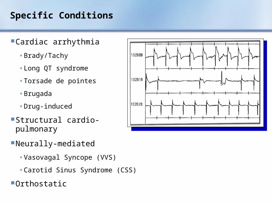
Specific Conditions
Cardiac arrhythmia
• Brady/Tachy
• Long QT syndrome
• Torsade de pointes
• Brugada
• Drug-induced
Structural cardio-pulmonary
Neurally-mediated
• Vasovagal Syncope (VVS)
• Carotid Sinus Syndrome (CSS)
Orthostatic

Cardiac Syncope
Includes cardiac arrhythmias and SHD
Often life-threatening
May be warning of critical CV disease
• Tachy and brady arrhythmias
• Myocardial ischemia, aortic stenosis, pulmonary hypertension, aortic dissection
Assess culprit arrhythmia or structural abnormality aggressively
Initiate treatment promptly
Brignole M, et al. Europace. 2004;6:467-537.

“…cardiac syncope can be a harbinger of sudden death.”
Survival with and without syncope
6-month mortality rate of greater than 10%
Cardiac syncope doubled the risk of death
Includes cardiac arrhythmias and SHD
No SyncopeVasovagal andOther CausesCardiac Cause
0 5 1015 Follow-Up (yr)
Pro
bab
ility
of
Sur
viva
l
1.0
0.8
0.6
0.4
0.2
0.0
Soteriades ES, et al. N Engl J Med. 2002;347:878.

Syncope Due to Structural Cardiovascular Disease: Principle Mechanisms
Acute MI/Ischemia
• 2° neural reflex bradycardia – Vasodilatation, arrhythmias, low output (rare)
Hypertrophic cardiomyopathy
• Limited output during exertion (increased obstruction, greater demand), arrhythmias, neural reflex
Acute aortic dissection
• Neural reflex mechanism, pericardial tamponade
Pulmonary embolus/pulmonary hypertension
• Neural reflex, inadequate flow with exertion
Valvular abnormalities
• Aortic stenosis – Limited output, neural reflex dilation in periphery
• Mitral stenosis, atrial myxoma – Obstruction to adequate flow
Brignole M, et al. Europace. 2004;6:467-537.

Syncope Due to Cardiac Arrhythmias
Bradyarrhythmias
• Sinus arrest, exit block
• High grade or acute complete AV block
• Can be accompanied by vasodilatation (VVS, CSS)
Tachyarrhythmias
• Atrial fibrillation/flutter with rapid ventricular rate (eg, pre-excitation syndrome)
• Paroxysmal SVT or VT
• Torsade de pointes
Brignole M, et al. Europace. 2004;6:467-537.

ILR Recordings
CASE: 28 year-old man presents to ER multiple times after falls resulting in trauma. VT: Ablated and medicated.
CASE: 83 year-old woman with syncope due to bradycardia: Pacemaker implanted.
Reveal ® ILR recordings; Medtronic data on file.

Cardiac Rhythms During Unexplained Syncope
Seidl K. Europace. 2000;2(3):256-262.Krahn AD. PACE. 2002;25:37-41.Medtronic ILR Replacement Data. FY03, 04. On file.
No Recurrence 36%
(31-48%)
Normal Sinus Rhythm 31%
(17-44%)
Other 11%
Arrhythmia 22%
(13-32%)
Tachycardia 6%(2-11%)
Bradycardia 16%
(11-21%)
Composite: N=133 to 7109

Long QT Syndromes
Mechanism
• Abnormalities of sodium and/or potassium channels
• Susceptibility to polymorphic VT (Torsade de pointes)
Prevalence
• Drug-induced forms – Common
• Genetic forms – Relatively rare, but increasingly being recognized
• “Concealed” forms:
− May be common
− Provide basis for drug-induced torsade
Schwartz P, Priori S. In: Zipes D and Jalife J, eds. Cardiac Electrophysiology. Saunders;2004:651-659.

Syncope: Torsade de Pointes
From the files of DG Benditt, MD. U of M Cardiac Arrhythmia Center

Long QT Syndromes: 12-Lead ECG
From the files of DG Benditt, MD. U of M Cardiac Arrhythmia Center

Drug-Induced QT Prolongation(List is continuously being updated)
Antiarrhythmics
• Class IA ...Quinidine, Procainamide, Disopyramide
• Class III…Sotalol, Ibutilide, Dofetilide, Amiodarone, NAPA*
Antianginal Agents
• Bepridil*
Psychoactive Agents
• Phenothiazines, Amitriptyline, Imipramine, Ziprasidone
Antibiotics
• Erythromycin, Pentamidine, Fluconazole, Ciprofloxacin and its relatives
Nonsedating antihistamines
• Terfenadine*, Astemizole
Others
• Cisapride*, Droperidol, Haloperidol
*Removed from U.S. Market
Brignole M, et al. Europace, 2004;6:467-537.

Treatment of Long QT
Suspicion and recognition are critical
Emergency treatment
• Intravenous magnesium
• Pacing to overcome bradycardia or pauses
• Isoproterenol to increase heart rate and shorten repolarization
• ICD if prior SCA or strong family history
• If drug induced:
− Reverse bradycardia
− Withdraw drug
− Avoid ALL long-QT provoking agents
• If genetic:
− Avoid ALL long-QT provoking agents
For more information visit www.longqt.orgSchwartz P, Priori S. In: Zipes D and Jalife J, eds. Cardiac Electrophysiology. Saunders;2004:651-659.

Treatment of Syncope Due to Bradyarrhythmia
Class I indication for pacing using dual chamber system wherever possible
Ventricular pacing in atrial fibrillation with slow ventricular response
ACC/AHA/NASPE 2002 Guideline Update. Circ. 2002;106:2145-2161.
nV
0.4
0.2
0.0
-0.2
-0.4
:45:44:43:42:41:40:39:38:37
:37:36:35:34:33:32:31:30:29
:29:28:27:26:25:24:23:22:21
08:23:21
8:23:29
08:23:37
0.4
0.2
0.0
-0.2
-0.4
0.4
0.2
0.0
-0.2
-0.4

Treatment of Syncope Due to Tachyarrhythmia
Atrial tachyarrhythmias
• AVRT due to accessory pathway – Ablate pathway
• AVNRT – Ablate AV nodal slow pathway
• Atrial fib – Pacing, linear/focal ablation for paroxysmal AF
• Atrial flutter – Ablate the IVC-TV isthmus of the re-entrant circuit for ‘typical’ flutter
Ventricular tachyarrhythmias
• Ventricular tachycardia – ICD or ablation where appropriate
• Torsade de pointes – Withdraw offending drug or implant ICD (long QT/Brugada/short QT)
Drug therapy may be an alternative in many cases
Brignole M, et al. Europace. 2004;6:467-537.

Neurally-Mediated Reflex Syncope
Vasovagal Syncope (VVS)
Carotid Sinus Syndrome (CSS)
Situational syncope
• Post-micturition
• Cough
• Swallow
• Defecation
• Blood drawing, etc.
Brignole M, et al. Europace, 2004;6:467-537.
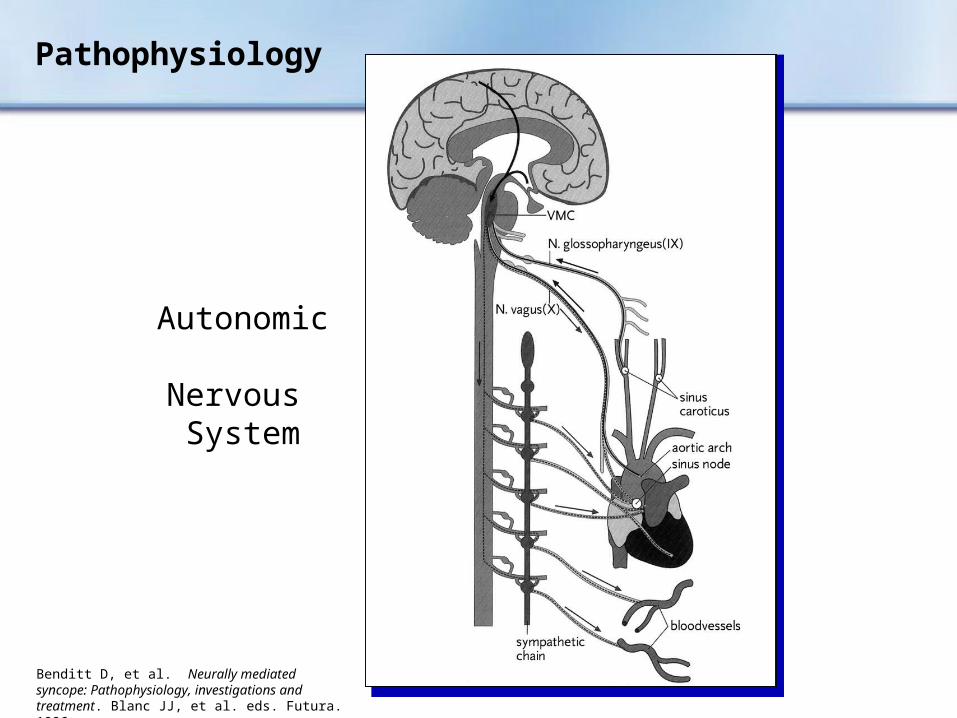
Pathophysiology
Autonomic Nervous System
Benditt D, et al. Neurally mediated syncope: Pathophysiology, investigations and treatment. Blanc JJ, et al. eds. Futura. 1996.

VVSClinical Pathophysiology
Neurally-mediated physiologic reflex mechanism with two components:
1. Cardioinhibitory (↓ HR)
2. Vasodepressor (↓ BP) despite heart beats, no significant BP generated
Both components are usually present
Wieling W, et al. In: Benditt D, et al. The Evaluation and Treatment of Syncope. Futura. 2003;11-22.
1
2

VVSIncidence
Most common form of syncope
• 8% to 37% (mean 18%) of syncope cases
Depends on population sampled
• Young without SHD, ↑ incidence
• Older with SHD, ↓ incidence
Linzer M, et al. Ann Intern Med. 1997;126:989.

VVS vs. CSS
In general:
• VVS patients younger than CSS patients
• Ages range from adolescence to older adults (median 43 years)
Linzer M, et al. Ann Intern Med. 1997;126:989.

VVS Recurrences
1Savage D, et al. STROKE. 1985;16:626-29. 2Sheldon R, et al. Circulation. 1996;93:973-81.
35% of patients report syncope recurrence during follow-up ≤3 years1
Positive HUT with >6 lifetime syncope episodes: recurrence risk >50% over 2 years2
1000
800
50
100
25
8
4
2
1
1 2 3 6 24 84 480
Months Since Symptoms Began
Two Year Risk
Tot
al N
umbe
r of
Syn
copa
l Epi
sode
s
> 75%
50-75%
25-50%
< 25%
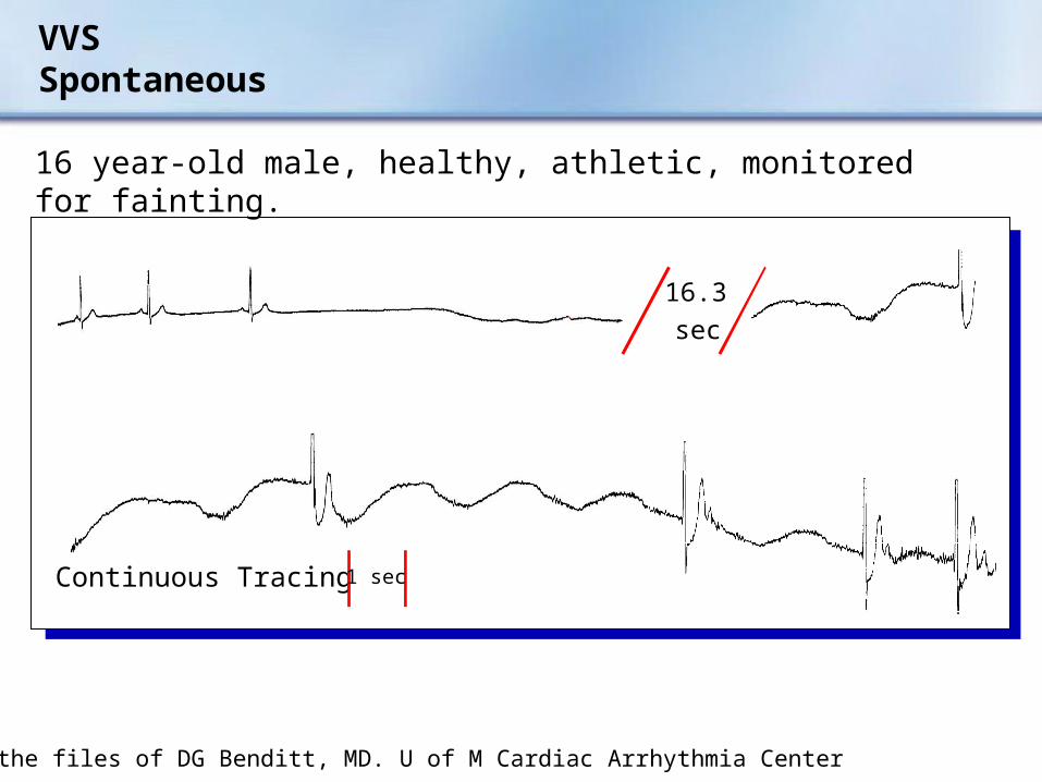
From the files of DG Benditt, MD. U of M Cardiac Arrhythmia Center
16.3
sec
Continuous Tracing 1 sec
VVS Spontaneous
16 year-old male, healthy, athletic, monitored for fainting.

VVSDiagnosis
History and physical exam, ECG and BP
Head-Up Tilt (HUT) – Protocol:
• Fast > 2 hours
• ECG and continuous blood pressure, supine, and upright
• Tilt to 70°, 20 minutes
• Isoproterenol/Nitroglycerin if necessary
• End point – Loss of consciousness
60° - 80°
Benditt D, et al. JACC. 1996;28:263-275.Brignole M, et al. Europace, 2004;6:467-537.

VVS General Treatment Measures
Optimal treatment strategies for VVS are a source of debate
Treatment goals
• Acute intervention− Physical maneuvers, eg,
crossing legs or tugging arms
− Lowering head
− Lying down
Long-term prevention
• Tilt training
• Education
• Diet, fluids, salt
• Support hose
• Drug therapy
• Pacing
Brignole M, et al. Europace, 2004;6:467-537.

VVS Tilt Training Protocol
Objectives
• Enhance orthostatic tolerance
• Diminish excessive autonomic reflex activity
• Reduce syncope susceptibility/recurrences
Technique
• Prescribed periods of upright posture against a wall
• Start with 3-5 min BID
• Increase by 5 min each week until a duration of 30 min is achieved
Reybrouck T, et al. PACE. 2000;23(4 Pt. 1):493-498.

VVS Tilt Training: Clinical Outcomes
Treatment of recurrent VVS
Reybrouck, et al.*: Long-term study
• 38 patients performed home tilt training
• After a period of regular tilt training, 82% remained free of syncope during the follow-up period
• However, at the 43-month follow-up, 29 patients had abandoned the therapy
• Conclusion: The abnormal autonomic reflex activity of VVS can be remedied. Compliance may be an issue.
*Reybrouck T, et al. PACE. 2000;23:493-498.

VVS Tilt Training: Clinical Outcomes
Foglia-Manzillo, et al.*: Short-term study
• 68 patients
– 35 tilt training
– 33 no treatment (control)
• Tilt table test conducted after 3 weeks
• 19 (59%) of tilt trained and 18 (60%) of controls had a positive test
• Tilt training was not effective in reducing tilt testing positivity rate
• Poor compliance in the majority of patients with recurrent VVS
*Foglio-Manzillo G, et al. Europace. 2004;6:199-204.

VVS Pharmacologic Treatment
Fludrocortisone
Beta-adrenergic blockers
• Preponderance of clinical evidence suggests minimal benefit1
SSRI (Selective Serotonin Re-Uptake Inhibitor)
• 1 small controlled trial2
Vasoconstrictors
• 1 negative controlled trial (etilefrine)3
• 2 positive controlled trials (midodrine)4,5
1Brignole M, et al. Europace, 2004;6:467-537.2Di Girolamo E, et al. JACC. 1999;33:1227-1230.3Raviele A, et al. Circ. 1999;99:1452-1457.
4Ward C, et al. Heart. 1998;79:45-49.5Perez-Lugones A, et al. J Cardiovasc Electrophysiol. 2001;12(8):935-938.

Midodrine for VVS
Perez-Lugones A, Schweikert R, Pavia S, et al. J Cardiovasc Electrophysiol. 2001;12(8):935-938.
Months
p < 0.001
Sym
pto
m-F
ree
Inte
rval
180160140120100806040200
100
80
60
40
20
0
Fluid
Midodrine

The Role of Pacing as Therapy for Syncope
VVS with +HUT and cardioinhibitory response:Class IIb indication for pacing
Three randomized, prospective trials reported benefits of pacing in select VVS patients:
• VPS I1
• VASIS2
• SYDIT3
Subsequent study results less clear
• VPS II4
• Synpace5
• INVASY6
1Connolly SJ. J Am Coll Cardiol. 1999;33:16-20.2Sutton R. Circulation. 2000;102:294-299.3Ammirati F. Circ. 2001;104:52-57.
4Connolly S. JAMA. 2003;289:2224-2229.5Giada F. PACE . 2003;26:1016 (abstract).6Occhetta E, et al. Europace. 2004;6:538-547.

VPS I (North American Vasovagal Pacemaker Study)
Objective: To evaluate pacemaker therapy for severe recurrent vasovagal syncope
Randomized, prospective, single center
N=54 patients
• 27: DDD pacemaker with rate drop response
• 27: No pacemaker
Inclusion: Vasodepressor response
Primary outcome: First recurrence of syncope
Connolly SJ. J Am Coll Cardiol. 1999;33:16-20.

100
90
80
70
60
50
40
30
20
10
0
0 3 6 9 12 15
Time in Months
No Pacemaker (PM)
2P=0.000022
PacemakerCu
mu
lati
ve R
isk
(%)
Connolly SJ. J Am Coll Cardiol. 1999;33:16-20.
Results: 6 (22%) with PM had recurrence vs. 19 (70%) without PM 84% RRR (2p=0.000022)
VPS I (North American Vasovagal Pacemaker Study)

VASIS (VAsovagal Syncope International Study)
Objective: To evaluate pacemaker therapy for severe cardioinhibitory tilt-positive neurally mediated syncope
Randomized, prospective, multi-center
N=42 patients
• 19: DDI pacemaker (80 bpm) with rate hysteresis (45 bpm)
• 23: No pacemaker
Inclusion: Positive cardioinhibitory response
Primary outcome: First recurrence of syncope
Sutton R. Circulation. 2000;102:294-299.

Sutton R. Circulation. 2000;102:294-299.
Pacemaker (PM)
No Pacemaker
p=0.0004
Years
% S
ynco
pe-
Fre
e
100
80
60
40
20
0 2 3 4 5 6
Results: 1 (5%) with PM had recurrence vs. 14 (61%) without PM
VASIS (VAsovagal Syncope International Study)

SYDIT (SYncope DIagnosis and Treatment)
Objective: To compare the effects of cardiac pacing with pharmacological therapy in patients with recurrent vasovagal syncope
Randomized, prospective, multi-center
N=93 patients
• 46: DDD pacemaker with rate drop response
• 47: Atenolol 100 mg/d
Inclusion: Positive HUT with relative bradycardia
Primary outcome: First recurrence of syncope
Ammirati F. Circulation. 2001;104:52-57.

SYDIT (SYncope DIagnosis and Treatment)
Ammirati F. Circulation. 2001;104:52-57.
0.6
0.7
0.8
0.9
1.0
0 100 200 300 400 500 600 700 800 900 1000
Drug
Pacemaker (PM)
Time (Days)
% S
ynco
pe-
Fre
e
p=0.0032
Results: 2 (4%) with PM had syncope recurrence vs. 12 (26%) without PM

VPS II (Vasovagal Pacemaker Study II)
Objective: To determine if pacing therapy reduces the risk of syncope in patients with vasovagal syncope
Randomized, double-blind, prospective, multi-center
N=100 patients
• 52: Only sensing without pacing
• 48: DDD pacemaker with rate drop response
Inclusion: Positive HUT with (HRxBP) < 6000/min x mm Hg
Primary outcome: First recurrence of syncope
Connolly S. JAMA. 2003;289:2224-2229.
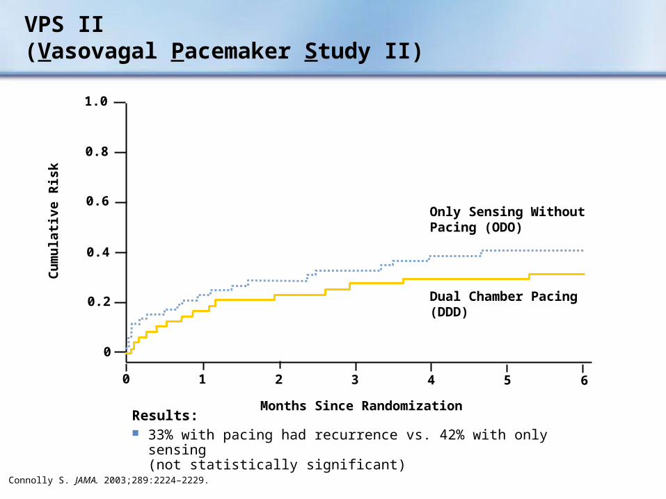
Dual Chamber Pacing (DDD)
Only Sensing Without Pacing (ODO)
1.0
0.8
0.6
0.4
0.2
0
Months Since Randomization
Cu
mu
lati
ve R
isk
6543210
Connolly S. JAMA. 2003;289:2224–2229.
Results: 33% with pacing had recurrence vs. 42% with only sensing
(not statistically significant)
VPS II (Vasovagal Pacemaker Study II)

SYNPACE(Vasovagal SYNcope and PACing)
Objective: To determine if pacing therapy will reduce syncope relapses in patients with recurrent vasovagal syncope, compared to those with a pacemaker programmed to OFF
Randomized, double-blind, prospective, multi-center, placebo-controlled
N=29 patients
• 16: DDD PM with rate drop response programmed ON
• 13: PM programmed OFF (OOO mode)
Inclusion: Recurrent VVS and +HUT with asystolic or mixed response
Primary outcome: First recurrence of syncope
Raviele A.. Europace. 2001;3:336–341.Raviele A, et al. Eur Heart J. 2004;25:1741-1748.

SYNPACE(Vasovagal SYNcope and PACing)
Raviele A, et al. Eur Heart J. 2004;25:1741-1748.
Results: 50% with pacing ON had recurrence vs. 38% with pacing OFF
(not statistically significant)
0.6
0.7
0.8
0.9
1.0
0 200 400 600 800 1000
Pacemaker OFF
% S
ynco
pe-
Fre
e
p=0.58
0.5
0.4
0.3
0.2
0.1
0.0
Pacemaker ON
Days Since Randomization
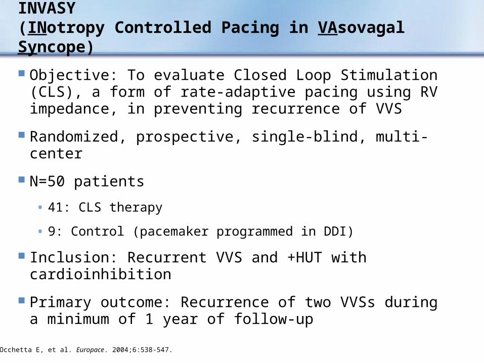
INVASY(INotropy Controlled Pacing in VAsovagal Syncope)
Objective: To evaluate Closed Loop Stimulation (CLS), a form of rate-adaptive pacing using RV impedance, in preventing recurrence of VVS
Randomized, prospective, single-blind, multi-center
N=50 patients
• 41: CLS therapy
• 9: Control (pacemaker programmed in DDI)
Inclusion: Recurrent VVS and +HUT with cardioinhibition
Primary outcome: Recurrence of two VVSs during a minimum of 1 year of follow-up
Occhetta E, et al. Europace. 2004;6:538-547.

INVASY(INotropy Controlled Pacing in VAsovagal SYncope)
20
40
60
0
100
% S
ynco
pe-
Fre
e
P < 0.0001
Closed Loop Stimulation (CLS)
Control (DDI only)
Time Since Randomization
3m 6m 9m 1y 2y 3y
Results: Patients with CLS had no syncope recurrence and improved quality of life
Occhetta E, et al. Europace. 2004;6:538-547.

Role of Pacing as Therapy for Syncope: Summary
Three earlier studies single blind – Bias?
Pacemaker implantation may modulate reflex syncope and autonomic responses1
Study results may differ based on pre-implant selection criteria and tilt-testing techniques
Pacing therapy is effective in some but not all (cardioinhibition vs. vasodepression)
In five pacing studies, syncope recurred in 33/156 (21%) of paced patients, 72/162 (44%) in non-paced patients (p<0.000)2
1Kapoor W. JAMA. 2003;289:2272-2275.2Brignole M, et al.. Europace. 2004;6:467-537.

CSSCarotid Sinus Syndrome
Syncope clearly associated with carotid sinus stimulation is rare (≤1% of syncope)
CSS may be an important cause of unexplained syncope/falls in older individuals
Prevalence higher than previously believed
Carotid Sinus Hypersensitivity (CSH)
• No symptoms
• No treatment
Kenny RA, et al. J Am Coll Cardiol. 2001;38:1491-1496.Brignole M, et al. Europace. 2004;6:467-537.Sutton R. In: Neurally Mediated Syncope: Pathophysiology, Investigation and Treatment. Blanc JJ, et al. eds. Armonk, NY: Futura;1996:138.

CSSEtiology
Sensory nerve endings in the carotid sinus walls respond to deformation
“Deafferentation” of neck muscles may contribute
Increased afferent signals tobrain stem
Reflex increase in efferent vagal activity and diminution of sympathetic tone results in bradycardia and vasodilatation
Carotid Sinus

Falls:Incidence, Recurrence, CSH*
1 J Am Geriatr Soc. 1995.2 Richardson D, et al. PACE. 1997;20:820.
0
25
50
75
Incidence> Age 65
Recurrence CSH* Presentin Fallers > Age 50Presenting at ER
30% 1
50% 1
23% 2
% o
f P
op
ula
tio
n
*Carotid Sinus Hypersensitivity

0
25
50
75
No Pacing Pacing
57%
%6
% R
ecu
rren
ce
CSS Role of Pacing – Syncope Recurrence Rate
Class I indication for pacing (AHA and BPEG)
Limit pacing to CSS that is:
• Cardioinhibitory
• Mixed
DDD/DDI superior to VVI
• Mean follow-up = 6 months
Brignole M, et al. Eur JCPE. 1992;4:247-254.

SAFE PACESyncope And Falls in the Elderly – Pacing And Carotid Sinus Evaluation
Objective
• Determine whether cardiac pacing reduces falls in older adults with carotid sinus hypersensitivity
Randomized controlled trial (N=175)
• Adults > 50 years, non-accidental fall, positive CSM
• Pacing (n=87) vs. No Pacing (n=88)
Results
• More than 1/3 of adults over 50 years presented to the Emergency Department because of a fall
• With pacing, falls 70%
• Syncopal events 53%
• Injurious events 70%
Kenny RA. J Am Coll Cardiol. 2001;38:1491-1496.

SAFE PACE
Conclusions
• Strong association between non-accidental falls and cardioinhibitory CSH
• These patients usually not referred for cardiac assessment
• Cardiac pacing significantly reduced subsequent falls
• CSH should be considered in all older adults who have non-accidental falls
Kenny RA, J Am Coll Cardiol. 2001; 38:1491-1496.

Orthostatic Hypotension
Etiology
Drug-induced (very common)
• Diuretics
• Vasodilators
Primary autonomic failure
• Multiple system atrophy
• Parkinson’s Disease
• Postural Orthostatic Tachycardia Syndrome (POTS)
Secondary autonomic failure
• Diabetes
• Alcohol
• Amyloid
Brignole M, et al. Europace, 2004;6:467-537.

Treatment Strategies for Orthostatic Intolerance
Patient education, injury avoidance
Hydration
• Fluids, salt, diet
• Minimize caffeine/alcohol
Sleeping with head of bed elevated
Tilt training, leg crossing, arm pull
Support hose
Drug therapies
• Fludrocortisone, midodrine, erythropoietin
Tachy-Pacing (probably not useful)
Brignole M, et al. Europace, 2004;6:467-537.

Section IV:
Special Issues

Syncope: Diagnostic Testing in Hospital Strongly Recommended
Suspected/known ‘significant’ heart disease
ECG abnormalities suggesting potential life-threatening arrhythmic cause
Syncope during exercise
Severe injury or accident
Family history of premature sudden death
Brignole M, et al. Europace. 2004;6:467-537.

SEEDS: Syncope Evaluation in the Emergency Department Study
Survival Free from Death
Shen W, et al. Circ. 2004;110(24):3636-3645.
Survival Free from Recurrence
Long-Term Clinical Outcomes
100%
90%
80%
70%
Years
210
P=0.30
Syncope Unit Group
Standard Care Group
100%
90%
80%
70%
Years
210
P=0.72
Syncope Unit Group
Standard Care Group
Results: Syncope unit improved diagnostic yield in the ED and reduced
hospital admission and length of stay

The Integrated Syncope Unit
To optimize the effectiveness of the evaluation and treatment of syncope patients at a given center
Best accomplished by:
• Cohesive, structured care pathway
• Multidisciplinary approach
• Core equipment available
• Preferential access to other tests or therapy
Majority of syncope evaluations – Out-patient or day cases
1Kenny RA, Brignole M. In: Benditt D, et al. eds. The Evaluation and Treatment of Syncope. Futura;2003:55-60. 2Brignole M, et al. Europace, 2004;6:467-537.

Conclusion
Syncope is a common symptom with many causes
Deserves thorough investigation and appropriate treatment
A disciplined approach is essential
ESC guidelines offer current best practices
Brignole M, et al. Europace, 2004;6:467-537.

Challenges of Syncope
Cost
Quality of life implications
Diagnosis and treatment
• Diagnostic yield and repeatability of tests
• Frequency and clustering of events
• Difficulty in managing/treating/controlling future events
• Appropriate risk stratification
• Complex etiology
Olshansky B. In: Grubb B and Olshansky B. eds. Syncope: Mechanisms and Management. Futura. 1998:15-71.Brignole M, et al. Europace, 2004;6:467-537.

Brief Statement
Indications
9526 Reveal® Plus Insertable Loop Recorder
The Reveal Plus ILR is an implantable patient- and automatically activated monitoring system that records subcutaneous ECG and is indicated for
Patients with clinical syndromes or situations at increased risk of cardiac arrhythmias
Patients who experience transient symptoms that may suggest a cardiac arrhythmia
6191 Activator
The Model 6191 Activator is intended for use in combination with a Medtronic Model 9526 Reveal Plus Insertable Loop Recorder.
Contraindications
There are no known contraindications for the implantation of the Reveal Plus ILR. However, the patient’s particular medical condition may dictate whether or not a subcutaneous, chronically implanted device can be tolerated.
Warnings/Precautions
9526 Reveal Plus Insertable Loop Recorder
Patients with the Reveal Plus ILR should avoid sources of magnetic resonance imaging, diathermy, high sources of radiation, electrosurgical cautery, external defibrillation, lithotripsy, and radiofrequency ablation to avoid electrical reset of the device, and/or inappropriate sensing.
6191 Activator
Operation of the Model 6191 Activator near sources of electromagnetic interference, such as cellular phones, computer monitors, etc., may adversely affect the performance of this device.
Potential Complications
Potential complications include, but are not limited to, body tissue rejection phenomena, including local tissue reaction, infection, device migration and erosion of the device through the skin.
2090 Programmer
The Medtronic/Vitatron CareLink programmer system is comprised of prescription devices indicated for use in the interrogation and programming of implantable medical devices. Prior to use, refer to the Programmer Reference Guide as well as the appropriate programmer software and implantable device technical manuals for more information related to specific implantable device models. Programming should be attempted only by appropriately trained personnel after careful study of the technical manual for the implantable device and after careful determination of appropriate parameter values based on the patient's condition and pacing system used. The Medtronic/Vitatron CareLink programmer must be used only for programming implantable devices manufactured by Medtronic or Vitatron.
See the device manual for detailed information regarding the implant procedure, indications, contraindications, warnings, precautions, and potential complications/adverse events. For further information, please call Medtronic at 1-800-328-2518 and/or consult Medtronic’s website at www.medtronic.com. To learn more about syncope, visit www.fainting.com.
Caution: Federal law (USA) restricts this device to sale by or on the order of a physician.


![Syncope AHD[1]](https://img.pdfslide.us/doc/110x75/577d36611a28ab3a6b92ec10/syncope-ahd1.jpg)