Embed Size (px)
Citation preview

Synchronous Unit Activity and Local Field Potentials Evoked in theSubthalamic Nucleus by Cortical Stimulation
Peter J. Magill,1 Andrew Sharott,2 Mark D. Bevan,1,3 Peter Brown,2 and J. Paul Bolam1
1Medical Research Council Anatomical Neuropharmacology Unit, University of Oxford, Oxford OX1 3TH; 2Sobell Department of MotorNeuroscience and Movement Disorders, Institute of Neurology, London WC1N 3BG, United Kingdom; and 3Department of Physiology,Feinberg School of Medicine, Northwestern University, Chicago, Illinois 60611-3008
Submitted 9 February 2004; accepted in final form 22 March 2004
Magill, Peter J., Andrew Sharott, Mark D. Bevan, Peter Brown,and J. Paul Bolam. Synchronous unit activity and local field po-tentials evoked in the subthalamic nucleus by cortical stimulation.J Neurophysiol 92: 700–714, 2004. First published March 24, 2004;10.1152/jn.00134.2004. The responses of single subthalamic nucleus(STN) neurons to cortical activation are complex and depend on therelative activation of several neuronal circuits, making theoreticalextrapolation of single neuron responses to the population leveldifficult. To understand better the degree of synchrony imposed onSTN neurons and associated neuronal networks by cortical activation,we recorded the responses of single units, pairs of neighboringneurons, and local field potentials (LFPs) in STN to discrete electricalstimulation of the cortex in anesthetized rats. Stimulation of ipsilateralfrontal cortex, but not temporal cortex, generated synchronized “mul-tiphasic” responses in neighboring units in rostral STN, usuallyconsisting of a brief, short-latency excitation, a brief inhibition, asecond excitation, and a long-duration inhibition. Evoked LFPs inSTN consistently mirrored unit responses; brief, negative deflectionsin the LFP coincided with excitations and brief, positive deflectionswith inhibitions. This characteristic LFP was dissimilar to potentialsevoked in cortex and structures surrounding STN and was resistant tofluctuations in forebrain activity. The short-latency excitation andassociated LFP deflection exhibited the highest fidelity to low-inten-sity cortical stimuli. Unit response failures, which mostly occurred incaudal STN, were not associated with LFPs typical of rostral STN.These data suggest that local populations of STN neurons can besynchronized by both direct and indirect cortical inputs. Synchronizedensemble activity is dependent on topography and input intensity.Finally, the stereotypical, multiphasic profile of the evoked LFPindicates that it might be useful for locating the STN in clinical as wellas nonclinical settings.
I N T R O D U C T I O N
The basal ganglia (BG) are a group of subcortical brainnuclei intimately involved in movement and cognition (De-Long 1990; Gerfen and Wilson 1996; Graybiel 1995). Thecerebral cortex, the principal afferent of the BG, directlytransfers information to the BG via projections to the neostri-atum (NS) and subthalamic nucleus (STN). Although notaccounted for in classic models of BG function (DeLong 1990;Wichmann and Delong 1996), recent studies contend that theinput operations of STN are critical for the processing ofcortical information in the BG, not least because the cortico-subthalamic pathway represents the swiftest route by which thecortex can influence the activity of STN and its targets, the
globus pallidus (GP) and the output nuclei of the BG (forreviews, see Mink 1996; Nambu et al. 2002; Smith et al. 1998).
The responses of STN neurons to cortical input can be“multiphasic” and vary according to the relative activation ofdistinct BG circuits, including the NS and the reciprocallyconnected STN-GP network (Fujimoto and Kita 1993; Kolo-miets et al. 2001; Maurice et al. 1998; Nambu et al. 2000). Thecorticostriatal projection can be quantitatively appreciated interms of the degrees of convergence/divergence and the spatialarrangements of connections between the neurons that areengaged by cortical activation (Kincaid et al. 1998; Zheng andWilson 2002). However, such an appreciation is not yet pos-sible for corticosubthalamic or striatopallidal projections or thereciprocal connections within the STN-GP network (for re-views, see Bevan et al. 2002b; Smith et al. 1998). It is thereforedifficult to predict how neighboring STN neurons will respondto cortical activation. Furthermore, although electrophysiolog-ical studies at the single-cell level in vitro suggest thatGABAergic inputs from the GP could act as a powerfulsynchronizing force in the STN (Bevan et al. 2002a,b), littleinformation is available concerning the response of STN neu-rons to cortical inputs and the interaction of cortical inputs withthe mechanisms underlying the autonomous activity and un-usual input-output properties of STN neurons (Beurrier et al.2000; Bevan and Wilson 1999; Hallworth et al. 2003; Otsukaet al. 2001). Indeed, cortical excitatory postsynaptic potentials(EPSPs) can be inefficiently coupled to the generation of actionpotentials in another class of autonomously active neuron inthe BG, the cholinergic interneuron of the NS (Bennett andWilson 1998).
The complex single-cell and network interactions withincortico-basal ganglia circuits, together with the paucity ofdetailed anatomical data, make the theoretical extrapolation ofsingle neuron responses to the population level a difficult task.Yet the manner in which local ensembles of STN neuronsmight be recruited and coordinated by cortical input is offundamental importance for our understanding of the roles ofthe STN and indeed, the BG, in behavior. Recent evidencesuggests that the activity of STN neurons may be highlysynchronized under some circumstances (Brown et al. 2002;Magill et al. 2000, 2001). The population response in STN, andparticularly the degree of synchronization associated with it, isall the more significant because cortical afferent activity itselfmay be highly synchronized during behavior (for reviews, see
Address for reprint requests and other correspondence: P. J. Magill, MRCAnatomical Neuropharmacology Unit, University of Oxford, Mansfield Road,Oxford OX1 3TH, UK (E-mail: [email protected]).
The costs of publication of this article were defrayed in part by the paymentof page charges. The article must therefore be hereby marked “advertisement”in accordance with 18 U.S.C. Section 1734 solely to indicate this fact.
J Neurophysiol 92: 700–714, 2004.First published March 24, 2004; 10.1152/jn.00134.2004.
700 0022-3077/04 $5.00 Copyright © 2004 The American Physiological Society www.jn.org

Engel and Singer 2001; Engel et al. 2001; MacKay 1997).Furthermore, inappropriate synchronization of neuronal en-sembles may underlie, or at least contribute to, dysfunction inBG diseases (for reviews, see Bergman et al. 1998; Bevan et al.2002b; Boraud et al. 2002; Brown 2003).
To gain a more complete understanding of the degree ofsynchrony that can be imposed on STN neurons and associatedneuronal networks by the cortex, we recorded the responses ofsingle units and pairs of neighboring neurons in STN to focalelectrical stimulation of the ipsilateral frontal cortex in anes-thetized rats. When the subthreshold and suprathreshold activ-ities of neural ensembles are sufficiently synchronized, it isoften evident at the level of the local field potential (LFP) (forreviews, see Hubbard et al. 1969; Mitzdorf 1985). Thus to testthe hypothesis that activity in STN can be synchronized to ahigh degree by cortical input, we simultaneously recorded theLFPs evoked together with the unit responses after corticalstimulation.
M E T H O D S
Electrophysiological recordings and labeling ofrecording sites
Experimental procedures were carried out on adult male Sprague-Dawley rats (Charles River, Margate, UK) and were conducted inaccordance with the Animals (Scientific Procedures) Act 1986(United Kingdom) and the American Physiological Society’s GuidingPrinciples in the Care and Use of Animals.
Electrophysiological recordings were made in 13 rats (230–350 g).Anesthesia was induced with isoflurane (Isoflo, Schering-Plough,Welwyn Garden City, UK) and maintained with urethan (1.3 g/kg ip;ethyl carbamate, Sigma, Poole, UK), and supplemental doses ofketamine (30 mg/kg ip; Ketaset, Willows Francis, Crawley, UK) andxylazine (3 mg/kg ip; Rompun, Bayer, Germany) as described previ-ously (Magill et al. 2001). All wound margins were infiltrated with thelocal anesthetic, bupivacaine (0.75% wt/vol; Astra, Kings Langley,UK), and corneal dehydration was prevented with application ofHypromellose eye drops (Norton Pharmaceuticals, Harlow, UK).Animals were then placed in a stereotaxic frame. Body temperaturewas maintained at 37 � 0.5°C (mean � SD) with the use of ahomeothermic heating device (Harvard Apparatus, Edenbridge, UK).Anesthesia levels were assessed by examination of the cortical elec-troencephalogram (EEG; see following text) and by testing reflexes toa cutaneous pinch or gentle corneal stimulation. Electrocardiographic(ECG) activity and respiration rate were also monitored constantly toensure the animals’ well being (see following text). Mineral oil orsaline solution (0.9% wt/vol NaCl) was applied to all areas of exposedcortex to prevent dehydration.
Parallel bipolar stimulating electrodes (constructed from nylon-coated stainless steel wires; California Fine Wire, Grover City, CA),with tip diameters of �100 �m, a tip separation of �150 �m, and animpedance of �10 k�, were implanted into the right frontal cortexand right temporal cortex and then affixed to the skull with dentalacrylic cement (Associated Dental Products, Swindon, UK). Thecoordinates of the frontal stimulation site [AP: �4.2 mm, ML: �3.5mm (bregma reference), and a depth of 2.3 mm below the dura](Paxinos and Watson 1986) correspond to the border region betweenthe lateral and medial agranular fields of the somatic sensorimotorcortex (Donoghue and Parham 1983; Donoghue and Wise 1982). Thecoordinates of the temporal stimulation site (AP: �5.2 mm, ML: �6.9mm, and a depth of 2.3 mm below the dura) approximately correspondto the primary auditory cortex (see Kolomiets et al. 2001).
The EEG was recorded via a 1 mm diam steel screw juxtaposed tothe dura mater above the right frontal cortex (AP: �4.5 mm, ML:
�2.0 mm, which corresponds to the medial agranular field of thesomatic sensorimotor cortex) (Donoghue and Wise 1982) and refer-enced against an indifferent electrode placed adjacent to the temporalmusculature. Raw EEG was band-pass filtered (0.1–2,000 Hz, �3 dBlimits) and amplified (2,000 times, NL104 preamplifier; Digitimer,Welwyn Garden City, UK) before acquisition. The ECG was differ-entially recorded via two silver wires that were inserted subcutane-ously into the ipsilateral forelimb and hindlimb. Raw ECG wasband-pass filtered (10–100 Hz) and amplified (5,000 times, NL104;Digitimer) before acquisition. The chest movements accompanyingrespiration were recorded using a miniature accelerometer (AP19, BaySystems, Somerset, UK) and charge amplifier (Type 5007; KistlerInstrumente AG, Winterthur, Switzerland). The signal from the ac-celerometer allowed the depth and rate of respiration to be accuratelyassessed on- and off-line.
Extracellular recordings of LFPs and action potentials in the ipsi-lateral STN were simultaneously made with glass electrodes (6–12M� measured at 10 Hz in situ, tip diameters of 2.0–3.0 �m) that werefilled with a 0.5 M NaCl solution containing 1.5% wt/vol Neurobiotin(Vector Labs, Peterborough, UK). Electrodes were lowered into thebrain using a computer-controlled stepper motor (Burleigh IW-711;Scientifica, Harpenden, UK), which allowed the electrode depth to bedetermined with a resolution of 0.5 �m. Extracellular signals from theelectrode were amplified (10 times) through the active bridge circuitof an Axoprobe-1A amplifier (Axon Instruments, Foster City, CA),bifurcated, and then differentially filtered to extract LFPs and unitactivity. The LFPs were recorded after further amplification (100times; NL-106 AC-DC Amp, Digitimer) and low-pass filtering (be-tween DC and 2 kHz; NL125 filters, Digitimer). Single units wererecorded following AC-coupling, further amplification (100 times;NL-106, Digitimer), and band-pass filtering (between 0.4 and 4 kHz;NL125, Digitimer). A HumBug unit (Quest Scientific, Vancouver,Canada) was used in place of a traditional “notch” filter to eliminatemains noise at 50 Hz (Brown et al. 2002). Action potentials weretypically between 0.4 and 1.2 mV in amplitude and always exhibitedan initial positive deflection.
The responses of the STN to cortical stimulation were determinedby focal electrical stimulation of the cortex (Kolomiets et al. 2001;Maurice et al. 1998). Electrical stimuli, which consisted of singlesquare-wave current pulses of 0.3 ms duration and variable amplitude(75–600 �A), were delivered to the ipsilateral frontal or temporalcortices at a frequency of 0.67 Hz using a constant current isolator(A360D; World Precision Instruments, Stevenage, UK) that was gatedby a programmable pulse generator (Master-8: AMPI, Jerusalem,Israel).
The final recording location in each experiment was marked by adiscrete, extracellular deposit of Neurobiotin [100 nA anodal current;1-s (50%) duty cycle for 60 min] (Magill et al. 2001). After a periodof 1–2 h for the uptake and transport of the Neurobiotin by neuronsand glia at the recording sites, animals were given a lethal dose ofketamine anesthetic and perfused via the ascending aorta with 100 mlof 0.01 M phosphate-buffered saline (PBS) at pH 7.4, followed by 300ml of 4% wt/vol paraformaldehyde and 0.1% wt/vol glutaraldehyde in0.1 M phosphate buffer, pH 7.4, and then by 150 ml of the samesolution without glutaraldehyde. Brains were then postfixed in thelatter solution at 4°C for �12 h before sectioning.
Histochemistry
Standard techniques were used to visualize the Neurobiotin depos-its (Kita and Armstrong 1991; Magill et al. 2001). Briefly, the fixedbrain was cut into 60 �m thick sections in the coronal plane on avibrating blade microtome (VT1000S; Leica Microsystems, MiltonKeynes, UK). Sections were washed in PBS and incubated overnightin avidin-biotin peroxidase complex (1:100; Vector) in PBS contain-ing 0.2% wt/vol Triton X-100 and 1% wt/vol bovine serum albumin(Sigma). After washing, the sections were incubated in hydrogen
701EVOKED ACTIVITY IN THE SUBTHALAMIC NUCLEUS
J Neurophysiol • VOL 92 • AUGUST 2004 • www.jn.org

peroxide (0.002% wt/vol; Sigma) and diaminobenzidine tetrahydro-chloride (0.025% wt/vol; Sigma) in the presence of nickel ammoniumsulfate (0.5% wt/vol; Sigma) dissolved in Tris buffer (0.05 M, pH 8.0)for 15–30 min. Neurobiotin-filled cells were intensely labeled with aninsoluble, black/blue precipitate. Finally, sections were dehydrated,cleared and mounted for light microscopy using standard techniques(Bolam 1992). All final recording sites and the locations of thestimulation electrodes were histologically verified.
Data acquisition and analysis
Evoked LFPs and unit activity were sampled at 5 and 10 kHz,respectively. The EEG signal was sampled at 5 kHz. The ECG andrespiration signals were each sampled at 400 Hz. All biopotentialswere digitized on-line with a PC running Spike2 acquisition andanalysis software (version 4; Cambridge Electronic Design, Cam-bridge, UK). Evoked LFPs and EEG were high-pass filtered at 0.25Hz off-line to remove slow “DC drift” (Spike2). Data from therecording session were first scrutinized for ECG-related artifacts; LFPdata contaminated with such artifacts were rejected. For recordings ofpairs of neurons, spikes were sorted off-line and under supervisionusing the automatic waveform discrimination function of Spike2 (seeLevy et al. 2002b). All paired recording data were visually inspectedto verify the high quality of spike sorting. Because some data sets didnot follow a normal distribution (1-sample Kolmogorov-Smirnov testfor “normality”), statistical comparisons of unpaired data were per-formed using the Mann-Whitney U test. The criterion for significancewas the 95% level (unless stated otherwise). Data are expressed asmeans � SD.
Peristimulus time histograms (PSTHs) were constructed from 200consecutive stimulation trials with a bin size of 1 ms. The cumulativesum (CUSUM) technique, a sensitive method for quantitatively as-sessing trends in PSTH profiles with respect to a prestimulus control,was used to statistically define the responses of units to corticalstimulation (Ellaway 1978). The criteria used to establish significantexcitatory or inhibitory responses were changes in CUSUM above orbelow, respectively, thresholds set at the mean CUSUM score duringthe 95 ms immediately preceding stimulation �2 SDs of this mean.Response latencies were calculated according to the first bin in whicha response reached significance. The “peak” of an excitation responsewas defined as the bin with the highest spike count. The peak of an
inhibition response was defined as the bin with the lowest spike countor, in the case of a cessation in firing, the bin in the middle of thestatistically significant inhibition. Peristimulus averages of the evokedLFPs were generated from the same 200 stimulation trials used for thePSTHs. Positive or negative deflections in the average evoked LFPwere considered significant if the peaks and troughs of such deflec-tions exceeded threshold voltages defined as �2 SD of the prestimu-lus mean. Normalized frequency histograms of the unit responses andevoked LFPs were calculated by assigning a score of 1 when theparameter in a given bin (1 ms duration) exceeded the upper signif-icance level (set at mean �2 SD) and a score of �1 when theparameter fell below the lower significance level (set at mean �2 SD).Bins in which the responses were not significantly different frombaseline were assigned a score of 0. LFPs were down-sampled to1,000 Hz (Spike2) for direct comparison of evoked LFPs with unitresponses. Significant relationships were tested by standard linearregression (Microsoft Excel).
R E S U L T S
Unit activity evoked by cortical stimulation
The responses of a total of 69 neurons to stimulation of theipsilateral frontal cortex were recorded throughout STN. Thespontaneous activities of these STN neurons were similar tothose previously described (Magill et al. 2000, 2001), i.e.,mean firing rates of �10 Hz, with tonic, irregular firing orperiodic, bursting patterns. The majority of the STN neurons(61%) responded to frontal cortical stimulation at an intensityof 300 �A (or 600 �A; see following text) in a typical,“multiphasic” fashion (Figs. 1–4). These neurons respondedwith, in turn, a short-latency brief excitation, a short-latencybrief inhibition (27 of 42 responsive neurons), or a markedreduction in firing (15 neurons), a long-latency excitation, andfinally a long-latency, long-duration inhibition (Fig. 1, A andB). The mean latencies of these ordered responses were 4.5 �1.6, 10.4 � 3.0, 16.6 � 4.7, and 30.6 � 5.5 ms, respectively.All neurons that responded in this characteristic manner werelocated in the rostral half of STN (Fig. 5). The responses of a
FIG. 1. The responses of neurons in the rostral half ofsubthalamic nucleus to stimulation of the ipsilateral frontalcortex are often multiphasic. Peristimulus time histograms(spike count vs. time) of the responses of single subthalamicnucleus (STN) neurons to frontal cortical stimulation (300 �A).In this and subsequent figures, stimulation occurred at time 0. A:typical “multiphasic” unit response that consisted of a short-latency brief excitation, a short-latency brief inhibition, a long-latency excitation, and, finally, a long-latency, long-durationinhibition. B: the two excitation peaks displayed by someneurons were separated by a marked reduction in firing, ratherthan a significant period of inhibition, during which firingceased. C and D: the responses of a small number of neuronsincluded only select phases of the stereotypical response, e.g.,no short-latency excitation (C) or a short-latency excitationfollowed by a long-lasting inhibition (D). Stimulation artifactsin this figure and subsequent figures are truncated for clarity.
702 MAGILL, SHAROTT, BEVAN, BROWN, AND BOLAM
J Neurophysiol • VOL 92 • AUGUST 2004 • www.jn.org
small minority of STN neurons (n � 8) were more variable;only some of the phases of the typical, multiphasic unitresponse were expressed, e.g., no short-latency excitation (Fig.1C), a short-latency excitation only (Fig. 1D), or a long-latencyexcitation and/or long-latency inhibition only (Figs. 6 and 10).Most of the STN neurons that did not respond to corticalstimulation (300 or 600 �A; 15 of 19 neurons) were located inthe caudal half of the nucleus (Fig. 6). These data suggest that,in agreement with previous studies (e.g., Kolomiets et al.2001), the responses of single STN neurons are complex andtopographically organized.
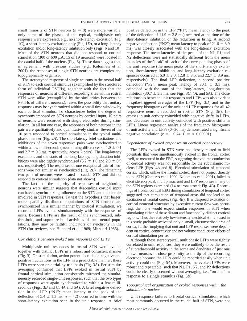
The stereotyped response of single neurons in the rostral halfof STN to each cortical stimulus (exemplified by the structuredform of individual PSTHs), together with the fact that theresponses of neurons at different recording sites within rostralSTN were alike (exemplified by the similarities between thePSTHs of different neurons), raises the possibility that unitaryresponses may be synchronized within a small time window byeach cortical stimulus. To characterize better the degree ofsynchrony imposed on STN neurons by cortical input, 10 pairsof neurons were recorded with single electrodes during stim-ulation. In all but one case, the responses of both neurons in thepair were qualitatively and quantitatively similar. Seven of the10 pairs responded to cortical stimulation in the typical multi-phasic manner (Fig. 2A). The short-latency brief excitations andinhibitions of the seven responsive pairs were synchronized towithin a few milliseconds (mean timing differences of 1.0 � 0.1and 2.7 � 0.5 ms, respectively, across 7 pairs). The long-latencyexcitations and the starts of the long-latency, long-duration inhi-bitions were also tightly synchronized (3.2 � 1.0 and 2.0 � 0.9ms, respectively). The responses of one exceptional pair of neu-rons were not similar or synchronized (Fig. 2B). The remainingtwo pairs of neurons were located in caudal STN and did notrespond to cortical stimulation (data not shown).
The fact that the majority of responses of neighboringneurons were similar suggests that descending cortical inputcan have a synchronizing influence on the STN and the circuitsinvolved in STN responses. To test the hypothesis that larger,more spatially distributed populations of STN neurons aresynchronized in a similar manner by cortical stimulation, werecorded LFPs evoked simultaneously with the responses ofunits. Because LFPs are the result of the synchronized, sub-threshold, and suprathreshold activities of local neural popu-lations, they may be faithful indicators of synchrony in theSTN (for reviews, see Hubbard et al. 1969; Mitzdorf 1985).
Correlations between evoked unit responses and LFPs
Multiphasic unit responses in rostral STN were evokedtogether with distinct LFPs in a robust and consistent manner(Fig. 3). On stimulation, action potentials rode on negative andpositive fluctuations in the LFP in a predictable manner; theseLFPs were seen on a trial-by-trial basis (Fig. 3A). Peristimulusaveraging confirmed that LFPs evoked in rostral STN byfrontal cortical stimulation consistently mirrored the simulta-neously recorded single-unit responses, such that the two typesof responses were again synchronized to within a few milli-seconds (Figs. 3B and C, 4A and 5A). A brief negative deflec-tion in the LFP (“N1”; mean latency to the peak of thedeflection of 5.4 � 1.3 ms; n � 42) occurred in time with theshort-latency excitation seen in the unit response. A brief
positive deflection in the LFP (“P1”; mean latency to the peakof the deflection of 11.9 � 2.8 ms) occurred at the time of theshort-latency inhibition or the reduction in firing. A secondnegative deflection (“N2”; mean latency to peak of 21.6 � 3.9ms) was closely associated with the long-latency excitationresponse. The mean latencies of the peaks of the N1, P1, andN2 deflections were not statistically different from the meanlatencies of the �peak� of each of the corresponding phases ofthe unit response (the mean peaks of the short-latency excita-tion, short-latency inhibition, and long-latency excitation re-sponses occurred at 6.0 � 2.0, 12.8 � 3.5, and 22.7 � 3.9 ms,respectively). The final LFP deflection, a second positivedeflection (“P2”; mean peak latency of 30.1 � 3.1 ms),coincided with the start of the long-latency, long-durationinhibition (30.7 � 5.3 ms; see Figs. 3C, 4A, and 5A). The closerelationship between unit responses and LFPs was also evidentin spike-triggered averages of the LFP (Fig. 3D) and in thefrequency histograms of the unit and LFP responses for all 42responsive neurons recorded in rostral STN (Fig. 3E). In-creases in unit activity coincided with negative shifts in LFPs,and decreases in unit activity coincided with positive shifts inLFPs. Linear regression analysis of the frequency histogramsof unit activity and LFPs (0–30 ms) demonstrated a significantnegative correlation (r � �0.74, P � � 0.00001).
Dependence of evoked responses on cortical connectivity
The LFPs evoked in STN were not closely related to fieldpotentials that were simultaneously evoked in the frontal cortexitself, as measured in the EEG, suggesting that volume conductionof cortical activity was not responsible for the subthalamic nu-cleus LFP (Figs. 4A and 8). Electrical stimulation of temporalcortex, which, unlike the frontal cortex, does not project directlyto the STN (Canteras et al. 1990; Kolomiets et al. 2001), failed toelicit stereotypical, multiphasic unit and LFP responses in any ofthe STN regions examined (14 neurons tested; Fig. 4B). Record-ings of frontal cortical EEG during stimulation of temporal cortexsuggested that stimulation of temporal areas did not lead to anexcitation of frontal cortex (Fig. 4B). If widespread excitation ofcortical neuronal structures by excessive current flow was occur-ring, then one would expect similar responses in STN whenstimulating either of these distant and functionally-distinct corticalregions. Thus the relatively low-intensity electrical stimuli used inthis study probably activated only a small, circumscribed area ofcortex, further implying that unit and LFP responses were depen-dent on cortical connectivity and not volume conduction effects ofcortical stimulation per se.
Although these stereotypical, multiphasic LFPs were tightlycorrelated to unit responses, they were unlikely to be the resultof suprathreshold activity in the soma and dendrites of just oneor two neurons in close proximity to the tip of the recordingelectrode because the LFPs could be recorded easily when unitactivity could not (Fig. 5A). Moreover, the evoked LFPs wererobust and repeatable, such that N1, P1, N2, and P2 deflectionscould be clearly discerned without averaging i.e., “on-line” inresponse to a single stimulus (Fig. 5B).
Topographical organization of evoked responses within thesubthalamic nucleus
Unit response failures to frontal cortical stimulation, whichmost commonly occurred in the caudal half of STN, were not
703EVOKED ACTIVITY IN THE SUBTHALAMIC NUCLEUS
J Neurophysiol • VOL 92 • AUGUST 2004 • www.jn.org
accompanied by the stereotypical evoked LFP (n � 16; Fig. 6),further indicating that these LFPs were topographically orga-nized and that the stimulation current was not excessive.
Although neurons in the caudal half of STN did not demon-strate the multiphasic response typical of cells in rostral STN,two neurons did respond with a long-latency excitation and a
FIG. 2. The responses of neighboring subthalamic nu-cleus neurons are often synchronized by frontal corticalstimulation. A, top: responses of 2 neighboring neurons inrostral STN that were simultaneously recorded using thesame electrode. Note that the response profiles were quali-tatively similar and that the peak excitations and inhibitionsof both neurons were synchronized to within a few millisec-onds. Bottom: rasterplots and longer peristimulus time his-tograms did not show significant increases or decreases infiring before cortical stimulation, suggesting that the dis-charges of the 2 neurons were not synchronized beforestimulation. B: the responses of the exceptional pair of rostralSTN neurons were not similar or synchronized. Calibrationbars for unit 1 also apply to unit 2. Calibration bar in A, top,also applies to B.
704 MAGILL, SHAROTT, BEVAN, BROWN, AND BOLAM
J Neurophysiol • VOL 92 • AUGUST 2004 • www.jn.org

FIG. 3. The unitary responses of subthalamic nucleusneurons are consistently evoked together with distinctlocal field potentials. A: typical unit responses and localfield potentials (LFPs, 1–4) evoked in rostral STN by 4sequential stimuli delivered to the frontal cortex (300 �Apulses at 0.67 Hz). Units (sharp spikes; see * in 1) andLFPs were recorded from the same electrode (0 to 2 kHzband-pass filtered). B: raster plot showing the typical“multiphasic” unit responses evoked by 200 sequentialstimuli. C: peristimulus time histogram of unit responsesand peristimulus average of LFPs evoked in the 200stimulation trials shown in B. D: spike-triggered averageof LFPs evoked in the 200 stimulation trials shown in B.Periods of increased likelihood of firing were coincidentwith significant negative deflections in the LFP. Thelarge deflection at time 0 corresponds to the spike in thewide-band filtered LFP. E: frequency histograms of theunit responses and evoked LFPs for all 42 responsiveneurons recorded in rostral STN demonstrated a stereo-typed, inverse relationship between unit activity andLFPs. In this and the following figures, positivity issignified as upward deflections of LFPs. Same STNneuron and recording site in A–D. Calibration bar in 1 ofA also applies to 2–4 in A.
705EVOKED ACTIVITY IN THE SUBTHALAMIC NUCLEUS
J Neurophysiol • VOL 92 • AUGUST 2004 • www.jn.org

weak, long-latency long-duration inhibition (Fig. 6). The ab-sence of short-latency responses was correlated at the level ofthe LFP by a lack of distinct N1 and P1 deflections. Moreover,the long-latency unit responses were associated with small N2and P2 deflections (also see Fig. 10).
Spatial specificity of evoked responses
To test whether the multiphasic evoked LFP was confined toSTN, unit and LFP responses were also recorded from neigh-boring brain regions. The LFP typically evoked in rostral STNwas not observed in surrounding structures, such as the zonaincerta, the cerebral peduncle and the internal capsule (Figs. 5Aand 7), supporting the idea that the LFP is due to the synchro-nous activity of neighboring STN neurons and is not stronglyinfluenced by activity in surrounding (proximal) structures.Most neurons in the ventral division of the zona incerta (ZIV)did not respond to cortical stimulation (13 of 20 neurons tested;Fig. 7A). Evoked LFPs recorded with nonresponsive ZIVneurons were of small amplitude and were unpredictable (Figs.5A and 7A). The discharges of the responsive ZIV neuronswere highly variable and were not clearly correlated with thewide variety of LFPs that were evoked in this region (Fig. 7,B–E). Neither the unit activity nor LFP bore a strong relation-ship to the responses in STN. Neurons in the cerebral peduncle(CP) were only rarely observed (n � 3). None of the CPneurons responded to cortical stimulation and evoked LFPswere relatively smooth and featureless (Figs. 5A and 7F).
Relationship of evoked responses to stimulus intensity
To ensure that stimulation at a current intensity of 300 �Awas effectively maximal and to test the possibility that differ-ent phases of the characteristic unit and LFP responses weredifferentially sensitive to input intensity, current-response re-lationships were studied. The profile of the LFP and the unitresponses evoked in STN by frontal cortical stimulation were
both dependent on the intensity of the current that was deliv-ered (Fig. 8). There were no qualitative differences in theresponse profiles evoked by stimulation at 600 and 300 �A(Fig. 8, A and B), and although the absolute magnitudes of thephasic responses varied slightly, no significant differences inlatencies were observed (n � 9 neurons). Thus the responses tostimulation at 300 �A were maximal with respect to the patternof the response. However, reducing the current intensity to 150or 75 �A resulted in distinct and corresponding changes in theunit and LFP responses (Fig. 8, C and D). The short-latencyresponses were attenuated, as were the associated LFP deflec-tions (N1 and P1), and the longer-latency unit responses tendedto fail together with the related LFP deflections (N2 and P2).The short-latency excitation and N1 responses were the last tofail at the lowest current intensity (Fig. 8D).
Relationship of evoked responses to brain state
The urethan-anesthetized rat is a good model for determin-ing the impact of extremes of forebrain activity on the BG(Magill et al. 2000, 2001). Activity in the cortex spontaneouslyshifts from “slow-wave activity” (Fig. 9A), which is similar toactivity observed during natural sleep, to a state of “globalactivation” (Fig. 9B), which contains patterns of activity thatare more analogous to those observed during the awake stateand vice versa (Steriade 2000). To test whether alterations inongoing forebrain activity could affect the profiles of theevoked responses in STN, we recorded and compared re-sponses evoked during both slow-wave activity and globalactivation. Spontaneous shifts in the global brain state of theanimal, as assessed from the cortical EEG, did not greatlyaffect the multiphasic responses to cortical stimulation (6neurons; Fig. 9, A and B). There were no qualitative differencesin the patterns of the unit responses or LFPs evoked bystimulation during the two brain states, and although theabsolute amplitudes of the multiphasic responses variedslightly, no significant differences in latencies were observed.
FIG. 4. Evoked unit activity in the subthalamic nucleus istightly correlated with LFPs and is organized according tocortical region. Top: schematic representations of the positionsof the stimulating electrodes (Stim) in the frontal (A) andtemporal (B) cortices. A: stimulation of the frontal cortex at 300�A evoked a characteristic unit response that was consistentlymirrored by contemporaneous deflections in the LFP (—, peri-stimulus average of LFP) recorded from the same electrode.Excitations at the single-unit level were accompanied by nega-tive deflections in the LFP (N1 and N2), while inhibitions wereassociated with positive deflections in the LFP (P1 and P2). TheLFP in STN was dissimilar to the field potential evoked in thefrontal cortex by the same stimuli [- - -; electroencephalogram(EEG)]. B: stimulation of the temporal cortex (600 �A) did notevoke a response from the same neuron. Accordingly, fieldpotentials in the STN and frontal cortex exhibited a different,smoother profile. Calibration bars in A also apply to B. AP(anterior-posterior) numbers denote positions with respect tobregma. M2, secondary motor cortex, Au1, primary auditorycortex.
706 MAGILL, SHAROTT, BEVAN, BROWN, AND BOLAM
J Neurophysiol • VOL 92 • AUGUST 2004 • www.jn.org

Atypical evoked responses
The unusual nature of the responses of a small population ofneurons was often reflected at the level of the evoked LFP.When STN neurons (n � 4) did not exhibit short-latencyexcitation or short-latency inhibition responses, then short-latency N1 and P1 deflections were small or absent from theLFP (Fig. 10, A and B); longer-latency unit responses were stillassociated with N2 and P2 deflections (Fig. 10, A and B).
D I S C U S S I O N
Taken together, these results suggest that cortical activationcan have a powerful synchronizing effect on spatially restricted
ensembles of STN neurons. Synchronized unit activity in STNwas consistently reflected at the local population level asmultiphasic LFPs, which were organized according to thetopography and intensity of cortical input. As such, LFPsevoked in the STN are good indicators of the functionalconnectivity of, and synaptic integration by, the underlyingneuronal population.
Circuit interactions underlying evoked unit responses in thesubthalamic nucleus
The STN receives monosynaptic inputs from select areas ofthe ipsilateral cerebral cortex, including prefrontal, premotor,
FIG. 5. Multiphasic unit responses and LFPs evoked in thesubthalamic nucleus by frontal cortical stimulation are stereo-typical and are not expressed in neighboring structures. A:schematic representation of recording sites in 1 vertical pass ofthe electrode through the brain at the level of the rostral half ofthe STN. Figures denote depths of recording sites from thecortical surface. Stereotypical, multiphasic unit and LFP re-sponses were observed throughout the dorsoventral axis of theSTN. Neurons located within a few hundred micrometers ofeach other shared similar response profiles (STN neurons atdepths of 7,881 and 8,085 �m). Multiphasic LFPs could still berecorded in the STN in the absence of unit activity (recordingsite at 8,020 �m). Vigorous LFP responses of the type seen inSTN were not observed in the ventral division of the zonaincerta (ZIV) nor the cerebral peduncle (CP); the small LFPdeflections in the two cases shown were not significant. B:stereotypical LFPs in STN were robust and repeatable. Fivesuperimposed LFP traces, each evoked sequentially by a singlestimulus (left); peristimulus average of 200 evoked LFPs withstandard errors (right). Calibration bar for LFP in STN (7,881�m) applies to all LFPs in A. Calibration bar in B, left, appliesto the right. AP (anterior-posterior) number denotes positionwith respect to bregma. ZID, dorsal division of the zonaincerta.
707EVOKED ACTIVITY IN THE SUBTHALAMIC NUCLEUS
J Neurophysiol • VOL 92 • AUGUST 2004 • www.jn.org

primary motor, cingulate and, to a lesser extent, somatosensorycortex (see review by Smith et al. 1998). The first response ofmost neurons in the rostral half of STN to stimulation of the
ipsilateral frontal cortex was a brief, but powerful, excitationwith a mean latency of 4.5 ms. This short-latency excitationhas been described by others and appears to be driven by the
FIG. 6. Unit responses and LFPs evoked in the subthalamicnucleus by frontal cortical stimulation are topographically or-ganized. Bottom left: schematic representation of recording sitesin 1 vertical pass of the electrode through the caudal half of theSTN. Figures denote depths of recording sites from the corticalsurface. These neurons were recorded from the same animal asshown in Fig. 5. Top left and right: the multiphasic unit and LFPresponses that were observed in rostral STN were not observedin caudal STN. Caudal STN neurons did not usually respond tofrontal cortical stimulation (STN neurons at depths of 7,920 and7,992 �m) and robust LFP responses of the kind recorded inrostral STN were not observed. Neurons located within a fewhundred micrometers of each other generally shared similar“response” profiles (neurons at 7,920 and 7,992 �m). However,2 cells did respond with a long-latency excitation and a weak,long-latency, long-duration inhibition, which were reflected inthe LFP (e.g., the neuron at 8,050 �m). Note the absence ofshort-latency responses was correlated at the level of the LFP bya lack of distinct N1 and P1 deflections. Calibration bars forunits and LFP in STN (7,920 �m) apply throughout. AP(anterior-posterior) number denotes position with respect tobregma. CP; cerebral peduncle, ZID, dorsal division of the zonaincerta; ZIV, ventral division of the zona incerta.
FIG. 7. Unit responses and LFPs evoked in the zona incertaand cerebral peduncle are variable and are not strongly corre-lated. A: most neurons in the ventral division of the zona incerta(ZIV) did not respond to cortical stimulation (300–600 �A).Evoked LFPs recorded with nonresponsive ZIV neurons were ofsmall amplitude and were unpredictable. ���, multiphasic LFPevoked in rostral STN for comparison. B–E: in contrast to thesubthalamic nucleus, the discharges of the responsive ZIV neu-rons were variable and were usually not clearly correlated withthe evoked LFPs. F: the small number of neurons recorded in thecerebral peduncle (CP) did not respond to cortical stimulation,and LFPs evoked in the peduncle were relatively smooth andfeatureless. Calibration bars in A apply to B–E.
708 MAGILL, SHAROTT, BEVAN, BROWN, AND BOLAM
J Neurophysiol • VOL 92 • AUGUST 2004 • www.jn.org

direct, excitatory corticosubthalamic projection (Bevan et al.1995; Fujimoto and Kita 1993; Kitai and Deniau 1981; Kolo-miets et al. 2001; Maurice et al. 1998; Nambu et al. 2000;Rouzaire-Dubois and Scarnati 1987; Ryan and Clark 1991,1992). The multiphasic nature of the typical unit responsedevelops from disynaptic and polysynaptic interactions, whichare slower to manifest. The second phase of the unit response,a brief, short-latency (mean: 10.4 ms) inhibition, probablyarises from feed-forward excitation of GP neurons by inputsfrom activated STN neurons and then feed-back inhibition ofSTN neurons by reciprocally connected neurons of the GP(Bevan et al. 2002a,b; Fujimoto and Kita 1993; Kita and Kitai1991; Maurice et al. 1998; Nambu et al. 2000; Ryan and Clark1991, 1992; Smith et al. 1998). Disinhibition of STN neurons,mediated by feed-forward connections through the NS and GPand then on to STN, has been proposed to account for the thirdphase of the response, a long-latency (mean: 16.6 ms) excita-
tion (Maurice et al. 1998; Nambu et al. 2000). Alternatively,this late excitation may represent the latter phase of a pro-longed barrage of cortical EPSPs and/or a long-lasting re-sponse of STN neurons to short-lasting cortical input (Fujimotoand Kita 1993; Otsuka et al. 2001; Ryan and Clarke 1992).
The short-latency excitation responses of most pairs ofneighboring STN neurons were similar such that they weresynchronized within a few milliseconds, and, when present,covaried; this demonstrates that the connectivity of the acti-vated corticosubthlamic projection could support the synchro-nous recruitment of neighboring target neurons. Because short-latency inhibitions were also synchronized, it is also likely thatthe activity of neurons in GP, and subsequently in STN, wassynchronized by the correlated discharges of STN neurons thatwere driven by corticosubthlamic input. Synchronous, long-latency excitations suggest that precisely timed, disinhibitoryprocesses were also widespread. These findings argue that,
FIG. 8. Unit responses and LFPs evoked in the subthalamicnucleus covary with stimulus intensity. A: typical responses ofa rostral unit, the LFP, and the cortical EEG (- - -) to frontalcortical stimulation at an intensity of 600 �A. B: reducing thestimulus intensity to 300 �A did not alter the qualitative natureof the responses although there were small, but commensurate,changes in the absolute magnitudes of the responses. C: afurther reduction in current intensity to 150 �A resulted in anattenuation of the short-latency excitation and inhibition re-sponses and a failure of the long-latency responses. Changes inthe evoked LFP were commensurate with these alterations in theunit response; the amplitudes of the N1 and P1 deflections werereduced, whereas the N2 and P2 responses virtually disap-peared. D: only the weak, short-latency excitation, with acorresponding small, negative deflection in the LFP, remainedat a stimulus intensity of 75 �A. Same neuron recorded in A–D.Calibration bars in A apply to all panels.
FIG. 9. Unit responses and LFPs evoked in the subthalamicnucleus by cortical stimulation are not strongly dependent onbrain state. A: during slow-wave activity, cortical activity(EEG) was dominated by a large-amplitude, slow oscillation.Unit activity in STN was closely related to the slow-waveactivity present in the cortex; STN neurons commonly exhib-ited low-frequency oscillations in firing. A: unit and LFPresponses evoked during slow-wave activity (a few secondsafter recording shown in A) were robust and were of stereo-typical, multiphasic natures. B: global activation was charac-terized by a prolonged loss of the large-amplitude, slow oscil-lation in the cortex and was associated with a change in theactivity of the STN neuron to irregular, tonic firing at a higherrate. B: the multiphasic responses of the same STN neuron andassociated LFP as evoked during global activation (a fewseconds after recording in B) did not substantially differ inpattern from responses evoked during slow-wave activity.Calibration bars in A apply to B, bars in A apply to B.
709EVOKED ACTIVITY IN THE SUBTHALAMIC NUCLEUS
J Neurophysiol • VOL 92 • AUGUST 2004 • www.jn.org

despite potentially complex circuit interactions, responseswithin small, local populations of neurons tend to be similarbecause the neurons are recruited by related afferent circuitry.
Unit responses were topographically organized within STN.A small minority of STN neurons, most of which were locatedin the caudal half of STN, did not respond to cortical stimu-lation. These findings are in good agreement with previousanatomical (Afsharpour 1985b; Canteras et al. 1990; Kolomi-ets et al. 2001) and physiological (Kolomiets et al. 2001)studies, which have shown that the frontal cortical areasstimulated in the present study project throughout most of thedorsoventral axis of the rostral two-thirds of STN only. Thecaudal one-third of the STN receives input from the caudalaspects of the medial frontal cortex (Afsharpour 1985b), whichwas presumably not activated by the electrical stimulus used inthis study. The present data are also in keeping with studiesdemonstrating that the (auditory) temporal cortex does notproject directly to STN (Canteras et al. 1990; Kolomiets et al.2001). Response differences were unlikely to be due to insuf-ficient current flow at the site of stimulation because responseswere maximal with respect to pattern at the stimulus intensitiesused. Similarly, the unresponsive nature of these neurons wasprobably not a function of anesthetic depth because the patternof responses did not dramatically alter across brain states. Thefact that unit and LFP responses to corticosubthalamic inputwere topographically organized and thus, did not conflict withthe known anatomy, adds further functional significance tothese stimulation data and implies that the currents used stim-ulated relatively restricted functional areas of cortex.
Neural basis of LFPs evoked in the subthalamic nucleus
The recording of LFPs evoked together with unit responsesallowed us to test the hypothesis that the responses of larger,more spatially distributed populations of STN neurons werealso synchronized to the high degree exhibited by pairs ofneighboring cells. A complex and nonlayered organization ofneurons and fibers, as occurs in STN (Afsharpour 1985a; Kitaet al. 1983; Smith et al. 1998; Yelnik and Percheron 1979), canmake the interpretation of current flows, and hence extracellu-lar potentials, challenging (Hubbard et al. 1969). Despite this,several temporal and spatial correlations between evoked LFPsand unit activity were reliably observed.
The first response of most STN neurons to cortical stimula-tion, i.e., the short-latency excitation, coincided with a prom-inent negative deflection (N1) in the LFP (Fig. 11A). Theoret-ical and experimental studies suggest that LFPs are a conse-quence of current flow related to synchronized, postsynapticpotentials rather than current flow across presynaptic andaxonal membranes (Hubbard et al. 1969; Mitzdorf 1985). Thus
we propose that the N1 deflection in the LFP was the result ofconcerted subthreshold and suprathreshold population activityin STN that was driven by monosynaptic cortical input (Fig.11, A and B). In agreement with this, the activity of neighbor-ing STN neurons was synchronously increased by the stimulus(also see Ryan et al. 1992) and, when single units did notrespond with a short-latency excitation, the N1 deflection wasmuch smaller or absent. The second phase of the unit responseto cortical stimulation, a brief reduction in activity, which waslikely caused by feed-back inhibition from GP neurons (seepreceding text) (Bevan et al. 2002b), was significantly associ-ated with a brief, positive deflection (P1) in the LFP (Fig. 11A).This positive deflection likely reflected the synchronous hy-perpolarization of STN neurons by pallidal inputs (Fig. 11, Aand C). Support for this comes from intracellular recordings(Fujimoto and Kita 1993; Kitai and Deniau 1981) and the factthat when single units did not respond with a short-latencyinhibition, the P1 deflection was not distinct. The subsequentlong-latency excitation, presumably due to the disinhibition ofSTN neurons and/or the continued excitation of STN neuronsby cortical input (see preceding text), was accompanied by asecond negative deflection (N2) in the LFP (Fig. 11, A and D).The cellular and synaptic mechanisms underlying the finalphase of the unit response, a long-duration inhibition, areunknown, although “cortical disfacilitation” has been hypoth-esized to be the cause (Fujimoto and Kita 1993). The start ofthis phase again was associated with a positive deflection (P2)in the LFP (Fig. 11A), a finding corroborated by previousintracellular data indicating that the long-duration inhibition isdue to membrane hyperpolarization (Kitai and Deniau 1981).These significant temporal correlations between unit and pop-ulation responses suggest that synchronous frontal corticalinputs can impose widespread synchronization within the ros-tral half of the STN and associated feed-back and feed-forwardneuronal circuits.
In agreement with the topographic organization of unitresponses, spatial correlations between units and LFPs werealso consistently observed in addition to temporal correlations.Neurons in the caudal half of STN did not respond in themultiphasic way that was typical of neurons in rostral STN. Inthese cases, the characteristic evoked LFP was either verysmall, or, more commonly, not seen at all. These observationssubstantiate the idea that the LFPs were the result of thesynchronous activity of ensembles of functionally related STNneurons and argue against a significant contribution to LFPsfrom volume-conducted activity.
The stereotypical unit responses and LFPs observed in STNwere not found in the zona incerta (ZI) or CP. Furthermore, inZI, unit responses were not clearly related to LFPs, despite the
FIG. 10. Atypical unit responses and LFPs evoked in thesubthalamic nucleus by cortical stimulation. A and B: a smallnumber of rostral neurons did not exhibit short-latency excita-tion or short-latency inhibition responses to stimulation (at300–600 �A) but did exhibit long-latency excitations andinhibitions. The LFPs evoked in these cases were also unusualbut still reflected unit activity; short-latency N1 and P1 deflec-tions were small or absent from the corresponding LFPs, but theN2 and P2 deflections were still present. Calibration bars in Aapply to B.
710 MAGILL, SHAROTT, BEVAN, BROWN, AND BOLAM
J Neurophysiol • VOL 92 • AUGUST 2004 • www.jn.org

fact that this area also receives monosynaptic inputs fromfrontal cortical areas (Mitrofanis and Mikuletic 1999). Thisimplies that the synchronization of unitary responses in STN isnot simply the result of the synchronized corticofugal volleysinduced by stimulation but must also entail some fundamentaldifferences in the organization of direct cortical inputs toneighboring neurons in STN as compared with neurons in ZIVand/or the intrinsic composition of these nuclei. The unit andpopulation responses recorded in ZI and the CP thus act asuseful controls for the data from STN recordings that supportthe idea that the multiphasic LFP in STN is the result of thesynchronized activity of a local population of neurons and, assuch, is characteristic of STN.
The high predictability and specificity of the evoked LFPs,together with their topographic nature, have two importantimplications. First, these data suggest that the geometry of therelevant local dipoles dictating current flow in STN i.e., thesomata and dendrites of neurons, may be more ordered thanpreviously thought. Second, the correlations we have observedbetween units and evoked LFPs may strengthen the interpre-tation of LFPs commonly observed in STN in other paradigms(see review by Brown 2003), particularly high-frequency LFPoscillations because rapid sequences of excitations and inhibi-tions at the unit level are reflected in the LFP with good timeresolution.
Functional implications
Although the electrical stimulation used in this study mayin itself ensure a synchronous cortical output, it does not fol-low that the subthreshold and suprathreshold responses ofneighboring STN neurons will also be synchronous. Glutama-tergic EPSPs and GABAergic inhibitory postsynaptic poten-
tials (IPSPs), whether they are compound or unitary, are ofteninefficient at phase-locking the responses of neurons (e.g.,Bennett and Wilson 1998; Carter and Regehr 2002; Fricker andMiles 2000). However, we found that activation of direct andindirect inputs to STN neurons could often synchronize thedischarges of specific neuronal populations in the STN. Ourfinding strongly suggests that the neuronal networks engagedby cortical activation are designed to promote precise couplingbetween synaptic potentials and action potentials in STN neu-rons, which, in turn, generate correlated activity in neighboringSTN neurons during synchronized cortical activity. The mech-anisms underlying this precise coupling are unknown but couldbe extrinsic, e.g., each STN neuron in a given functionaldomain is innervated to a similar degree by the associatedregions of cortex and GP. Alternatively, or additionally, themechanism may be intrinsic in nature, e.g., STN neuronsexhibit an unusual enhanced sensitivity to excitatory input andlittle spike-frequency adaptation during high-frequency firing(Beurrier et al. 2000; Bevan and Wilson 1999; Hallworth et al.2003; Wilson et al. 2004). Whatever the mechanism(s) respon-sible for precise coupling in STN, it is important to note thatthe same stimuli did not have the same effect on the ZI despitethe fact that it also receives inputs from frontal and/or prefron-tal cortices (Mitrofanis and Mikuletic 1999).
The significance of our findings is supported by studies thatsuggest that the STN, under normal and pathological condi-tions, is subject to synchronous cortical inputs in vivo. Neuronsin cortical areas projecting to the STN exhibit a wide range ofsynchronous network activity during natural behaviors (Aoki etal. 1999; Donoghue et al. 1998; Kristeva-Feige et al. 1993;Murthy and Fetz 1992, 1996). In particular, synchronizedoscillations, which are caused by periodic, phase-locked dis-charges of cortical cell ensembles, are frequently observed in
FIG. 11. Hypothetical scheme of the neuro-nal circuits that likely underlie the corticalstimulation-evoked LFP in subthalamic nu-cleus. A: the stereotypical, multiphasic profileof the LFP evoked in STN. The LFP was likelya consequence of activity in monosynaptic andpolysynaptic circuits. B–D: times are meanlatencies to peak deflections. Shaded basalganglia nuclei contribute to that particularphase of the response in STN; white nuclei donot contribute. Dark gray arrows, excitatorypathways; light gray arrows, inhibitory path-ways. B: the short-latency, negative deflection,N1, was probably due to activation of thedirect corticosubthalamic pathway. C: theshort-latency, positive deflection, P1, probablyarose as a consequence of feed-forward exci-tation of globus pallidus (GP) by STN and thenfeed-back inhibition of STN by GP. D: thelong-latency, negative deflection, N2, wasmost likely due to disinhibition of STNthrough inhibition of GP by neostriatum (NS)in a feed-forward manner. Arrow with dashedline represents disinhibition. The circuits thatmay underlie the long-latency (mean: 30.1ms), positive deflection, P2, are unknown.
711EVOKED ACTIVITY IN THE SUBTHALAMIC NUCLEUS
J Neurophysiol • VOL 92 • AUGUST 2004 • www.jn.org

cortex during sensory-motor integration and other complexbehaviors that are likely to involve the basal ganglia (Engel andSinger 2001; Engel et al. 2001; MacKay 1997; Roelfsema et al.1997). Corticosubthalmic input might therefore be highly syn-chronized during these behaviors, and, in turn, generate syn-chronous activity in the STN. Consistent with this hypothesis,emergent cortical oscillations synchronize (oscillatory) unitactivity in STN (Allers et al. 2002; Magill et al. 2000, 2001;Wichmann et al. 2001). Furthermore, the STN may expresssynchronous oscillations, as evinced in unit or LFP recordings,under both normal and pathological conditions (Boraud et al.2002; Brown et al. 2002; Brown 2003; Levy et al. 2002a).Some oscillations in STN are related to movement and aredependent on dopamine, suggesting they are of functionalsignificance (Brown et al. 2001, 2002; Levy et al. 2002a;Williams et al. 2002, 2003). Moreover, simultaneous record-ings have shown that oscillatory population activity in STNmay be significantly coherent with that in cortex and that thetime lags between oscillations are consistent with the cortexplaying a role in synchronizing activity in the basal ganglia(Cassidy et al. 2002; Marsden et al. 2001; Williams et al.2002).
The present data highlight two additional considerations ofimportance. The response evoked in the cortex itself, as mea-sured in the EEG, was entirely different to the evoked STNresponse, as recorded in the LFP, suggesting that BG circuitsare able to generate a different form of activity than thatexpressed by the cortex. Second, the fact that the short-latencyexcitation response of STN neurons and associated LFP de-flection (N1) were last to fail on reduction of stimulus intensitysuggests that STN neurons receiving monosynaptic corticalinput are likely to respond most vigorously and reliably to thatinput when activated, compared with subsequent inputs de-rived from polysynaptic circuit interactions. Taken together,the current data add weight to the proposed importance of thecorticosubthalamic projection (also see Magill et al. 2001;Mink 1996; Nambu et al. 2002) and synchronous ensembleactivity in information processing in cortico-basal gangliacircuits (Engel et al. 2001).
Clinical implications
The STN is an important target for the surgical treatment ofParkinson’s disease and perhaps, in the future, intractableepilepsy, partly because of the proven clinical benefits of “deepbrain stimulation,” in which neuronal activity is modified byelectrical stimulation through electrodes implanted in STN(Chabardes et al. 2002; Kumar et al. 1998; Limousin et al.1998; Loddenkemper et al. 2001). However, surgical implan-tation of electrodes is challenging because of the difficulties ofunequivocally locating the STN and key neighboring structures(Starr 2002; Voges et al. 2002). The finding that LFPs evokedin STN by stimulation of the cerebral cortex are distinct fromthose evoked in surrounding structures indicates that evokedLFPs could act as electrophysiological “fingerprints” thatmight greatly aid the identification of the STN and, perhapsmore importantly, specific regions within or above STN (Saint-Cyr et al. 2002; Voges et al. 2002). Electrophysiologicalrecordings of STN activity are now routinely used in implan-tation surgery to refine the positioning of the stimulationelectrode (Priori et al. 2003; Starr 2002) and because the
evoked LFPs are relatively resistant to changes in brain state,patient anesthesia may not be an issue. Furthermore, LFPs canbe recorded in STN from microelectrodes, or the macroelec-trodes that are used for deep brain stimulation (Brown et al.2001; Dinner et al. 2002; Levy et al. 2002a; Liu et al. 2002),and importantly, evoked LFPs could be evaluated after only afew cortical stimuli (i.e., within a few seconds intraopera-tively). There is also some evidence to suggest that our ap-proach could potentially be extrapolated for use with a nonin-vasive stimulation technique, such as transcranial magneticstimulation, thereby increasing its potential clinical value (Ku-mar et al. 1999). Finally, our definitive demonstration of theneural basis of LFPs in the STN confirms that LFP studies inhumans with implanted electrodes provide valuable insight intothe pathophysiological mechanisms underlying idiopathic Par-kinson’s disease (see Brown 2003).
A C K N O W L E D G M E N T S
We are grateful to Drs. J. Csicsvari, T. Klausberger, M. Ungless, X. Lin, T.Aziz, and J. Stein for valuable comments. We thank L. Norman, B. Micklem,and C. Francis for technical assistance.
G R A N T S
This work was supported by the Medical Research Council United King-dom and the Brain Research Trust (A. Sharott) and by National Institute ofNeurological Disorders and Stroke Grant NS-41280 (M. D. Bevan). P. J.Magill holds a Fellowship by Examination at Magdalen College, Oxford, UK.
R E F E R E N C E S
Afsharpour S. Light microscopic analysis of Golgi-impregnated rat subtha-lamic neurons. J Comp Neurol 236: 1–13, 1985a.
Afsharpour S. Topographical projections of the cerebral cortex to the sub-thalamic nucleus. J Comp Neurol 236: 14–28, 1985b.
Allers KA, Ruskin DN, Bergstrom DA, Freeman LE, Ghazi LJ, TierneyPL, and Walters JR. Multisecond periodicities in basal ganglia firing ratescorrelate with theta bursts in transcortical and hippocampal EEG. J Neuro-physiol 87: 1118–1122, 2002.
Aoki F, Fetz EE, Shupe L, Lettich E, and Ojemann GA. Increasedgamma-range activity in human sensorimotor cortex during performance ofvisuomotor tasks. Clin Neurophysiol 110: 524–537, 1999.
Bennett BD and Wilson CJ. Synaptic regulation of action potential timing inneostriatal cholinergic interneurons. J Neurosci 18: 8539–8549, 1998.
Bergman H, Feingold A, Nini A, Raz A, Slovin H, Abeles M, and VaadiaE. Physiological aspects of information processing in the basal ganglia ofnormal and parkinsonian primates. Trends Neurosci 21: 32–38, 1998.
Beurrier C, Bioulac B, and Hammond C. Slowly inactivating sodium current(INaP) underlies single-spike activity in rat subthalamic neurons. J Neuro-physiol 83: 1951–1957, 2000.
Bevan MD, Francis CM, and Bolam JP. The glutamate-enriched cortical andthalamic input to neurons in the subthalamic nucleus of the rat: convergencewith GABA-positive terminals. J Comp Neurol 361: 491–511, 1995.
Bevan MD, Magill PJ, Hallworth NE, Bolam JP, and Wilson CJ. Regula-tion of the timing and pattern of action potential generation in rat subtha-lamic neurons in vitro by GABAA IPSPs. J Neurophysiol 87: 1348–1362,2002a.
Bevan MD, Magill PJ, Terman D, Bolam JP, and Wilson CJ. Move to therhythm: oscillations in the subthalamic nucleus-external globus pallidusnetwork. Trends Neurosci 25: 525–531, 2002b.
Bolam JP. (Editor). Experimental Neuroanatomy. Oxford, UK: Oxford Univ.Press, 1992.
Boraud T, Bezard E, Bioulac B, and Gross CE. From single extracellularunit recording in experimental and human Parkinsonism to the developmentof a functional concept of the role played by the basal ganglia in motorcontrol. Prog Neurobiol 66: 265–283, 2002.
Brown P. Oscillatory nature of human basal ganglia activity: relationship tothe pathophysiology of Parkinson’s disease. Mov Disord 18: 357–363, 2003.
Brown P, Kupsch A, Magill PJ, Sharott A, Harnack D, and Meissner W.Oscillatory local field potentials recorded from the subthalamic nucleus ofthe alert rat. Exp Neurol 177: 581–585, 2002.
712 MAGILL, SHAROTT, BEVAN, BROWN, AND BOLAM
J Neurophysiol • VOL 92 • AUGUST 2004 • www.jn.org

Brown P, Oliviero A, Mazzone P, Insola A, Tonali P, and Di Lazzaro V.Dopamine dependency of oscillations between subthalamic nucleus andpallidum in Parkinson’s disease. J Neurosci 21: 1033–1038, 2001.
Canteras NS, Shammah-Lagnado SJ, Silva BA, and Ricardo JA. Afferentconnections of the subthalamic nucleus: a combined retrograde and antero-grade horseradish peroxidase study in the rat. Brain Res 513: 43–59, 1990.
Carter AG and Regehr WG. Quantal events shape cerebellar interneuronfiring. Nat Neurosci 512: 1309–1318, 2002.
Cassidy M, Mazzone P, Oliviero A, Insola A, Tonali P, Di Lazarro V, andBrown P. Movement-related changes in synchronization in the human basalganglia. Brain 125: 1235–1246, 2002.
Chabardes S, Kahane P, Minotti L, Koudsie A, Hirsch E, and BenabidAL. Deep brain stimulation in epilepsy with particular reference to thesubthalamic nucleus. Epilepsy Disord 4: S82–S93, 2002.
DeLong MR. Primate models of movement disorders of basal ganglia origin.Trends Neurosci 13: 281–285, 1990.
Dinner DS, Neme S, Nair D, Montgomery EB Jr, Baker KB, Rezai A, andLuders HO. EEG and evoked potential recording from the subthalamicnucleus for deep brain stimulation of intractable epilepsy. Clin Neurophysiol113: 1391–1402, 2002.
Donoghue JP and Wise SP. The motor cortex of the rat: cytoarchitecture andmicrostimulation mapping. J Comp Neurol 212: 76–88, 1982.
Donoghue JP and Parham C. Afferent connections of the lateral agranularfield of the rat motor cortex. J Comp Neurol 217: 390–404, 1983.
Donoghue JP, Sanes JN, Hastopoulos NG, and Gaal G. Neural dischargeand local field potential oscillations in primate motor cortex during volun-tary movements. J Neurophysiol 79: 159–173, 1998.
Engel AK, Fries P, and Singer W. Dynamic predictions: oscillations andsynchrony in top-down processing. Nat Rev Neurosci 2: 704–716, 2001.
Engel AK and Singer W. Temporal binding and the neural correlates ofsensory awareness. Trends Cog Sci 5: 16–25, 2001.
Ellaway PH. Cumulative sum technique and its application to the analysis ofperistimlus time histograms. J Physiol 45: 320–304, 1978.
Fricker D and Miles R. EPSP amplification and the precision of spike timingin hippocampal neurons. Neuron 28: 559–569, 2000.
Fujimoto K and Kita H. Response characteristics of subthalamic neurons tothe stimulation of the sensorimotor cortex in the rat. Brain Res 609:185–192, 1993.
Gerfen CR and Wilson CJ. The basal ganglia. In: Handbook of ChemicalNeuroanatomy. Integrated Systems of the CNS III, edited by Swanson LW,Bjorklund A, and Hokfelt, T. London: Elsevier, 1996, vol. 12, p. 371–468.
Graybiel AM. Building action repertoires: memory and learning functions ofthe basal ganglia. Curr Opin Neurobiol 5: 733–741, 1995.
Hallworth NE, Wilson CJ, and Bevan MD. Apamin-sensitive small conduc-tance calcium-activated potassium channels, through their selective cou-pling to voltage-gated calcium channels, are critical determinants of theprecision, pace, and pattern of action potential generation in rat subthalamicnucleus neurons in vitro. J Neurosci 23: 7525–7542, 2003.
Hubbard JI, Llinas R, and Quastel DMJ. Extracellular field potentials in thecentral nervous system. In: Electrophysiological Analysis of Synaptic Trans-mission. London: Edward Arnold, 1969, p. 265–293.
Kincaid AE, Zheng T, and Wilson CJ. Connectivity and convergence ofsingle corticostriatal axons. J Neurosci 18: 4722–4731, 1998.
Kita H and Armstrong WE. A biocytin-containing compound N-(2-amino-ethyl)biotinimide for intracellular labeling and neuronal tracing studies:comparison with biocytin. J Neurosci Methods 37: 141–150, 1991.
Kita H, Chang HT, and Kitai ST. The morphology of intracellularly labeledrat subthalamic neurons: a light microscopic analysis. J Comp Neurol 215:245–257, 1983.
Kita H and Kitai ST. Intracellular study of rat globus pallidus neurons:membrane properties and responses to neostriatal, subthalamic and nigralstimulation. Brain Res 564: 296–305, 1991.
Kitai ST and Deniau JM. Cortical inputs to the subthalamus: intracellularanalysis. Brain Res 214: 411–415, 1981.
Kolomiets BP, Deniau JM, Mailly P, Menetrey A, Glowinski J, andThierry AM. Segregation and convergence of information flow through thecortico-subthalamic pathways. J Neurosci 21: 5764–5772, 2001.
Kristeva-Feige R, Feige B, Makeig S, Ross B, and Elbert T. Oscillatorybrain activity during a motor task. Neuroreport 4: 1291–1294, 1993.
Kumar R, Chen R, and Ashby P. Safety of transcranial magnetic stimulationin patients with implanted deep brain stimulators. Mov Disord 14: 157–158,1999.
Kumar R, Lozano AM, Kim YJ, Hutchison WD, Sime E, Halket E, andLang AE. Double-blind evaluation of subthalamic nucleus deep brainstimulation in advanced Parkinson’s disease. Neurology 51: 850–855, 1998.
Levy R, Ashby P, Hutchison WD, Lang AE, Lozano AM, and DostrovskyJO. Dependence of subthalamic nucleus oscillations on movement anddopamine in Parkinson’s disease. Brain 125: 1196–1209, 2002a.
Levy R, Hutchison WD, Lozano AM, and Dostrovsky JO. Synchronizedneuronal discharge in the basal ganglia of parkinsonian patients is limited tooscillatory activity. J Neurosci 22: 2855–2861, 2002b.
Limousin P, Krack P, Pollak P, Benazzouz A, Ardouin C, Hoffmann D,and Benabid AL. Electrical stimulation of the subthalamic nucleus inadvanced Parkinson’s disease. N Engl J Med 339: 1105–1111, 1998.
Liu X, Ford-Dunn HL, Hayward GN, Nandi D, Miall RC, Aziz TZ, andStein J. The oscillatory activity in the Parkinsonian subthalamic nucleusinvestigated using the macro-electrodes for deep brain stimulation. ClinNeurophysiol 113: 1667–1672, 2002.
Loddenkemper T, Pan A, Neme S, Baker KB, Rezai AR, Dinner DS,Montgomery EB Jr, and Luders HO. Deep brain stimulation in epilepsy.J Clin Neurophysiol 18: 514–532, 2001.
MacKay WA. Synchronized neuronal oscillations and their role in motorprocesses. Trends Cog Sci 1: 176–183, 1997.
Magill PJ, Bolam JP, and Bevan MD. Relationship of activity in thesubthalamic nucleus-globus pallidus network to cortical electroencephalo-gram. J Neurosci 20: 820–833, 2000.
Magill PJ, Bolam JP, and Bevan MD. Dopamine regulates the impact of thecerebral cortex on the subthalamic nucleus-globus pallidus network. Neu-roscience 106: 313–330, 2001.
Marsden JF, Limousin-Dowsey P, Ashby P, Pollak P, and Brown P.Subthalamic nucleus, sensorimotor cortex and muscle interrelationships inParkinson’s disease. Brain 124: 378–388, 2001.
Maurice N, Deniau JM, Glowinski J, and Thierry AM. Relationshipsbetween the prefrontal cortex and the basal ganglia in the rat: physiology ofthe corticosubthalamic circuits. J Neurosci 18: 9539–9546, 1998.
Mink JW. The basal ganglia: focused selection and inhibition of competingmotor programs. Prog Neurobiol 50: 381–425, 1996.
Mitrofanis J and Mikuletic L. Organisation of the cortical projection to thezona incerta of the thalamus. J Comp Neurol 412: 173–185, 1999.
Mitzdorf U. Current-source density method and application in cat cerebralcortex: investigation of evoked potentials and EEG phenomena. Physiol Rev65: 37–100, 1985.
Murthy VN and Fetz EE. Coherent 25- and 35- Hz oscillations in thesensorimotor cortex of awake behaving monkeys. Proc Natl Acad Sci USA89: 5670–5674, 1992.
Murthy VN and Fetz EE. Synchronization of neurons during local fieldpotential oscillations in sensorimotor cortex of awake monkeys. J Neuro-physiol 76: 3968–3982, 1996.
Nambu A, Tokuno H, Hamada I, Kita H, Imanishi M, Akazawa T, IkeuchiY, and Hasegawa N. Excitatory cortical inputs to pallidal neurons via thesubthalamic nucleus in the monkey. J Neurophysiol 84: 289–300, 2000.
Nambu A, Tokuno H, and Takada M. Functional significance of thecortico-subthalamo-pallidal “hyperdirect” pathway. Neurosci Res 43: 111–117, 2002.
Otsuka T, Murakami F, and Song WJ. Excitatory postsynaptic potentialstrigger a plateau potential in rat subthalamic neurons at hyperpolarizedstates. J Neurophysiol 86: 1816–1825, 2001.
Paxinos G and Watson C. The Rat Brain in Stereotaxic Coordinates (2nded.). Sydney, Australia: Academic, 1986.
Priori A, Egidi M, Pesenti A, Rohr M, Rampini P, Locatelli M, Tamma F,Caputo E, Chiesa V, and Barbieri S. Do intraoperative microrecordingsimprove subthalamic nucleus targeting in stereotactic neurosurgery forParkinson’s disease? J Neurosurg Sci 47: 56–60, 2003.
Roelfsema PR, Engel AK, Konig P, and Singer W. Visuomotor integrationis associated with zero time-lag synchronization among cortical areas.Nature 385: 157–161, 1997.
Rouzaire-Dubois B and Scarnati E. Pharmacological study of the cortical-induced excitation of subthalamic nucleus neurons in the rat: evidence foramino acids as putative neurotransmitters. Neuroscience 21: 429–440,1987.
Ryan LJ and Clark KB. The role of the subthalamic nucleus in the responseof globus pallidus neurons to stimulation of the prelimbic and agranularfrontal cortices in rats. Exp Brain Res 86: 641–651, 1991.
Ryan LJ and Clark KB. Alteration of neuronal responses in the subthalamicnucleus following globus pallidus and neostriatal lesions in rats. Brain ResBull 29: 319–327, 1992.
713EVOKED ACTIVITY IN THE SUBTHALAMIC NUCLEUS
J Neurophysiol • VOL 92 • AUGUST 2004 • www.jn.org
Ryan LJ, Sanders DJ, and Clark KB. Auto- and cross-correlation analysis ofsubthalamic nucleus neuronal activity in neostriatal- and globus pallidal-lesioned rats. Brain Res 583: 253–261, 1992.
Saint-Cyr JA, Hoque T, Pereira LC, Dostrovsky JO, Hutchison WD,Mikulis DJ, Abosch A, Sime E, Lang AE, and Lozano AM. Localizationof clinically effective stimulating electrodes in the human subthalamicnucleus on magnetic resonance imaging. J Neurosurg 97: 1152–1166, 2002.
Smith Y, Bevan MD, Shink E, and Bolam JP. Microcircuitry of the directand indirect pathways of the basal ganglia. Neuroscience 86: 353–387, 1998.
Starr PA. Placement of deep brain stimulators into the subthalamic nucleus orglobus pallidus internus: technical approach. Stereotact Funct Neurosurg79: 118–145, 2002.
Steriade M. Corticothalamic resonance, states of vigilance and mentation.Neuroscience 101: 243–76, 2000.
Voges J, Volkmann J, Allert N, Lehrke R, Koulousakis A, Freund HJ, andSturm V. Bilateral high-frequency stimulation in the subthalamic nucleusfor the treatment of Parkinson disease: correlation of therapeutic effect withanatomical electrode position. J Neurosurg 96: 269–279, 2002.
Wichmann T and DeLong MR. Functional and pathophysiological models ofthe basal ganglia. Curr Opin Neurobiol 6: 751–758, 1996.
Wichmann T, Kliem MA, and Soares J. Correlation between neuronaldischarge in the basal ganglia and EEG in normal and parkinsonian pri-mates. Soc Neurosci Abstr 31: 749.23, 2001.
Williams D, Kuhn A, Kupsch A, Tijssen M, Van Bruggen G, Speelman H,Hotton G, Yarrow K, and Brown P. Behavioral cues are associated withmodulations of synchronous oscillations in the human subthalamic nucleus.Brain 126: 1975–1985, 2003.
Williams D, Tijssen M, Van Bruggen G, Bosch A, Insola A, Di LazzaroV, Mazzone P, Oliviero A, Quartarone A, Speelman H, and Brown P.Dopamine-dependent changes in the functional connectivity betweenbasal ganglia and cerebral cortex in humans. Brain 125: 1558 –1569,2002.
Wilson CJ, Weyrick A, Terman D, Hallworth NE, and Bevan MD. Amodel of reverse spike frequency adaptation and repetitive firing of subtha-lamic nucleus neurons. J Neurophysiol 91: 1963–1980, 2004.
Yelnik J and Percheron G. Subthalamic neurons in primates: a quantitativeand comparative analysis. Neuroscience 4: 1717–1743, 1979.
Zheng T and Wilson CJ. Corticostriatal combinatorics: the implications ofcorticostriatal axonal arborizations. J Neurophysiol 87: 1007–1017, 2002.
714 MAGILL, SHAROTT, BEVAN, BROWN, AND BOLAM
J Neurophysiol • VOL 92 • AUGUST 2004 • www.jn.org