Embed Size (px)
Citation preview
Synaptic Vesicle Cycle
quantal release has many advantages
but imposes several requirements
high potential for regulation
huge variation in release probability
multiple modes of release
process information

vesicular transport: a general mechanism
transport vesicles form at compartment A
fuse with compartment B
what are alternative possibilities?
usually constitutive
vesicles do not accumulate
cannot isolate key intermediate
--unless process regulated
SV
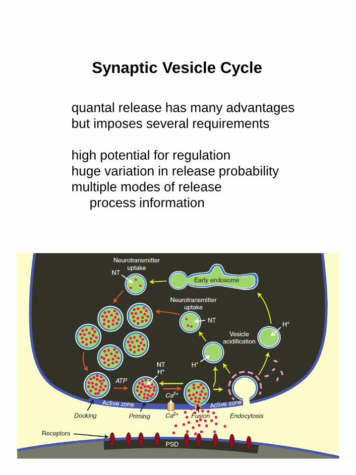
SV Purification
synaptic vesicles the smallest biological membranes
homogeneous in size, shape
--separate by density (equilibrium sedimentation)
and size (velocity sedimentation, size ex-
exclusion chromatography)
(size)
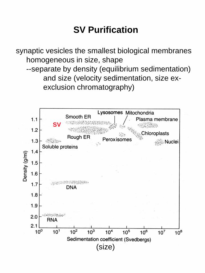
proteins excised from gel, sequenced:
mechanisms of membrane association
single TMD (type 2 synaptobrevin, 1 synaptotagmin)
polytopic (synaptophysin, SV2)
peripheral membrane proteins (synapsin, synuclein)
lipid-anchored (rab3, cysteine string protein)
proteomics has quantified components
the function of most remain unknown
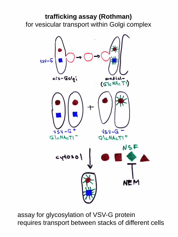
trafficking assay (Rothman)
for vesicular transport within Golgi complex
assay for glycosylation of VSV-G protein
requires transport between stacks of different cells

activity requires many proteins
identified by functional complementation
inactivate extract with NEM (-SH reagent)
rescue with untreated extract
purify rescuing component:
--NEM-sensitive factor (NSF)
NSF is an ATPase--used to find associated proteins:
binding in non-hydrolyzable ATP
elute with ATP--releases only those bound
dependent on ATP
--soluble NSF attachment receptors (SNAREs)

functionclostridial toxins block NT release
--Zn-dependent proteases
(A. Brunger)
specifically cleave SNAREs
--less effect on spontaneous than
evoked release
structure

revised model
zippering mechanism provides energy
N-termini of v- and t-SNAREs interact first
energy for fusion provided by binding
SNARE complex very stable
--dissociates only by boiling in SDS
OR addition of ATP to NSF
to dissociate SNAREs before endocytosis,
leaving t-SNAREs on plasma membrane
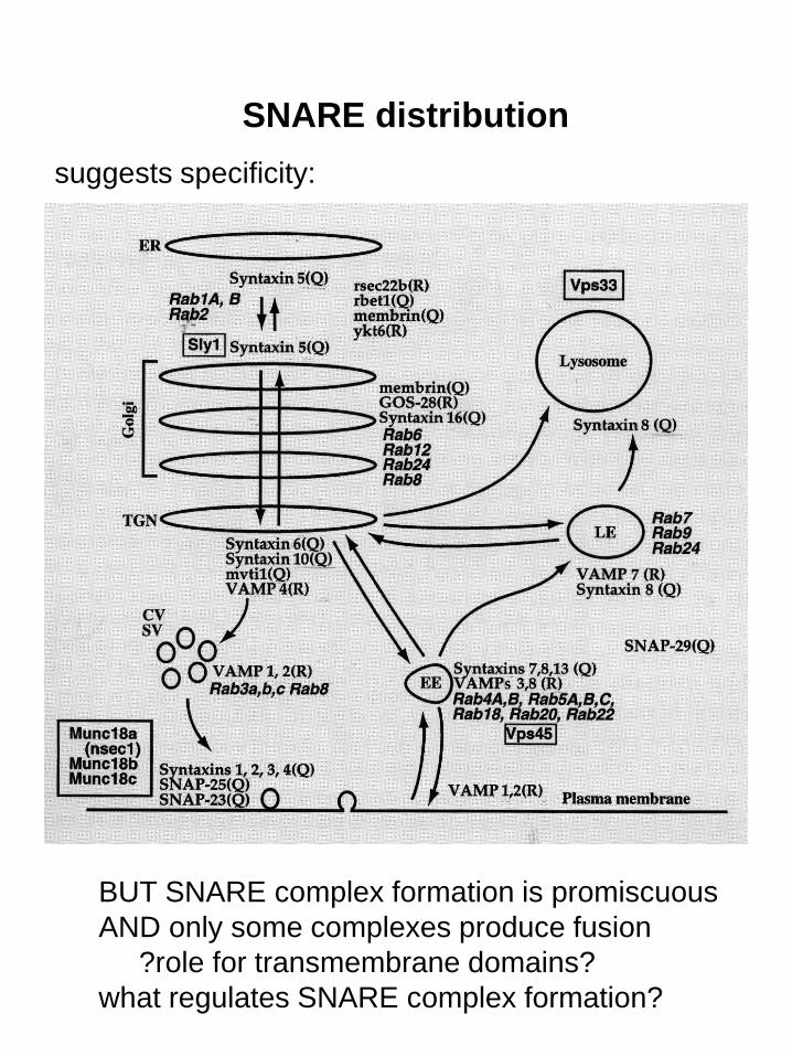
SNARE distribution
BUT SNARE complex formation is promiscuous
AND only some complexes produce fusion
?role for transmembrane domains?
what regulates SNARE complex formation?
suggests specificity:
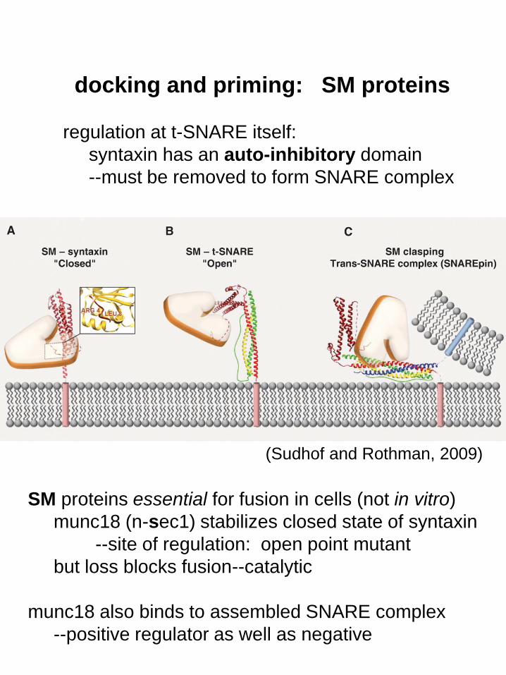
SM proteins essential for fusion in cells (not in vitro)
munc18 (n-sec1) stabilizes closed state of syntaxin
--site of regulation: open point mutant
but loss blocks fusion--catalytic
munc18 also binds to assembled SNARE complex
--positive regulator as well as negative
regulation at t-SNARE itself:
syntaxin has an auto-inhibitory domain
--must be removed to form SNARE complex
(Sudhof and Rothman, 2009)
docking and priming: SM proteins
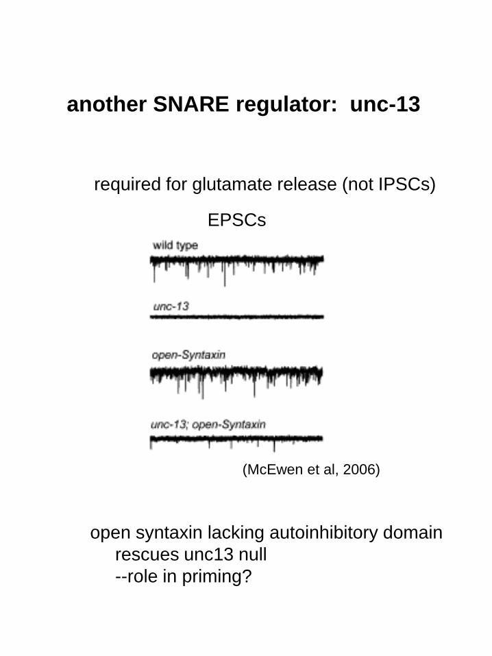
another SNARE regulator: unc-13
open syntaxin lacking autoinhibitory domain
rescues unc13 null
--role in priming?
EPSCs
required for glutamate release (not IPSCs)
(McEwen et al, 2006)

(Rosenmund et al, 2002)
munc13
double KO = no release
rescue with different isoforms
alone confers different forms
of short-term plasticity
(due to changes in Pr)

what confers specificity to SNARE assembly?
not SNAREs
only a few SM proteins (operate at multiple sites)
?rabs
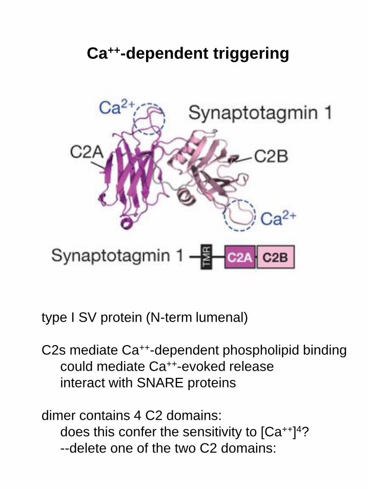
Ca++-dependent triggering
type I SV protein (N-term lumenal)
C2s mediate Ca++-dependent phospholipid binding
could mediate Ca++-evoked release
interact with SNARE proteins
dimer contains 4 C2 domains:
does this confer the sensitivity to [Ca++]4?
--delete one of the two C2 domains:
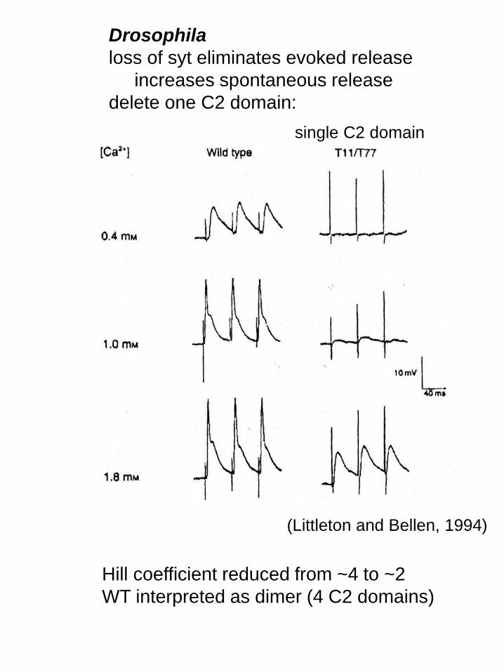
(Littleton and Bellen, 1994)
single C2 domain
Hill coefficient reduced from ~4 to ~2
WT interpreted as dimer (4 C2 domains)
Drosophila
loss of syt eliminates evoked release
increases spontaneous release
delete one C2 domain:
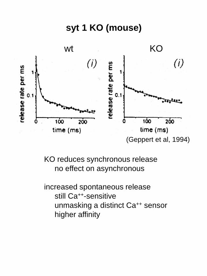
syt 1 KO (mouse)
wt KO
KO reduces synchronous release
no effect on asynchronous
increased spontaneous release
still Ca++-sensitive
unmasking a distinct Ca++ sensor
higher affinity
(Geppert et al, 1994)

multiple synaptotagmins
differ in Ca++ affinity
Syt7 on LDCVs and lysosomes
required for facilitation: how?
contributes to regulated secretion
function of others unknown
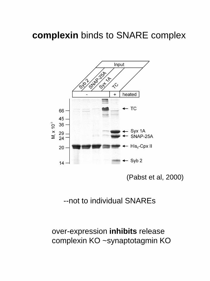
complexin binds to SNARE complex
(Pabst et al, 2000)
--not to individual SNAREs
over-expression inhibits release
complexin KO ~synaptotagmin KO
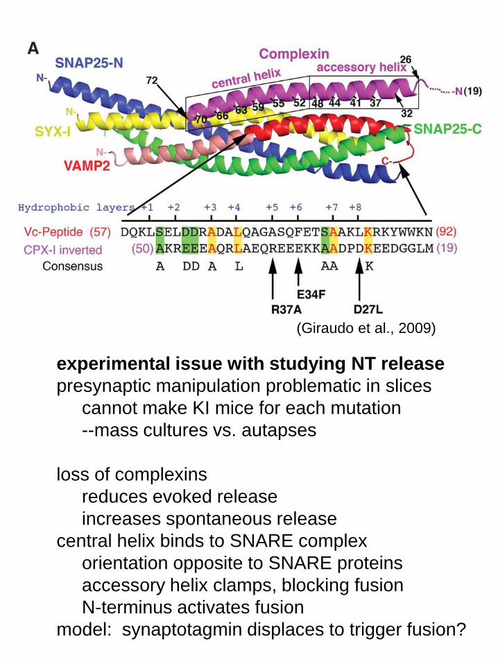
(Giraudo et al., 2009)
experimental issue with studying NT release
presynaptic manipulation problematic in slices
cannot make KI mice for each mutation
--mass cultures vs. autapses
loss of complexins
reduces evoked release
increases spontaneous release
central helix binds to SNARE complex
orientation opposite to SNARE proteins
accessory helix clamps, blocking fusion
N-terminus activates fusion
model: synaptotagmin displaces to trigger fusion?
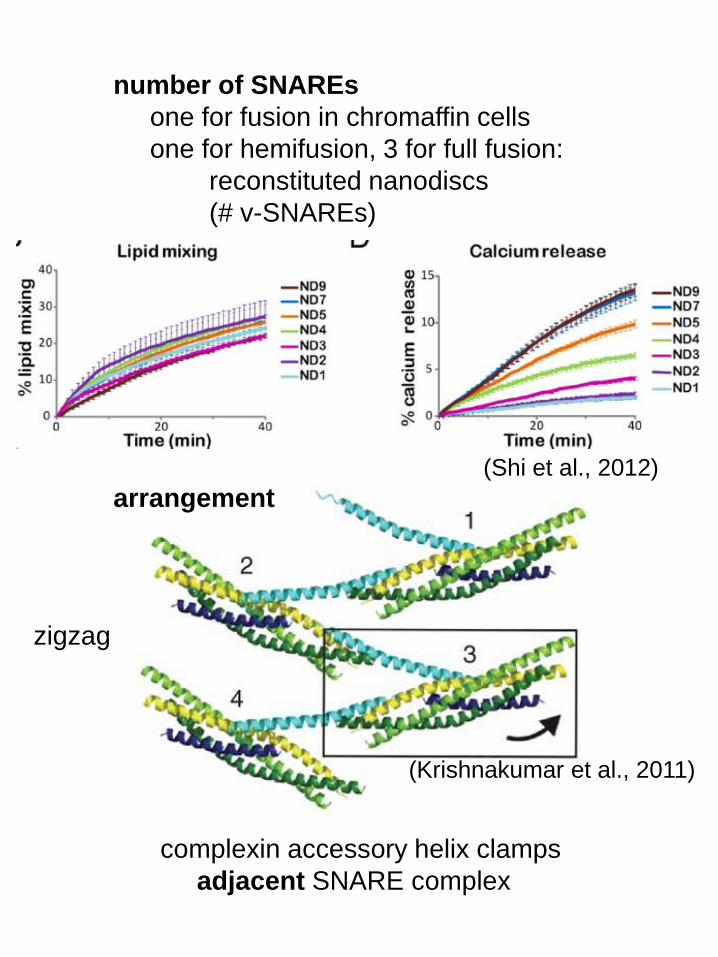
number of SNAREs
one for fusion in chromaffin cells
one for hemifusion, 3 for full fusion:
reconstituted nanodiscs
(# v-SNAREs)
arrangement
(Shi et al., 2012)
complexin accessory helix clamps
adjacent SNARE complex
zigzag
(Krishnakumar et al., 2011)
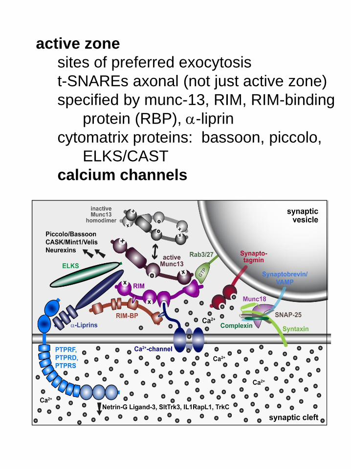
active zone
sites of preferred exocytosis
t-SNAREs axonal (not just active zone)
specified by munc-13, RIM, RIM-binding
protein (RBP), a-liprin
cytomatrix proteins: bassoon, piccolo,
ELKS/CAST
calcium channels
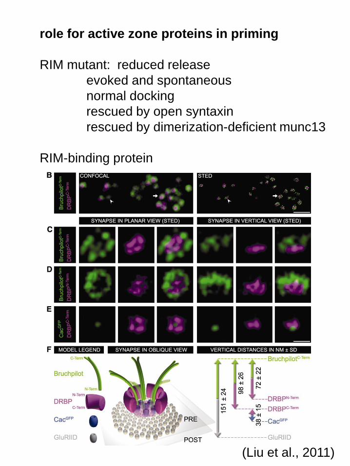
role for active zone proteins in priming
RIM mutant: reduced release
evoked and spontaneous
normal docking
rescued by open syntaxin
rescued by dimerization-deficient munc13
RIM-binding protein
(Liu et al., 2011)

loss of RBP disrupts Brp (ELKS)
RBP mutant recovers with stimulus train
(Liu et al., 2011)
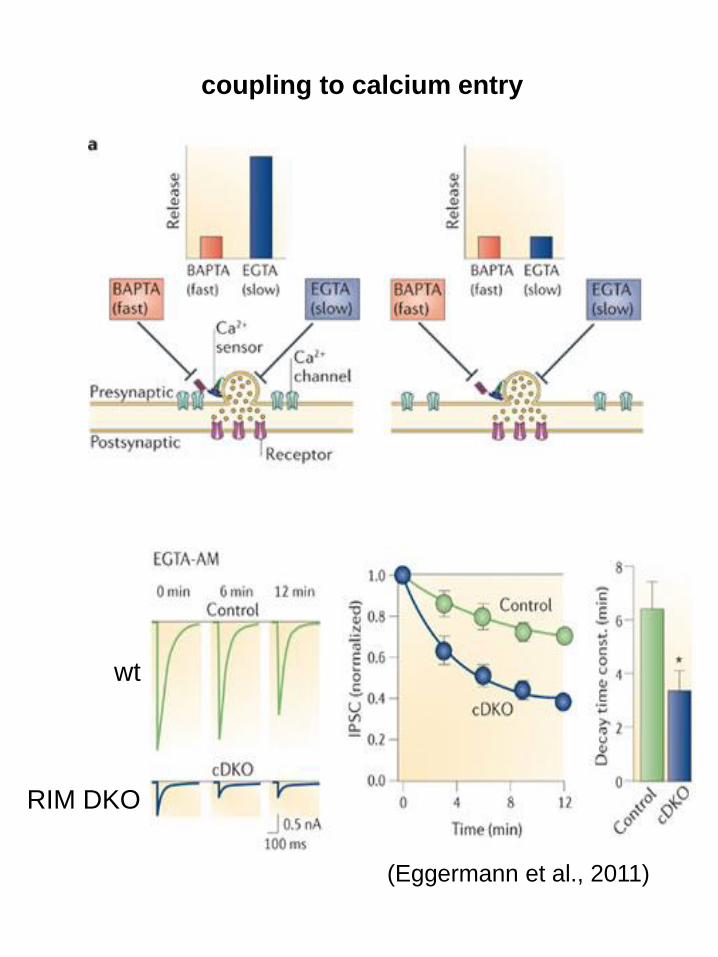
(Eggermann et al., 2011)
coupling to calcium entry
wt
RIM DKO
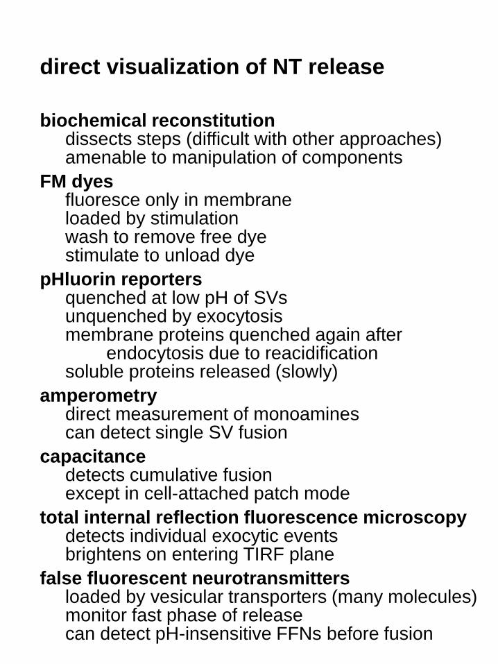
direct visualization of NT release
biochemical reconstitutiondissects steps (difficult with other approaches)amenable to manipulation of components
FM dyesfluoresce only in membraneloaded by stimulationwash to remove free dyestimulate to unload dye
pHluorin reporters quenched at low pH of SVsunquenched by exocytosismembrane proteins quenched again after
endocytosis due to reacidificationsoluble proteins released (slowly)
amperometrydirect measurement of monoaminescan detect single SV fusion
capacitancedetects cumulative fusionexcept in cell-attached patch mode
total internal reflection fluorescence microscopydetects individual exocytic eventsbrightens on entering TIRF plane
false fluorescent neurotransmittersloaded by vesicular transporters (many molecules)monitor fast phase of releasecan detect pH-insensitive FFNs before fusion
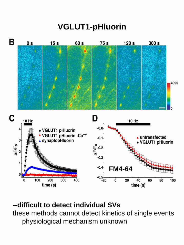
VGLUT1-pHluorin
FM4-64
--difficult to detect individual SVs
these methods cannot detect kinetics of single events
physiological mechanism unknown

large dense core vesicles (LDCVs): release kinetics
capacitance: oscillating voltage
individual vesicles in mast cells--
amperometry
spikes
stand-alone foot
foot
(Breckenridge and Almers, 1987)
capacitance
fluore
scence
preloaded quinacrine
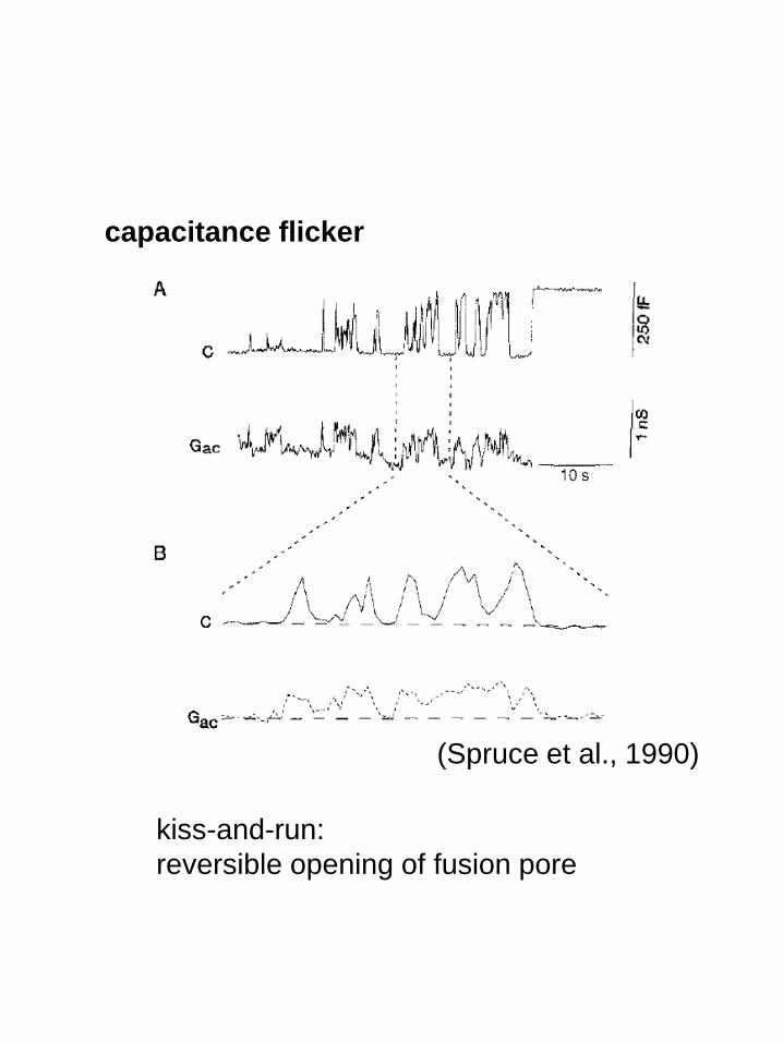
kiss-and-run:
reversible opening of fusion pore
capacitance flicker
(Spruce et al., 1990)
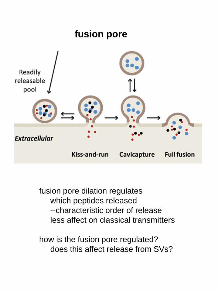
fusion pore
fusion pore dilation regulates
which peptides released
--characteristic order of release
less affect on classical transmitters
how is the fusion pore regulated?
does this affect release from SVs?
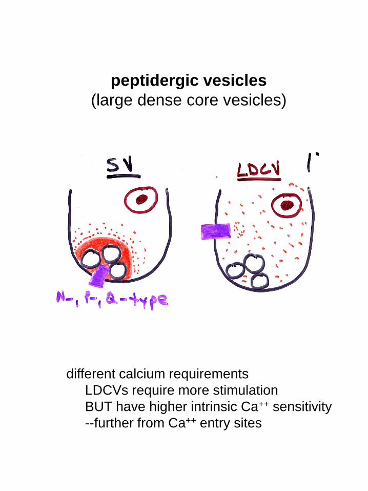
peptidergic vesicles
(large dense core vesicles)
different calcium requirements
LDCVs require more stimulation
BUT have higher intrinsic Ca++ sensitivity
--further from Ca++ entry sites
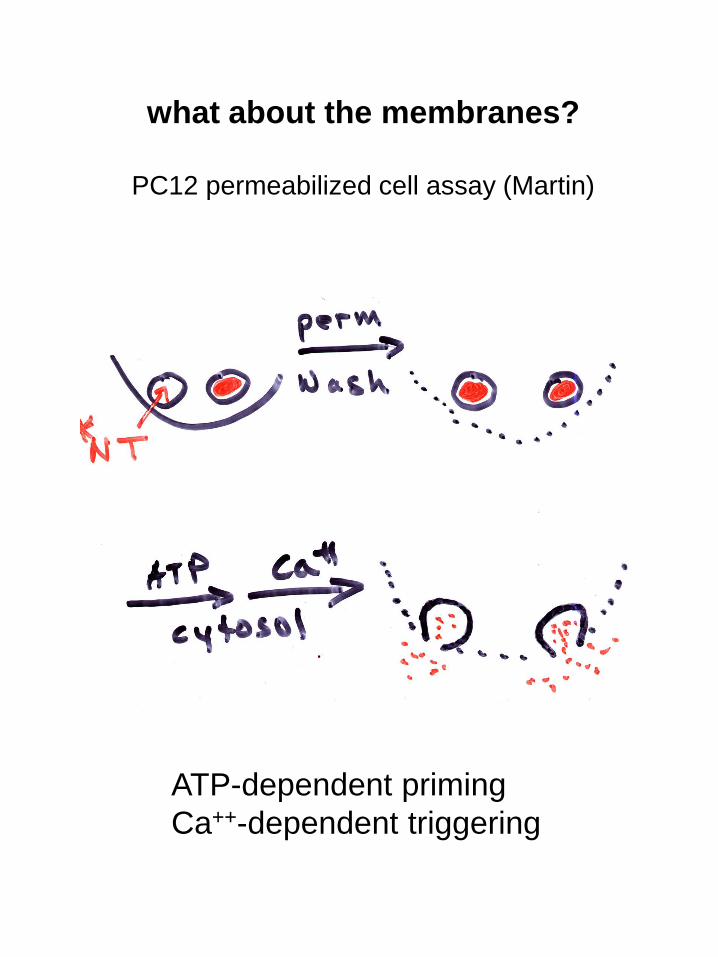
what about the membranes?
PC12 permeabilized cell assay (Martin)
ATP-dependent priming
Ca++-dependent triggering
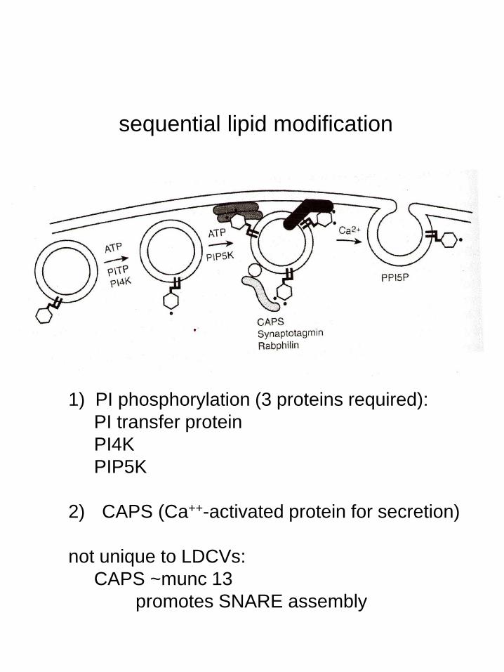
1) PI phosphorylation (3 proteins required):
PI transfer protein
PI4K
PIP5K
2) CAPS (Ca++-activated protein for secretion)
not unique to LDCVs:
CAPS ~munc 13
promotes SNARE assembly
sequential lipid modification
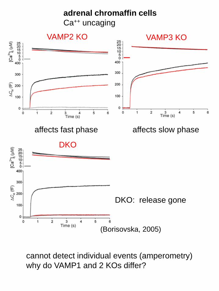
VAMP2 KO VAMP3 KO
DKO
affects fast phase affects slow phase
DKO: release gone
cannot detect individual events (amperometry)
why do VAMP1 and 2 KOs differ?
(Borisovska, 2005)
adrenal chromaffin cells
Ca++ uncaging
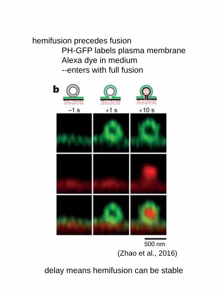
hemifusion precedes fusion
PH-GFP labels plasma membrane
Alexa dye in medium
--enters with full fusion
(Zhao et al., 2016)
delay means hemifusion can be stable
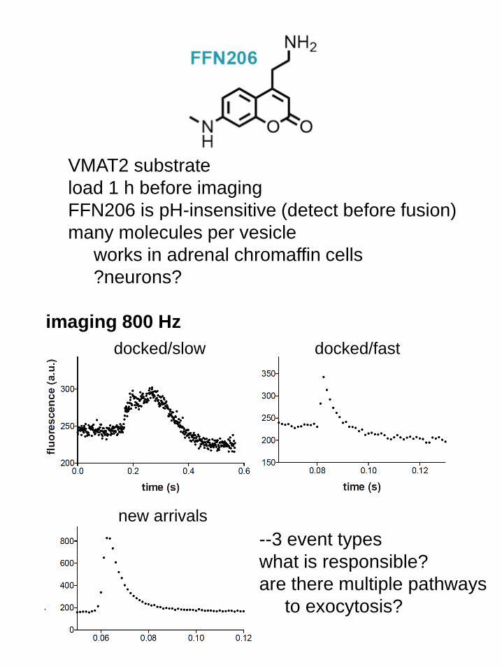
VMAT2 substrate
load 1 h before imaging
FFN206 is pH-insensitive (detect before fusion)
many molecules per vesicle
works in adrenal chromaffin cells
?neurons?
docked/slow docked/fast
new arrivals
imaging 800 Hz
--3 event types
what is responsible?
are there multiple pathways
to exocytosis?

Reading: The Synapse, edited by Sheng, Sabatini and Sudhof, pp. 49-78
ReferencesBorisovska M, Zhao Y, Tsytsyura Y, Glyvuk N, Takamori S, Matti U, Rettig J,Sudhof T, Bruns D (2005) v-SNAREs control exocytosis of vesicles from priming tofusion. EMBO J 24:2114-2126.Chen, X., Tomchick, D.R., Kovrigin, E., Arac, D., Machius, M., Sudhof, T.C., andRizo, J. (2002). Three-dimensional structure of the complexin/SNARE complex.Neuron 33, 397-409.Eggermann, E., Bucurenciu, I., Goswami, S.P., and Jonas, P. (2012). Nanodomaincoupling between Ca(2)(+) channels and sensors of exocytosis at fast mammaliansynapses. Nature reviews. Neuroscience 13, 7-21.Geppert, M., Goda, Y., Hammer, R.E., Li, C., Rosahl, T.W., Stevens, C.F., andSudhof, T.C. (1994). Synaptotagmin I: a major Ca++ sensor for transmitterrelease at a central synapse. Cell 79, 717-727.Giraudo, C.G., Eng, W.S., Melia, T.J. and Rothman, J.E. 2006. A clampingmechanism Involved in SNARE-dependent exocytosis. Science 313: 676-80.Giraudo CG, Garcia-Diaz A, Eng WS, Chen Y, Hendrickson WA, Melia TJ,Rothman JE (2009) Alternative zippering as an on-off switch for SNARE-mediatedfusion. Science 323:512-516.Graf, E.R., R.W. Daniels, R.W. Burgess, T.L. Schwarz, and A. DiAntonio. 2009.Rab3 dynamically controls protein composition at active zones. Neuron. 64:663-77.Jackman, S.L., Turecek, J., Belinsky, J.E. & Regehr, W.G. The calcium sensorsynaptotagmin 7 is required for synaptic facilitation. Nature 529, 88-91 (2016).Krishnakumar, S.S., Radoff, D.T., Kummel, D., Giraudo, C.G., Li, F., Khandan, L.,Baguley, S.W., Coleman, J., Reinisch, K.M., Pincet, F., et al. (2011). Aconformational switch in complexin is required for synaptotagmin to triggersynaptic fusion. Nat. Struct. Mol. Biol. 18, 934-940.Hay, J.C., and Martin, T.F.J. (1992). Resolution of regulated secretion intosequential MgATP-dependent and calcium-dependent stages mediated by distinctcytosolic proteins. J. Cell Biol. 119, 139-151.Hu, Z., Hom, S., Kudze, T., Tong, X.J., Choi, S., Aramuni, G., Zhang, W., andKaplan, J.M. (2012). Neurexin and neuroligin mediate retrograde synapticinhibition in c. Elegans. Science 337, 980-984.Hu, Z. Tong, X.J., Kaplan, J.M. 2013. UNC-13L, UNC-13S and tomosyn for aprotein code for fast and slow neurotransmitter release in Caenorhabditis elegans.eLife e00967.Jackman, S. L., Turecek, J., Belinsky, J. E. & Regehr, W. G. 2016. Thecalcium sensor synaptotagmin 7 is required for synaptic facilitation. Nature529, 88-91.Jahn, R., and Fasshauer, D. (2012). Molecular machines governing exocytosis ofsynaptic vesicles. Nature 490, 201-207.Kaeser PS, Regehr WG (2014) Molecular mechanisms for synchronous,asynchronous, and spontaneous neurotransmitter release. Annu. Rev. Physiol.76:333-363.Krishnakumar, S.S., Radoff, D.T., Kummel, D., Giraudo, C.G., Li, F., Khandan, L.,Baguley, S.W., Coleman, J., Reinisch, K.M., Pincet, F., et al. (2011). Aconformational switch in complexin is required for synaptotagmin to triggersynaptic fusion. Nat Struct Mol Biol 18, 934-940.Lee, H.-K., Yang, H. Su, Z. et al. 2010. Dynamic calcium-dependent stimulationof vesicle fusion by membrane-anchored synaptotagmin 1. Science 328, 760-3.
Littleton, J.T., Stern, M., Perin, M., and Bellen, H.J. (1994). Calcium dependenceof neurotransmitter release and rate of spontaneous vesicle fusions are alteredin Drosophila synaptotagmin mutants. Proc. Natl. Acad. Sci. USA 91, 10888-10892.Liu, K. S. et al. 2011. RIM-binding protein, a central part of the active zone, isessential for neurotransmitter release. Science 334, 1565-1569.Ma, C., Su, L., Seven, A. Xu, Y. and Rizo, J. 2013. Reconstitution of the vitalfunctions of munc18 and 13 in neurotransmitter release. Science 339, 421-5.Maximov A, Tang J, Yang X, Pang ZP, Sudhof TC. 2009. Complexin controls theforce transfer from SNARE complexes to membranes in fusion. Science323:516-521.McNew, J.A., Parlati, F., Fukuda, R., Johnston, R.J., Paz, K., Paumet, F., Sollner,T.H., and Rothman, J.E. (2000). Compartmental specificity of cellular membranefusion encoded in SNARE proteins. Nature 407, 153-159.Mohrmann, R., de Wit, H., Verhage, M., Neher, E., and Sorensen, J.B. (2010).Fast vesicle fusion in living cells requires at least three SNARE complexes.Science 330, 502-505.Pabst, S., Hazzard, J.W., Antonin, W., Sudhof, T.C., Jahn, R., Rizo, J., andFasshauer, D. (2000). Selective interaction of complexin with the neuronalSNARE complex. Determination of the binding regions. J. Biol. Chem. 275,19808-19818.Rosenmund, C., Sigler, A., Augustin, I., Reim, K., Brose, N., and Rhee, J.S.(2002). Differential control of vesicle priming and short-term plasticity by Munc13isoforms. Neuron 33, 411-424.Schiavo, G., Benfenati, F., Poulain, B., Rossetto, O., Polverino de Laureto, P.,DasGupta, B.R., and Montecucco, C. (1992). Tetanus and botulinum-Bneurotoxins block neurotransmitter release by proteolytic cleavage ofsynaptobrevin. Nature 359, 832-835.Schonn JS, Maximov A, Lao Y, Sudhof TC, Sorensen JB (2008) Synaptotagmin-1 and -7 are functionally overlapping Ca2+ sensors for exocytosis in adrenalchromaffin cells. Proc Natl Acad Sci U S A 105:3998-4003.Shi, L., Shen, Q.T., Kiel, A., Wang, J., Wang, H.W., Melia, T.J., Rothman, J.E.,and Pincet, F. (2012). Snare proteins: One to fuse and three to keep the nascentfusion pore open. Science 335, 1355-1359.Sollner, T., et al. 1993. SNAP receptors implicated in vesicle targeting and fusion.Nature 362, 318-324.Spruce, A. E., Breckenridge, L. J., Lee, A. K. & Almers, W. 1990. Properties ofthe fusion pore that forms during exocytosis of a mast cell secretory vesicle.Neuron 4, 643-654.Sudhof, T.C. (2012). The presynaptic active zone. Neuron 75, 11-25.Sudhof TC, Rothman JE (2009) Membrane fusion: grappling with SNARE andSM proteins. Science 323:474-477.Sutton, R.B., Fasshauer, D., Jahn, R., and Brunger, A.T. (1998). Crystal structureof a SNARE complex involved in synaptic exocytosis at 2.4 A resolution. Nature395, 347-353.Walent, J.H., Porter, B.W., and Martin, T.F. (1992). A novel 145 kd brain cytosolicprotein reconstitutes Ca++-regulated secretion in permeable neuroendocrinecells. Cell 70, 765-775.Zenisek, D., Steyer, J.A., and Almers, W. (2000). Transport, capture andexocytosis of single synaptic vesicles at active zones. Nature 406, 849-854.Zhao, W. D. et al. 2016. Hemi-fused structure mediates and controls fusion andfission in live cells. Nature 534, 548-552.