Embed Size (px)
Citation preview
LUND UNIVERSITY
PO Box 117221 00 Lund+46 46-222 00 00
Symptomatic cluster headache: a review of 63 cases.
Edvardsson, Bengt
Published in:SpringerPlus
DOI:10.1186/2193-1801-3-64
2014
Link to publication
Citation for published version (APA):Edvardsson, B. (2014). Symptomatic cluster headache: a review of 63 cases. SpringerPlus, 3(1), [64].https://doi.org/10.1186/2193-1801-3-64
General rightsCopyright and moral rights for the publications made accessible in the public portal are retained by the authorsand/or other copyright owners and it is a condition of accessing publications that users recognise and abide by thelegal requirements associated with these rights.
• Users may download and print one copy of any publication from the public portal for the purpose of private studyor research. • You may not further distribute the material or use it for any profit-making activity or commercial gain • You may freely distribute the URL identifying the publication in the public portalTake down policyIf you believe that this document breaches copyright please contact us providing details, and we will removeaccess to the work immediately and investigate your claim.
a SpringerOpen Journal
Edvardsson SpringerPlus 2014, 3:64http://www.springerplus.com/content/3/1/64
REVIEW Open Access
Symptomatic cluster headache: a review of63 casesBengt Edvardsson
Abstract
Cluster headache is a primary headache by definition not caused by any known underlying structural pathology.Symptomatic cases have been described, for example tumours, dissections and infections, but a causal relationshipbetween the underlying lesion and the headache is difficult to determine in many cases. The proper diagnosticevaluation of cluster headache is an issue unresolved. The literature has been reviewed for symptomatic clusterheadache or cluster headache-like cases in which causality was likely. The review also attempted to identify clinicalpredictors of underlying lesions in order to formulate guidelines for neuroimaging. Sixty-three cluster headache or"cluster headache-like"/"cluster-like headache" cases in the literature were identified which were associated with anunderlying lesion. A majority of the cases had a non-typical presentation that is atypical symptomatology andabnormal examination (including Horner’s syndrome). A striking finding in this appraisal was that a significantproportion of CH cases were secondary to diseases of the pituitary gland or pituitary region. Another notablefinding was that a proportion of cluster headache cases were associated with arterial dissection. Even typical clusterheadaches can be caused by structural lesions and the response to typical cluster headache treatments does notexclude a secondary form. It is difficult to draw definitive conclusions from this retrospective review of case reportsespecially considering the size of the material. However, based on this review, I suggest that neuroimaging, preferablycontrast-enhanced magnetic resonance imaging/magnetic resonance angiography should be undertaken in patientswith atypical symptomatology, late onset, abnormal examination (including Horner’s syndrome), or those resistant tothe appropriate medical treatment. The decision to perform magnetic resonance imaging in cases of typical clusterheadache remains a matter of medical art.
Keywords: Cluster headache; Neuroimaging; Secondary; Symptomatic; Magnetic resonance imaging;Differential diagnosis
IntroductionCluster headache (CH) is a primary headache, by definitionnot caused by any underlying structural pathology and be-longing to the group of trigeminal-autonomic cephalalgias(Headache Subcommittee of the International HeadacheSociety 2004). CH is the most frequent syndrome in thisgroup. The characteristic symptoms are strictly unilateralhead pain (mainly around orbital and temporal regions)and associated ipsilateral cranial autonomic features. Theheadache usually lasts 45 to 90 minutes, but can rangebetween 15 and 180 minutes. CH is an excruciatingheadache and probably one of the most painful head-ache syndromes. A circannual and circadian pattern is
Correspondence: [email protected] of Clinical Sciences Lund, Faculty of Medicine, Neurology, SkaneUniversity Hospital, S-221 85 Lund, Sweden
© 2014 Edvardsson; licensee Springer. This is aAttribution License (http://creativecommons.orin any medium, provided the original work is p
typical. Most patients have the episodic form characterizedby bouts lasting >1 week and separated by remissions last-ing longer than 4 weeks. A minority of about 10% to 20%have the chronic form, with no remission within a year orremission periods lasting <1 month (Headache Subcommit-tee of the International Headache Society 2004; Cittadini &Matharu 2009). The disease primarily emerges between theages of 20 to 40 years and is more prevalent in men. Pooleddata from epidemiological studies give CH a lifetime preva-lence of 0.12%. Furthermore, the condition has a heritabletendency in some families (Nesbitt & Goadsby 2012). Thepathophysiology of CH is not well known. The most widelyaccepted theory is that primary CH is characterized byhypothalamic activation with secondary activation of thetrigeminal-autonomic reflex, probably by a trigeminal-hypothalamic pathway (Nesbitt & Goadsby 2012). Cluster
n Open Access article distributed under the terms of the Creative Commonsg/licenses/by/2.0), which permits unrestricted use, distribution, and reproductionroperly cited.

Edvardsson SpringerPlus 2014, 3:64 Page 2 of 8http://www.springerplus.com/content/3/1/64
headache has also been associated with heart disease. Dataindicate a higher prevalence of right-left shunt in patientswith cluster headache compared with controls (Morelliet al. 2005). The great majority of cases of CH are primary.There are many case reports of symptomatic/secondarycases of CH in the literature (Table 1), but a causal rela-tionship between the underlying lesion and the head-ache is difficult to determine in many cases (Cittadini &Matharu 2009). The proper diagnostic evaluation of CH isan issue unresolved. The true prevalence of symptomaticcases of CHs is unknown because there are no pro-spective population-based studies including neuroradi-ology. Thus, the question arises whether patients withCH should have a diagnostic evaluation to rule out anunderlying structural lesion. To address this question, Ireviewed the English language literature on symptom-atic/secondary cases of CHs. The review also attemptsto identify clinical predictors of underlying lesions inorder to formulate guidelines for neuroimaging.
Table 1 Conditions associated with cluster headache
• Arterial aneurysm [West & Todman 1991, Todo & Inoya 1991, McBeath &
• Arteriovenous malformation-/cavernous hemangioma [Mani & Deeter 1982,
• Subclavian steal syndrome [Piovesan et al. 2001]
• Carotid artery thrombosis [Ashkenazi & Brown 2008]
• Cerebral venous thrombosis [Park et al. 2006, Peterlin et al. 2006, Georgia
• Carotid-/vertebral dissection [Mainardi et al. 2002, Frigerio et al. 2003, HaHardmeier et al. 2007, Straube et al. 2007 case 2, Godeiro-Junior et al. 20
• Pituitary tumours [Tfelt-Hansen et al. 1982, Greve & Mai 1988 case 3, Milo2005, Favier et al. 2007a case 2 and 4, Levy et al. 2012, Edvardsson 2013]
• Meningeoma [Kuritzky 1984, Hannerz 1989, Taub et al. 1995, Alty et al. 20
• Glioblastoma multiforme [Edvardsson & Persson 2012]
• Hemangiopericytoma [Fontaine et al. 2013]
• Nasopharynx carcinoma [Appelbaum & Noronha 1989]
• Angiomyolipoma [Messina et al. 2013]
• Epidermoid tumour [Levyman et al. 1991, Massie et al. 2006, Eimil-Ortiz e
• Inflammatory myofibroblastic tumour [Bigal et al. 2003]
• Lipoma [Cologno et al. 2008]
• Arachnoid cyst [Edvardsson & Persson 2013a]
• Sinusitis [Takeshima et al. 1988, Edvardsson & Persson 2013b]
• Aspergilloma [Zanchin et al. 1995]
• Granolomatous pituitary involvement [Favier et al. 2007b]
• Orbital pseudotumour [Harley & Ahmed 2008]
• Cervical spinal epidural abscess [Liu & Su 2009]
• Multiple sclerosis [Gentile et al. 2007]
• Foreign body in the maxillary sinus [Scorticati et al. 2002]
• Cervical syringomyelia and Arnold -Chiari malformation [Seijo-Martinez e
• Sarcoidosis [van der Vlist et al. 2013]
*References.
Materials and methodsA literature search of English-language articles in PubMedusing the keywords "cluster headache", "secondary", "symp-tomatic", "infection", "inflammation", "multiple sclerosis","tumour", "vascular", "malformation", "infarction", "malig-nancy" was conducted. Four own published cases were alsoincluded in the review. Only articles with a diagnosis of"cluster headache" or "cluster headache-like/cluster-likeheadache" were included. The search has been carried outfrom 1993 to May 2013, but also older mentioned publica-tions (in articles) were included. Both original articles andreview articles were evaluated. The purpose of the searchwas to identify symptomatic headaches caused by a re-ported underlying lesion. Cases of headache which devel-oped in the context of or was directly associated withtrauma, stroke, and operations/interventions such as dentalsurgeries, neck surgery and eye surgery were excluded.Only articles with a clear description of the localization ofthe underlying lesion and headache were included and only
Nanda 2000, Gentile et al. 2006, Valença et al. 2007 case 1 and 2]*
Munoz et al. 1996 case 1 and 2, Favier et al. 2007a case 3, Sewell et al. 2009]
dis et al. 2007, Rodríguez et al. 2008]
nnerz et al. 2005, Razvi et al. 2006, Rigamonti et al. 2007 case 1 and 2,08, Tobin & Flitman 2008, Kim et al. 2008]
s et al. 1996, Porta-Etessam et al. 2001, Minguzzi et al. 2003, Negoro et al.
08, Robbins et al. 2009]
t al. 2008]
t al. 2004]

Edvardsson SpringerPlus 2014, 3:64 Page 3 of 8http://www.springerplus.com/content/3/1/64
articles where a therapeutic intervention directed at theunderlying lesion had resulted in a significant improve-ment or resolution of the headache. A causal relationshipin all these cases is likely but unproven.
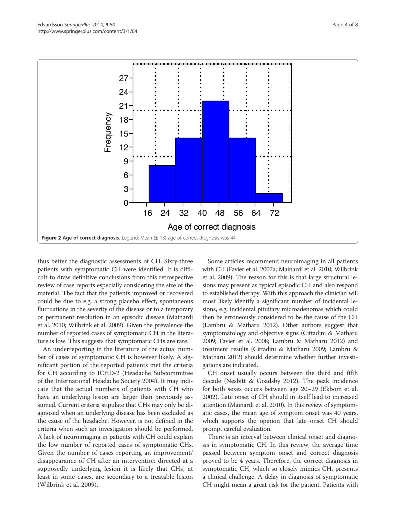
ResultsThe initial search identified 375 papers of cluster head-ache or "cluster headache-like"/"cluster-like headache".Sixty-three cluster headache or "cluster headache-like"/"cluster-like headache" cases (including 4 of my own)were found (Table 1). All cases had a clear description ofthe localization of the underlying lesion and were onlyincluded if a therapeutic intervention directed at theunderlying lesion had resulted in a significant improve-ment or resolution of the headache. All other cases wereexcluded due to insufficient data or lack of relevance in thepapers. Forty-eight (76%) of the sample were male and 15female, the M: F ratio being 3.2:1. The mean age of symp-tom onset was 40 years ± 14 (range: 13–76 years) (Figure 1).The mean age of correct diagnosis was 44 years ± 13(range: 17–76 years) (Figure 2).In 28 patients (44%) a vascular cause was identified,
including arterial aneurysms, arteriovenous malformation/cavernous angioma, venous sinus thrombosis, carotid/vertebral dissection, subclavian steal syndrome, cavernouscarotid artery thrombosis, moyamoya disease, of which 11had a dissection. Twenty-five patients (40%) had a tumourincluding 10 with pituitary tumours. An arachnoid cystand a meningioma were also found in the pituitary region.Inflammation/infection accounted for 7 cases (11%), of
16 24 32 4
Age of s
0
2
4
6
8
10
12
14
16
18
Fre
quen
cy
Figure 1 Age of symptom onset. Legend: Mean (± 14) age of symptom
which 1 with granulomatous hypophysitis and 1 withhypothalamic sarcoidosis. The remaining 3 patients hadmultiple sclerosis, foreign body and Arnold-Chiari malfor-mation with cervical syringomyelia.Thirty patients (48%) satisfied the criteria for CH
(Headache Subcommittee of the International HeadacheSociety 2004). The remaining 33 patients had an atypicalpresentation (Table 2).Fourteen patients had episodic CH (4, 12, 16, 17: case
2; 18, 19, 21, 30, 31, 34: case 2; 49, 51, 52, 54), 14 pa-tients had chronic CH (5, 7: case 3; 10, 11, 15, 20, 23,27, 32, 33, 34: cases 3 and 4; 35, 61). In the remaining 35patients it was not possible to classify CH mainly becausethe patients were diagnosed and treated within 1 year.Thirty patients of the 63 patients had a disappearance of
the headache after medical therapy aimed at the structurallesion. Different treatments were used as antibiotics(sinusitis), corticosteroids (multiple sclerosis, orbital pseu-dotumor, sarcoidosis), anticoagulation/antiplatelet treat-ment (dissection, thrombosis), dopamine agonist treatment(prolactinoma), radiotherapy/chemotherapy (nasopharynxcarcinoma).Fifty-three of all patients had an ipsilateral lesion. Nine
patients had a bilateral lesion (with unilateral attacks)and in 1 patient the lesion was central (Arnold-Chiarimalformation with syringomyelia).
DiscussionThe aim of this review was to identify clinical signs andsymptoms predictive of underlying abnormalities and
0 48 56 64 72
ymptom onset onset was 40.
16 24 32 40 48 56 64 72
Age of correct diagnosis
0
3
6
9
12
15
18
21
24
27
Fre
quen
cy
Figure 2 Age of correct diagnosis. Legend: Mean (± 13) age of correct diagnosis was 44.
Edvardsson SpringerPlus 2014, 3:64 Page 4 of 8http://www.springerplus.com/content/3/1/64
thus better the diagnostic assessments of CH. Sixty-threepatients with symptomatic CH were identified. It is diffi-cult to draw definitive conclusions from this retrospectivereview of case reports especially considering the size of thematerial. The fact that the patients improved or recoveredcould be due to e.g. a strong placebo effect, spontaneousfluctuations in the severity of the disease or to a temporaryor permanent resolution in an episodic disease (Mainardiet al. 2010; Wilbrink et al. 2009). Given the prevalence thenumber of reported cases of symptomatic CH in the litera-ture is low. This suggests that symptomatic CHs are rare.An underreporting in the literature of the actual num-
ber of cases of symptomatic CH is however likely. A sig-nificant portion of the reported patients met the criteriafor CH according to ICHD-2 (Headache Subcommitteeof the International Headache Society 2004). It may indi-cate that the actual numbers of patients with CH whohave an underlying lesion are larger than previously as-sumed. Current criteria stipulate that CHs may only be di-agnosed when an underlying disease has been excluded asthe cause of the headache. However, is not defined in thecriteria when such an investigation should be performed.A lack of neuroimaging in patients with CH could explainthe low number of reported cases of symptomatic CHs.Given the number of cases reporting an improvement/disappearance of CH after an intervention directed at asupposedly underlying lesion it is likely that CHs, atleast in some cases, are secondary to a treatable lesion(Wilbrink et al. 2009).
Some articles recommend neuroimaging in all patientswith CH (Favier et al. 2007a; Mainardi et al. 2010; Wilbrinket al. 2009). The reason for this is that large structural le-sions may present as typical episodic CH and also respondto established therapy. With this approach the clinician willmost likely identify a significant number of incidental le-sions, e.g. incidental pituitary microadenomas which couldthen be erroneously considered to be the cause of the CH(Lambru & Matharu 2012). Other authors suggest thatsymptomatology and objective signs (Cittadini & Matharu2009; Favier et al. 2008; Lambru & Matharu 2012) andtreatment results (Cittadini & Matharu 2009; Lambru &Matharu 2012) should determine whether further investi-gations are indicated.CH onset usually occurs between the third and fifth
decade (Nesbitt & Goadsby 2012). The peak incidencefor both sexes occurs between age 20–29 (Ekbom et al.2002). Late onset of CH should in itself lead to increasedattention (Mainardi et al. 2010). In this review of symptom-atic cases, the mean age of symptom onset was 40 years,which supports the opinion that late onset CH shouldprompt careful evaluation.There is an interval between clinical onset and diagno-
sis in symptomatic CH. In this review, the average timepassed between symptom onset and correct diagnosisproved to be 4 years. Therefore, the correct diagnosis insymptomatic CH, which so closely mimics CH, presentsa clinical challenge. A delay in diagnosis of symptomaticCH might mean a great risk for the patient. Patients with

Table 2 Atypical presentation/atypical symptoms associated with cluster headache
• Atypical attacks duration [Mani & Deeter 1982, West & Todman 1991, Todo & Inoya 1991, Zanchin et al. 1995, Milos et al.1996, Mainardi et al. 2002, Razvi et al. 2006, Park et al. 2006, Massie et al. 2006, Favier et al. 2007a: case 2, Rigamonti et al.2007 case 1, Hardmeier et al. 2007, Cologno et al. 2008, Tobin & Flitman 2008, Eimil-Ortiz et al. 2008, Robbins et al. 2009, Liu &Su 2009]*
• Atypical attack frequency [Todo & Inoya 1991, Favier et al. 2007a: case 2 and 3, Alty et al. 2008, Eimil-Ortiz et al. 2008]
• Atypical attack duration and frequency [Todo & Inoya 1991, Favier et al. 2007a: case 2, Eimil-Ortiz et al. 2008]
• Atypical attack duration and abnormal findings on neurologic examination [Mainardi et al. 2002, Razvi et al. 2006, Parket al. 2006, Massie et al. 2006, Favier et al. 2007a: case 2, Rigamonti et al. 2007 case 1, Hardmeier et al. 2007, Tobin &Flitman 2008, Liu & Su 2009]
• Did not meet the criterion of five attacks [Todo & Inoya 1991]
• Continuous headache or a background headache [Hannerz 1989, West & Todman 1991, Todo & Inoya 1991, Taub et al.1995, Frigerio et al. 2003, Favier et al. 2007a: case 3, Hardmeier et al. 2007, Kim et al. 2008, Harley & Ahmed 2008]
• Atypical symptoms:
Impotence [Tfelt-Hansen et al. 1982]
Symptoms of acromegaly [Milos et al. 1996]
Episodes of altered consciousness [Munoz et al. 1996: case 2, Favier et al. 2007a: case 2]
Headache triggered by sitting or standing [Piovesan et al. 2001]
Fever, and purulent nasal discharge [Scorticati et al. 2002]
Acute weakness in the upper extremity [Liu & Su 2009]
• Physical abnormalities onclinical examination:
Testicular atrophy [Tfelt-Hansen et al. 1982]
Ophthalmoplegia [Hannerz 1989, Todo & Inoya 1991, Taub et al. 1995, Mainardi et al. 2002, Frigerio et al. 2003, Hannerzet al. 2005, Razvi et al. 2006, Park et al. 2006, Favier et al. 2007a: case 2: Favier et al. 2007b, Rigamonti et al. 2007 case 1and 2, Hardmeier et al. 2007, Straube et al. 2007 case 2, Valença et al. 2007 case 1 and 2, Godeiro-Junior et al. 2008, Tobin& Flitman 2008, Ashkenazi & Brown 2008]
Optic atrophy [Tfelt-Hansen et al. 1982]
Papilloedema [Park et al. 2006]
Bitemporal hemianopia [Favier et al. 2007b]
Adie syndrome [Favier et al. 2007a: case 2]
Persistent partial or complete Horner syndrome [Mainardi et al. 2002, Frigerio et al. 2003, Hannerz et al. 2005, Razvi et al.2006 Rigamonti et al. 2007 case 1 and 2, Hardmeier et al. 2007, Straube et al. 2007 case 2, Godeiro-Junior et al. 2008,Tobin & Flitman 2008]
Signs of acromegaly [Milos et al. 1996]
Absent radial pulse [Piovesan et al. 2001]
Trigeminal distribution numbness [Massie et al. 2006, Gentile et al. 2007, Ashkenazi & Brown 2008, Alty et al. 2008, Liu &Su 2009]
Swelling of the eye [Favier et al. 2007a: case 3, Ashkenazi & Brown 2008, Kim et al. 2008]
Absent nasal tickle reflex [Massie et al. 2006]
Purulent nasal discharge [Scorticati et al. 2002]
*References.
Edvardsson SpringerPlus 2014, 3:64 Page 5 of 8http://www.springerplus.com/content/3/1/64
a presumed diagnosis of CH should therefore be accur-ately evaluated to rule out symptomatic CH.The ratio between male and female is 4.4:1 in clinical
populations of CH. The ratio has decreased in the lastdecades, possibly due to increased awareness that alsofemales can suffer from CH (Olesen et al. 2006). Fur-thermore, it has been suggested that this change may bedue to alterations in lifestyle of both genders over thepast few decades. A study from 2002 reported that theoverall male-to-female ratio in the sample was 2.5:1
(Bahra et al. 2002). In this study, gender ratio was 3.2:1,which is in line with most of the patient sample studies.However, male preponderance is not expected in symp-tomatic CH. The underlying lesions as a whole are notconsidered to be gender related or genetically determined.The observed male preponderance in symptomatic CHsneeds to be elucidated in further studies.In many articles there was only limited information
about the response to specific headache therapy. The re-sponse of the headache to sumatriptan and oxygen and

Edvardsson SpringerPlus 2014, 3:64 Page 6 of 8http://www.springerplus.com/content/3/1/64
other typical CH medications does not exclude a second-ary form. The headache attacks may be clinically indistin-guishable from the primary form (Favier et al. 2007a).Symptomatic CHs responsive to this therapy have beendescribed (Ad Hoc. Committee on Classification ofHeadache of the National Institutes of Health 1962;Cremer et al. 1995).Of the 63 cases reported in this review, 30 had a dis-
appearance of the headache after medical therapy aimedat the structural lesion. It is important to stress that be-cause of the temporal pattern of CH, the disappearanceof the headache attacks might be attributed to the remis-sion of the active phase in the episodic form, even in thecases with a documented pathology. A spontaneous re-mission of an episodic CH could be misinterpreted asbeing an effect of therapy aimed at the structural lesion.All the patients in the review were reported as showingresolution of the headache syndrome after treatment ofthe underlying pathology though the follow-up periodwas not stated in all cases and was fairly short in othercases. However, many patients have remained free of CHattacks at the follow-up after many years. This fact pointsto an association between the intervention and the reso-lution of the headache.The exact pathophysiology in these cases of symptom-
atic CH is unknown. A structural lesion may cause auto-nomic imbalance, resulting in periodic fluctuations inthe activity of the autonomic nervous system, ultimatelyleading to an attack-wise presentation of the symptoms(Wilbrink et al. 2009). Differences in the individual thresh-old for triggering the parasympathetic trigeminal reflexesmay also play a role (Straube et al. 2007). The pain mech-anism in secondary CH seems ascribable to irritation ofpain-sensitive structures and activation of trigeminal nerveendings (Leone & Bussone 2009).The low prevalence of glial tumours associated with
symptomatic CHs is remarkable. Only one report hasbeen published showing an association between CH andglioma (Edvardsson & Persson 2012). A possible mech-anism could be the infiltrating nature of a glial tumourwhich lowers its potential to act on structures triggeringsymptomatic CH (Mainardi et al. 2010).A striking finding in this appraisal was that a signifi-
cant proportion of CH cases were secondary to dis-eases of the pituitary gland or pituitary region. Of the63 cases, 14 (22%) were diseases in this region, with 10cases of pituitary tumours, 1 case report of a granuloma-tous hypophysitis, 1 case report of an arachnoid cyst, 1case report of a hypothalamic sarcoidosis and 1 case re-port of a meningioma. In all 10 cases of pituitary tumoursthe headache resolved completely after treatment. In 7cases there was medical treatment (dopamine agonisttreatment). Two cases had surgery. One case had surgeryand radiotherapy.
Other articles have reported similar findings (Favier et al.2007a; Favier et al. 2008; Wilbrink et al. 2009; Cittadini &Matharu 2009; Mainardi et al. 2010; Lambru & Matharu2012). Headache is a common symptom of pituitary dis-ease. In a large observational study of pituitary tumoursand headache (84 patients), 4% had CH. Functioning ratherthan nonfunctioning adenomas were more likely to beassociated with CH. However, the study was conductedin a tertiary referral neurosurgical centre and, therefore,does not give a meaningful indication of the prevalenceof these headaches in patients with pituitary disorders(Levy et al. 2005; Cittadini & Matharu 2009; Lambru &Matharu 2012). It is still unclear whether the prevalenceof pituitary tumours is higher in CH patients as clarify-ing studies are lacking. Approximately one in 10 of thegeneral population has an incidental pituitary microade-noma (<1 cm diameter) on routine MRI, and up to one in500 will have a macroadenoma (Ezzat et al. 2004; Cittadini& Matharu 2009; Lambru & Matharu 2012). Thus, it isnot uncommon that MRI reveals a pituitary lesion inheadache patients. The diagnostic workup of CH remainsunclear. In view of this, it is reasonable to recommendthat all patients with CH should be carefully assessed forsymptoms/objective signs of pituitary gland/pituitary re-gion disease and further investigations should be under-taken when needed.Another notable finding was that a proportion of CH
cases were associated with arterial dissection. Pain in in-ternal carotid artery dissection is postulated to be causedby stimulation of the trigeminovascular system and itcan mimic different primary headaches, including CH(Biousse et al. 1994). Examination during both primaryCH and internal carotid artery dissection may demon-strate a Horner’s syndrome. Persistent ptosis and miosisbetween headaches are widely accepted as features ofprimary CH. Duration of headache more than three hours,absence of daily periodicity, neck pain, and no worseningfrom alcohol should increase the degree of suspicion ofdissection (Razvi et al. 2006; Godeiro-Junior et al. 2008).A non-typical presentation was seen in 52% of the
cases. Some of the cases developed objective signs andatypical symptomatology during the course of the dis-ease. Thus, the presence of atypical symptomatologysuch as abnormal attack duration (17 patients), develop-ment of continuous headache or background headache(9 patients), other abnormal symptoms of primary head-ache (8 patients) and abnormal clinical examination (29patients) in the initial stage or during the course of thedisease should prompt further investigations and shouldbe considered as warning signs of secondary headaches.It is noteworthy that 48% of the cases had a typical
CH according to the criteria without evidence of anyunderlying pathology in the initial stage. Furthermore, inmany cases, it took several years before the underlying

Edvardsson SpringerPlus 2014, 3:64 Page 7 of 8http://www.springerplus.com/content/3/1/64
cause was identified. This was in many cases due to thefact that the patients initially had a typical CH and laterdeveloped objective signs/symptoms during the courseof the disease which prompted further investigations(Cittadini & Matharu 2009). In 3 cases, the initial evalu-ation with computer tomography was normal but a sub-sequent MRI revealed the underlying pathology. Whatimplications does this have for the management of patientswith cluster headache i.e. what investigations should becarried out and which patients should be assessed? The an-swer to that question will have to wait until a large pro-spective study is carried out. On the basis of availableinformation, it is recommended that MRI including mag-netic resonance angiography (MRA) should be carried outin all patients with atypical symptoms, abnormal clinicalexamination, late onset of symptoms and in patients withtherapy resistance to appropriate treatments.In patients with typical CH with respect to age at onset,
symptoms, clinical examination, and response to therapythe question of investigation is more in dispute. The deci-sion to perform neuroimaging in these patients remainsa matter of medical art. If no further investigations areundertaken, the patient may be followed up in order todetect any abnormalities later during the course of thedisease.
ConclusionsCH is a primary headache. The great majority of casesare primary. In the initial assessment, medical historyand clinical examination are of vital importance and canpoint to secondary causes of the headache. In patientswith typical CH with respect to age at onset, symptoms,clinical examination, and response to therapy the pa-tients may be followed up in order to detect any abnor-malities during the course of the disease. However, somearticles recommend that all patients with CH should beinvestigated with MRI. A significant portion of the casesin the review were secondary to diseases of the pituitary/pituitary region and arterial dissection. All patients withCH should be especially assessed for the possibility ofpituitary region disease/arterial dissection. MRI includ-ing MRA should be undertaken in patients with atyp-ical symptoms, abnormal clinical examination (includingHorner's syndrome), late onset of symptoms and in pa-tients with therapy resistance to appropriate treatments.Prospective studies are needed to identify the prevalenceof symptomatic CHs.
Competing interestsThe author declares that there are no competing interests regarding thepublication of this article.
Authors’ contributionsBE is the only contributor to the study.
Received: 4 November 2013 Accepted: 28 January 2014Published: 3 February 2014
ReferencesAd Hoc. Committee on Classification of Headache of the National Institutes of
Health (1962) Classification of headache. JAMA 179:717–718. doi: 10.1001/jama.1962.03050090045008
Alty J, Kempster P, Raghav S (2008) Cluster-like headache secondary to trigeminalmeningioma. Neurology 70:1938
Appelbaum J, Noronha A (1989) Pericarotid cluster headache. J Neurol 236:430–431Ashkenazi A, Brown F (2008) Images from headache: cluster-like headache associated
with intra-cavernous carotid artery thrombosis. Headache 48:1214–1215Bahra A, May A, Goadsby PJ (2002) Cluster headache: a prospective clinical study
with diagnostic implications. Neurology 58:354–361Bigal ME, Rapoport A, Camel M (2003) Cluster headache as a manifestation of
intracranial inflammatory myofibroblastic tumour: a case report withpathophysiological considerations. Cephalalgia 23:124–128
Biousse V, D'Anglejan-Chatillon J, Massiou H, et al. (1994) Head pain in non-traumaticcarotid artery dissection: a series of 65 patients. Cephalalgia 14:33–36
Cittadini E, Matharu MS (2009) Symptomatic trigeminal autonomic cephalalgias.Neurologist 15:305–312
Cologno D, Buzzi MG, Cicinelli P, et al. (2008) Symptomatic cluster-like headachetriggered by forehead lipoma: a case report and review of the literature.Neurol Sci 29:331–335
Cremer PD, Halmagyi GM, Goadsby PJ (1995) Secondary cluster headacheresponsive to sumatriptan. J Neurol Neurosurg Psychiatry 59:633–634
Edvardsson B (2013) Cluster headache and pituitary prolactinoma. J Med Cases4:523–525
Edvardsson B, Persson S (2012) Cluster headache and parietal glioblastomamultiforme. Neurologist 18:206–207
Edvardsson B, Persson S (2013a) Cluster headache and arachnoid cyst.Springerplus 2:4
Edvardsson B, Persson S (2013b) Cluster headache and acute maxillary sinusitis.Acta Neurol Belg: Epub ahead of print
Eimil-Ortiz M, María-Salgado F, Fontán-Tirado C, et al. (2008) Pseudo-cluster-likeheadache secondary to contralateral epidermoid cyst of the pontocerebellarangle. Headache 48:471–472
Ekbom K, Svensson DA, Träff H, et al. (2002) Age at onset and sex ratio in clusterheadache: observations over three decades. Cephalalgia 22:94–100
Ezzat S, Asa SL, Couldwell WT, et al. (2004) The prevalence of pituitary adenomas:a systematic review. Cancer 101:613–619
Favier I, Haan J, van Duinen SG, et al. (2007a) Typical cluster headache caused bygranulomatous pituitary involvement. Cephalalgia 27:173–176
Favier I, van Vliet JA, Roon KI, et al. (2007b) Trigeminal autonomic cephalgiasbecause of structural lesions: a review of 31 cases. Arch Neurol 64:25–31
Favier I, Haan J, Ferrari MD (2008) Cluster headache: to scan or not to scan.Curr Pain Headache Rep 12:128–131
Fontaine D, Almairac F, Mondot L, et al. (2013) Cluster-Like Headache Secondaryto Parasagittal Hemangiopericytoma. Headache 53:1496–1498
Frigerio S, Bühler R, Hess CW, et al. (2003) Symptomatic cluster headache ininternal carotid artery dissection–consider anhidrosis. Headache 43:896–900
Gentile S, Fontanella M, Giudice RL, et al. (2006) Resolution of cluster headacheafter closure of an anterior communicating artery aneurysm: the role ofpericarotid sympathetic fibres. Clin Neurol Neurosurg 108:195–198
Gentile S, Ferrero M, Vaula G, et al. (2007) Cluster headache attacks and multiplesclerosis. J Headache Pain 8:245–247
Georgiadis G, Tsitouridis I, Paspali D, et al. (2007) Cerebral sinus thrombosispresenting with cluster-like headache. Cephalalgia 27:79–82
Godeiro-Junior C, Kuster GW, Felício AC, et al. (2008) Internal carotid arterydissection presenting as cluster headache. Arq Neuropsiquiatr 66:763–764
Greve E, Mai J (1988) Cluster headache-like headaches: a symptomatic feature? Areport of three patients with intracranial pathologic findings. Cephalalgia8:79–82
Hannerz J (1989) A case of parasellar meningioma mimicking cluster headache.Cephalalgia 9:265–269
Hannerz J, Arnardottir S, Bro Skejø HP, et al. (2005) Peripheral postganglionicsympathicoplegia mimicking cluster headache attacks. Headache 45:84–86
Hardmeier M, Gobbi C, Buitrago C, et al. (2007) Dissection of the internal carotidartery mimicking episodic cluster headache. J Neurol 254:253–254
Harley JS, Ahmed F (2008) Cluster-like headache heralding inflammatory orbitalpseudotumour. Cephalalgia 28:401–402
Edvardsson SpringerPlus 2014, 3:64 Page 8 of 8http://www.springerplus.com/content/3/1/64
Headache Subcommittee of the International Headache Society (2004) Theinternational classification of headache disorders, 2nd edition. Cephalalgia24(suppl 1):9–160
Kim JT, Lee SH, Choi SM, et al. (2008) Spontaneous vertebral artery dissectionmimicking cluster headache. Cephalalgia 28:671–673
Kuritzky A (1984) Cluster headache-like pain caused by an upper cervicalmeningioma. Cephalalgia 4:185–186
Lambru G, Matharu MS (2012) Trigeminal autonomic cephalalgias: a review ofrecent diagnostic, therapeutic and pathophysiological developments.Ann Indian Acad Neurol 15:S51–S61
Leone M, Bussone G (2009) Pathophysiology of trigeminal autonomiccephalalgias. Lancet Neurol 8:755–764
Levy MJ, Matharu MS, Meeran K, et al. (2005) The clinical characteristics ofheadache in patients with pituitary tumours. Brain 128:1921–1930
Levy MJ, Robertson I, Howlett TA (2012) Cluster headache secondary tomacroprolactinoma with ipsilateral cavernous sinus invasion. Case ReportNeurol Med 2012:830469. doi: 10.1155/2012/830469. Epub 2012 Sep 23
Levyman C, Dagua Filho Ados S, Volpato MM, et al. (1991) Epidermoid tumourof the posterior fossa causing multiple facial pain - a case report. Cephalalgia11:33–36
Liu KT, Su CS (2009) Cluster-like headache as an opening symptom of cervicalspinal epidural abscess. Am J Emerg Med 27:370. e5-370.e6
Mainardi F, Maggioni F, Dainese F, et al. (2002) Spontaneous carotid arterydissection with cluster-like headache. Cephalalgia 22:557–559
Mainardi F, Trucco M, Maggioni F, et al. (2010) Cluster-like headache. A comprehensivereappraisal. Cephalalgia 30:399–412
Mani S, Deeter J (1982) Arteriovenous malformation of the brain presenting as acluster headache: a case report. Headache 22:184–185
Massie R, Sirhan D, Andermann F (2006) Chronic cluster-like headache secondaryto an epidermoid clival lesion. Can J Neurol Sci 33:421–422
McBeath JG, Nanda A (2000) Case reports: sudden worsening of clusterheadache: a signal of aneurysmal thrombosis and enlargement. Headache40:686–688
Messina G, Rizzi M, Cordella R, et al. (2013) Secondary chronic cluster headachetreated by posterior hypothalamic deep brain stimulation: first reported case.Cephalalgia 33:136–138
Milos P, Havelius U, Hindfelt B (1996) Clusterlike headache in a patient with apituitary adenoma: with a review of the literature. Headache 36:184–188
Minguzzi E, Cevoli S, Pierangeli G, et al. (2003) Cluster headache (CH) associatedwith pituitary prolactinoma. Cephalalgia 23:66
Morelli N, Gori S, Cafforio G, et al. (2005) Prevalence of right-to-left shunt inpatients with cluster headache. J Headache Pain 6:244–246
Munoz C, Diez-Tejedor E, Frank A, et al. (1996) Cluster headache syndrome associatedwith middle cerebral artery arteriovenous malformation. Cephalalgia 16:202–205
Negoro K, Kawai M, Tada Y, et al. (2005) A case of postprandial cluster-like headachewith prolactinoma: dramatic response to cabergoline. Headache 45:604–606
Nesbitt AD, Goadsby PJ (2012) Cluster headache. BMJ 344:e2407Olesen J, Tfelt-Hansen P, Welch KMA, Goadsby PJ, Ramadan NM (2006) The headaches,
3rd edn. Lippincott Williams & Wilkins, PhiladelphiaPark KI, Chu K, Park JM, et al. (2006) Cluster-like headache secondary to cerebral
venous thrombosis. J Clin Neurol 2:70–73Peterlin BL, Levin M, Cohen JA, et al. (2006) Secondary cluster headache:
a presentation of cerebral venous thrombosis. Cephalalgia 26:1022–1024Piovesan EJ, Lange MC, Werneck LC, et al. (2001) Clusterlike headache. A case
secondary to the subclavian steal phenomenon. Cephalalgia 21:850–851Porta-Etessam J, Ramos-Carrasco A, Berbel-Garcia A, et al. (2001) Clusterlike
headache as first manifestation of a prolactinoma. Headache 41:723–725Razvi SS, Walker L, Teasdale E, et al. (2006) Cluster headache due to internal
carotid artery dissection. J Neurol 253:661–663Rigamonti A, Iurlaro S, Zelioli A, et al. (2007) Two symptomatic cases of cluster
headache associated with internal carotid artery dissection. Neurol Sci 28:S229–S231
Robbins MS, Tarshish S, Napchan U, et al. (2009) Images from headache: atypicalcluster headache secondary to giant meningioma. Headache 49:1052–1053
Rodríguez S, Calleja S, Morís G (2008) Cluster-like headache heralding cerebralvenous thrombosis. Cephalalgia 28:906–907
Scorticati MC, Raina G, Federico M (2002) Cluster-like headache associated to aforeign body in the maxillary sinus. Neurology 59:643–644
Seijo-Martinez M, Castro del Río M, Conde C, et al. (2004) Cluster-like headache:association with cervical syringomyelia and Arnold-Chiari malformation.Cephalalgia 24:140–142
Sewell RA, Johnson DJ, Fellows DW (2009) Cluster headache associated withmoyamoya. J Headache Pain 10:65–67
Straube A, Freilinger T, Rüther T, et al. (2007) Two cases of symptomatic cluster-likeheadache suggest the importance of sympathetic/parasympathetic balance.Cephalalgia 27:1069–1073
Takeshima T, Nishikawa S, Takahashi K (1988) Cluster headache like symptomsdue to sinusitis: evidence for neuronal pathogenesis of cluster headachesyndrome. Headache 28:207–208
Taub E, Argoff CE, Winterkorn JM, et al. (1995) Resolution of chronic clusterheadache after resection of a tentorial meningioma: case report.Neurosurgery 37:319–321
Tfelt-Hansen P, Paulson OB, Krabbe AA (1982) Invasive adenoma of the pituitarygland and chronic migrainous neuralgia. A rare coincidence or a causalrelationship? Cephalalgia 2:25–28
Tobin J, Flitman S (2008) Cluster-like headaches associated with internal carotidartery dissection responsive to verapamil. Headache 48:461–466
Todo T, Inoya H (1991) Sudden appearance of a mycotic aneurysm of theintracavernous carotid artery after symptoms resembling cluster headache:case report. Neurosurgery 29:594–598
Valença MM, Andrade-Valença LP, Martins C, et al. (2007) Cluster headache andintracranial aneurysm. J Headache Pain 8:277–282
van der Vlist SH, Hummelink BJ, Westerga J, et al. (2013) Cluster-like headacheand a cystic hypothalamic tumour as first presentation of sarcoidosis.Cephalalgia 33:421–424
West P, Todman D (1991) Chronic cluster headache associated with a vertebralartery aneurysm. Headache 31:210–212
Wilbrink LA, Ferrari MD, Kruit MC, et al. (2009) Neuroimaging in trigeminalautonomic cephalgias: when, how, and of what? Curr Opin Neurol 22:247–253
Zanchin G, Rossi P, Licandro AM, et al. (1995) Clusterlike headache. A case ofsphenoidal aspergilloma. Headache 35:494–497
doi:10.1186/2193-1801-3-64Cite this article as: Edvardsson: Symptomatic cluster headache: a reviewof 63 cases. SpringerPlus 2014 3:64.
Submit your manuscript to a journal and benefi t from:
7 Convenient online submission
7 Rigorous peer review
7 Immediate publication on acceptance
7 Open access: articles freely available online
7 High visibility within the fi eld
7 Retaining the copyright to your article
Submit your next manuscript at 7 springeropen.com