Embed Size (px)
Citation preview
Symmetry breakage in the vertebrate embryo: When does it happenand how does it work?
Martin Blum a,n, Axel Schweickert a, Philipp Vick a, Christopher V.E. Wright b,Michael V. Danilchik c
a University of Hohenheim, Institute of Zoology (220), Garbenstrasse 30, D-70593 Stuttgart, Germanyb Department of Cell and Developmental Biology, Vanderbilt University, Nashville, TN 37232-0494, USAc Department of Integrative Biosciences, Oregon Health & Science University, Portland, OR 97239-3098, USA
a r t i c l e i n f o
Article history:Received 25 February 2014Received in revised form8 June 2014Accepted 17 June 2014Available online 24 June 2014
Keywords:Left–right asymmetryCiliaLeftward flowIon-flux modelSymmetry breakage
a b s t r a c t
Asymmetric development of the vertebrate embryo has fascinated embryologists for over a century.Much has been learned since the asymmetric Nodal signaling cascade in the left lateral plate mesodermwas detected, and began to be unraveled over the past decade or two. When and how symmetry isinitially broken, however, has remained a matter of debate. Two essentially mutually exclusive modelsprevail. Cilia-driven leftward flow of extracellular fluids occurs in mammalian, fish and amphibianembryos. A great deal of experimental evidence indicates that this flow is indeed required for symmetrybreaking. An alternative model has argued, however, that flow simply acts as an amplification step forearly asymmetric cues generated by ion flux during the first cleavage divisions. In this review wecritically evaluate the experimental basis of both models. Although a number of open questions persist,the available evidence is best compatible with flow-based symmetry breakage as the archetypical modeof symmetry breakage.& 2014 The Authors. Published by Elsevier Inc. This is an open access article under the CC BY-NC-ND
license (http://creativecommons.org/licenses/by-nc-nd/3.0/).
Introduction
Establishment of left–right asymmetry of animal body plans isof the utmost importance for embryonic development and adulthealth. During vertebrate embryogenesis, the cardiovascular sys-tem, the organs of the chest and abdomen, and even the brain,develop morphological and/or functional asymmetries (Aizawa,2013; Burn and Hill, 2009; Franco et al., 2014; Perloff, 2011;Roussigne et al., 2012). Developmental defects in laterality speci-fication and asymmetric morphogenesis are sometimes compati-ble with embryogenesis, and occasionally, fully mirror-imageindividuals develop to term (Bartram et al., 2005; Peeters andDevriendt, 2006; Shiraishi and Ichikawa, 2012; Sutherland andWare, 2009). Left–right (L–R) defects are often much less pervasiveand usually strike organs at random, resulting in severe visceralmisalignment, organ malformations and malfunctions (Burdineand Caspary, 2013; Hirokawa et al., 2012; Yoshiba and Hamada,2014). Asymmetric organ morphogenesis is preceded by an asym-metric signaling cascade, which initiates during neurulation in theleft lateral plate mesoderm (LPM). This so-called Nodal cascade
consists of the TGFβ-type growth factor Nodal, its secreted feed-back repressor Lefty (also known as Antivin) and the home-odomain transcription factor Pitx2 (Marjoram and Wright, 2011;Schier, 2009; Shen, 2007). Expression of this cascade is bothnecessary and sufficient to induce the correct asymmetric place-ment of organs (situs solitus) (Hamada et al., 2002; Hirokawaet al., 2012).
How the Nodal cascade becomes asymmetrically expressedconstitutes a conceptual cell-biological problem, because zygotestypically lack recognizable morphological or functional asymme-tries that could initiate it. Brown and Wolpert (1990) proposed theconcept of an intrinsic biochemical-structural chirality (repre-sented in their model by an “F-molecule”), even though uniformlydistributed, such a molecule would operate by undergoing chiralalignment against the A–P and D–V axes of the embryo. Thenother molecular interactions feeding off the deduced L–R vectorwould eventually lead (via an unknown number of steps) to themorphogenetic process of asymmetric organ development (Brownand Wolpert, 1990). Amongst the animals classified as the bila-teria, two cytoskeleton-dependent chiralities have been identified,whose mechanism of action fulfill the conceptual nature of theF-molecule hypothesis (though not being single molecules in theinitial invocation of the model). Interestingly, although expressedat two different developmental stages, both of these instances
Contents lists available at ScienceDirect
journal homepage: www.elsevier.com/locate/developmentalbiology
Developmental Biology
http://dx.doi.org/10.1016/j.ydbio.2014.06.0140012-1606/& 2014 The Authors. Published by Elsevier Inc. This is an open access article under the CC BY-NC-ND license (http://creativecommons.org/licenses/by-nc-nd/3.0/).
n Corresponding author.E-mail address: [email protected] (M. Blum).
Developmental Biology 393 (2014) 109–123
result in asymmetric activation of the Nodal pathway. In spirallycleaving snail embryos, asymmetric positioning of the spindleapparatus during cleavage induces Nodal asymmetry, ostensiblyby repositioning maternally synthesized factors (Grande and Patel,2009). In embryos of most vertebrates, including fish, frogs andmammals, chiral rotation of cilia polarized to the posterior pole ofcells produces a vectorial leftward flow of extracellular fluids. Thisflow is necessary and sufficient for Nodal-dependent symmetrybreakage (Fig. 1A–E; Basu and Brueckner, 2008; Blum et al., 2009b;Hirokawa et al., 2012), and substantial enough to sweep fluores-cent latex microbeads across from one side of the ciliatedepithelium to the other (Essner et al., 2005; Kramer-Zuckeret al., 2005; Nonaka et al., 1998; Okada et al., 2005; Schweickertet al., 2007). Although both mechanisms lead to asymmetric Nodalactivity, they seem to have little else in common, raising thequestion as to why and how flow-type symmetry breakage hasevolved. We have recently addressed this problem in a hypothesisarticle (Blum et al., 2014), and will not repeat this issue here. Onemajor difference between the two strategies is that one (spiralcleavage in spiralian protostomes) is determined maternally andacts during very early cleavage stages, while the other (leftwardflow) operates much later, and depends on zygotic gene expres-sion during neurula stages.
Although most of the vertebrates examined so far utilize cilia-generated flow to initiate the asymmetric Nodal cascade, twoalternative strategies have also been observed. First, in the chick,large-scale whole-cell repositioning during gastrulation results ina significant asymmetry in the morphology of Hensen's node,and this appears to play a role in the asymmetric expression ofspecific intercellular signaling molecules (Dathe et al., 2002; Groset al., 2009; Wetzel, 1929). Second, in amphibians, asymmetriclocalization of determinants has been proposed to act during earlycleavage stages (Fig. 1F and G; for a recent review see Vandenbergand Levin, 2013). According to this view, cytoskeletal motorproteins asymmetrically transport a maternal deposit of the ionpump ATP4 (as mRNA and/or translated protein), changing itsdistribution from a symmetric to an asymmetric one (Levin et al.,2002). This asymmetry is hypothesized to generate an intracellularpH and voltage gradient, along which the small charged mono-amine, serotonin, transfers via gap junctional communication(GJC) to blastomeres on the right side of the cleavage stage embryo(Fig. 1F and G; Fukumoto et al., 2005b). Almost one day later,when the embryo consists of thousands of cells, this right-sidedserotonin asymmetry, by an unknown epigenetic mechanism, isproposed to repress Nodal activity on the right side, therebyinitiating the left-asymmetric activation of the Nodal cascade
Fig. 1. Prevailing models of symmetry breakage in the frog Xenopus. (A–E) Leftward flow. (A) Schematic representation of a stage 17 archenteron roof in ventral perspective.Flow occurs from the right to the left side of the ciliated gastrocoel roof plate (GRP; red). Nodal and Coco are co-expressed at the lateral GRP margins on both sides (purple).Flow represses Coco, activating Nodal by release of repression. bp, blastopore. (B) GRP at higher magnification. Polarized and flow-producing cilia at the GRP center arebordered by Nodal/Coco-positive cells (purple) which harbor unpolarized, sensory cilia. (C and D) Coco expression during (C) and following (D) leftward flow. Note thedecrease in signal intensity on the left at post-flow stage 20 (D). (E) Schematic depiction of events on the left and right side leading up to asymmetric Nodal cascadeinduction in the left lateral plate mesoderm (LPM). (F and G) Ion-flux. (F) Asymmetrically expressed ion pumps create a voltage gradient in the 4-cell embryo which initiatesthe electrogenic transfer of serotonin through gap junctional communication to the ventral-right lineage at the 32-cell stage. Serotonin accumulates in this lineage becausethe ventral midline is devoid of GJC. (F) Schematic depiction of events on the left and right side leading up to asymmetric Nodal cascade induction in the left LPM. Questionmarks indicate unproven interactions and mechanisms.
M. Blum et al. / Developmental Biology 393 (2014) 109–123110
(Fig. 1G; Vandenberg and Levin, 2013). This mechanism forsymmetry breakage will be referred to herein as the “ion-flux”model. Consistent with this model, asymmetries in serotonin,ATP4 and ATP6 were reported in early cleavage stage embryos(Adams et al., 2006; Fukumoto et al., 2005b; Levin et al., 2002). Inaddition, blockage of GJC or mild interference with cytoskeletaldynamics reportedly disrupts L–R development (Levin andMercola, 1999, 1998; Lobikin et al., 2012).
When cilia-driven leftward flow was found in the neurula ofthe Xenopus embryo (Schweickert et al., 2007), as observedpreviously in mouse (Nonaka et al., 1998), rabbit (Okada et al.,2005), zebrafish (Essner et al., 2005) and medaka (Okada et al.,2005), the question arose as to which mechanism is principallyinstructive for breaking symmetry in the amphibian embryo.Advocates of the ion-flux model have suggested that, throughoutthe animal kingdom, symmetry breakage occurs very early, i.e. inthe zygote or during the first two cell divisions, and that thefunction of cilia-driven fluid flow must therefore be restricted to alater-stage amplification step (Vandenberg and Levin, 2013). Here,we present our view of the conceptual problems with the ion-fluxmodel, and evaluate the salient experimental support for eachof the two opposing models. For detailed reviews on other aspectsof the two models we refer to recent comprehensive reviews(Hirokawa et al., 2012; Nakamura and Hamada, 2012; Vandenberg,2012; Vandenberg and Levin, 2013, 2010a; Yoshiba and Hamada,2014).
Embryological considerations: Spemann's organizerand left–right asymmetry
A mechanism that breaks symmetry during early cleavagestages is likely to be independent of L–R orientational cues thatderive from the gastrula, or Spemann's organizer. In contrast, amechanism that operates during or after gastrulation is likely to bestrongly influenced by, or even be mandatorily dependent on, theorganizer. Experimental analysis of organizer function on L–Rasymmetry should thus provide an answer as to when symmetryis broken (Schweickert et al., 2012).
Spemann's and the left–right organizer: how are they related?
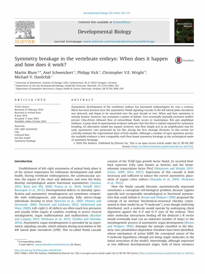
In frogs, symmetry breakage via cilia-driven leftward flow isintrinsically tied to Spemann's organizer. The ciliated gastrocoelroof plate (GRP), where leftward flow develops during neurulation,is derived from the superficial mesoderm (SM) of the gastrula. TheSM constitutes the superficial cell layer of a region that sits aboveSpemann's organizer during early gastrulation (Fig. 2A and B). It isthus sandwiched between prospective neuroectoderm and themore vegetally located epithelial layer of the organizer (Shooket al., 2004). In the early gastrula, the SM expresses foxj1, the maincontrol gene for motile cilia (Stubbs et al., 2008; Fig. 2C and D).In addition, both the organizer and the central part of the GRPcontain cells with eventual notochordal fate, reflecting their closerelationship (Shook et al., 2004). Ciliated, flow-generating epithe-lia in other vertebrates, despite their common function, display awide morphological variety (Blum et al., 2007). In this review theywill be referred to as left–right organizers (LRO). In rabbit and frog,for example, the LRO develops as a flattened epithelial plate, whileit appears as a concavity in the mouse, as a raised dome inmedaka, and as a completely enclosed, hollow vesicle in zebrafish(Blum et al., 2009b, 2007).
We have hypothesized that the evolution of LROs and thenotochord are intricately linked in the chordates (Blum et al.,2014), and it might be worthwhile to evaluate the LRO lineage infish and mammals. In fact, embryos with defects in notochord
formation are particularly prone to develop L–R defects: either dueto defective morphogenesis of the ciliated LRO and the resultantinterference with leftward flow, and/or due to absent midlinebarrier function of Lefty (Bisgrove et al., 1999; Cheng et al., 2000;Danos and Yost, 1996; Hamada et al., 2002; King et al., 1998;Lenhart et al., 2011; Melloy et al., 1998; see below). The classicalBrachyury mouse mutant and the corresponding no tail mutationin zebrafish exemplify the relationship between LRO and noto-chord. In both species, this T-box transcription factor is requiredfor notochordal integrity and development (Schulte-Merker et al.,1994; Wilkinson et al., 1990). Besides notochordal defects, brachy-ury (T) and no tail mutants display laterality defects (King et al.,1998). Mutant LROs are smaller and overall malformed, andciliogenesis is strongly affected (Amack and Yost, 2004). Conse-quently, mutant embryos lack flow (Essner et al., 2005; ourunpublished results). In Xenopus T (brachyury) morphant embryos,GRP development is similarly disturbed, underscoring the con-served dependence of symmetry breakage on ciliary flow (ourunpublished data). These examples demonstrate that a subset ofthe cells comprising the organizer proper is required for L–R
Fig. 2. Structural and functional relationship of Spemann's organizer and super-ficial mesoderm. (A) Schematic depiction of superficial mesoderm (SM; red) andorganizer (O; green) in whole-mount stage 10þ gastrula embryo shown in dorsalview. (B) Arrangement of organizer and SM in a sagittal section. (C) SM foxj1expression in a whole-mount gastrula embryo. (D) Sagittal section (plane indicatedby dashed line in C) demonstrates foxj1 mRNA in the SM (arrowhead). (E and F)Loss of SM foxj1 expression (E) in UV-ventralized gastrula embryo (F), demonstrat-ing the dependence of SM specification on organizer function.
M. Blum et al. / Developmental Biology 393 (2014) 109–123 111
development. These cells organize into an epithelial tissue, differ-entiate transiently into the ciliated LRO, produce the pattern-specifying leftward flow and, at least in Xenopus, finally contributeto the notochord (Shook et al., 2004).
Loss of organizer function invariably leads to L–R defects
The organizer is responsible for the explicit development of theembryo's overall dorso-anterior to ventro-posterior axis. Manipula-tion of organizer function could therefore impact not only onleftward flow, but on an additional, separate process: the midlinebarrier. A multitude of experiments and analyses of mutant embryoshave provided compelling evidence that the midline is crucial for L–Rdevelopment (Bisgrove et al., 2000; Danos and Yost, 1996; Hamada etal., 2002; Izraeli et al., 1999; Lee and Anderson, 2008; Lohr et al.,1997; Zhang et al., 2012). Besides the notochord (Lee and Anderson,2008), the floor plate of the neural tube serves as a functional barrier;Lefty expression there prevents Nodal secreted from the left LPMfrom functioning on the right side (Shiratori and Hamada, 2006).
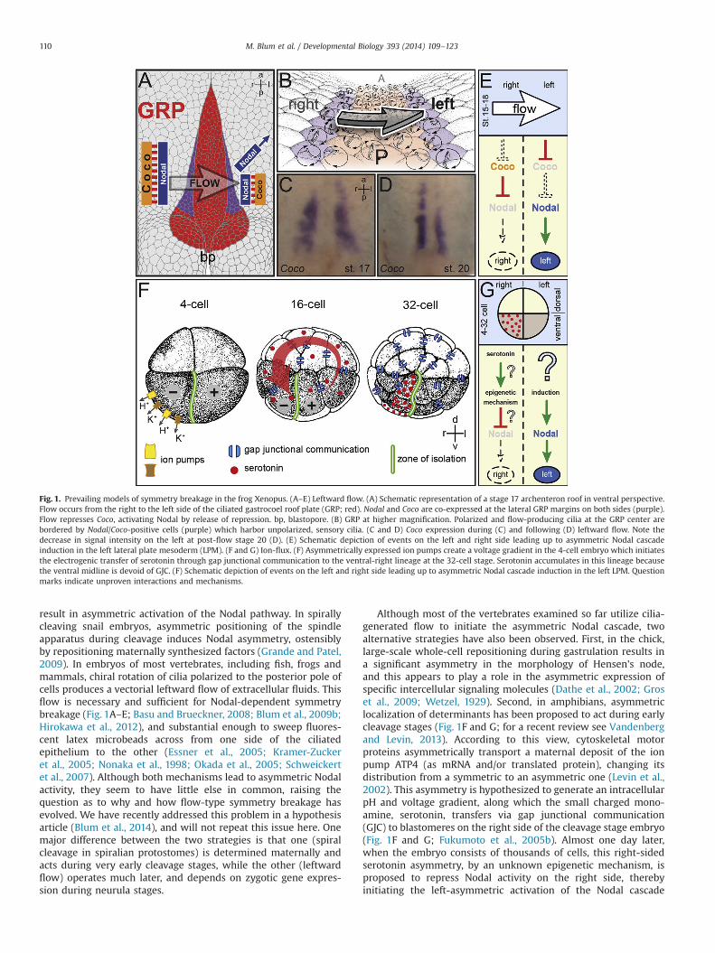
Two classical kinds of experiments in Xenopus have reinforcedthe functional connection between dorso-anterior and L–R devel-opment: (1) UV irradiation at the one-cell stage to prevent theorganizer from forming in the first place (Fig. 3); and (2) mis-expression of DNA expression constructs to manipulate organizerfunction after it has formed following the mid-blastula transition(MBT). The vegetal outer cortex of the zygote contains determi-nants necessary to establish the dorsal organizer (Ku and Melton,1993; Tao et al., 2005; Zhang and King, 1996). Normally, thevegetal cortex rotates in a microtubule-dependent manner awayfrom the site of sperm entry, depositing these determinants on thefuture dorsal side, thus establishing the dorso-ventral axis (Kaoand Elinson, 1988; Miller et al., 1999; Vincent et al., 1986). UV-irradiation of the vegetal cortex disrupts the microtubules neededfor cortical rotation, thereby preventing dorsal accumulation ofdeterminants and resulting in organizer-deprived, ventralizedembryos (Gerhart et al., 1989). In contrast, manipulation oforganizer function by dorsally targeted injection of DNA expres-sion vectors affects the embryo after MBT, when zygotic transcrip-tion is released from repression shortly before the onset ofgastrulation. Activation of the canonical Wnt pathway in theorganizer territory during blastula/early gastrula stages counter-acts both organizer specification and function (De Robertis et al.,2000). Although operating at different developmental stages, bothtreatments (the earlier UV treatment and the later Wnt pathwaydisruption) interfere with dorso-anterior development, becausethe organizer does not form or act properly. Naturally, it is notpossible to observe L–R asymmetry in an embryo lacking a dorsalbody axis, but it is important to ask what happens to the process oflaterality specification when dorsal development has been par-tially disrupted. Significantly, the specification of the L–R axisappears randomized in embryos mildly ventralized by eithermanipulation (Danos and Yost, 1995). Thus, Spemann's organizerappears to govern not only dorsal axial but also L–R specification.
[Intriguingly, mildly ventralized tadpoles fail to form a notochordspecifically in the anterior region (Danos and Yost, 1995). Duringgastrulation, anterior notochordal cells involute first, accompanied bythe overlying SM cells (Shook et al., 2004), suggesting that the GRP isunable to form in these ventralized individuals. Indeed, the SM markerfoxj1 was absent in UV-irradiated specimens (Fig. 2E and F), demon-strating that LRO specification is linked to general organizer functions.]
The concept of “early” vs. “late” induced organizers is irrelevantfor L–R asymmetry
The above discussion indicates that when the endogenousorganizer is damaged, laterality becomes randomized. What happens
to L–R specification when a new organizer is introduced into a fullyventralized background? Organizer reconstitution has long been avaluable tool to elucidate signaling pathways that are involved inorganizer induction and specification. Two convenient methods areavailable in Xenopus embryos. First, by injecting mRNAs at the 4–32cell stage of ventralized embryos, many factors, including compo-nents of the canonical Wnt pathway, or targets like the homeoboxgene siamois, can completely rescue the ventralizing effects of UV byde novo organizer induction (Fig. 3; Fan and Sokol, 1997; Sokol et al.,1991). Second, mimicking the effects of cortical rotation can be usedto reintroduce organizer function into irradiated embryos. Gravity-driven artificial rotation of the cortex relative to the core of thezygote after UV treatment relocates vegetal dorsal determinantsasymmetrically by gravity (tipping, Fig. 3). This method effectivelyrescues dorso-anterior development in UV embryos (Scharf andGerhart, 1980; Weaver and Kimelman, 2004).
Both methods, schematically summarized in Fig. 3, have beenapplied by proponents of the ion-flux model (Vandenberg andLevin, 2010b). The hypothesis of those experiments has been thatL–R determination in cleavage-stage embryos is independent ofthe organizer or imprints L–R positional information onto theorganizer (Vandenberg and Levin, 2010b). Because experimentallyinduced organizers are introduced at random positions comparedto the initial primary dorsal axis, early (i.e. already fixed) L–Rdeterminants should be mis-localized with respect to a newlyintroduced organizer. However, the results of these experimentsdo not support the hypothesis: as judged by the organ situs oftadpoles, laterality was basically restored by both tipping (95% ofcases) and localized siamois mRNA injections (75% of specimens;Schweickert et al., 2012; Vandenberg and Levin, 2010b). Therefore,the organizer effect is dominant over any early instructive “bias”,which leads to the conclusion that the deterministic event isrelated to the organizer and to structures carrying instructiveinfluence that are derived from it, i.e., the GRP/LRO.
Surprisingly, Vandenberg and Levin arrived at an unusual inter-pretation of their results. In short, rescues were categorized as“early” or “late” according to the presumed interval of their activity(Vandenberg and Levin, 2010b). As the transcription factor Siamoisis only able to activate organizer target genes after MBT it wascoined a “late induced organizer”, while tipping of the zygote wasreferred to as an “early induced organizer” (Vandenberg and Levin,2010b). The slightly lower rescue efficiency of siamois was taken assupporting the view that “late induced organizers” lack early L–Rcues, implying that they must be present in the “early inducedorganizer”. This interpretation is insupportable for (at least) threereasons. First, it is problematic to compare two completely differentmethods, an invasive (injection) to a non-invasive one (tipping;Fig. 3). We are not aware of any injection experiment that results inone hundred percent efficiency of rescue – it is a technical fact oflife that the intracellular distribution of an injected RNA can onlyapproximate that of the endogenous transcript. In addition, becausemildly UV-ventralized embryos are profoundly impaired for later-ality specification (Fig. 3F; Danos and Yost, 1995; Lohr et al., 1997),the rescue of 75% wild-type organ situs in siamois-injectedUV-treated embryos (Vandenberg and Levin, 2010b) appearsvery high. Researchers familiar with rescue of morpholinooligonucleotide-mediated gene knockdowns in most cases have tolive with lower (yet significant) rescue efficiencies. It would beinteresting to know whether the organizer function was fullyrescued in all cases, i.e. whether specimens with heterotaxia hadnormal heads and body axes. Unfortunately, this important infor-mation was not reported (Vandenberg and Levin, 2010b). Second,the result of the siamois rescue is not in accordance with theauthors' own hypothesis, which predicts a loss of laterality uponloss of the early asymmetric cues in UV irradiated embryos.Vandenberg and Levin (2010b) scored three asymmetric organs of
M. Blum et al. / Developmental Biology 393 (2014) 109–123112
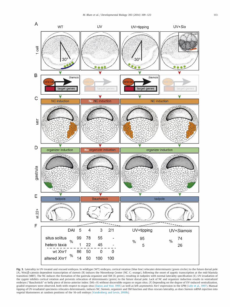
Fig. 3. Laterality in UV-treated and rescued embryos. In wildtype (WT) embryos, cortical rotation (blue line) relocates determinants (green circles) to the future dorsal pole(A). Wnt/β-catenin dependent transcription of siamois (B) induces the Nieuwkoop Center (NC; C; orange), following the onset of zygotic transcription at the mid-blastulatransition (MBT). The NC induces the formation of the gastrula organizer and SM (D, green), resulting in tadpoles with normal laterality specification (E). UV-irradiation ofthe zygote inhibits cortical rotation and prevents relocation of determinants (green) to the future dorsal pole. Lack of NC and organizer induction results in ventralizedembryos (“Bauchstück” or belly piece of dorso-anterior index: DAI¼0) without discernible organs or organ situs. (F) Depending on the degree of UV-induced ventralization,graded responses were observed, both with respect to organ situs (Danos and Yost, 1995) as well as left-asymmetric Xnr1 expression in the LPM (Lohr et al., 1997). Manualtipping of UV-irradiated specimens relocates determinants, induces NC, Siamois, organizer and SM function and thus rescues laterality, as does Siamois mRNA injection intovegetal blastomeres at random positions of the 16-cell embryo (Vandenberg and Levin, 2010b).
M. Blum et al. / Developmental Biology 393 (2014) 109–123 113
the frog tadpole, heart, gut and gall bladder. As each organ canindividually adopt a wild type or reversed orientation, the maximaltheoretical percentage of situs solitus in batches of embryosshowing a completely random assortment of these independentorgan locations should be 12.5% (Fukumoto et al., 2005b), close tothe value observed in UV embryos when scored for Nodal cascadeinduction. Situs solitus in three out of four siamois-injected UVembryos thus rather reflects an efficient restoration of lateralitythan a loss of L–R identity. Third, the idea that leftward flowamplifies early L–R cues does not help the argument – if cuesare gone, what is available to be amplified? Vandenberg andLevin (2010b) reported high efficiencies of UV-treatments, affecting94–97% of specimens in any given experiment. If L–R cues were thateffectively destroyed, what could leftward flow amplify in siamois-induced “late” organizer specimens?
We therefore contend that the organizer's activity as a vectorialinstructor of laterality does not require any preceding cues for itsactivity. In normal embryos, factors absolutely restricted to earlycleavage stages of activity have not been identified, and there is nostrong requirement for such cues in order to yield L–R asymmetry.But even if hypothetical early determinants were to be alignedwith the organizer's vector of action, the requirement for thesedeterminants needs to be experimentally shown. We concludefrom these lines of arguments that setting up the L–R axis is anintrinsic feature of the organizer, a notion initially put forwardalmost 20 years ago (Nascone and Mercola, 1997). The lateralityprogram is not executed unless the organizer is functioning, and itemerges de novo from the activity of the organizer or tissues thatare readily derived from it.
Nodal cascade induction in the LPM depends on flow
One additional point of embryological reasoning must beaddressed here, which has been used to suggest that Nodalcascade induction occurs prior to cilia-dependent flow. Becausethe ion-flux model assumes a constitutive early induction of theNodal cascade in the LPM on both sides of the neurula stageembryo, Levin et al. used LPM explants to test whether Nodalinduction is independent of leftward flow. They isolated left LPMexplants of embryos at stages 13, 18 or 22, i.e. before, during andafter leftward flow. Because activation of Nodal cascade genes waslater detected in “the vast majority of explants” (Vandenberg et al.,2013a), it was argued that a cilia-dependent induction of Nodalexpression in the left LPM could not have occurred in stage 13explants. We do not subscribe to these conclusions and herewithprovide a robust alternative explanation. As previously reported(Lohr et al., 1997), at least some isolated LPM explants indeed havethe ability to activate the Nodal gene cascade at stages 23 and 24.However, we know that this phenomenon is highly sensitive to amicrosurgically introduced experimental variable – preciselywhere the explant is made, relative to Nodal's primary secretionsite at the posterior midline, the lateral cells of the LRO (Ohi andWright, 2007). In addition, L–R asymmetry is highly sensitive to,and dependent upon, dynamic interactions between the leftand right sides, which of course can only be true in intact andundisturbed embryos.
Both sides seem to be completely competent to activate Nodalexpression initially. The asymmetry comes from the specificationof the left side as having a small advantage over the right side, andthe necessity to work continually to suppress the activation of theNodal auto-regulatory loop on the right side. Indeed, right-sidedexpression of Nodal, even in whole normal embryos, is detected atlow levels, while the targets Lefty and Pitx2 are not (Ohi andWright, 2007; our unpublished qRT-PCR results). Therefore,explantation needs to take into account the loss of the suppressive
influence from the left side, and especially the timing of theexplantation. In our own hands, explants varying by “only a stageor so” (i.e., a few hours) can produce dramatically different effects.Moreover, the in situ hybridization detection method requires realattention to detail with certain probes that are subject to “back-ground artifacts”, and Nodal is one of these – especially in frogembryos.
Since Nodal stimulates its own expression, early explants thathappen to contain the very posterior paraxial part of the embryo,directly adjacent to the zone of bilateral Nodal expression, wouldsubsequently be able to auto-activate Nodal in the LPM at stage 24.In contrast, precisely cut early explants, only comprised of centralparts of the LPM, and clearly free of posterior-most structures, donot activate Nodal (Ohi and Wright, 2007). Importantly, the samekind of explants in post-flow stages express Nodal perfectly (on inleft-side explants, off in right-side ones). The most plausibleexplanation for the left-sided induction of Nodal in explantsexcised in early, pre-flow stages is that these stage 13 explantsmust have included parts of the tissue that in later stagescomprises the bilateral Nodal domain (or that this type of tissuearises post-explantation by a regulative process), and thereforeautonomously secretes Nodal. A close look at the published stage24 specimen, which was explanted at stage 13, endorses thisnotion, as this embryo clearly has head structures and a cementgland, indicating imperfect preparation of at least this specimen(Fig. 1D in Vandenberg et al., 2013a). Several additional pointscritiquing the conclusions of Vandenberg and Levin (2013) arereserved for the specialist reader in a Supporting information.In summary, we state that asymmetric LPM Nodal cascade induc-tion strictly depends on the physical presence of Spemann'sorganizer and cilia-driven leftward flow. Following these embry-ological considerations, we now turn to a discussion of asymme-trically expressed factors and their relationship to L–R axisformation.
Asymmetric expression of mRNAs and proteins duringembryonic development
Most species with an asymmetric body plan, with the markedexception of ecdysozoa, express the Nodal cascade asymmetrically.This is true for snails (Grande and Patel, 2009; Vandenberg andLevin, 2013), as representatives of the protostomes, as well as forall deuterostomes, including primitive chordates (amphioxus andCiona) and the vertebrates (Blum et al., 2014; Chea et al., 2005).In deuterostomes, Nodal is exclusively found on the left side.A potential exception to this general pattern was presented by thesea urchin pluteus larva, in which expression was described asright-sided (Molina et al., 2013). However, this may not necessarilyreflect a discrepancy, but rather how the pluteus body axes aremapped onto the more familiar vertebrate body plan. The align-ment of the D–V axis of the larva conventionally follows theoral–aboral polarity, i.e. placing the mouth on the ventral side.However, because the oral ectoderm expresses typical dorsalorganizer genes such as chordin and goosecoid (Li et al., 2013,2012), the older convention should be functionally inverted, andthus the mouth of the larva more properly represents the dorsalpole of the embryo (Blum et al., 2014, 2009b). With this topolo-gical reassignment, the pluteus larva's left and right sidesare reversed and, relative to the organizer, the Nodal cascade isexpressed in a left-asymmetric manner, consistent with all otherhigher organisms investigated so far.
Embryos using leftward flow during early neurulation display asecond consistent molecular asymmetry. This asymmetry precedesinduction of the Nodal cascade in the LPM. It is found in cellsbordering the LRO on both the left and right side and relates to the
M. Blum et al. / Developmental Biology 393 (2014) 109–123114
dand5 gene which encodes an inhibitor of Nodal. It is known bydifferent names – Coco in Xenopus, charon in fish and Dante/Cerl2in mammals (Hojo et al., 2007; Marques et al., 2004; Vonica andBrivanlou, 2007). In all these organisms, dand5 is co-expressedwith Nodal during early neurula stages. However, as a conse-quence of leftward flow, dand5 mRNA expression becomesdown-regulated on the left side, resulting in a right-asymmetricexpression pattern. Down-regulation of dand5 has been shown tobe essential for the subsequent induction of the Nodal cascade inthe left LPM (Hojo et al., 2007; Nakamura et al., 2012; Schweickertet al., 2010; Fig. 1C–E).
Further asymmetries in gene expression have been reported forchick and Xenopus embryos. In the chick, asymmetries have beenobserved at gastrula stages (Levin, 2005; Levin et al., 1995; Raya andIzpisúa-Belmonte, 2004). Most of these result from the above-mentioned chiral (left-sided) cell migration at Hensen's node.Mechanical or pharmacological interference with node rotationprevents establishment of these asymmetries, i.e. they are the resultand not the cause of symmetry breakage in the chick (Gros et al.,2009). In Xenopus, in contrast, a large inventory of molecularasymmetries has been reported during early cleavage stages, manyof which have been integrated into different versions of the ion-fluxmodel (Fig. 1F and G; Table 1). These can be grouped into ionchannels (ATP4, ATP6, KCNQ1, KCNE1), motor proteins (Dnah11,KIF3B), cytoskeletal components (acetylated and detyrosinatedtubulin) and others (serotonin, 14-3-3E, disheveled-2/dvl2, inversin,IFT88/polaris). In a recent review, the advocates of the ion-flux
model remarked that “molecular evidence for early models inmammalian model systems remains one of the major unaddressedopportunities in this field” (Vandenberg and Levin, 2013). Actually,we have in the past quite rigorously analyzed the possibility of pHand voltage gradients, i.e. the basis of the ion-flux model. To wit, wemade very careful measurements to detect potential pH and voltagegradients across the large, flat, and easily imaged rabbit blastodisc.Our thorough investigations of embryos of different stages using thepH-sensitive fluorescent dye BCECF-AM failed to detect consistentimbalances between left and right sides (Feistel, 2007).
The early-asymmetry model in its current version (or “iteration”)requires the asymmetric localization of two crucial components(Fig. 1F): (1) ATP4, as mRNA or protein, has to localize asymmetricallyto the ventral right lineages before the first cleavages partition thezygote's cytoplasm; (2) serotonin has to be driven through functionalgap junctions to “pile up” in a single early ventral-right blastomereby the 32–64 cell stages. To test this model directly, we carried outsome basic descriptive investigations for serotonin and ATP4.
ATP4
ATP4 was found in the animal hemisphere of embryos from thezygote throughout late cleavage stages. L–R differences were neverencountered (Walentek et al., 2012). In a publication subsequent tothe original description Levin and colleagues reported “significantvariability of in situ signal in embryos from different females” (Awet al., 2008). We therefore analyzed hundreds of embryos from
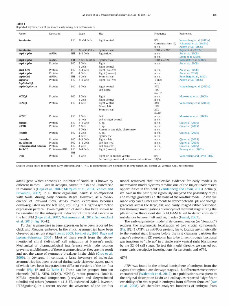
Table 1Reported asymmetries of presumed early acting L–R determinants.
Factor Detection Stage Site Frequency References
Serotonin IHC 32–64 Cells Right ventral 8/8 Vandenberg et al. (2013a)Consensus (n¼10) Fukumoto et al. (2005b)n. sp. Adams et al. (2006)
Serotonin IF 32–256 Cells Symmetrical 100% n4200 Beyer et al. (2012a)atp4 alpha mRNA ISH 2–4 Cells Right-sided n. sp. Aw et al. (2008)
n. sp. Levin et al. (2002)atp4 alpha mRNA ISH 2 Cell-blastula Symmetrical 100% n¼320 Walentek et al. (2012)atp4 alpha Protein IHC 2 Cells Right n. sp. Aw et al. (2008)
4 Cells Right ventralatp4 beta Protein IHC 2–4 Cells Right (doþve) n. sp. Aw et al. (2008)atp4 alpha Protein IF 4 Cells Right (doþve) n. sp. Aw et al., 2010)atp6v0a2 mRNA ISH 4 Cells Symmetrical n. sp. Rutenberg et al., 2002)atp6v0c Protein IHC 2–8 Cells Right (doþve) 480% Adams et al. (2006)Atp6v1a/b/f n. sp.atp6v0c/ductin Protein IHC 4 Cells Right ventral 55% Vandenberg et al. (2013b)
Left dorsal 11%n¼110
KCNQ1 Protein IHC 2 Cells Right n. sp. Morokuma et al. (2008)4 Cells Right ventral n. sp.
KCNQ1 Protein IHC 4 Cells Right ventral 34% Vandenberg et al. (2013b)Dorsal left 18%Symmetrical 27%
n¼85KCNE1 Protein IHC 2 Cells Left n. sp. Morokuma et al. (2008)
4 Cells Left or right ventral n. sp.dnah11 Protein IHC 2–8 Cells n. sp. n. sp. Qiu et al. (2005)KiF3B Protein IHC 2 Cells n. sp. n. sp. Qiu et al. (2005)
4 Cells Absent in one right blastomere n. sp.Polaris Protein IHC 2 Cells n. sp. n. sp. Qiu et al. (2005)
4 Cells Symmetric n. sp.Inversin Protein IHC 4–8 Cells Right4 left n. sp. Qiu et al. (2005)ac. tubulin Protein IHC 2–4 Cells Left (doþve) n. sp. Qiu et al. (2005)Detyrosinated tubulin Protein IHC 4 Cells Left (doþve) n. sp. Qiu et al. (2005)14-3-3E ProteinþmRNA IHC 2–4 Cells Right (doþve) n. sp. Bunney et al. (2003)
ISHDvl2 Protein IF 4 Cells Right-ve in animal-vegetal 8/9 Vandenberg and Levin (2012)
Sections symmetrical in transversal sections 14/14
Studies which failed to reproduce early serotonin and ATP4 L–R asymmetries are highlighted in gray shade. do, dorsal; ve, ventral; n.sp., not specified.
M. Blum et al. / Developmental Biology 393 (2014) 109–123 115
different females from our own frog colony and from a separatecolony of a colleague, but still did not find a single embryo withL–R asymmetric ATP4 expression (Table 1; Walentek et al., 2012).Of course, we cannot exclude that asymmetries occur in very rarecases. It seems inescapable that infrequent events cannot qualifyas the basis for robust laterality formation in the amphibianembryo.
Serotonin
Serotonin asymmetry should be easily detectable, but it is not.We initially became interested in the ion-flux hypothesis becausean immunocytochemically assayed lineage-specific enrichment ofserotonin (as reported by Fukumoto et al., 2005b) would havebeen an extremely useful readout for studying experimentalperturbations of early localization mechanisms (Danilchik et al.,2006). Disappointingly, and following considerable effort, we werenever able to demonstrate a consistent enrichment of endogenousserotonin (Beyer et al., 2012a). Further, microinjected serotonin,easily detected by whole-mount immunocytochemistry, failed torelocalize to uninjected blastomeres (Beyer et al., 2012a), demon-strating that it rapidly becomes sequestered into some bound form(e.g. vesicles), which cannot pass through gap junctions (cf. Fig. 4,below). Thus, we are forced to question both the early localizationof an electrogenic maternal factor (such as ATP4) as well as thedirected passage of serotonin through gap junctions. A third, little-discussed possibility, that serotonin might be selectively degradedin some particular lineage, also must be discounted, since exogen-ous serotonin observably persists in any lineage into which it ismicroinjected (Beyer et al., 2012a).
Previously published reports of maternal asymmetric localizations
Unfortunately, it is difficult to develop a comprehensive picturefrom published reports – in most cases the numbers of analyzedspecimens and frequencies of observed asymmetries were notreported (Table 1). For three particular factors, numerical data areavailable – right-side asymmetries were reported for KCNQ1 in34% of cases (Morokuma et al., 2008), for the ductin subunit ofATP6 in 55% of analyzed specimens (Vandenberg et al., 2013b), aswell as in 8/9 embryos analyzed for Disheveled-2 (Dvl2) expression(Vandenberg and Levin, 2012; Table 1). With the exception of Dvl2,these rates are too low to explain the normally robust (495%)expression of situs solitus in undisturbed Xenopus embryos. TheDvl2 data set, aside from the low number of analyzed embryos,suffers from another complication. In Vandenberg and Levin(2012) two planes of sections of Dvl2-analyzed specimens areshown. Along the animal–vegetal axis, Dvl2 protein immunode-tection showed uniform expression in the animal hemisphere ofthe 4-cell embryo, i.e. both cells of the section contain protein. Thisreportedly was observed in 14/14 cases. In transverse sections, incontrast, right asymmetries were reported to occur in 8/9 cases.Only one pattern can be correct – if Dvl2 is found in both cells insections along the animal–vegetal axis, transverse sections at thelevel of staining in the animal half of the embryo should show allfour cells positive for Dvl2. Or, conversely, a consistent ventral-right asymmetry in transverse sections would mean that speci-mens sectioned along the animal–vegetal axis would also revealthis clearly asymmetric pattern. The published results on Dvl2protein expression need to be carefully reconsidered as potentiallymisinterpreted from a slightly oblique plane of section relative tothe transition between Dvl2-containing and Dvl2-lacking regionsalong the animal–vegetal axis. Clearly, whole-mount type ana-lyses, marked carefully with respect to the dorso-ventral axis,could negate this criticism. In summary, there is presently no
compelling evidence for consistent cleavage-stage asymmetries inmaternal factors with causal roles in L–R determination.
Taken together, the available data on asymmetric expression ofmRNAs and proteins support, rather than discount, the pivotalroles of the Nodal cascade and, in the case of leftward flow, ofthe Nodal inhibitor dand5 for laterality determination. Other,presumably early-acting factors including Dvl2 might of coursebe involved in the different steps of laterality determination,which has been shown for quite a number of them in the past(see below). It is not necessary, however, that such factors areasymmetrically expressed, in particular if they are involved in thespecification or morphogenesis of the symmetrical LRO.
Genetic evidence supports cilia-based laterality determinationin the vertebrates
There is overwhelming genetic evidence that malformation ofcilia structure and function causes laterality defects in fish, mouseand humans. Many excellent reviews have covered this topic(Bisgrove et al., 2003; McGrath and Brueckner, 2003; Norris andGrimes, 2012; Oh and Katsanis, 2012; Shiraishi and Ichikawa,2012; Sutherland and Ware, 2009). Human ciliopathies, i.e. syn-dromes characterized by ciliary malfunctions, frequently displaylaterality defects. The first laterality syndrome, caused by immotilecilia, was Kartagener syndrome, in which patients show defects inthe ciliary motor protein Dnah11 (formerly known as left–rightdynein, lrd; (McGrath and Brueckner, 2003)). Kartagener syn-drome belongs to the larger group of primary ciliary dyskinesia(PCD) syndromes. Other ciliopathies with left–right defectsinvolve, among others, polycystic kidney disease, Bardet–Biedelsyndrome, Meckel–Gruber syndrome, Joubert syndrome andnephronophthisis (Norris and Grimes, 2012).
Various genetic screens in mouse and zebrafish underscore theimportance of cilia for L–R axis formation. Dominic Norris (Har-well) performed an ENU-based screen for L–R patterning defectsin mouse and identified mostly cilia-related genes (Ermakov et al.,2009; Field et al., 2011). Cecilia Lo (Pittsburgh), in a recessivescreen for congenital heart disease, scanned 465,000 fetuses byultrasound in utero, identified 43000 abnormal embryos andestablished 192 lines. Of the 69 laterality-defective individualmouse lines derived, 82% showed mutations in cilia-related genes(Liu et al., 2013; Cecilia Lo, personal communication). Finally,knockouts of genes encoding intraflagellar transport proteins(IFT), motor proteins (dyneins and kinesins) or transcriptionfactors involved in ciliogenesis (i.e. foxj1, Rfx) inevitably resultedin laterality defects (for a recent review see Yoshiba and Hamada,2014). In zebrafish, general L–R screens are difficult to performbecause of the very high rates of background laterality defects innon-isogenic lines, which can be as high as 10–15% (RebeccaBurdine, Princeton, personal communication), possibly related tosome subtle but fairly pervasive environmental disturbance. Still,re-screening of the Tübingen collection for L–R patterning defectsin the heart uncovered primarily cilia genes (Chen et al., 2001;Panizzi et al., 2012; Schottenfeld et al., 2007; Serluca et al., 2009;Sullivan-Brown et al., 2008). In addition, a genetic screen for cystickidneys identified cilia genes, and many of the mutants displayL–R defects as well (Sun et al., 2004). These genetic screenscertainly cement the importance of cilia in laterality deter-mination.
As mentioned above, it has been proposed that cilia-drivenleftward flow merely acts as an amplification step for asymmetrythat is initially defined by ion-flux during early cleavage(Vandenberg and Levin, 2013). This proposal predicts that asym-metry defined by ion-flux should be present in the absence offlow, unless it is mandatorily gated through the cilia/LRO machine.
M. Blum et al. / Developmental Biology 393 (2014) 109–123116
Two lines of experimentation have addressed that question.The first approach manually ablated the ciliated epithelium orthe precursor tissue, respectively. When Kupffer's vesicle wasmechanically destroyed in Medaka fish (Oryzias latipes), massivelaterality defects occurred (Bajoghli et al., 2007). Removal of theSM in the gastrula Xenopus embryo (Blum et al., 2009a), or laserablations of dorsal forerunner cells in zebrafish (Essner et al.,
2005), from which the ciliated LROs (GRP and KV) develop duringearly neurulation, resulted in the same outcome. Remarkably, in allcases no other morphological defects arose besides disruption oflaterality (Bajoghli et al., 2007; Blum et al., 2009a; Essner et al.,2005). The second type of experiment interfered with flowdirectly, without manipulation of tissues, cilia or genes involvedin setting up flow. Methylcellulose was applied to the ciliated
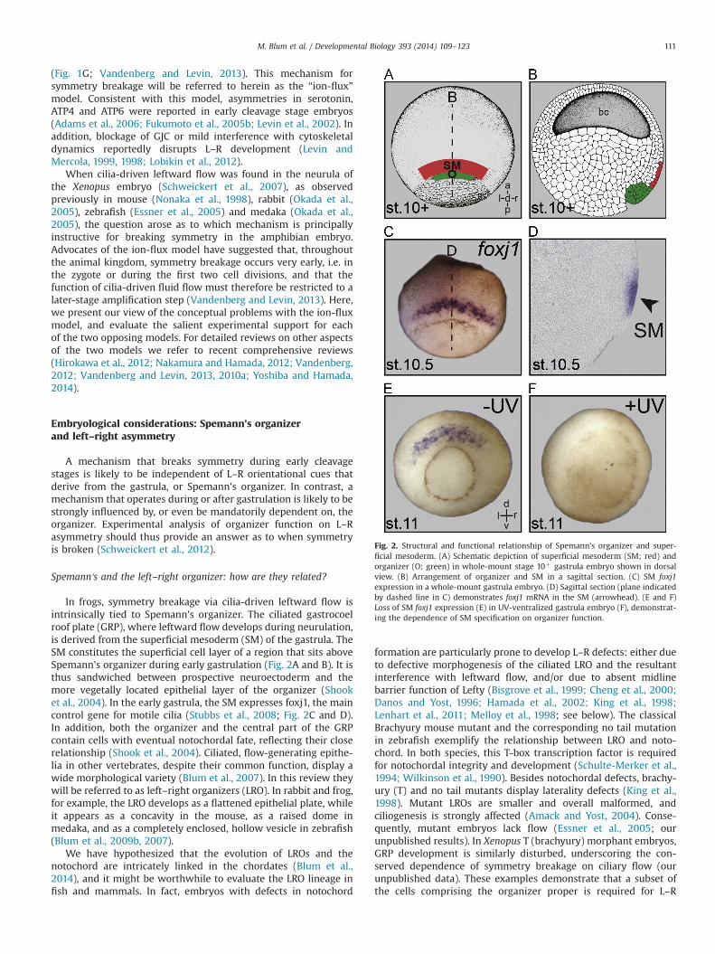
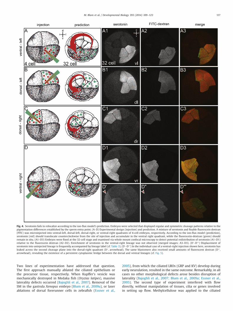
Fig. 4. Serotonin fails to relocalize according to the ion-flux model's prediction. Embryos were selected that displayed regular and symmetric cleavage patterns relative to thepigmentation differences established by the sperm entry point. (A–D) Experimental design (injection) and prediction. A mixture of serotonin and fixable fluorescein dextran(FITC) was microinjected into ventral-left, dorsal-left, dorsal-right, or ventral-right quadrants of 4-cell embryos, respectively. According to the ion-flux model (prediction),serotonin (red) should translocate counterclockwise from the site of injection and accumulate in the ventral right quadrant, while the fluorescein-dextran (green) shouldremain in situ. (A1–D3) Embryos were fixed at the 32-cell stage and examined via whole mount confocal microscopy to detect potential redistribution of serotonin (A1–D1)relative to the fluorescein dextran (A2–D2). Enrichment of serotonin in the ventral-right lineage was not observed (merged images; A3–D3). (D0–D0″) Displacement ofserotonin into uninjected lineage is frequently accompanied by lineage label (cf. Table 3). (D0–D0″) In the individual case of a ventral-right injection shown here, serotonin hasleaked across the second cleavage plane into the dorsal-right quadrant (D″, arrowhead). The same blastomere also received small amounts of fluorescent dextran (D″0 ,arrowhead), revealing the existence of a persistent cytoplasmic bridge between the dorsal and ventral lineages (cf. Fig. 5).
M. Blum et al. / Developmental Biology 393 (2014) 109–123 117
epithelia in cultured frog (Schweickert et al., 2007) and mouse(Nakamura et al., 2012) embryos to hamper fluid transport byincreasing the viscosity of the medium. These manipulationsprevented the induction of the Nodal cascade in a high andstatistically significant percentage of cases.
[The fact that procedures to ablate flow do not reach one hundredpercent efficiency is intrinsic to all experimental manipulations ofembryos and can be readily explained. The time point of treatmentin some cases might have been too late, after fixation of lateralitycues. Targeting of methylcellulose to the ciliated LRO is challengingin frog, as it has to be applied blindly directly into the gastrocoel.In the great majority of cases in which procedures did work,however, potentially early-acting ion-flux based mechanismswould have been active, and thus would have left a mark ofasymmetry in every single case. Since such an outcome was notobserved in all of these experiments, early ion-flux cannot beinvoked as the responsible mechanism].
In summary, experimental LRO manipulations together withhuman, mouse and zebrafish genetics seem to indicate beyondreasonable doubt that motile cilia are required for the formation ofsitus solitus, and do not act by amplifying some earlier determi-nistic laterality signal.
Role of serotonin, ATP4 and GJC in laterality specification
In the light of these genetic data, what role might “early”determinants play in the process of laterality specification? A largebody of published results from one laboratory, mostly involvingpharmacological manipulation in early frog embryos, suggestsinvolvement of maternal factors in L–R axis formation (reviewedin Vandenberg and Levin, 2013). It has been suggested that someof these factors, such as motor proteins, act during early cleavagestages prior to formation of any cilia. The implication is thereforethat ciliary proteins must be carrying out laterality-specifyingfunctions in the cytoplasm (Vandenberg and Levin, 2013). Indeed,the ion-flux model requires that motor proteins move mRNAs andproteins to the ventral-right blastomere in the early cleavage-stageembryo. In Xenopus, often huge amounts of many mRNAs andproteins of maternal origin are present in the zygote. However, inno single case have the consequences of pharmacological manip-ulations of “early” factors on flow-related processes been analyzed.After the discovery of the frog LRO and flow (Schweickert et al.,2007), we wondered whether early determinants and leftwardflow acted sequentially or in parallel, and chose to analyze thecentral components of the ion-flux model, ATP4, serotonin and GJCin the context of flow. In the following, we briefly summarizeresults of these recently published studies.
Serotonin signaling
Flow and asymmetry were lost in embryos in which serotoninsignaling was down-regulated (Beyer et al., 2012a). Importantly,
we found that serotonin accumulated in the epithelial cell layer ofthe blastula from the 128-cell stage onwards, i.e. in the same layerwhere the SM forms on the dorsal side (Beyer et al., 2012a).Molecularly, the SM, from which the LRO derives in the frog, ischaracterized by the expression of nodal3 and foxj1. Both geneswere down-regulated upon loss of serotonin signaling. Nodal3induction depends on canonical Wnt signaling. We showed thatserotonin acts as a competence factor for canonical Wnt signaling,i.e. that the role of serotonin in L–R axis formation lies in the Wnt-dependent specification of the superficial mesoderm (Beyer et al.,2012a).
ATP4
Gene knock-down or pharmacological inhibition of ATP4 com-promised organ situs, asymmetric gene expression and leftwardflow. The GRP analysis revealed fewer, shortened and misalignedcilia. FoxJ1 was down-regulated in the SM. ATP4 was requiredduring consecutive steps of L–R axis formation – for Wnt/β-catenin regulated Foxj1 induction in the SM and for Wnt/PCPdependent cilia polarization in the GRP (Walentek et al., 2012).In summary, our work on serotonin and ATP4 defines a new SM/GRP/Flow (SGF) module for symmetry breakage in frogs (Table 2).This module spans the time period from late blastula/earlygastrula (SM specification) to the flow-dependent down-regula-tion of the Nodal inhibitor Coco at the left margin of the GRP atstage 19, which is the first molecular asymmetry and instrumentalfor Nodal cascade induction in the left LPM (Schweickert et al.,2010).
GJC and serotonin localization
We and others recently showed that GJC is required later indevelopment for the transfer of asymmetric cue(s) from themidline to the lateral plate mesoderm in frog and mouse (Beyeret al., 2012b; Viotti et al., 2012). In the frog, connexin26 expressionin the endoderm between the LRO/GRP and the LPM is required forNodal cascade induction downstream of flow. Inhibitor experi-ments in addition strongly argue against earlier functions of GJC inlaterality determination (Beyer et al., 2012b). However, it remainsa formal possibility that GJC also acts much earlier in develop-ment, i.e., during the first few cleavage divisions. Indeed, the ion-flux model requires that GJC exists between nearly all of the earlyblastomeres, except those along the incipient ventral midline.
Because early blastomeres begin a new cleavage furrow beforehaving fully completed the preceding round of cytokinesis, it hasbeen experimentally difficult, using small-molecule lineage tra-cers, to demonstrate authentic GJC against the background of opencytoplasmic bridges. Thus, the degree of physiological coupling ofblastomeres via gap junctions in the earliest cleavage stages hasremained controversial (Guthrie, 1984; Landesman et al., 2000).To revisit GJC in the context of a potential early localizationof serotonin, we microinjected single blastomeres at the late4-cell stage with a mixture of serotonin and a fixable fluorescent
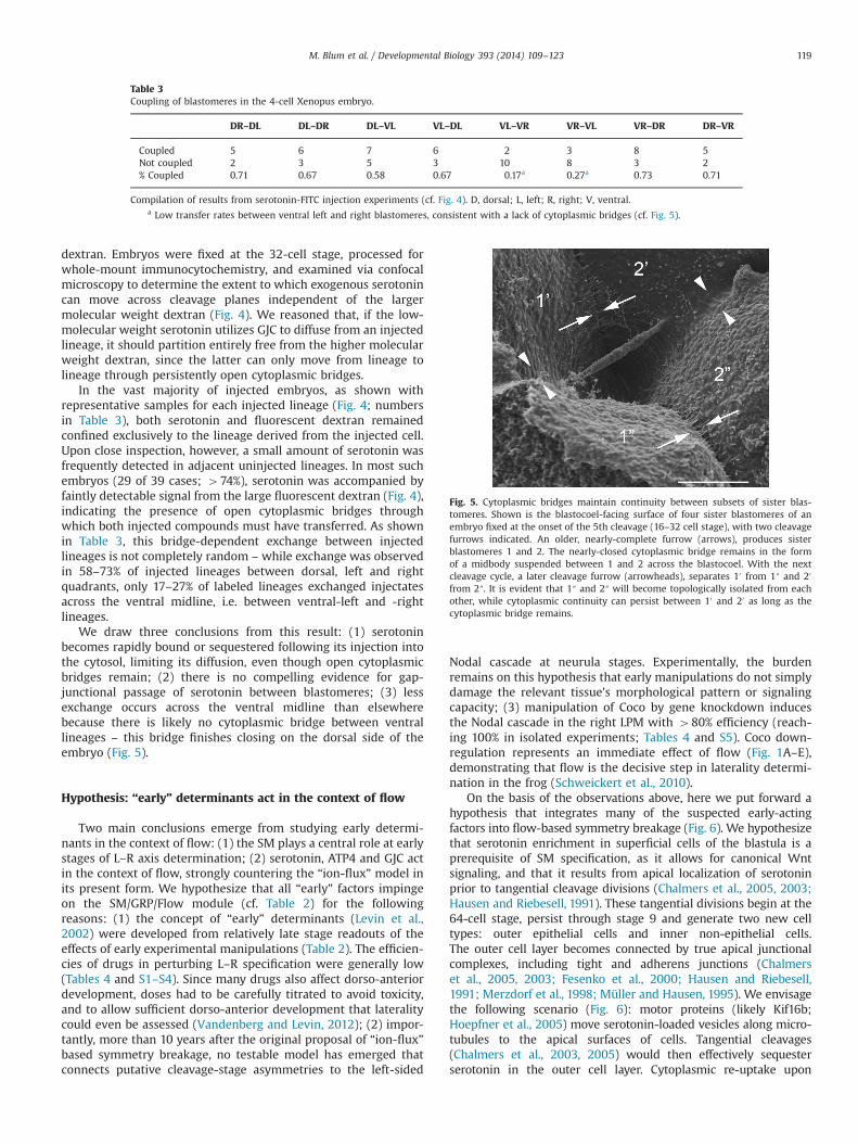
Table 2Module of L–R development. Timing of developmental stages and L–R modules.
Stage St. 1–3 St. 6–9 St. 10–14 St. 15–18 St. 19–45h 0–2 h 3–7 h 9–20 h 17–20 h 20–106 h
1–4 Cells Blastula Gastrula–early neurula Neurula Tadpole
LR module “Ion-flux” Serotonin localization SGF Nodal cascade and organ situsReadout Nodal cascade and organ situs SGF SM GRP Flow
Xnr3 Ciliation QualityFoxj1 Morphology Directionality
Marker genes Coco
M. Blum et al. / Developmental Biology 393 (2014) 109–123118
dextran. Embryos were fixed at the 32-cell stage, processed forwhole-mount immunocytochemistry, and examined via confocalmicroscopy to determine the extent to which exogenous serotonincan move across cleavage planes independent of the largermolecular weight dextran (Fig. 4). We reasoned that, if the low-molecular weight serotonin utilizes GJC to diffuse from an injectedlineage, it should partition entirely free from the higher molecularweight dextran, since the latter can only move from lineage tolineage through persistently open cytoplasmic bridges.
In the vast majority of injected embryos, as shown withrepresentative samples for each injected lineage (Fig. 4; numbersin Table 3), both serotonin and fluorescent dextran remainedconfined exclusively to the lineage derived from the injected cell.Upon close inspection, however, a small amount of serotonin wasfrequently detected in adjacent uninjected lineages. In most suchembryos (29 of 39 cases; 474%), serotonin was accompanied byfaintly detectable signal from the large fluorescent dextran (Fig. 4),indicating the presence of open cytoplasmic bridges throughwhich both injected compounds must have transferred. As shownin Table 3, this bridge-dependent exchange between injectedlineages is not completely random – while exchange was observedin 58–73% of injected lineages between dorsal, left and rightquadrants, only 17–27% of labeled lineages exchanged injectatesacross the ventral midline, i.e. between ventral-left and -rightlineages.
We draw three conclusions from this result: (1) serotoninbecomes rapidly bound or sequestered following its injection intothe cytosol, limiting its diffusion, even though open cytoplasmicbridges remain; (2) there is no compelling evidence for gap-junctional passage of serotonin between blastomeres; (3) lessexchange occurs across the ventral midline than elsewherebecause there is likely no cytoplasmic bridge between ventrallineages – this bridge finishes closing on the dorsal side of theembryo (Fig. 5).
Hypothesis: “early” determinants act in the context of flow
Two main conclusions emerge from studying early determi-nants in the context of flow: (1) the SM plays a central role at earlystages of L–R axis determination; (2) serotonin, ATP4 and GJC actin the context of flow, strongly countering the “ion-flux” model inits present form. We hypothesize that all “early” factors impingeon the SM/GRP/Flow module (cf. Table 2) for the followingreasons: (1) the concept of “early” determinants (Levin et al.,2002) were developed from relatively late stage readouts of theeffects of early experimental manipulations (Table 2). The efficien-cies of drugs in perturbing L–R specification were generally low(Tables 4 and S1–S4). Since many drugs also affect dorso-anteriordevelopment, doses had to be carefully titrated to avoid toxicity,and to allow sufficient dorso-anterior development that lateralitycould even be assessed (Vandenberg and Levin, 2012); (2) impor-tantly, more than 10 years after the original proposal of “ion-flux”based symmetry breakage, no testable model has emerged thatconnects putative cleavage-stage asymmetries to the left-sided
Nodal cascade at neurula stages. Experimentally, the burdenremains on this hypothesis that early manipulations do not simplydamage the relevant tissue's morphological pattern or signalingcapacity; (3) manipulation of Coco by gene knockdown inducesthe Nodal cascade in the right LPM with 480% efficiency (reach-ing 100% in isolated experiments; Tables 4 and S5). Coco down-regulation represents an immediate effect of flow (Fig. 1A–E),demonstrating that flow is the decisive step in laterality determi-nation in the frog (Schweickert et al., 2010).
On the basis of the observations above, here we put forward ahypothesis that integrates many of the suspected early-actingfactors into flow-based symmetry breakage (Fig. 6). We hypothesizethat serotonin enrichment in superficial cells of the blastula is aprerequisite of SM specification, as it allows for canonical Wntsignaling, and that it results from apical localization of serotoninprior to tangential cleavage divisions (Chalmers et al., 2005, 2003;Hausen and Riebesell, 1991). These tangential divisions begin at the64-cell stage, persist through stage 9 and generate two new celltypes: outer epithelial cells and inner non-epithelial cells.The outer cell layer becomes connected by true apical junctionalcomplexes, including tight and adherens junctions (Chalmerset al., 2005, 2003; Fesenko et al., 2000; Hausen and Riebesell,1991; Merzdorf et al., 1998; Müller and Hausen, 1995). We envisagethe following scenario (Fig. 6): motor proteins (likely Kif16b;Hoepfner et al., 2005) move serotonin-loaded vesicles along micro-tubules to the apical surfaces of cells. Tangential cleavages(Chalmers et al., 2003, 2005) would then effectively sequesterserotonin in the outer cell layer. Cytoplasmic re-uptake upon
Table 3Coupling of blastomeres in the 4-cell Xenopus embryo.
DR–DL DL–DR DL–VL VL–DL VL–VR VR–VL VR–DR DR–VR
Coupled 5 6 7 6 2 3 8 5Not coupled 2 3 5 3 10 8 3 2% Coupled 0.71 0.67 0.58 0.67 0.17a 0.27a 0.73 0.71
Compilation of results from serotonin-FITC injection experiments (cf. Fig. 4). D, dorsal; L, left; R, right; V, ventral.a Low transfer rates between ventral left and right blastomeres, consistent with a lack of cytoplasmic bridges (cf. Fig. 5).
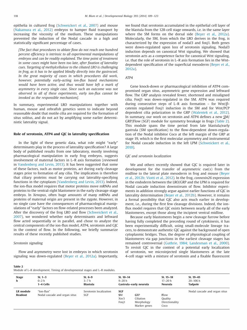
Fig. 5. Cytoplasmic bridges maintain continuity between subsets of sister blas-tomeres. Shown is the blastocoel-facing surface of four sister blastomeres of anembryo fixed at the onset of the 5th cleavage (16–32 cell stage), with two cleavagefurrows indicated. An older, nearly-complete furrow (arrows), produces sisterblastomeres 1 and 2. The nearly-closed cytoplasmic bridge remains in the formof a midbody suspended between 1 and 2 across the blastocoel. With the nextcleavage cycle, a later cleavage furrow (arrowheads), separates 10 from 1″ and 20
from 2″. It is evident that 1″ and 2″ will become topologically isolated from eachother, while cytoplasmic continuity can persist between 10 and 20 as long as thecytoplasmic bridge remains.
M. Blum et al. / Developmental Biology 393 (2014) 109–123 119
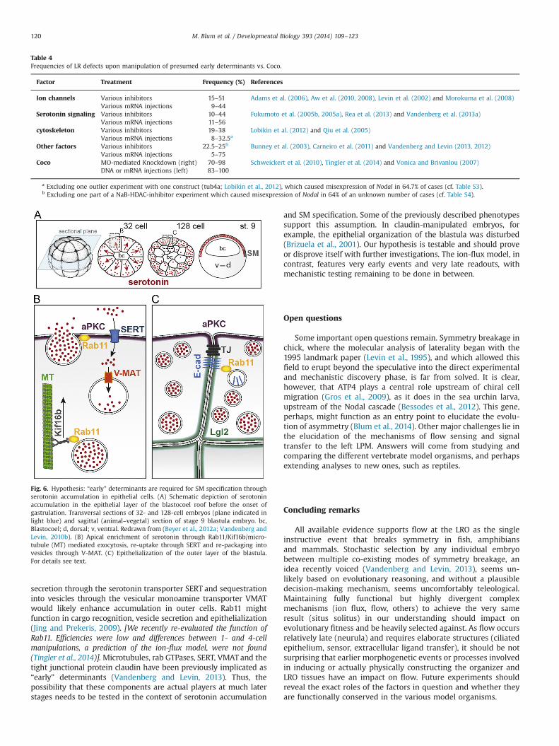
secretion through the serotonin transporter SERT and sequestrationinto vesicles through the vesicular monoamine transporter VMATwould likely enhance accumulation in outer cells. Rab11 mightfunction in cargo recognition, vesicle secretion and epithelialization(Jing and Prekeris, 2009). [We recently re-evaluated the function ofRab11. Efficiencies were low and differences between 1- and 4-cellmanipulations, a prediction of the ion-flux model, were not found(Tingler et al., 2014)].Microtubules, rab GTPases, SERT, VMAT and thetight junctional protein claudin have been previously implicated as“early” determinants (Vandenberg and Levin, 2013). Thus, thepossibility that these components are actual players at much laterstages needs to be tested in the context of serotonin accumulation
and SM specification. Some of the previously described phenotypessupport this assumption. In claudin-manipulated embryos, forexample, the epithelial organization of the blastula was disturbed(Brizuela et al., 2001). Our hypothesis is testable and should proveor disprove itself with further investigations. The ion-flux model, incontrast, features very early events and very late readouts, withmechanistic testing remaining to be done in between.
Open questions
Some important open questions remain. Symmetry breakage inchick, where the molecular analysis of laterality began with the1995 landmark paper (Levin et al., 1995), and which allowed thisfield to erupt beyond the speculative into the direct experimentaland mechanistic discovery phase, is far from solved. It is clear,however, that ATP4 plays a central role upstream of chiral cellmigration (Gros et al., 2009), as it does in the sea urchin larva,upstream of the Nodal cascade (Bessodes et al., 2012). This gene,perhaps, might function as an entry point to elucidate the evolu-tion of asymmetry (Blum et al., 2014). Other major challenges lie inthe elucidation of the mechanisms of flow sensing and signaltransfer to the left LPM. Answers will come from studying andcomparing the different vertebrate model organisms, and perhapsextending analyses to new ones, such as reptiles.
Concluding remarks
All available evidence supports flow at the LRO as the singleinstructive event that breaks symmetry in fish, amphibiansand mammals. Stochastic selection by any individual embryobetween multiple co-existing modes of symmetry breakage, anidea recently voiced (Vandenberg and Levin, 2013), seems un-likely based on evolutionary reasoning, and without a plausibledecision-making mechanism, seems uncomfortably teleological.Maintaining fully functional but highly divergent complexmechanisms (ion flux, flow, others) to achieve the very sameresult (situs solitus) in our understanding should impact onevolutionary fitness and be heavily selected against. As flow occursrelatively late (neurula) and requires elaborate structures (ciliatedepithelium, sensor, extracellular ligand transfer), it should be notsurprising that earlier morphogenetic events or processes involvedin inducing or actually physically constructing the organizer andLRO tissues have an impact on flow. Future experiments shouldreveal the exact roles of the factors in question and whether theyare functionally conserved in the various model organisms.
Table 4Frequencies of LR defects upon manipulation of presumed early determinants vs. Coco.
Factor Treatment Frequency (%) References
Ion channels Various inhibitors 15–51 Adams et al. (2006), Aw et al. (2010, 2008), Levin et al. (2002) and Morokuma et al. (2008)Various mRNA injections 9–44
Serotonin signaling Various inhibitors 10–44 Fukumoto et al. (2005b, 2005a), Rea et al. (2013) and Vandenberg et al. (2013a)Various mRNA injections 11–56
cytoskeleton Various inhibitors 19–38 Lobikin et al. (2012) and Qiu et al. (2005)Various mRNA injections 8–32.5a
Other factors Various inhibitors 22.5–25b Bunney et al. (2003), Carneiro et al. (2011) and Vandenberg and Levin (2013, 2012)Various mRNA injections 5–75
Coco MO-mediated Knockdown (right) 70–98 Schweickert et al. (2010), Tingler et al. (2014) and Vonica and Brivanlou (2007)DNA or mRNA injections (left) 83–100
a Excluding one outlier experiment with one construct (tub4a; Lobikin et al., 2012), which caused misexpression of Nodal in 64.7% of cases (cf. Table S3).b Excluding one part of a NaB-HDAC-inhibitor experiment which caused misexpression of Nodal in 64% of an unknown number of cases (cf. Table S4).
Fig. 6. Hypothesis: “early” determinants are required for SM specification throughserotonin accumulation in epithelial cells. (A) Schematic depiction of serotoninaccumulation in the epithelial layer of the blastocoel roof before the onset ofgastrulation. Transversal sections of 32- and 128-cell embryos (plane indicated inlight blue) and sagittal (animal–vegetal) section of stage 9 blastula embryo. bc,Blastocoel; d, dorsal; v, ventral. Redrawn from (Beyer et al., 2012a; Vandenberg andLevin, 2010b). (B) Apical enrichment of serotonin through Rab11/Kif16b/micro-tubule (MT) mediated exocytosis, re-uptake through SERT and re-packaging intovesicles through V-MAT. (C) Epithelialization of the outer layer of the blastula.For details see text.
M. Blum et al. / Developmental Biology 393 (2014) 109–123120
Acknowledgments
We are grateful to Rebecca Burdine (Princeton) and Cecilia Lo(Pittsburgh) for sharing unpublished results and observations.The generous help from Matthias Tisler and Tim Ott in preparingall figures was much appreciated and is gratefully acknowledged.Work in the Blum lab was supported by DFG Grants BL285/9-2 andBL285/10-1, PV was the recipient of a DFG return fellowship(VI-574/2-1), and CW was supported by NIH R01-GM56238.
Appendix A. Supporting information
Supplementary data associated with this article can be found inthe online version at http://dx.doi.org/10.1016/j.ydbio.2014.06.014.
References
Adams, D.S., Robinson, K.R., Fukumoto, T., Yuan, S., Albertson, R.C., Yelick, P., Kuo, L.,McSweeney, M., Levin, M., 2006. Early, Hþ-V-ATPase-dependent proton flux isnecessary for consistent left–right patterning of non-mammalian vertebrates.Development 133, 1657–1671.
Aizawa, H., 2013. Habenula and the asymmetric development of the vertebratebrain. Anat. Sci. Int. 88, 1–9.
Amack, J.D., Yost, H.J., 2004. The T box transcription factor no tail in ciliated cellscontrols zebrafish left–right asymmetry. Curr. Biol. 14, 685–690.
Aw, S., Adams, D.S., Qiu, D., Levin, M., 2008. H,K-ATPase protein localization andKir4.1 function reveal concordance of three axes during early determination ofleft–right asymmetry. Mech. Dev. 125, 353–372.
Aw, S., Koster, J.C., Pearson, W., Nichols, C.G., Shi, N.-Q., Carneiro, K., Levin, M., 2010.The ATP-sensitive K(þ)-channel (K(ATP)) controls early left-right patterning inXenopus and chick embryos. Dev. Biol. 346, 39–53.
Bajoghli, B., Aghaallaei, N., Soroldoni, D., Czerny, T., 2007. The roles of Groucho/Tlein left–right asymmetry and Kupffer's vesicle organogenesis. Dev. Biol. 303,347–361.
Bartram, U., Wirbelauer, J., Speer, C.P., 2005. Heterotaxy syndrome – asplenia andpolysplenia as indicators of visceral malposition and complex congenital heartdisease. Biol. Neonate 88, 278–290.
Basu, B., Brueckner, M., 2008. Cilia multifunctional organelles at the center ofvertebrate left–right asymmetry. Curr. Top. Dev. Biol. 85, 151–174.
Bessodes, N., Haillot, E., Duboc, V., Röttinger, E., Lahaye, F., Lepage, T., 2012.Reciprocal signaling between the ectoderm and a mesendodermal left–rightorganizer directs left–right determination in the sea urchin embryo. PLoSGenet. 8, e1003121.
Beyer, T., Danilchik, M., Thumberger, T., Vick, P., Tisler, M., Schneider, I., Bogusch, S.,Andre, P., Ulmer, B., Walentek, P., Niesler, B., Blum, M., Schweickert, A., 2012a.Serotonin signaling is required for Wnt-dependent GRP specification andleftward flow in Xenopus. Curr. Biol. 22, 33–39.
Beyer, T., Thumberger, T., Schweickert, A., Blum, M., 2012b. Connexin26-mediatedtransfer of laterality cues in Xenopus. Biol. Open 1, 473–481.
Bisgrove, B.W., Essner, J.J., Yost, H.J., 1999. Regulation of midline development byantagonism of lefty and nodal signaling. Development 126, 3253–3262.
Bisgrove, B.W., Essner, J.J., Yost, H.J., 2000. Multiple pathways in the midlineregulate concordant brain, heart and gut left–right asymmetry. Development127, 3567–3579.
Bisgrove, B.W., Morelli, S.H., Yost, H.J., 2003. Genetics of human laterality disorders:insights from vertebrate model systems. Annu. Rev. Genomics Hum. Genet. 4,1–32.
Blum, M., Andre, P., Muders, K., Schweickert, A., Fischer, A., Bitzer, E., Bogusch, S.,Beyer, T., van Straaten, H.W.M., Viebahn, C., 2007. Ciliation and gene expressiondistinguish between node and posterior notochord in the mammalian embryo.Differentiation 75, 133–146.
Blum, M., Beyer, T., Weber, T., Vick, P., Andre, P., Bitzer, E., Schweickert, A., 2009a.Xenopus, an ideal model system to study vertebrate left–right asymmetry. Dev.Dyn. 238, 1215–1225.
Blum, M., Weber, T., Beyer, T., Vick, P., 2009b. Evolution of leftward flow. Semin. CellDev. Biol. 20, 464–471.
Blum, M., Feistel, K., Thumberger, T., Schweickert, A., 2014. The evolution andconservation of left–right patterning mechanisms. Development 141, 1603–1613.
Brizuela, B.J., Wessely, O., De Robertis, E.M., 2001. Overexpression of the Xenopustight-junction protein claudin causes randomization of the left–right body axis.Dev. Biol. 230, 217–229.
Brown, N.A., Wolpert, L., 1990. The development of handedness in left/rightasymmetry. Development 109, 1–9.
Bunney, T.D., De Boer, A.H., Levin, M., 2003. Fusicoccin signaling reveals 14-3-3protein function as a novel step in left–right patterning during amphibianembryogenesis. Development 130, 4847–4858.
Burdine, R.D., Caspary, T., 2013. Left–right asymmetry: lessons from Cancun.Development 140, 4465–4470.
Burn, S.F., Hill, R.E., 2009. Left–right asymmetry in gut development: what happensnext? Bioessays 31, 1026–1037.
Carneiro, K., Donnet, C., Rejtar, T., Karger, B.L., Barisone, G.A., Díaz, E., Kortagere, S.,Lemire, J.M., Levin, M., 2011. Histone deacetylase activity is necessary for left–right patterning during vertebrate development. BMC Dev. Biol. 11, 29.
Chalmers, A.D., Pambos, M., Mason, J., Lang, S., Wylie, C., Papalopulu, N., 2005. aPKC,Crumbs3 and Lgl2 control apicobasal polarity in early vertebrate development.Development 132, 977–986.
Chalmers, A.D., Strauss, B., Papalopulu, N., 2003. Oriented cell divisions asymme-trically segregate aPKC and generate cell fate diversity in the early Xenopusembryo. Development 130, 2657–2668.
Chea, H.K., Wright, C.V., Swalla, B.J., 2005. Nodal signaling and the evolution ofdeuterostome gastrulation. Dev. Dyn. 234, 269–278.
Chen, J.N., van Bebber, F., Goldstein, A.M., Serluca, F.C., Jackson, D., Childs, S.,Serbedzija, G., Warren, K.S., Mably, J.D., Lindahl, P., Mayer, A., Haffter, P.,Fishman, M.C., 2001. Genetic steps to organ laterality in zebrafish. Comp. Funct.Genomics 2, 60–68.
Cheng, A.M., Thisse, B., Thisse, C., Wright, C.V., 2000. The lefty-related factor Xatvacts as a feedback inhibitor of nodal signaling in mesoderm induction and L–Raxis development in Xenopus. Development 127, 1049–1061.
Danilchik, M.V., Brown, E.E., Riegert, K., 2006. Intrinsic chiral properties of theXenopus egg cortex: an early indicator of left–right asymmetry? Development133, 4517–4526.
Danos, M.C., Yost, H.J., 1995. Linkage of cardiac left–right asymmetry and dorsal-anterior development in Xenopus. Development 121, 1467–1474.
Danos, M.C., Yost, H.J., 1996. Role of notochord in specification of cardiac left–rightorientation in Zebrafish and Xenopus. Dev. Biol. 177, 96–103.
Dathe, V., Gamel, A., Männer, J., Brand-Saberi, B., Christ, B., 2002. Morphologicalleft–right asymmetry of Hensen's node precedes the asymmetric expression ofShh and Fgf8 in the chick embryo. Anat. Embryol. 205, 343–354.
De Robertis, E.M., Larraín, J., Oelgeschläger, M., Wessely, O., 2000. The establish-ment of Spemann's organizer and patterning of the vertebrate embryo. Nat.Rev. Genet. 1, 171–181.
Ermakov, A., Stevens, J.L., Whitehill, E., Robson, J.E., Pieles, G., Brooker, D.,Goggolidou, P., Powles-Glover, N., Hacker, T., Young, S.R., Dear, N., Hirst, E.,Tymowska-Lalanne, Z., Briscoe, J., Bhattacharya, S., Norris, D.P., 2009. Mousemutagenesis identifies novel roles for left–right patterning genes in pulmonary,craniofacial, ocular, and limb development. Dev. Dyn. 238, 581–594.
Essner, J.J., Amack, J.D., Nyholm, M.K., Harris, E.B., Yost, H.J., 2005. Kupffer's vesicleis a ciliated organ of asymmetry in the zebrafish embryo that initiates left–rightdevelopment of the brain, heart and gut. Development 132, 1247–1260.
Fan, M.J., Sokol, S.Y., 1997. A role for Siamois in Spemann organizer formation.Development 124, 2581–2589.
Feistel, K., 2007. Determination of Laterality in the Rabbit Embryo: Studies onCiliation and Asymmetric Signal Transfer. ⟨https://opus.uni-hohenheim.de/volltexte/2007/177/⟩.
Fesenko, I., Kurth, T., Sheth, B., Fleming, T.P., Citi, S., Hausen, P., 2000. Tight junctionbiogenesis in the early Xenopus embryo. Mech. Dev. 96, 51–65.
Field, S., Riley, K.-L., Grimes, D.T., Hilton, H., Simon, M., Powles-Glover, N., Siggers, P.,Bogani, D., Greenfield, A., Norris, D.P., 2011. Pkd1l1 establishes left–rightasymmetry and physically interacts with Pkd2. Development 138, 1131–1142.
Franco, D., Christoffels, V.M., Campione, M., 2014. Homeobox transcription factorPitx2: the rise of an asymmetry gene in cardiogenesis and arrhythmogenesis.Trends Cardiovasc. Med. 24, 23–31.
Fukumoto, T., Blakely, R., Levin, M., 2005a. Serotonin transporter function is anearly step in left–right patterning in chick and frog embryos. Dev. Neurosci. 27,349–363.
Fukumoto, T., Kema, I.P., Levin, M., 2005b. Serotonin signaling is a very early step inpatterning of the left–right axis in chick and frog embryos. Curr. Biol. 15,794–803.
Gerhart, J., Danilchik, M., Doniach, T., Roberts, S., Rowning, B., Stewart, R., 1989.Cortical rotation of the Xenopus egg: consequences for the anteroposteriorpattern of embryonic dorsal development. Development 107 (Suppl.), S37–S51.
Grande, C., Patel, N.H., 2009. Nodal signalling is involved in left–right asymmetry insnails. Nature 457, 1007–1011.
Gros, J., Feistel, K., Viebahn, C., Blum, M., Tabin, C.J., 2009. Cell movements atHensen's node establish left/right asymmetric gene expression in the chick.Science 324, 941–944.
Guthrie, S.C., 1984. Patterns of junctional communication in the early amphibianembryo. Nature 311, 149–151.
Hamada, H., Meno, C., Watanabe, D., Saijoh, Y., 2002. Establishment of vertebrateleft–right asymmetry. Nat. Rev. Genet. 3, 103–113.
Hausen, P., Riebesell, M., 1991. The Early Development of Xenopus leavis. Springer-Verlag, Berlin, Heidelberg, New York.
Hirokawa, N., Tanaka, Y., Okada, Y., 2012. Cilia, KIF3 molecular motor and nodalflow. Curr. Opin. Cell Biol. 24, 31–39.
Hoepfner, S., Severin, F., Cabezas, A., Habermann, B., Runge, A., Gillooly, D.,Stenmark, H., Zerial, M., 2005. Modulation of receptor recycling and degrada-tion by the endosomal kinesin KIF16B. Cell 121, 437–450.
Hojo, M., Takashima, S., Kobayashi, D., Sumeragi, A., Shimada, A., Tsukahara, T., Yokoi,H., Narita, T., Jindo, T., Kage, T., Kitagawa, T., Kimura, T., Sekimizu, K., Miyake, A.,Setiamarga, D., Murakami, R., Tsuda, S., Ooki, S., Kakihara, K., Naruse, K., Takeda,H., 2007. Right-elevated expression of charon is regulated by fluid flow inmedaka Kupffer's vesicle. Dev. Growth Differ. 49, 395–405.
M. Blum et al. / Developmental Biology 393 (2014) 109–123 121
Izraeli, S., Lowe, L.A., Bertness, V.L., Good, D.J., Dorward, D.W., Kirsch, I.R., Kuehn, M.R.,1999. The SIL gene is required for mouse embryonic axial development and left–right specification. Nature 399, 691–694.
Jing, J., Prekeris, R., 2009. Polarized endocytic transport: the roles of Rab11 andRab11-FIPs in regulating cell polarity. Histol. Histopathol. 24, 1171–1180.
Kao, K.R., Elinson, R.P., 1988. The entire mesodermal mantle behaves as Spemann'sorganizer in dorsoanterior enhanced Xenopus laevis embryos. Dev. Biol. 127,64–77.
King, T., Beddington, R.S., Brown, N.A., 1998. The role of the brachyury gene in heartdevelopment and left–right specification in the mouse. Mech. Dev. 79, 29–37.
Kramer-Zucker, A.G., Olale, F., Haycraft, C.J., Yoder, B.K., Schier, A.F., Drummond, I.A.,2005. Cilia-driven fluid flow in the zebrafish pronephros, brain and Kupffer'svesicle is required for normal organogenesis. Development 132, 1907–1921.
Ku, M., Melton, D.A., 1993. Xwnt-11: a maternally expressed Xenopus wnt gene.Development 119, 1161–1173.
Landesman, Y., Goodenough, D.A., Paul, D.L., 2000. Gap junctional communicationin the early Xenopus embryo. J. Cell Biol. 150, 929–936.
Lee, J.D., Anderson, K.V., 2008. Morphogenesis of the node and notochord: thecellular basis for the establishment and maintenance of left–right asymmetryin the mouse. Dev. Dyn. 237, 3464–3476.
Lenhart, K.F., Lin, S.-Y., Titus, T.A., Postlethwait, J.H., Burdine, R.D., 2011. Twoadditional midline barriers function with midline lefty1 expression to maintainasymmetric Nodal signaling during left–right axis specification in zebrafish.Development 138, 4405–4410.
Levin, M., 2005. Left–right asymmetry in embryonic development: a comprehen-sive review. Mech. Dev. 122, 3–25.
Levin, M., Johnson, R.L., Sterna, C.D., Kuehn, M., Tabin, C., 1995. A molecularpathway determining left–right asymmetry in chick embryogenesis. Cell 82,803–814.
Levin, M., Mercola, M., 1998. Gap junctions are involved in the early generation ofleft–right asymmetry. Dev. Biol. 203, 90–105.
Levin, M., Mercola, M., 1999. Gap junction-mediated transfer of left–right pattern-ing signals in the early chick blastoderm is upstream of Shh asymmetry in thenode. Development 126, 4703–4714.
Levin, M., Thorlin, T., Robinson, K.R., Nogi, T., Mercola, M., 2002. Asymmetries inHþ/Kþ-ATPase and cell membrane potentials comprise a very early step inleft–right patterning. Cell 111, 77–89.
Li, E., Materna, S.C., Davidson, E.H., 2012. Direct and indirect control of oralectoderm regulatory gene expression by Nodal signaling in the sea urchinembryo. Dev. Biol. 369, 377–385.
Li, E., Materna, S.C., Davidson, E.H., 2013. New regulatory circuit controlling spatialand temporal gene expression in the sea urchin embryo oral ectoderm GRN.Dev. Biol. 382, 268–279.
Liu, X., Tobita, K., Francis, R.J.B., Lo, C.W., 2013. Imaging techniques for visualizingand phenotyping congenital heart defects in murine models. Birth Defects Res.C: Embryo Today 99, 93–105.
Lobikin, M., Wang, G., Xu, J., Hsieh, Y.-W., Chuang, C.-F., Lemire, J.M., Levin, M., 2012.Early, nonciliary role for microtubule proteins in left–right patterning isconserved across kingdoms. Proc. Natl. Acad. Sci. USA 109, 12586–12591.
Lohr, J.L., Danos, M.C., Yost, H.J., 1997. Left–right asymmetry of a nodal-related geneis regulated by dorsoanterior midline structures during Xenopus development.Development 124, 1465–1472.
Marjoram, L., Wright, C., 2011. Rapid differential transport of Nodal and Lefty onsulfated proteoglycan-rich extracellular matrix regulates left–right asymmetryin Xenopus. Development 138, 475–485.
Marques, S., Borges, A.C., Silva, A.C., Freitas, S., Cordenonsi, M., Belo, J.A., 2004. Theactivity of the Nodal antagonist Cerl-2 in the mouse node is required for correctL/R body axis. Genes Dev. 18, 2342–2347.
McGrath, J., Brueckner, M., 2003. Cilia are at the heart of vertebrate left–rightasymmetry. Curr. Opin. Genet. Dev. 13, 385–392.
Melloy, P.G., Ewart, J.L., Cohen, M.F., Desmond, M.E., Kuehn, M.R., Lo, C.W., 1998. Noturning, a mouse mutation causing left–right and axial patterning defects. Dev.Biol. 193, 77–89.
Merzdorf, C.S., Chen, Y.H., Goodenough, D.A., 1998. Formation of functional tightjunctions in Xenopus embryos. Dev. Biol. 195, 187–203.
Miller, J.R., Rowning, B.A., Larabell, C.A., Yang-Snyder, J.A., Bates, R.L., Moon, R.T.,1999. Establishment of the dorsal–ventral axis in Xenopus embryos coincideswith the dorsal enrichment of dishevelled that is dependent on corticalrotation. J. Cell Biol. 146, 427–437.
Molina, M.D., de Crozé, N., Haillot, E., Lepage, T., 2013. Nodal: master andcommander of the dorsal. Curr. Opin. Genet. Dev. 23, 445–453.
Morokuma, J., Blackiston, D., Levin, M., 2008. KCNQ1 and KCNE1 Kþ channelcomponents are involved in early left–right patterning in Xenopus laevisembryos. Cell. Physiol. Biochem. 21, 357–372.
Müller, H.A., Hausen, P., 1995. Epithelial cell polarity in early Xenopus development.Dev. Dyn. 202, 405–420.
Nakamura, T., Hamada, H., 2012. Left–right patterning: conserved and divergentmechanisms. Development 139, 3257–3262.
Nakamura, T., Saito, D., Kawasumi, A., Shinohara, K., Asai, Y., Takaoka, K., Dong, F.,Takamatsu, A., Belo, J.A., Mochizuki, A., Hamada, H., 2012. Fluid flow andinterlinked feedback loops establish left–right asymmetric decay of Cerl2mRNA. Nat. Commun. 3, 1322.
Nascone, N., Mercola, M., 1997. Organizer induction determines left–right asym-metry in Xenopus. Dev. Biol. 189, 68–78.
Nonaka, S., Tanaka, Y., Okada, Y., Takeda, S., Harada, A., Kanai, Y., Kido, M., Hirokawa, N.,1998. Randomization of left–right asymmetry due to loss of nodal cilia generating
leftward flow of extraembryonic fluid in mice lacking KIF3B motor protein. Cell 95,829–837.
Norris, D.P., Grimes, D.T., 2012. Mouse models of ciliopathies: the state of the art.Dis. Model. Mech. 5, 299–312.
Oh, E.C., Katsanis, N., 2012. Cilia in vertebrate development and disease. Develop-ment 139, 443–448.
Ohi, Y., Wright, C.V.E., 2007. Anteriorward shifting of asymmetric Xnr1 expressionand contralateral communication in left–right specification in Xenopus. Dev.Biol. 301, 447–463.
Okada, Y., Takeda, S., Tanaka, Y., Belmonte, J.-C.I., Hirokawa, N., 2005. Mechanism ofnodal flow: a conserved symmetry breaking event in left–right axis determina-tion. Cell 121, 633–644.
Panizzi, J.R., Becker-Heck, A., Castleman, V.H., Al-Mutairi, D.A., Liu, Y., Loges, N.T., Pathak,N., Austin-Tse, C., Sheridan, E., Schmidts, M., Olbrich, H., Werner, C., Häffner, K.,Hellman, N., Chodhari, R., Gupta, A., Kramer-Zucker, A., Olale, F., Burdine, R.D., Schier,A.F., O'Callaghan, C., Chung, E.M.K., Reinhardt, R., Mitchison, H.M., King, S.M., Omran,H., Drummond, I.A., 2012. CCDC103 mutations cause primary ciliary dyskinesia bydisrupting assembly of ciliary dynein arms. Nat. Genet. 44, 714–719.
Peeters, H., Devriendt, K., 2006. Human laterality disorders. Eur. J. Med. Genet. 49,349–362.
Perloff, J.K., 2011. The cardiac malpositions. Am. J. Cardiol. 108, 1352–1361.Qiu, D., Cheng, S.-M., Wozniak, L., McSweeney, M., Perrone, E., Levin, M., 2005.
Localization and loss-of-function implicates ciliary proteins in early, cytoplas-mic roles in left–right asymmetry. Dev. Dyn. 234, 176–189.
Raya, A., Izpisúa-Belmonte, J.C., 2004. Unveiling the establishment of left–rightasymmetry in the chick embryo. Mech. Dev. 121, 1043–1054.
Rea, A.C., Vandenberg, L.N., Ball, R.E., Snouffer, A.A., Hudson, A.G., Zhu, Y., McLain, D.E.,Johnston, L.L., Lauderdale, J.D., Levin, M., Dore, T.M., 2013. Light-activatedserotonin for exploring its action in biological systems. Chem. Biol. 20,1536–1546.
Roussigne, M., Blader, P., Wilson, S.W., 2012. Breaking symmetry: the zebrafish as amodel for understanding left–right asymmetry in the developing brain. Dev.Neurobiol. 72, 269–281.
Rutenberg, J., Cheng, S.-M., Levin, M., 2002. Early embryonic expression of ionchannels and pumps in chick and Xenopus development. Dev. Dyn. 225,469–484.
Scharf, S.R., Gerhart, J.C., 1980. Determination of the dorsal–ventral axis in eggs ofXenopus laevis: complete rescue of uv-impaired eggs by oblique orientationbefore first cleavage. Dev. Biol. 79, 181–198.
Schier, A.F., 2009. Nodal morphogens. Cold Spring Harb. Perspect. Biol. 1, a003459.Schottenfeld, J., Sullivan-Brown, J., Burdine, R.D., 2007. Zebrafish curly up encodes a
Pkd2 ortholog that restricts left-side-specific expression of southpaw. Devel-opment 134, 1605–1615.
Schulte-Merker, S., van Eeden, F.J., Halpern, M.E., Kimmel, C.B., Nüsslein-Volhard, C.,1994. No tail (ntl) is the zebrafish homologue of the mouse T (Brachyury) gene.Development 120, 1009–1015.
Schweickert, A., Vick, P., Getwan, M., Weber, T., Schneider, I., Eberhardt, M., Beyer,T., Pachur, A., Blum, M., 2010. The nodal inhibitor coco is a critical target ofleftward flow in Xenopus. Curr. Biol. 20, 738–743.
Schweickert, A., Walentek, P., Thumberger, T., Danilchik, M., 2012. Linking earlydeterminants and cilia-driven leftward flow in left–right axis specification ofXenopus laevis: a theoretical approach. Differentiation 83, S67–S77.
Schweickert, A., Weber, T., Beyer, T., Vick, P., Bogusch, S., Feistel, K., Blum, M., 2007.Cilia-driven leftward flow determines laterality in Xenopus. Curr. Biol. 17,60–66.
Serluca, F.C., Xu, B., Okabe, N., Baker, K., Lin, S.-Y., Sullivan-Brown, J., Konieczkowski, D.J., Jaffe, K.M., Bradner, J.M., Fishman, M.C., Burdine, R.D., 2009. Mutations inzebrafish leucine-rich repeat-containing six-like affect cilia motility and result inpronephric cysts, but have variable effects on left–right patterning. Development136, 1621–1631.
Shen, M.M., 2007. Nodal signaling: developmental roles and regulation. Develop-ment 134, 1023–1034.
Shiraishi, I., Ichikawa, H., 2012. Human heterotaxy syndrome – from moleculargenetics to clinical features, management, and prognosis. Circ. J. 76, 2066–2075.
Shiratori, H., Hamada, H., 2006. The left–right axis in the mouse: from origin tomorphology. Development 133, 2095–2104.
Shook, D.R., Majer, C., Keller, R., 2004. Pattern and morphogenesis of presumptivesuperficial mesoderm in two closely related species, Xenopus laevis andXenopus tropicalis. Dev. Biol. 270, 163–185.
Sokol, S., Christian, J.L., Moon, R.T., Melton, D.A., 1991. Injected Wnt RNA induces acomplete body axis in Xenopus embryos. Cell 67, 741–752.
Stubbs, J.L., Oishi, I., Izpisúa-Belmonte, J.C., Kintner, C., 2008. The forkhead proteinFoxj1 specifies node-like cilia in Xenopus and zebrafish embryos. Nat. Genet.40, 1454–1460.
Sullivan-Brown, J., Schottenfeld, J., Okabe, N., Hostetter, C.L., Serluca, F.C., Thiberge,S.Y., Burdine, R.D., 2008. Zebrafish mutations affecting cilia motility sharesimilar cystic phenotypes and suggest a mechanism of cyst formation thatdiffers from pkd2 morphants. Dev. Biol. 314, 261–275.
Sun, Z., Amsterdam, A., Pazour, G.J., Cole, D.G., Miller, M.S., Hopkins, N., 2004.A genetic screen in zebrafish identifies cilia genes as a principal cause of cystickidney. Development 131, 4085–4093.
Sutherland, M.J., Ware, S.M., 2009. Disorders of left–right asymmetry: heterotaxyand situs inversus. Am. J. Med. Genet. C: Semin. Med. Genet. 151C, 307–317.
Tao, Q., Yokota, C., Puck, H., Kofron, M., Birsoy, B., Yan, D., Asashima, M., Wylie, C.C.,Lin, X., Heasman, J., 2005. Maternal wnt11 activates the canonical wnt signalingpathway required for axis formation in Xenopus embryos. Cell 120, 857–871.
M. Blum et al. / Developmental Biology 393 (2014) 109–123122
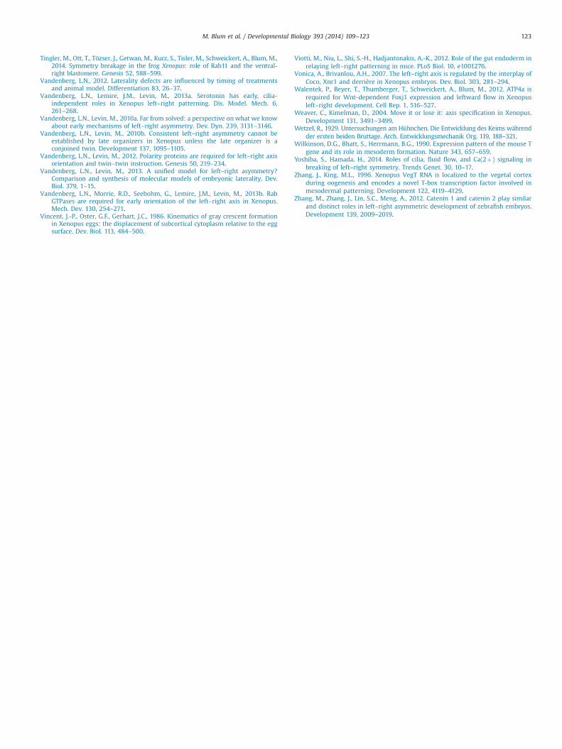
Tingler, M., Ott, T., Tözser, J., Getwan, M., Kurz, S., Tisler, M., Schweickert, A., Blum, M.,2014. Symmetry breakage in the frog Xenopus: role of Rab11 and the ventral-right blastomere. Genesis 52, 588–599.
Vandenberg, L.N., 2012. Laterality defects are influenced by timing of treatmentsand animal model. Differentiation 83, 26–37.
Vandenberg, L.N., Lemire, J.M., Levin, M., 2013a. Serotonin has early, cilia-independent roles in Xenopus left–right patterning. Dis. Model. Mech. 6,261–268.
Vandenberg, L.N., Levin, M., 2010a. Far from solved: a perspective on what we knowabout early mechanisms of left–right asymmetry. Dev. Dyn. 239, 3131–3146.
Vandenberg, L.N., Levin, M., 2010b. Consistent left–right asymmetry cannot beestablished by late organizers in Xenopus unless the late organizer is aconjoined twin. Development 137, 1095–1105.
Vandenberg, L.N., Levin, M., 2012. Polarity proteins are required for left–right axisorientation and twin–twin instruction. Genesis 50, 219–234.
Vandenberg, L.N., Levin, M., 2013. A unified model for left–right asymmetry?Comparison and synthesis of molecular models of embryonic laterality. Dev.Biol. 379, 1–15.
Vandenberg, L.N., Morrie, R.D., Seebohm, G., Lemire, J.M., Levin, M., 2013b. RabGTPases are required for early orientation of the left–right axis in Xenopus.Mech. Dev. 130, 254–271.
Vincent, J.-P., Oster, G.F., Gerhart, J.C., 1986. Kinematics of gray crescent formationin Xenopus eggs: the displacement of subcortical cytoplasm relative to the eggsurface. Dev. Biol. 113, 484–500.
Viotti, M., Niu, L., Shi, S.-H., Hadjantonakis, A.-K., 2012. Role of the gut endoderm inrelaying left–right patterning in mice. PLoS Biol. 10, e1001276.
Vonica, A., Brivanlou, A.H., 2007. The left–right axis is regulated by the interplay ofCoco, Xnr1 and derrière in Xenopus embryos. Dev. Biol. 303, 281–294.
Walentek, P., Beyer, T., Thumberger, T., Schweickert, A., Blum, M., 2012. ATP4a isrequired for Wnt-dependent Foxj1 expression and leftward flow in Xenopusleft–right development. Cell Rep. 1, 516–527.
Weaver, C., Kimelman, D., 2004. Move it or lose it: axis specification in Xenopus.Development 131, 3491–3499.
Wetzel, R., 1929. Untersuchungen am Hühnchen. Die Entwicklung des Keims währendder ersten beiden Bruttage. Arch. Entwicklungsmechanik Org. 119, 188–321.
Wilkinson, D.G., Bhatt, S., Herrmann, B.G., 1990. Expression pattern of the mouse Tgene and its role in mesoderm formation. Nature 343, 657–659.
Yoshiba, S., Hamada, H., 2014. Roles of cilia, fluid flow, and Ca(2þ) signaling inbreaking of left–right symmetry. Trends Genet. 30, 10–17.
Zhang, J., King, M.L., 1996. Xenopus VegT RNA is localized to the vegetal cortexduring oogenesis and encodes a novel T-box transcription factor involved inmesodermal patterning. Development 122, 4119–4129.
Zhang, M., Zhang, J., Lin, S.C., Meng, A., 2012. Catenin 1 and catenin 2 play similarand distinct roles in left–right asymmetric development of zebrafish embryos.Development 139, 2009–2019.
M. Blum et al. / Developmental Biology 393 (2014) 109–123 123