Embed Size (px)
Citation preview

MICROBIAL DIVERSITY 1996
ISOLATION AND CHARACTERIZATION OF
SYMBIOTIC BACTERIA FROM THE HERMIT CRAB
Rachel Greedy Stefan RateringDept. Microbiol. & Immunol. Max-Planck Institute fuerUniversity of Leicester Terrestrische MikrobiologieUniversity Road Karl Frisch Str.Leicester LE1 9HN 35043 Marburg
U.K. Germanye-mail: rsg 1 @ le.ac.uk ratering @ mailer.uni-marburg.de
INTRODUCTION
In terrestrial ecosystems symbiotic relationships, between bacteria and vertebrate or invertebrate
animals, have been well studied. Examples are the gut symbionts of ruminants and termites. The
presence of symbionts in the guts of some aquatic invertebrates has been known of for a long time,
for example the presence of Cristispira in the crystalline style of Saxidames gigantes (Berkeley
1959) and the cellulose-degrading bacteria in the gut of Teredo or shipworm (Hidaka 1954).
Endosymbiotic relationships in corals and in deep sea invertebrates have also been well studied
(Conway et al 1989, Herry et al 1989).
However, very little attention has been paid to the possible symbiotic relationships between aquaticinvertebrates and the bacteria present in the gut. Harris (1993) suggests that gut bacteria maycontribute significantly to nutrient gain by aquatic hosts. Some organisms have been shown tomaintain a permanent and consistent microbiota in the gut, which is significantly different from thatof the surroundings. These organisms include the giant prawn Macrobrachium rosenbergi (Colorni1985), the polychaete Thiepus setosus (Duchene et at 1988), prawns Upogenbia africana andCallinessa kraussi (Harris et al 1991), halothuroid Psychropoles sp. (Denning et al 1981), deep seaamphipod Lyssianssidae hirondella (Schwartz et at 1976) and sea urchin Echinus exilentus(Unkles 1977). In contrast another study showed an absence of bacteria in the gut of marine,wood-boring crustacea (Boyle & Mitchell 1978). Two students on the Microbial diversity Cousrein 1993 attempted to isolate bacteria from spider crabs and marine isopods but were unsuccessful.In many other studies there is no indication to suggest the bacteria isolated from the gut areanything other than ingested, transient bacteria ( Harris 1993),, Nagasawa & Nemoto (1988)suggest that bacteria may be an important food source for some marine invertebrates . The gutmicrobiota of Crustacea has not been studied in detail, but the most commonly isolated bacterialgenera are Vibrio, Pseudomonas, Flavobacterium, Aeromonas, Micrococcus, and Staphlycoccus(Harris 1993).
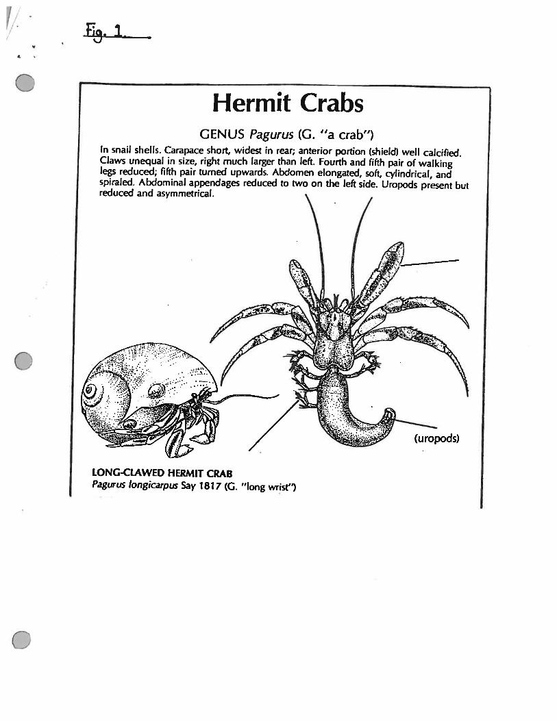
Hermit Crabs (Family: Paguridae) are a large group of Crustacea. They are a quite separate groupfrom the true crabs or Brachyura. Hermit Crabs have a hard cephalothorax and a soft twisted,abdomen which is hidden within a discarded, snail shell (See Fig. 1) They are omnivorousdetrivores and scavengers (Bliss 1982). The gut of a hermit crab is very different from that of atrue crab or lobster. Typically a crab or lobster has a very short anterior gut leading into thestomach. From the stomach the food particles then pass into the hepato-pancreas. Undigested foodis compacted in a short intestine and then passed out of the anus (Berrick 1986). However in theHermit crab the anterior gut leading to the stomach and hepatopancreas is proportionally muchlonger. (Bullis pers. comm.). This may suggest that some pre-digestion of ingested food involving
-
Hermit CrabsGENUS Pagurus (G. “a crab”)
In snail shells. Carapace short, widest in rear; anterior portion (shield) well calcified.Claws unequal in size, right much larger than left. Fourth and fifth pair of walkinglegs reduced; fifth pair turned upwards. Abdomen elongated, soft, cylindrical, andspiraled. Abdominal appendages reduced to two on the left side. Uropods present butreduced and asymmetrical.
LONG-CLAWED HERMIT CRABPagurus longicarpus Say 1817 (G. “long wrist”)
(uropods)
a resident bacterial population could be occurring. To our knowledge there has been no previous
study of the microbiota of the gut of the Hermit Crab.
The aims of this project are isolate and characterize bacteria from the different regions of the Hermit
Crab gut. Bacteria will also be isolated from the water and sediment in the tank, where the hermit
crabs are maintained, in an attempt to ascertain whether bacteria in the Hermit crab gut are resident
or merely ingested from the environment.
MATERIAL AND METHODS
1. Isolation of Bacteria
A Hermit Crab was obtained from the Marine Resurces Centre, MBL. The crab was anesthetised
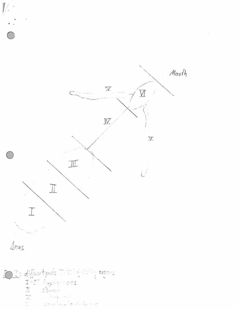
for 2 hours at -20 °C prior to dissection. The hermit crab digestive tract was removed and dividedinto five part (Fig 2). Each part was transferred into sterile seawater (1 ml) and homogenized.Homogenate was examined using phase contrast microscopy, DAPI was then added to samplesand examined using flouresence microscopy. F420 and chlorophyll flouresence was also lookedfor. 100 ul of homogenate was spread onto plates of three different media (SWC, CAA SW, BHISW -Appendix 1). Further more sediment and sand from the bottom of the aquarium and scrapingsfrom the Hermit Crab shell suspended in sterile sea water were spread directly onto plates (SWC,BHI SW, CAA SW, YEG SW). 10 ml aliquots of sea water from the Hermit Crab Aquarium werefiltered on to 0.2um filters. The filters were then placed onto plates of SWC and YEG-SWC
Incubation conditions were aerobic and anaerobic at 20°C.
After growth the different colonies were picked and transferred on fresh plates of the same media.To get pure cultures the isolates were streaked several times. The morphology of the bacteria andpurity of the cultures were examined with the microscope.
4
///
r
—
4
2A /4
Vc
-
Mi
NLi
U
N
-N
2. Phenotypic Tests
Oxidase test
One colony of each isolate was picked and streaked on a filter paper. To show the oxidase activityone drop of tetramethyl p-phenylenediamine (Difco laboratories) was dropped on the colony.Development of a blue colouration indicates oxidase activity.
Luminescence
All isolates of the SWC plates were stored in the dark and examined for luminescence.
Fluorescence-
All isolates were stored in the dark and irradiated with short wave UV light to look for flouresence.
Growth on Different MediaPure cultures were spread on to plates of the different media descibed in Appendix 1.
Growth Temperature
Growth at 4, 15, 20, 30, 37 and 42°C was investigated.
Requirement of Salt for GrowthThe requirement for salt was tested by comparing growth of isolates on BHI made with sea waterto growth on media made with distelled water.
Hydrolysis of Starch and Milk ProteinThis was investigated by spreading isolates on Skim Milk plates and BHI + Starch plates(Appendix 1) and looking for zones of clearing around the colonies.
Aerobic/Anaerobic GrowthIsolates obtained aerobically were tested for their ability to grow anerobically, and likewise thoseisolated anaerobically were tested for their ability to grow aerobically.
Production of Acid or Alkali Under Anaerobic GrowthProduction of acid or alkali under anaerobic growth was tested by placing pH paper next to thecolonies on plates.
Characterisation of Pigments
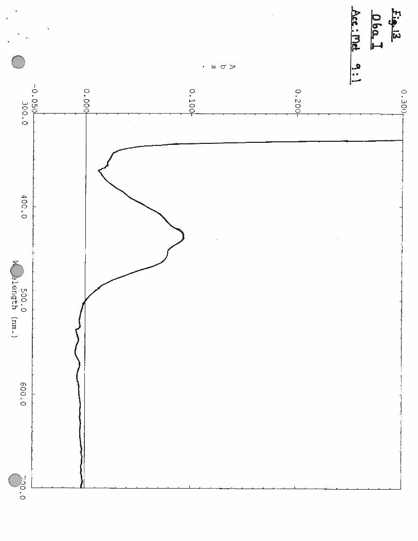
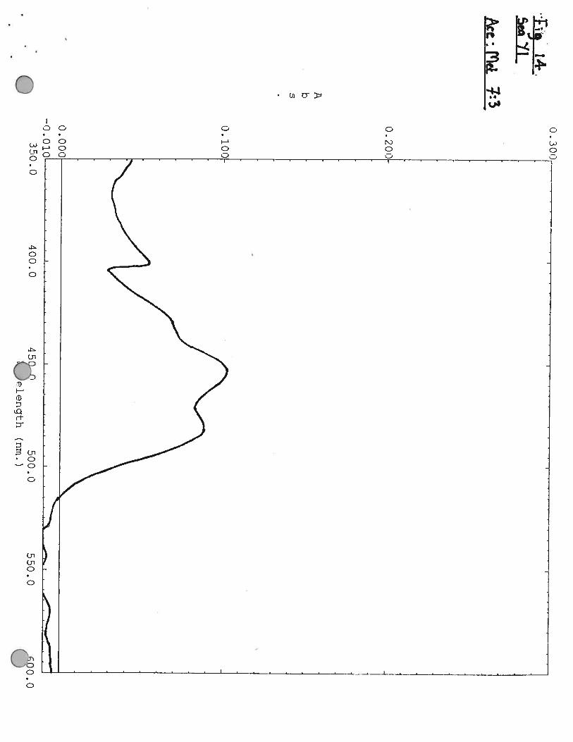
Pigments were extrated from cells by mixing a small amount of cells with ethanol: methanol (70:30 V:V). A spectra was then made of the pigment.
3. Genotypic Tests
16S rRNA in situ hybridization
1 or 2 colonies were suspended in 1 ml of PBS. After centrifugation the pellets were washed twotimes with ice cold PBS and resuspended in 1 ml PBS. 15 ul from a dilution (1: 100) of the cells
was applied to subbed slides (0,1 % gelantine, 0,01 % CrK(S04)212 H20) and dried in 37 °C
incubator.
The slides were treaded with ethanol and formaldehyde (90/10 v/v) for 5 mm. to fix the cells and
than washed two 2 times for 2 mm. with H20 in a coplin jar. After drying at 37 °C 40 ul of the
probe (340 nglul) in hybridization mix was dropped on the cells and hybridized overnight at 37
C. At the next morning the slides were washed three times in 1 X SET at 37 ° C and dried
vertically in a dark place. On each spot a tiny drop of mounting fluid was added and thecoversliped was placed on the slide.
Solutions
lOX SET: l.5MNaC1
200 mM Tris Cl pH 7.8
10 mM EDTA
hybridization mix: 400 ul 2 % BSA - 1 g/ 50 ml
400 ul 0.1 % PAA - 0.1 g/ 100 sterile water
2.0 ml 10 X SET
1.2 ml Dextran sulfate
probes: Proteobacteria (Enteric, Alphas, Betas, Gammas),Flavobacteria
Universal probe.
PCR AND SEQUENCING OF 16S rRNA GENE
Several different approaches were investigated for lysis of cells to release DNA:
(1) 1 or 2 colonies were suspended in 50 ul of TE buffer and 17.5 ul of GeneReleaser was added.
Cells were vortexed for 20 sec followed by centrifugation for 20 sec.and then 20 ul of mineral oil
was overlaid in the tubes. Tubes were heated in the microwave for 5 mm
(2) As above except tubes were heated in the thermocycler as descibed in the GeneReleaser
protocol.
(3) Cells were suspended in TE buffer as above. Proteinase K was added to the tubes to give a
concentration of 200ug/ml. Tubes were heated at 55°C for 2 hours, followed by heating to 95°C
for 10 minutes.
(4) Cells were suspended in TE buffer as above and Lysozyme was added to a concentration of 0.5
mg/mi. Tubes were heated for 1 minute at 95°C.
(5) Cells were added directly to the PCR master-mix ( see below). Tubes were then heated at 94°C
for 10 minutes directly prior to addition of Taq. polymerase.
(6) Cells were suspended in lOul of TE buffer and 25 ul of GeneReleaser. Tubes were then heated
in the thermocycler as described in the GeneReleaser protocol.
PCR was then carried out as follows:
Eppendorf tubes with 25 ul of master mix 1 were placed in the Temp cycler and Ampliwax beads
(2-3) were added. After the Temp cycler has headed to 80 ° C and than cooled to 35°C, 54u1 of
master mix 2 and 3 ul of DNA was added. A negative control with 3 ul H20 was also carried out.

The following program of the Thermo-cycler was used. 35 cycles were carried out:
95 0 C for 45 sec (denaturing step)
50 ° C for 50 sec (annealing step)
72 ° C for 2 mm. (elongation step)
After the end of the 35 cycles the temperaturec was held at 4 C. 5 ul of the sample was run on
0.8% agarose ethidiumbromide gel for approximately 1 h at 100 mV to check the PCR product.
Solutions
master mix 1: 2.5 ul lox PCR buffer
10 ul MgC12
2.5 ul H20
2.0 ul dATP, dCTP, dTTP, dGTP
1.0 iii forward primer (universal)
1.0 ul reverse primer (universal)
master mix 2: 5.7 ul iox PCR buffer
0.5 ul Taq enzyme
47.8 ul H20
The PCR products were cleaned using Wizard kit. The amount of DNA was measured using theDNA Dipstick kit. DNA was then diluted if neccessary to a concentration of 50 ng/ul. This wasthen used for sequencing.
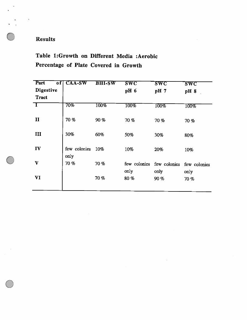
Results
Table 1:Growth on Different Media :Aerobic
Percentage of Plate Covered in Growth
Part of CAA-SW BHI-SW SWC SWC SWCDigestive pH 6 pH 7 pH 8Tract
I 70% 100% 100% 100% 100%
II 70 % 90 % 70 % 70 % 70 %
III 30% 60% 50% 30% 80%
IV few colonies 10% 10% 20% 10%only
V 70 % 70 % few colonies few colonies few coloniesonly only only
VI 70% 80% 90% 70%
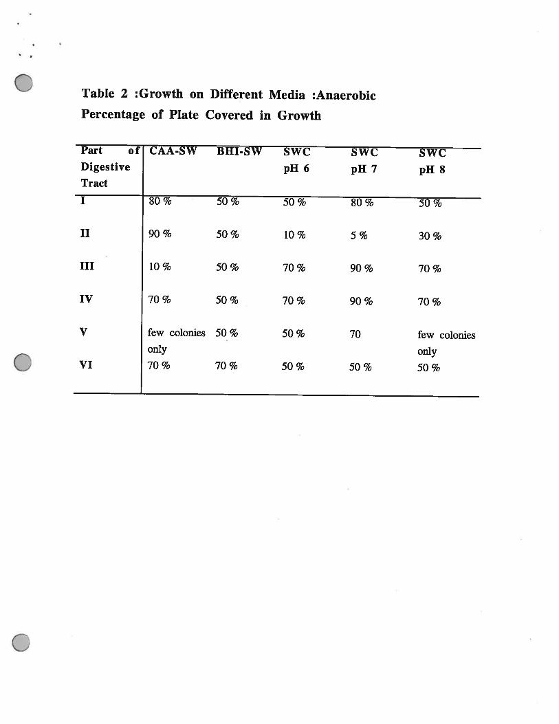
Table 2 :Growth on Different Media :Anaerobic
Percentage of Plate Covered in Growth
Part of CAA-SW BHI-SW SWC SWC SWCDigestive pH 6 pH 7 pH 8Tract
I 80% 50% 50% 80% 50%
II 90% 50% 10% 5% 30%
III 10 % 50 % 70 % 90 % 70 %
IV 70 % 50 % 70 % 90 % 70 %
V few colonies 50 % 50 % 70 few coloniesonly only
VI 70% 70% 50% 50% 50%
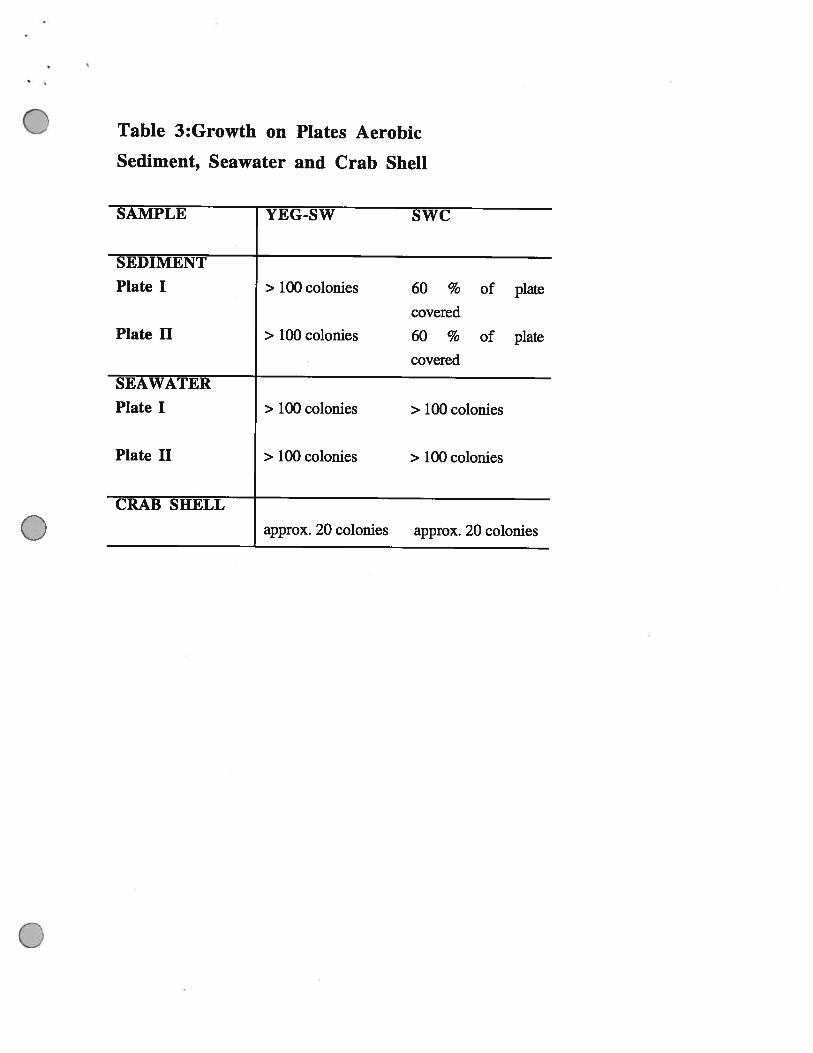
Table 3:Growth on Plates Aerobic
Sediment, Seawater and Crab Shell
SAMPLE YEG-SW SWC
SEDIMENT
Plate I > 100 colonies 60 % of plate
coveredPlate II > 100 colonies 60 % of plate
covered
SEAWATER
Plate I > 100 colonies > 100 colonies
Plate II > 100 colonies > 100 colonies
CRAB SHELL
approx. 20 colonies approx. 20 colonies

Tab. 4 and 5: Isolates part I
isolate pigment colony T ° c bacteria Motility An- Oxidase acidshape 1520 30 3742 shape aerobic production
BHI N small ? + ? ? ? rods no + i negative pH 5AN 2 circular — — — — — spores? —
BHI A B circular - + -‘ - - short + ? + positiveraised rods
BHI B Y circular - + -i -1 - short + ? + positiveraised rods
CAA PY flat - + - - - long + + + positive1.1 rodsCAA YO flat ? 1- i i ? ? + F positive1.2CAA 2a YO flat = i- = = small ? T positive
rodsCAA 2b N flat - + - - - small ? + positive
rodsow6a N circular ? + -i -i - rods no + negative
raisedr8an N flat ‘? + -, ? ? rods + ± positive not7an N flat 7 ± - 7 7 rods + ± positive noo6a YO circular ? + - - - rods no + positive
. raisedw8a N circular ? + - -I - small no + positive
raised rods
isolate Colony CAA YEG SWC BHI SW BHI skim-color SW SW milk
BHIAN2 N + ? ? + no noBHJA WB no ? no + no noBUlB CP no ? + + no noCAA1.1 CS ? no no no noCAA1.2 CY + ? no no no noCAA2a T + ? ? + no noCAA2b TD + ? + no no noow6a W + + + + + +r8an W ? ? + + ? 7t7an WT 7 7 + + ? ?o6a 0 + + + + no now8a W + + + + + no

Tab. 6: Isolates part II
isolate pigment colony T ° C bacteria Motility An- ?5 Oxidase acidshape 1520 30 3742 shape aerobic production
BHI C B regular H + - - - short no ± positiveflat rods
CAA YO flat - + - - - small no no + positv4.0 —— - —— rods —
CAA PY rand - + - - - small no no + positive4.1 slights rods
raisedBHI N ‘? ! ? small + + iE positive noAn3 —— - —— rods —
o8a N circular ? + - -I - rods no + positiveraised
g7a N flat ? ± - - rods ? no ± positivewt6a N circular ? +
-
- rods no ± positivew8a circular 7 + - rods no ± positivet6a N circular 7 + - - rods 7 no ± positivew7a N circular 7 + - - rods 7 no ± positivet8a N flat 7 + - - rods 7 no ± positivew6a N circular 7 + - H - rods ? no + negative
raisedw8an N flat i- ‘?
7rods + F positive no
t7an N flat 7 + - 7 ‘? cocci ? + ± positive not6an N flat 7 ± - 7 7 rods +0 ± positive nowg6an N circular 7 ± - 7 7 rods 7 + ± positive now6an N circular 7 + - ? 7 rods 7 + + positive no
raised
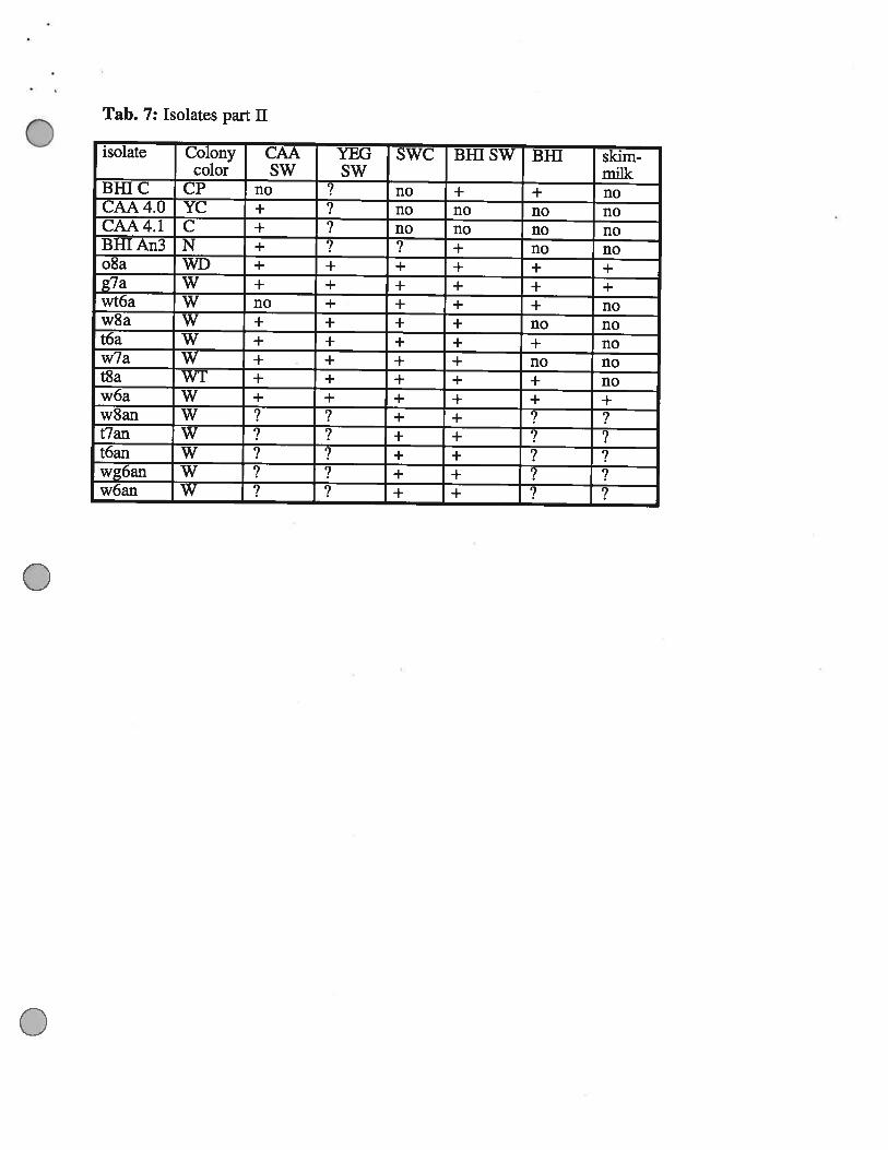
Tab. 7: Isolates part II
isolate Colony CAA YEG SWC Bifi SW BHI skim-color SW SW milk
BBIC CP no ?____ no + + noCAA4.O YC + no no no noCAA4.l C ÷ no no no noBR[An3 N + + no noo8a WD ÷ + + + + +g7a W + + + + + +wt6a W no + + + + now8a W + + + + no not6a W + + + + + now7a W + + + + no not8a WT ÷ + + + + now6a W + + + + + +w8an W + + ?t7an W + + ?t6an W + +wg6an W + + ?w6an W + +
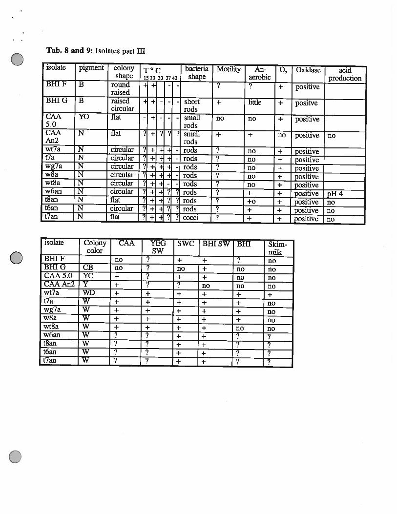
Tab. 8 and 9: Isolates part ifi
isolate pigment colony T ° c bacteria Motility An- Oxidase acidshape 1520 30 3742 shape aerobic production
BHI F B round - + -- ? T positive
raisedBHI G B raised - + - - - short + little + positve
circular rodsCAA YO flat - + - - - small no no + positive5.0 rodsCAA N flat i i- ? ?‘? small + ÷ i positive noAn2 —— - —— rods —
wt7a N circular ? + - - rods no ± positivet7a N circular ? +- -
- rods no ± positivewg7a N circular ? + - - rods no ± positivew8a N circular ‘? ± -
rods no + positivewt8a N circular .Y ± = - rods no + positivew6an N circular ? ± ‘1 ? rods + ± positive pH 4t8an N flat ? ± ? 7 rods +0 + positive not6an N circular J ± 7 7 rods + + positive not7an N flat ? ÷ ? ? cocci ? + ÷ positive no
isolate Colony CAA YEG SWC Bill SW BHI Skim-color SW milk
BHIF no ? + + ? noBHIG CB no no + no noCAA5.0 YC + + + no noCAAAn2 Y + ? no no nowt7a WD + + + + + +t7a W + + + + + nowg7a W + + + + + now8a W + + + + + nowt8a W + + + + no now6an W ? + +t8an W + + ?t6an W + +t7an W ? + + ?
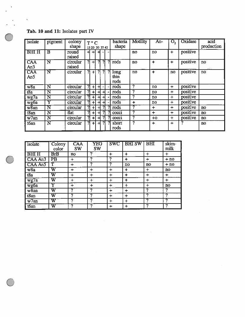
Tab. 10 and 11: Isolates part IV
isolate pigment colony T° c bacteria Motility An- • Oxidase acidshape 1520 30 3742 shape production
BHI H B round = + H - - no no + positiveraised
CAA N circular ‘? - ? ? ? rods no + positive noAn3 raisedCAA N circular ‘? - ? ? ? long no + i positive noAn5 thin
rodsw8a N circular ? + - - rods ? no ± positivet8a N circular Y ± -
- rods no ± positivewg7a N circular
-. ± -
- rods no ± positivewg6a Y circular 7 ÷ - rods + no ± positivew8an N circular 9 ± 2 ? rods + + positive not8an N flat ? + 7 7 cocci ? + ± positive now7an N circular ? ÷ ‘1 7 cocci +0 + positive not6an N circular ? + - ? ? short ? + + ? no
rods
isolate Colony CAA YEG SWC Bill SW BHI skim-color SW SW milk
BHIH BrB no ? + + + +
CAAAn3 PB + ? ? + + +noCAAAn5 T + ? no no +now8a W + + + + + not8a W + + + + + +
wg7a W + + + + + +
wg6a Y + + + + + now8an W + + ?t8an W + + ?w7an W + + ? ?t6an W ? + + ?
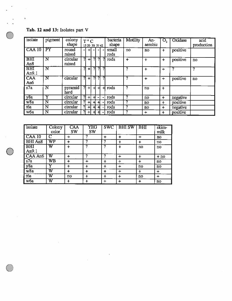
Tab. 12 and 13: Isolates part V
isolate pigment colony T ° c bacteria Motility An- • Oxidase acidshape 1520 30 3742 shape aerobic production
CAA 10 PY round - + - - - small no no •E positiveraised rods
BHI N circular ? + ? ? ? rods + + + positive noAn8 raisedBHI N +An9. 1CAA N circular ! - ! i ‘i + positive noAn6s7a N pyramid i 1- rods ? no
hardy8a Y circular 3 ± - rods ? no ± negativew8a N circular ? ± - rods no + positivet6a N circular ? ± - H - rods no ± negativew6a N circular ? ± - - rods + + positive
isolate Colony CAA YEG SWC Bill SW BHI skim-color SW SW milk
CAA1O C + + + + noBHIAn8 WP + + + noBHI W + + no noAn9.lCAAAn6 W + ? + + +nos7a WB + + + + + noy8a Y + + + + no now8a W + + + + + +t6a W no + + + no +w6a W + + + + + no
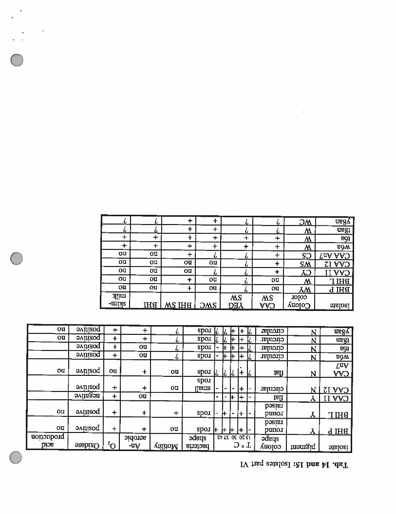
Tab.14and15:IsolatespartVI
isolatepigmentcolonyT°cbacteriaMotilityAn-Oxidaseacid
shape20303742shapeaerobicproductionBillPYround=+-‘rodsno+Epositiveno
raisedBillLYround-+--I-rods+++positiveno
raisedCAA11Yflat---no±negativeCAA12Ncircular-+---smallno++positive
rodsCAANflat?I-?!?rodsno+iEpositivenoAn7w6aNcircular!i-ErodsnoEpositivet6aNcircular?÷-rodsno±positivet8anNcircular7÷-7‘1rods7+±positivenoy8anNcircular?+-77rods7+±positiveno
0
0
0
isolateColonyCAAYEGSWCBillSWBHIskim-colorSWSWmilk
BHIPWYno?no+nonoBHILWno?no+nonoCAA11CY+7nononoCAA12WS+?nonononoCAAAn7CS+77+nonow6aW++++++t6aW++++++t8anW7++y8anWC7?++?7
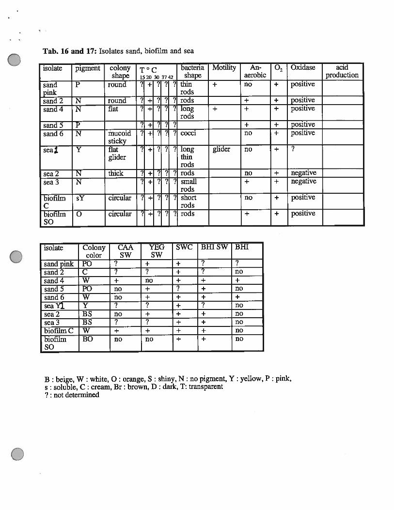
Tab. 16 and 17: Isolates sand, biofilm and sea
isolate pigment colony T ° c bacteria Motility An- Oxidase acidshape 1520 30 3742 shape aerobic production
sand P round ? + ? 7 ? thin + no + positivepink —— - rods —sand 2 N round 7 -i- ? ? ‘? rods + ± positivesand 4 N flat 7 + 7 ? 7 long + + + positive
rodssand 5 P
7? ? ? + T positive
sand 6 N mucoid 7 + 7 ? 7 cocci no + positivesticky
seal Y flat 7 + 7 ? 7 long glider no + 7glider thin
rodssea 2 N thick ? i- ? ?
7rods no T negative
sea3 N 7 + 7 ? ? small + + negativerods
biofilm sY circular ? ? ? ? short no T positiveC rodsbiofilm 0 circular ? ? ‘? rods + T positiveSo
isolate Colony CAA YEG SWC Bill SW BHTcolor SW SW
sandpink P0 ? + + ? ?sand2 C 7 7 + 7 nosand4 W + no + + +
sand5 P0 no + 7 + nosand6 W no + + + +
seaY]. Y ? 7 + 7 nosea2 BS no + + + nosea3 BS 7 7 + + nobiofthnC W + ÷ + + nobiofilm BO no no + + noSo
B : beige, W : white, 0: orange, S : shiny, N: no pigment, Y: yellow, P: pink,s: soluble, C: cream, Br: brown, D: dark, T: transparent7 : not determined
Tab. 18: In situ hybridisation
Isolate Universal Enteric Prote x Proteo 3 Proteo ft( Flavo
BHI H yes no no no n. d. n. d.sand 5 yes no no no n. d. n. d.CAA An3 yes no no no n. d. n. d.Bill An2 yes yes no no n. d. n. d.w3a, ifi yes no n. d. n. d. no noo6a, I yes no n. d. n. d. no nos7a, V yes no n. d. n. d. no noBHI G yes no n. d. n. d. no noBHI L yes no n. d. n. d. no noCAA4.l yes no n. d. n. d. no noCAA 2b yes no n. d. n. d. no noBHI SO yes no n. d. n. d. no no
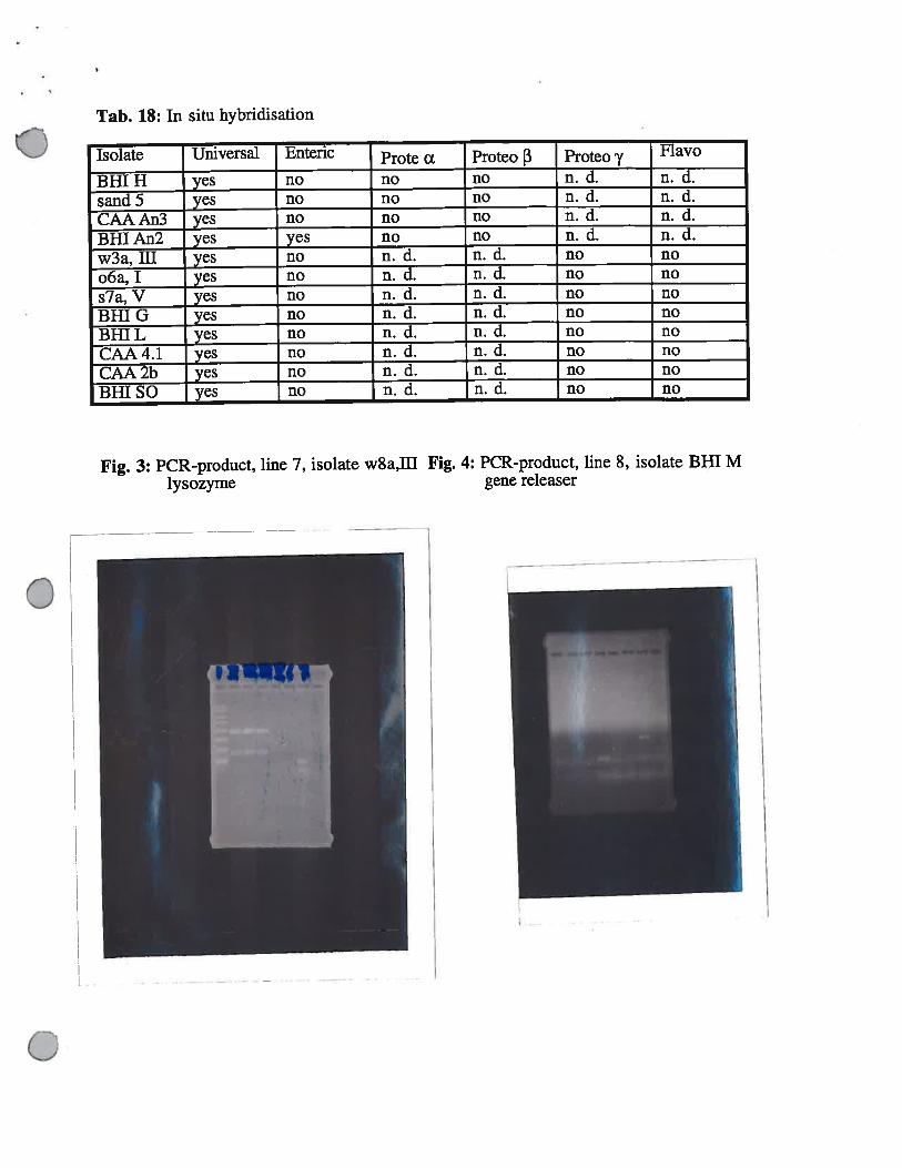
Fig. 3: PCR-product, line 7, isolate w8a,ffllysozyme
Fig. 4: PCR-product, line 8, isolate BHI Mgene releaser
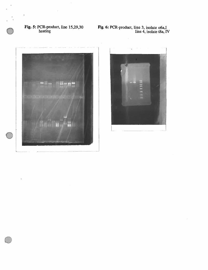
Fig. 5: PCR-product, line 15,29,30heating
Fig. 6: PCR-product, line 3, isolate o6a,Iline 4, isolate t8a, TV
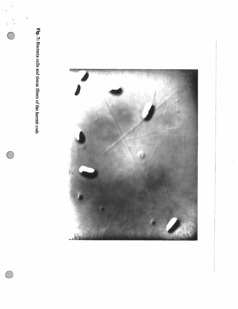
Fig.7:
Bacteria
cellsand
tissuefibers
ofthe
hermitcrab
U0
S%
e•
——
I.•
•%S
o—
0.
dIP
::_•P-,.
*_
.-.,
._•
.•.•
.•_
:.4:0
0’.
j•i;
•
“4
Fig.
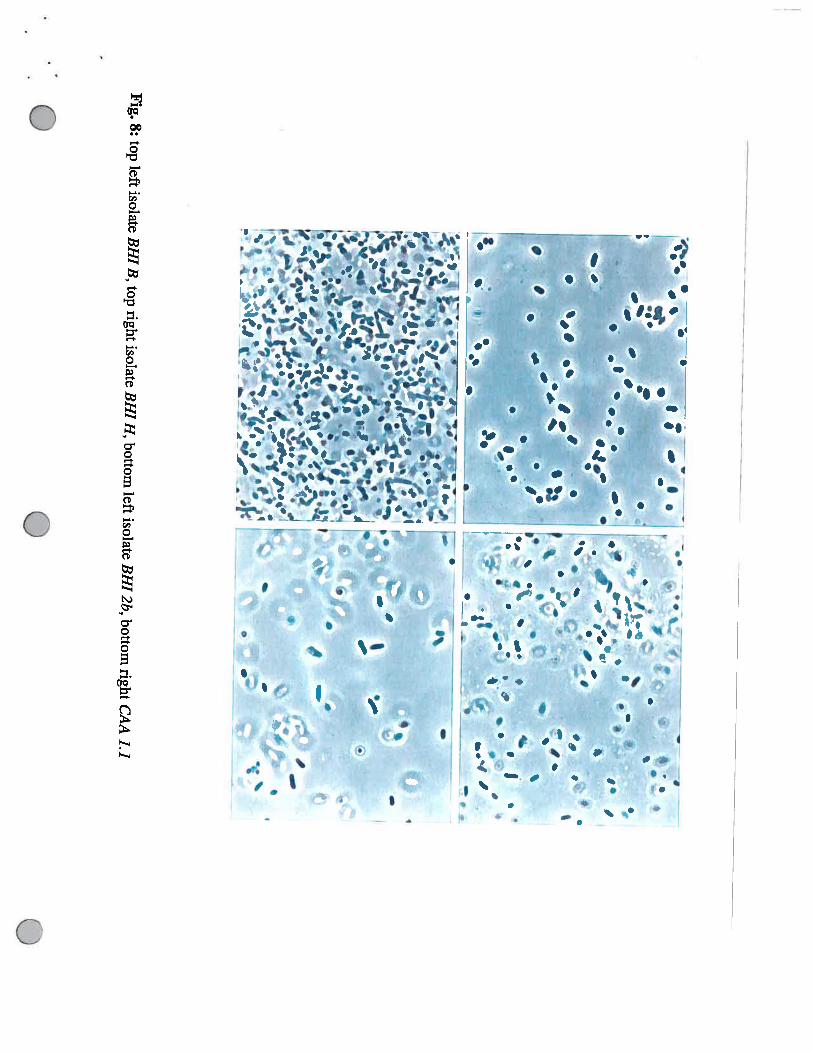
8:top
leftisolate
BH
IB
,top
rightisolate
BH
IH
,bottom
leftisolate
BH
I2b,
bottomright
CA
A1.1
0
—
‘..-‘-,.
1-•_
F‘‘
------—
---—
—.a
&-a
•II
C
I4
••
—I.
I
I
4II
S
•4
.,,
•.
.4-
I’.
a.
UU
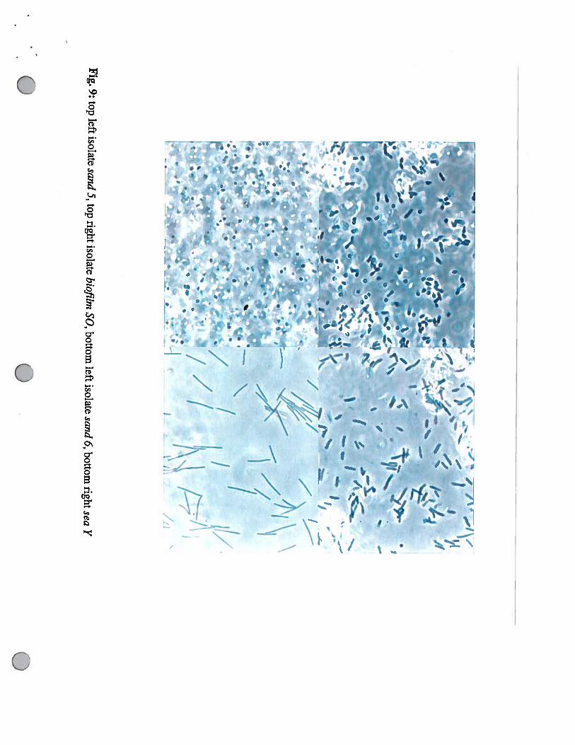
Fig.
9:top
leftisolatesand
5,toprightisolate
bioflimSO
,bottomleft
isolatesand
6,bottomrightsea
Y
U
0
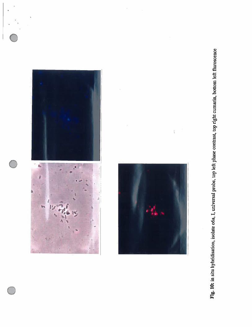
Fig
.10
:in
situ
hybr
idis
atio
n,is
olat
eo6
a,I,
univ
ersa
lpr
obe,
top
left
phas
eco
ntra
st, t
opri
ghtc
umar
in, b
otto
mle
ftfl
uros
cenc
e
dl,p
0
, 0’—1’
—.
0
IS.,
p.’
4
F,.,.’
I
‘1
)
II—’ I
a,
‘4
0
‘1’
p V
. —I
I
-I0 —
•
U
—
w--—
—.
II
a
.iw
I“a
S
S
‘II
I.
aS.
.
I
•
‘
‘1I0’
‘I
—b.
S
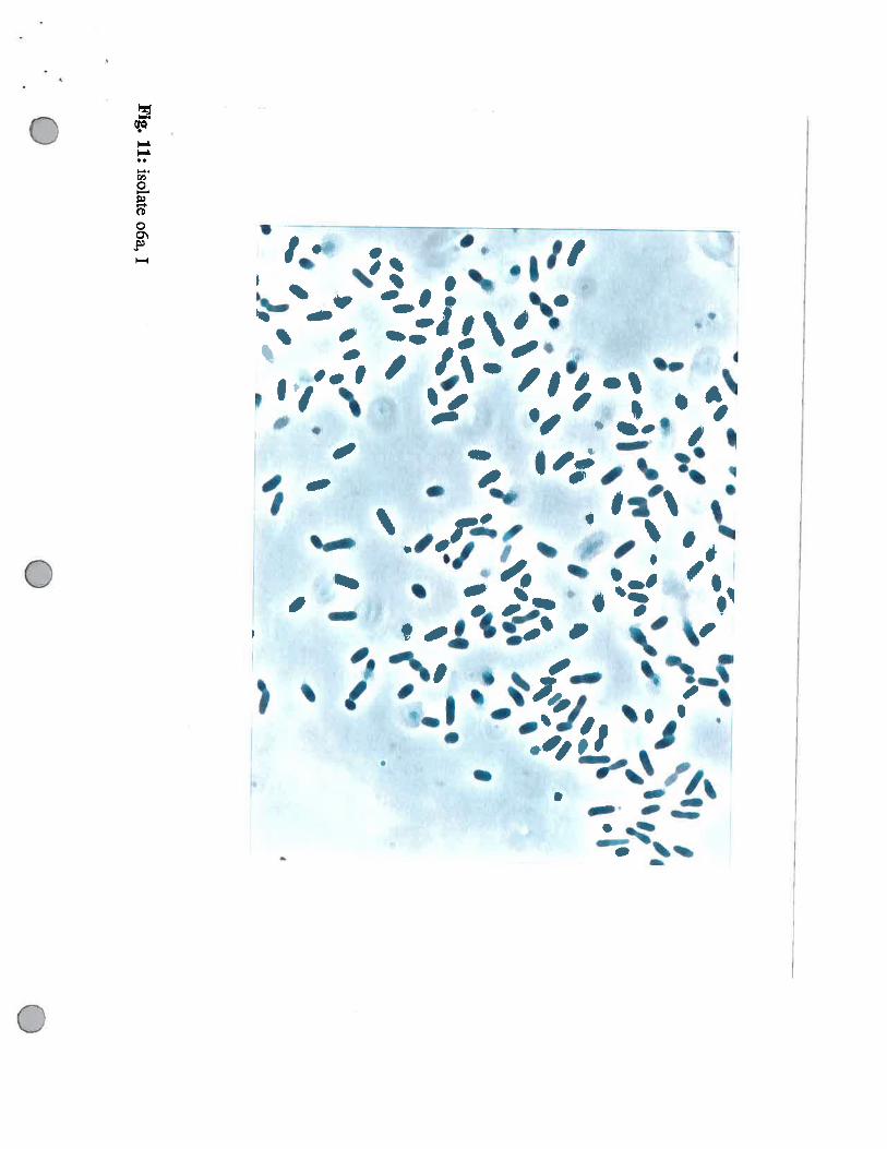
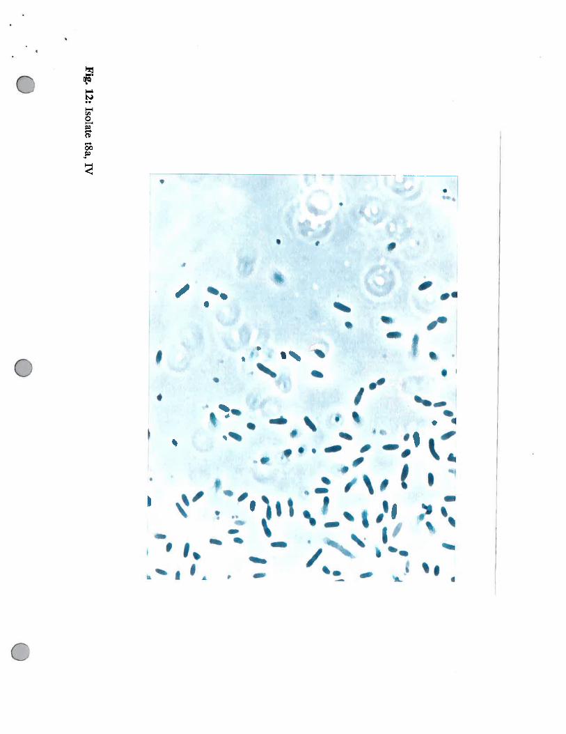
Fig.
12:Isolate
t8a,IV
U
50
0.0
tQ1
en
gth
(nm.
0.
3Qt
Ace:rTe
0.2
AhS
0.1
0.
—0
3C’0
.04
00
.06
00.0
o.o
Q0•0
99
(uxu)
o.oo
qU—
o900
0fi
•0—
-o
0 S V
03•0
ct
‘tI
0
(% Difference)
Wbrio fisher/i --MBL isolate #40
Vibrio fisher/i
Vibrio aestuarianus
Vibrio cincinnatiensis
Photobacterium phosphoreum
Aeromonas sobria
Aeromonas veronll
Escherich/a coli
Proteus vulgar/s
Vibrio harvey/i
Vibrio carchiariea
Vibrio campbelli
Wbrio a/ginolyticus
0 1 0’ 2 3 4 5
10 20 30 40 50 60 70
5 5’ 6 7 8
80 90 100 110 120 130 140 150
8 8’ 9 9’ 10 10
UEJCG CAAAGGA-UUCG-CCU
160 170 180 190 200 210
7’ 11 11’ 12
220 230 240 250 260 270 280 290
12’ 6’ 13 13’ 14 14’ 4’ 15
300 310 320 330 340 350 360 370
15 15’ 3’ 16 16’ 17
-
390 400 410 420 430 440 450
17 17’ 18 18’I I
II xxx vv xxxv’UGUUGUAUTThUAGUGCAGCAUUUGACGUUACCUACAGAAAWCACCGGCUAACUCCGU CCACAGCCCGGçAA460 470 480 490 500 510 520 530
18’ 2’ P18 19 P19
540 550 560 570 580 590 600 610
P19’ 20I I I 00 0—0GGGCUCAACCUCGGAACCGCAUEJUGAAACUGGUGAACUAGAGUGCUGUAGAGGGGGGUAGAAUUUCAGGUGUAGC UGA
620 630 640 650 660 670 680 690
20’ 19I 0—0= 0—ZZ ZZO 00— = I IAAUGCGUAGAGAUCUGAAGGAAUACCAGUGGCGAAGGCGGCCCCCUGGACAGACAC G CACUCAGA-UGCGAAAGCGUG
700 710 720 730 740 750 76 0 770

cf4p1 21 21’ P21-i 11P21-2 P21
- - CtJtTGA- - GGAG780 790 800 810 820 830 840 8
nP21-3 P21-3’ P21-i’ P18’ 22 22’ 1’ 23
860 870 880 890 900 910 920 9
24 25 26 26’ 27 28= I I Ci I I ii i I 01
AGAA-UU940 950 960 970 980 990 1000
28 28’ 28’ 29 30 31 31
CCUUC& -JCUCUG-
1010 1020 1030 1040 1050 1060 1070 1080
32 32’ 30’ 33 34 34!
- GUAAUG -
1090 1100 1110 1120 1130 1140 1150
35 35’ 33’ 29’ 27’ 25’I I 11 I II =-=—---- I I 0CçGGUGAuAAcçG - CACACGGC ACAA
1170 1180 1190 1200 1210 1220 1230 12
36 36’ 37
40 1250 1260 1270 1280 1290 1300 1310 13
37’ 24’ 38 38’ 23’00O01 I I 0 Ci ICUçGACUCCAUGAGUCGGAUçGCUAGU UçGUG UCAG AUGCUACGGGAAUACGUUçCCGGGCCUUUACACA
1330 1340 1350 1360 1370 1380 1390
39 39’0 EFO—-----000CCçCCGUCACACçAUGGGAGUGGCUGCAAANAAGUGNGNAgNUUAACc-uuçG- GNGG
1400 1410 1420 1430 1440 1450 1460 1470
39’ 40 40’—°°=°-————-—--———=—-—°,-i 0 ii Ii ii
1480 1490 1500 1510 1520 1530 1540

Discussion
The aims of this project were to isolate and characterise bacteria from the Hermit crab gut. Previous
investigators have described the crustacean gut as sterile. (Boyle & Mitchell 1978). Boyle and
Mitchell examined the digestive tract of two marine and one terrestrial wood- boring isopods and
one wood inhabiting amphipod using SEM and saw no evidence of any microbial life. Hughes
(Microbial Diversity 1993) examined the hind gut of two species of marine isopods
microscopically and revealed no visible microbiota. We also observed very little microbial life
microscopically.
However we have isolated a total of 83 strains of bacteria from the Hermit Crab gut. Ten strains
were also isolated either from the sea water of the tank, the sediment at the bottom of the tank and
the Hermit Crab shell. Each part of the crab gut was homogenized in 1 ml of sterile sea water and
then 100 ul of this was spread directly onto the various plates. Some plates were estimated to be
covered with up to 2000 colonies giving a figure of 20 000 bacteria per ml of homogenate. This
figure is low compared to numbers of bacteria found in the ruminant gut (1011 per ml) or the
termite hind gut but it may explain why we saw very little microscopically, especially considering
the large amounts of tissue and other gut contents present. Bacteria may also be attached to the wall
of the gut also making then more difficult to observe. In some microscopic examinations of
colonies we observed very narrow long fibres of what we presumed to be crab tissue. (Fig. 7)
Often the bacteria were attached to these fibres. Other plates had much lower numbers of colonies
on them.
We were interested to compare the numbers of bacteria isolated from each region of the crab gut.
The highest numbers were isolated from Parts I and II (Hepato-pancreas) and VI ( anterior gut).
The highest diversity was also isolated from parts I and II. Lowest numbers of bacteria were from
Parts ifi ( Anterior end of the Hepato-Pancreas) and 1V (Stomach). Interestingly although there

was very little growth of bacteria from Part IV aerobically there was much higher numbers
anaerobically. Unfortunately because we did not carry out dilutions, accurate counts of numbers
present in each part could not be carried out.
Using a variety of phenotypic tests we tried to characterize the isolates. It was very diificult to
group the organisms with the tests we used. We found that the majority of isolates were different
from each other. Significantly we found the isolates from the tank and from the surface of the
Hermit Crab shell were phenotypicafly very different from any strains isolated from the gut of the
Hermit Crab. This could support the idea that the Hermit Crab has a symbiotic population of
bacteria resident in the gut.
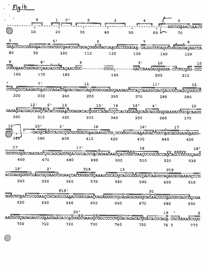
Using 16S rRNA sequencing, presumptive identitiesand relationships between bacteria isolated
can be made more easily. However we found that not one single method of cell lysis was suitable
for all our isolates. PCR products were obtained from only three of our isolates and only one of
these isolates was successfully sequenced. There has been much debate recently about the
advantages of genotypic versus phenotypic typing of organisms. It is agreed that the culture
methods used to isolate bacteria introduce a large amount of bias into the study, selecting only for
bacteria which can be cultured under a particular set of conditions. This study illustrates that the
protocol used for PCR also introduces bias into the study selecting for only the bacteria which can
be lysed using a particular protocol or whose DNA can be amplified using a particular primer. The
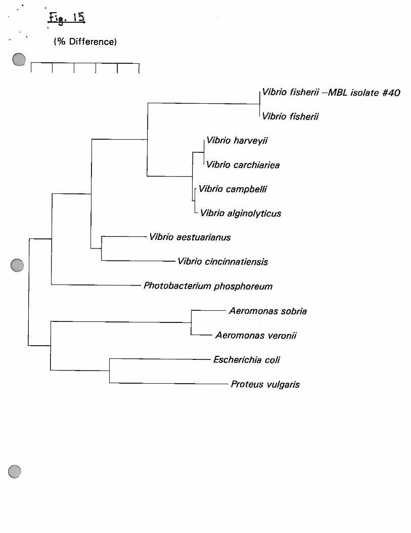
16S rRNA sequence was found to be idenlicle to that of Vibrio fisherii. This is a commonly
isolated from coastal and open ocean sea water, from the surfaces of squid and fish, from
luminous organs and from seafood. Many strains are bioluminescent and do not grow well at
37°C. However our isolate was not bioluminescent and it also grew at 37°C.
The Hermit Crab is a detrivore and scavenger so the diet would include protein, polysaccharide and
other complex carbohydrates. We have shown that bacteria isolated from the Hermit Crab gut can
hydrolyse protein and that they grow on complex media. This could suggest that they play a role in
the digestion of the Hermit Crab. Most isolates grew best between 15°C to 30°C and many
required seawater media. These isolates are therefore well adapted to the marine environment.
Before any conclusions can be drawn about whether any of these bacteria are indeed symbionts of
the Hermit Crab or are merely ingested with the food of the Hermit Crab many further
investigations need to be carried out. Further physiological studies of the bacteria need to be carried
out to look for enzyme activities which might be of benefit to the crab for example cellulose and
polysaccharide degradation. A more complete survey of the environment may reveal more
similarities between bacteria isolated from the Hermit Crab gut and those in the environment.
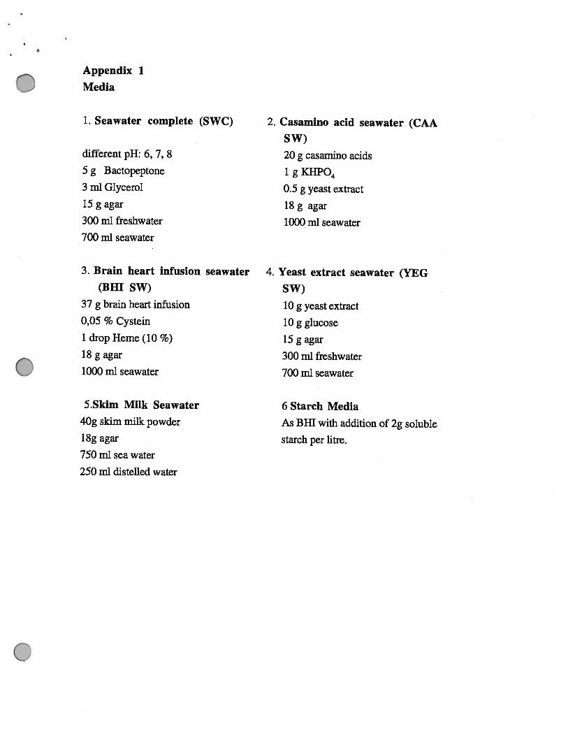
Appendix 1
Media
1. Seawater complete (SWC)
different pH: 6,7,8
5 g Bactopeptone
3 ml Glycerol
15 g agar
300 ml freshwater
700 ml seawater
3. Brain heart infusion seawater 4.(BHI SW)
37 g brain heart infusion
0,05 % Cystein
1 drop Heme (10 %)
l8gagar
1000 ml seawater
5.Skim Milk Seawater
40g skim milk powder
l8gagar
750 nil sea water
250 ml distelled water
2. Casamino acid seawater (CAA
SW)
20 g casamino acids
1gKHPO4
0.5 g yeast extract
18g agar
1000 ml seawater
Yeast extract seawater (YEG
SW)
10 g yeast extract
10 g glucose
l5gagar
300 ml freshwater
700 ml seawater
6 Starch Media
As BHI with addition of 2g soluble
starch per litre.
References
Berkeley C (1959): Some observations of Cristispira in the crystalline style ofSaxidomasgiganteus and in that of sone other Lameffibranchiata.
Can. 3. Zool. 37: 53 - 58-
Berrick S (1986): Crabs of Cape Cod
Cape Cod Museum of Natural History, Brewster, Massachusetts
Bliss DE (1982): Shrimps, Lobsters and Crabs.
New Century Publishers Inc.
Boyle PJ, Mitchell R (1978): The absence of microorganisms in the crustacean digestivetract.
Science 200:1157- 1159
Bullis R (1996): Personal Communication.Marine Resource Center, MBL
Colon A (1985): A study on the bacterial flora of giant prawn Macrobrachium rosenbergiilarvae fed with Artemia sauna nauplli.
Aquaculture 49:1 - 10
Conway N, Capuzzo JM, Fry B (1989): Role of endosymbiotic bacteria in the nutrition ofSoleyma velum: evidence from stabir isotope analysis of endosymbionts and host.Lininl Oceanogr 34(1) : 249 - 258
Denning JW, Coiwell RR (1981): Barophilic bacteria associated with deep sea animals.Bioscience 31:507-511
Duchene et a! (1988): Associated bacterial flora of a subantarctic polychaete worm Thelepussetosus.
Arch. Hydrobiol 112 : 221 - 231
Harris et a! (1991): Gut microflora of two saltmarsh detritivore Thalssinid prawns, Upogebiaafricana and Calinanassa kraussi.
4
Microbiol. Ecol. 21: 63 - 84
Harris (1993): The presence, nature and role of gut microflora in Aquatic Invertebrates : ASynthesis.
Microb Ecol 25: 195 - 231
Berry et al (1989): Chemoautotrophic symionts and translocations of fixed carbon frombacteria to to host tissues in the littoral bivalve Loripes lucinalis.
Mar Biol 101: 305-312
Hidaka (1954): On the cellulose degrading bacteria in the digestive organs of Teredo.Mem Fac Fish Kagoshima Univ 5: 172- 177
Nagasawa S, Nemoto T (1988): Presence of bacteria in guts of marine crustaceans and ontheir fecal pellets.
I. Plankton Res 10: 559 - 564
Schwartz et a! (1976): Metabolic activities of the intestinal microflora of a deep seainvertebrate. Appi. Environ. Microbiol 3 1(1): 46-48
Unkis SE (1977): Bacterial flora of the sea urchin Echinus esculentus.Appi. Environ. Microbiol 34: 347 - 350