Embed Size (px)
Citation preview
2003: Elected FRS 1959 28 February−−Sylvia Agnes Sophia Tait. 8 January 1917
Derek A. Denton and Iain MacIntyre
, 379-399, published 1 December 2006522006 Biogr. Mems Fell. R. Soc.
Supplementary datahttp://rsbm.royalsocietypublishing.org/content/suppl/2009/05/01/52.0.379.DC1"Data Supplement"
Email alerting serviceherethe top right-hand corner of the article or click
Receive free email alerts when new articles cite this article - sign up in the box at
http://rsbm.royalsocietypublishing.org/subscriptions, go to: Biogr. Mems Fell. R. Soc.To subscribe to
on October 29, 2018http://rsbm.royalsocietypublishing.org/Downloaded from on October 29, 2018http://rsbm.royalsocietypublishing.org/Downloaded from
SYLVIA AGNES SOPHIA TAIT8 January 1917 — 28 February 2003
Biogr. Mems Fell. R. Soc. 52, 379–399 (2006)
on October 29, 2018http://rsbm.royalsocietypublishing.org/Downloaded from
on October 29, 2018http://rsbm.royalsocietypublishing.org/Downloaded from
SYLVIA AGNES SOPHIA TAIT
8 January 1917 — 28 February 2003
Elected FRS 1959
BY DEREK A. DENTON1 FRS AND IAIN MACINTYRE2 FRS
1Department of Physiology, University of Melbourne, Parkville,
Victoria 3010, Australia2William Harvey Research Institute, Charterhouse Square, London EC1M 6BQ, UK
Sylvia Agnes Sophia Tait was born on 8 January 1917 in Tumen, Siberia, Russia. She was thedaughter of James Wardropper, an agronomist and trader, working in Russia. It seems thatJames Wardropper worked there with his elder brother, Robert (Huntford 1997). The wife ofJames Wardropper, Ludmilla, was a Russian who had the rare distinction of graduating inmathematics from the University of Moscow in the time of the reign of the Tsar. James andLudmilla Wardropper adopted a Russian girl, Pasha; she became part of the family and helpedto look after Sylvia. During the revolution, in 1920 the whole family, including Pasha (but notincluding Robert) left Russia from Vladivostok for the UK, where James Wardropper eventu-ally became a successful civil engineer. The fate of Robert Wardropper remains a mystery. Theother Wardroppers first stayed in the UK in Ealing, London, where Sylvia attended the localsecondary school, the Ealing County School for Girls. In her senior years there, she mainlystudied languages, particularly German but also French and Latin. The Wardroppers had rela-tives in Germany and, before World War II, Sylvia spent some time in Germany, includingBerlin, which improved her German. In addition, of course, at that time she spoke fairly flu-ent Russian with her mother and step-sister, Pasha. Sylvia had considerable trouble in estab-lishing her citizenship because of her birthplace but eventually was officially declared British.Because of the nature of the father’s history as a Scottish engineer in Russia and also theeffects of the revolution, Sylvia never met her maternal grandparents and knew little aboutthem.
Sylvia was quite a keen netball player at school until she tore a knee cartilage. This trou-bled her for the rest of her life and involved her in several fairly serious operations with threeknee replacements. After leaving school, she had a short period at King’s College, London,taking mainly courses in the German Department. However, she soon transferred to University
doi:10.1098/rsbm.2006.0026 381 © 2006 The Royal Society
on October 29, 2018http://rsbm.royalsocietypublishing.org/Downloaded from
College London where, after first qualifying in courses on science subjects, she eventuallytook a second-class honours degree in zoology in 1939, which was a worthy achievement inview of her earlier specialization in languages. In 1940 she married Flight-Lieutenant AnthonySimpson, a fellow student at University College, who then flew in the RAF Coastal Command.After winning a DFC, he was subsequently killed in action near Bergen, Norway, in 1941.Professionally, Sylvia took his name of Simpson (rather than her maiden name of Wardropper)until she married J. F. Tait in 1956. Soon after the death of her first husband, Sylvia A. S.Simpson joined the team of J. Z. Young (FRS 1945) in Oxford, who were working on nerveregeneration (1)*. In the same building, P. B. (later Sir Peter) Medawar (FRS 1949), who hadgraduated under J. Z. Young, was starting his well-known work on transplantation immunityfor the MRC.
In 1944, after about three years in Oxford, Sylvia Simpson took up a more permanent posi-tion in the Courtauld Institute of Biochemistry, Middlesex Hospital Medical School, London,UK, as an assistant to P. C. Williams, who was then Head of the Biological Unit on the fifthfloor, which included the animal house. As a war effort, a Courtauld team, including SylviaSimpson, E. C. (later Sir Charles) Dodds FRS, W. Lawson and P. C. Williams, tested syntheticanalgesics as alternatives for opiates (2). She also worked with Williams and a chemist, A. E.Wilder-Smith, mainly on oestrogens (3–5, 7, 8). Later, Sylvia Simpson took over fromWilliams when he retired. Incidentally, Hans Selye, who had proposed the adaptation syn-drome with a crucial role for a then unknown adrenal mineralocorticoid (Selye & Horava1953), had been a visitor in the department and had taught Williams various surgical tech-niques, such as hypophysectomy. The animal house in the Courtauld Institute was very wellequipped with surgical, rat breeding and constant-temperature rooms and a highly competentteam of technicians. This was due largely to the reliance of the work of Dodds and W. Lawson,a chemist, on stilboestrol in the Courtauld Institute on reliable oestrogen bioassays (Dickens1975). Later, this work was performed in collaboration with Sir Robert Robinson FRS atOxford University (Dickens 1975). Sylvia Simpson did not take part in the original work onstilboestrol but the Courtauld Animal House continued to perform bioassays of oestrogen, usu-ally under her supervision. This included a bioassay of synthetic oestrogens other than stil-boestrol, including the isolation and identification of genistein, an oestrogen in clover inAustralia (6). Genistein, an isoflavone, was another non-steroidal oestrogen. Incidentally, thiscompound, an undesirable abortificant in Australian sheep, has recently been found in soyabeans and to inhibit prostate growth in mice. It has been suggested that the low rate of cancerof the prostate in Japanese men is due to the relatively high quantities of isoflavones in thediet. The bioassay of material from extracts of the clover, supervised by Sylvia Simpson,played a vital role in this successful pioneering work on genistein in the UK. The early periodof her career in the Courtauld coincided with the assault on London by V1 and V2 rockets (thefirst V2 exploded nearby in Tottenham Court Road). Sylvia took a full part in air-raid duties,as did Dodds (Ranger 1985). The animal house in the Courtauld was particularly vulnerableduring this time.
In 1948 a clinician, B. Lewis (known as ‘Bruin Lewis’), in the Department of Medicine,Middlesex Hospital, read a paper by Dorfman et al. (1947), which proposed a bioassay foradrenal mineralocorticoids (using deoxycorticosterone as a model steroid) from effects on the
382 Biographical Memoirs
* Numbers in this form refer to the bibliography at the end of the text.
on October 29, 2018http://rsbm.royalsocietypublishing.org/Downloaded from
urinary excretion of 24Na in adrenalectomized rats. Lewis thought it would be useful for sucha bioassay to be developed and applied in the Middlesex. He therefore separately approachedSylvia Simpson and a medical physicist in the Physics Department, James Tait (FRS 1959), tosuggest that they collaborate to achieve this. As previously described, Sylvia Simpson was anexperienced bioassayist seeking to broaden her work from the use of oestrogen assays. Taitwas in charge of the use of radioactive isotopes in the Middlesex and was interested in expand-ing their application there. They therefore both accepted the suggested project and started tocollaborate to develop the initial studies of Dorfman et al. (1947). This turned out to be ademanding task, particularly in view of their extensive routine duties. Incidentally, by anextraordinary coincidence, during this work it emerged that the father and grandfather ofJames Tait, marine engineers, were interned in Russia (in Odessa) in 1917, when theWardroppers, including Sylvia, were also living in Russia. After about three years, SylviaSimpson and James Tait succeeded in devising a satisfactory bioassay by measuring the effectof adrenal steroids on the urinary 24Na/42K ratios in adrenalectomized rats after the injectionof trace amounts of the radioactive isotopes (9, 11). It was found to be important to avoid load-ing the rats with electrolytes, as would have been necessary with the use of the insensitiveflame photometers then available, to measure the appropriate non-radioactive electrolytes.With the injection of trace amounts of electrolytes, all adrenal steroids tested acted unidirec-tionally and this simplified the interpretation of the results when assaying mixtures of steroidsfrom adrenal extracts before extensive purification. Sylvia Simpson had already used quanti-tative methods, involving the analysis of variance, in bioassays and these were applied rou-tinely in the 24Na/42K assay even though this involved the rather hectic use of mechanicalcalculators between assays. The excellent facilities in the Courtauld animal house meant thatthe rats used were bred in house in excellent conditions, and a narrow weight range of animalscould be selected. The assays were conducted in conditions of constant temperature andhumidity available in certain experimental rooms in the animal house, which was importantfor assays involving electrolyte metabolism. The development of this sensitive, specific andreliable assay was undoubtedly the key factor in the subsequent success of its application atthe Middlesex, particularly with the initially surprising and potentially confusing results.Later, the Mayo Clinic group, led by H. L. Mason and V. R. Mattox (Mason et al. 1936;Mattox et al. 1953), applied a similar bioassay but measured the non-radioactive Na/K ratiowith a sensitive flame photometer. This gave the same results for the activity of steroidsbetween the Middlesex and Mayo Clinic groups (72), as was established by direct compar-isons of the two types of assay at the Middlesex (21).
With the collaboration of Hilary Grundy, Simpson and Tait found that a commerciallyavailable ox adrenal extract (Eucortone, from Allen & Hanbury) showed high activity in the24Na/42K assay (10), as had the amorphous fraction obtained by earlier workers in the adrenalfield after the crystallization of the known adrenal steroids (Mason et al. 1936; Kuizenga1944). A crucial technique in these studies was the use of ultraviolet light (254 nm) to detectthe usual adrenal steroids on the paper chromatograms non-destructively (66). As it lateremerged, this technique was developed and used independently by Ian Bush (Bush 1952) andthe Upjohn group (Haines & Drake 1950). As in the earlier work on the amorphous fraction,this activity in Eucortone could not have been due to deoxycorticosterone, the model miner-alocorticoid synthesized by Tadeus Reichstein (ForMemRS 1952), as it was present in negli-gible quantities (10) (Kuizenga 1944). However, the activity could theoretically have been dueto a synergistic action between known adrenal steroids, as was suggested as a possibility for
Sylvia Agnes Sophia Tait 383
on October 29, 2018http://rsbm.royalsocietypublishing.org/Downloaded from
the amorphous fraction in an influential review by Sayers (1950). The Middlesex team thenfractionated the adrenal extract using the recently published Zaffaroni paper chromatographicsystem (propylene glycol/toluene) (Zaffaroni et al. 1950; Burton et al. 1951) (12, 14). At thetime, the clinical groups of Conn and Albright had proposed a unitarian theory of adrenalsecretion, with cortisol being both the natural glucocorticoid and mineralocorticoid (Fourmanet al. 1950; Conn et al. 1951). This theory arose mainly because the therapeutic use of largeamounts of cortisone (equivalent to the administration of similar quantities of cortisol) hadcaused marked effects on sodium and potassium metabolism. Therefore, when the assay of theZaffaroni chromatogram (propylene glycol/toluene) of adrenal extract by the Middlesex teamshowed that nearly all the electrolyte activity ran at exactly the same speed as cortisone and itcould not be separated from cortisone under the usual conditions (running for three days), thisresult might have seemed to provide a confirmation of the unitarian theory (10, 12, 14).However, experience with the electrolyte assay had shown that the amount of cortisone in theelution could not account for the activity (10, 11). It was an advantage in this situation that, inthis bioassay, all known adrenal steroids acted unidirectionally to decrease the 24Na/42K uri-nary ratio. Eventually it was found possible to separate the electrolyte activity and cortisonein the Zaffaroni system by running the system for seven days (12, 14). In addition, in the verydifferent paper chromatographic system devised by Bush (1952) (12, 14, 15) the active com-pound ran clearly between cortisone and cortisol with the solvent system benzene/aqueousmethanol. This type of solvent system, used by Bush for paper chromatography (at raised tem-peratures), was also employed by Morris and co-workers for the partition column chromatog-raphy of steroids at room temperatures (Butt et al. 1949; Morris & Williams 1953). Anotherunique property of the active compound was that, after acetylation, the derivative chro-matographed as a polyacetate, which was inactive in the 24Na/42K assay. However, after acidhydrolysis, some activity, presumably due to the free compound, could be regenerated by acidhydrolysis, enabling the chromatographic properties of the polyacetate to be established (15).These results proved that the activity in the adrenal extract (and probably also in the earlieramorphous fraction) was due to a single previously unknown highly biologically active com-pound. This was termed ‘electrocortin’ by the Middlesex team. During these studies it was ten-tatively concluded that electrocortin did not possess a �4-3-oxo structure because the relevanteluates of the paper chromatograms did not have an ultraviolet absorption peak at about 240nm (12). However, later application of the sensitive and specific Bush soda fluorescence test(Bush 1952) indicated that it did have such a group (15). The previous negative result was dueto the unexpectedly high biological activity (and therefore the very small amounts) of elec-trocortin present in the eluates. There was also a contaminating phenol (with an ultravioletpeak at 280 nm) that ran at about the same speed as electrocortin. This was later isolated andidentified in Basle but was shown not to have any type of biological activity (22). As a resultof this ‘error’ at the Middlesex, it was found that allotetrahydro metabolites of �4-3-oxosteroids, for example allotetrahydrocortisol or Reichstein’s compound C (allotetrahydrocorti-sol), were active in the 24Na/42K assay (half as active as the parent �4-3-oxo steroids) (72).The biological activity of allotetrahydrocortisol confirmed the earlier results of Reichsteinwith a mineralocorticoid assay (72). The importance of this is still not clear but it means thatan active glucocorticoid, such as cortisol, can be converted to a mineralocorticoid, allote-trahydrocortisol, after metabolism in the liver (Hechter & Pincus 1954).
In his review, Sayers (1950) had also suggested that the electrolyte activity in the amor-phous fraction could be a compound that was an artefact present in adrenal extract but not nec-
384 Biographical Memoirs
on October 29, 2018http://rsbm.royalsocietypublishing.org/Downloaded from
essarily secreted. The Middlesex team then collaborated with Bush, who was fortunatelyworking at the time in the nearby MRC Laboratories in Mill Hill, to investigate this possibil-ity. In addition to devising paper chromatographic methods for adrenal steroids, as describedpreviously (Bush 1952), Bush had successfully made in vivo preparations of perfused adrenalglands for various animals based on the earlier work of Martha Vogt (FRS 1952) (Vogt 1943).Bush had analysed their secretion for specific adrenal steroids with his paper chromatographicmethods (Bush 1952) and found a marked species variation in the ratio of cortisol to corti-costerone (Bush 1953). However, although aware of the possible significance of active com-pounds in the amorphous fraction and not believing in the unitarian theory, he did not have asuitable bioassay to enable him to investigate this aspect. The Middlesex and Mill Hill teamsthen collaborated and in a short period of intensive work showed that electrocortin wassecreted (13). It was found that the electrolyte-active compound in the secretions of monkeyand dog adrenal glands behaved chromatographically in exactly the same way as electrocortinobtained from adrenal extract. In addition, this active compound in an extract of dog perfusedblood showed the same unique properties as electrocortin after acetylation. Meanwhile, JohnLuetscher in the USA had shown mineralocorticoid activity in human urine (Deming &Luetscher 1950; Luetscher et al. 1954), which was revealed, after correspondence by theMiddlesex team with John Luetscher, to be identical with electrocortin. Therefore, in 1952, itwas proved definitively that electrocortin was secreted by the mammalian adrenal gland andwas, by the usual definition in endocrinology, a new hormone.
The Middlesex team then prepared 1 mg of pure electrocortin by using column partitionchromatography (15), devised for separating steroids in blood by Morris and co-workers (Buttet al. 1949; Morris & Williams 1953). This preparation showed the same infrared spectra (bothas the free compound and as the acetyl derivative) as the crystalline material prepared later byReichstein. The application of specific chemical micromethods in the Middlesex (including byDr Kellie’s group in the Courtauld) showed that electrocortin had both the �4-3-oxo and the�-ketol sidechain structure possessed by most other biologically active adrenal steroids (15).By using 14C-carboxy-labelled acetic anhydride, it was established by the Middlesex team thatelectrocortin had a total of two acylable groups, one at position 21 and another at an unknownposition (15). Infrared spectra indicated that this carboxyl group in the electrocortin diacetatewas in proximity to the 21-carboxyl group in the side chain, such as at the 16 position, but itwas not further characterized at this time (15). As a result of these preliminary findings, 16-hydroxydeoxycorticosterone was synthesized in several laboratories. It was found to be activeas a mineralocorticoid but later 16-oxygenated steroids were found to cause salt excretion.
At this point it became clear that much larger quantities of electrocortin must be preparedin order to perform classical degradation studies to establish the structure rigorously. As aresult mainly of the good offices of Sir Charles Dodds, Simpson and Tait then collaboratedfully with Reichstein to achieve this goal. The Ciba team of A. Wettstein, R. Neher and O.Schindler in Basle was also involved, and Organon (Holland) was essential in supplying thelarge amounts of adrenal extract required. The collaboration is fully documented in the exten-sive correspondence between Reichstein and the Middlesex group (72). A summary of thiswas published (72) and the original letters are in the Wellcome Museum of the History ofMedicine, London. This correspondence indicates that the use of the 24Na/42K bioassay inLondon was crucial in guiding the isolation work after, at a critical point, the failure of paperchromatographic methods of analysis in Basle (72). Eventually, 21 mg of crystalline electro-cortin was obtained in Reichstein’s laboratory and subsequently smaller amounts of crystalline
Sylvia Agnes Sophia Tait 385
on October 29, 2018http://rsbm.royalsocietypublishing.org/Downloaded from
material in the Middlesex and in the Ciba laboratories in Basle (16, 18). The 21 mg obtainedby Reichstein was sufficient for him to arrive at the correct unexpected structure, a remark-able achievement in those days (17, 19). The finding of �4-3-oxo and �-ketol groups in thestructure of electrocortin by the Middlesex team, was confirmed by more rigorous methods.However, the extra acylable hydroxyl group was found to be at the 18 position in a hemiac-etal structure with 11�-hydroxyl and 18-aldehyde groups (Fieser & Fieser 1959) (17, 19).Both these groups were concealed (for example in the infrared spectra) by the hemiacetalformation, which is probably formed nearly 100% in solution (Neher 1979) (17, 19). Thestructure of electrocortin was finally elucidated as 11�,21-dihydroxy-18-oxo-pregn-4-ene-3,20 dione and, with the agreement of Simpson and Tait, Reichstein renamed electrocortinaldosterone (17, 19). Only a few weeks after the successful efforts of Reichstein, the structureof aldosterone was confirmed by similar chemical studies by Mason and co-workers at theMayo Clinic (Mason et al. 1936; Mattox et al. 1953) and later by Sarrett and co-workers(Harman et al. 1954; Ham et al. 1955). Finally, aldosterone was synthesized by Schmidlin andco-workers at the Ciba laboratories (Schmidlin et al. 1955). The rigorous elucidation of theunique structure of aldosterone was due almost entirely to Reichstein. The main contributionof Simpson and Tait in this later work on the structure was the previous establishment of thefully acetylated derivative as the diacetate, which was a crucial question at one point in dis-cussions held in Basle between the collaborating groups (15, 72). This acetyl derivative wasnever crystallized satisfactorily, but the analysis with [14C]acetic anhydride at the Middlesexsupplied the crucial information accurately (15). This radioactive acetic anhydride method(preferably with 3H-labelled acetic anhydride with [4-14C]aldosterone as an indicator) waslater used, for analysing small amounts of steroids in biologically material by Simpson andTait and co-workers (20). It was later particularly successfully employed by Peterson and co-workers (Kliman & Peterson 1960), who used the commercial liquid scintillation countersthen available in the USA to analyse the very small amounts of aldosterone in biologicalfluids. Nearly all the extensive international studies (for example at the Howard FloreyInstitute of Experimental Physiology and Medicine in Melbourne) on the mode of control ofaldosterone used the radioactive acetic anhydride method to measure aldosterone.
After the elucidation of the structure of aldosterone, P. Ayres and P. Gould, at theMiddlesex, in collaboration with Simpson and Tait, found that aldosterone was produced onlyin the zona glomerulosa of rat and beef adrenals and cortisol by the zona fasciculata-reticu-laris of beef glands. Corticosterone was produced by all regions of the adrenal cortex in bothspecies (38). At about the same time, Giroud and co-workers (Giroud et al. 1956), in Montreal,found the same distribution of steroids produced in the rat adrenal. Later, the Middlesex teamfound a similar distribution of steroids produced by the human adrenal (39). These results con-firmed conclusions from the more indirect earlier studies of H. W. Deane, R. O. Greep andothers (Deane et al. 1948).
In biosynthetic studies, the Middlesex team, led by Simpson and Tait, and partly in collab-oration with the team of Oscar Hechter at the Worcester Foundation for Experimental Biology(WFEB), Shrewsbury, MA, USA, found, by using radioactive steroids as substrates in adrenalincubations, that progesterone, deoxycorticosterone and corticosterone were major precursorsof aldosterone (41, 69). The result with corticosterone was unexpected because Hechter in hisprevious classical studies of the biosynthesis of corticoids at the WFEB had concluded that‘the adrenal enzymes regarded 11� hydroxylation as the trade mark of an end product’(Hechter & Pincus 1954).
386 Biographical Memoirs
on October 29, 2018http://rsbm.royalsocietypublishing.org/Downloaded from
The metabolism of aldosterone was also studied extensively by the Middlesex team.[3H]aldosterone, specifically labelled at the 16 position, was biosynthesized with, as substrate,tritiated [16-3H]progesterone synthesized by W. H. Pearlman, who was then working inLondon (23, 40). Pearlman had also been interested in steroid dynamics (Pearlman et al.1954), which influenced work at the Middlesex. The tritiated aldosterone had a high specificradioactivity and could be injected in trace amounts to study metabolism. It was found that thislabelled aldosterone had a high volume of distribution and metabolic clearance rate asexpected from its low-affinity binding to proteins such as transcortin in human blood and incontrast with the metabolism of cortisol (23–25, 27). A method of measuring aldosteronesecretion rates was devised from the specific radioactivity of a urinary metabolite (23, 24) (theacid-labile metabolite of aldosterone itself discovered by J. Luetscher (Deming & Luetscher1950; Luetscher et al. 1954)). This method, based on the earlier work of Pearlman, who usedoestrogens labelled with non-radioactive deuterium to estimate oestrogen secretion rates (Pitt2003), was subsequently used generally in the steroid field as a relatively non-invasive methodto measure secretion rates of steroids in humans. The theoretical basis of the method was stud-ied later by the Taits at the WFEB (28, 29).
Sylvia Simpson and James Tait married in 1957, but Sylvia continued to use her marriedname professionally. This meant a change in name from Simpson to Tait, which has causedsome confusion to newcomers in the field. In 1958, as a result of interdepartmental politicaldifficulties in the Middlesex and because they knew and admired scientists in their intendednew workplace, the Taits decided to accept the invitation from Gregory Pincus to move to theWFEB in the USA. Pincus was best known for his work on the contraceptive pill (Pincus1965). However, early preparations of the pill containing compounds with combined oestro-genic and progestational effects had increased the secretion of aldosterone, a potentially harm-ful side effect (Pincus 1965). At this point, the use of the 24Na/42K assay, which had done suchnoble service, was terminated in the tradition—expounded by Sir Henry Dale FRS—that thebest use of a bioassay method was to eliminate itself.
At the WFEB, the Taits first continued their studies on aldosterone metabolism with [7-3H]aldosterone and [414C]aldosterone synthesized there in collaboration with Marcel Gut, achemist and former student of Reichstein’s (25, 30–34). The properties of the acid-hydrolysable metabolite of aldosterone were investigated in collaboration with R. Underwood(29). These properties were explained by the structure, proposed by Underwood and con-firmed by Maddox and Mason, as being the 18-glucuronide. With the collaboration ofA. Brodie, A. Riondel, R. Horton and B. Little, the studies on the metabolism of aldosterone,both experimental and theoretical, were extended to other steroids, such as progesterone,androstenedione and testosterone. The method of continuous infusion of trace amounts oflabelled steroids to estimate their metabolic clearance and interconversion rates was applied(27, 32). In addition, labelled reagent methods, using [3H]acetic anhydride, [14C]acetic anhy-dride and [35S]thiosemicarbazide, were employed to measure the peripheral plasma concen-trations of the steroids (31, 35, 36). These measurements allowed the calculation of plasmaproduction rates and the contribution of precursors to these production rates. These studiesconcluded that when there was peripheral interconversion of steroids, as for androstenedioneand testosterone, the measurement of plasma production was easier to interpret than valuesobtained from analysing urinary metabolites. It was also concluded that androstenedione couldbe a prehormone, which was important to young women in determining local concentrationsof testosterone.
Sylvia Agnes Sophia Tait 387
on October 29, 2018http://rsbm.royalsocietypublishing.org/Downloaded from
At the WFEB, the Taits also advised on the study of the effects of the contraceptive pill andits constituent compounds on adrenocortical metabolism, particularly the secretion and metab-olism of aldosterone and cortisol (Layne et al. 1962) (26). It was found that the secretion ofaldosterone was increased as a result of the effects of the progestational component and theplasma binding of adrenal steroids by the oestrogenic element (Layne et al. 1962). Theseeffects could readily be normalized by reducing the dose of the components of the pill with-out significantly affecting its contraceptive efficiency.
An indirect non-invasive method employing the administration of oral [14C]aldosteroneand intravenous [3H]aldosterone followed by the measurement of the isotopic ratio of urinaryaldosterone predicted the nearly 100% hepatic extraction of aldosterone in normal subjects. Incollaboration with J. Bougas and B. Little, the Taits measured the hepatic extraction and meta-bolic clearance rate of aldosterone directly in patients with congestive heart failure (34). It wasfound that the hepatic extraction of aldosterone was lowered in congestive heart failure,depending on the severity of the condition. This was also found by Luetscher and co-workersat about the same time (Cheville et al. 1966). This finding may be relevant to the escape oflowered concentrations of aldosterone in patients with congestive heart failure after treatmentwith angiotensin-converting enzyme (ACE) inhibitors and the success of therapy with aldos-terone inhibitors such as spironolactone (Sayers 1950; Selye & Horava 1953; Luetscher et al.1954; Schmidlin et al. 1955).
After about two years at the WFEB, the Taits studied the biosynthesis of aldosterone in vivoin sheep at the Physiology Department, University of Melbourne, Australia, headed byProfessor D. Wright. These studies were performed with a research group in the department,led by Derek Denton (FRS 1999), who had successfully transplanted the left adrenal gland tothe neck of Merino sheep. The right adrenal gland had been removed at an earlier operation.Studies of the group indicated that these glands functioned normally. The advantage of thesetransplanted glands was that they were readily accessible so that the biosynthesis of aldos-terone could be studied under various in vivo conditions, such as in different states of sodiumbalance, without stressing the animal. It was found that in the lower range of aldosteronesecretion with moderate salt deficiency, the rate of conversion of corticosterone to aldosteroneincreased at about the same rate as the increase in aldosterone output (42, 43). However, withsevere sodium deficiency, the conversion rate decreased markedly with increasing aldosteroneoutput (42, 43). Later the group at the Howard Florey Institute suggested that, although aftermoderate salt depletion corticosterone was probably an important precursor of aldosterone,after severe salt depletion an alternative pathway, such as that involving 18-hydroxycorticos-terone, might be activated (Boon et al. 1996). This stay in Australia was an invaluable ifexhausting experience, both at work and play, that the Taits never forgot. It made for fruitfulcollaboration and friendship with members of the group, particularly Derek (Dick) Denton,John Coghlan and Marelyn Winter-Coghlan, for the rest of their lives. Unfortunately, both theleading animal surgeons involved, Douglas Wright and Jim Goding, died prematurely. Shortlybefore Wright’s death, the Taits were privileged to give the Wright Lecture and Seminars inthe University of Melbourne.
Although Sylvia Tait played a full role in the experimental work, the studies of steroiddynamics at the WFEB, particularly the theoretical aspects, were mainly the concern of JamesTait. However, eventually the Taits also decided to study isolated adrenal cells, particularlyzona glomerulosa (ZG) cells from rat glands, with a view to helping to elucidate the molecu-lar basis for the control of aldosterone production. The WFEB was an appropriate place to do
388 Biographical Memoirs
on October 29, 2018http://rsbm.royalsocietypublishing.org/Downloaded from
such work: Oscar Hechter and co-workers had pioneered the use of isolated dispersed cellsgenerally. In the studies on isolated adrenal cells, although James Tait had a significant role,for example in planning experiments, this line of research became the major interest of SylviaTait, and she supervised this experimental work. Ray Haning, a Steroid Training CourseFellow at the WFEB, had a vital role in these pioneering studies on ZG cells. Eventually, areliable system using collagenase was developed to produced isolated ZG cells from capsularstrippings of rat adrenals. The aldosterone output of these cells in vitro responded reasonablyto the known in vivo stimuli, such as K+, angiotensin II and corticotropin (ACTH), and also toserotonin (44). Unfortunately, the usual preparation of ZG cells obtained from rat adrenal cap-sular strippings contained 5% of contaminating zona fasciculata-reticularis (ZF-ZR) cells.This complicated the interpretation of the results of experiments with stimuli such as ACTH,which increased the output of both ZG and ZF-ZR cells. This is because, in the usual in vitroarrangement, the corticosterone produced by ZF-ZR cells can act as a precursor for the pro-duction of aldosterone by the ZG cells. Presumably this does not occur in vivo with the usualroutes of circulation of blood through the adrenal gland. Using cells from capsular strippings,ACTH gave the greatest response of aldosterone to any stimuli tested, but this was probablylargely due to the contaminating ZF-ZR cells. However, the characteristics of the response ofthe capsular cells to K+, with a maximum effect at about 8 mM K+, was established and thiswas not affected by the contaminating ZF-ZR cells, whose steroid response did not respond toincreases in K+ concentration (44).
In 1970 the Taits returned to the Middlesex Hospital Medical School. The main reason wasthat there were family problems in England that required their attention. Gregory Pincus, towhom they had obligations, had died and a new suitable Director of the WFEB, MahlonHoagland, had been appointed. James Tait returned to the Middlesex as Head of theDepartment of Physics as Applied to Medicine, and a Biophysical Endocrinology Unit, led bythe Taits and mainly supported by the UK MRC, was established within the department. Thework in the Biophysical Endocrinology Unit concentrated on the studies with the isolatedadrenal cells, which had been Sylvia’s main interest (50, 54). However, one of the first tasksof the new team was to use the resources of the Physics Department to purify the rat adrenalcells by the method of unit gravity sedimentation. The cells were detected by Coulter count-ing with pulse height analysis. Vital to this work was the collaboration of Peter Gould, an elec-tron microscopist in the Biology Department, who had participated in the early adrenalmorphological work of the Taits at the Middlesex. A biochemist in the Biophysical Unit, JanetBell, also collaborated from the start of the work on isolated cells and later, P. Hyatt, anotherbiochemist with interests in morphology, also joined the team. Using unit gravity sedimenta-tion, ZG cells from rat capsular strippings were purified satisfactorily (45). There was a largeloss of cells due to clumping, which was difficult to prevent. However, the remaining cellswere in a better state than in the original preparation, according to electron microscopy. Withthe purified ZG cells it was shown that ZG cells respond to ACTH as expected from in vivoresults. The maximum response to ACTH was now similar to that of stimuli, such as serotoninand K+, that increased the aldosterone output of ZG cells only. At high concentrations, thepreparation of angiotensin II used initially increased the aldosterone output of unpurified cap-sular cells to about the same extent as ACTH. However, mainly through the work of D.Schulster, who was then at the MRC laboratories at Hampstead, this was shown to be due tocontamination of the angiotensin II, probably by ACTH, in this preparation. A pureangiotensin II preparation stimulated the aldosterone output of capsular and pure ZG cells to
Sylvia Agnes Sophia Tait 389
on October 29, 2018http://rsbm.royalsocietypublishing.org/Downloaded from
the same extent as the other stimuli and did not stimulate rat ZF-ZR adrenal cells, as was alsofound by Kevin Catt (57). By then Catt and co-workers also were studying isolated adrenalcells after collagenase treatment in the NIH laboratories in Bethesda, MD, USA. Therefore, tosummarize, it was shown at the Middlesex that the aldosterone output of purified ZG cellsfrom rat adrenal cells responded to K+, ACTH, angiotensin and serotonin at reasonable con-centrations of the stimuli and that the maximum responses to all these stimuli were similar.This corresponded to in vivo results when the comparison could be made (45–49, 51, 56).
As a less demanding method compared with unit gravity sedimentation, John McDougall(from the Howard Florey Institute and holder of an overseas fellowship financially supportedby the Royal Society) and B. Williams had found that ZG and ZF-ZR cells could be separatedby column filtration (53). These workers also found that the Ca2+ efflux of superfused ZG cellsheld on a column was increased by angiotensins II and III but not by K+, serotonin and cyclicAMP (58).
As a rather fortuitous bonus to the work to obtain pure ZG cells, it was found readily pos-sible in the same sedimentation run to obtain pure ZR cells from the capsular cells (52, 55,56). In addition, using unit gravity sedimentation, these ZR cells could be obtained in greaterquantities from preparations of rat decapsulated adrenals, which consisted nearly entirely ofZF-ZR cells with only a few contaminating ZG cells. These ZF-ZR cells produced mainly cor-ticosterone, the major glucocorticoid in the rat, but no aldosterone. At the time there were twomain theories about the function of the ZR cells. The cell migration theory stated that theregion of the adrenal containing ZR cells was one of low mitotic activity, where the adrenalcells migrated to die. In contrast, the functional theory proposed that the ZR cells secretedsteroids with a different function from that of the ZF cells; for example the ZR cells producedthe adrenal androgens preferentially (Zaffaroni et al. 1950). It was found with the preparationsof pure cells obtained at the Middlesex that there was a deficiency in 11� hydroxylating activ-ity by ZR compared with ZF cells. This would lead to the preferential production of 11-deoxysteroids, including adrenal androgens such as androstenedione, in ZR cells with a diminishedproduction of corticosterone. Additionally, the response of corticosterone (and cyclic AMP)output to ACTH was lower in the ZR cells. These observations were the first direct demon-stration that the functional theory for ZR cells was probably correct, although it did notexclude the cell migration theory from applying simultaneously. These results were extendedto studies with guinea pig adrenals, which had the advantage that the ZF-ZR cells producedcortisol as for human adrenals. This later work on ZR cells, which confirmed the earlier workat the Middlesex on rat adrenal cells, became the special interest of P. Hyatt, who had joinedthe Unit.
As regards studies on the molecular basis of the mode of action of ZG stimuli, theMiddlesex team, with the collaboration of B. Brown and R. Ekins in the Nuclear MedicineDepartment of Middlesex Hospital, first studied the role of cyclic AMP. It was found thatACTH and serotonin definitely stimulated cyclic AMP in the ZG cells. As in studies with ZF-ZR adrenal tissue, there was some dichotomy in the steroid and cyclic AMP responses but thiscould be explained theoretically. When pure angiotensin II was used as an aldosterone stimu-lus, cyclic AMP was not increased in rat ZG cells, as was first established by the group of K.Catt in Bethesda. Actually, both groups found that the cyclic AMP output was decreasedslightly but significantly by pure angiotensin II. However, the effect of increases in K+ con-centration on the cyclic AMP output of rat ZG cells remained controversial. The Middlesexteam found that increases in K+ concentration did increase cyclic AMP significantly but the
390 Biographical Memoirs
on October 29, 2018http://rsbm.royalsocietypublishing.org/Downloaded from
corresponding results of the Catt’s group were negative. Eventually, it was accepted generallyin the field that K+ did increase cyclic AMP. The negative results of the Bethesda group wereprobably due to the use of phosphodiesterase inhibitors, which had unexpected effects.
Also in the Tait laboratories, G. St J. Whitley and co-workers found that, in ZG cells, theangiotensins markedly increased the incorporation of [32P]phosphate into phospholipids, suchas phosphatidylinositol and phosphatidic acid, and also [3H]inositol into inositol monophos-phate, bisphosphate and trisphosphate (59, 60), indicating an effect on phospholipase C activ-ity. ACTH, K+ or serotonin were nearly equally effective as the angiotensins in stimulatingsteroidogenesis in ZG cells but had no such specific effects on the production of inositol phos-phates. ACTH in ZF-ZR cells also had little specific effect on phospholipid metabolism.However, the angiotensins had a marked effect of incorporation of labelled inositol into theinositol phosphates in ZF-ZR cells, although the pure peptides did not stimulate steroidogen-esis. This suggested that there was a lack of receptors for the inositol phosphates in ZF-ZRcells for these messengers to effect the stimulation of steroidogenesis with the angiotensins.Of all the stimuli of steroidogenesis in adrenal cells, only the angiotensins in ZG cells usedinositol phosphates as effective messengers. �-Melanocyte-stimulating hormone (�-MSH)preferentially increased the steroidogenesis in ZG cells. However, as with ACTH, there wasconcomitant stimulation of cyclic AMP with �-MSH but not of phospholipase C in either ZGor ZF-ZR cells (61).
At this point, in 1982, both the Taits decided to retire from the Middlesex. A major factorin this decision was that the atmosphere in London University, with the constant pressures toeliminate at least one Medical School, was not conducive to conducting good research. Apartfrom the waste of time in the relevant political discussions, this made it difficult to retainresearch staff of high quality; potential collaborating colleagues such as Professor T. Powell,Physiology Department, University of Oxford, who had succeeded in producing a viablepreparation of isolated heart cells, left the department for an appointment in OxfordUniversity. After the Taits retired to their house in East Boldre in the New Forest, Hampshire,P. Hyatt continued studies on ZG cells in the Physics Department, particularly the role of Ca2+
in the action of ZG stimuli, such as increased K+ concentration (62). The Taits, who had goodmodern facilities, including computing, in East Boldre, were in close contact with the workand helped to interpret the data and design of experiments. It was concluded that changes ininternal cellular Ca2+ concentration were important in the action of increased K+ to stimulatealdosterone production through cyclic AMP and other mechanisms (62).
In the later 12 years of studies at the Middlesex on the molecular basis of the mode ofaction of stimuli of ZG cells, the Taits and co-workers showed the following. First, ACTH, �-MSH and serotonin stimulate steroidogenesis to different extents in ZG and in ZF cells.Serotonin stimulates the steroidogenesis of ZG cells only. ACTH is the most active stimulatorof ZF compared with ZG cells. �-MSH is more of a ZG stimulator than ACTH is, but is asspecific as serotonin. These different activities in stimulating steroidogenesis in the ZG, ZFand ZR cells may be correlated with binding properties of the stimuli to these cells, althoughthis has not yet been established. All these three stimuli use cyclic AMP as a messenger in ZG,ZF and ZR cells. Second, AII, AIII and angiotensin analogues stimulate phospholipase C inZG cells by means of changes in the levels of intracellular Ca2+ and the inositol phosphates.Phospholipase C is also increased in ZF cells by the angiotensins. However, only in ZG cellsare the inositol phosphates used as messengers to increase steroidogenesis. Third, K+ acts toincrease steroidogenesis in ZG cells through changes in intracellular Ca2+, which probably
Sylvia Agnes Sophia Tait 391
on October 29, 2018http://rsbm.royalsocietypublishing.org/Downloaded from
acts as the primary messenger. These changes in Ca2+ may also increase cyclic AMP levels butthe relative importance of this is not known.
In ‘retirement’ in East Boldre, the Taits continued to publish scientific papers even aftertheir research unit in the Middlesex closed. Using computer simulation (with two Apple IIepersonal computers operating in parallel), the effect of the binding of steroids to albumin incirculating blood on the hepatic extraction of steroids was modelled (37). It was concludedthat an active process must be involved to explain the high hepatic extraction of steroidsstrongly bound to albumin (37). The Taits also published scientific reviews, such as on theeffects of K+ on cyclic AMP in ZG cells (73), and a theory to explain the hypertension ofNIDDM patients (70). They also wrote historical scientific reviews on the development ofthe concept of metabolic clearance rate (71) and continued to write, mainly on request,accounts of the discovery and identification of electrocortin (aldosterone) (63–65, 67, 68,72, 74). The last joint publication of the Taits was written in collaboration with JohnCoghlan for the meeting in London celebrating the 50th anniversary of the discovery andidentification of aldosterone (73). This meeting was held on 24 April 2003 at the RoyalSociety building in Carlton House Terrace, London. Unfortunately, Sylvia Tait died on28 February 2003 and James Tait was in the Royal Bournemouth Hospital being treatedsurgically for the long-term effects of diabetes. Therefore, neither of the Taits could attendthe London meeting and their paper was given by Coghlan. There was much interest in thismeeting clinically because it had been established, mainly in Brisbane, Australia, thatprimary aldosteronism was a much more common condition than had been supposed (occur-ring in 10–15% of all hypertensive subjects) (Stowasser & Gordon 2003). Conn firstdescribed the condition (Conn 1955) and proposed its relatively high frequency and occur-rence even with nearly normal blood K+ concentrations. The more recent confirmation ofthe relatively high rate of occurrence of the condition was due mainly to the use of the ratioof renin to aldosterone concentration in peripheral blood as a diagnostic tool (Stowasser &Gordon 2003). Surgical removal of the relevant tumour (usually an adenoma without metas-tases) has made primary aldosteronism the most treatable form of hypertension (Young etal. 1994). In addition, it had been found that treatment of patients with congestive heart fail-ure (using ACE inhibitors) with an anti-aldosterone compound, such as spironolactone (oreplerenone), markedly reduced mortality and hospital admissions (by about 30% forspironolactone) (Pitt et al. 1999, 2003; Jessop 2003; Pitt 2003). This is probably becausealdosterone can produce cardiac dysfunction, such as that due to fibrosis in the heart (Younget al. 1994). Ironically, after suffering from very painful leg ulcers for about two years,Sylvia Tait developed a heart condition which seems to have been possibly suitable for treat-ment with spironolactone. However, the treatment actually used proved to be increasinglyineffective and she died of renal and heart failure in Lymington Hospital, Hampshire, justbefore the 50th anniversary meeting. James Tait, temporarily allowed out of the RoyalBournemouth Hospital, visited her bedside on her last day. After a service at BournemouthCrematorium, Sylvia’s ashes were placed in Lymington Cemetery in a small plot with asimple marker, where those of James Tait will also be placed. When she died, she was thesenior woman Fellow of the Royal Society living in Britain. The senior woman Fellow atthe time, Martha Vogt, who was then living in San Diego, had also been in the adrenal field,as described above (Vogt 1943).
Those who collaborated with Sylvia have warm and enduring memories of her enthusi-asm, her direct and outspoken objective analysis of the data, and the pleasures of debate with
392 Biographical Memoirs
on October 29, 2018http://rsbm.royalsocietypublishing.org/Downloaded from
her. She disciplined herself with regard to any emotional attachment of her own hypotheses;accordingly, discussion often far into the night with Sylvia and James was relaxed thoughanimated. She tended often to defer to ‘Jimmy’ on matters of everyday life and plans, but onissues of science it was an egalitarian partnership. In the early days there was division ofresponsibility with their work, with Sylvia taking major responsibility for the biologicalassay work and running the animal facility. She had an excellent team of technicians there,led by the Graves brothers, and she had a knack of inspiring loyalty in them and others. Hertraining as a zoologist was crucial in the organization of the animal experiments, particularlyin as far as it involved adrenalectomized animals. The rats used were bred on site, and thisenabled the careful selection of animals that contributed to the success of the critical bio-assay. James Tait developed physicochemical methods for simultaneously measuringradioactive sodium and potassium as used in the bioassay, for the ultraviolet location ofsteroids on paper, and also the labelled acetic anhydride work that determined the number ofacylable groups in the molecule. These data were noted by Reichstein as vital to the struc-tural work on aldosterone. They were both equivalently involved in the theoretical interpre-tation of data, as was the case with the early investigation involving the discovery ofaldosterone. This pattern of shared responsibility was evident when they worked with theHoward Florey group in Australia.
As a husband and wife team at the forefront of scientific discovery with great implicationin general biology and medicine they were exceptional. Sylvia has left a legacy in the annalsof scientific discovery, and is and will be honoured for it.
POSITIONS HELD
1941–44 Department of Anatomy, Oxford; Assistant to Professor J. Z. Young1944–55 Biological Assistant, Courtauld Institute of Biochemistry, Middlesex Hospital
Medical School, London, UK1955–58 External Scientific Staff, Medical Research Council, UK1958–70 Senior Scientist, Worcester Foundation for Experimental Biology, Shrewsbury,
MA, USA1970–85 Middlesex Hospital Medical School, London, UK, Department of Medical Physics
(Research Associate 1970–82; Joint Head (with J. F. Tait) of BiophysicalEndocrinology Unit 1970–85)
1985– Member at Large, Howard Florey Institute of Experimental Physiology andMedicine, Melbourne, Australia
1996– Honorary Member, Department of Molecular Endocrinology, University CollegeLondon
APPOINTMENTS
1981–83 Royal Society Sectional Committee 91985–89 Royal Society Library Committee
Sylvia Agnes Sophia Tait 393
on October 29, 2018http://rsbm.royalsocietypublishing.org/Downloaded from
AWARDS
1959 Elected Fellow of the Royal Society of London1976 Tadeus Reichstein Award of the International Endocrine Society1977 Gregory Pincus Memorial Medal
Ciba Award, Council for High Blood Pressure1979 Dale Medal, Society for Endocrinology
Honorary DSc, Hull University1989 The R. Douglas Wright Lecture and Medallion
MEMBERSHIP OF SOCIETIES
Society for Endocrinology, UKEndocrine Society, USAAmerican Association for the Advancement of ScienceRoyal Society of London
ACKNOWLEDGEMENTS
The authors are deeply appreciative of help and consultation provided by Professor J. F. Tait FRS, and of his agree-ment to use published material.
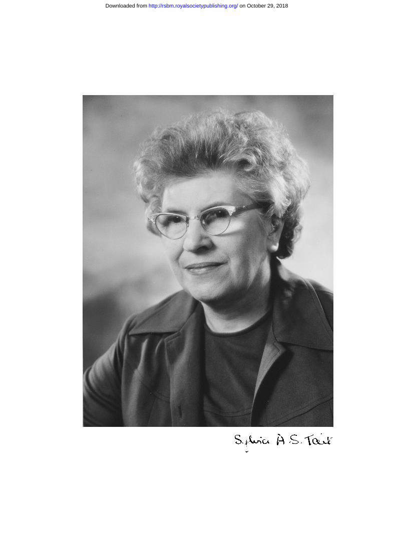
The frontispiece photograph was taken in 1977 by Godfrey Argent, and is reproduced with permission.
REFERENCES TO OTHER AUTHORS
Boon, W. C., McDougall, J. G. & Coghlan, J. P. 1996 Control of aldosterone secretion. ‘Towards the molecularidiom’. In Adrenal glands, vascular systems and hypertension (ed. V. P. Vinson & D. C. Anderson), pp.159–185. Bristol: J Endocrinol Ltd.
Burton, R. B., Zaffaroni, A. & Keutmann, E. H. 1951 Paper chromatography of steroids. II. Corticosteroids andrelated compounds. J. Biol. Chem. 188, 763–771.
Bush, I. E. 1952 Methods of paper chromatography of steroids applicable to the study of steroids in mammalian bloodand tissues. Biochem. J. 50, 370–398.
Bush, I. E. 1953 Species differences and other factors influencing adrenocortical secretion. (Ciba FoundationColloquia on Endocrinology, vol. VII: Synthesis and metabolism of adrenocortical steroids) (ed. W. Klyne, G.Wolstenholme & M. P. Cameron). London: J & A. Churchill Ltd.
Butt, W. R., Morris, P. & Morris, C. J. O. R. 1949 Determination of �4-3-ketosteroids in blood. In First InternationalCongress of Biochemistry, Cambridge, pp. 405–406.
Cheville, R. A., Luetscher, J. A., Hancock, E. W., Dowdy, A. J. & Nokes, G. W. 1966 Distribution, conjugation, andexcretion of labeled aldosterone in congestive heart failure and in controls with normal circulation: develop-ment and testing of a model with an analog computer. J. Clin. Invest. 45, 1302–1316.
Conn, J. 1955 Presidential Address. Part I. Painting background; Part II. Primary aldosteronism, a new clinical syn-drome. J. Lab. Clin. Med. 45, 3–17.
Conn, J. W., Lewis, I. H. & Fajans, S. S. 1951 The probability of compound F (17 hydroxycorticosterone) is the hor-mone produced by the normal human adrenal cortex. Science 113, 713–714.
Deane, H. W., Shaw, J. S. & Greep, R. O. 1948 The effect of altered sodium and potassium intake on the width andcytochemistry of the cat’s adrenal cortex. Endocrinology 43, 133–153.
394 Biographical Memoirs
on October 29, 2018http://rsbm.royalsocietypublishing.org/Downloaded from
Deming, Q. B. & Luetscher, J. A. 1950 Bioassay of deoxycorticosterone-like material in urine. Proc. Soc. Exp. Biol.73, 171–175.
Dickens, F. 1975 Edward Charles Dodds. Biogr. Mem. Fell. R. Soc. 21, 227–267.Dorfman, R. I., Potts, A. M. & Feil, M. L. 1947 Studies on the bioassay of hormones. The use of radiosodium for the
detection of small quantities of deoxycorticosterone. Endocrinology 41, 464–469.Fieser, L. F. & Fieser, M. 1959 Steroids, pp. 713–720. London: Reinhold, Chapman & Hall.Fourman, P., Bartter, F. C., Albright, F., Dempsey, E., Carroll, E. & Alexander, J. 1950 Effect of 17-hydroxycorticos-
terone (compound F) in man. J. Clin. Invest. 19, 1462–1473.Giroud, C. J. P., Stachenko, J. & Venning, E. H. 1956 Secretion of aldosterone by the zona glomerulosa of rat adre-
nal in vitro. Proc. Soc. Exp. Med. 92, 154–158.Haines, W. J. & Drake, N. A. 1950 Fluorescent scanner for the evaluation of papergrams of adrenal cortical hormones.
Fedn Proc. 9, 180–182.Ham, E. A., Harman, R. E., DeYoung, J. J., Brink, N. G. & Sarrett, L. H. 1955 Studies on the chemistry of aldos-
terone. J. Am. Chem. Soc. 77, 1637–1641.Harman, R. E., Ham, E. A., DeYoung, J. J., Brink, N. G. & Sarrett, L. H. 1954 Isolation of aldosterone (Electrocortin).
J. Am. Chem. Soc. 76, 5035–5036.Hechter, O. & Pincus, G. 1954 Genesis of the adrenocortical secretion. Physiol. Rev. 34, 459–495.Huntford, R. 1997 Nansen: the explorer as hero. London: Duckworth.Jessop, M. 2003 Aldosterone blockade and heart failure. New Engl. J. Med. 348, 1380–1388.Kliman, B. & Peterson, R. E. 1960 Double isotope derivative assay of aldosterone in biological extracts. J. Biol.
Chem. 235, 1639–1648.Kuizenga, M. H. 1944 The isolation and chemistry of the adrenal hormones. In The chemistry and physiology of hor-
mones (ed. F. R. Moulton), pp. 57–68. Washington.Layne, D., Meyer, C. J., Vaishwaner, P. S. & Pincus, G. 1962 The secretion and metabolism of cortisol and aldos-
terone in normal and in steroid-treated women. J. Clin. Endocrinol. Metab. 22, 107–118.Luetscher, J. A. Jr, Johnson, B. B., Dowdy, A., Harvey, J., Lew, W. & Poo, L. J. 1954 Chromatographic separation of
the sodium-retaining corticoid from the urine of children with nephrosis compared with observations on nor-mal children. J. Clin. Invest. 33, 276–286.
Mason, H., Myers, C. S. & Kendall, E. C. 1936 The chemistry of crystalline substances isolated from the suprarenalgland. J. Biol. Chem. 114, 613–631.
Mattox, V. R., Mason, H. L., Albert, A. & Code, J. C. 1953 Properties of a sodium-retaining principle from beef adre-nal extract. J. Am. Chem. Soc. 75, 4869–4870.
Morris, C. J. O. R. & Williams, D. 1953 The polarographic estimation of steroid hormones. 6. Determination of indi-vidual adrenocortical in human peripheral blood. Biochem. J. 54, 470–475.
Neher, R. 1979 Aldosterone: chemical aspects and related enzymology. J. Endocrinol. 81, 25P–35P.Pearlman, W. H. 1957 Circulating steroid hormone levels in relation to steroid hormone production. Ciba Found.
Colloq. Endocr. Horm. Blood 111, 233–251.Pearlman, W. H., Pearlman, M. R. J. & Rakoff, A. E. 1954 Estrogen metabolism in pregnancy: a study with the aid of
deuterium. J. Biol. Chem. 209, 803–812.Pincus, G. 1965 The control of fertility. New York: Academic Press.Pitt, B. 2003 Effect of aldosterone blockade in patients with systolic left ventricular dysfunction: implications of the
RALES and EPHESUS studies. In 50th Anniversary of the Discovery of Aldosterone Meeting (ed. G. Vinson& J. Coghlan), pp. 53–58. London: Elsevier.
Pitt, B., Zannad, F., Remme, W. J., Cody, R., Castaigne, A., Perez, A., Palensky, J. & Wittes, J. 1999 The effect ofspironolactone on morbidity and mortality in patients with severe heart failure. New Engl. J. Med. 341,709–717.
Pitt, B., Remme, W., Zannad, F., Neaton, J., Martinez, F., Roniker, B., Bittman, R., Hurley, S., Kleiman, J. & Gatlin,M. 2003 Eplerenone, a selective aldosterone blocker in patients with left ventricular dysfunction after myocar-dial infarction. New Engl. J. Med. 348, 1309–1382.
Ranger, D. 1985 The Middlesex Hospital Medical School Centenary to Sesquicentenary 1935–1985. London:Hutchinson Benham.
Sayers, G. 1950 The adrenal cortex and homeostasis. Physiol. Rev. 30, 241–320.
Sylvia Agnes Sophia Tait 395
on October 29, 2018http://rsbm.royalsocietypublishing.org/Downloaded from
Schmidlin, J., Anner, G., Billeter, J. R. & Wettstein, A. 1955 Über Synthesen in der Aldsterons-Reihe. Experientia 40,365–368.
Selye, H. & Horava, A. 1953 The stress concept in 1953. In Third Annual Report on Stress (ed. H. Selye & A.Horava), vol. 3, pp. 17–65. Montreal: Acta Inc.
Stowasser, M. & Gordon, R. D. 2003 Primary aldosteronism—careful investigation is essential and rewarding. In 50thAnniversary of the Discovery of Aldosterone Meeting (ed. G. Vinson & J. Coghlan), pp. 33–39. London:Elsevier.
Vogt, M. 1943 The output of cortical hormones by the mammalian suprarenal. J. Physiol. 102, 341–356.Young, M., Fullerton, M., Dilley, R. & Funder, J. 1994 Mineralocorticoids, hypertension and cardiac fibrosis. J. Clin.
Invest. 93, 2578–2583.Zaffaroni, A., Burton, R. B. & Keutmann, E. G. 1950 Adrenal cortical hormones: analysis by paper partition chro-
matography and occurrence in the urine of normal persons. Science 111, 6–8.
BIBLIOGRAPHY
The following publications are those referred to directly in the text. A full bibliography isavailable as electronic supplementary material at http://dx.doi.org/10.1098/rsbm.2006.0026 orvia http://www.journals.royalsoc.ac.uk.
(1) 1945 (With J. Z. Young) Regeneration of fibre diameter after cross-unions of visceral and somatic nerves. J.Anat. 79, 48–65.
(2) (With E. C. Dodds, W. Lawson & P. C. Williams) Testing diphenylethylamine compounds for anal-gesic action. J. Physiol. 104, 47–51.
(3) 1946 (With P. C. Williams) Increased pituitary weight produced by oestrone in intact and castrated rats.Endocrinology 39, 272–274.
(4) 1948 (With P. C. Williams) Improved method of getting rats’ eggs from the fallopian tubes. Nature 161, 237.(5) (With A. E. Wilder-Smith) The isolation and properties of the monoglucuronides of stilboestrol, hex-
oestrol and dienoestrol. Biochem. J. 42, 258–260.(6) (With S. Bartlett, S. J. Folley, S. J. Rowland & D. H. Curnow) Oestrogens in grass and their possible
effects on milk secretion. Nature 161, 845.(7) 1949 (With P. C. Williams) Mating of spayed-adrenalictomized rats given oestrogen. Endocrinology 6,
169–170.(8) (With A. E. Wilder-Smith) The excretion of synthetic oestrogens as ethereal sulphates and monoglu-
curonides in the rabbit and in man. Biochem. J. 44, 366–368.(9) 1950 (With J. F. Tait) Dose response studies of the effect of deoxycorticosterone acetate (DOCA) on the
sodium excretion of adrenalectomized rats. Endocrinology 47, 308–310.(10) 1952 (With J. F. Tait & H. M. Grundy) The effect of adrenal extract on mineral metabolism. Lancet i,
122–124.(11) (With J. F. Tait) A quantitative method for the bioassay of the effect of adrenal cortical steroids on min-
eral metabolism. Endocrinology 50, 150–161.(12) (With H. M. Grundy & J. F. Tait) Isolation of a highly active mineralocorticoid from beef adrenal
extract. Nature 169, 795–797.(13) (With J. F. Tait & I. E. Bush) The secretion of a salt-retaining hormone by the mammalian adrenal cor-
tex. Lancet ii, 226–228.(14) (With H. M. Grundy, J. F. Tait & M. Woodford) Further studies on the properties of a highly active
mineralocorticoid. Acta Endocrinol., Copenh. 11, 199–220.(15) 1953 (With J. F. Tait) Physico-chemical methods of detection of a previously unidentified adrenal hormone.
Mem. Soc. Endocrinol. 2, 9–24.(16) (With J. F. Tait, A. Wettstein, R. Neher, J. von Euw & T. Reichstein) Isolierung eines neuen
kristallisierten Hormons aus Nebennieren mit besonders hoher Wirksamkeit auf denMineralstoffwechsel. Experientia 9, 333–335.
396 Biographical Memoirs
on October 29, 2018http://rsbm.royalsocietypublishing.org/Downloaded from
(17) 1954 (With J. F. Tait, A. Wettstein, R. Neher, J. von Euw, O. Schindler & T. Reichstein) Konstitution desAldosterons, des neuen Minerolocorticoids. Experientia 10, 132–133.
(18) (With J. F. Tait, A. Wettstein, R. Neher, J. von Euw, O. Indler & T. Reichstein) Aldosteron, Isolierungund Eigenschaften. Uber Bestandteile der Nebennierenrinde und verwandte Stoffe. 91 Mitteilung.Helv. Chim. Acta 37, 1163–1200.
(19) (With J. F. Tait, A. Wettstein, R. Neher, J. von Euw, O. Schindler & T. Reichstein) Die Konstitution desAldosterons. Uber Bestandteile der Nebennierenrinde und verwandte Stoffe. 92 Mitteilung. Helv.Chim. Acta 37, 1200–1223.
(20) (With P. Avivi, J. F. Tait & J. K. Whitehead) The use of 3H and 14C labeled acetic anhydride as analyt-ical reagents in micorbiochemistry. In Proc. 2nd Radioisotope Conf. (ed J. E. Johnston), pp. 313–324.Oxford: Butterworths Sci. Pub.
(21) 1956 (With R. N. Jones & J. F. Tait) The assay of aldosterone and other adrenal steroids by the 24Na/42Kmethod. Analyst 81, 439–440.
(22) 1959 (With J von Euw, R. Neher, T. Reichstein, J. F. Tait & A. Wettstein) Substanz Z. Uber Bestandteile derNebennierenrinde und verwandte Stoffe. 100. Mitteilung. Helv. Chim. Acta 42, 1817–1829.
(23) 1957 (With P. J. Ayres, O. Garrod, J. F. Tait, G. Walker & W. H. Pearlman) The use of 16-3H aldosterone instudies on human peripheral blood. Ciba Found. Colloq. Endocr. Horm. Blood 11, 309–326.
(24) 1958 (With P. J. Ayres, J. Barlow, O. Garrod, A. E. Kellie, J. F. Tait & G. Walker). The metabolism of 16-3Haldosterone in man. In Int. Symp. Aldosterone (ed. A. F. Muller & C. M. O’Connor), pp. 73–99.London: J. & A. Churchill.
(25) 1960 (With J. F. Tait, B. Little & K. Laumas) The metabolism of aldosterone in man. In Proc. Conf. HumanAdrenal Cortex. Glasgow (ed A. E. Currie, T. Symington & J. K. Grant), pp. 107–123. Edinburgh: E.& S. Livingstone.
(26) 1961 (With C. Flood, D. S. Layne, S. Ramcharan, E. Rossipal & J. F. Tait) An investigation of the urinarymetabolites and secretion rates of aldosterone and cortisol in man and a description of methods fortheir measurement. Acta Endocrinol., Copenh. 36, 237–264.
(27) (With J. F. Tait, B. Little & K. Laumas) The disappearance of 7-H3-d-aldosterone in the plasma of nor-mal subjects. J. Clin. Invest. 40, 72–80.
(28) (With K. Laumas & J. F. Tait) The validity of the calculation of secretion rates from the specific activ-ity of a urinary metabolite. Acta Endocrinol., Copenh. 36, 265–280.
(29) (With K. Laumas & J. F. Tait) Further considerations on the calculations of secretion rates: a correc-tion. Acta Endocrinol., Copenh. 38, 469–472.
(30) (With R. H. Underwood, C. A. Flood & J. F. Tait) A comparison of methods for the acid hydrolysis ofa urinary conjugate of aldosterone. J. Clin. Endocrinol. Metab. 21, 1092–1098.
(31) 1962 (With M. Gut, R. Underwood, J. F. Tait, A. Riondel, A. L. Southren & B. Little) The synthesis andproperties of steroidal thiosemicarbazones and of their 2,4-diacetyl derivatives. First Int. Cong. Horm.Steroids (Excerpta Medica Int Congr Series no. 51), p. 129. Amsterdam: Excerpta Medica.
(32) (With B. Little, J. F. Tait & C. Flood) The metabolic clearance rate of aldosterone in pregnant and non-pregnant subjects estimated by both single-injection and constant-infusion methods. J. Clin.Endocrinol. Metab. 41, 2093–2100.
(33) 1964 (With J. Bougas, C. Flood, B. Little, J. F. Tait & R. Underwood) Dynamic aspects of aldosteronemetabolism. In Aldosterone. A Symposium, Prague (ed. E. E. Baulieu & P. Robel), pp. 25–50. Oxford:Blackwell Science.
(34) 1965 (With J. Bougas, B. Little, J. F. Tait & C. Flood) Splanchnic extraction and clearance of aldosterone insubjects with minimal and marked cardiac dysfunction. J. Clin. Endocrinol. Metab. 25, 219–228.
(35) (With A. Riondel, J. F. Tait, M. N. Gut & B. Little) Estimation of progesterone in human peripheralblood using 35S-thiosemicarbazide. J. Clin. Endocrinol. Metab. 25, 229–242.
(36) (With A. H. Brodie, N. Shimizu & J. F. Tait) A method for the measurement of aldosterone in periph-eral plasma using 3H acetic anhydride. J. Clin. Endocrinol. Metab. 27, 997–1011.
(37) 1991 (With J. F. Tait) The effect of plasma protein binding on the metabolism of steroid hormones. J.Endocrinol. 131, 339–357.
(38) 1956 (With P. J. Ayres, R. P. Gould & J. F. Tait) The in vitro demonstration of differential corticosteroid pro-duction within the ox adrenal gland. Biochem. Soc. Trans. 63, 19P.
Sylvia Agnes Sophia Tait 397
on October 29, 2018http://rsbm.royalsocietypublishing.org/Downloaded from
(39) 1958 (With P. J. Ayres, O. Garrod & J. F. Tait) Primary aldosteronism (Conn’s syndrome). In Symposium onAldosterone (ed. A. F. Muller & C. M. O’Connor), pp. 143–154. London: Churchill.
(40) (With P. J. Ayres, W. H. Pearlman & J. F. Tait) The biosynthetic preparation of 16-3H-aldosterone and16-3H-corticosterone. Biochem. J. 70, 230–236.
(41) 1960 (With P. J. Ayres, J. Eichhorn, O. Hechter, N. Saba & J. F. Tait) Some studies on the biosynthesis ofaldosterone and other adrenal steroids. Acta Endocrinol., Copenh. 33, 27–58.
(42) 1968 (With S. Baniukiewicz, A. Brodie, C. Flood, M. Motta, M. Okamoto, J. F. Tait, J. R. Blair-West, J. P.Coghlan, D. A. Denton, J. R. Goding, B. A. Scoggins, E. M. Wintour & J. D. Wright) Adrenal biosyn-thesis of steroids in vitro and in vivo using continuous superfusion and infusion procedures. InFunctions of the adrenal cortex (ed. R. W. McKerns), pp. 153–232. New York: Appleton-Century-Crofts.
(43) 1970 (With J. R. Blair-West, A. Brodie, J. P. Coghlan, D. A. Denton, C. Flood, J. R. Goding, B. A.Scoggins, J. F. Tait, E. M. Wintour & R. D. Wright) Studies on the biosynthesis of aldosterone usingthe sheep adrenal transplant. Effect of sodium depletion on the conversion of corticosterone to aldos-terone. J. Endocrinol. 46, 453–476.
(44) (With R. Haning & J. F. Tait) In vitro effects of ACTH, serotonin and potassium on steroid output andconversion of corticosterone to aldosterone in isolated adrenal cells. Endocrinology 87, 1147–1167.
(45) 1974 (With J. F. Tait, R. P. Gould & M. S. R. Mee) The properties of adrenal glomerulosa cells after purifi-cation by gravitational sedimentation. Proc. R. Soc. B 185, 375–407.
(46) (With J. D. M. Albano, B. L. Brown, R. P. Ekins & J. F. Tait) The effects of potassium, 5-hydrox-ytryptamine, adrenocorticotrophin and angiotensin II on the concentration of adenosine 3�,5� cyclicmonophosphate in suspensions of dispersed rat adrenal zona glomerulosa and zona fasciculata cells.Biochem. J. 142, 391–400.
(47) (With J. F. Tait, R. P. Gould, B. L. Brown & J. D. M. Albano) The preparation and use of purified andunpurified dispersed adrenal cells and a study of the relationship of their cAMP and steroid output. J.Steroid Biochem. 5, 775–787.
(48) (With J. F. Tait, R. P. Gould, J. D. M. Albano & B. L. Brown) Properties of enzymatically dispersedadrenal cells after purification by sedimentation at 1g. Biochem. Soc. Trans. 2, 847–851.
(49) 1975 (With J. F. Tait, J. D. M. Albano, B. L. Brown & F. Mendelsohn) The response of purified zonaglomerulosa cells of the rat adrenal to stimulation by KD+U, serotonin, ACTH, angiotensin II andcAMP. In Trans. VIth Meeting for Steroid Hormones (ed. H. B. Brewer, A. Hughes, A. Klopper, C.Conti & P. Gungblut), vol. 6, pp. 19–33. Amsterdam: North-Holland.
(50) 1976 (With J. F. Tait) The effect of changes in potassium concentration on the maximal steroidogenicresponse of purified zona glomerulosa cells to angiotensin II. J. Steroid Biochem. 7, 687–690.
(51) 1977 (With C. M. Mackie, E. R. Simpson, M. S. R. Mee & J. F. Tait) Intracellular potassium and steroido-genesis of isolated rat adrenal cells: effect of potassium ions and angiotensin II on purified zonaglomerulosa cells. Clin. Sci. Mol. Med. 53, 289–296.
(52) 1979 (With J. B. G. Bell, R. P. Gould, P. J. Hyatt & J. F. Tait) Properties of rat adrenal zona reticularis cells:production and stimulation of certain steroids. J. Endocrinol. 83, 435–447.
(53) (With J. G. McDougall, B. C. Williams, P. J. Hyatt, J. B. G. Bell & J. F. Tait) Purification of dispersedrat adrenal cells by column filtration. Proc. R. Soc. B 206, 15–32.
(54) 1980 (With J. F. Tait, J. B. G. Bell, P. J. Hyatt & B. C. Williams) Further studies on the stimulation of ratadrenal capsular cells: four types of response. J. Endocrinol. 87, 11–27.
(55) (With J. B. G. Bell, K. Bhatt, P. J. Hyatt & J. F. Tait) Properties of adrenal zona reticularis cells. InAdrenal androgens (ed. A. R. Genazzani et al.), pp. 1–6. New York: Raven Press.
(56) (With J. F. Tait & J. B. G. Bell) Steroid hormone production by mammalian adrenocortical dispersedcells. Essays Biochem. 16, 99–174.
(57) 1981 (With J. B. G. Bell, J. F. Tait, G. D. Barnes & B. L. Brown) Lack of effect of angiotensin on levels ofcyclic AMP in isolated adrenal zona glomerulosa cells from the rat. J. Endocrinol. 91, 145–154.
(58) (With B. C. Williams, J. G. McDougall & J. F. Tait) Calcium efflux and steroid output from superfusedrat adrenal cells: effects of potassium, adrenocorticotropic hormone, 5-hydroxytryptamine, adenosine3�:5� cyclic monophosophate and angiotensins II and III. Clin. Sci. 61, 541–551.
398 Biographical Memoirs
on October 29, 2018http://rsbm.royalsocietypublishing.org/Downloaded from
(59) 1984 (With G. St J. Whitley, J. B. G. Bell, F. W. Chu & J. F. Tait) The effects of ACTH, serotonin, K+ andangiotensin analogues on 32P incorporation into phospholipids of the rat adrenal cortex: basis for anassay method using zona glomerulosa cells. Proc. R. Soc. B 222, 273–294.
(60) 1985 (With J. B. G. Bell, P. J. Hyatt, J. F. Tait & G. St J. Whitley) Phospholipid metabolism in the adrenalcortex. Biochem. Soc. Trans. 13, 64–67.
(61) (With P. J. Hyatt, J. B. G. Bell, K. Bhatt, F. W. Chu, J. F. Tait & G. St J. Whitley) Effects of alpha-melanocyte-stimulating hormone on the cyclic AMP and phospholipid metabolism of rat adrenocorti-cal cells. J. Endocrinol. 110, 405–416.
(62) 1986 (With P. J. Hyatt & J. F. Tait) The mechanism of the effect of K+ on the steroidogenesis of rat zonaglomerulosa cells of the adrenal cortex: role of cyclic AMP. Proc. R. Soc. B 227, 21–42.
(63) 1978 (With J. F. Tait) A short history of aldosterone. Trends Biochem. Sci 3, N273–N275.(64) 1979 (With J. F. Tait) Recent perspectives on the history of the adrenal cortex. The Sir Henry Dale Lecture
for 1979. J. Endocrinol. 83, 1P–24P.(65) (With J. F. Tait) Opening Address at the Symposium on the 25th Anniversary of the Discovery of
Aldosterone. J. Endocrinol. 81, 1P–3P.(66) 1987 (With J. F. Tait) Obituary. Ian Elcock Bush. Lancet i, 56. (Also published in The Times (1986) and J.
Chromatogr. (1987).)(67) 1988 (With J. F. Tait) A decade (or more) of electrocortin (aldosterone). Steroids 51, 213–250.(68) 1990 (With J. F. Tait) A decade (and even more) of aldosterone and other adrenal steroids. In Endocrine
hypertension (ed. E. Biglieri), pp. 5–27. New York: Raven Press.(69) 1956 (With P. J. Ayres, O. Hechter, N. Saba & J. F. Tait) Intermediates in the biosynthesis of aldosterone by
capsule strippings of ox adrenal gland. Biochem. J. 65, 22P.(70) 1997 (With J. F. Tait) Insulin, the renin-angiotensin-aldosterone system and hypertension. Perspect. Biol.
Med. 40, 246–259.(71) 1998 (With J. F. Tait) A personal history of the early development of the concept and methods of measure-
ment of the metabolic clearance rate, particularly of steroid hormones. Clin. Exp. Pharmacol. Physiol.25 (suppl.), S101–S118.
(72) (With J. F. Tait) Personal history. The correspondence of S. A. S. Simpson and J. F. Tait with T.Reichstein during their collaborative work on the isolation and elucidation of the structure of electro-cortin (later aldosterone). Steroids 64, 440–453.
(73) 1999 (With J. F. Tait) A brief review. Role of cAMP in the effects of K+ on the steroidogenesis of zonaglomerulosa cells. Clin. Exp. Pharmacol. Physiol 26, 947–955.
(74) 2004 The discovery, isolation and identification of aldosterone: reflections on energy regulation and func-tion. Mol. Cell. Endocrinol. 217, 3–31.
Sylvia Agnes Sophia Tait 399
on October 29, 2018http://rsbm.royalsocietypublishing.org/Downloaded from

on October 29, 2018http://rsbm.royalsocietypublishing.org/Downloaded from