Embed Size (px)
Citation preview
8/3/2019 Sylvain Vauthey et al- Molecular self-assembly of surfactant-like peptides to form nanotubes and nanovesicles
http://slidepdf.com/reader/full/sylvain-vauthey-et-al-molecular-self-assembly-of-surfactant-like-peptides 1/6
Molecular self-assembly of surfactant-like peptides toform nanotubes and nanovesiclesSylvain Vauthey*, Steve Santoso*†, Haiyan Gong*‡, Nicki Watson§, and Shuguang Zhang*¶
*Center for Biomedical Engineering, 56-341, Massachusetts Institute of Technology, 77 Massachusetts Avenue, Cambridge, MA 02139-4307; †Department ofBiology, Massachusetts Institute of Technology, Cambridge, MA 02139; §W. M. Keck Imaging Facility, Whitehead Institute, Cambridge, MA 02142; and‡Department of Ophthalmology, Boston University School of Medicine, 715 Albany Street, Boston, MA 02118
Communicated by John M. Buchanan, Massachusetts Institute of Technology, Cambridge, MA, February 13, 2002 (received for review November 17, 2001)
Several surfactant-like peptides undergo self-assembly to form
nanotubes and nanovesicles having an average diameter of 30–50
nm with a helical twist. The peptide monomer contains 7–8
residues and has a hydrophilic head composed of aspartic acid and
a tail of hydrophobic amino acids such as alanine, valine, or leucine.
The length of each peptide is 2 nm, similar to that of biological
phospholipids. Dynamic light-scattering studies showed structures
with very discrete sizes. The distribution becomes broader over
time, indicating a very dynamic process of assembly and disassem-
bly. Visualization with transmission electron microscopy of quick-
freezedeep-etch sample preparation revealed a network of open-
ended nanotubes and some vesicles, with the latter being able to
‘‘fuse’’ and ‘‘bud’’ out of the former. The structures showed some
tail sequence preference. Many three-way junctions that may actas links between the nanotubes have been observed also. Studies
of peptide surfactantmolecules have significant implicationsin the
design of nonlipid biological surfactants and the understanding of
the complexity and dynamics of the self-assembly processes.
amino acids charged and hydrophobic residues nonlipid
surfactants simplicity to complexity prebiotic enclosures
Molecular self-assembly recently has attracted consider-able attention for its use in the design and fabrication of
nanostructures leading to the development of advanced ma-terials (1, 2). The self-assembly of biomolecular buildingblocks plays an increasingly important role in the discoveryof new materials and scaffolds (3, 4), with a wide range of
applications in nanotechnology and medical technologies suchas regenerative medicine and drug delivery systems (5, 6).Recently, Hartgerink et al. (7) reported the design of achimeric material consisting of a hydrophobic alkyl tail and ahydrophilic peptide containing phosphorylated serine with anRGD motif that facilitates directional alignment of mineral-ization of hydroxyapatite.
We previously described a class of ionic self-complementarypeptide that spontaneously self-assemble to form interwovennanofibers in the presence of monovalent cations (8–10).These nanofibers further form a hydrogel consisting of greaterthan 99.5% water. The constituent of the hydrogel scaffold ismade of peptides with alternating hydrophilic and hydrophobicamino acids. Such a sequence has a tendency to form anunusually stable -sheet structure in water (8–10). When thepeptides form a -sheet, they exhibit two surfaces, a hydro-philic surface consisting of charged ionic side chains and ahydrophobic surface with hydrophobic side chains. As a result,the self-assembly of these peptides is facilitated by electrostaticinteractions on one side and the hydrophobic interaction onthe other, in addition to the conventional -sheet hydrogenbond along the backbones. The self-assembling peptide scaf-folds have been demonstrated to serve as substrate for tissue-cell attachment, extensive neurite outgrowth, and formation of active nerve connections. Thus, the nanofiber hydrogel can beused as a permissive biological material for culturing cells ina three-dimensional environment (9).
In another attempt to exploit the intrinsic self-assembly of peptides as an avenue to emerging materials, Aggeli et al. (11, 12)have designed different short peptides that self-assemble innonaqueous solvent into long, semiflexible, polymeric -sheetpeptide nanotapes. These systems were designed rationally toprovide strong cross-strand-attractive forces between the sidechains such as electrostatic, hydrophobic, or hydrogen-bondinginteractions. In another study, Ghadiri and coworkers (13–15)produced self-assembling nanotubes made from alternating D,L--peptides and cyclic -peptide. They first showed that D,L-peptides {cyclo-[-(L-Gln-D-Ala-L-Glu-D-Ala)2-]} adopt flat,ring-shaped conformations and stack through backbone–backbone hydrogen bonding to form extended cylindrical struc-
tures with a diameter 1 nm in the single peptide tube. Theyfurther showed that this type of peptide nanotubes forms poresin the cell membrane (15).
We are interested in broadening the diversity of the buildingblocks of self-assembling peptides for scaffolds and biologicalmaterials. We therefore designed another class of amphiphilicsurfactant-like peptides. These 7–8-residue peptides (Fig. 1),each 2 nm in length, have properties very similar to thoseobserved in biological surfactant molecules. They have a hydro-philic head group of negatively charged aspartic acid at the Cterminus, thus containing two negative charges (one from theside chain carboxyl group and the other from the C terminus)and a lipophilic tail made of hydrophobic amino acids such asalanine, valine, or leucine (Fig. 1). The N terminus is acetylated,making it uncharged. When dissolved in water, these surfactant-
like peptides tend to self-assemble to isolate the hydrophobic tailfrom contact with water. Similar to lipids and fatty acids, thesupramolecular structure is characterized by the formation of apolar interface that sequesters the hydrophobic tail from water.
Materials and Methods
Surfactant-Like Peptides. All peptides were synthesized with Wangresin and acetylated at their N termini. All were synthesizedcommercially (Synpep, Dublin, CA, or Research Genetics,Huntsville, AL) and solubilized in water to a concentration of 4–5 mM, and their pH values were neutralized with 0.1 NNaOH in small scintillation glass vials. The solutions weresonicated for 10 min in an Aquasonic Model 50HT water bath(VWR Scientific). All peptide solutionswere stored and handledat room temperature.
Dynamic Light Scattering (DLS). An aliquot of 150–200 l of thepeptide solution was used to perform DLS experiments by usingPDDLSBatch (Precision Detectors, Franklin, MA). Intensitydata from each sample were collected in five replicates andanalyzed by using the PRECISION DECONVOLVE program and
Abbreviations: DLS, dynamic light-scattering; TEM, transmission electron microscopy.
¶To whom reprint requests should be addressed. E-mail: [email protected].
The publication costs of this article were defrayed in part by page charge payment. This
article must therefore be hereby marked “advertisement ” in accordance with 18 U.S.C.
§1734 solely to indicate this fact.
www.pnas.orgcgidoi10.1073pnas.072089599 PNAS April 16, 2002 vol. 99 no. 8 5355–5360
8/3/2019 Sylvain Vauthey et al- Molecular self-assembly of surfactant-like peptides to form nanotubes and nanovesicles
http://slidepdf.com/reader/full/sylvain-vauthey-et-al-molecular-self-assembly-of-surfactant-like-peptides 2/6
yielded size-versus-fraction distribution plots. It was found thatnanostructures formed in a narrow range of 30–50 nm (Fig. 2).DLS is a rapid screening method for defined nanostructures.Without discrete peak intensity, there are no nanostructuresobserved subsequently. These observations encouraged us tofurther study the structures by using a quick-freezedeep-etchtechnique to preserve the structures as they appear in aqueousstate and to examine them by using transmission electronmicroscopy (TEM).
Quick-FreezeDeep-Etch Sample Preparation. Quick-freezedeep-etch sample preparation for TEM followed the protocol de-scribed by Magid (16). Briefly, aliquots (1–2 l) of peptides in
water were placed on 3-mm gold specimen carriers. The samplesthen were frozen rapidly in liquid propane (180 to190°C) byusing the TFD 010 plunge-freeze and transfer device (BAL-TEC, Balzers, Principality of Liechtenstein). The frozen samples
were stored in liquid nitrogen and transferred onto a cold stage(180°C) in the CFE-60 freeze fracture system (CressingtonScientific Instruments, Cranberry, PA). The sample holder was
warmed to100°C and the sample surface was etched for 30 minby placing a cooled knife (180°C) directly above the samples.
After etching, the specimens were rotary-shadowed with a 20°platinum-carbon gun. The estimated electron-dense coatingthickness using this method was 1.5–2.0 nm as determined bya quartz crystal thin-filmmonitor. Replicas were strengthenedbyevaporation of carbon at an angle of 90°. The thickness of the
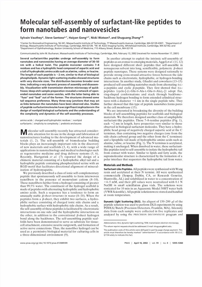
Fig.1. Space-filling molecularmodelsof surfactant peptide.( A) A6D. (B) V6D.
(C ) V6D2. (D) L6D2. D (aspartic acid) bears negative charges, and A (alanine), V
(valine), and L (leucine) constitute the hydrophobic tails with increasing
hydrophobicity. Green, carbons; red, oxygen; blue, nitrogen; white, hydro-
gen. Each peptide is
2–3 nm in length, similar to biological phospholipids.
Fig. 2. DLSmeasurementof surfactant peptidenanostructures.The peptides
V6D, V6D2, and A6D gave similar results. Intensity data were collected five
times, each looking nearly identical to the rest. The x axis is the size in
nanometers, and the y axis is the fraction distribution. The average diameter
(D) is30–50 nm. ( A) A6D. (B) V6D. The other dimension along the length of
the nanotube is beyond the range of DLS measurement.
5356 www.pnas.orgcgidoi10.1073pnas.072089599 Vauthey et al.
8/3/2019 Sylvain Vauthey et al- Molecular self-assembly of surfactant-like peptides to form nanotubes and nanovesicles
http://slidepdf.com/reader/full/sylvain-vauthey-et-al-molecular-self-assembly-of-surfactant-like-peptides 3/6
carbon surface was 15–20 nm. After shadowing, the sample with the replica coating was stored in methanol overnight andthen treated in 5% sodium hypochloride containing 10 –15%potassium hydroxide or bleach to degrade the peptides. Theremaining replicas were washed several times in distilled waterand f loated onto copper Gilder grids (Electron MicroscopySciences, Fort Washington, PA). The replicas were examined byusing a Philips-300 or Philips EM-410 TEM (Philips, Eindhoven,the Netherlands).
Results and Discussion
Design of Surfactant Peptides. The sequences and molecular mod-els of several surfactant peptides are shown in Fig. 1. Thehydrophilic moiety of the molecule is provided by 1–2 asparticacids at the C termini such that the peptide would have two orthree negative charges, similar to biological phospholipids. Thehydrophobic tails of the peptides consist of six consecutivehydrophobic amino acids with an acetylated N terminus, elim-inating the positive charge. The lengths and overall hydropho-
bicity of these peptides can be fine-tuned by modifying thealiphatic side groups of the amino acids. The calculated pI forthe investigated peptides range from3.56 to 3.8depending on thenumber of aspartic acids constituting the polar head group. Atneutral pH, the aspartic acids are negatively charged, but whenthe pH of the peptide solutions is below 3.5, the aspartic acidsbecome protonated, the peptides are uncharged, and theirsolubility in water becomes compromised substantially.
The hydrophobic tails of the peptides contain alanine (A), valine (V), or leucine (L) with increasing size of the hydrocarbonside chain and thus hydrophobicity. Various residues have
influence in the intermolecular interactions and packing of thetails. In the surfactant peptides, V6D2 and L6D2, the hydrophilicheads have a common aspartic acid, whereas the tails varied inhydrophobicity (Fig. 1). After self-assembly, these peptides mayform different structures w ith different physical parameters,because packing of the hydrophobic side chains depends on thesizes of their corresponding van der Waals surface areas. Like-
wise, by looking at both V6D and V6D2 we tested whether theaddition of one aspartic acid to the head group would make adifference in the size andor shape of the aggregates formed.This phenomenon is well described in the surfactant field as thepacking parameter by Israelachvili et al. (17) [ P v( al)], where P is the packing parameter, v is the molecular volume, l is themolecular length, and a is the cross-sectional area of the polarhead group. It should be noted that A6D and V6D carry twonegative charges at the C termini, and A6D2, A6D, V6D2, andL6D2 consist of three negative charge groups. A simple modifi-cation of the peptide sequence will result in the change of thecharge ratio w ith a significant impact in its solubility. If there is
too much charge, they become too soluble and may repel eachother electrostatically. On the other hand, if there are too manyhydrophobic residues, the peptides w ill become insoluble in
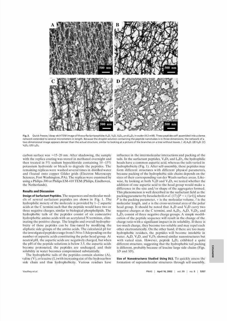
water. A6D, V6D, and V6D2 showed similar nanostructures but with varied sizes. However, peptide L6D2 exhibited a quitedifferent structure, suggesting that the hydrophobic tail packingis different, probably because of leucine large side chains (Figs.1 D and 3 D).
Size of Nanostructures Studied Using DLS. To quickly assess theformation of supramolecular structures through self-assembly,
Fig. 3. Quick-freezedeep-etchTEM image of thesurfactantpeptides A6D, V6D, V6D2, an d L6D2 in water (4.3 mM). These peptides self-assembled into a dense
network extended to several micrometers in length. Because the droplet solution containing the peptide nanotubes is in three dimensions, the network of a
two-dimensional image appears denser than the actual structure, similar to looking at a picture of the branches on a tree without leaves. ( A) A6D. (B) V6D. (C )
V6D2. (D) L6D2.
Vauthey et al. PNAS April 16, 2002 vol. 99 no. 8 5357
8/3/2019 Sylvain Vauthey et al- Molecular self-assembly of surfactant-like peptides to form nanotubes and nanovesicles
http://slidepdf.com/reader/full/sylvain-vauthey-et-al-molecular-self-assembly-of-surfactant-like-peptides 4/6
we performed DLS experiments. The observation of a mono-disperse population with sizes in the range of 30 –50 nm for A6D,V6D, and V6D2 suggested the formation of regular structure(s)from the peptide building blocks (Fig. 2 A and B, not shown forV6D2). As indicated above, we expected the packing parametersof these peptides to affect the shape and size of the assembliesthat they formed significantly. In this study, the polar head groupdid not significantly influence the size of the nanostructures,suggesting that either more aspartic acids or a charged amino
acid with a longer carboxylic acid tether is needed. Interestingly,the distribution of structures became more polydisperse overtime (data not shown), implying the process in which thesepeptides interact with each other is very dynamic. The poly-disperse samples also did not produce regular nanostructures
when examined by using the quick-freezedeep-etch methodunder TEM.
TEM Examination of Samples Prepared Through the Quick-FreezeDeep-Etch Procedure. The quick-freezedeep-etch sample prep-aration procedure has minimal disturbance of the structuresformed in solution. TEM images revealed that the surfactantpeptides formed a dense network of nanotubes and nanovesicles,
with diameters ranging from 30 to 50 nm (Figs. 3 and 4), whichis consistent with the measurement from DLS. The peptideassemblies represent a three-dimensional network similar to
flexible polymers in their semidilute or concentrated solutions(18). It should be pointed out that under the TEM, only atwo-dimensional projection of the specimen could be imaged.The network below and above the focal point in the space couldbe relatively distant in three dimensions while appearing super-imposed in the two-dimensional projection, thus appearingdenser than it actually is. This is similar to looking at a pictureof the branches on a tree without leaves; the network seemsdenser on a two-dimensional photo than on the actual object.TEM images of A6D, V6D, and V6D2 exhibit very similar tubularmorphologies. These self-assemblies have high axial ratios andcan extend in length to tens of micrometers (Fig. 3). Further-more, 3-fold junctions or branches connecting the nanotubesforming the network can be identified, similar to those observedin tree branches (Fig. 4).
The possibility of branched supramolecular organizations has
attracted considerable interest (19 –21). Evidence of branchinghas been reported mainly for aqueous surfactant solutions, butreversed structure such as lecithin organogels also can formthree-way junctions (22). Branch points produce patches havinga mean curvature opposite to that of the portion far from the
junction(22). Many reports claimed the importance of branchingpoints in the visco-elastic properties of polymer-like systems.Cates (21, 22) provided a statistical description of branches
versus entanglements and Lequeux (23) modeled the expectedeffects on the rheological properties.
TEM images show a dense network of nanotubes 30–50 nmin diameter linked by a number of 3-fold junctions. L6D2 is themost hydrophobic molecule, with a tail c ontaining six leucines.
Aqueous solution of L6D2 exhibits a heterogeneous populationof nanotubes and entangled rod-like micelles and vesicles (Fig.3 D). These molecular assemblies are similar to string-like mi-cellar systems made from traditional amphiphilic molecules suchas cetyltrimethylammonium bromide (CTAB; refs. 24 and 25).In this case, DLS did not provide detailed insight into the sizeof any regular structures formed and perhaps only captured theaverage size of the entanglements.
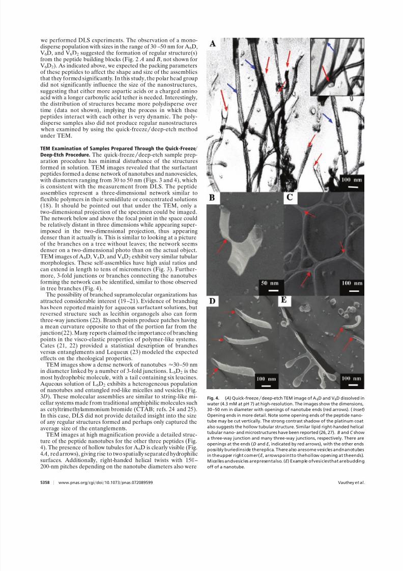
TEM images at high magnification provide a detailed struc-ture of the peptide nanotubes for the other three peptides (Fig.4). The presence of hollow tubules for A6D is clearly visible (Fig.4 A, red arrows), giving rise to two spatially separated hydrophilicsurfaces. Additionally, right-handed helical twists with 150–200-nm pitches depending on the nanotube diameters also were
Fig. 4. ( A) Quick-freezedeep-etch TEM image of A6D and V6D dissolved inwater (4.3 mM at pH 7) at high-resolution. The images show the dimensions,
30 –50 nm in diameter with openings of nanotube ends (red arrows). (Inset )
Opening ends in more detail. Note some opening ends of the peptide nano-
tube may be cut vertically. The strong contrast shadow of the platinum coat
also suggests the hollow tubular structure. Similar lipid right-handed helical
tubular nano- and microstructures have been reported (26, 27). B and C show
a three-way junction and many three-way junctions, respectively. There are
openings at the ends (D and E , indicated by red arrows), with the other ends
possibly buriedinside thereplica. There also aresome vesicles andnanotubes
in theupper right corner(E , arrowspointto thehollow opening at theends).
Micelles andvesicles arepresentalso. (E ) Example ofvesiclesthat arebudding
off of a nanotube.
5358 www.pnas.orgcgidoi10.1073pnas.072089599 Vauthey et al.
8/3/2019 Sylvain Vauthey et al- Molecular self-assembly of surfactant-like peptides to form nanotubes and nanovesicles
http://slidepdf.com/reader/full/sylvain-vauthey-et-al-molecular-self-assembly-of-surfactant-like-peptides 5/6
observed (Fig. 4 A, blue arrows). Open-ended tubes and athree-way junction also are shown in high magnification (Fig. 4 B and C). Interestingly, we also observed plausible budding orfusion of nanovesicles from the V6D nanotube (Fig. 4 D), sug-gesting the dynamic behavior of the self-assembly process of these surfactant peptides.
Other Structural Aspects. Circular dichroism studies revealed asingle minimum spectrum at 220 nm that does not resembleeither -sheet or -helical structure (data not shown). This
observation may reflect an unusual chirality of the assembledstructures that do not have typical -sheet packing. Spector et al.(26) also used circular dichroism to study the chirality of diacetylenic lipid tubules formed from lipid bilayer membranes.Schnur and coworkers (26, 27) developed a theory based onmolecular chirality to explain the presence of helical markings onthe twist of lipid tubules.
It should be pointed out that the light-scattering experimentssuggest that self-assembly is a dynamic process, and the sizedistributions are not static over long periods. Although it seemsthat the structures of A6D and V6D are rather similar except the
initial diameter size, they also change over time from constantassembly and disassembly.
Molecular Modeling. How could these simple surfactant-like pep-tides form such well ordered nanotubes and nanovesicles? Thereare molecular and chemical similarities between lipids and thepeptides, because both have a hydrophilic head and a hydro-phobic tail. The packing between lipids and peptides are likelyto be quite different, however. In lipids, the hydrophobic tailspack tightly against each other to completely displace water,
precluding the formation of hydrogen bonds. On the other hand,in addition to hydrophobic tail packing between the amino acidside chains, surfactant peptides also may interact through inter-molecular hydrogen bonds along the backbone. To understandhow each peptide molecule interacts with one another, wemodeled the nanotube from the constituent building block. Itshould be noted that a complete molecular simulation of theself-assembly process is beyond our current computationalcapability.
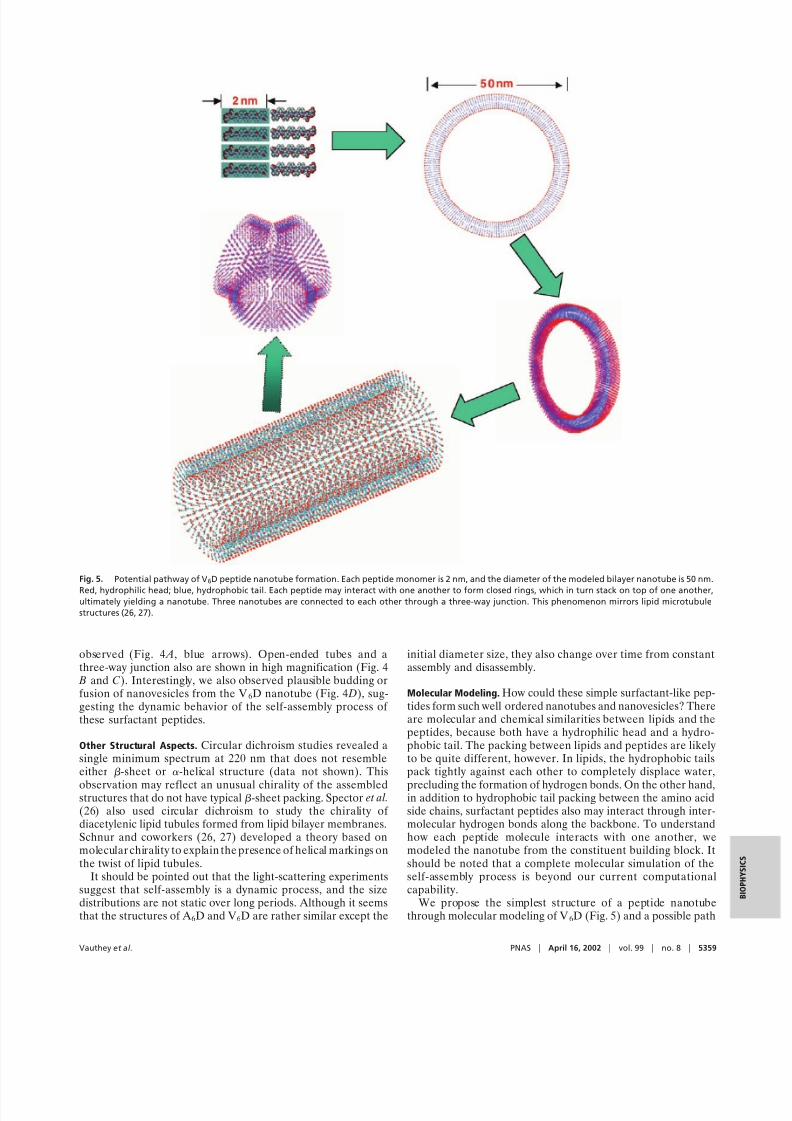
We propose the simplest structure of a peptide nanotubethrough molecular modeling of V6D (Fig. 5) and a possible path
Fig. 5. Potential pathway of V6D peptide nanotube formation. Each peptide monomer is 2 nm, and the diameter of the modeled bilayer nanotube is 50 nm.
Red, hydrophilic head; blue, hydrophobic tail. Each peptide may interact with one another to form closed rings, which in turn stack on top of one another,ultimately yielding a nanotube. Three nanotubes are connected to each other through a three-way junction. This phenomenon mirrors lipid microtubule
structures (26, 27).
Vauthey et al. PNAS April 16, 2002 vol. 99 no. 8 5359
8/3/2019 Sylvain Vauthey et al- Molecular self-assembly of surfactant-like peptides to form nanotubes and nanovesicles
http://slidepdf.com/reader/full/sylvain-vauthey-et-al-molecular-self-assembly-of-surfactant-like-peptides 6/6
from the monomer state to the assemblies. Each peptide mono-mer is 2 nm, with the diameter of the observed tube being 50 nm.Two peptides form dimeric tail-to-tail packing to form a bilayercreating a unilamellar shell with a maximum thickness of 4 nm.Had they formed a multilamellar structure, the opening endsmay not have exhibited a clearly observed single-shell ringstructure when viewed with the TEM. Furthermore, the strongcontrast shadow of the platinum coat also suggested that thetubes are hollow, similar to the lipid microtubes. These surfac-
tant peptides may form tubular structures akin to those found inlipid systems (27). Individual peptides are intrinsically twistedsuch that assembled structures will likely have a curvature. Oneof the simplest pathways of formation may be that the mono-meric peptides form small segments of the bilayer ring, withhydrophobic tails packing together to avoid water and hydro-philic heads exposed to water on the inner and outer portion of the tube. Through continuous dynamic energy minimization,they grow into single subunit rings and multirings, in whichaspartic acid hydrophilic groups remain exposed to the contin-uous aqueous medium. The tubular arrays may subsequentlystack through noncovalent interactions to form longer nano-tubes. This proposed model remains to be clarified experimen-tally and computationally.
Synthetic Peptide Surfactant. Although there are several types of
nanotubes ranging from carbon, D,L-cyclical peptides (13–15),and lipids (26, 27), our system is complementary to them. Thesurfactant peptides are relatively inexpensive and chemicallyfacile to modify, leading to potential tailoring of new materialsfor a broad spectrum of applications, including serving asscaffolds to organize conducting and semiconducting nanocrys-tals into high-density ordered structures; incorporating otherbiomolecules on their surfaces; encapsulating molecules formolecular deliveries; and forming a scaffold for cell encapsula-tion. Moreover, the molecular self-assembly process of surfac-
tant peptide mirrors the lipid-surfactant assemblies. Thus a wealth of literature and methods can be used as a guide forfurther studies.
Peptides in general have not been considered seriously to beuseful materials as scaffolds because of their small size and lessdefined structures at the individual monomeric level. However,several self-assembling peptide systems have not only introduceddesigned peptides as molecular building blocks but also mayinspire others to uncover and develop additional self-assembling
systems. Nanotubes and nanovesicles from self-assembly of thisclass of surfactant-like peptides are one of the simplest systemsthat can lead to formation of the well defined complex structures.The phenomena from simplicity to complexity also occur inother natural self-assembly systems including nucleic acids,lipids, saccharides, and proteins. It is anticipated that self-assemblies and fine-tuning of the surfactant peptide buildingblocks will lead to construction of a wide range of nanostruc-tures, fostering innovative avenues for the development of scaffold and biologically inspired materials.
This surfactant peptide system also may capture the prebioticenvironment’s simple origin and complex outcome. It is possibleto study molecular reorganizations, replications, and evolution ina simple enclosed environment of a peptide nanovesicle and aclosed-ended nanotube.
We thank Geoffrey von Maltzahn for sharing unpublished results andWonmuk Hwang and Davide Marini for molecular modeling of thesurfactant peptide nanotubes. We also thank Alexander Rich, CarlosSemino, Joseph Jacobson, Hyman Hartman, Jianping Shi, Kim Hamad,Shuwang An, Mark Spector, Joel Schnur, and Daniel Blankschtein forstimulating and helpful discussions. This work is supported in part bygrants from the U.S. Army Research Office, Naval Research LabsDe-fense Advanced Research Project Agency, and National Institutes of Health. We also thank Intel Corporation for educational donation of high-speed computers for molecular modeling.
1. Whitesides, G. M., Mathias, J. P. & Seto, C. T. (1991) Science 254, 1312–1319.2. Lehn, J. M. (1993) Science 260, 1762–1763.3. Alivisatos, A. P., Johnsson, K. P., Peng, X., Wilson, T. E., Loweth, C. J.,
Bruchez, M. P., Jr. & Schultz, P. G. (1996) Nature ( London) 382, 609–611.4. Mirkin, C. A., Letsinger, R. L., Mucic, R. C. & Storhoff, J. J. (1996) Nature
( London) 382, 607–609.5. Langer, R. S. & Vacanti, J. P. (1993) Science 260, 920–926.6. Hubbell, J. A. (1999) Curr. Opin. Biotechnol. 10, 123–129.7. Hartgerink, J. D., Beniash, E. & Stupp, S. I. (2001) Science 294, 1684 –1688.8. Zhang, S., Holmes, T., Lockshin, C. & Rich, A. (1993) Proc. Natl. Acad. Sci.
USA 91, 1345–1349.9. Holmes, T. C., De Lecalle, S., Su, X., Liu, G., Rich, A. & Zhang, S. (2000) Proc.
Natl. Acad. Sci. USA 97, 6728 –6733.10. Caplan, M., Moore, P., Zhang, S., Kamm, R. D. & Lauffenburger, D. A. (2001)
Biomacromolecules 4, 627–631.11. Aggeli, A., Bell, M., Boden, N., Keen, J. N., Knowles, P. F., McLeish, T. C. B.,
Pitkeathly, M. & Radford, S. E. (1997) Nature ( London) 386, 259–262.12. Aggeli, A., Nyrkova, I. A., Bell, M., Harding, R., Carrick, L., McLeish, T. C. B.,
Semenov, A. N. & Boden, N. (2001) Proc. Natl. Acad. Sci. USA 98, 11857–11862.
13. Ghadiri, M. R., Granja, J. R. & Buehler, L. K. (1994) Nature ( London) 369,
301–304.
14. Bong, D. T., Clark, T. D., Granja, J. R. & Ghadiri, M. R. (2001) Angew. Chem.
Int. Ed. Engl. 40, 988–1011.
15. Fernandez-Lopez, S., Kim, H. S., Choi, E. C., Delgado, M., Granja, J. R.,
Khasanov,A., Kraehenbuehl,K., Long, G.,Weinberger, D.A., Wilcoxen, K.M.
& Ghadiri, M. R. (2001) Nature ( London) 41, 452–455.
16. Magid, J. L. (1998) J. Phys. Chem. B 102, 4064 –4074.17. Israelachvili, J. N., Mitchell, D. J. & Ninham, B. W. (1976) J. Chem. Soc.
Faraday Trans. 2 72, 1525–1568.
18. Shchipunov, Y. A. & Hoffmann, H. (1998) Langmuir 14, 6350–6360.
19. Drye, T. J. & Cates, M. E. J. (1993) J. Chem. Phys. 98, 9790 –9797.
20. Shikata, T. & Imai, S. (2000) Langmuir 16, 4840 –4845.
21. Cates, M. E. (1987) Macromolecules 20, 2289–2296.
22. Cates, M. E. (1988) J. Phys. 49, 1593–1600.
23. Lequeux, F. (1996) Curr. Opin. Colloid Interface Sci. 1, 341–344.
24. Nemoto, N., Kuwahara, M., Yao, M. L. & Osaki, K. (1995) Langmuir 11,
30 –36.
25. Hassan, P. A. & Yakhmi, J. V. (2000) Langmuir 16, 7187–7191.
26. Spector, M. S., Easwaran, K. R., Jyothi, G., Selinger, J. V., Singh, A. & Schnur,
J. M. (1996) Proc. Natl. Acad. Sci. USA 93, 12943–12946.
27. Selinger, J.V., MacKintosh,F. C.& Schnur, J. M.(1996) Phys.Rev. E Stat. Phys.
Plasmas Fluids Relat. Interdiscip. Top. 53, 3804 –3818.
5360 www.pnas.orgcgidoi10.1073pnas.072089599 Vauthey et al.


![[MBF2] API Uber par Sylvain Andrieu](https://img.pdfslide.us/doc/110x75/55a64e0d1a28abef028b4865/mbf2-api-uber-par-sylvain-andrieu.jpg)