Embed Size (px)
Citation preview

8/13/2019 SutuSuturing Principles for Dentoalveolar Surgeonring Principles for Dentoalveolar Surgeon
http://slidepdf.com/reader/full/sutusuturing-principles-for-dentoalveolar-surgeonring-principles-for-dentoalveolar 1/23
S u t u r i n g P r i n c i p l e sf o r t h e D e n t o a l v e o l a r
S u r g e o nM. Todd Brandt, DDS, MD
a,*, W. Scott Jenkins, DMD, MDb
Successful dentoalveolar surgery begins with a detailed presurgical assessment,
consideration of potential postoperative sequelae, proper selection of surgical instru-
mentation and materials, and sound surgical technique using those instruments and
materials. One of the most important aspects for general practitioners who routinely
perform dentoalveolar surgery to master is adequate closure of the surgical wound.
Dentists who begin to perform more extensive dentoalveolar surgery often encounter
surgical cases in which the removal of the tooth, excision of a mass or lesion, or place-
ment of a graft or implant is less technically demanding than the wound closure. Inad-
equate or improper wound closure can lead to inadequate or delayed healing, or
worse, including surgical failure. Thus, proper healing requires proper positioning of the soft tissues closest to their original position in a stable fashion, with the least
amount of tension. Closure in this fashion decreases fibrous scarring, decreases the
risk for infection, provides enhanced cosmesis, and facilitates hemostasis. The variety
of suture material available is expansive and many companies manufacture sutures
with seemingly unlimited sizes, needle designs, and materials. Dentists have tradition-
ally selected sutures based on materials that were available during training. However,
a decision made in this manner may overlook the distinct physical properties of a given
suture or its effect on the surrounding tissues. This article focuses on the physical
properties of suture materials and their tissue reactivity, and reviews various suturing
techniques used in contemporary dentoalveolar surgery.
SUTURE MATERIAL
A suture is a strand of material used to ligate vessels and reapproximate lacerated or
incised tissue. Evidence of suture use dates back to 50,000 BC.1 Materials historically
The authors have nothing to disclose.a
Blue Ridge Oral and Maxillofacial Surgery, 54 South Medical Park Drive, Fishersville, VA22939, USAb Jenkins and Morrow, PLLC, 216 Fountain Court, 110, Lexington, KY 40509, USA* Corresponding author.E-mail address: [email protected]
KEYWORDS
Sutures Suturing Dentoalveolar Oral surgery
Dent Clin N Am 56 (2012) 281–303doi:10.1016/j.cden.2011.08.004 dental.theclinics.com
0011-8532/12/$ – see front matter 2012 Elsevier Inc. All rights reserved.

8/13/2019 SutuSuturing Principles for Dentoalveolar Surgeonring Principles for Dentoalveolar Surgeon
http://slidepdf.com/reader/full/sutusuturing-principles-for-dentoalveolar-surgeonring-principles-for-dentoalveolar 2/23
used have included linen, horsehair, flax, silkworm gut, kangaroo tendon, umbilical
tape, ligament, cotton, iron wire, bark fibers, stainless steel, gold, and silver.1–3
Synthetic fibers, such as nylon and polyester, first appeared in the 1940s.2 Polygly-
colic acid (Dexon) and polyglactin 910 (Vicryl) were developed in the early 1970s
and polydioxanone (PDS) was introduced in the 1980s.2 Suture materials are classified
by performance, size, and physical configuration. Suture performance is categorized
as either absorbable or nonabsorbable. The United States Pharmacopeia (USP) and
the European Pharmacopeia (EP) govern the size or diameter of sutures and needles
and prescribe the maximum and minimum standards and the diameter tolerances to
which each manufacturer must adhere.4 Physical configuration describes whether the
suture is a monofilament or in braided, multifilament form.1,3,5 The various suture char-
acteristics all contribute to tissue reactivity, breaking strength, tensile strength,
breaking strength retention, knot security, extensibility, memory, and absorbability
( Tables 1–3 ).1,2,5–15 A thorough knowledge of these properties is essential for the
selection and proper use of the most appropriate suture for a specific clinical use.
Ideally, suture material should persist and retain adequate tensile strength after
surgery, until healing has reached a stage at which wound separation is unlikely to
occur.1,2,5–15 The chosen suture material should have adequate and easy handling
qualities and excellent knot security.1,2,5–15 The material should not impede healing
or elicit an inflammatory response or toxic effect.3,5,10–16 Ideal sutures should also
be affordable, available, easily sterilized, and nonconducive to bacterial growth.2
Dentists should also be able to predict the absorption or encapsulation of the suture
in the tissue.1,2,8 No single ideal suture material exists, so dentists must choose
a suture material that has most of the aforementioned qualities.
PHYSICAL PROPERTIES
The physical properties of sutures must be reviewed before discussing individual
suture characteristics.
Tissue Reactivity
Sutures are foreign bodies that elicit an inflammatory response. Tissue reaction can be
slight, minimal, or severe.1,2,8–16 Specific perisutural cellular and enzymatic changes
vary with each suture material.16
Breaking Strength
Breaking strength is the maximum force applied to a suture at the point of breaking or
disruption.16
Tensile Strength
Tensile strength is the ratio of maximum load a suture can withstand without breaking
while being stretched (breaking strength) to the original cross-sectional area of the
given material.5,7 The suture first deforms and then returns to the original size or shape
when a stress less than the tensile strength is applied and then removed.5,7
Tensilestrength is measured in units of force per unit area.5,7
Breaking Strength Retention
Breaking strength retention is a measure of the tensile strength retained by a suture
in vivo over time,5,7 and is typically measured as a percent loss of the tensile strength
calculated from peri-implantation tensile strength.5,7
Brandt & Jenkins282

8/13/2019 SutuSuturing Principles for Dentoalveolar Surgeonring Principles for Dentoalveolar Surgeon
http://slidepdf.com/reader/full/sutusuturing-principles-for-dentoalveolar-surgeonring-principles-for-dentoalveolar 3/23
Knot Security
Knot security is a force defined as the force applied to a loop (knot) suture at the point
of knot slippage or disruption.5,7,8
Extensibility
During closure of a wound, sutures inevitably stretch during knot tying. This stretch in
a suture varies for each material; as surgeons become familiar with a suture material’s
extensibility, their knot-tying ability improves, with less breakage of that suture
material.5,8
Memory
Most materials used in dentoalveolar surgery have memory, that is, the tendency to
not lay flat but to return to an original shape set by the material’s manufacturing
process or packaging.5 This must be remembered during wound closure and knot
tying.
Absorbable Sutures
The USP defines an absorbable suture as a “flexible strand prepared from collagen
derived from health mammals, or from a synthetic polymer. It is capable of being
absorbed by living mammalian tissue, but may be treated to modify its resistance to
absorption. It may be impregnated or treated with a suitable coating, softening, or
antimicrobial agent. It may be colored by a color additive approved by the Food
and Drug Administration (FDA).”4
Nonabsorbable SuturesThe USP defines a nonabsorbable suture as a “flexible strand of material that is suit-
ably resistant to the action of living mammalian tissue. It may be impregnated or
treated with a suitable coating, softening, or antimicrobial agent. It may be colored
by a color additive approved by the Food and Drug Administration (FDA).”4
Monofilament Sutures
Monofilament sutures are a single strand or filament.
Multifilament Sutures
Multifilament sutures are made of several braided or twisted strands or filaments.1,3,5
BIOLOGIC RESPONSE TO SUTURE MATERIALS
Regardless of their physical composition, all sutures implanted in the human body act
as foreign bodies.1–3,5,6,9–16 Intraoral placement of sutures produces a different inflam-
matory response than that witnessed elsewhere in the body.16 Confounding factors in
the oral cavity include humidity and an indigenous flora, which increases the likelihood
for bacterial migration along the suture, resulting in infection.14,16 Natural absorbable
sutures are generally digested by enzymatic and macrophage activity.14,15 This
produces a greater degree of tissue reaction in the breakdown of synthetic absorbablesutures, which occur by hydrolysis.3,16 Water gradually penetrates the synthetic
suture, causing a breakdown in the polymer chain.3,16
Some generalities exist regarding suture tissue reactivity.1 Multifilament sutures
elicit a greater inflammatory response than monofilament sutures.2 Polypropylene
and steel elicit the least inflammatory response, whereas nylon, polyester, cotton,
and silk all cause an increased tissue response.1,2 Histologic analysis shows discrete
Dentoalveolar Suturing 283
8/13/2019 SutuSuturing Principles for Dentoalveolar Surgeonring Principles for Dentoalveolar Surgeon
http://slidepdf.com/reader/full/sutusuturing-principles-for-dentoalveolar-surgeonring-principles-for-dentoalveolar 4/23
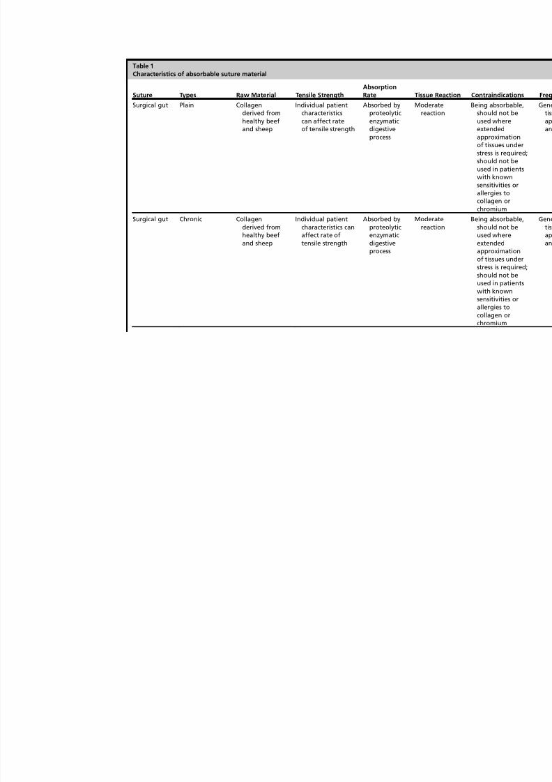
Table 1
Characteristics of absorbable suture material
Suture Types Raw Material Tensile Strength
Absorption
Rate Tissue Reaction
Surgical gut Plain Collagenderived fromhealthy beefand sheep
Individual patientcharacteristicscan affect rateof tensile strength
Absorbed byproteolyticenzymaticdigestiveprocess
Moderatereaction
Surgical gut Chronic Collagenderived fromhealthy beefand sheep
Individual patientcharacteristics canaffect rate oftensile strength
Absorbed byproteolyticenzymaticdigestiveprocess
Moderatereaction

8/13/2019 SutuSuturing Principles for Dentoalveolar Surgeonring Principles for Dentoalveolar Surgeon
http://slidepdf.com/reader/full/sutusuturing-principles-for-dentoalveolar-surgeonring-principles-for-dentoalveolar 5/23
Polyglactin910 Vicryl(Ethicon)
Braidedmonofilament
Copolymer oflactide andglycolidecoated withpolyglactin370 andcalcium
stearate
w75% remains at2 wk; w50%remains at 3 wk
Completebetween56 and 90 dAbsorbed byhydrolysis
Minimal acuteinflammatoryreaction
Polyglycolicacid Dexon(USS/DG)
Braided (coated) Polyglycolic acidpolycaprolatecoating system(copolymer ofglycolide and 3-caprolactone)
w65% remains at2 wk; w35%remains at 3 wk
Essentiallycompletebetween 60and 90 d,absorbedby hydrolysis
Minimal acuteinflammatoryreaction
8/13/2019 SutuSuturing Principles for Dentoalveolar Surgeonring Principles for Dentoalveolar Surgeon
http://slidepdf.com/reader/full/sutusuturing-principles-for-dentoalveolar-surgeonring-principles-for-dentoalveolar 6/23
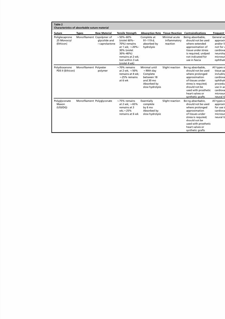
Table 2
Characteristics of absorbable suture material
Suture Types Raw Material Tensile Strength Absorption Rate Tissue Reaction Co
Poliglecaprone25 Monocryl(Ethicon)
Monofilament Copolymer ofglycolide and 3-caprolactone
w50%–60%(violet 60%–70%) remainsat 1 wk; w20%–30% (violet30%–40%)remains at 2 wk;lost within 3 wk(violet 4 wk)
Complete at91–119 d;absorbed byhydrolysis
Minimal acuteinflammatoryreaction
Beiswatinu
PolydioxanonePDS II (Ethicon)
Monofilament Polyesterpolymer
w70% remainsat 2 wk; w50%
remains at 4 wk;w25% remainsat 6 wk
Minimal untilw90th day
Completebetween 18and 30 moAbsorbed byslow hydrolysis
Slight reaction Bes
waossuhs
PolyglyconateMaxon(USS/DG)
Monofilament Polyglyconate w75% remainsat 2 wk; w65%remains at 3
wk;w
25%remains at 6 wk
Essentiallycompleteby 6 mo
Absorbed byslow hydrolysis
Slight reaction Besw
aossuhs

8/13/2019 SutuSuturing Principles for Dentoalveolar Surgeonring Principles for Dentoalveolar Surgeon
http://slidepdf.com/reader/full/sutusuturing-principles-for-dentoalveolar-surgeonring-principles-for-dentoalveolar 7/23
Table 3
Characteristics of nonabsorbable suture material
Suture Types Raw Material Tensile Strength Absorption Rate Tissue Reaction
Silk suture Braided Organic proteincalled fibroin
Progressivedegradation offiber may resultin gradual lossof tensilestrengthover time
Gradualencapsulationby fibrousconnectivetissue
Acuteinflammatoryreaction
Nylon sutureEthilon (Ethicon)Dermalon
(USS/DG)
Monofilament Long-chainaliphaticpolymers
nylon 6 ornylon 6,6
Progressivehydrolysis mayresult in
a gradual lossof tensilestrengthover time
Gradualencapsulationby fibrous
connectivetissue
Minimalinflammatoryreaction
Polyester fibersuture ersiline(Ethicon) Dacron(USS/DG)
Braidedmonofilament
Poly (ethyleneterephthalate)
No significantchange knownto occur in vivo
Gradualencapsulationby fibrousconnectivetissue
Minimalinflammatoryreaction
Polypropylenesuture Prolene
(Ethicon)Surgiline(USS/DG)
Monofilament Isotacticcrystalline
stereoisomer ofpolypropylene
Not subject todegradation
or weakeningby action oftissue enzymes
Nonabsorbable Minimalinflammatory
reaction

8/13/2019 SutuSuturing Principles for Dentoalveolar Surgeonring Principles for Dentoalveolar Surgeon
http://slidepdf.com/reader/full/sutusuturing-principles-for-dentoalveolar-surgeonring-principles-for-dentoalveolar 8/23
temporal phases of tissue reactivity around sutures.16 Selvig and colleagues16 found
distinct concentric perisutural zones after histologic processing and analysis. An
acute-phase response of neutrophil infiltration was observed up to 3 days, mainly
reflecting the initial surgical trauma in suture placement. This response was compa-
rable in all suture materials tested.16 The neutrophilic infiltration is soon replaced by
chronic cellular infiltrates, including monocytes, plasma cells, and lymphocytes.13
These stages of tissue reactivity remove cellular debris and suture material.13 Peak
tissue reaction occurs between the second and seventh days.16 Theoretically, in
favorable healing conditions, this acute phase should be replaced by the formation
of granulation tissue in the absence of inflammatory cells.16 Progressive inflammatory
reactions caused by sutures may persist for as long as 7 to 14 days.16
Bacterial migration along the suture track has been documented.2,3,16 Although
braided suture has been reported to promote bacterial retention and growth because
of its physical composition, Selvig and colleagues16 found bacteria plaque migration
extending more than 100 mm into suture channels at 14 days regardless of the suture
material tested, except for gut, which had rapidly dissipated by this time. Sutures that
remain in intraoral wounds, such as silk, cause epithelial tracks, thereby increasing the
propensity for bacterial migration.16 In general, sutures should be removed no later
than 7 to 10 days.16 The loss of tensile strength and rate of absorption are separate
and distinct phenomena.3 Sutures may rapidly lose adequate tensile strength, but
be absorbed slowly, or vice versa. Fever, infection, or protein-deficient states may
accelerate the absorption process and cause an increase in loss of tensile strength.2,7
Moist or fluid-filled tissue such as the oral cavity, or soaking sutures in saline for
extended periods, may also accelerate the absorption process.2,3,7
SUTURE SELECTION
When selecting a suture material, consideration must be given to the duration the
suture must remain and the relationship the suture has with adjacent tissue. The small-
est suture that couples the least immunogenicity with the highest tensile strength is
preferable.5 This article examines suture performance by degree of absorbability.
Nonabsorbable sutures resist enzymatic activity and hydrolysis. One of the most
widely used nonabsorbable sutures is silk. This raw fiber is harvested while the silk-
worm larvae are spinning the cocoons. This material is processed into a braided fiber,
sterilized, and used in a variety of surgical settings. Although it is classified as a nonab-sorbable suture, histologic analysis of silk in vivo after 2 years revealed no evidence of
remaining suture.5 Synthetic absorbable sutures include nylon, polypropylene, and
polyester. These sutures are chemically synthesized polymers that vary in their phys-
ical properties and chemical structure, and are manufactured as monofilament or
braided strands. Nylon is a polyamide polymer that may either be monofilament or
braided and can be dyed in a variety of colors.5 Polyester sutures are polyethylene
terephthalate braided multifilament strands that produce little inflammatory response
but tend to create more tissue tearing.5 Polypropylene-based sutures have low immu-
nogenicity compared with most other nonabsorbable sutures with high breaking
strength. As with nonabsorbable sutures, absorbable sutures are classified as either monofil-
ament or multifilament. The natural form of absorbable sutures is surgical gut that may
be further subdivided as plain or chromic. Gut suture is rendered from bovine or sheep
submucosal intestinal layer and processed to the desired size. Once processed, the
gut suture is either packaged as plain gut or is treated to lengthen the absorption
time. In vivo, gut is digested by proteolytic enzymes in macrophage activity. This
Brandt & Jenkins288

8/13/2019 SutuSuturing Principles for Dentoalveolar Surgeonring Principles for Dentoalveolar Surgeon
http://slidepdf.com/reader/full/sutusuturing-principles-for-dentoalveolar-surgeonring-principles-for-dentoalveolar 9/23
absorption can be prolonged if the suture is treated with acromion salt solution. Thus,
in infected wounds, dentists may choose to use chromic gut suture for wound closure
because of the delayed absorption.
Synthetic absorbable sutures were designed to bypass problems encountered
with gut suture immunogenicity and unpredictability in absorption. Historically, the
synthetic absorbable sutures include polyglycolic acid (Dexon), polyglactin 910
(Vicryl), and polydioxanone (PDS). Polyglycolic acid (Dexon) was the first synthetic
absorbable suture with handling characteristics similar to silk, but has greater
tensile strength than gut.2 It is produced in a braided multifilament form. Polyglactin
910 (Vicryl) is a copolymer of lactic and polyglycolic acid that has a 50% tensile
strength for 3 weeks and then resorbs within 90 days. It is produced in a braided
multifilament form. Poliglecaprone 25 (Monocryl) is composed of glycoside and
3-caprolactone. It offers high initial tensile strength and absorbs by 119 days.
Another synthetic material suture is PDS, a polydioxanone polymer that forms
a monofilament suture with enhanced flexibility and significantly greater tensile
strength than both polyglycolic acid and polyglactin 910.2 Absorbable synthetic
suture can also be obtained in a coated form that inhibits bacterial colonization of
the suture. Ethicon Vicryl Plus coated antibacterial suture contains Irgacare MP,
a pure form of triclosan, which is a broad-spectrum antibacterial agent that creates
a zone of inhibition, preventing bacterial colonization by the pathogens that most
often cause surgical site infections.
NEEDLE SELECTION
The purpose of the needle is to transport the chosen suture material through the softtissues with the least amount of traumatic injury. A balance must be maintained
between needle rigidity and flexibility. Too rigid a needle may fracture when met
with resistance, whereas one that is too flexible may not accurately travel to the desig-
nated exit point.5 The material of choice in contemporary needle design is a corrosion-
resistant stainless steel alloy. The needle itself has 3 designated regions: the eye, the
body, and the point. The entire complex begins as a sharp point and expands in diam-
eter to the eye. The eye of the needle harbors the interface of the suture material. There
are 3 types of needle eyes: closed, split (French), and swaged. The closed and split
types are less desirable because the junction of the needle and the suture is often
enlarged, increasing the risk of tissue trauma, and must be threaded, which is timeconsuming. The needle of choice today is a swaged eyeless needle, in which the junc-
tion of the needle and suture is uniform and permanent. The body of the needle is man-
ufactured in a variety of sizes, shapes, and curvatures. Needles may be round, flat,
triangular, oval, or tapered. The needles most frequently used in dentoalveolar surgery
are curved needles that range in shape from 1/4 to 5/8 of a circle ( Fig. 1 ). The appro-
priate needle must be based on the dimensions of the wound to be closed. The point
of the needle is the initial contact point of the needle with the tissue. Three basic nee-
dle points exist: tapered, blunt, or cutting. In dentoalveolar surgery, cutting needles
are preferred because of the thickness, resilience, and resistance of the gingiva and
oral mucosa. The 2 basic cutting needles used in dentoalveolar surgery are conven-tional (cutting) and reverse cutting needles ( Fig. 2 ). Each has 3 cutting edges with 2
edges opposing each other. The conventional has a third edge facing upwards
(toward the inside of the circle), whereas, in the reverse cutting needle, it faces down.
Suture materials have their own classification system for needle size and shape.
When selecting the desired suture and needle combination, close attention must be
paid to each manufacturer’s supply-order charts for the proper needle selection.
Dentoalveolar Suturing 289

8/13/2019 SutuSuturing Principles for Dentoalveolar Surgeonring Principles for Dentoalveolar Surgeon
http://slidepdf.com/reader/full/sutusuturing-principles-for-dentoalveolar-surgeonring-principles-for-dentoalveolar 10/23
Fig. 1. Degree of needle curvature: ( A) 1/4 circle, (B) 3/8 circle, (C ) 1/2 circle, (D) 3/4 circle.(From Peterson LJ, editor. Contemporary oral and maxillofacial surgery. 3rd edition: MosbyYear Book; 1998. p. 54; with permission.)
Fig. 2. Needles used in dentoalveolar surgery. (Left ) Conventional. (Right ) Reverse cutting,regular cutting. (From Peterson LJ, editor. Contemporary oral and maxillofacial surgery.3rd edition: Mosby Year Book; 1998. p. 54; with permission.)
Brandt & Jenkins290

8/13/2019 SutuSuturing Principles for Dentoalveolar Surgeonring Principles for Dentoalveolar Surgeon
http://slidepdf.com/reader/full/sutusuturing-principles-for-dentoalveolar-surgeonring-principles-for-dentoalveolar 11/23
INSTRUMENTATION
Intraoral suturing requires 2 main instruments: the needle driver and the suture scis-
sors. Tissue forceps may occasionally be used to ensure proper needle entrance
through tissue by ensuring stability of the soft tissue flap. These tissue forceps are
manufactured in various sizes, with and without teeth. The most common tissue
forceps used is a single-toothed 7.6-cm (3-inch) Adson forceps ( Fig. 3 ). The needle
driver most commonly used is the 15.2-cm (6-inch) version of the Hegar-Mayo type
( Fig. 4 ).17 Needle drivers are also manufactured with a variety of sizes and beaks,
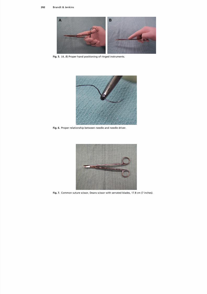
with or without teeth. A needle driver is most effective when using proper hand posi-
tion. The needle driver is held in the palm with the fourth (ring) finger and the thumb
within the rings of the instrument. The second finger is placed along the lower straight
arm for stabilization, and the third ring finger is laid passively outside the ring of the
fourth finger ( Fig. 5 ). The beaks of the needle driver should be perpendicular to the
needle and held one-third of the distance from the origin of the suture ( Fig. 6 ). The
weakest part of the needle is located at the junction of the needle body in the portion
where the suture is affixed to the needle. If the beaks of the needle driver are placed
too close to the swaged end, bending or breakage of the needle may occur on
insertion in the tissue. A bend in the needle may not be visible to the clinician, but
Fig. 3. Common tissue forceps, single-toothed Adson, 7.6 cm (3 inches).
Fig. 4. Common needle driver, Hegar-Mayo type, 15.2 cm (6 inches).
Dentoalveolar Suturing 291
8/13/2019 SutuSuturing Principles for Dentoalveolar Surgeonring Principles for Dentoalveolar Surgeon
http://slidepdf.com/reader/full/sutusuturing-principles-for-dentoalveolar-surgeonring-principles-for-dentoalveolar 12/23
Fig. 5. ( A, B) Proper hand positioning of ringed instruments.
Fig. 6. Proper relationship between needle and needle driver.
Fig. 7. Common suture scissor, Deans scissor with serrated blades, 17.8 cm (7 inches).
Brandt & Jenkins292

8/13/2019 SutuSuturing Principles for Dentoalveolar Surgeonring Principles for Dentoalveolar Surgeon
http://slidepdf.com/reader/full/sutusuturing-principles-for-dentoalveolar-surgeonring-principles-for-dentoalveolar 13/23
nonetheless can cause sufficient metal fatigue to result in unexpected needle
breakage. Holding the needle too close to the sharp tip limits the length of needle
available to pass through tissue. The most common suture scissor is the Dean scissor,
an instrument that is 17.8 cm (7 inches) long and has offset serrated blades ( Fig. 7 ).17 It
should be held in the same fashion as the needle driver. It is most efficient when
Fig. 8. Most intraoral sutures are tied with instrument tie. ( A) Suture is pulled through tissueuntil short tail of suture (approximately 1.3–5 cm long) remains. Needle holder is held hor-izontally by right hand in preparation for knot-tying procedure. (B) Left hand then wrapslong end of suture around needle holder twice in clockwise direction to make 2 loops ofsuture around needle holder. (C ) Surgeon then opens needle holder and grasps short endof suture near its end. (D) Ends of suture are then pulled to tighten knot. Needle holdershould not pull until knot is nearly tied, to avoid lengthening that portion of suture. ( E )End of first step of surgeon’s knot. Note that double wrap has resulted in double overhandknot, which increases friction in knot and keeps wound edges together until second portion
of knot is tied. (F ) Needle holder is then released from short end of suture and held in sameposition as when knot-tying procedure began. Left hand then makes single wrap in coun-terclockwise direction. (G) Needle holder then grasps short end of suture at its end. (H )This portion of knot is completed by pulling this loop firmly down against previous portionof knot. (I ) This completes surgeon’s knot. Double loop of first pass holds tissue togetheruntil second portion of square knot can be tied. ( J ) Most surgeons add a third throw to theirinstrument tie. Needle holder is repositioned in original position, and 1 wrap is placedaround needle holder in original clockwise direction. Short end of suture is grasped andtightened down firmly to form second square knot. Final throw of 3 knots is tightenedfirmly. (From Peterson LJ, editor. Contemporary oral and maxillofacial surgery, 3rd edition:Mosby Year Book; 1998. p. 188–9; with permission.)
Dentoalveolar Suturing 293
8/13/2019 SutuSuturing Principles for Dentoalveolar Surgeonring Principles for Dentoalveolar Surgeon
http://slidepdf.com/reader/full/sutusuturing-principles-for-dentoalveolar-surgeonring-principles-for-dentoalveolar 14/23
cutting the suture perpendicular to its blades. The Dean scissor is also a general-
purpose scissor that may be used in trimming or removing tissue.
SUTURE TECHNIQUES
There is a multitude of suturing techniques available to the dentist. The techniqueselected depends on the breadth and length of the wound, the closure tension
required, and the distance to which wound edges must move. Techniques are broadly
categorized as interrupted and continuous: interrupted techniques include simple
interrupted, horizontal/vertical mattress, and sling; continuous suturing is divided
into running, locking, and continuous sling. Choosing between an interrupted and
continuous technique requires striking a balance between the ease and rapidity of
the continuous techniques and the additional stability and control of wound edges
offered by interrupted techniques.
Simple Interrupted
This is the most universal technique in practice today and may be used for small
wounds, or evenly spaced or continuous to close larger wounds. Placement requires
entrance of the suture through both wound margins, with the surgeons knot for
stability. Placed correctly, the suture should slightly evert the wound edges ( Fig. 8 ).18
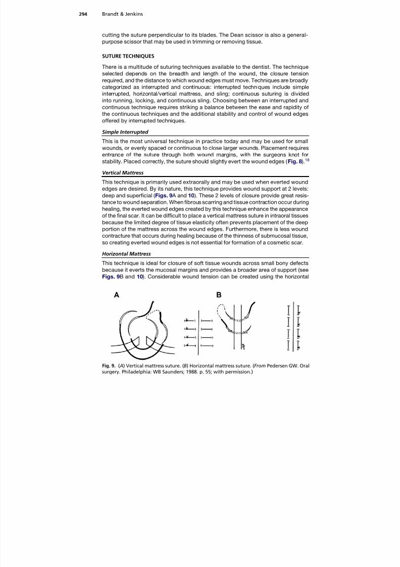
Vertical Mattress
This technique is primarily used extraorally and may be used when everted wound
edges are desired. By its nature, this technique provides wound support at 2 levels:
deep and superficial ( Figs. 9 A and 10 ). These 2 levels of closure provide great resis-tance to wound separation. When fibrous scarring and tissue contraction occur during
healing, the everted wound edges created by this technique enhance the appearance
of the final scar. It can be difficult to place a vertical mattress suture in intraoral tissues
because the limited degree of tissue elasticity often prevents placement of the deep
portion of the mattress across the wound edges. Furthermore, there is less wound
contracture that occurs during healing because of the thinness of submucosal tissue,
so creating everted wound edges is not essential for formation of a cosmetic scar.
Horizontal Mattress
This technique is ideal for closure of soft tissue wounds across small bony defectsbecause it everts the mucosal margins and provides a broader area of support (see
Figs. 9B and 10 ). Considerable wound tension can be created using the horizontal
Fig. 9. ( A) Vertical mattress suture. (B) Horizontal mattress suture. (From Pedersen GW. Oralsurgery. Philadelphia: WB Saunders; 1988. p. 55; with permission.)
Brandt & Jenkins294

8/13/2019 SutuSuturing Principles for Dentoalveolar Surgeonring Principles for Dentoalveolar Surgeon
http://slidepdf.com/reader/full/sutusuturing-principles-for-dentoalveolar-surgeonring-principles-for-dentoalveolar 15/23
Fig. 10. ( A) A 58-year-old man with a large area of bone loss over the maxillary right centralincisor. The tooth was mobile and had a draining fissure present over the labial surface ofthe tooth at the level of the apex of the tooth. (B) Periapical radiograph showing significantbone loss to approximately 3 mm from the apex of the tooth. This large restoration hadbeen stable for 14 years before the current problem. The patient was placed on antibioticsand prescribed a mouth rinse to decrease the bacteria flora and was appointed for surgery.(C ) The tooth was extracted easily after incisions were made around the neck of the tooth.After the tooth was removed, there was a large area of bone loss, extending 9 mm from thegingival margin. Even with the 9-mm pocket that was present on the labial aspect of thetooth, the gingiva form matched the level on the adjacent tooth. (D) A graft of humanmineralized bone was placed into the defect and compacted to recreate root form anatomyand the labial aspect of the socket. (E ) A piece of collagen was placed and retained by a hori-zontal mattress suture. (F ) The area approximately 4 months after graft placement, indi-cating sufficient form of the gingiva and root prominence. (G) After a crestal incision andsmall reflection in the sulci of the adjacent teeth, sufficient bone was found for placementof a 4-mm diameter implant. (H ) The implant was placed approximately 3 mm apical to theadjacent gingival margin. After the implant was placed, bone harvested from the drills wasplaced over the labial aspect to further augment the site. (I ) The site was closed with 2vertical mattress sutures everting the interdental papilla and to advance the flaps coronally.
( J ) A central incisor was extracted with loss of a significant amount of labial bone. There wasvertical palatal bone present but no labial bone superior to the nasal floor. (K ) Bovine miner-alized bone was compacted into the site to recreate the root prominence and to fill thespace that was previously occupied by the root of the tooth. ( L) A collagen material (Colla-plug) was placed over the bovine graft and was retained in position with 2 horizontalmattress silk sutures. (From Block MS, Jackson WC. Techniques for grafting the extractionsite in preparation for dental implant placement. Atlas of oral and maxillofacial surgery.vol. 14. Elsevier; 2006. p. 2; with permission.)
Dentoalveolar Suturing 295

8/13/2019 SutuSuturing Principles for Dentoalveolar Surgeonring Principles for Dentoalveolar Surgeon
http://slidepdf.com/reader/full/sutusuturing-principles-for-dentoalveolar-surgeonring-principles-for-dentoalveolar 16/23
mattress technique; the mattress spreads the forces along the horizontal band of
tissue instead of focusing at a single point, thus decreasing the chance for the suture
to pull through and lacerate tissue during wound approximation and knot tying. The
horizontal mattress technique is frequently used during closure of an oroantral
opening, where tension-free, watertight wound closure is required to prevent
exchange of air, saliva, and mucus between the sinus and oral cavity.
Figure-eight
This technique can be used effectively to close the tissue of an extraction site.
Although not frequently used to gain primary closure, it can provide a barrier to
dislodgment of a clot after tooth extraction and may help stabilize materials placed
into an extraction socket, such as Gelfoam, bone grafting materials, collagen plugs,
or other packing materials ( Figs. 11 and 12 ).
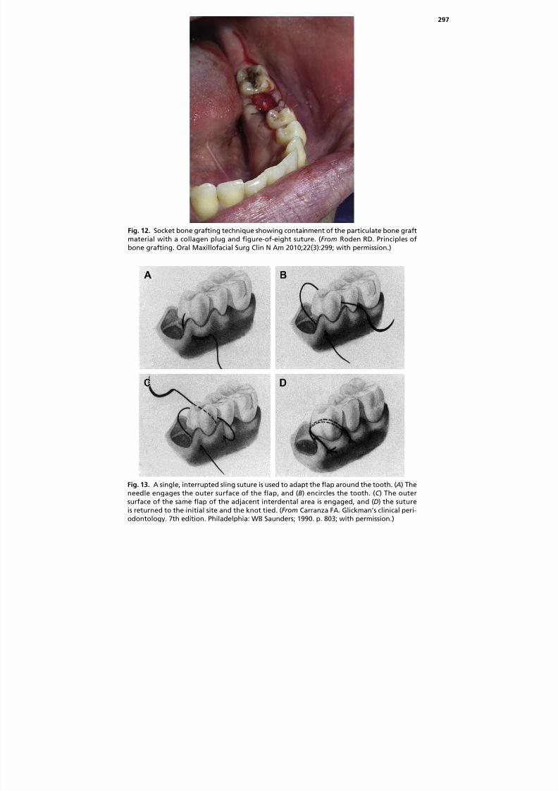
Sling Ligation
This technique is ideally used in surgery where a flap is elevated only on one side of the
alveolus. This technique allows repositioning of the flap without entering the opposing
intact soft tissue ( Figs. 13 and 14 ).
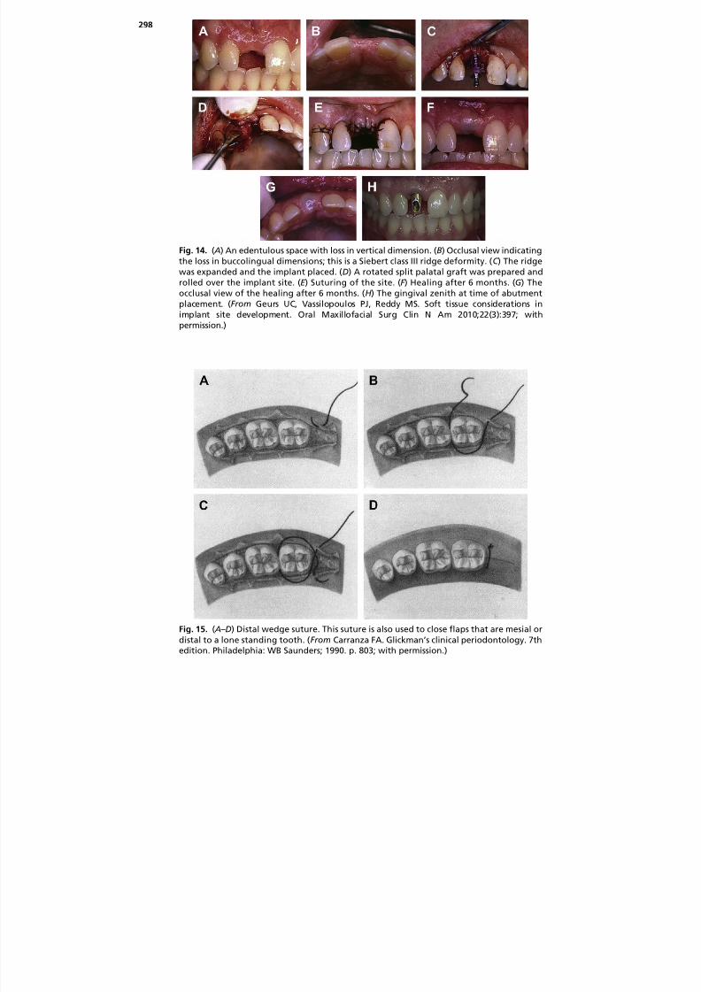
Anchor Suture
This technique is indicated for closure of mucosa in edentulous areas either mesial or
distal to a tooth. This technique offers tight closure of the buccal and lingual soft
tissues and causes increased adaptation to the adjacent tooth ( Fig. 15 ).
Continuous SuturesThis technique is commonly used in dentoalveolar surgery when longer wounds result.
Dentists often use this technique following multiple or full-mouth tooth extractions that
leave a wound that spans the entire alveolus. This method offers quicker closure
because few knots are placed over the entire length of the wound. A major disadvan-
tage in this type of suturing is that, if one knot fails, the closure is compromised. This
simple technique has a potential to obliquely apply pressure along the length of the
wound, whereas the locked technique has a lesser potential ( Figs. 16 and 17 ).18
Fig. 11. Figure-of-eight suture techniques. (From Kwon PH, Laskin DM. Clinician’s manual oforal and maxillofacial surgery. 2nd edition: Quintessence Publishing; 1997. p. 249; withpermission.)
Brandt & Jenkins296
8/13/2019 SutuSuturing Principles for Dentoalveolar Surgeonring Principles for Dentoalveolar Surgeon
http://slidepdf.com/reader/full/sutusuturing-principles-for-dentoalveolar-surgeonring-principles-for-dentoalveolar 17/23
Fig. 12. Socket bone grafting technique showing containment of the particulate bone graftmaterial with a collagen plug and figure-of-eight suture. (From Roden RD. Principles ofbone grafting. Oral Maxillofacial Surg Clin N Am 2010;22(3):299; with permission.)
Fig. 13. A single, interrupted sling suture is used to adapt the flap around the tooth. ( A) Theneedle engages the outer surface of the flap, and (B) encircles the tooth. (C ) The outersurface of the same flap of the adjacent interdental area is engaged, and (D) the sutureis returned to the initial site and the knot tied. (From Carranza FA. Glickman’s clinical peri-odontology. 7th edition. Philadelphia: WB Saunders; 1990. p. 803; with permission.)
297
8/13/2019 SutuSuturing Principles for Dentoalveolar Surgeonring Principles for Dentoalveolar Surgeon
http://slidepdf.com/reader/full/sutusuturing-principles-for-dentoalveolar-surgeonring-principles-for-dentoalveolar 18/23
Fig. 14. ( A) An edentulous space with loss in vertical dimension. (B) Occlusal view indicatingthe loss in buccolingual dimensions; this is a Siebert class III ridge deformity. (C ) The ridgewas expanded and the implant placed. (D) A rotated split palatal graft was prepared androlled over the implant site. (E ) Suturing of the site. (F ) Healing after 6 months. (G) Theocclusal view of the healing after 6 months. (H ) The gingival zenith at time of abutmentplacement. (From Geurs UC, Vassilopoulos PJ, Reddy MS. Soft tissue considerations inimplant site development. Oral Maxillofacial Surg Clin N Am 2010;22(3):397; withpermission.)
Fig. 15. ( A–D) Distal wedge suture. This suture is also used to close flaps that are mesial ordistal to a lone standing tooth. (From Carranza FA. Glickman’s clinical periodontology. 7thedition. Philadelphia: WB Saunders; 1990. p. 803; with permission.)
298

8/13/2019 SutuSuturing Principles for Dentoalveolar Surgeonring Principles for Dentoalveolar Surgeon
http://slidepdf.com/reader/full/sutusuturing-principles-for-dentoalveolar-surgeonring-principles-for-dentoalveolar 19/23
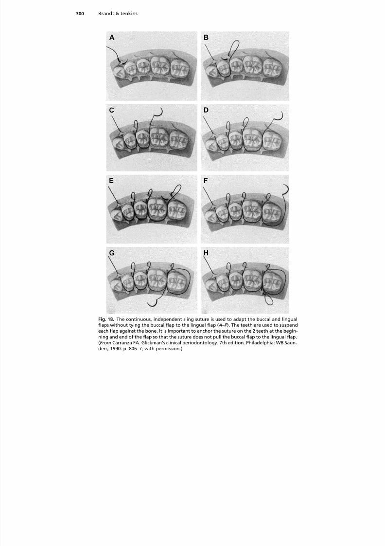
Continuous Sling
This technique is the continuous version of the isolated sling technique, and may be
used when both buccal and lingual flaps have been reflected. This closure allows
both flaps to be repositioned independently because of their anchors or the abutment
teeth at either end of the wound ( Fig. 18 ).19,20
Combination
This technique may include a continuous suture with overlaying vertical or horizontal
mattress sutures and/or simple interrupted sutures ( Fig. 19 ). The continuous suture is
often absorbable with a nonabsorbable or more slowly absorbable suture placed over
Fig. 16. When multiple sutures are to be placed, the incision can be closed with running orcontinuous sutures. ( A) First, the papilla is closed and the knot tied in the usual way. Thelong end of the suture is held, and the adjacent papilla is sutured, without the knot beingtied but just with the suture being pulled firmly through the tissue. (B) Succeeding papillaeare then sutured until the final one is sutured and the final knot is tied. Final appearance iswith the suture going across each empty socket. (C ) Continuous locking stitch can be madeby passing the long end of the suture underneath the loop before it is pulled through thetissue. (D) This stage puts the suture on both deep periosteal and mucosal surfaces directlyacross the papilla and may aid in more direct apposition of tissues. (From Peterson LJ, editor.Principles of complicated exodontia. In: Contemporary oral and maxillofacial surgery. 3rdedition. Mosby Year Book; 1998. p. 191; with permission.)
Fig. 17. Continuous chromic gut suture with overlaid simple, interrupted silk sutures in theanterior maxilla.
Dentoalveolar Suturing 299
8/13/2019 SutuSuturing Principles for Dentoalveolar Surgeonring Principles for Dentoalveolar Surgeon
http://slidepdf.com/reader/full/sutusuturing-principles-for-dentoalveolar-surgeonring-principles-for-dentoalveolar 20/23
Fig. 18. The continuous, independent sling suture is used to adapt the buccal and lingualflaps without tying the buccal flap to the lingual flap ( A–P ). The teeth are used to suspendeach flap against the bone. It is important to anchor the suture on the 2 teeth at the begin-ning and end of the flap so that the suture does not pull the buccal flap to the lingual flap.(From Carranza FA. Glickman’s clinical periodontology. 7th edition. Philadelphia: WB Saun-ders; 1990. p. 806–7; with permission.)
Brandt & Jenkins300

8/13/2019 SutuSuturing Principles for Dentoalveolar Surgeonring Principles for Dentoalveolar Surgeon
http://slidepdf.com/reader/full/sutusuturing-principles-for-dentoalveolar-surgeonring-principles-for-dentoalveolar 21/23
the first layer of superficial suturing. This technique helps prevent wound dehiscence if
the continuous suture resorbs before initial wound healing has occurred. Dentists
often use this technique with larger dentoalveolar defects and cases that require graft-
ing (soft and hard tissue).
Fig. 18. (continued )
Dentoalveolar Suturing 301

8/13/2019 SutuSuturing Principles for Dentoalveolar Surgeonring Principles for Dentoalveolar Surgeon
http://slidepdf.com/reader/full/sutusuturing-principles-for-dentoalveolar-surgeonring-principles-for-dentoalveolar 22/23
SUMMARY
When suturing is required for wound closure, dentists should be aware of the charac-
teristics of suture material so the most appropriate material can be selected, and thetechnique used should provide effectiveness and ease. The authors frequently recom-
mend that dentists who routinely perform dentoalveolar surgery, such as the extraction
of teeth, should have at least 1 type of absorbable and 1 type of nonabsorbable suture
readily available within their operatory supply. Commonly, 3-0 or 4-0 chromic gut
suture and 3-0 or 4-0 silk suture can be used successfully to close nearly any type of
intraoral wound following dentoalveolar surgery. Familiarity with the concepts
presented in this article and continuous practice of the surgical skills presented
enhances surgical acumen and allows for improved healing, increased postoperative
comfort, and successful surgery.
REFERENCES
1. Macht SD. Sutures and suturing-current concepts. J Oral Surg 1978;36:710–2.
2. Gutman JL, Harrison JW. Surgical endodontics. Saint Louis (MO): Ishiyaku Euro-
America; 1994. p. 278–99.
3. Wound closure manual. Somerville (NJ): Ethicon; 2007.
4. Unites States Pharmacopeia (USP). 24th edition. National Formulary. 17th edition.
Philadelphia: National Publishing; 1999. p. 1584–6.
5. Knot tying manual. Somerville (NJ): Ethicon; 2007.
6. Romfh RF, Cramer FS. Technique and the use of surgical tools, 2nd edition.Norwalk (CT): Appleton and Lang; 1992. p. 33–124.
7. Herrman JB. Changes in tensile strength and knot security of surgical sutures
in vivo. Arch Surg 1973;106:707–10.
8. Greenwald D, Shumway S, Albear P, et al. Mechanical comparison of 10 suture
materials before and after in vivo incubation. J Surg Res 1994;56:372–7.
9. Maves TJ, Pechman PS, Gebhart GF, et al. Possible chemical contribution from
chromic gut sutures produces disorder of pain sensation like those seen in
man. Pain 1993;54:57–69.
10. Wallace WR, Maxwell GR, Cacalaris CJ. Comparison of polyglycolic acid suture to
black silk, chromic and plain gut in human oral tissues. J Oral Surg 1970;28:739–46.11. Lilly GE. Reaction of oral tissues to suture materials. Oral Surg Oral Med Oral
Pathol 1968;26:128–33.
12. Lilly GE, Armstrong JH, Salem GE, et al. Reaction of oral tissues to suture
materials. Part II. Oral Surg Oral Med Oral Pathol 1968;26:592–9.
13. Lilly GE, Salem JE, Armstrong JH, et al. Reaction of oral tissues to suture
materials. Part III. Oral Surg Oral Med Oral Pathol 1969;28:432–8.
Fig. 19. Combination of absorbable and nonabsorbable simple, interrupted sutures,mattress sutures, and continuous sutures for wound closure in the anterior mandible.
Brandt & Jenkins302

8/13/2019 SutuSuturing Principles for Dentoalveolar Surgeonring Principles for Dentoalveolar Surgeon
http://slidepdf.com/reader/full/sutusuturing-principles-for-dentoalveolar-surgeonring-principles-for-dentoalveolar 23/23
14. Lilly GE, Cutcher JL, Jones JC, et al. Reaction of oral tissues to suture materials.
Part IV. Oral Surg Oral Med Oral Pathol 1972;33:152–7.
15. DeNardo GA, Brown AN, Trenka-Benthin S, et al. Comparison of seven different
suture materials in the feline oral cavity. J Am Anim Hosp Assoc 1996;32:164–72.
16. Selvig KA, Biagiotti GR, Leknes KN, et al. Oral tissue reactions to suture mate-
rials. Int J Periodontics Restorative Dent 1998;18:475–87.
17. Pedersen GW. Oral surgery. Philadelphia: WB Saunders; 1998. p. 47–81.
18. Moore UJ. Principles of oral and maxillofacial surgery. Mauldin (MA): Blackwell
Science; 2001. p. 77–81.
19. Newman MG, Takei H, Carranza FA, et al. Glickman’s clinical periodontology. 10th
edition. Philadelphia: WB Saunders; 2006. p. 800–10.
20. Unites States Surgical Corporation, a unit of Covidien (formerly a division of Tyco
Healthcare Group, LP). Products by material. Available at: www.syneture.com.
Accessed July 21, 2011.
Dentoalveolar Suturing 303