Embed Size (px)
Citation preview
u n i ve r s i t y o f co pe n h ag e n
Sustained involuntary muscle activity in cerebral palsy and stroke
same symptom, diverse mechanisms
Forman, Christian Riis; Svane, Christian; Kruuse, Christina; Gracies, Jean-Michel; Nielsen,Jens Bo; Lorentzen, Jakob
Published in:Brain Communications
DOI:10.1093/braincomms/fcz037
Publication date:2019
Document versionPublisher's PDF, also known as Version of record
Document license:CC BY
Citation for published version (APA):Forman, C. R., Svane, C., Kruuse, C., Gracies, J-M., Nielsen, J. B., & Lorentzen, J. (2019). Sustainedinvoluntary muscle activity in cerebral palsy and stroke: same symptom, diverse mechanisms. BrainCommunications, 1(1), [37]. https://doi.org/10.1093/braincomms/fcz037
Download date: 09. jan.. 2022
Sustained involuntary muscle activity incerebral palsy and stroke: same symptom,diverse mechanisms
Christian Riis Forman,1,* Christian Svane,1,* Christina Kruuse,2 Jean-Michel Gracies,3
Jens Bo Nielsen1,4 and Jakob Lorentzen1,4
* These authors contributed equally to this work.
Individuals with lesions of central motor pathways frequently suffer from sustained involuntary muscle activity. This symptom
shares clinical characteristics with dystonia but is observable in individuals classified as spastic. The term spastic dystonia has been
introduced, although the underlying mechanisms of involuntary activity are not clarified and vary between individuals depending
on the disorder. This study aimed to investigate the nature and pathophysiology of sustained involuntary muscle activity in adults
with cerebral palsy and stroke. Seventeen adults with cerebral palsy (Gross Motor Function Classification System I–V), 8 adults
with chronic stroke and 14 control individuals participated in the study. All individuals with cerebral palsy or stroke showed
increased resistance to passive movement with Modified Ashworth Scale >1. Two-minute surface EMG recordings were obtained
from the biceps muscle during attempted rest in three positions of the elbow joint; a maximally flexed position, a 90-degree pos-
ition and a maximally extended position. Cross-correlation analysis of sustained involuntary muscle activity from individuals with
cerebral palsy and stroke, and recordings of voluntary isometric contractions from control individuals were performed to examine
common synaptic drive. In total, 13 out of 17 individuals with cerebral palsy and all 8 individuals with stroke contained sustained
involuntary muscle activity. In individuals with cerebral palsy, the level of muscle activity was not affected by the joint position. In
individuals with stroke, the level of muscle activity significantly (P< 0.05) increased from the flexed position to the 90 degree and
extended position. Cumulant density function indicated significant short-term synchronization of motor unit activities in all record-
ings. All groups exhibited significant coherence in the alpha (6–15 Hz), beta (16–35 Hz) and early gamma band (36–60 Hz). The
cerebral palsy group had lower alpha band coherence estimates, but higher gamma band coherence estimates compared with the
stroke group. Individuals with increased resistance to passive movement due to cerebral palsy or stroke frequently suffer sustained
involuntary muscle activity, which cannot exclusively be described by spasticity. The sustained involuntary muscle activity in both
groups originated from a common synaptic input to the motor neuron pool, but the generating mechanisms could differ between
groups. In cerebral palsy it seemed to originate more from central mechanisms, whereas peripheral mechanisms likely play a larger
role in stroke. The sustained involuntary muscle activity should not be treated simply like the spinal stretch reflex mediated symp-
tom of spasticity and should not either be treated identically in both groups.
1 Department of Neuroscience, University of Copenhagen, 2200 Copenhagen, Denmark2 Department of Neurology, Neurovascular Research Unit, Herlev Gentofte Hospital, 2730 Herlev Gentofte, Denmark3 EA 7377 BIOTN, Universite Paris-Est Creteil, Hospital Albert Chenevier-Henri Mondor, Service de Reeducation Neurolocomotrice,APHP, Creteil, France4 Elsass Institute, 2830 Charlottenlund, Denmark
Received April 08, 2019. Revised October 24, 2019. Accepted October 28, 2019. Advance Access publication November 25, 2019VC The Author(s) (2019). Published by Oxford University Press on behalf of the Guarantors of Brain.
This is an Open Access article distributed under the terms of the Creative Commons Attribution Non-Commercial License (http://creativecommons.org/licenses/by-nc/4.0/), which per-
mits non-commercial re-use, distribution, and reproduction in any medium, provided the original work is properly cited. For commercial re-use, please contact
BBRAIN COMMUNICATIONSAIN COMMUNICATIONSdoi:10.1093/braincomms/fcz037 BRAIN COMMUNICATIONS 2019: Page 1 of 13 | 1
Dow
nloaded from https://academ
ic.oup.com/braincom
ms/article/1/1/fcz037/5640495 by R
oyal Library Copenhagen U
niversity user on 20 October 2020

Correspondence to: Jakob Lorentzen, PT, PhD, Department of Neuroscience, University of Copenhagen,
Panum Institute 33.3 Blegdamsvej 3, 2200 Copenhagen, Denmark
E-mail: [email protected]
Keywords: spastic dystonia; coherence; cerebral palsy; stroke
Abbreviations: CP¼ cerebral palsy; GMFCS¼Gross Motor Function Classification System; MAS¼Modified Ashworth Scale;
RMS¼ root mean square; SCPE¼ Surveillance of Cerebral Palsy in Europe
IntroductionA lesion to the central motor pathways is often followed
by a development of muscle overactivity that causes body
deformities and decreases joint mobility (Gracies, 2005;
Sheean and McGuire, 2009; Lorentzen et al., 2018).
The term muscle overactivity is not precise as it
includes several different conditions that although pre-
sumed pathologically distinct are not always easily distin-
guishable. This can be the cause of confusion and
misunderstanding among clinicians and researchers. The
confusion is frequently encountered in the discussion of
symptoms and classifications of cerebral palsy (CP) but
can also be related to other conditions of central motor
lesions such as stroke.
In the clinic, definitions by the Surveillance of Cerebral
Palsy in Europe (SCPE) are commonly used to distinguish
types of CP. The SCPE characterizes spastic CP by the
symptoms abnormal pattern of posture and/or movement,
persistently increased muscle tone and pathological
reflexes (Cans, 2008). In research settings, however, the
term muscle tone is often not considered scientifically pre-
cise enough, and we therefore prefer to use the term
increased resistance to passive movement, which we
believe refers to the same phenomenon. The definition of
spasticity, as an ‘enhancement of the velocity-dependent
stretch responses’ (Lance, 1980; Gracies, 2005), which is
frequently used in research publications, is therefore con-
tained within this classification but does not cover the
full range of symptoms in spastic CP. We believe that the
persistent increase in muscle tone described in the SCPE
classification of spastic CP is partly due to dystonia,
which in relation to the spastic movement disorder could
be referred to as spastic dystonia (Denny-Brown, 1966;
Sheean and McGuire, 2009; Lorentzen et al., 2018;
Trompetto et al., 2019). The condition of spastic dys-
tonia has received little attention in the scientific litera-
ture but is described as an inability to relax the muscle,
persisting despite a lack of voluntary neural activation
(Gracies, 2005; Sheean and McGuire, 2009; Lorentzen
et al., 2018; Trompetto et al., 2019). It has therefore
been suggested as an important contributor to body
deformities, abnormal joint mobility and increased resist-
ance during externally applied movement of affected
joints (Sheean and McGuire, 2009; Lorentzen et al.,
2018). It is our understanding, that the conditions of
muscle overactivity causing abnormal pattern of posture
and/or movement in spastic movement disorder include
Graphical Abstract
2 | BRAIN COMMUNICATIONS 2019: Page 2 of 13 C. R. Forman et al.
Dow
nloaded from https://academ
ic.oup.com/braincom
ms/article/1/1/fcz037/5640495 by R
oyal Library Copenhagen U
niversity user on 20 October 2020

spastic dystonia, spasticity, spastic co-contraction and
spasms (Gracies, 2005; Fig. 1).
Historically, muscle overactivity following a central
motor lesion has been viewed as an afferent phenomenon
only, demonstrated by dorsal root section abolishing in-
voluntary muscle activity in some animal studies
(Sherrington, 1898) and individuals with spastic paralysis
(Foerster, 1911). However, later studies (Pollock and
Davis, 1930; Denny-Brown, 1966) found that tonic invol-
untary muscle contractions, indicative of spastic dystonia,
persisted despite section of the dorsal roots in brain-
lesioned cats and monkeys. This led to the hypothesis
that the pathophysiology of spastic dystonia is, at least in
part, independent of spinal stretch reflex activity. Despite
these early observations on tonic muscle contractions
(Pollock and Davis, 1930; Denny-Brown, 1966), the
exact nature and cause of spastic dystonia remain to be
explored further. Some authors propose that the severity
of spastic dystonia is sensitive to the amount and dur-
ation of maintained muscle stretch (Gracies, 2005;
Trompetto et al., 2019) even though the phenomenon
also exists in the absence of stretch or effort.
Several mechanisms have been hypothesized to be
involved in spastic dystonia; changes in descending cor-
tical- or subcortical drive [e.g. involving brainstem
descending pathways (Miller et al., 2014; Sukal-Moulton
et al., 2014a, b)], plastic changes in spinal interneuron
networks and upregulation of persistent inward currents
in motoneurons (Fig. 1; for reviews, see Gracies, 2005 or
Lorentzen et al., 2018). The treatment options for muscle
overactivity are diverse, and the clinical population of
individuals with muscle overactivity following central
motor lesion represents several different types of lesions.
We hypothesize that the mechanisms responsible for in-
voluntary muscle activity may vary according to the type
of central motor lesion. This would promote the need for
increased focus on treatment individualization. The study
will, therefore, focus on two different groups with lesions
to the descending motor pathways, CP and chronic
stroke, differing mainly by the timing of the lesions. The
brain damage during early development in CP might lead
to different adaptations compared with brain damage
during late adulthood in stroke. The aim of this study is,
therefore, to investigate and identify the nature and
pathophysiology of sustained involuntary muscle activity
in individuals with CP or stroke and to relate this to the
phenomenon of spastic dystonia.
Materials and methods
Participants
Two groups of individuals with movement disorder and
increased resistance to passive movement [defined in this
study as Modified Ashworth Scale (MAS) >1] due to
central motor lesions and one group of healthy control
individuals participated in the project. One group con-
sisted of 17 adults with CP [corresponding to SCPE def-
inition of spastic CP (Cans, 2008)] aged (mean 6 SD)
43 6 10 years (10 male, 7 female) with a Gross Motor
Function Classification System (GMFCS) score from I to
V. A second group consisted of eight adults with chronic
stroke (>6 months since injury) and spastic hemiparesis
aged 62 6 5 years (four male, four female), all receiving
treatment by botulinum toxin (onabotulinumtoxin) for
muscle overactivity in the upper extremities. In this
group, the injury had occurred 1–10 years ago. All
experiments in the stroke group were carried out immedi-
ately before botulinum toxin injections to ensure the least
Figure 1 Muscle overactivity in spastic movement disorder. Muscle overactivity in spastic movement disorder causing abnormal
patterns of posture and/or movement secondary to a central motor lesion. Adapted from Lorentzen et al. (2018) with permission from Elsevier.
What causes spastic dystonia? BRAIN COMMUNICATIONS 2019: Page 3 of 13 | 3
Dow
nloaded from https://academ
ic.oup.com/braincom
ms/article/1/1/fcz037/5640495 by R
oyal Library Copenhagen U
niversity user on 20 October 2020
possible effect of the previous injections. The botulinum
toxin injections were administered approximately every
12 weeks. No participants had undergone brachial biceps
surgery (e.g. tenotomy).
The group of healthy adult control individuals aged
37 6 13 years (five male, nine female) was recruited to
obtain measurements of voluntary muscle activity to com-
pare with the involuntary muscle overactivity in the CP
and stroke group. Informed consent was obtained from
all individuals participating in the study. The study was
performed in accordance with the Declaration of Helsinki
and approved by the ethics committee for the capital re-
gion of Denmark (Approval no. H-16028528).
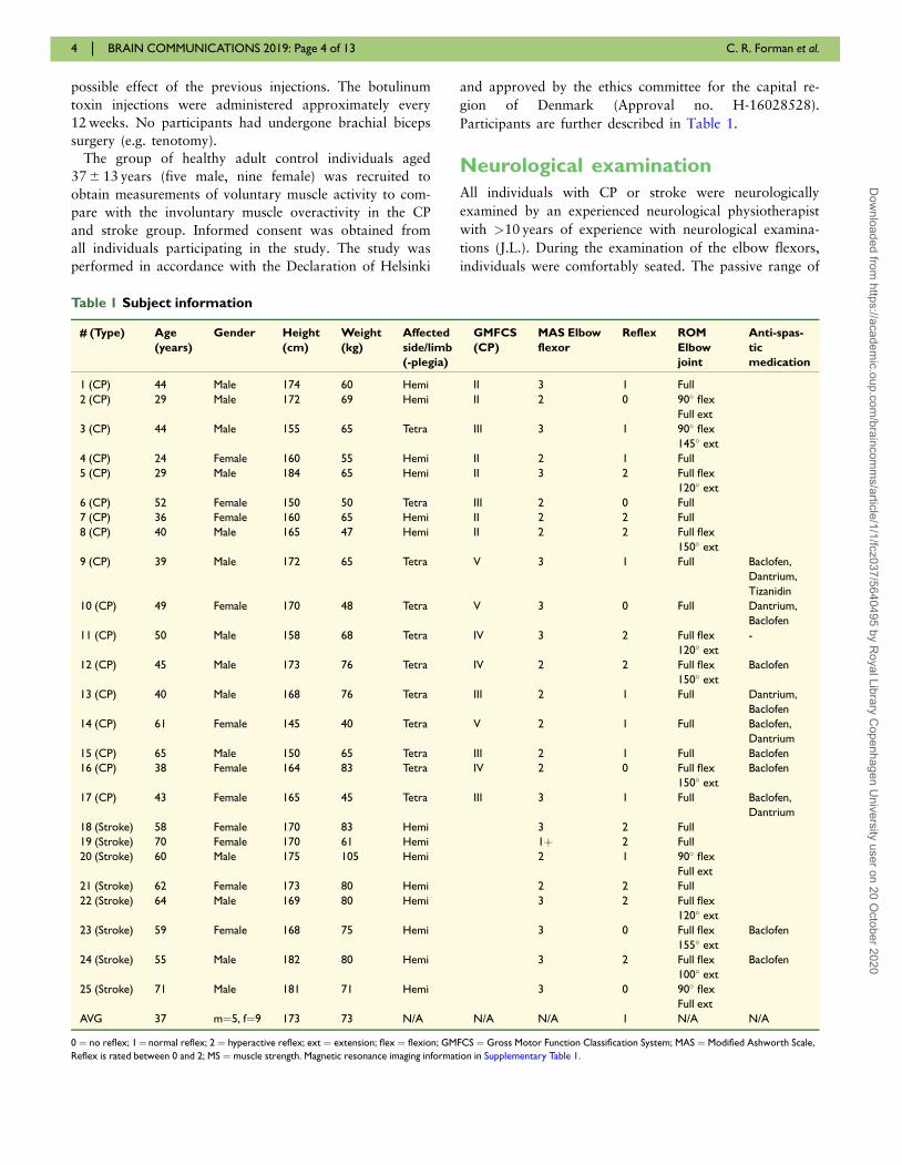
Participants are further described in Table 1.
Neurological examination
All individuals with CP or stroke were neurologically
examined by an experienced neurological physiotherapist
with >10 years of experience with neurological examina-
tions (J.L.). During the examination of the elbow flexors,
individuals were comfortably seated. The passive range of
Table 1 Subject information
# (Type) Age
(years)
Gender Height
(cm)
Weight
(kg)
Affected
side/limb
(-plegia)
GMFCS
(CP)
MAS Elbow
flexor
Reflex ROM
Elbow
joint
Anti-spas-
tic
medication
1 (CP) 44 Male 174 60 Hemi II 3 1 Full
2 (CP) 29 Male 172 69 Hemi II 2 0 90� flex
Full ext
3 (CP) 44 Male 155 65 Tetra III 3 1 90� flex
145� ext
4 (CP) 24 Female 160 55 Hemi II 2 1 Full
5 (CP) 29 Male 184 65 Hemi II 3 2 Full flex
120� ext
6 (CP) 52 Female 150 50 Tetra III 2 0 Full
7 (CP) 36 Female 160 65 Hemi II 2 2 Full
8 (CP) 40 Male 165 47 Hemi II 2 2 Full flex
150� ext
9 (CP) 39 Male 172 65 Tetra V 3 1 Full Baclofen,
Dantrium,
Tizanidin
10 (CP) 49 Female 170 48 Tetra V 3 0 Full Dantrium,
Baclofen
11 (CP) 50 Male 158 68 Tetra IV 3 2 Full flex -
120� ext
12 (CP) 45 Male 173 76 Tetra IV 2 2 Full flex Baclofen
150� ext
13 (CP) 40 Male 168 76 Tetra III 2 1 Full Dantrium,
Baclofen
14 (CP) 61 Female 145 40 Tetra V 2 1 Full Baclofen,
Dantrium
15 (CP) 65 Male 150 65 Tetra III 2 1 Full Baclofen
16 (CP) 38 Female 164 83 Tetra IV 2 0 Full flex Baclofen
150� ext
17 (CP) 43 Female 165 45 Tetra III 3 1 Full Baclofen,
Dantrium
18 (Stroke) 58 Female 170 83 Hemi 3 2 Full
19 (Stroke) 70 Female 170 61 Hemi 1þ 2 Full
20 (Stroke) 60 Male 175 105 Hemi 2 1 90� flex
Full ext
21 (Stroke) 62 Female 173 80 Hemi 2 2 Full
22 (Stroke) 64 Male 169 80 Hemi 3 2 Full flex
120� ext
23 (Stroke) 59 Female 168 75 Hemi 3 0 Full flex Baclofen
155� ext
24 (Stroke) 55 Male 182 80 Hemi 3 2 Full flex Baclofen
100� ext
25 (Stroke) 71 Male 181 71 Hemi 3 0 90� flex
Full ext
AVG 37 m¼5, f¼9 173 73 N/A N/A N/A 1 N/A N/A
0 ¼ no reflex; 1¼ normal reflex; 2 ¼ hyperactive reflex; ext ¼ extension; flex ¼ flexion; GMFCS ¼ Gross Motor Function Classification System; MAS ¼ Modified Ashworth Scale,
Reflex is rated between 0 and 2; MS ¼ muscle strength. Magnetic resonance imaging information in Supplementary Table 1.
4 | BRAIN COMMUNICATIONS 2019: Page 4 of 13 C. R. Forman et al.
Dow
nloaded from https://academ
ic.oup.com/braincom
ms/article/1/1/fcz037/5640495 by R
oyal Library Copenhagen U
niversity user on 20 October 2020

motion was evaluated by slowly moving the elbow joint
through the movement range, noting the positions of
maximal flexion and extension using a goniometer. These
positions were reached without causing discomfort to the
participants due to contractures and other physical con-
straints. Subsequently, a MAS of the elbow flexors was
determined and the presence and possible exaggeration of
the biceps reflex evaluated using a reflex hammer.
Electromyographic recording
In all hemiplegic individuals, EMG was recorded from
the hemiplegic side. In tetraplegic individuals, EMG was
recorded from the side with the highest elbow flexor
MAS score. EMG was recorded from the biceps muscle
using two sets of surface electrodes (3.0 � 2.2 cm, Ambu
Bluesensor N, Denmark), one set placed proximally and
medially on the short head of the biceps brachii and one
set placed distally and laterally on the long head of the
biceps brachii. These positions were chosen to minimize
the risk of cross-talk between the electrodes by maximiz-
ing the distance between them. A reference electrode was
placed over the lateral epicondyle of the humerus. In a
single instance, increased resistance to passive movement
was atypically observed in the elbow extensors only (sub-
ject 25). Though this is an unusual pattern, the individual
was clearly found to have increased resistance to passive
movement associated with an overactive muscle and cor-
responding to a spastic catch. The individual was there-
fore included. Here, the medial set was placed on the
long head of the triceps, while the lateral set was placed
on the lateral head of the triceps. The distance between
electrodes inside pairs was 2 cm and the distance between
pairs varied according to muscle size and length. To min-
imize EMG noise factors, the skin was softly sanded with
a very fine grain sandpaper (3M red dot). EMG record-
ings were made using a portable device fitted to a fore-
arm orthosis. The device contains two EMG channels
and samples at 1024 Hz. Data are then transferred to a
computer via Bluetooth. The technical properties of the
device are explained more thoroughly in Yamaguchi
et al. (2018).
Characterizing sustained involuntarymuscle activity
All EMG recordings of attempted rest lasted 2 min. They
were visually examined and recordings that contained
continuous muscle activity for at least 30 s were identi-
fied. In identifying muscle activity from electrical back-
ground noise, a steady presence of EMG spikes with high
amplitude and variability in the firing pattern was
sought. This EMG pattern is likely to be consistent with
involuntary muscle contractions and unlikely to be con-
sistent with electrical background noise. All identified
EMG recordings of muscle activity were then rectified
and a root mean square (RMS) of the full recording
calculated. The two effects of muscle stretch were investi-
gated as follows. First, the effect of elbow extension on
the sustained involuntary muscle activity (RMS of the
three different recordings from each individual) was nor-
malized to the recording in the maximally flexed position.
This was done to enable evaluation of stretch sensitivity
across groups despite differences in raw EMG amplitudes.
As both increases and decreases in muscle activity were
found with increased elbow extension, we performed
logarithm transformation of the EMG data to ensure
equal mathematical weighing of increasing and decreasing
factors in the group averaging. Secondly, the effects of
maintaining the muscle in a position of stretch, on the in-
voluntary muscle activity, were investigated by dividing
the 2-min recordings into four separate 30-s time periods
(‘bins’) and testing if the mean RMS EMG would differ
between the bins.
Cross-correlation analysis
To examine the common synaptic drive to the two EMG
channels of the biceps, a cross-correlation analysis was
undertaken using the methods outlined in Halliday et al.
(1995). The standard practice of full-wave rectification
was adopted, and each recording was divided into non-
overlapping segments with a duration of 1 s (1024 sam-
ples). A fast Fourier transformation was performed on
each segment for frequencies up to 300 Hz and then aver-
aged to construct estimations of the auto spectra, denoted
fxx(j) and fyy(j), for each EMG channel and the cross-
spectra, denoted fxy(j), ‘j’ referring to the given frequency
being analysed. The cross-correlation analysis delivers
results in form of the coherence, phase and cumulant
density measures. ‘Coherence’ describes the correlation
between frequency components of two processes
(Rosenberg et al., 1989) and is defined for a given fre-
quency as the absolute square of the cross-spectrum, nor-
malized to the auto spectra of the two channels (Grosse
et al., 2002). Because of this normalization procedure,
the coherence values are bound to produce results be-
tween 0 and 1, with 1 meaning perfect linear association
of the signals and 0 meaning no association. The object-
ive of coherence values in this study is to estimate
whether a common synaptic input to the motor neuron
pool represents a significant driving force of the muscle
activity, and which frequency components characterize
this common synaptic drive. Where coherence describes
the association in the frequency domain of the signals,
the ‘cumulant density function’ describes the linear associ-
ation in the time domain and is defined as the inverse
Fourier transform of the cross-spectrum (Halliday et al.,
1995). The cumulant density is an unbound measure
describing the statistical dependence between the two sig-
nals with 0 meaning complete independence of the proc-
esses. A peak in the cumulant density function describes
that the two signals are synchronized in time and are,
therefore, used to validate the presence of a common
What causes spastic dystonia? BRAIN COMMUNICATIONS 2019: Page 5 of 13 | 5
Dow
nloaded from https://academ
ic.oup.com/braincom
ms/article/1/1/fcz037/5640495 by R
oyal Library Copenhagen U
niversity user on 20 October 2020
synaptic input to the motor neuron pool. The EMG–
EMG coherence values and cumulant density functions
were compared between the voluntary muscle activity of
the control group and the involuntary muscle activity in
both the CP and the stroke groups. To make the com-
parison, all results from individual recordings of muscle
activity in each group were ‘pooled’. Pooled coherence
and cumulant density function are single representative
group estimates from combining independent coherence
estimates and the interpretation, therefore, is similar, ex-
cept that it relays information about the group.
Recordings that were contaminated by both sets of elec-
trodes picking up the same signals (cross-talk) were iden-
tified by elevated coherence in all bands and zero phase
delay (Grosse et al., 2004, 2002). Five recordings from
individuals with CP, 2 recordings from individuals with
stroke and 14 recordings from control individuals were
excluded due to cross-talk.
Experimental design
In all individuals, paired 120-s EMG recordings from the
investigated upper limb were obtained during attempted
rest in three different positions; a maximally flexed pos-
ition of the elbow, a 90-degree joint angle and a max-
imally extended position. The experimenter fixated the
arm in the positions by supporting the subject’s arm. In
the flexed and extended positions, the experimenters
made sure that the position did not cause any discomfort
to the individual due to contractures and physical con-
straints of joint position. Between each resting recording
the arm was held in a maximally flexed position for a
minimum of 20 s before being placed in a new position.
EMG recordings were started as soon as the individual’s
arm was placed in a new position. Furthermore, all con-
trol individuals were asked to perform 120 s of low-force
static contractions against resistance from the experiment-
er corresponding to approximately 10% of maximum
voluntary contraction in each position at the end of the
experiment. These recordings were performed to obtain a
measure of the EMG–EMG coherence from an isometric
voluntary contraction of the biceps muscle that was com-
parable with the EMG–EMG coherence measures
obtained from individuals with CP or stroke exhibiting
sustained muscle activity during rest.
Statistical analysis
In analysing normalized and logarithm transformed EMG
levels in different positions of the elbow joint and the dif-
ference between the 30-s bin groups mean a one-way
repeated measures ANOVA was used. All data were
tested for normality and equal variance before ANOVA
analysis using the Shapiro–Wilk test and Brown–Forsythe
test, respectively. If data failed tests for normality or
equal variance, data were rank transformed before statis-
tical analysis. It is noted in the figure legend, if the
statistical analysis was performed by ANOVA on ranks.
Multiple pairwise comparisons were performed using the
Holm–Sidak test. Both pooled coherence and group com-
parisons of coherence were compared by including a chi-
squared test, which denotes the difference required for
statistical significance in the frequency distribution.
Significance was in all cases determined at a P-value of
0.05 and all values are given as means 6 SD. Analyses
were performed using Sigmaplot 13 (SYSTAT software)
and MATLAB R2017a (The Mathworks Inc.).
Data availability
The data that support the findings of this study are avail-
able from the corresponding author, upon request.
Results
Electromyographic recordings
In total, 36 recordings from 13 out of 17 individuals with
CP and 17 recordings from all 8 individuals with stroke
contained sustained involuntary muscle activity (see
Materials and methods section for identification criteria).
The four individuals with CP who did not show signs of
involuntary muscle activity were individuals #2, 3, 7 and
12 (Table 1). None of the 42 recordings from 14 control
individuals contained sustained involuntary muscle activity
at rest. The characteristics of the observed sustained invol-
untary muscle activity differed considerably, both between
CP and stroke, between individuals in the same group
and between individual recordings in different positions of
the elbow joint from the same individual (Fig. 2A).
Whereas some individuals (e.g. subject 4) exhibited invol-
untary muscle activity with sudden increases and
decreases, other individuals (e.g. subject 24) exhibited
more constant and stable involuntary muscle activity.
Figure 2B presents the effect of elbow joint position on
the biceps muscle EMG levels. The RMS EMG of one
position of the elbow joint is normalized to the same
individual’s flexed position RMS amplitude and then
logarithm transformed. In the CP group, a repeated
measures ANOVA showed no significant (P> 0.05) dif-
ferences between RMS EMG levels in the investigated
positions, indicating no effect of elbow joint position.
However, in the stroke group, EMG levels increased with
extension of the elbow joint from the flexed position to
the 90-degree position (P¼ 0.05) and the extended pos-
ition (P¼ 0.01). No difference was found comparing the
90-degree and the extended position (P¼ 0.36).
Figure 2C presents the development of involuntary
muscle activity following the positioning of the joint in
the maximally extended joint position calculated as 30-s
mean RMS EMG amplitude bins and then normalized to
the first 30 s bin. In the CP group, the mean RMS EMG
amplitudes were found to be significantly lower, when
6 | BRAIN COMMUNICATIONS 2019: Page 6 of 13 C. R. Forman et al.
Dow
nloaded from https://academ
ic.oup.com/braincom
ms/article/1/1/fcz037/5640495 by R
oyal Library Copenhagen U
niversity user on 20 October 2020

compared to bin 1, in both bin 2 (1 6 0 versus
0.83 6 0.29, P¼ 0.03), bin 3 (1 6 0 versus 0.64 6 0.25,
P< 0.001) and in bin 4 (1 6 0 versus 0.66 6 0.24,
P< 0.001). Furthermore, bin 3 was found to be signifi-
cantly lower compared with bin 2 (P¼ 0.03). In the
stroke group, no differences were found between the
bins. In the flexed- and 90-degree joint positions, no dif-
ferences were found between the bins in either group.
Cross-correlation analysis
In Fig. 3, the pooled coherence results from CP, stroke
and control individuals (A–C) are presented. In all
groups, significant coherence was found in the alpha (6–
15 Hz), beta (16–35 Hz) and early gamma band (36–
60 Hz).
The pooled coherence estimates are compared group-
wise in Fig. 4A–C. When compared to the control group
Figure 2 EMG activity in different positions. (A) Raw EMG activity in the flexed, 90 degree and extended position of the elbow in subject
4, 10 and 24, respectively. In subject 4 and 10, the recordings were identified with sustained involuntary muscle activity. In subject 24, the
recordings performed in the 90 degree and extended position were identified with sustained involuntary muscle activity, the recording in the
flexed position was not. (B) RMS EMG from different elbow positions normalized to the individuals’ flexed position RMS amplitude and
logarithm transformed including group means (large circles connected by dotted lines). Asterisk (*) signifies a significant difference compared
with the flexed position. (C) The CP and stroke group mean RMS EMG amplitudes divided into four 30-s periods (bins) and then normalized to
Bin 1. Bin 1: 0–30 s. Bin 2: 30–60 s. Bin 3: 60–90 s. Bin 4: 90–120 s. Asterisk (*) signifies a significant difference compared with Bin 1. The
statistical testing in Fig. 2c was performed by an ANOVA on ranks test. The means of Fig. 2c are made from all individual recordings containing
muscle activity, and therefore contain multiple recordings from some participants. In the stroke group, n¼ 16 recordings. In the CP group,
n¼ 31 recordings.
What causes spastic dystonia? BRAIN COMMUNICATIONS 2019: Page 7 of 13 | 7
Dow
nloaded from https://academ
ic.oup.com/braincom
ms/article/1/1/fcz037/5640495 by R
oyal Library Copenhagen U
niversity user on 20 October 2020

(4B) and to the CP group (4 C), an increased coherence
in the alpha band of the stroke group is visible mainly in
the 8–11 Hz band. In the CP group, coherence is larger
than in stroke in the early gamma band around 40 Hz
(Fig. 4C). The pooled cumulant density functions
(Fig. 3D–F) all show a significant central peak of syn-
chronization. The average duration of the central peaks
of synchronization was 19.5 (67.2) ms for the CP group,
22.7 (67.5) ms for the stroke group and 20.8 (68.7) for
the control group. No significant differences were found
in either the percentage of individual recordings showing
a significant central peak (100% for the CP and control
group, 93.4% for the stroke group), the duration of the
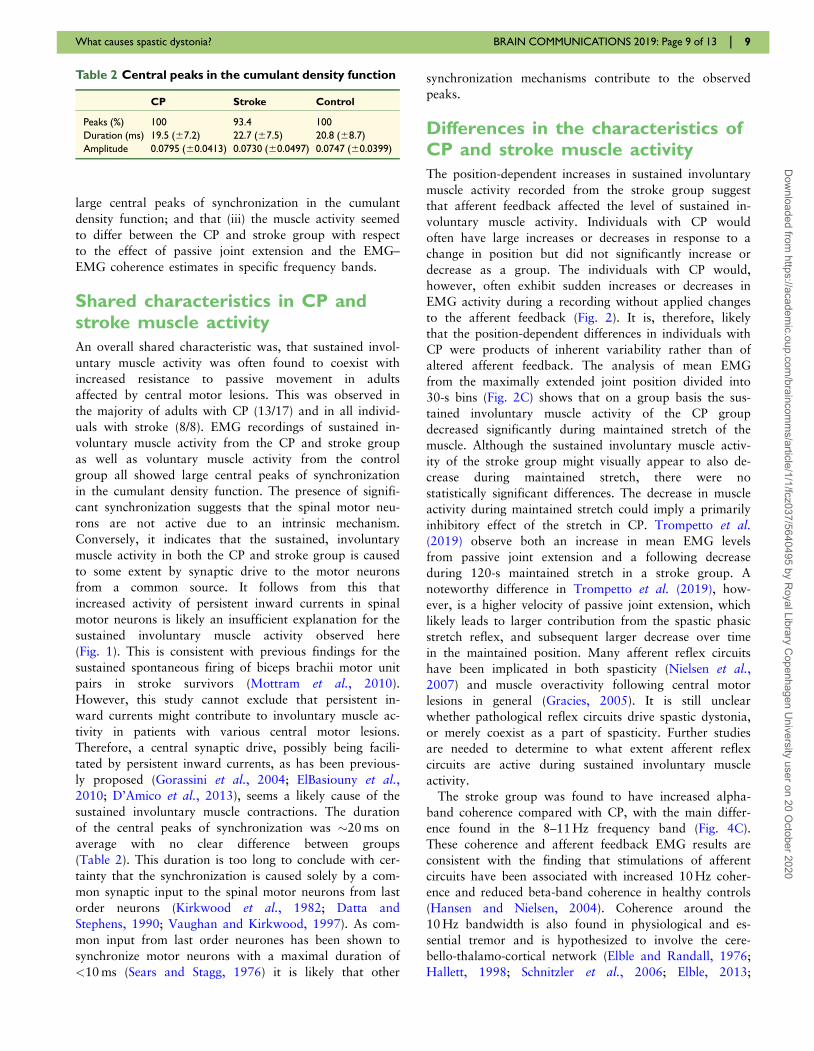
peak or the amplitude of the peak (Table 2).
DiscussionThe primary findings of this study are that (i) sustained
involuntary muscle activity exists in individuals with
movement disorder due to lesions of the descending
motor pathways and increased resistance to passive
movement (MAS> 1) due to CP or stroke; (ii) the muscle
activity of both individuals with CP and stroke showed
Figure 3 Pooled coherence. A–C depicts the pooled coherence in the CP (A), stroke (B) and control group (C). The dotted lines note the
chi-squared test level. D–F depicts pooled cumulant density functions from the CP group (D) stroke group (E) and control group (F).
Figure 4 Difference in pooled coherence. A–C depicts differences in pooled coherence between the CP and control group (A), the stroke
and control group (B) and the CP and stroke group (C). The dotted lines note the chi-squared test level.
8 | BRAIN COMMUNICATIONS 2019: Page 8 of 13 C. R. Forman et al.
Dow
nloaded from https://academ
ic.oup.com/braincom
ms/article/1/1/fcz037/5640495 by R
oyal Library Copenhagen U
niversity user on 20 October 2020
large central peaks of synchronization in the cumulant
density function; and that (iii) the muscle activity seemed
to differ between the CP and stroke group with respect
to the effect of passive joint extension and the EMG–
EMG coherence estimates in specific frequency bands.
Shared characteristics in CP andstroke muscle activity
An overall shared characteristic was, that sustained invol-
untary muscle activity was often found to coexist with
increased resistance to passive movement in adults
affected by central motor lesions. This was observed in
the majority of adults with CP (13/17) and in all individ-
uals with stroke (8/8). EMG recordings of sustained in-
voluntary muscle activity from the CP and stroke group
as well as voluntary muscle activity from the control
group all showed large central peaks of synchronization
in the cumulant density function. The presence of signifi-
cant synchronization suggests that the spinal motor neu-
rons are not active due to an intrinsic mechanism.
Conversely, it indicates that the sustained, involuntary
muscle activity in both the CP and stroke group is caused
to some extent by synaptic drive to the motor neurons
from a common source. It follows from this that
increased activity of persistent inward currents in spinal
motor neurons is likely an insufficient explanation for the
sustained involuntary muscle activity observed here
(Fig. 1). This is consistent with previous findings for the
sustained spontaneous firing of biceps brachii motor unit
pairs in stroke survivors (Mottram et al., 2010).
However, this study cannot exclude that persistent in-
ward currents might contribute to involuntary muscle ac-
tivity in patients with various central motor lesions.
Therefore, a central synaptic drive, possibly being facili-
tated by persistent inward currents, as has been previous-
ly proposed (Gorassini et al., 2004; ElBasiouny et al.,
2010; D’Amico et al., 2013), seems a likely cause of the
sustained involuntary muscle contractions. The duration
of the central peaks of synchronization was �20 ms on
average with no clear difference between groups
(Table 2). This duration is too long to conclude with cer-
tainty that the synchronization is caused solely by a com-
mon synaptic input to the spinal motor neurons from last
order neurons (Kirkwood et al., 1982; Datta and
Stephens, 1990; Vaughan and Kirkwood, 1997). As com-
mon input from last order neurones has been shown to
synchronize motor neurons with a maximal duration of
<10 ms (Sears and Stagg, 1976) it is likely that other
synchronization mechanisms contribute to the observed
peaks.
Differences in the characteristics ofCP and stroke muscle activity
The position-dependent increases in sustained involuntary
muscle activity recorded from the stroke group suggest
that afferent feedback affected the level of sustained in-
voluntary muscle activity. Individuals with CP would
often have large increases or decreases in response to a
change in position but did not significantly increase or
decrease as a group. The individuals with CP would,
however, often exhibit sudden increases or decreases in
EMG activity during a recording without applied changes
to the afferent feedback (Fig. 2). It is, therefore, likely
that the position-dependent differences in individuals with
CP were products of inherent variability rather than of
altered afferent feedback. The analysis of mean EMG
from the maximally extended joint position divided into
30-s bins (Fig. 2C) shows that on a group basis the sus-
tained involuntary muscle activity of the CP group
decreased significantly during maintained stretch of the
muscle. Although the sustained involuntary muscle activ-
ity of the stroke group might visually appear to also de-
crease during maintained stretch, there were no
statistically significant differences. The decrease in muscle
activity during maintained stretch could imply a primarily
inhibitory effect of the stretch in CP. Trompetto et al.
(2019) observe both an increase in mean EMG levels
from passive joint extension and a following decrease
during 120-s maintained stretch in a stroke group. A
noteworthy difference in Trompetto et al. (2019), how-
ever, is a higher velocity of passive joint extension, which
likely leads to larger contribution from the spastic phasic
stretch reflex, and subsequent larger decrease over time
in the maintained position. Many afferent reflex circuits
have been implicated in both spasticity (Nielsen et al.,
2007) and muscle overactivity following central motor
lesions in general (Gracies, 2005). It is still unclear
whether pathological reflex circuits drive spastic dystonia,
or merely coexist as a part of spasticity. Further studies
are needed to determine to what extent afferent reflex
circuits are active during sustained involuntary muscle
activity.
The stroke group was found to have increased alpha-
band coherence compared with CP, with the main differ-
ence found in the 8–11 Hz frequency band (Fig. 4C).
These coherence and afferent feedback EMG results are
consistent with the finding that stimulations of afferent
circuits have been associated with increased 10 Hz coher-
ence and reduced beta-band coherence in healthy controls
(Hansen and Nielsen, 2004). Coherence around the
10 Hz bandwidth is also found in physiological and es-
sential tremor and is hypothesized to involve the cere-
bello-thalamo-cortical network (Elble and Randall, 1976;
Hallett, 1998; Schnitzler et al., 2006; Elble, 2013;
Table 2 Central peaks in the cumulant density function
CP Stroke Control
Peaks (%) 100 93.4 100
Duration (ms) 19.5 (67.2) 22.7 (67.5) 20.8 (68.7)
Amplitude 0.0795 (60.0413) 0.0730 (60.0497) 0.0747 (60.0399)
What causes spastic dystonia? BRAIN COMMUNICATIONS 2019: Page 9 of 13 | 9
Dow
nloaded from https://academ
ic.oup.com/braincom
ms/article/1/1/fcz037/5640495 by R
oyal Library Copenhagen U
niversity user on 20 October 2020

Albanese and Del Sorbo, 2016). As tremor can also be
affected by the function of sensory afferents (Sanes,
1985), this could indicate afferent differences in stroke
and CP, perhaps through different projections to the cere-
bello-thalamo-cortical network.
Coherence in the beta and gamma bands are often
assumed to relate to activity originating in the primary
motor cortex (Grosse et al., 2002), and could therefore
imply reduced neural drive from the primary motor cor-
tex in stroke compared with CP (Fig. 4). Reduced corti-
cospinal input in stroke is consistent with observations of
decreased corticospinal excitability (Dimyan and Cohen,
2010; Cortes et al., 2012). It is an interesting result, that
the corticospinal input in sustained involuntary muscle
activity in CP should be different from stroke. An explan-
ation of this difference could be the maturation of the
central nervous system at the time of injury in CP and
stroke. CP might differ from stroke through extensive
cortical and spinal adaptive reorganization following le-
sion during early development. One line of evidence sup-
porting this hypothesis is that ipsilateral corticospinal
projections from the non-affected hemisphere to the
muscles of the affected side have been found in both chil-
dren and adults with unilateral CP (Carr, 1996;
Marneweck et al., 2018). The same has not been
observed in individuals suffering from stroke (Brouwer
and Ashby, 1990; Palmer et al., 1992). These cortical
adaptations have been suggested beneficial for regaining
some voluntary function in the paretic side during devel-
opment (Carr, 1996; Bleyenheuft et al., 2015; Friel et al.,
2016) but could also lead to disorganized motor control
causing sustained involuntary muscle activity such as that
observed here.
Information regarding the exact individual sites of the
central motor lesion is of great interest, as lesions to the
basal ganglia are a frequently cited likely cause of invol-
untary movements in both CP (Aravamuthan and
Waugh, 2016) and stroke (Ghika-Schmid et al., 1997).
As the participants with stroke had magnetic resonance
imaging scans performed in relation to the injury, this in-
formation was available. Indeed, all eight individuals
with stroke were found to have some degree of damage
to the basal ganglia (Supplementary Table 1). As we did
not have access to an magnetic resonance imaging scan-
ner, it was unfortunately not possible to obtain this infor-
mation from the individuals with CP. Basal ganglia
lesions, however, are unlikely to provide the full explan-
ation of sustained involuntary muscle activity following
central motor lesions (Neychev et al., 2011). Results from
interventions using deep brain stimulation to reduce dys-
tonia in CP (Koy et al., 2013) and in stroke (Elias et al.,2018) have seen large effects in some individuals but no
effect in others. This is consistent with the idea that the
basal ganglia might contribute to sustained involuntary
muscle activity, but that the complete origin of the condi-
tion involves more network-based complex causes such as
maladaptive neural plasticity or defects in sensorimotor
integration (Neychev et al., 2011; Quartarone and
Hallett, 2013; Liuzzi et al., 2016).
As individuals with stroke were generally older than
the individuals with CP, we are not able to exclude that
the observed differences between the two groups could be
partly due to age differences.
How does the sustained involuntarymuscle activity compare to spasticdystonia?
In this study, we have attempted to depict with EMG,
how the involuntary muscle activity presents itself in two
different groups of individuals with central motor lesions.
This has been done to illustrate the complex nature of
the clinical examination of these populations. We present
here, evidence that many individuals with movement dis-
order due to lesions of the descending motor pathways,
experience involuntary muscle activity during rest, which
should not be labelled spasticity. Whether the introduced
condition of spastic dystonia can fully explain the sus-
tained involuntary muscle activity in this study is how-
ever not clear. The sustained involuntary muscle activity
resembling that of a dystonia exists to some degree in
both populations, but the seemingly different patterns of
involuntary muscle activity in the two groups point to
other conditions of muscle overactivity also being present.
Although no blatant choreoathetosis was found during
the neurological examination, the variable pattern of in-
voluntary muscle activity in the CP group could be inter-
preted as a sign hereof. Both dystonia and
choreoathetosis have been accepted to constitute separate
subclassifications of dyskinetic CP (Cans, 2008), but ra-
ther than interpreting this as an indicator of misclassifica-
tion of this study’s individuals with CP, our findings
should exemplify the complexity and overlap of the clas-
sifications and symptoms of individuals with central
motor lesions. The SCPE classifications are an attempt to
characterize the difference between spastic and dyskinetic
CP as whether the increase in involuntary muscle activity
is persistent or varying (Cans, 2008), but many defini-
tions of dystonia following central motor lesions would
refer to it as long-lasting muscle contractions causing sus-
tained abnormal posturing (Sanger et al., 2010;
Siniscalchi et al., 2012; Albanese et al., 2013). We believe
it is important to recognize, that the symptom of spasti-
city (Lance, 1980; Gracies, 2005) only is a part of the
clinical picture in the individuals with central motor
lesions, who are often characterized by the word spastic.
Sustained involuntary muscle activity, perhaps referred to
as spastic dystonia, should be considered as a separate
symptom with a separate pathophysiology in this popula-
tion. The distinction is important, as the study provides
evidence suggesting that the sustained involuntary muscle
activity is driven by a common synaptic drive to the
motor neuron pool. This indicates that the treatment for
10 | BRAIN COMMUNICATIONS 2019: Page 10 of 13 C. R. Forman et al.
Dow
nloaded from https://academ
ic.oup.com/braincom
ms/article/1/1/fcz037/5640495 by R
oyal Library Copenhagen U
niversity user on 20 October 2020
the spinal stretch reflex mediated symptom of spasticity
might not be the same as the treatment for the centrally
driven sustained involuntary muscle activity.
Clinical implications
This study found evidence suggesting that the underlying
mechanisms causing sustained involuntary muscle activity
differed between the CP and stroke groups. Whereas the
sustained involuntary muscle activity was increased by af-
ferent input in individuals with stroke, it appeared to
have larger contributions from the motor cortex in indi-
viduals with CP. It, therefore, seems likely that the opti-
mal treatment option in the CP group would not be
identical to that of the stroke group. The involuntary
muscle activity of the individuals with CP could originate
from complex disorganized motor control following cor-
tical adaptations to the lesion. This emphasizes the need
for motor learning rehabilitation following a central
motor lesion, not only to regain function for activities of
daily living, but also to reduce involuntary muscle activ-
ity. It has previously been reported that even purely par-
ietal lesions in stroke frequently lead to dystonia, perhaps
due to the reduced integration of sensorimotor inputs to
the motor cortex (Ghika et al., 1998). Sensorimotor inte-
gration of afferent inputs has also been suggested as a
mechanism for causing various types of focal dystonia
(Neychev et al., 2011; Avanzino and Fiorio, 2014; Patel
et al., 2014; Avanzino et al., 2015; Liuzzi et al., 2016).
Regaining the sensorimotor integration in individuals suf-
fering from involuntary muscle activity following a stroke
is therefore an important therapeutic intervention.
Although this study has shed some light on the under-
lying mechanisms causing sustained involuntary muscle
activity in individuals with CP or stroke, there is an ur-
gent need for future studies to further explore these
mechanisms in order to improve current treatment.
ConclusionThis study found that sustained involuntary muscle activ-
ity, like that described in spastic dystonia, was frequently
present alongside increased resistance to passive move-
ment in individuals with movement disorder due to
lesions of the descending motor pathways. The sustained
involuntary muscle activity of both CP and stroke was
found to contain a common synaptic drive to the motor
neuron pool, but coherence estimates indicate, that the
origin of this common synaptic drive differed between
the groups. Stroke seemed to have increased muscle activ-
ity from afferent neural feedback and an increased alpha-
band coherence. CP was not found to have increased
muscle activity from afferent neural feedback, but instead
had increased gamma-band coherence, indicating contri-
butions from cortical motor regions. We find these results
to indicate that the sustained involuntary muscle activity
may require different treatment in the two groups. In
some individuals, treatment should focus on plastic adap-
tations to central motor control, whereas other individu-
als might instead be affected by deficits in the integration
of sensory feedback.
Supplementary materialSupplementary material is available at Brain
Communications online.
AcknowledgementsWe are grateful to the staff from the department of neur-
ology at the Herlev Gentofte Hospital for helping with the
recruitment of individuals suffering from chronic stroke and
to the staff at Jonstrupvang for helping with the recruitment
of individuals with CP.
FundingThe study was supported by a grant from the Elsass
Foundation.
Competing interestsThe authors report no competing interests.
ReferencesAlbanese A, Bhatia K, Bressman SB, DeLong MR, Fahn S, Fung VSC,
et al. Phenomenology and classification of dystonia: a consensus up-
date. Mov Disord NIH Disord 2013; 28: 863–73.Albanese A, Del Sorbo F. Dystonia and tremor: the clinical syn-
dromes with isolated tremor. Tremor Other Hyperkinet Mov
2016; 6: 319.
Aravamuthan BR, Waugh JL. Localization of basal ganglia and thal-
amic damage in dyskinetic cerebral palsy. Pediatr. Neurol. 2016; 54:
11–21.Avanzino L, Fiorio M. Proprioceptive dysfunction in focal dystonia:
from experimental evidence to rehabilitation strategies. Front Hum
Neurosci 2014; 8: 1–7.Avanzino L, Tinazzi M, Ionta S, Fiorio M. Sensory-motor integration
in focal dystonia. Neuropsychologia 2015; 79: 288–300.Bleyenheuft Y, Dricot L, Gilis N, Kuo H, Grandin C, Bleyenheuft C,
et al. Capturing neuroplastic changes after bimanual intensive re-
habilitation in children with unilateral spastic cerebral palsy: a com-
bined DTI, TMS and fMRI pilot study. Res Dev Disabil 2015;
43–44: 136–49.Brouwer B, Ashby P. Do injuries to the developing human brain alter
corticospinal projections? Neurosci Lett 1990; 108: 225–30.
Cans C. Surveillance of cerebral palsy in Europe: a collaboration of
cerebral palsy surveys and registers. Dev Med Child Neurol 2008;
42: 816–24.Carr LJ. Development and reorganization of descending motor path-
ways in children with hemiplegic cerebral palsy. Acta Paediatr 1996;
85: 53–7.
What causes spastic dystonia? BRAIN COMMUNICATIONS 2019: Page 11 of 13 | 11
Dow
nloaded from https://academ
ic.oup.com/braincom
ms/article/1/1/fcz037/5640495 by R
oyal Library Copenhagen U
niversity user on 20 October 2020

Cortes M, Black-Schaffer RM, Edwards DJ. Transcranial magnetic
stimulation as an investigative tool for motor dysfunction and recov-
ery in stroke: an overview for neurorehabilitation clinicians.
Neuromodulation 2012; 15: 316–25.D’Amico JM, Murray KC, Li Y, Chan KM, Finlay MG, Bennett DJ,
et al. Constitutively active 5-HT 2/a 1 receptors facilitate muscle
spasms after human spinal cord injury. J. Neurophysiol 2013; 109:
1473–84.
Datta AK, Stephens JA. Synchronization of motor unit activity
during voluntary contraction in man. J Physiol 1990; 422:
397–419.Denny-Brown D. 1966. The cerebral control of movement. Liverpool:
Liverpool University Press.Dimyan MA, Cohen LG. Contribution of transcranial magnetic stimu-
lation to the understanding of functional recovery mechanisms after
stroke. Neurorehabil Neural Repair 2010; 24: 125–35.ElBasiouny SM, Schuster JE, Heckman CJ. Persistent inward currents
in spinal motoneurons: important for normal function but potential-
ly harmful after spinal cord injury and in amyotrophic lateral scler-
osis. Clin Neurophysiol 2010; 121: 1669–79.Elble RJ. What is essential tremor? Curr Neurol Neurosci Rep 2013;
13: 353.Elble RJ, Randall JE. Motor-unit activity responsible for 8- to 12-Hz
component of human physiological finger tremor. J. Neurophysiol
1976; 39: 370–83.
Elias GJ, Namasivayam AA, Lozano AM. Deep brain stimulation for
stroke: Current uses and future directions. Brain Stimul 2018; 11:
3–28.
Foerster. Resection of the posterior spinal nerve-roots in the treatment
of gastric crises and spastic paralysis. Proc R Soc Med 1911; 4:
226–46.
Friel KM, Kuo HC, Fuller J, Ferre CL, Brand~ao M, Carmel JB, et al.
Skilled bimanual training drives motor cortex plasticity in children
with unilateral cerebral palsy. Neurorehabil Neural Repair 2016;
30: 834–44.
Ghika J, Ghika-Schmid F, Bogousslasvky J. Parietal motor syndrome:
a clinical description in 32 patients in the acute phase of pure par-
ietal strokes studied prospectively. Clin Neurol Neurosurg 1998;
100: 271–82.Ghika-Schmid F, Ghika J, Regli F, Bogousslavsky J. Hyperkinetic
movement disorders during and after acute stroke:The Lausanne
Stroke Registry. J Neurol Sci 1997; 146: 109–16.Gorassini MA, Knash ME, Harvey PJ, Bennett DJ, Yang JF. Role of
motoneurons in the generation of muscle spasms after spinal cord
injury. Brain 2004; 127: 2247–58.
Gracies JM. Pathophysiology of spastic paresis. II: emergence of
muscle overactivity. Muscle Nerve 2005; 31: 552–71.
Grosse P, Cassidy MJ, Brown P. EEG-EMG, MEG-EMG and EMG-
EMG frequency analysis: physiological principles and clinical appli-
cations. Clin Neurophysiol 2002; 113: 1523–31.Grosse P, Edwards M, Tijssen MAJ, Schrag A, Lees AJ, Bhatia KP,
et al. Patterns of EMG-EMG coherence in limb dystonia. Mov
Disord 2004; 19: 758–69.Hallett M. Overview of human tremor physiology. Mov Disord 1998;
13: 43–8.Halliday DM, Rosenberg JR, Amjad AM, Breeze P, Conway BA,
Farmer SF. A framework for the analysis of mixed time series/point
process data-Theory and application to the study of physiological
tremor, single motor unit discharges and electromyograms. Prog
Biophys Mol Biol 1995; 64: 237–78.Hansen NL, Nielsen JB. The effect of transcranial magnetic stimulation
and peripheral nerve stimulation on corticomuscular coherence in
humans. J Physiol (Lond) 2004; 561: 295–306.Kirkwood PA, Sears TA, Tuck DL, Westgaard RH. Variations in the
time course of the synchronization of intercostal motoneurons in the
cat. J Physiol 1982; 327: 105–35.
Koy A, Hellmich M, Pauls KAM, Marks W, Lin J-P, Fricke O, et al.
Effects of deep brain stimulation in dyskinetic cerebral palsy: a
meta-analysis. Mov Disord 2013; 28: 647–54.Lance JW. Symposium synopsis. In: RG Feldman, RR Young, WP
Koella, editors. Spasticity: disordered motor control. Chicago:
Yearbook Medical Publishers; 1980. p. 485–94.Liuzzi D, Gigante AF, Leo A, Defazio G. The anatomical basis of
upper limb dystonia: lesson from secondary cases. Neurol Sci 2016;
37: 1393–8.Lorentzen J, Pradines M, Gracies J-M, Bo Nielsen J. On Denny-
Brown’s ‘spastic dystonia’—what is it and what causes it? Clin
Neurophysiol 2018; 129: 89–94.Marneweck M, Kuo HC, Smorenburg ARP, Ferre CL, Flamand VH,
Gupta D, et al. The Relationship between hand function and over-
lapping motor representations of the hands in the contralesional
hemisphere in unilateral spastic cerebral palsy. Neurorehabil Neural
Repair 2018; 32: 62–72.
Miller DM, Klein CS, Suresh NL, Rymer WZ. Asymmetries in vestibu-
lar evoked myogenic potentials in chronic stroke survivors with
spastic hypertonia: evidence for a vestibulospinal role. Clin
Neurophysiol 2014; 125: 2070–8.
Mottram CJ, Wallace CL, Chikando CN, Rymer WZ. Origins of
spontaneous firing of motor units in the spastic—paretic biceps bra-
chii muscle of stroke survivors. J. Neurophysiol 2010; 104:
3168–79.Neychev VK, Gross RE, Lehericy S, Hess EJ, Jinnah HA. The function-
al neuroanatomy of dystonia. Neurobiol Dis 2011; 42: 185–201.
Nielsen JB, Crone C, Hultborn H. The spinal pathophysiology of spas-
ticity—from a basic science point of view. Acta Physiol 2007; 189:
171–80.
Palmer E, Ashby P, Hajek VE. Ipsilateral fast corticospinal pathways
do not account for recovery in stroke. Ann Neurol 1992; 32:
519–25.
Patel N, Jankovic J, Hallett M. Sensory aspects of movement disorders.
Lancet Neurol 2014; 13: 100–12.Pollock LJ, Davis L. The reflex activities of a decerebrate animal.
J Comp Neurol 1930; 50: 377–411.Quartarone A, Hallett M. Emerging concepts in the physiological basis
of dystonia. Mov Disord 2013; 28: 958–67.
Rosenberg JR, Amjad A. M, Breeze P, Brillinger DR, Halliday DM.
The Fourier approach to the identification of functional coupling be-
tween neuronal spike trains. Prog Biophys Mol Biol 1989; 53: 1–31.
Sanes JN. Absence of enhanced physiological tremor in patients with-
out muscle or cutaneous afferents. J Neurol Neurosurg Psychiatry
1985; 48: 645–9.
Sanger TD, Chen D, Fehlings D, Hallett M, Lang AE, Mink JW, et al.
Definition and classification of hyperkinetic movements in child-
hood. Mov Disord 2010; 25: 1538–49.
Schnitzler A, Timmermann L, Gross J. Physiological and pathological
oscillatory networks in the human motor system. J Physiol Paris
2006; 99: 3–7.
Sears TA, Stagg D. Short-term synchronization of intercostal moto-
neurone activity. J. Physiol 1976; 263: 357–81.Sheean G, McGuire JR. Spastic hypertonia and movement disorders:
pathophysiology, clinical presentation, and quantification. Phys Med
Rehabil 2009; 1: 827–33.Sherrington CS. Decerebrate rigidity, and reflex coordination of move-
ments. J Physiol 1898; 22: 319–37.
Siniscalchi A, Gallelli L, Labate A, Malferrari G, Palleria C, Sarro GD.
Post-stroke movement disorders: clinical manifestations and
pharmacological management. Curr Neuropharmacol 2012; 10:
254–62.Sukal-Moulton T, Krosschell KJ, Gaebler-Spira DJ, Dewald JPA.
Motor impairment factors related to brain injury timing in early
hemiparesis, part I: expression of upper-extremity weakness.
Neurorehabil Neural Repair 2014a; 28: 13–23.
12 | BRAIN COMMUNICATIONS 2019: Page 12 of 13 C. R. Forman et al.
Dow
nloaded from https://academ
ic.oup.com/braincom
ms/article/1/1/fcz037/5640495 by R
oyal Library Copenhagen U
niversity user on 20 October 2020

Sukal-Moulton T, Krosschell KJ, Gaebler-Spira DJ, Dewald JPA.
Motor impairments related to brain injury timing in early hemipar-esis. part II: abnormal upper extremity joint torque synergies.Neurorehabil Neural Repair 2014b; 28: 24–35.
Trompetto C, Curra A, Puce L, Mori L, Serrati C, Fattapposta F, et al.Spastic dystonia in stroke subjects: prevalence and features of the
neglected phenomenon of the upper motor neuron syndrome. Clin.Neurophysiol 2019; 130: 521–7.
Vaughan CW, Kirkwood PA. Evidence from motoneurone
synchronization for disynaptic pathways in the control ofinspiratory motoneurones in the cat. J Physiol 1997; 503:673–89.
Yamaguchi T, Hvass Petersen T, Kirk H, Forman C, Svane C, Kofoed-Hansen M, et al. Spasticity in adults with cerebral palsy and mul-
tiple sclerosis measured by objective clinically applicable technique.Clin Neurophysiol 2018; 129: 2010–21.
What causes spastic dystonia? BRAIN COMMUNICATIONS 2019: Page 13 of 13 | 13
Dow
nloaded from https://academ
ic.oup.com/braincom
ms/article/1/1/fcz037/5640495 by R
oyal Library Copenhagen U
niversity user on 20 October 2020