Embed Size (px)
Citation preview

Sustained Expression of a1-Antitrypsin afterTransplantation of Manipulated HematopoieticStem Cells
Andrew A. Wilson1, Letty W. Kwok1, Avi-Hai Hovav2, Sarah J. Ohle1, Frederic F. Little1, Alan Fine1,and Darrell N. Kotton1
1The Pulmonary Center, Boston University School of Medicine, Boston, Massachusetts; and 2The Hebrew University, Institute of Dental Sciences,
Jerusalem, Israel
Inherited mutations in the human a1-antitrypsin (AAT) gene leadto deficient circulating levels of AAT protein and a predispositionto developing emphysema. Gene therapy for individuals deficient inAAT is an attractive goal, because transfer of a normal AAT gene intoany cell type able to secrete AAT should reverse deficient AAT levelsand attenuate progression of lung disease. Here we present anapproach for AAT gene transfer based on the transplantation oflentivirally transduced hematopoietic stem cells (HSCs). We developa novel dual-promoter lentiviral system to transfer normal humanAAT cDNA as well as a fluorescent tracking ‘‘reporter gene’’ intomurine HSCs. After transplantation of 3,000 transduced HSCs intoirradiated mouse recipients, we demonstrate simultaneous andsustained systemic expression of both genes in vivo for at least 31weeks. The stem cells transduced with this protocol maintain multi-potency, self-renewal potential, and the ability to reconstitute thehematopoietic systems of both primary and secondary recipients.This lentiviral-based system may be useful for investigations re-quiring the systemic secretion ofanti-proteases or cytokines relevantto the pathogenesis of a variety of lung diseases.
Keywords: gene therapy; bone marrow; a1-antitrypsin; lung; stem cells
a1-antitrypsin (AAT) deficiency is one of the most commonhereditary diseases worldwide (1, 2). A variety of inheritedmutations in the AAT gene have been shown to cause de-creased levels of circulating AAT resulting in a predispositionto developing panacinar emphysema and cirrhosis. The pulmo-nary disease phenotype is thought to result directly from unbal-anced protease activity in the lung and subsequent destructionof elastin, extracellular matrix, and basement membrane com-ponents (3–5). The standard of care for patients with AAT de-ficiency and impaired lung function is weekly infusion of AATprotein derived from pooled human plasma (6). The high cost,inconvenient route of administration, risk of bloodborne in-fection, and time-consuming nature of this therapy provide animpetus to develop alternative treatment modalities such asgene therapy.
Because AAT deficiency results from inherited AAT muta-tions, transfer of a normal AAT gene into any cell type able tosecrete AAT should reverse deficient AAT levels and attenuate
progression of lung disease. Over the past two decades,numerous attempts to apply gene therapy to AAT deficiencyhave been made (7–16); however, the majority have been unsuc-cessful due to transduction of insufficient target cell numbers (7,9, 14) or failure to produce sustained gene expression (10, 15).
Stem cell–based therapy has been proposed as a novelapproach to treating degenerative or genetic diseases such asAAT deficiency. Stem cells undergo indefinite self-renewal andmaintain broad differentiative capacity, making them idealvehicles for permanent delivery of normal genes to thoseaffected by loss-of-function genetic mutations. For example, he-matopoietic stem cells (HSCs), which give rise indefinitely to allblood cells of an organism, have been employed as gene de-livery vehicles in clinical trials (17). Development of an HSC-based therapy for AAT deficiency, in particular, is attractive forseveral reasons. First, engraftment of genetically altered HSCspotentially establishes a self-renewing supply of virtually un-limited numbers of circulating cells secreting normal AATprotein into the bloodstream. Second, transplantation of HSCs,the stem cell source of alveolar macrophages, results in a per-manent supply of local resident cells potentially able to secreteAAT directly into the epithelial lining fluid bathing all alveoli.Such a local source of AAT would efficiently reach its site ofaction in the lung in contrast to circulating blood AAT, onlya small percentage of which diffuses into the epithelial liningfluid (ELF) (18, 19). As a result, lower levels of AAT wouldneed to be produced than with other systemic approaches.
Because HSCs are quiescent, gene transfer into these cellshas been inefficient, and most approaches have required use ofstimulating culture conditions that result in differentiation andloss of multipotency (20). The capacity of lentiviruses to trans-duce quiescent cells, such as stem cells, offers a potential solu-tion. We have previously developed a method whereby smallnumbers of hematopoietic stem cells (200–2,000) can be in-fected ex vivo before transplantation into recipient animals,where they undergo clonal expansion and life-long reconstitu-tion of all hematopoietic lineages (20). This results in life-longpropagation of the integrated transgene in all blood cells,leading to significant increases in overall gene expression.
Here, we present a lentiviral system engineered for the sus-tained expression of normal human AAT together with a track-ing ‘‘reporter gene.’’ We target transplantable HSCs to establisha self-renewing stem cell source of circulating cells expressingboth transgenes. The approach presented may also be useful for
CLINICAL RELEVANCE
This study shows that lentivirally transduced hematopoieticstem cells can be used to deliver a gene systemically, eitheras a cell-based gene therapy or for laboratory researchpurposes.
(Received in original form April 18, 2007 and in final form February 6, 2008)
This work was funded by: American Lung Association of Maine/Alpha-1 Foundation
RT-22452-N; Flight Attendant Medical Research Institute 062572_YCSA; American
Thoracic Society/Alpha-1 Foundation A-05-005; NIH/NHLBI: K08HL71640 (to
D.N.K.); and NIH/NHLBI: 5R21HL086414-02 (to D.N.K.).
Correspondence and requests for reprints should be addressed to Andrew A.
Wilson, M.D., Boston University School of Medicine, The Pulmonary Center,
R-304, 715 Albany St., Boston, MA 02118. E-mail: [email protected]
This article has an online supplement, which is accessible from this issue’s table of
contents at www.atsjournals.org
Am J Respir Cell Mol Biol Vol 39. pp 133–141, 2008
Originally Published in Press as DOI: 10.1165/rcmb.2007-0133OC on March 6, 2008
Internet address: www.atsjournals.org

laboratory investigations requiring the systemic secretion of anti-proteases, cytokines, or other transgenes relevant to the path-ogenesis of a variety of lung diseases.
MATERIALS AND METHODS
Generation of Dual Promoter and Internal Ribosome Entry
Site–Containing Lentiviral Constructs for Dual Transgenesis
Lentiviral constructs utilized the third generation, self-inactivating,replication incompetent lentiviral backbone vector originally publishedas pHR9 (21) and subsequently modified into the ‘‘pHAGE’’ vector(22), a generous gift of Dr. Richard C. Mulligan (Harvard MedicalSchool, Boston, MA). The pHAGE vector was modified for dualtransgenesis as follows: cDNA encoding a variant of the red fluorescentprotein adapted from Discosoma sp. (DsRed-Express; Clontech, Moun-tain View, CA) was amplified by PCR attaching NotI and BamH1restriction sites to 59 and 39 ends, respectively. This amplicon wascloned into the pHAGE backbone in the first gene expression positionby ligation to NotI/BamH1 cohesive ends. Next, enhanced greenfluorescence protein (GFP; Clontech) cDNA was generated by PCRattaching NdeI and ClaI sites to the 59 and 39 ends, respectively, forligation into the second gene position of pHAGE. Immediatelyupstream of the dsRed or GFP ATG start site, the indicated promoterfragment (cytomegalovirus [CMV], 584 bp; phosphoglycerate kinase[PGK], 464 bp; ubiquitin C [UBC], 397 bp; or elongation factor 1 a
[EF1a], 228 bp [22]) was inserted by standard cloning techniques asillustrated in Figure 1. For bicistronic vectors, the internal ribosomeentry site (IRES) from the encephalomyocarditis virus was inserted(Figure 1; sequences available for download at www.kottonlab.com)immediately upstream of the second cistron’s ATG start site.
Generation of AAT-Containing Lentiviral Constructs and
Viral Packaging
Lentiviral constructs for human AAT expression were generated usingfull-length (1,257-bp) human AAT (hAAT) cDNA cloned from thec-AT plasmid, a generous gift of Drs. Terry R. Flotte and Sihong Song(University of Florida, Gainesville, FL; Figure 1) (8). PCR was used toadd NotI and BglII restriction sites upstream and downstream of the59 hAAT ATG start site and 39 TAA stop site, respectively. Thisamplicon was ligated into the first gene position of the indicated singleor dual transgenesis pHAGE, lentiviral construct (Figure 1) bystandard directional cloning into compatible NotI and BamH1 sites.
Vesicular stomatitis virus (VSV) pseudotyped lentivirus was gener-ated by quintuple transfection of 293T cells with the lentiviral back-bone construct together with four helper plasmids encoding the viralgenes Gag-Pol, Tat, Rev, and VSV-G (20, 21). Cell supernatantscontaining virus were concentrated by centrifugation (90 min; 48,960 3
g). Titers of fluorochrome-expressing lentiviruses were calculated as ‘‘293-transducing units’’ per ml (TU/ml) based on flow cytometry of infected293 cells. Titers of viruses lacking a fluorochrome were determined by p24enzyme-linked immunosorbent assay (ELISA) kit (Cell Biolabs, SanDiego, CA) according to the manufacturer’s instructions and equivalentTU/ml were calculated.
HSC Isolation, Transduction, and Transplantation
Donor 11-week old B6.SJL-PtprcaPep3b/BoyJ (CD45.1; Jackson Labs,Bar Harbor, ME) mice expressing the Ly5.1 (CD45.1) isoform of CD45were killed and their bone marrow extracted from tibias, femurs, andiliac crests using a mortar and pestle. HSCs were purified from marrowby selection of Hoechst effluxing ‘‘side population’’ (SP) cells by flowcytometry as published by our lab and others (23–26). Sorted HSCswere incubated overnight in culture (378C, 5%CO2) with lentivirus ata multiplicity of infection (MOI) of 200 under ‘‘minimally stimulatingconditions’’ (20) defined as serum-free Stempro 34 media (Invitrogen)supplemented with 10 ng/ml murine stem cell factor (SCF; R&DSystems, Minneapolis, MN), 100 ng/ml human thrombopoietin (TPO;R&D Systems), and 5 mg/ml polybrene (Sigma, St. Louis, MO). Imme-diately after overnight infection, 3,000 HSCs were transplanted withoutselection of transduced cells into 8-week-old C57BL/6J (CD45.2;Jackson Labs) recipient mice by retroorbital intravenous injection.All recipients were prepared for transplant by exposure to 9.5 Gy of
ionizing radiation 24 hours before HSC injection. Twenty-four weeksafter primary transplant, mice were killed and blood, bone marrow,bronchoalveolar lavage (BAL), and lung tissues were harvested foranalysis. Bone marrow was harvested from one mouse in each groupfor secondary transplantation. Ten million unfractionated whole bonemarrow cells from each of these mice were secondarily transplantedinto four 9-week-old irradiated (9.5 Gy) C57BL/6J recipients. Periph-eral blood chimerism and gene expression levels in secondarily trans-planted recipients were followed for 7 weeks after transplant. Allanimal studies were approved by the Institutional Animal Care andUse Committee (IACUC) at Boston University.
Measurement of In Vitro Gene Expression
In vitro gene expression was measured after lentiviral infection of eitherthe FG293 cell line or primary murine HSCs in culture at the MOIindicated. Cultured cells were harvested and stained with propidiumiodide (PI) to exclude dead cells. PI fluorescence and reporter fluoro-chrome transgene expression (dsRed, GFP, or ZsGreen) were assessedby flow cytometry (BD FACScan; BD Biosciences, San Jose, CA, andFlowJo analysis software, Treestar, Ashland, OR). Cell supernatantswere collected after 6 days in culture to measure human AAT proteinexpression by ELISA as detailed below. The presence of hAAT in cellsupernatants was further demonstrated by immunoblot as follows:2.5 mg of total protein was loaded per sample and proteins were separatedby 10% sodium dodecyl sulfate/polyacrylaimde gel electrophoresis(SDS-PAGE). hAAT was detected by Western Blot analysis usinga rabbit anti-hAAT primary antibody (RDI, Concord, NH; protocolgenerously provided by Drs. Yuanqing Lu and Sihong Song, Universityof Florida, Gainsville, FL). Serial dilutions of cell supernatants wereincubated with human neutrophil elastase (Calbiochem, San Diego,CA) in the presence of methoxysuccinyl-ala-ala-pro-val-paranitroani-lide (Sigma) to measure bioactivity. Colorimetric change in thepresence of substrate was measured using a 96-well plate reader setat 405 nm (protocol generously provided by Drs. McGarry Houghtonand Steven Shapiro, University of Pittsburgh, Pittsburgh, PA).
Measurement of In Vivo Gene Expression
At 6-week intervals, blood cells and plasma samples were obtainedfrom the retroorbital venous plexus of anesthetized recipient miceusing heparinized capillary tubes (Drummond Scientific, Broomall,PA). After exposure to red blood cell lysis buffer (Sigma), bloodsamples were stained with phycoerythrin (PE)-conjugated anti-CD45.1(BD Biosciences #553776) and biotinylated anti-CD45.2 monoclonalantibodies (BD Biosciences #553771) followed by streptavidin-labeledperidinin-chlorophyll-protein complex (Per-CP; BD Biosciences#554064) and 2 mg/ml PI before flow cytometry analysis. BAL wasperformed on the right lung of each animal with two consecutive 500-mlaliquots of PBS. BAL fluid (BALF) was stored at 2808C beforeanalysis of urea and human AAT protein content. The contralaterallung from each animal was then harvested for enzyme digestion withCollagenase A and Dispase II (Roche, Basel, Switzerland) as pre-viously published (27) to prepare lung single-cell suspensions. Cellsuspensions were stained with PE-conjugated rat anti-mouse CD45monoclonal antibody (BD Biosciences) or nonspecific isotype controlIgG and 2 mg/ml PI for flow cytometry (27).
Measurement of hAAT Protein Levels
hAAT protein expression was measured by dual Ab, sandwich ELISAspecific for human AAT (protocol generously provided by Drs.Roberto Calcedo and Joanita Figueredo, University of Pennsylvania,Philadelphia, PA). A detailed ELISA protocol is included in the onlinesupplement. The lower limit of detection for hAAT was 0.39 ng/ml.Calculation of the hAAT level in the ELF of selected animals wasperformed using the urea dilution method (Quantichrom Urea Assay;Bioassay Systems, Hayward, CA) (28).
Splenocyte Stimulation and Intracellular Cytokine Staining
Splenocytes were harvested from individual mice 18 weeks after HSCtransplant. T cells were stimulated by incubation with anti-CD28 (2 mg/ml), anti-CD49 d (2 mg/ml), and hAAT whole protein (4 mg/ml).Unstimulated cells were incubated with all the above reagents except
134 AMERICAN JOURNAL OF RESPIRATORY CELL AND MOLECULAR BIOLOGY VOL 39 2008

for the protein. As a positive control, splenocytes were incubated for6 hours with Phorbol 12-myristate 13-acetate (PMA, 2 mg/ml; Sigma) andionomycin (10 mg/ml). After stimulation, standard intracellular cyto-kine staining was performed by exposure to Golgiplug (BD Bioscien-ces) followed by cell fixation/permeabilization and staining withfluorescence-conjugated monoclonal antibodies specific to CD4, CD8,IFN-g, and IL-2 (29, 30). Data were collected on an LSR II instrumentand analyzed using FlowJo software to quantify the percentage ofCD41 and CD81 lymphocytes producing IFN-g and IL-2 in responseto hAAT stimulation. T cell proliferation in response to hAATstimulation was quantified by standard CFSE staining (CFSE CellProliferation Kit; Molecular Probes) of stimulated and unstimulatedsplenocytes and further culturing of stained cells for 4 days afterstimulation. Additional details of the T- cell stimulation protocol areavailable in the online supplement.
Anti-hAAT Serology
Blood serum was collected from HSC recipient mice via RV puncture18 weeks after HSC transplant. Anti-hAAT-specific IgG antibodieswere measured by ELISA for each mouse. Absorption was read at405 nm using an ELISA reader (SPECTRA max PLUS). A detailedELISA protocol is included in the online supplement.
RESULTS
Generation of Lentiviral Vectors for Dual Transgenesis
To develop a stem cell–based therapy for AAT deficiency we co-expressed a normal hAAT gene together with a fluorescentreporter molecule that would allow tracking of transplanted stemcells and their progeny. Although transferring multiple genes intothe same cell is necessary for combined genetic correction,tracking, and selection of transduced cells, most existing genetransfer technologies do not reliably accomplish this in HSCs(31). For this reason, our initial studies focused on in vitro screensof novel promoter combinations that might allow effective in vivodual transgene expression from a single lentiviral vector. Vectorsconsisting of combinations of the viral CMV promoter andmammalian UBC, PGK, and EF1a promoters were generatedto express combinations of the dsRed and GFP reporter genes intandem (vector sequences available for download at www.kottonlab.com). As shown in Figure 1, only a combination of aninternal CMV promoter in the first gene position followed by theUBC promoter in the second gene position resulted in co-
Figure 1. Testing of dual promoter lentiviral constructs for simultaneous expression of two genes. (A) Schematic of five lentiviral constructs used to
infect 293 cells. The activity of three different promoters (EF1a, PGK, or CMV) is compared based on expression levels of gene position 1 (dsRed).
Gene position 2 expression (green fluorescence protein [GFP]) is driven by the human ubiquitin C (UBC) promoter in each vector. (B) Flowcytometry analysis of 293 cells 3 days after infection with the lentiviruses shown in A. Multiplicity of infection (MOI) 5 0.5 is employed to achieve an
average single copy integration of each vector in approximately 1/3 of cells; at this MOI, approximately 2/3 of cells are predicted to remain
untransduced (29). Only the combination of a cytomegalovirus (CMV) promoter in position 1 followed by a UBC promoter in position 2 effectively
expresses both dsRed and GFP reporter genes in transduced cells. Cells transduced with CMV-dsRed alone or CMV-Mock-UBC-GFP are used as singlecolor controls. Results are representative of experiments repeated three times at varying MOI.
Wilson, Kwok, Hovav, et al.: Sustained AAT Expression after HSC Transplant 135

expression of both reporter genes in the majority of transducedcells. Hence this vector was selected for further testing, and thedsRed cassette was replaced with human AAT cDNA (Figure2C).
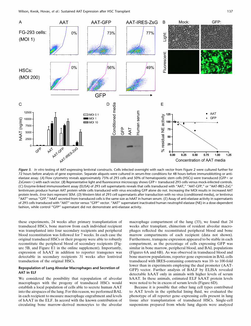
Next we tested our dual promoter vector (AAT-GFP),a bicistronic vector (AAT-IRES-ZSGreen), and a single pro-moter vector (AAT) by infecting 293 cells and freshly purifiedmurine HSCs in vitro (Figures 2 and 3). The ZsGreen fluoro-chrome rather than GFP was utilized for the bicistronic IRES-containing vector based on brighter FL1 channel fluorescence(data not shown). Two additional bicistronic vectors (CMV-dsRed-IRES-GFP and CMV-dsRed-IRES-ZsGreen) were notadapted for AAT expression based on low level in vivoexpression from the second cistron in HSCs (data not shown).Cells infected with each vector were analyzed by flow cytom-etry, fluorescence microscopy, and ELISA of cell supernatantsto assess relative levels of hAAT and reporter gene expression(Figure 3). Supernatants of infected cells were further analyzedby Western Blot and assay for anti-elastase activity to evaluatehAAT protein size and functional capacity (Figures 3D and3E). GFP or ZsGreen expression was easily detected aftertransduction of both the 293 cell line and primary HSCs, andwas used to demonstrate effective transduction of the majorityof cells (Figures 3A and 3B). High levels of hAAT protein weredocumented in all cell supernatants, providing in vitro confir-mation that dual trangenesis (i.e., expression of both hAATprotein and the fluorescent reporter) was achieved using dualpromoter constructs as well as the bicistronic IRES-containingconstruct (Figure 3C). hAAT protein secreted by transducedcells was the same size as hAAT from human serum, indicatingthat proper post-translational processing of the transgeneoccurred (Figure 3D). Supernatants of cells transduced withAAT vectors had the capacity to inactivate human neutrophilelastase in contrast to supernatants from cells transduced withcontrol (GFP-expressing) vectors (Figure 3E).
Transplantation of Lentivirally Transduced HSCs Results in
Long-Term Expression of hAAT In Vivo
Next, we tested in vivo gene expression after transplantation of3,000 HSCs infected with each AAT-expressing lentiviral vector.Because growth factors present in serum are known to stimulatedifferentiation of HSCs, serum-free culture conditions supple-
mented with low levels of SCF and TPO were used duringovernight lentiviral infection before transplantation (20). Trans-duced HSCs reconstituted the hematopoietic systems of allrecipients and expression of reporter genes as well as hAATprotein was tracked in the peripheral blood of each recipient for24 weeks (Figure 4). At all time points after transplant, 60 to90% of peripheral blood cells were donor-derived. The dualpromoter construct developed for these experiments accom-plished stable and efficient transduction of 87 6 10% (avg. 6
SD) of donor-derived cells as determined by flow cytometry forGFP expression (Figure 4B). In addition, circulating hAATprotein levels were easily detected for all vectors tested for this24-week period (Figure 4C). Although IRES-containing vectorsachieved stable hAAT levels in the peripheral blood, fluores-cent reporter gene expression from the second cistron incirculating cells was low or below detectable threshold (Figure4), consistent with prior reports of inefficient activity of IRES-containing lentiviral vectors in hematopoietic cells (31, 32).
Maintenance of Self-Renewal and Multipotency in
Transduced Stem Cells
Hematopoietic stem cells are defined by a remarkable capacityfor indefinite self-renewal as well as differentiation into allblood cell lineages. Although we have previously demonstratedthat our lentiviral transduction protocol does not diminish self-renewal or differentiation capacities of stem cells (20), we specif-ically tested whether stem cells transduced to overexpress AATstill possessed these defining stem cell characteristics. For thisreason the ability of transduced HSCs to contribute to myeloid,erythroid, and lymphoid lineages in the peripheral blood ofall recipients was followed for 14 weeks after transplantation.Recipients of HSCs transduced with either AAT-GFP or AAT-IRES-ZsG vectors demonstrated robust multilineage reconsti-tution of circulating myeloid (CD11b, GR-1), lymphoid (B220,CD3), and erythroid (TER-119) blood lineages (Figure 5). InAAT-GFP–transduced recipients, reporter transgene expres-sion (percent GFP1 and mean fluorescence intensity) wasmaintained in all peripheral blood lineages and did not appearto differ from recipients of HSCs transduced with control GFPvectors.
Maintenance of self-renewing undifferentiated HSCs wasdemonstrated by performing secondary transplantation. For
Figure 2. Schematic of lentiviral constructs emp-loyed for stem cell transduction. (A) Negative
control lentiviral vector (‘‘GFP’’) expresses only
the GFP reporter gene driven by an internal CMV
promoter (CMVp). (B) The ‘‘AAT’’ vector expressesthe human a1-antitrypsin gene (hAAT) driven by
CMVp. (C) Dual promoter vector ‘‘AAT-GFP’’
expresses hAAT driven by CMVp as well as the
GFP gene driven by a human UBC promoter(UBCp). (D) A bicistronic vector ‘‘AAT-IRES-ZsG’’
expresses a single mRNA encoding hAAT and
ZsGreen genes linked by an internal ribosome entrysite (IRES). All vectors include the woodchuck
hepatitis virus post-transcriptional regulatory ele-
ment (WPRE) to augment gene expression levels.
LTR: long terminal repeat. C: Psi packaging signal.
136 AMERICAN JOURNAL OF RESPIRATORY CELL AND MOLECULAR BIOLOGY VOL 39 2008
these experiments, 24 weeks after primary transplantation oftransduced HSCs, bone marrow from each individual recipientwas transplanted into four secondary recipients and peripheralblood reconstitution was followed for 7 weeks. In each case theoriginal transduced HSCs or their progeny were able to robustlyreconstitute the peripheral blood of secondary recipients (Fig-ure 5B, and Figure E1 in the online supplement). Importantly,expression of hAAT in addition to reporter transgenes wasdetectable in secondary recipients 31 weeks after lentiviraltransduction of the original HSCs.
Repopulation of Lung Alveolar Macrophages and Secretion of
AAT in ELF
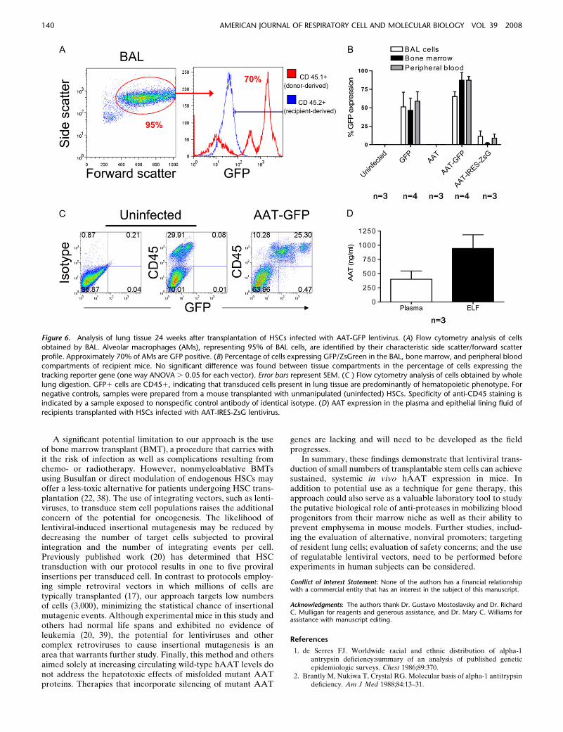
We considered the possibility that repopulation of alveolarmacrophages with the progeny of transduced HSCs wouldestablish a local population of cells able to secrete human AATinto the airspaces of the lung. For this reason, we performed BALin each recipient to measure macrophage engraftment and levelsof hAAT in the ELF. In accord with the known contribution ofcirculating bone marrow–derived monocytes to the alveolar
macrophage compartment of the lung (33), we found that 24weeks after transplant, chimerism of resident alveolar macro-phages reflected the reconstituted peripheral blood and bonemarrow compartments of each recipient (data not shown).Furthermore, transgene expression appeared to be stable in eachcompartment, as the percentage of cells expressing GFP wassimilar in bone marrow, peripheral blood, and BAL populations(Figures 6A and 6B). As was observed in transduced blood andbone marrow populations, reporter gene expression in BAL cellstransduced with IRES-containing constructs was 10- to 100-foldlower than in experiments employing the dual promoter (AAT-GFP) vector. Further analysis of BALF by ELISA revealeddetectable hAAT only in animals with higher levels of serumhAAT. In these animals, estimated ELF hAAT protein levelswere noted to be in excess of serum levels (Figure 6D).
Because it is possible that other lung cell types contributedto AAT levels in the epithelial lining fluid, we evaluated thephenotype of all reporter gene–expressing cells present in lungtissue after transplantation of transduced HSCs. Single-cellsuspensions prepared from whole lung digests were analyzed
Figure 3. In vitro testing of AAT-expressing lentiviral constructs. Cells infected overnight with each vector from Figure 2 were cultured further for
72 hours before analysis of gene expression. Separate aliquots were cultured in serum-free conditions for 48 hours before immunoblotting or anti-
elastase assay. (A) Flow cytometry reveals approximately 75% of 293 cells and 50% of hematopoietic stem cells (HSCs) were transduced (GFP1 orZsGreen1) with each vector. (B) Representative light and fluorescence microscopy shows GFP1 transduced 293 cells versus mock-infected controls.
(C) Enzyme-linked immunosorbent assay (ELISA) of 293 cell supernatants reveals that cells transduced with ‘‘AAT,’’ ‘‘AAT-GFP,’’ or ‘‘AAT-IRES-ZsG’’
lentiviruses produce human AAT protein while cells transduced with virus encoding GFP alone do not. Increasing the MOI results in increased AAT
protein levels. Error bars represent SEM. (D) Western blot of 293 cell supernatants after transduction with no virus (conditioned media), or lentivirus‘‘AAT’’ versus ‘‘GFP.’’ hAAT secreted from transduced cells is the same size as hAAT in human serum. (E) Assay of anti-elastase activity in supernatants
of 293 cells transduced with ‘‘AAT’’ vector versus ‘‘GFP’’ vector. ‘‘AAT’’ supernatant inactivated human neutrophil elastase (NE) in a dose-dependent
fashion, while control ‘‘GFP’’ supernatant did not demonstrate anti-elastase activity.
Wilson, Kwok, Hovav, et al.: Sustained AAT Expression after HSC Transplant 137

by flow cytometry for expression of reporter transgenes and thepan-hematopoietic marker, CD45 (Figure 6C). In each recipient(n 5 11), more than 99% of cells in the lung expressing reportertransgenes also expressed CD45, demonstrating that stem cellstransduced with either GFP, GFP-AAT, or AAT-IRES-ZsGreen constructs had predominantly given rise to hemato-poietic derivatives rather than epithelial or endothelial celltypes after transplantation.
Absence of Murine Immune Response to Circulating
Human Transgene
To determine whether expression of hAAT protein resultedin activation of a cellular or humoral immune response, wescreened for the presence of circulating anti-hAAT antibodies,and analyzed CD41 and CD81 T cells isolated from the spleensof experimental recipient animals (Figure E2). There were nodetectable circulating anti-hAAT IgG antibodies in any of thesamples tested. Similarly, ex vivo stimulation of splenocytes withhAAT protein did not reveal evidence of a cell-mediatedresponse as determined by intracellular cytokine staining (ICS)for IFN-g and IL-2 in CD41 or CD81 lymphocytes. Further-more, stimulation with hAAT protein did not result in T cellproliferation by CFSE assay (data not shown).
DISCUSSION
The concept of cell-based therapy for AAT deficiency wasinitially demonstrated by Garver and coworkers, who employeda retrovirus to introduce human AAT cDNA into murine fibro-blasts in culture prior to intraperitoneal transplantion into nudemice (7). Recent advances in stem cell biology and the de-velopment of lentiviral vectors allow us to extend this approachto transplantable stem cells with well-characterized self-renewaland differentiation capacities.
Our results support a novel approach to the systemic deliveryof AAT. This approach harnesses the considerable capacity ofHSCs to expand in vivo and uses lentiviruses to integrate genesinto the HSC genome. In doing so, we adapt our previouslyreported method for stem cell transduction to accomplish pro-longed expression of circulating human AAT in vivo. This worksuggests stem cells in general and HSCs in particular as potentialtargets for gene therapy of AAT deficiency.
HSCs transduced to overexpress AAT appear to retain theircapacity for engraftment and long-term multilineage bloodreconstitution. The ability of transduced HSCs to contributeto all peripheral blood cell lineages is particularly significantgiven recent publications regarding the putative role of AAT inthe regulation of bone marrow cell populations (34). AAT is
Figure 4. Peripheral blood chimerism and in vivo gene expression after HSC infection and transplant. HSCs were infected overnight with each
indicated lentiviral vector, and 3,000 HSCs were transplanted into each irradiated mouse recipient (n 5 4 mice per group). Six, 12, and 24 weeks
after transplantation, peripheral blood from each recipient was analyzed by flow cytometry to detect the percentage of donor-derived cells (blood
chimerism; A) and the percentage of donor-derived cells expressing GFP or ZSGreen reporter genes (B). ELISA of blood plasma was used to quantifycirculating human AAT protein levels (C). Error bars represent SEM. (D) Representative flow cytometry analysis from one mouse in each group, 24
weeks after HSC transplant: nucleated peripheral blood cells are analyzed after staining with antibodies that identify donor-derived cells (CD45.1)
versus recipient-derived cells (CD45.2). A subgate of only donor-derived blood cells (CD45.11/CD45.22; represented as a red histogram) illustrates
the percentage of donor-derived cells expressing the GFP or ZSGreen tracking reporter genes. The blue histogram overlay illustrates the absence ofGFP expression in recipient cells (CD45.12/CD45.21) in each sample.
138 AMERICAN JOURNAL OF RESPIRATORY CELL AND MOLECULAR BIOLOGY VOL 39 2008

produced by a variety of hematopoietic cell types in the bonemarrow, and down-regulation of AAT expression in the marrowto allow protease-based cleavage of matrix anchors may beimportant for the release of hematopoietic progenitors into theperipheral blood (34). If true, overexpression of AAT in themarrow could interfere with progenitor cell mobilization. Wefound that HSCs transduced to overexpress AAT contributed toall peripheral blood lineages, and the proportion of cells thatwere GFP1 remained constant as cells were mobilized frombone marrow to blood compartments. Although transduced(GFP1) cells competed well with untransduced (GFP-) cells intheir ability to contribute to the peripheral blood over time,cytokine mobilization experiments would be required to prop-erly test for any effect of AAT overexpression on the kinetics ofblood progenitor mobilization from marrow.
Despite accomplishing transduction of the majority of thehematopoietic system, our method did not result in circulatinghAAT levels in mice above the theoretical ‘‘protective thresh-old’’ of 800 mg/ml in human serum, the level thought to protectpatients from developing emphysema (18, 35–37). This contrastswith previous studies in which transduction of muscle fibers withadeno-associated viral vectors (AAV) was able to achieve ‘‘pro-tective’’ levels of circulating hAAT in mice (8). The levelsachieved may reflect intrinsic secretory limitations of hemato-pietic cell types compared with hepatocytes and myocytes, ormay be a consequence of limited promoter activity levels in ourstudies. Low circulating levels do not appear to have resultedfrom either a cell-mediated or from a humoral immune re-
sponse directed against the hAAT protein, in contrast to priorgene therapy approaches (8). This result is consistent with theobservation that HSC transplantation and resultant immunereconstitution typically abrogates humoral and cellular immu-nity against heterologous proteins expressed by the donor-derived stem cells.
Future studies employing lentiviral transduction of otherprogenitor cell populations, such as hepatic oval cells, musclesatellite cells, or embryonic stem cell–derived hepatocytes mayreveal better targets for cell-based therapies of AAT deficiencyand result in higher levels of AAT expression. Alternatively,evaluation of additional nonviral promoters may reveal candi-dates able to drive higher levels of gene expression. Indeed, inour studies, the highest level of hAAT expression in the plasmaresulted from HSCs transduced with AAT under control of thehuman UBC promoter.
It is notable that estimated hAAT expression in the ELFexceeded plasma expression in some animals. This result raisesthe possibility that a local pool of transduced cells, such asalveolar macrophages, contributed to hAAT production inthese recipients. A lung-localized hAAT-producing cell couldbe more effective than a distant one in suppressing proteaseactivity in the lung. It is known that only a fraction of circulatingAAT reaches the lung interstitium, where it purportedly exertsits protective benefit (18). While the serum AAT thresholdnecessary to provide protection from lung damage is widely ac-cepted to be approximately 800 mg/ml, the level required in thelung itself to provide this benefit is likely to be much lower (18, 19).
Figure 5. Assay of HSC functional capacity after lentiviral transduction. (A) Multilineage blood differentiation is illustrated by representative flow
cytometry analysis of peripheral blood 14 weeks after transplantation of 3,000 HSCs infected with ‘‘AAT-GFP’’ lentivirus. Robust engraftment ofdifferentiated lymphoid (CD31 or B2201), myeloid (CD11b1 or GR-11), and erythroid (Ter-1191) hematopoietic lineages derives from the
transduced, transplanted donor cells (GFP1). Peripheral blood ELISA at this time point demonstrated an hAAT level of 149 ng/ml. (B) Analysis of
peripheral blood drawn 13 weeks after primary transplant reveals 80% of all peripheral blood cells are donor-derived cells transduced with the ‘‘AAT-GFP’’ lentiviral construct (CD45.11/GFP1; hAAT 5 245 ng/ml). Twenty-four weeks after the initial transplant, 10 3 106 bone marrow cells from this
primary recipient were transplanted into an irradiated secondary recipient. Seven weeks later, 83% of peripheral blood cells are derived from the
original donor stem cells and continue to express both the GFP reporter gene (CD45.11/GFP1).
Wilson, Kwok, Hovav, et al.: Sustained AAT Expression after HSC Transplant 139
A significant potential limitation to our approach is the useof bone marrow transplant (BMT), a procedure that carries withit the risk of infection as well as complications resulting fromchemo- or radiotherapy. However, nonmyeloablative BMTsusing Busulfan or direct modulation of endogenous HSCs mayoffer a less-toxic alternative for patients undergoing HSC trans-plantation (22, 38). The use of integrating vectors, such as lenti-viruses, to transduce stem cell populations raises the additionalconcern of the potential for oncogenesis. The likelihood oflentiviral-induced insertional mutagenesis may be reduced bydecreasing the number of target cells subjected to proviralintegration and the number of integrating events per cell.Previously published work (20) has determined that HSCtransduction with our protocol results in one to five proviralinsertions per transduced cell. In contrast to protocols employ-ing simple retroviral vectors in which millions of cells aretypically transplanted (17), our approach targets low numbersof cells (3,000), minimizing the statistical chance of insertionalmutagenic events. Although experimental mice in this study andothers had normal life spans and exhibited no evidence ofleukemia (20, 39), the potential for lentiviruses and othercomplex retroviruses to cause insertional mutagenesis is anarea that warrants further study. Finally, this method and othersaimed solely at increasing circulating wild-type hAAT levels donot address the hepatotoxic effects of misfolded mutant AATproteins. Therapies that incorporate silencing of mutant AAT
genes are lacking and will need to be developed as the fieldprogresses.
In summary, these findings demonstrate that lentiviral trans-duction of small numbers of transplantable stem cells can achievesustained, systemic in vivo hAAT expression in mice. Inaddition to potential use as a technique for gene therapy, thisapproach could also serve as a valuable laboratory tool to studythe putative biological role of anti-proteases in mobilizing bloodprogenitors from their marrow niche as well as their ability toprevent emphysema in mouse models. Further studies, includ-ing the evaluation of alternative, nonviral promoters; targetingof resident lung cells; evaluation of safety concerns; and the useof regulatable lentiviral vectors, need to be performed beforeexperiments in human subjects can be considered.
Conflict of Interest Statement: None of the authors has a financial relationshipwith a commercial entity that has an interest in the subject of this manuscript.
Acknowledgments: The authors thank Dr. Gustavo Mostoslavsky and Dr. RichardC. Mulligan for reagents and generous assistance, and Dr. Mary C. Williams forassistance with manuscript editing.
References
1. de Serres FJ. Worldwide racial and ethnic distribution of alpha-1antrypsin deficiency:summary of an analysis of published geneticepidemiologic surveys. Chest 1986;89:370.
2. Brantly M, Nukiwa T, Crystal RG. Molecular basis of alpha-1 antitrypsindeficiency. Am J Med 1988;84:13–31.
Figure 6. Analysis of lung tissue 24 weeks after transplantation of HSCs infected with AAT-GFP lentivirus. (A) Flow cytometry analysis of cells
obtained by BAL. Alveolar macrophages (AMs), representing 95% of BAL cells, are identified by their characteristic side scatter/forward scatter
profile. Approximately 70% of AMs are GFP positive. (B) Percentage of cells expressing GFP/ZsGreen in the BAL, bone marrow, and peripheral blood
compartments of recipient mice. No significant difference was found between tissue compartments in the percentage of cells expressing thetracking reporter gene (one way ANOVA . 0.05 for each vector). Error bars represent SEM. (C ) Flow cytometry analysis of cells obtained by whole
lung digestion. GFP1 cells are CD451, indicating that transduced cells present in lung tissue are predominantly of hematopoietic phenotype. For
negative controls, samples were prepared from a mouse transplanted with unmanipulated (uninfected) HSCs. Specificity of anti-CD45 staining isindicated by a sample exposed to nonspecific control antibody of identical isotype. (D) AAT expression in the plasma and epithelial lining fluid of
recipients transplanted with HSCs infected with AAT-IRES-ZsG lentivirus.
140 AMERICAN JOURNAL OF RESPIRATORY CELL AND MOLECULAR BIOLOGY VOL 39 2008

3. Stockley RA. The pathogenesis of chronic obstructive lung diseases:implications for therapy. QJM 1995;88:141–146.
4. Sandhaus RA Elastase May Play a Central Role in the NeutrophilMigration Through Connective Tissue. In: Taylor JC, Mittman C,editors. Pulmonary emphysema and proteolysis Orlando, FL: Aca-demic Press; 1997. p. 227.
5. Larsson C. Natural history and life expectancy in severe alpha-1-antitrypsin deficiency, PiZ. Acta Med Scand 1978;204:345–351.
6. American Thoracic Society. Guidelines for the approach to the patientwith severe hereditary alpha-1-antitrypsin deficiency. Am Rev RespirDis 1989;140:1494–1497.
7. Garver RI Jr, Chytil A, Courtney M, Crystal RG. Clonal gene therapy:transplanted mouse fibroblast clones express human alpha 1-antitrypsingene in vivo. Science 1987;237:762–764.
8. Song S, Morgan M, Ellis T, Poirier A, Chesnut K, Wang J, Brantly M,Muzyczka N, Byrne BJ, Atkinson M, et al. Sustained secretion ofhuman alpha-1-antitrypsin from murine muscle transduced withadeno-associated virus vectors. Proc Natl Acad Sci USA 1998;95:14384–14388.
9. Ferkol T, Mularo F, Hilliard J, Lodish S, Perales JC, Ziady A, KonstanM. Transfer of the human alpha1-antitrypsin gene into pulmonarymacrophages in vivo. Am J Respir Cell Mol Biol 1998;18:591–601.
10. Kay MA, Baley P, Rothenberg S, Leland F, Fleming L, Ponder KP, LiuT, Finegold M, Darlington G, Pokorny W, et al. Expression of humanalpha 1-antitrypsin in dogs after autologous transplantation of retro-viral transduced hepatocytes. Proc Natl Acad Sci USA 1992;89:89–93.
11. Duan YY, Wu J, Zhu JL, Liu SL, Ozaki I, Strayer DS, Zern MA. Genetherapy for human alpha1-antitrypsin deficiency in an animal modelusing SV40-derived vectors. Gastroenterology 2004;127:1222–1232.
12. Song S, Embury J, Laipis PJ, Berns KI, Crawford JM, Flotte TR. Stabletherapeutic serum levels of human alpha-1 antitrypsin (AAT) afterportal vein injection of recombinant adeno-associated virus (RAAV)vectors. Gene Ther 2001;8:1299–1306.
13. Lemarchand P, Jaffe HA, Danel C, Cid MC, Kleinman HK, Stratford-Perricaudet LD, Perricaudet M, Pavirani A, Lecocq JP, Crystal RG.Adenovirus-mediated transfer of a recombinant human alpha 1-antitrypsin CDNA to human endothelial cells. Proc Natl Acad SciUSA 1992;89:6482–6486.
14. Rosenfeld MA, Siegfried W, Yoshimura K, Yoneyama K, Fukayama M,Stier LE, Paakko PK, Gilardi P, Stratford-Perricaudet LD,Perricaudet M, et al. Adenovirus-mediated transfer of a recombinantalpha 1-antitrypsin gene to the lung epithelium in vivo. Science 1991;252:431–434.
15. Setoguchi Y, Jaffe HA, Chu CS, Crystal RG. Intraperitoneal in vivogene therapy to deliver alpha 1-antitrypsin to the systemic circulation.Am J Respir Cell Mol Biol 1994;10:369–377.
16. Stecenko AA, Brigham KL. Gene therapy progress and prospects:alpha-1 antitrypsin. Gene Ther 2003;10:95–99.
17. Hacein-Bey-Abina S, le Deist F, Carlier F, Bouneaud C, Hue C, deVillartay JP, Thrasher AJ, Wulffraat N, Sorensen R, Dupuis-GirodS, et al. Sustained correction of X–linked severe combined immuno-deficiency by ex vivo gene therapy. N Engl J Med 2002;346:1185–1193.
18. Hubbard RC, Crystal RG. Augmentation therapy of alpha 1-antitrypsindeficiency 4. Eur Respir J Suppl 1990;9:44s–52s.
19. Wewers MD, Casolaro MA, Sellers SE, Swayze SC, McPhaul KM,Wittes JT, Crystal RG. Replacement therapy for alpha 1-antitrypsindeficiency associated with emphysema. N Engl J Med 1987;316:1055–1062.
20. Mostoslavsky G, Kotton DN, Fabian AJ, Gray JT, Lee JS, Mulligan RC.Efficiency of transduction of highly purified murine hematopoietic
stem cells by lentiviral and oncoretroviral vectors under conditions ofminimal in vitro manipulation. Mol Ther 2005;11:932–940.
21. Naldini L, Blomer U, Gallay P, Ory D, Mulligan RC, Gage FH, VermaIM, Trono D. In vivo gene delivery and stable transduction ofnondividing cells by a lentiviral vector. Science 1996;272:263–267.
22. Mostoslavsky G, Fabian AJ, Rooney S, Alt FW, Mulligan RC. Completecorrection of murine artemis immunodeficiency by lentiviral vector-mediated gene transfer. Proc Natl Acad Sci USA 2006;103:16406–16411.
23. Summer R, Kotton DN, Sun X, Fitzsimmons K, Fine A. Translationalphysiology: origin and phenotype of lung side population cells. Am JPhysiol Lung Cell Mol Physiol 2004;287:L477–L483.
24. Goodell MA, Brose K, Paradis G, Conner AS, Mulligan RC. Isolationand functional properties of murine hematopoietic stem cells that arereplicating in vivo. J Exp Med 1996;183:1797–1805.
25. Kotton DN, Fabian AJ, Mulligan RC. A novel stem cell population inadult liver with potent hematopoietic reconstitution activity. Blood2005;106:1574–1580.
26. Balazs AB, Fabian AJ, Esmon CT, Mulligan RC. Endothelial protein Creceptor (CD201) explicitly identifies hematopoietic stem cells inmurine bone Marrow. Blood 2006;107:2317–2321.
27. Kotton DN, Fabian AJ, Mulligan RC. Failure of bone marrow toreconstitute lung epithelium. Am J Respir Cell Mol Biol 2005;33:328–334.
28. Rennard SI, Basset G, Lecossier D, O’Donnell KM, Pinkston P, MartinPG, Crystal RG. Estimation of volume of epithelial lining fluidrecovered by lavage using urea as marker of dilution. J Appl Physiol1986;60:532–538.
29. Hovav AH, Panas MW, Rahman S, Sircar P, Gillard G, Cayabyab MJ,Letvin NL. Duration of antigen expression in vivo following DNAimmunization modifies the magnitude, contraction, and secondaryresponses of CD8+ T lymphocytes. J Immunol 2007;179:6725–6733.
30. Hovav AH, Panas MW, Osuna CE, Cayabyab MJ, Autissier P, LetvinNL. The impact of a boosting immunogen on the differentiation ofsecondary memory CD8+ T cells. J Virol 2007;81:12793–12802.
31. Amendola M, Venneri MA, Biffi A, Vigna E, Naldini L. Coordinatedual-gene transgenesis by lentiviral vectors carrying synthetic bi-directional promoters. Nat Biotechnol 2005;23:108–116.
32. Borman AM, Le MP, Girard M, Kean KM. Comparison of picornaviralIRES-driven internal initiation of translation in cultured cells ofdifferent origins. Nucleic Acids Res 1997;25:925–932.
33. Thomas ED, Ramberg RE, Sale GE, Sparkes RS, Golde DW. Directevidence for a bone marrow origin of the alveolar macrophage inman. Science 1976;192:1016–1018.
34. Winkler IG, Hendy J, Coughlin P, Horvath A, Levesque JP. Serineprotease inhibitors serpina1 and serpina3 are down-regulated in bonemarrow during hematopoietic progenitor mobilization. J Exp Med2005;201:1077–1088.
35. Morse JO. Alpha1-antitrypsin deficiency (first of two parts). N Engl JMed 1978;299:1045–1048.
36. Morse JO. Alpha1-antitrypsin deficiency (second of two parts). N Engl JMed 1978;299:1099–1105.
37. Kueppers F, Black LF. Alpha1-antitrypsin and its deficiency. Am RevRespir Dis 1974;110:176–194.
38. Adams AB, Durham MM, Kean L, Shirasugi N, Ha J, Williams MA,Rees PA, Cheung MC, Mittelstaedt S, Bingaman AW, et al. Cos-timulation blockade, busulfan, and bone marrow promote titratablemacrochimerism, induce transplantation tolerance, and correct ge-netic hemoglobinopathies with minimal myelosuppression. J Immu-nol 2001;167:1103–1111.
39. Woods NB, Bottero V, Schmidt M, von Kalle C, Verma IM. Genetherapy: therapeutic gene causing lymphoma. Nature 2006;440:1123.
Wilson, Kwok, Hovav, et al.: Sustained AAT Expression after HSC Transplant 141