1
Poster Design & Printing by Genigraphics ® - 800.790.4001 Miss Azrina Zaman Doncaster Royal Infirmary Email: [email protected] Phone: +447863117046 Suspension suture technique in Nasal Valve Collapse Objective: To assess efficacy of alar suspension sutures in the management of nasal valve collapse Method: Retrospective casenote review of patients who had alar suspension sutures inserted for management of nasal valve collapse in Doncaster Royal Infirmary from January 2009 to December 2010. Subjective nasal congestion reported by patients pre and post procedure was measured using Visual analogue scale (VAS 010). Objective measurement using Peak Inspiratory Flow Rate (PIFR) before and at least three months after the operation was also performed. Results: Thirty five patients who had alar suspension sutures inserted were identified. Nine patients were excluded from the study as 6 had concurrent procedures carried out on septum, turbinates and lacrimal duct and 3 patients’ casenotes were unavailable. 26 patients were included in our study. The average VAS score improvement was by 5 points (pvalue=0.00) and PIFR increased by 63.7% (p value= 0.00) Conclusion: Our study shows a significant improvement in patient’s symptoms following insertion of alar suspension suture. This is a short and well tolerated procedure with minimal side effects. Suspension Suture Technique in Nasal Valve Collapse Azrina Zaman, MRCS DOHNS; Chee-Yean Eng, FRCS (ORL-HNS); Ullas Raghavan FRCS (ORL-HNS) Doncaster Royal Infirmary, United Kingdom This study has shown the efficacy of this surgical technique in improving symptoms in patients with nasal valve collapse by way of improvement in both the objective measurement of Peak Inspiratory Flow Rate (PIFR) and the subjective measurement of Visual analogue scale. (VAS). This is supported by the findings of Lee et al 3 and Paniello et al 5 . Ala suspension sutures are well tolerated and has minimal side effects compared to other surgical techniques. For example, when using grafts this can be complicated by migration and change in external appearance of nose 6 . Graft insertion is technically more demanding and requires more theatre time, compared to insertion of ala suspension sutures hence this makes suspension sutures more cost effective. The improvement in nasal air flow also correlated with symptomatic improvement for obstruction unlike septoplasty as shown by Tompos et al 7 . This leads us to conclude that ala suspension suture is a reliable, safe and effective technique in managing nasal valve collapse. Patients are assessed following topical decongestion using the Modified Cottle’s Test. The site where maximal symptom relief is achieved by Modified Cottle’s Test is then carefully noted. A30 Prolene suture is inserted at this site in the nasal vestibule between skin and the lateral crura. A 1 cm incision is made in the medial end of the inferior orbital margin after infiltration with lignocaine and adrenaline reaching the muscular layer. A straightened 42G Mayo’s Needle is used to pass both ends the suture through their exit wound respectively in the vestibule to ensure the suture is not exposed and is brought out through the incision. A 32G Mayo’s Needle is then used to pass the suture through the periosteum, where it is tightened and tied. The orbital margin incision is the closed in 2 layers using 5O Vicryl Rapide. The patient is discharged home with 1 week course of oral antibiotics and topical polymyxin Ointment. The concept of the nasal valve was first described by Mink in 1903. In 1970 Bridger then defined it as the ‘flow limiting segment’ of the nasal airway. The nasal valve is a complex structure and is prone to collapse if there is a loss of cartilage resilience. This can be due to surgery, trauma, ageing process or congenital weakness of cartilage. There are different surgical options for the management of nasal valve collapse 1 and this can include graft insertion 2 , flare or suspension suture insertion 3,4 . AIM The aim of our study is to evaluate the efficacy of using our technique of inserting alar suspension sutures in the management of nasal valve collapse. INTRODUCTION SURGICAL TECHNIQUE 1. Wittkopf M, et al. Feb 2008. The diagnosis and treatment of nasal valve collapse. Current Opinion in Otoloaryngology & Head & Neck Surgery. 16: 1013 2. Wallace H, et al. 2009. Management of the narrow nose. The Journal of Laryngology and Otology 123: 945951. 3. Lee DS, et al .Oct 2011. Correction of nasal valve stenosis with lateral suspension suture. Archives of Facial Plastic Surgery 3: 237240. 4. Apaydin, F. Apr 2011. Nasal Valve Surgery. Facial Plastic Surgery 27: 179191 5. Paniello RC, et al. 1996. Nasal Valve suspension an effective treatment for nasal valve collapse. Arch Otolaryngology and Head and Neck Surgery 122(12): 13421346 6. Valerio C. et al. 2009. Alar Batten Cartilage graft: Treatment of internal and external nasal valve collapse. Aesthetic Plastic Surgery. 33:625634 7. Tompos T et al.Dec 2010. Sensation of nasal patency compared to rhinometric results after septoplasty. European Archives of Otolaryngology 267(12): 18871991 CONCLUSIONS REFERENCES ABSTRACT CONTACT A retrospective case note review was conducted on patients who had alar suspension sutures inserted between January 2009 to December 2010 in Doncaster Royal Infirmary. Patients who had concurrent procedures carried out on the septum, turbinates and lacrimal duct were excluded from the study. The Visual analogue scale (VAS) was used to assess patients’ symptoms subjectively and peak inspiratory flow rates (PIFR) were used to assess symptoms objectively. The VAS scale: 0‐‐‐‐‐‐‐‐‐‐‐‐‐‐‐‐‐‐‐‐‐‐‐‐‐‐‐‐‐‐‐‐‐‐‐‐‐‐‐‐‐‐‐‐‐‐‐‐‐‐‐‐‐‐‐‐‐‐‐‐‐‐‐‐‐‐‐‐‐10 No congestion Completely blocked nose Peak inspiratory Flow Rate Measure: METHOD Preop: Alar position at rest Preop: Alar collapse when sniffing Endoscopic appearance Right nasal airway pre op under GA Endoscopic appearance Left nasal airway preop under GA Prolene 3/0 suture inserted between mucosa and lateral crura 1cm incision in the medial end of inferior orbital margin Straightening of 42G Needle Peak inspiratory flow rate measured using peak inspiratory flow meter RESULTS 35 patients identified 9 patients excluded (6 concurrent procedures, 3 –casenotes not found 26 patients included in study 15 male and 11 female 23 patients had bilateral suture insertion 2 had left suture insertion and 1 had right suture insertion The mean difference in VAS is 4.97 (CI 4.485.46) p value= 0.00 The mean difference is 25.5L/min (CI 22.3328.67) p value= 0.00 Complications 2 (8%) developed slipped sutures 2 (8%) developed post operative neuralgia controlled with low dose amitriptyline. 0 2 4 6 8 VAS 7.7 2.9 PRE POST Post op: Alar position at rest Post op: Alar position during sniffing End appearance of suture Sutures tightened Suture brought out of incision Using 42G Mayo’s needle to pass suture through exit wound

Suspension Suture Technique in Nasal Valve Collapse · Suspension suture technique in Nasal Valve Collapse Objective: To assess efficacy of alar suspension sutures in the management

Embed Size (px)

Citation preview

Poster Design & Printing by Genigraphics® - 800.790.4001

Miss Azrina ZamanDoncaster Royal InfirmaryEmail: [email protected]: +447863117046

Suspension suture technique in Nasal Valve Collapse

Objective:  To  assess  efficacy  of  alar  suspension  sutures  in the management of nasal valve collapse

Method:  Retrospective  case‐note  review  of  patients  who had  alar  suspension  sutures  inserted  for  management  of nasal  valve  collapse  in  Doncaster  Royal  Infirmary  from January  2009  to  December  2010.  Subjective  nasal congestion  reported  by  patients  pre  and  post  procedure was  measured  using  Visual  analogue  scale  (VAS  0‐10). Objective  measurement  using  Peak  Inspiratory  Flow  Rate (PIFR) before and at  least three months after the operation was also performed.

Results: Thirty five patients who had alar suspension sutures inserted were  identified. Nine patients were excluded from the  study  as  6  had  concurrent  procedures  carried  out  on septum,  turbinates and  lacrimal duct and 3 patients’ case‐notes were  unavailable.  26  patients were  included  in  our study. The average VAS score improvement was by 5 points (p‐value=0.00) and PIFR increased by 63.7% (p value= 0.00)

Conclusion: Our  study  shows  a  significant  improvement  in patient’s  symptoms  following  insertion  of  alar  suspension suture.  This  is  a  short  and well  tolerated  procedure with minimal side effects.

Suspension Suture Technique in Nasal Valve CollapseAzrina Zaman, MRCS DOHNS; Chee-Yean Eng, FRCS (ORL-HNS); Ullas Raghavan FRCS (ORL-HNS)

Doncaster Royal Infirmary, United Kingdom

This  study  has  shown  the  efficacy  of  this  surgical  technique  in  improving symptoms  in  patients with  nasal  valve  collapse  by way  of  improvement  in both the objective measurement of Peak Inspiratory Flow Rate (PIFR) and the subjective measurement of  Visual analogue scale. (VAS). This is supported by the findings of Lee et al3 and Paniello et al5. 

Ala  suspension  sutures  are  well  tolerated  and  has  minimal  side  effects compared to other surgical  techniques. For example, when using   grafts  this can be complicated by migration and change in external appearance of nose6 .

Graft  insertion  is  technically  more  demanding  and  requires  more theatre time,  compared  to  insertion  of  ala  suspension  sutures  hence  this makes suspension sutures more cost effective. 

The  improvement  in  nasal  air  flow  also  correlated  with    symptomatic improvement for obstruction unlike septoplasty as shown by Tompos et al7. 

This  leads us to conclude that ala suspension suture  is a reliable,  safe and effective  technique  in managing nasal valve collapse.

Patients are assessed following topical decongestion using the Modified Cottle’s Test. The site where maximal symptom relief is achieved by Modified Cottle’s Test is then carefully noted.

A 3‐0 Prolene suture is inserted at this site in the nasal vestibule between skin and the lateral crura.

A 1 cm incision is made in the medial end of the inferior orbital margin after infiltration with lignocaine and adrenaline reaching the muscular layer.

A straightened 42G Mayo’s Needle  is used  to pass both ends  the suture  through  their exitwound respectively  in the vestibule to ensure the suture  is not exposed and  is brought out through the incision.

A 32G Mayo’s Needle  is  then used  to pass  the  suture  through  the periosteum, where  it  is tightened and tied.

The orbital margin incision is the closed in 2 layers using 5O Vicryl Rapide.

The patient is discharged home with 1 week course of oral antibiotics and topical polymyxin Ointment.

The  concept  of  the  nasal  valve was  first  described  by Mink  in  1903.  In  1970  Bridger  then defined it as the ‘flow limiting segment’ of the nasal airway. 

The nasal valve  is a complex structure and  is prone to collapse  if there  is a  loss of cartilage resilience.  This  can  be  due  to  surgery,  trauma,  ageing  process  or  congenital weakness  of cartilage.

There are different surgical options for the management of  nasal valve collapse1 and this can include graft insertion2, flare or suspension suture insertion3,4.

AIMThe  aim  of  our  study  is  to  evaluate  the  efficacy  of  using  our  technique  of  inserting  alar suspension sutures in the management of nasal valve collapse.

INTRODUCTION

SURGICAL TECHNIQUE

1. Wittkopf M, et al. Feb 2008. The diagnosis and treatment of nasal valve  collapse. Current Opinion in Otoloaryngology & Head & Neck Surgery. 16: 10‐13

2. Wallace H, et al. 2009. Management of the narrow nose. The Journal of Laryngology and Otology 123: 945‐951.

3. Lee DS,  et al .Oct 2011. Correction of nasal valve stenosis with lateral suspension suture. Archives of Facial Plastic Surgery 3: 237‐240. 

4. Apaydin, F. Apr 2011. Nasal Valve Surgery. Facial Plastic Surgery 27: 179‐1915. Paniello RC, et al. 1996. Nasal Valve suspension an effective treatment for nasal  

valve collapse. Arch Otolaryngology and Head and Neck Surgery 122(12): 1342‐13466. Valerio C. et al. 2009. Alar Batten Cartilage graft: Treatment of internal and external 

nasal valve collapse. Aesthetic Plastic Surgery. 33:625‐6347. Tompos T et al.Dec 2010. Sensation of nasal patency compared to rhinometric results

after septoplasty. European Archives of Otolaryngology 267(12): 1887‐1991

CONCLUSIONS

REFERENCES

ABSTRACT

CONTACT

A  retrospective  case  note  review was  conducted  on  patients who  had  alar suspension  sutures  inserted  between  January  2009  to  December  2010  in Doncaster Royal Infirmary.

Patients  who  had  concurrent  procedures  carried  out  on  the  septum, turbinates and lacrimal duct were excluded from the study.

The  Visual  analogue  scale  (VAS)  was  used  to  assess  patients’ symptoms subjectively  and  peak  inspiratory  flow  rates  (PIFR)  were  used  to assess symptoms objectively. 

The VAS scale:0‐‐‐‐‐‐‐‐‐‐‐‐‐‐‐‐‐‐‐‐‐‐‐‐‐‐‐‐‐‐‐‐‐‐‐‐‐‐‐‐‐‐‐‐‐‐‐‐‐‐‐‐‐‐‐‐‐‐‐‐‐‐‐‐‐‐‐‐‐10

No congestion Completely blocked nose

Peak inspiratory Flow Rate Measure:

METHOD

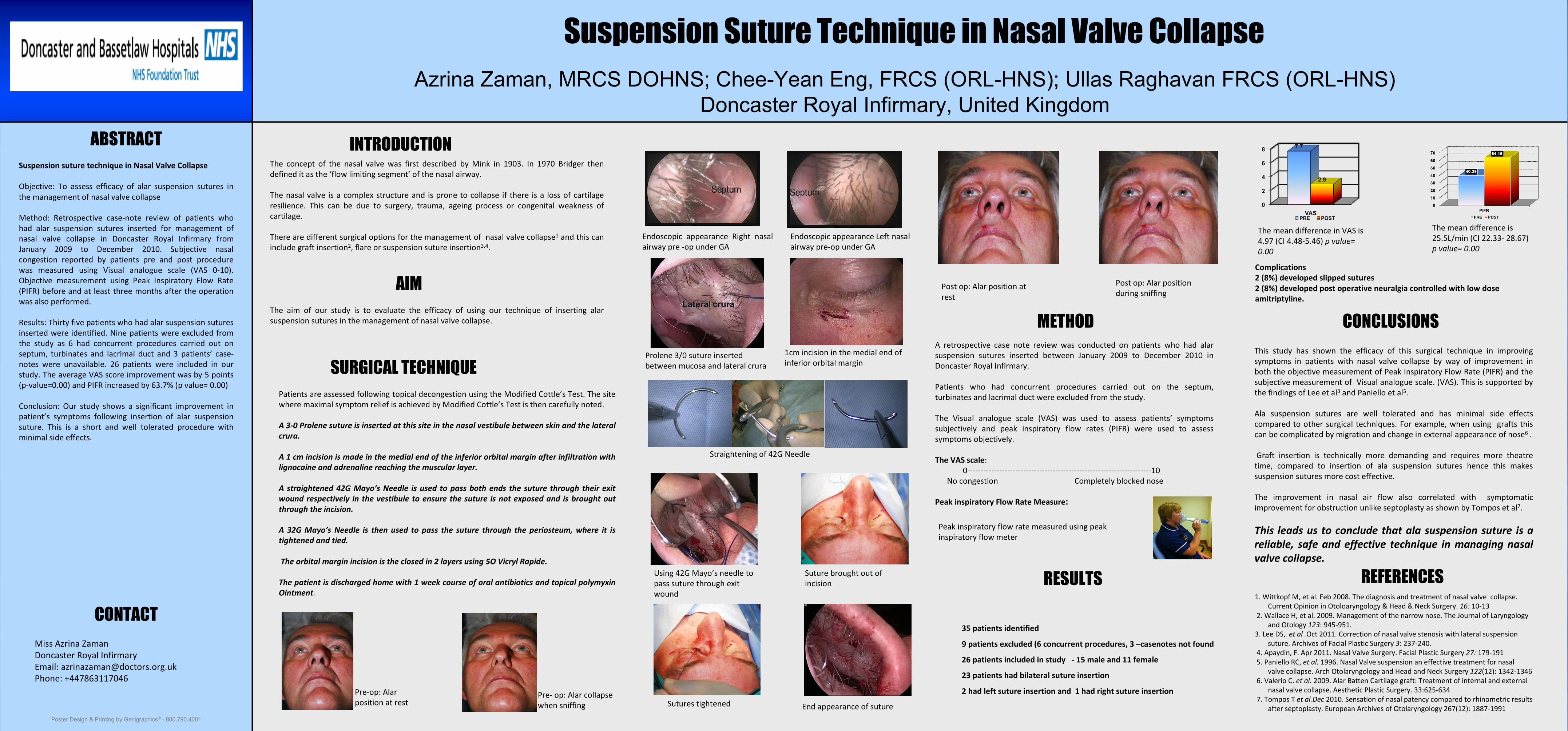
Pre‐op: Alar position at rest

Pre‐ op: Alar collapse when sniffing

Endoscopic  appearance  Right  nasal airway pre ‐op under GA

Endoscopic appearance Left nasal airway pre‐op under GA

Prolene 3/0 suture inserted  between mucosa and lateral crura

1cm incision in the medial end of inferior orbital margin 

Straightening of 42G Needle

Peak inspiratory flow rate measured using peak inspiratory flow meter

RESULTS

35 patients identified

9 patients excluded (6 concurrent procedures, 3 –casenotes not found

26 patients included in study   ‐ 15 male and 11 female

23 patients had bilateral suture insertion 

2 had left suture insertion and  1 had right suture insertion

The mean difference in VAS is 4.97 (CI 4.48‐5.46) p value= 0.00

The mean difference is 25.5L/min (CI 22.33‐ 28.67) p value= 0.00

Complications 2 (8%) developed slipped sutures2 (8%) developed post operative neuralgia controlled with low dose amitriptyline.

0

2

4

6

8

VAS

7.7

2.9

PRE POST

Post op: Alar position at rest

Post op: Alar position during sniffing 

End appearance of sutureSutures tightened

Suture brought out of incision

Using 42G Mayo’s needle to pass suture through exit wound