Embed Size (px)
Citation preview
SURVEILLANCE OF BACTERIAL PNEUMONIA AND MENINGITIS IN CHILDREN AGED UNDER
5 YEARS
Field guideSecond edition
SURVEILLANCE OF BACTERIAL PNEUMONIA AND MENINGITIS IN CHILDREN AGED UNDER
5 YEARS
Field guideSecond edition
Washington, D.C.2019
Surveillance of Bacterial Pneumonia and Meningitis in Children Aged Under 5 Years: Field Guide. Second Edition
© Pan American Health Organization, 2021ISBN: 978-92-75-12188-7 (print)ISBN: 978-92-75-12189-4 (pdf) Some rights reserved. This work is available under the Creative Commons Attribution-NonCommercial-ShareAlike 3.0 IGO license (CC BY-NC-SA 3.0 IGO; https://creativecommons.org/licenses/by-nc-sa/3.0/igo).
Under the terms of this license, this work may be copied, redistributed, and adapted for non-commercial purposes, provided the new work is issued using the same or equivalent Creative Commons license and it is appropriately cited, as indicated below. In any use of this work, there should be no suggestion that the Pan American Health Organization (PAHO) endorses any specific organization, product, or service. Use of the PAHO logo is not permitted.
Adaptations: If this work is adapted, the following disclaimer should be added along with the suggested citation: “This is an adaptation of an original work by the Pan American Health Organization (PAHO). Views and opinions expressed in the adaptation are the sole responsibility of the author(s) of the adaptation and are not endorsed by PAHO.”
Translation: If this work is translated, the following disclaimer should be added along with the suggested citation: “This translation was not created by the Pan American Health Organization (PAHO). PAHO is not responsible for the content or accuracy of this translation.”
Suggested citation. Surveillance of Bacterial Pneumonia and Meningitis in Children Aged Under 5 Years: Field Guide. Second Edition. Washington, D.C.: Pan American Health Organization; 2021. License: CC BY-NC-SA 3.0 IGO. https://doi.org/10.37774/9789275121894.
Cataloguing-in-Publication (CIP) data. CIP data are available at http://iris.paho.org.Sales, rights, and licensing. To purchase PAHO publications, write to [email protected]. To submit requests for commercial use and queries on rights and licensing, visit http://www.paho.org/permissions.
Third-party materials. If material that is attributed to a third party, such as tables, figures, or images, is reused from this work, it is the user’s responsibility to determine whether permission is needed for that reuse and to obtain permission from the copyright holder. The risk of claims resulting from infringement of any third-party owned material or component from this work rests solely with the user.
General disclaimers. The designations employed and the presentation of the material in this publication do not imply the expression of any opinion whatsoever on the part of PAHO concerning the legal status of any country, territory, city, or area or of its authorities, or concerning the delimitation of its frontiers or boundaries. Dotted and dashed lines on maps represent approximate border lines for which there may not yet be full agreement.The mention of specific companies or of certain manufacturers’ products does not imply that they are endorsed or recommended by PAHO in preference to others of a similar nature that are not mentioned. Errors and omissions excepted, the names of proprietary products are distinguished by initial capital letters.
All reasonable precautions have been taken by PAHO to verify the information contained in this publication. However, the published material is being distributed without warranty of any kind, either expressed or implied. The responsibility for the interpretation and use of the material lies with the reader. In no event shall PAHO be liable for damages arising from its use. FPL/IM/2021
INDEXIndex.................................................................................................................................iiiPreface for the 1st edition................................................................................................ivPreface for the 2nd edition..................................................................................................vAcknowledgements..........................................................................................................viAbbreviations and acronyms...........................................................................................vii
1. INTRODUCTION.........................................................................................................................11.1 Epidemiological Situation in the Region of the Americas1.2 Epidemiology
1.2.1 Etiologic agents1.2.2 Reservoir1.2.3 Transmission1.2.4 Distribution and seasonality1.2.5 Susceptibility and risk factors1.2.6 Immunity1.2.7 Carrier status
2. BACTERIAL PNEUMONIA................................................................................................92.1 Clinical aspects
2.1.1 Differential diagnosis2.1.2 Complications
2.2 Radiological diagnosis2.2.1 X-ray quality
2.3 Laboratory diagnosis of bacterial pneumonia 2.3.1 Biological samples
2.4 Treatment
3. BACTERIAL MENINGITIS............................................................................................193.1 Clinical aspects
3.1.1 Differential diagnosis3.1.2 Complications
3.2 Laboratory diagnosis of bacterial meningitis3.2.1 Biological samples
3.3 Treatment
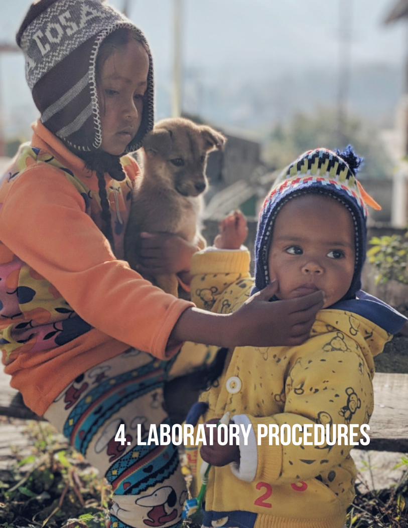
4. LABORATORY PROCEDURES........................................................................................244.1 Biosafety
4.1.1 Health program for personnel4.2 Hemoculture
4.2.1 Collecting blood specimens for hemoculture
4.2.2 Transporting blood culture bottles4.2.3 Reception of blood culture bottles by laboratory4.2.4 Incubating blood culture bottles4.2.5 Blood culture gram staining 4.2.6 Subculturing of blood culture bottles4.2.7 Subculturing techniques4.2.8 Differentiating bacteremia from contamination
4.3 Cerebrospinal fluid4.3.1 Collecting csf specimens4.3.2 Transporting csf specimens4.3.3 Reception of csf specimens by laboratory 4.3.4 Analyzing csf specimens4.3.5 Delivering csf analysis results
4.4 Pleural fluid4.4.1 Collecting pf specimens4.4.2 Transporting pf specimens4.4.3 Reception of pf specimens by laboratory 4.4.4 Pl cytology and chemistry
4.5 General considerations for csf and pf specimens4.6 S. Pneumoniae, h. Influenzae and n. Meningitidis identification
4.6.1 Bacterial isolates or strains4.6.2 Culture4.6.3 Antimicrobial susceptibility testing4.6.4 Immunological and molecular diagnostic methods
4.7 Quality control 4.7.1 Internal quality control program 4.7.2 Responsibilities of the laboratory network
5. SURVEILLANCE OF BACTERIAL PNEUMONIA AND MENINGITIS .................................475.1 Surveillance objectives5.2 Surveillance strategies
5.2.1 Target population surveillance 5.2.2 Type of surveillance5.2.3 Criteria for the selection of sentinel hospitals
5.3 Sentinel hospital-based bacterial pneumonia surveillance5.3.1 Case definitions5.3.2 Steps of sentinel hospital-based bacterial pneumonia surveillance5.3.3 Data required for sentinel hospital-based bacterial pneumonia surveillance
5.4 Sentinel hospital-based bacterial meningitis surveillance5.4.1 Case definitions5.4.2 Steps in sentinel hospital-based bacterial meningitis surveillance5.4.3 Data to be collected for sentinel hospital-based bacterial meningitides surveillance
5.5 Data analysis
5.6 Information Flow and Reporting Periodicity5.7 Functional Structure of the Surveillance System5.8 Evaluating the Surveillance System5.9 Feedback5.10 Investigating Meningitis Cases5.11 Intervention Measures
5.11.1 protecting contacts
6. VACCINES........................................................................................................................626.1 Pneumococcal Vaccines 6.2 Hib Vaccine 6.3 Meningococcal Vaccines
GLOSSARY........................................................................................................................71
BIBLIOGRAPHY.................................................................................................................74
ANNEX 1. Bacterial pneumonia case report form...........................................................79ANNEX 2. Bacterial meningitis case report form............................................................80ANNEX 3. Functions of those responsible for surveillance..............................................81

Preface for the 1st Edition
The Expanded Program on Immunization is viewed as one of the most successful public health experiences in the Americas because it has played a pivotal role in reducing infant mortality from vaccine-preventable diseases in the Region. In fact, since the program was launched, our countries stopped the transmission of wild poliovirus in the Region in 1991 and interrupted indigenous measles transmission in November 2002; they also are making significant gains in the battle to eliminate rubella and congenital rubella syndrome. In addition, national immunization programs are undertaking extraordinary efforts to identify at-risk populations and overcome inequities in vaccination. To maintain these advances and to cope with new challenges, such as the introduction of new vaccines, partnerships will have to be strengthened among governments, donor agencies, the private sector, scientific associations, and society as a whole.
To this end, PAHO is promoting the best technical quality by issuing these practical field guides, which have been prepared by the Immunization Unit in the Family and Community Health Area. The most recent techniques presented in the field guides, coupled with useful illustrations, will aid health workers in their efforts to control, eliminate, or eradicate diseases such as poliomyelitis, neonatal tetanus, yellow fever, diphtheria, pertussis, tetanus, Haemophilus influenzae type b infections, hepatitis B, measles, and rubella. The field guides also include standardized methods and procedures for conducting epidemiologic surveillance and maintaining an up-to-date information system that will make it possible to make timely and effective decisions.
These field guides are based on the latest scientific information, and they pool the experience of prominent health professionals in the field. As a result, they are particularly suitable for promoting strategies that have already proven to be effective. The strengthening of prevention activities, the reduction of health inequities, and the promotion of technical expertise in vaccination services were the principles that guided the preparation of the guides.
The Expanded Program on Immunization, a joint effort by all the countries of the Americas, effectively contributes to the attainment of the Millennium Development Goals.
Dr. Mirta Roses PeriagoDirector, PAHO, 2009
Preface FOR THE 2ND EdiTiOn
The Comprehensive Family Immunization Unit of the Family, Health Promotion and Life Course (FPL/IM) Department of the Pan American Health Organization (PAHO) has been promoting the implementation of hospital-based sentinel surveillance of bacterial pneumonia and meningitis in children under 5 in Latin America and the Caribbean (LAC) since 2007. Progress has been made over the years and, in 2014, this type of surveillance was incorporated into the Global Surveillance Network led by the World Health Organization (WHO).
There have been many challenges and many changes since 2008, when the first field guide was published. With the experience gained and the participation of LAC in the WHO Global Surveillance Network, as well as the introduction of pneumococcal conjugate vaccines in many countries and the meningococcal conjugate vaccine in a few, PAHO has seen the need to update and review this field guide.
The principal aim of this second edition of the field guide on Surveillance of Bacterial Pneumonia and Meningitis in Children Aged Under 5 Years is to provide an update on some new concepts. It also describes new procedures to improve the quality of data collected for epidemiological purposes, bringing them into line with the performance criteria defined for the WHO Global Network.
Some of the most significant updates in this edition include more detailed information on radiological imaging of the most common infectious pulmonary diseases in childhood; updates on laboratory diagnosis, including additional testing methods for pleural and cerebrospinal fluid. Special attention has been given to advances in more sensitive molecular techniques for the diagnosis of the etiologic agents under surveillance: Streptococcus pneumoniae (pneumococcus), Haemophilus influenzae and Neisseria meningitidis (meningococcus). New vaccines now available for pneumococcus and meningococcus are also described.
Surveillance of these diseases must continue to be strengthened and improved. Etiologic agents can change and be replaced by new ones, hence the importance of timely and complete reporting. Epidemiological surveillance is key for the control and prevention of vaccine-preventable diseases, since it informs decision-making on whether a new vaccine should be introduced or if a vaccine or vaccination program requires adjustment.
Cuauhtémoc Ruiz MatusComprehensive Family Immunization Unit Family, Health Promotion and Life Course DepartmentPan American Health Organization, 2019

ACKNOWLEDGEMENTS
The Pan American Health Organization is grateful for the valuable contributions to this second edition made by Maria Tereza da Costa Oliveira, Loriana Castillo, Alvaro Whittembury, Andrea Villalobos, Lely Guzman, Irene Leal, and Jennifer Sanwogou. For their careful review of its content, PAHO would also like to thank Carolina Danovaro, Clara Ines Agudelo, and Valeska Stempliuk.
Coordination:Lucia Helena de OliveiraGloria Rey-BenitoCuauhtémoc Ruiz-Matus
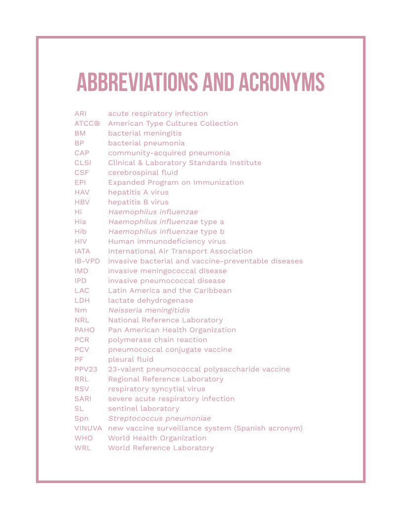
ABBREVIATIONS AND ACRONYMSARI acute respiratory infectionATCC® American Type Cultures CollectionBM bacterial meningitisBP bacterial pneumoniaCAP community-acquired pneumoniaCLSI Clinical & Laboratory Standards InstituteCSF cerebrospinal fluidEPI Expanded Program on ImmunizationHAV hepatitis A virusHBV hepatitis B virusHi Haemophilus influenzae Hia Haemophilus influenzae type a Hib Haemophilus influenzae type b HIV Human immunodeficiency virusIATA International Air Transport AssociationIB-VPD invasive bacterial and vaccine-preventable diseasesIMD invasive meningococcal diseaseIPD invasive pneumococcal diseaseLAC Latin America and the CaribbeanLDH lactate dehydrogenase Nm Neisseria meningitidis NRL National Reference LaboratoryPAHO Pan American Health OrganizationPCR polymerase chain reactionPCV pneumococcal conjugate vaccinePF pleural fluidPPV23 23-valent pneumococcal polysaccharide vaccineRRL Regional Reference LaboratoryRSV respiratory syncytial virus SARI severe acute respiratory infectionSL sentinel laboratorySpn Streptococcus pneumoniae VINUVA new vaccine surveillance system (Spanish acronym)WHO World Health OrganizationWRL World Reference Laboratory


- 1 -
1. INTRODUCtioN
- 2 -
1.1. Epidemiological Situation in the Region of the Americas
Acute respiratory infections (ARI), especially community-acquired pneumonia (CAP), are the leading cause of hospitalization and death among children under 5 years in developing countries.
Over 95% of all episodes of clinical pneumonia and over 99% of deaths from pneumonia among children under age 5 worldwide occur in low- and middle-income countries. Pneumococcus caused an estimated 8.9 million pneumonia cases in 2015, of which 3.5 million were severe or very severe. It is the second leading cause of CAP requiring hospitalization, after respiratory syncytial virus (RSV), but the first in the number of deaths: approximately 300,000 deaths per year worldwide in children under 5 years. Associated bacteremia does not occur in most pneumococcal pneumonia cases (80%); nevertheless, pneumonia cases with bacteremia constitute most invasive infections caused by the pneumococcus (90%). Among the causes of deaths due to pneumococcal infections, pneumonia represents 81% and meningitis 12%. The global mortality rate in 2015 due to pneumococcal disease was 45 deaths (29-56) per 100,000 children under 5 years of age.
Haemophilus influenzae type b (Hib) caused 0.9 million pneumonia cases in 2015, of which approximately 300,000 were severe or very severe. It is the second leading cause of deaths due to pneumonia in the under-5 age group worldwide, with approximately 30,000 deaths.
The pneumococcal conjugate vaccine (PCV) was initially introduced in the Region of the Americas, Canada, and the United States in 2000 and by December 2019, 37 countries and territories had included the vaccine in their regular immunization programs. Globally, the number of pneumococcal pneumonia cases is estimated to have fallen by more than one third, and deaths from pneumococcal infections by 51% between 2000 and 2015, following the introduction of the PCV in many countries.
There are approximately 1.2 million cases of bacterial meningitis in children under 5 each year, with 180,000 deaths. In an analysis published in 2013, the Region of the Americas had the lowest burden of disease worldwide, with an incidence of 17 cases per 100,000 children per year. This is likely due to the introduction of the Hib vaccine in national programs in all countries of the Region several years earlier. The Region of the Americas presented the second largest decline in the number of Hib deaths (96%) during the 2000-2015 period. Globally, it is estimated that over 90% of bacterial meningitis is caused by S. pneumoniae, H. influenzae and N. meningitidis. At present, S. pneumoniae is the primary cause of bacterial meningitis in the Region of the Americas.
In 1993, the Region of the Americas established a network of laboratories responsible for the surveillance of bacterial meningitis and pneumonia in the Region. It is known as
- 3 -
SIREVA (Regional System for Vaccines) and up to 19 countries participate in it. SIREVA has identified the three principal agents responsible for bacterial pneumonia and meningitis: Haemophilus influenzae (Hi), Neisseria meningitidis (meningococcus) and Streptococcus pneumoniae (pneumococcus). It has also characterized the serotypes and circulating serogroups of these bacteria, as well as establishing their susceptibility to most used antibiotics. The network, however, lacked epidemiological data to support its laboratory data.
To meet this need, in 2007 PAHO set in motion an initiative to coordinate a sentinel surveillance network for bacterial pneumonia and meningitis in children under 5. Later, in 2014, this regional network was invited to form part of the Global Surveillance Network, led by the World Health Organization.
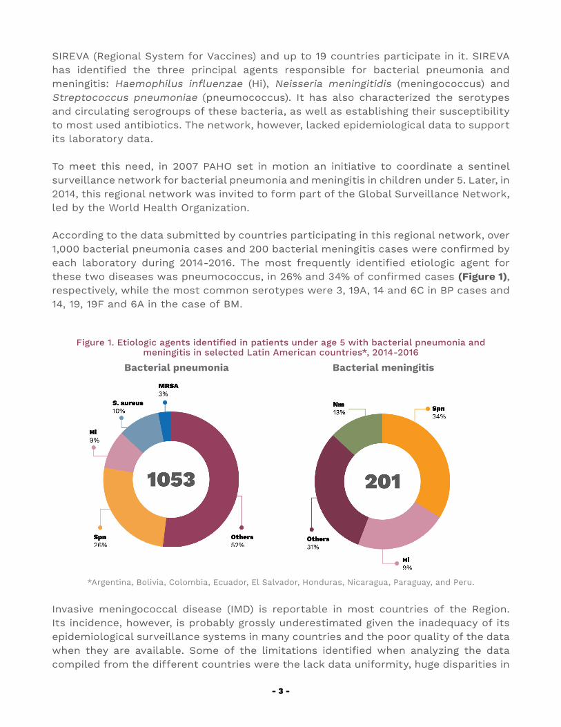
According to the data submitted by countries participating in this regional network, over 1,000 bacterial pneumonia cases and 200 bacterial meningitis cases were confirmed by each laboratory during 2014-2016. The most frequently identified etiologic agent for these two diseases was pneumococcus, in 26% and 34% of confirmed cases (Figure 1), respectively, while the most common serotypes were 3, 19A, 14 and 6C in BP cases and 14, 19, 19F and 6A in the case of BM.
Figure 1. Etiologic agents identified in patients under age 5 with bacterial pneumonia and meningitis in selected Latin American countries*, 2014-2016
Bacterial pneumonia Bacterial meningitis
*Argentina, Bolivia, Colombia, Ecuador, El Salvador, Honduras, Nicaragua, Paraguay, and Peru.
Invasive meningococcal disease (IMD) is reportable in most countries of the Region. Its incidence, however, is probably grossly underestimated given the inadequacy of its epidemiological surveillance systems in many countries and the poor quality of the data when they are available. Some of the limitations identified when analyzing the data compiled from the different countries were the lack data uniformity, huge disparities in
- 4 -
morbidity and mortality records, extremely low incidence rates due to underreporting, and a high number of meningitis cases with no bacterial identification where collecting a specimen was not feasible.
Given the increased availability of pneumococcal and meningococcal vaccines, it has become necessary to strengthen and integrate the surveillance of these vaccine-preventable diseases to ensure that laboratory findings are supplemented with standardized epidemiological data at all levels. All sentinel hospitals and countries participating in the network must, therefore, work together in a more uniform and systematic manner so that the information generated can be reliably used to inform decision-making on the feasibility of introducing new vaccines on national immunization programs, of adjusting current vaccination series, as well as monitoring of the impact of such actions.
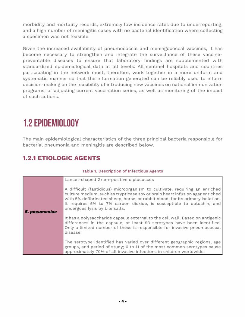
1.2 EPIDEMIOLOGY The main epidemiological characteristics of the three principal bacteria responsible for bacterial pneumonia and meningitis are described below.
1.2.1 ETIOLOGIC AGENTS
Table 1. Description of Infectious Agents
S. pneumoniae
Lancet-shaped Gram-positive diplococcus
A difficult (fastidious) microorganism to cultivate, requiring an enriched culture medium, such as trypticase soy or brain heart infusion agar enriched with 5% defibrinated sheep, horse, or rabbit blood, for its primary isolation.It requires 5% to 7% carbon dioxide, is susceptible to optochin, and undergoes lysis by bile salts.
It has a polysaccharide capsule external to the cell wall. Based on antigenic differences in the capsule, at least 93 serotypes have been identified. Only a limited number of these is responsible for invasive pneumococcal disease.
The serotype identified has varied over different geographic regions, age groups, and period of study; 6 to 11 of the most common serotypes cause approximately 70% of all invasive infections in children worldwide.
- 5 -
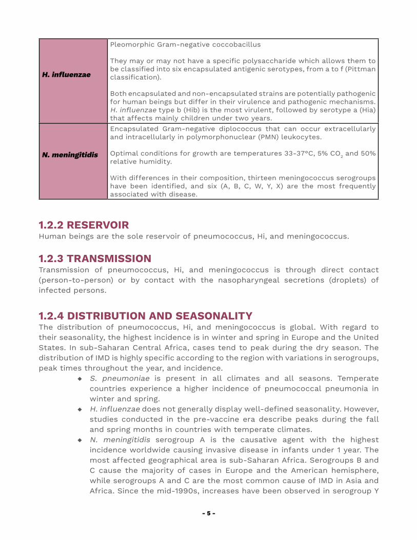
H. influenzae
Pleomorphic Gram-negative coccobacillus
They may or may not have a specific polysaccharide which allows them to be classified into six encapsulated antigenic serotypes, from a to f (Pittman classification).
Both encapsulated and non-encapsulated strains are potentially pathogenic for human beings but differ in their virulence and pathogenic mechanisms. H. influenzae type b (Hib) is the most virulent, followed by serotype a (Hia) that affects mainly children under two years.
N. meningitidis
Encapsulated Gram-negative diplococcus that can occur extracellularly and intracellularly in polymorphonuclear (PMN) leukocytes.
Optimal conditions for growth are temperatures 33-37°C, 5% CO2 and 50% relative humidity.
With differences in their composition, thirteen meningococcus serogroups have been identified, and six (A, B, C, W, Y, X) are the most frequently associated with disease.
1.2.2 RESERVOIRHuman beings are the sole reservoir of pneumococcus, Hi, and meningococcus.
1.2.3 TRANSMISSIONTransmission of pneumococcus, Hi, and meningococcus is through direct contact (person-to-person) or by contact with the nasopharyngeal secretions (droplets) of infected persons.
1.2.4 DISTRIBUTION AND SEASONALITYThe distribution of pneumococcus, Hi, and meningococcus is global. With regard to their seasonality, the highest incidence is in winter and spring in Europe and the United States. In sub-Saharan Central Africa, cases tend to peak during the dry season. The distribution of IMD is highly specific according to the region with variations in serogroups, peak times throughout the year, and incidence.
◆ S. pneumoniae is present in all climates and all seasons. Temperate countries experience a higher incidence of pneumococcal pneumonia in winter and spring.
◆ H. influenzae does not generally display well-defined seasonality. However, studies conducted in the pre-vaccine era describe peaks during the fall and spring months in countries with temperate climates.
◆ N. meningitidis serogroup A is the causative agent with the highest incidence worldwide causing invasive disease in infants under 1 year. The most affected geographical area is sub-Saharan Africa. Serogroups B and C cause the majority of cases in Europe and the American hemisphere, while serogroups A and C are the most common cause of IMD in Asia and Africa. Since the mid-1990s, increases have been observed in serogroup Y
- 6 -
IMD cases in the United States and Israel, while serogroup X caused local epidemics local in sub-Saharan Africa. Furthermore, an increasing number of infections with serogroup W has been identified in the Region of the Americas since 2007.
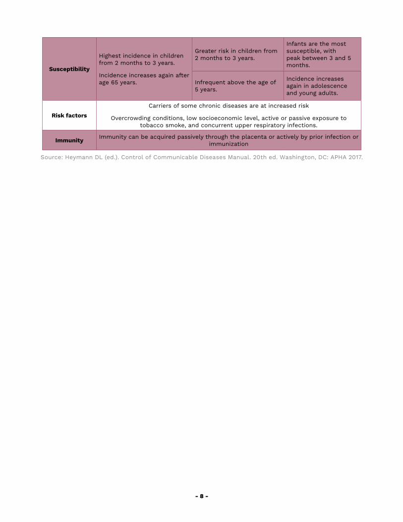
1.2.5 SUSCEPTIBILITY AND RISK FACTORSSusceptibility to pneumococcus, Hi and meningococcus infection is universal. In other words, all people are susceptible to the infections caused by these agents. However, certain conditions can increase a person’s susceptibility to these bacteria and the invasive illnesses that they cause.
Pneumococcus infection is most frequent in children from the age of 2 months to 3 years, although it declines after 18 months. The risk rises again from the age of 65 years. The risk of Hi infection is also greatest in children aged between 2 months and 3 years and declines after the age of 2 years. In developing countries, the greatest incidence is in children under 6 months of age while in developed countries this peak is observed in children aged 6 to 12 months. Infection is uncommon after the age of 5 years. Regarding meningococcus, the highest case rates are reported in children under 1 year, with a peak in this group in the 3-to-5-month range. It can also, however, affect adolescents and young adults.
Like other airborne infectious microorganisms, in addition to age, other conditions also increase the risk of pneumococcus, Hi, and meningococcus infections: overcrowding, poverty, active or passive tobacco exposure, and concurrent upper respiratory infections. Carriers of some chronic diseases are also sat greater risk of infections caused by these bacteria.
1.2.6 IMMUNITYImmunity to pneumococcus, Hi, and meningococcus can be acquired passively through the placenta, or actively by previous infection or immunization.
Newborns may have antibodies against pneumococcus due to passive transmission from the mother. These antibodies disappear within a few months, coincidentally with the increase in invasive disease. After the age of 18 months, children show specific immune responses to most circulating pneumococcus serotypes because of repeated exposure.
As of age 5 years, most unvaccinated children have anticapsular H. influenzae antibodies due to exposure to the bacterium.
Regarding meningococcus, there is an immune response of unknown duration following clinical and subclinical infections and which increases with age.
- 7 -
1.2.7 CARRIER STATUSPneumococcus, Hi, and meningococcus are generally nasopharyngeal colonizing agents in asymptomatic people, who are considered carriers.
Between 4% and 35% of non-immunized healthy adults are estimated to be Hi carriers. The percentage of carriers is higher among preschool children. Hi can remain in the nasopharynx for months.
Pneumococcal disease is preceded by asymptomatic nasopharyngeal colonization of varying duration. The period in which a person is a carrier and source of person-to-person transmission has often been shown to be between one month and five years (average six months). The prevalence of pneumococcus carriage is higher in children, especially those attending day-care centers, and in adults in close contact with them. It is estimated that practically all children have been a carrier of pneumococcus on at least one occasion during the preschool stage. The conjugate vaccines have been seen to diminish the number of carriers with strains included in the vaccine and that seems to bear a direct relation with the capacity to produce IgA and IgG antipolysaccharide antibodies. However, studies conducted after the introduction of the vaccine did not detect a reduction in the percentage of carriers in the population but found that the serotypes were replaced by other non-vaccine serotypes.
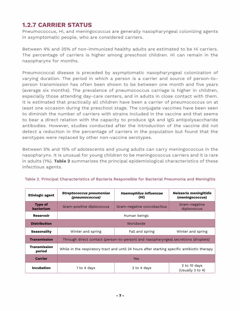
Between 5% and 15% of adolescents and young adults can carry meningococcus in the nasopharynx. It is unusual for young children to be meningococcus carriers and it is rare in adults (1%). Table 2 summarizes the principal epidemiological characteristics of these infectious agents.
Table 2. Principal Characteristics of Bacteria Responsible for Bacterial Pneumonia and Meningitis
Etiologic agent Streptococcus pneumoniae (pneumococcus)
Haemophilus influenzae(Hi)
Neisseria meningitidis (meningococcus)
Type of bacterium Gram-positive diplococcus Gram-negative coccobacillus Gram-negative
diplococcus
Reservoir Human beings
Distribution Worldwide
Seasonality Winter and spring Fall and spring Winter and spring
Transmission Through direct contact (person-to-person) and nasopharyngeal secretions (droplets)
Transmission period While in the respiratory tract and until 24 hours after starting specific antibiotic therapy
Carrier Yes
Incubation 1 to 4 days 2 to 4 days2 to 10 days
(Usually 3 to 4)
- 8 -
Susceptibility
Highest incidence in children from 2 months to 3 years.
Incidence increases again after age 65 years.
Greater risk in children from 2 months to 3 years.
Infants are the most susceptible, with peak between 3 and 5 months.
Infrequent above the age of 5 years.
Incidence increases again in adolescence and young adults.
Risk factors
Carriers of some chronic diseases are at increased risk
Overcrowding conditions, low socioeconomic level, active or passive exposure to tobacco smoke, and concurrent upper respiratory infections.
Immunity Immunity can be acquired passively through the placenta or actively by prior infection or immunization
Source: Heymann DL (ed.). Control of Communicable Diseases Manual. 20th ed. Washington, DC: APHA 2017.
2. BACTERIAL PNEUMONIA
- 10 -
2.1 CLINICAL ASPECTS
Pneumonia is an infection of the pulmonary parenchyma that can be caused by a variety of microorganisms (viral, bacterial, or others). Pneumonias of different etiologies can present very similar clinical symptoms.
In infants and young children, pneumonia tends to begin with acute fever. Numerous studies have attempted to determine what specific and sensitive clinical signs most reliably indicate the presence of pneumonia. Most of these studies agreed that tachypnea (rapid breathing) is the most effective predictive sign.
The strategy known as the Integrated Management of Childhood Illness (IMCI) classifies pneumonia according to its clinical manifestations as pneumonia, severe pneumonia, and very severe pneumonia. Pneumonia is suspected when the physical examination reveals that the child is coughing or has difficulty breathing as well as rapid breathing. Rapid breathing is defined as:
◆ Under 2 months: over 60 breaths/minute◆ Age 2 to 11 months: over 50 breaths/minute◆ Age 12 months to 5 years: over 40 breaths/minute.
Other signs that can be detected through thoracic auscultation include crepitant stertors, reduced respiratory sounds, or areas of bronchial breathing. Oxygen saturation over 95%, measured using a pulse oximeter at atmospheric pressure, is an important parameter in determining breathing difficulty.
The most severe pneumonia cases are of bacterial origin, and these are responsible for most hospitalizations and deaths of children under 5 years.
Table 3 shows the symptoms that define pneumonia by severity.
Table 3. Classification of Pneumonia by Severity
Basic pneumonia symptoms Severe pneumonia Very severe pneumonia
•Difficulty breathing •Rapid breathing •Cough
Basic symptoms plus: •Nasal flaring •Whizzing sounds (in younger infants) •Retraction of inferior thoracic wall (subcostal and/or supraclavicular retraction)
In addition to the previous, plus: •Central cyanosis •Convulsions, lethargy, or loss of consciousness •Severe difficulty breathing (for example, with head nodding) •Inability to breast-feed or drink •Vomiting everything ingested
Source: Diagnóstico y Tratamiento de las Enfermedades Prevalentes Graves en la Infancia [Diagnosis and Treatment of Serious Prevalent Illnesses in Childhood] PAHO, 2004
- 11 -
2.1.1 DIFFERENTIAL DIAGNOSISRespiratory viral infections are common in children under 5 years, and tend to cause cough, fever, mouth breathing, and nasal secretion.
Some viruses cause bronchial hyperreactivity, which causes episodes of wheezing, especially in young children. Bronchiolitis is a viral infection of the lower respiratory tract. It is frequent and relatively severe in infants. Most cases are caused by respiratory syncytial virus (RSV), although other viruses such as influenza and parainfluenza can also cause bronchiolitis. The disease is characterized by obstruction of the respiratory tract and episodes of wheezing that respond poorly to bronchodilators. Secondary bacterial infection can occur.
Asthma is a chronic inflammatory disorder with reversible obstruction of the respiratory tract. It is characterized by recurrent episodes of wheezing with cough, and sometimes with lower intercostal retraction and tachypnea. Fever only occurs in cases of concurrent viral or bacterial infectious processes. It responds well to treatment with bronchodilators and anti-inflammatories.
Other respiratory viruses, such as adenovirus, influenza and parainfluenza viruses, can also cause viral pneumonia, but they tend to cause upper respiratory tract infections rather than pneumonia. Other examples of viruses that can cause viral pneumonia are measles, chickenpox, and RSV.
2.1.2 COMPLICATIONSPleural effusion (up to 50% of cases), empyema, atelectasis, and hypertensive pneumothorax are among the complications of bacterial pneumonia.
If a child with severe pneumonia does not receive proper and timely treatment with specific antibiotics, respiratory insufficiency can become acute and lead to death.
2.2 radiological diagnosis
Radiographic analysis is an important tool in diagnosing severe and very severe pneumonia as it helps to differentiate between bacterial or viral etiologies, and to determine whether complications such as pleural effusion or atelectasis are present.
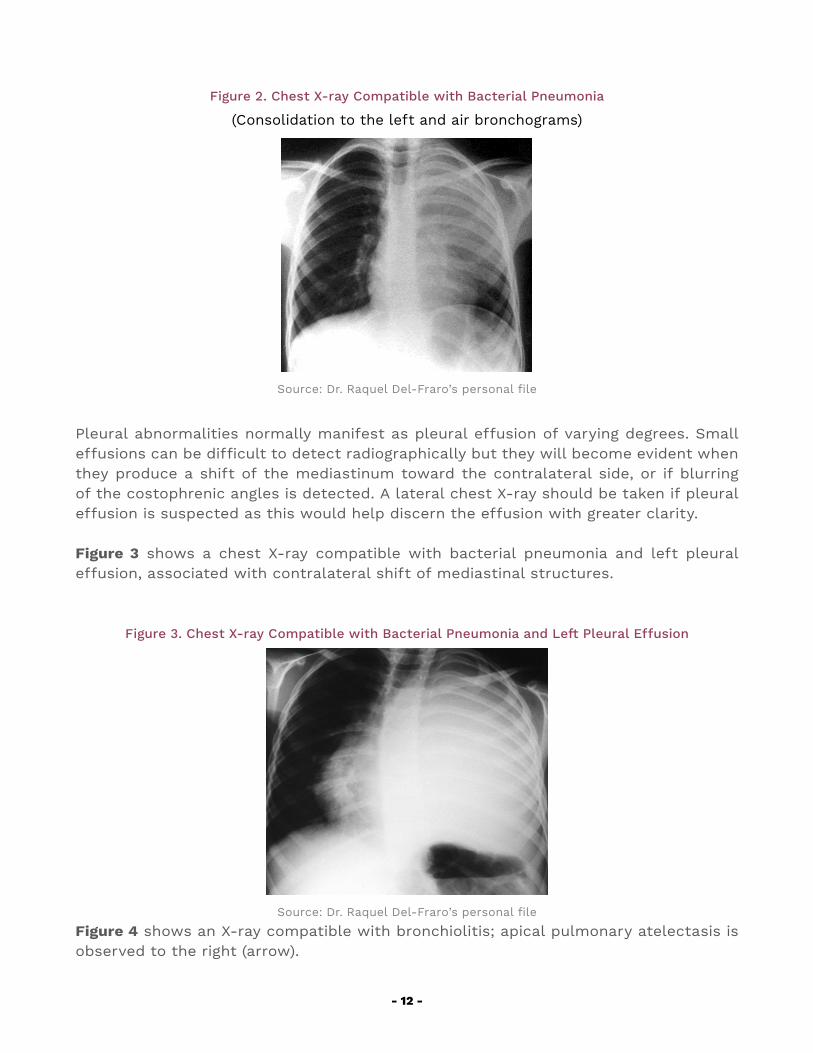
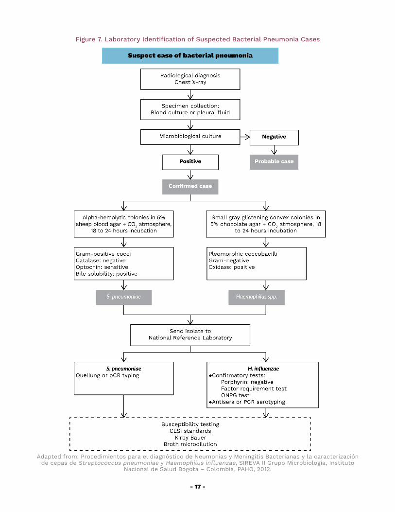
According to the criteria and definitions established by WHO for interpreting chest X-ray of children with pneumonia, bacterial pneumonia present a dense cottony appearance (alveolar infiltrate), reflecting that one or more segments or pulmonary lobes, or a complete lung, are partially or totally compromised. With these infiltrates, there are often areas of air bronchogram, sometimes in conjunction with pleural effusion. Figures 2 and 3 show radiological images compatible with bacterial pneumonia.
- 12 -
Figure 2. Chest X-ray Compatible with Bacterial Pneumonia
(Consolidation to the left and air bronchograms)
Source: Dr. Raquel Del-Fraro’s personal file
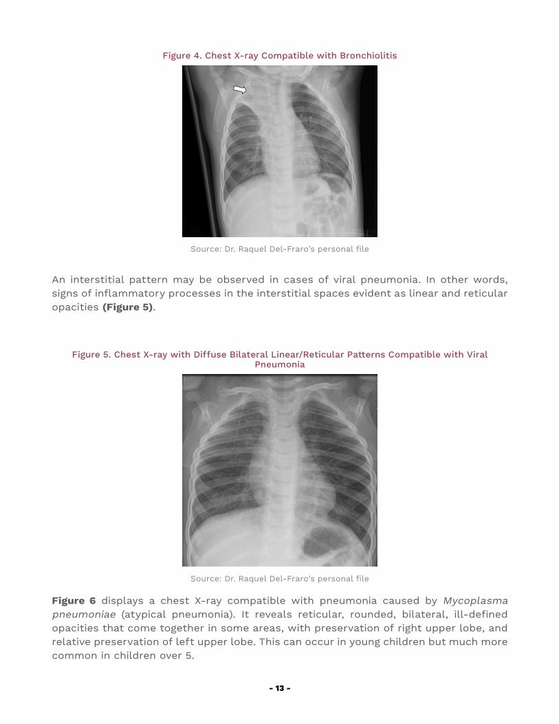
Pleural abnormalities normally manifest as pleural effusion of varying degrees. Small effusions can be difficult to detect radiographically but they will become evident when they produce a shift of the mediastinum toward the contralateral side, or if blurring of the costophrenic angles is detected. A lateral chest X-ray should be taken if pleural effusion is suspected as this would help discern the effusion with greater clarity.
Figure 3 shows a chest X-ray compatible with bacterial pneumonia and left pleural effusion, associated with contralateral shift of mediastinal structures.
Figure 3. Chest X-ray Compatible with Bacterial Pneumonia and Left Pleural Effusion
Source: Dr. Raquel Del-Fraro’s personal file
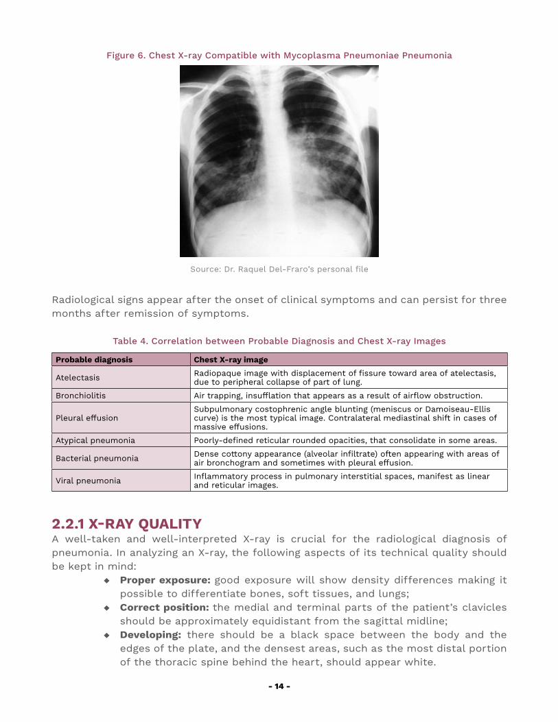
Figure 4 shows an X-ray compatible with bronchiolitis; apical pulmonary atelectasis is observed to the right (arrow).
- 13 -
Figure 4. Chest X-ray Compatible with Bronchiolitis
Source: Dr. Raquel Del-Fraro’s personal file
An interstitial pattern may be observed in cases of viral pneumonia. In other words, signs of inflammatory processes in the interstitial spaces evident as linear and reticular opacities (Figure 5).
Figure 5. Chest X-ray with Diffuse Bilateral Linear/Reticular Patterns Compatible with Viral Pneumonia
Source: Dr. Raquel Del-Fraro’s personal file
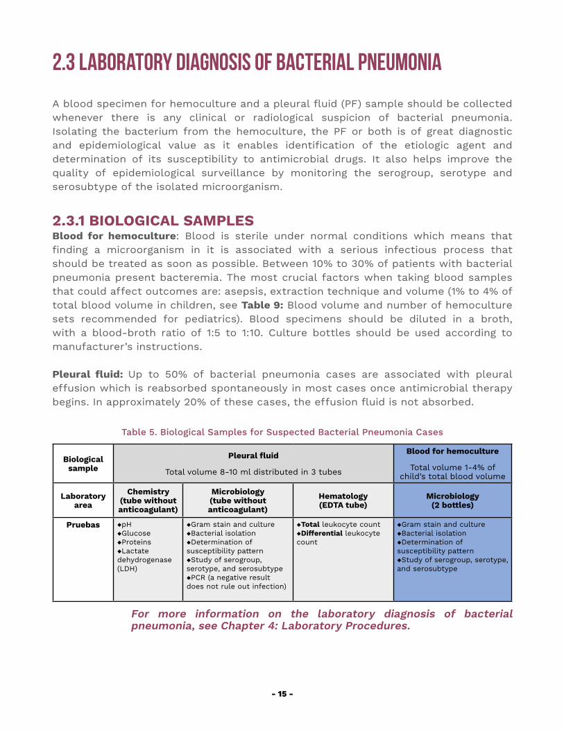
Figure 6 displays a chest X-ray compatible with pneumonia caused by Mycoplasma pneumoniae (atypical pneumonia). It reveals reticular, rounded, bilateral, ill-defined opacities that come together in some areas, with preservation of right upper lobe, and relative preservation of left upper lobe. This can occur in young children but much more common in children over 5.
- 14 -
Figure 6. Chest X-ray Compatible with Mycoplasma Pneumoniae Pneumonia
Source: Dr. Raquel Del-Fraro’s personal file
Radiological signs appear after the onset of clinical symptoms and can persist for three months after remission of symptoms.
Table 4. Correlation between Probable Diagnosis and Chest X-ray Images
Probable diagnosis Chest X-ray image
Atelectasis Radiopaque image with displacement of fissure toward area of atelectasis, due to peripheral collapse of part of lung.
Bronchiolitis Air trapping, insufflation that appears as a result of airflow obstruction.
Pleural effusionSubpulmonary costophrenic angle blunting (meniscus or Damoiseau-Ellis curve) is the most typical image. Contralateral mediastinal shift in cases of massive effusions.
Atypical pneumonia Poorly-defined reticular rounded opacities, that consolidate in some areas.
Bacterial pneumonia Dense cottony appearance (alveolar infiltrate) often appearing with areas of air bronchogram and sometimes with pleural effusion.
Viral pneumonia Inflammatory process in pulmonary interstitial spaces, manifest as linear and reticular images.
2.2.1 X-RAY QUALITY A well-taken and well-interpreted X-ray is crucial for the radiological diagnosis of pneumonia. In analyzing an X-ray, the following aspects of its technical quality should be kept in mind:
◆ Proper exposure: good exposure will show density differences making it possible to differentiate bones, soft tissues, and lungs;
◆ Correct position: the medial and terminal parts of the patient’s clavicles should be approximately equidistant from the sagittal midline;
◆ Developing: there should be a black space between the body and the edges of the plate, and the densest areas, such as the most distal portion of the thoracic spine behind the heart, should appear white.
- 15 -
2.3 Laboratory Diagnosis of Bacterial Pneumonia
A blood specimen for hemoculture and a pleural fluid (PF) sample should be collected whenever there is any clinical or radiological suspicion of bacterial pneumonia. Isolating the bacterium from the hemoculture, the PF or both is of great diagnostic and epidemiological value as it enables identification of the etiologic agent and determination of its susceptibility to antimicrobial drugs. It also helps improve the quality of epidemiological surveillance by monitoring the serogroup, serotype and serosubtype of the isolated microorganism.
2.3.1 BIOLOGICAL SAMPLESBlood for hemoculture: Blood is sterile under normal conditions which means that finding a microorganism in it is associated with a serious infectious process that should be treated as soon as possible. Between 10% to 30% of patients with bacterial pneumonia present bacteremia. The most crucial factors when taking blood samples that could affect outcomes are: asepsis, extraction technique and volume (1% to 4% of total blood volume in children, see Table 9: Blood volume and number of hemoculture sets recommended for pediatrics). Blood specimens should be diluted in a broth, with a blood-broth ratio of 1:5 to 1:10. Culture bottles should be used according to manufacturer’s instructions. Pleural fluid: Up to 50% of bacterial pneumonia cases are associated with pleural effusion which is reabsorbed spontaneously in most cases once antimicrobial therapy begins. In approximately 20% of these cases, the effusion fluid is not absorbed.
Table 5. Biological Samples for Suspected Bacterial Pneumonia Cases
Biological sample
Pleural fluid
Total volume 8-10 ml distributed in 3 tubes
Blood for hemoculture
Total volume 1-4% of child’s total blood volume
Laboratory area
Chemistry (tube without anticoagulant)
Microbiology (tube without anticoagulant)
Hematology(EDTA tube)
Microbiology (2 bottles)
Pruebas ◆pH◆Glucose◆Proteins◆Lactate dehydrogenase (LDH)
◆Gram stain and culture◆Bacterial isolation◆Determination of susceptibility pattern◆Study of serogroup, serotype, and serosubtype◆PCR (a negative result does not rule out infection)
◆Total leukocyte count◆Differential leukocyte count
◆Gram stain and culture◆Bacterial isolation ◆Determination of susceptibility pattern◆Study of serogroup, serotype, and serosubtype
For more information on the laboratory diagnosis of bacterial pneumonia, see Chapter 4: Laboratory Procedures.
- 16 -
Possible meningitis or pneumonia specimens must be processed following the highest biosafety risk standards required for the pathogens that could be present: N. meningitidis specimens.
- 17 -
Figure 7. Laboratory Identification of Suspected Bacterial Pneumonia Cases
Adapted from: Procedimientos para el diagnóstico de Neumonías y Meningitis Bacterianas y la caracterización de cepas de Streptococcus pneumoniae y Haemophilus influenzae, SIREVA II Grupo Microbiología, Instituto
Nacional de Salud Bogotá – Colombia, PAHO, 2012.
- 18 -
2.4 TrEATMENT
In general, children with pneumonia can be treated on an outpatient basis, following the medical instructions specific to each case.
Children with severe or very severe pneumonia should be hospitalized. For more details on recommended treatment, consult national protocols for the management of patients with bacterial pneumonia.
3. BACTERIAL MENINGITIS
- 20 -
3.1 CLINICAL ASPECTS
Meningitis is an inflammation of the membranes around the brain, the cerebellum and the bone marrow, the anatomical sites surrounded by the subarachnoid space, where the cerebrospinal fluid (CSF) circulates.
It is characterized by fever and signs of meningeal inflammation. Table 6 lists the signs and symptoms by age.
Meningococcemia is accompanied by an initial erythematous and macular cutaneous exanthema that rapidly leads to petechial eruption and ultimately ecchymoses.
Convulsions tend to occur in 20% of cases.
Table 6. Signs and Symptoms of Meningitis by Age
Children under 1 year Children over 1 year and adultsBulging fontanelle
Convulsions
Loss of appetite
Irritability without justification or clinical cause
Lethargy
Vomiting
Altered state of consciousness
Convulsions
Headache
Photophobia
Lethargy
Stiff neck or other signs of meningeal inflammation, or both
Prominent signs of hyperactivity
Projectile vomiting (explosive)
3.1.1 DIFFERENTIAL DIAGNOSISThe incidence of bacterial meningitis has declined in recent decades following the introduction of the Hib and pneumococcal vaccines.
Non-bacterial meningitis is usually of viral etiology and is the leading cause of neuroinfection, with a global incidence of 10.9 cases per 100,000 population annually. Non-polio enteroviruses account for almost 90% of cases, and herpes simplex for 0.5% to 3%.
The human enterovirus (HEV) genus belongs to the Picornaviridae family that includes over 90 serotypes including poliovirus (PV), coxsackie A (CAV) and B (CBV) virus, echovirus (E) and the new enteroviruses (EV). These are ubiquitous viruses. Humans are the only known reservoir and transmission is primarily fecal-oral. In temperate climates, HEV infections occur primarily during the summer and fall months. HEV cause a wide range
- 21 -
of diseases including aseptic meningitis, paralytic disease, myocarditis, pleurodynia and various febrile and exanthem type symptoms. Most infections are asymptomatic.
In clinical practice the differential diagnosis of bacterial and non-bacterial meningitis is challenging, but of utmost importance in order to initiate correct treatment. Both clinical and laboratory parameters must be considered. The Boyer Score, for example, as proposed in 1980 by Thomé et al., has been used to guide the differential diagnosis of bacterial versus viral meningitis and the prescription of antibiotic therapy. According to various studies, its sensitivity is in the region of 70-80% and its specificity between 90 and 100%. Three clinical and five analytical parameters are evaluated, four of which using CSF.
A positive CSF culture is considered “the gold standard” for diagnosing bacterial etiology. When available, both rapid tests and molecular testing are useful to guide diagnosis and early treatment options.
3.1.2 COMPLICATIONSMeningitis can evolve rapidly toward stupor, coma, and death.
As many as 30% of bacterial meningitis survivors can suffer permanent sequelae. The most common is sensorineural hearing loss. Others include language disorders, mental retardation, motor anomalies, convulsions, and visual disorders. Sequelae are more frequent in meningitis caused by pneumococcus, as shown in Table 7.
Table 7. Percentage of Bacterial Meningitis Sequelae and Case-Fatality by Etiologic Agent
Etiologic agent
Sequelae (%)Case-fatality
(%)Deafness Mental
retardationSpasticity/
paresis Convulsions
H. influenzae 10 6 5 6 3-6
N. meningitidis 6 2 2 6 8-15
S. pneumoniae 28 17 12 14 10-30
Source: Baraff LJ, Lee SI, Schriger DL. Outcomes of bacterial meningitis in children: a meta-analysis. Pediatr Infect Dis J, 1993;12:392.
3.2 Laboratory Diagnosis of Bacterial Meningitis
In the event of clinical suspicion of meningitis, one CSF and two blood samples should be collected for hemocultures, if possible, before initiating antibiotic treatment. Isolating the bacterium in the CSF or the blood is of great diagnostic and epidemiological value
- 22 -
as it enables identification of the etiologic agent and determination of its susceptibility to antimicrobial drugs. It also helps improve the quality of epidemiological surveillance by monitoring the serogroup, serotype and serosubtype of the isolated microorganism.
3.2.1 BIOLOGICAL SAMPLES
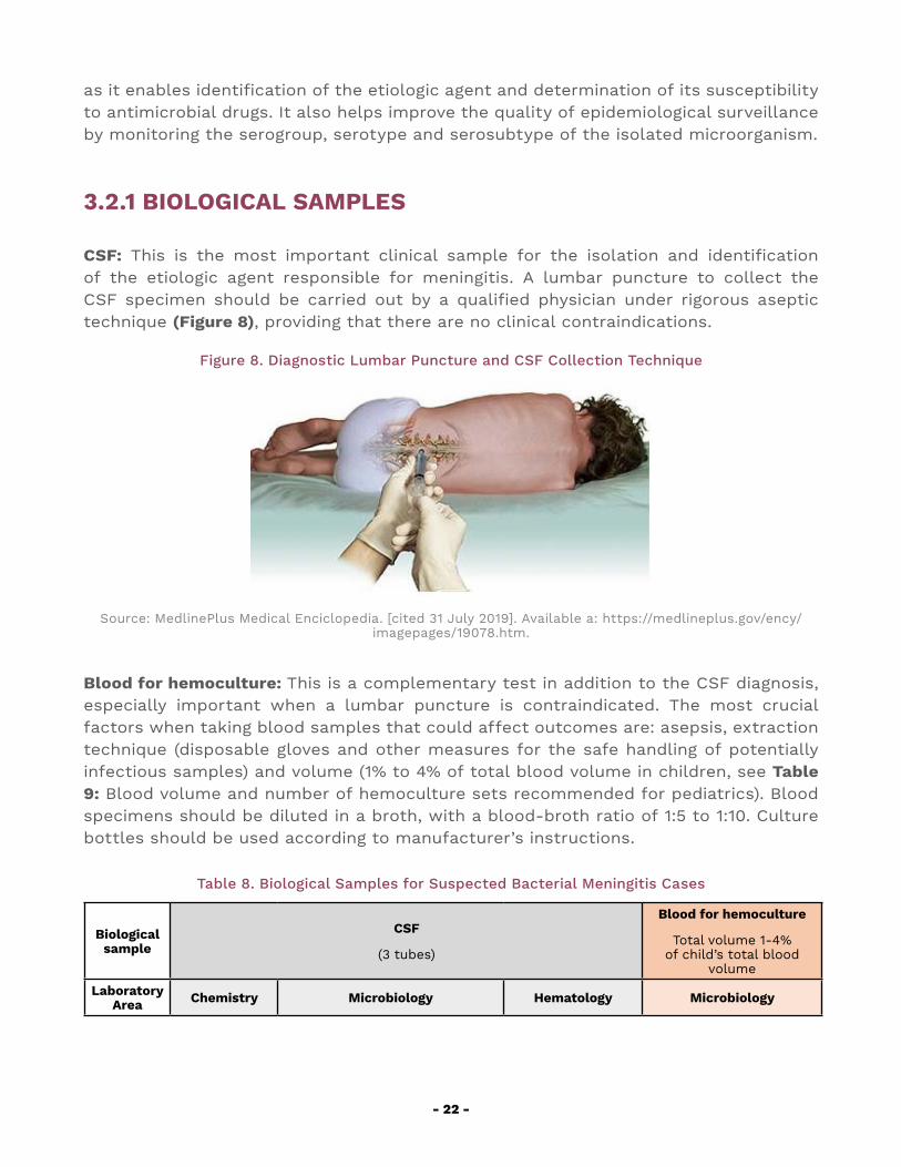
CSF: This is the most important clinical sample for the isolation and identification of the etiologic agent responsible for meningitis. A lumbar puncture to collect the CSF specimen should be carried out by a qualified physician under rigorous aseptic technique (Figure 8), providing that there are no clinical contraindications.
Figure 8. Diagnostic Lumbar Puncture and CSF Collection Technique
Source: MedlinePlus Medical Enciclopedia. [cited 31 July 2019]. Available a: https://medlineplus.gov/ency/imagepages/19078.htm.
Blood for hemoculture: This is a complementary test in addition to the CSF diagnosis, especially important when a lumbar puncture is contraindicated. The most crucial factors when taking blood samples that could affect outcomes are: asepsis, extraction technique (disposable gloves and other measures for the safe handling of potentially infectious samples) and volume (1% to 4% of total blood volume in children, see Table 9: Blood volume and number of hemoculture sets recommended for pediatrics). Blood specimens should be diluted in a broth, with a blood-broth ratio of 1:5 to 1:10. Culture bottles should be used according to manufacturer’s instructions.
Table 8. Biological Samples for Suspected Bacterial Meningitis Cases
Biological sample
CSF
(3 tubes)
Blood for hemoculture
Total volume 1-4% of child’s total blood
volume
Laboratory Area Chemistry Microbiology Hematology Microbiology
- 23 -
Tests ◆Appearence* ◆Glucose◆Proteins
◆Gram stain and culture◆Bacterial isolation◆Susceptibility testing◆Serogroup, serotype, and serosubtype identification◆Latex agglutination◆Immunochromatography (Binax NOW®)◆PCR (a negative result does not rule out infection)
◆Total leukocyte count ◆Differential leukocyte count
◆Gram stain and culture◆Bacterial isolation◆Determination of susceptibility pattern ◆Study of serogroup, serotype, and serosubtype
*See the description of appearance at collection in medical record, if it has a blood-tinged appearance, include red blood cell count which will aid clinical interpretation.
For more information on the laboratory diagnosis of bacterial meningitis, see Chapter 4: Laboratory Procedures
Possible meningitis or pneumonia specimens must be processed following the highest biosafety risk standards required for the pathogens that could be present: N. meningitidis specimens.
3.3 TrEATMENT Every child with meningitis should be referred to the closest hospital for treatment. For more details on the treatment of bacterial meningitis, consult national protocols for the management of patients with bacterial meningitis.
4. Laboratory Procedures
- 25 -
4. Laboratory ProceduresMicrobiologists play a critical role in gathering data for both clinical and public health decision-making. A role that is essential to preventing morbidity and mortality from bacterial pneumonia and meningitis1.
All laboratories at sentinel hospitals where this surveillance is conducted must: ◆ operate around the clock, 24 hours a day 365 days a year ◆ have qualified technical personnel available 24 hours a day 365 days a year ◆ have standardized operational procedures and good laboratory practices,
and ◆ have the necessary materials for the procedure, in accordance with the
standardized manual of procedures of the laboratory
4.1 BiosAFETY
Biosafety standards for laboratories handling samples and strains associated with the laboratory diagnosis of bacterial pneumonia and meningitis and likely to contain S. pneumoniae, H. influenzae and N. meningitidis, are clearly defined in the biosafety manual for processing of samples and strains associated with the laboratory diagnosis of bacterial pneumonia and meningitis caused by S. pneumoniae and H. influenzae published by PAHO in 20082. This manual must be read and understood by all temporary staff likely to handle biological samples (clinical samples and isolates) that could contain any of these three microorganisms. A “read and understood” statement should be signed, and the head of the laboratory should verified that the procedure is duly followed.
It is important to be aware that good laboratory practices (GLPs) are organizational and operational procedures under which tests are planned, performed, monitored, recorded, and reported. It must be fully understood that good operating procedures are indispensable for biosafety2. Before any possible risk of exposure to HIV, HBV, and HCV or other diseases, personnel must be familiar with the action to take and the communication channel to use for immediate notification, so that the medical team can assess the need for emergency prophylaxis.
With regard to the laboratory procedures for the diagnosis of bacterial pneumonia and meningitis, several cases of invasive meningococcal disease have been described following occupational exposure. All samples should be considered as potentially infectious, although the risk of transmission is not the same for these three causative agents, S. pneumoniae, H. influenzae and N. meningitidis. The increased risk for personnel 1 WHO. Laboratory Methods for the Diagnosis of Meningits caused for Neisseria meningitidis, Streptococcus pneumoniae and Haemophilus influenzae. Manual 2nd Edition, 2011.2 PAHO. Manual de bioseguridad para el procesamiento de muestras y cepas relacionadas con el diagnóstico de laboratorio de las neumonías y meningitis por N. meningitidis, S. pneumoniae y H. influenzae. 2008.
- 26 -
handling N. meningitidis is clearly documented, including the development of serogroup B meningococcal disease with fatal outcome.3
Biosafety Level 2 (BSL-2) is required for laboratories processing potentially infectious N. meningitidis samples. A biosafety cabinet should be used to protect the user and the environment from risks associated with the handling of infectious material. This should be a class II cabinet. The verification or certification of this type of cabinets includes a series of tests that ensure that the cabinet is safe and suitable for tasks to be performed in it.
The risk reduction or prevention measures for laboratory-acquired pneumococcal infections should focus on the use of biological safety cabinets when handling potentially infectious material, and should include a risk analysis, supported by good working practices, skilled personnel, training, competence, and immunization policies at each laboratory.3
4.1.1 HEALTH PROGRAM FOR PERSONNELVaccination of laboratorians should form part of a broad occupational health program that should also include post-exposure protocols and training on the use of personal protective equipment and accident prevention.
Laboratorians working with samples that could potentially contain these viable microorganisms should receive the following vaccines:
◆ BCG (against tuberculosis)◆ Hepatitis B ◆ Hib ◆ Seasonal influenza◆ Meningococcal and ◆ Pneumococcal
For the hepatitis B vaccine series, the worker’s immune status should be checked 1.5 months after the third dose using the anti-hepatitis B surface antigen antibody test. If the result is <10 mUI/ml, the 3-dose vaccine should be repeated. If the result is still <10 mUI/ml after the second series, the person should be considered as a “non-responder”. In this case, the person must be considered as unimmunized and proceed with the HBV post-exposure protocol.
For staff who may have been vaccinated against Hib during childhood, a booster dose is advisable before beginning work in the laboratory.
The seasonal influenza vaccine should be offered every year.
Laboratory workers should receive meningococcal and pneumococcal vaccines to protect them against all serogroups/serotypes identified in the country depending on currently 3 Borrow R, Findlow J, Gray S, Taylor S and Kaczmarski E. Safe laboratory handling of Neisseria meningitis. Elsevier. J Infect 2014; 68 305-212
- 27 -
available vaccines. For further details on pneumococcus, Hib, and meningococcus vaccine, see chapter on “Vaccines”.
Note: If the law prohibits mandatory vaccination, any staff member who refuses vaccination must sign a statement that he/she recognizes and assumes the risks associated with not having been vaccinated.
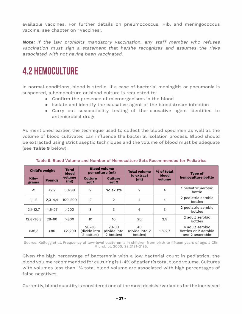
4.2 HemocultUREIn normal conditions, blood is sterile. If a case of bacterial meningitis or pneumonia is suspected, a hemoculture or blood culture is requested to:
◆ Confirm the presence of microorganisms in the blood ◆ Isolate and identify the causative agent of the bloodstream infection ◆ Carry out susceptibility testing of the causative agent identified to
antimicrobial drugs
As mentioned earlier, the technique used to collect the blood specimen as well as the volume of blood cultivated can influence the bacterial isolation process. Blood should be extracted using strict aseptic techniques and the volume of blood must be adequate (see Table 9 below).
Table 9. Blood Volume and Number of Hemoculture Sets Recommended for Pediatrics
Child’s weight Toral blood
volume (ml)
Blood volume per culture (ml) Total volume
to extract (ml)
% of total blood
volume
Type of hemoculture bottleKilo-
grams Pounds Culture set 1
Culture set 2
<1 <2,2 50-99 2 No existe 2 4 1 pediatric aerobic bottle
1,1-2 2,3-4,4 100-200 2 2 4 4 2 pediatric aerobic bottles
2,1-12,7 4,5-27 >200 3 3 6 3 2 pediatric aerobic bottles
12,8-36,3 28-80 >800 10 10 20 2,5 2 adult aerobic bottles
>36,3 >80 >2-20020-30
(divide into 2 bottles)
20-30(divide into 2 bottles)
40(divide into 2
bottles)1,8-2,7
4 adult aerobic bottles or 2 aerobic
and 2 anaerobic
Source: Kellogg et al. Frequency of low-level bacteremia in children from birth to fifteen years of age. J Clin Microbiol. 2000; 38:2181-2185.
Given the high percentage of bacteremia with a low bacterial count in pediatrics, the blood volume recommended for culturing is 1-4% of patient’s total blood volume. Cultures with volumes less than 1% total blood volume are associated with high percentages of false negatives.
Currently, blood quantity is considered one of the most decisive variables for the increased
- 28 -
positivity of hemocultures. It is now known that most bacteremias are low-level (< 1 to 10 cfu/ml). Hence, the larger the volume of the sample, the higher the sensitivity of the hemoculture. It is also known that for each additional milliliter inoculated in the bottle, positivity increases by 2-5%. Finally, the importance of blood volume also applies when using automated processing systems.
Blood culture bottles are designed to allow for the recommended blood-broth ratio (1:5 to 1:10) with an optimal blood volume. The use of hemocultures enriched with substances that facilitate bacterial growth with nourishing requirements are recommended as well as those with substances that inhibit factors found in blood and hinder bacterial growth. Suitable blood volume per bottle is essential for optimal microbial recovery. Check the manufacturer’s instructions for the hemoculture bottles available at each hospital (these can vary depending on the bottle type, the child’s weight, among others).
In summary, the critical steps when collecting blood samples for hemoculture are: ◆ Collect sample before starting antibiotic therapy. ◆ Ensure proper cleaning and asepsis before collecting the sample. ◆ Check the sample volume collected and distribute it accordingly. ◆ Transport specimens at room temperature. ◆ Deliver to laboratory within 2 hours of collection.
4.2.1 COLLECTING BLOOD SPECIMENS FOR HEMOCULTURE ◆ Collect the blood specimen before starting antimicrobial therapy, but do
not wait for sampling before starting antibiotics. If patient is receiving antibiotics, use resin-containing hemoculture bottles (automated systems) that neutralize the drugs administered.
◆ Wash hands following surgical hand washing procedures. ◆ Ensure aseptic techniques throughout the entire procedure. ◆ Use an aseptic field to avoid contact with surrounding areas that could
pose a contamination risk. ◆ Place mask on patient. ◆ Disinfect the puncture site. Once skin has been prepared, do not re-palpate
the vein unless wearing sterile gloves. ◆ Use new sterile gloves for every venipuncture. ◆ Collect two blood culture specimens at least 30 minutes apart. Alternatively,
to avoid delays in starting antimicrobial therapy collect at the same time (one after another) from different anatomical sites.
◆ Do not change the needle when filling blood culture bottles. ◆ For patients on antibiotics, collect samples in resin-containing blood
culture bottles. ◆ Disinfect the top of the culture bottle with 70% alcohol before proceeding
with bottle inoculation. ◆ Observe strict aseptic techniques at puncture site. There is strong
conclusive evidence that hemoculture contamination rates are significantly lower when samples are collected by experienced personnel employing
- 29 -
effective aseptic techniques. ◆ Avoid unnecessary movements of personnel to prevent errors. ◆ Collect sample from a new peripheral (venous or arterial) site; this should
be the first if there is evidence of other tests. ◆ Avoid drawing blood specimens for an existing intravenous line (this can
produce false positives due to microbial skin colonization after 48 hours). ◆ Prepare all materials and supplies necessary for hemoculture collection
in advance. ◆ The volumes usually collected are: 1-2 ml for newborns, 2-3 ml for
infants (1 month to 2 years), 3-5 ml for children over 2 years and 10 ml for adolescents. However, the required blood volumes vary according to the child’s weight (See Table 9: Blood volume and number of hemoculture sets recommended for pediatrics). Follow the instructions given depending on the bottle type and manufacturer.
◆ Draw the required volume of blood using an appropriate needle and syringe for the vein caliber and the volume of blood as defined above. Needle and syringe are preferred, since vacuum extraction systems are not as accurate in terms of volume extracted and there is a risk of backflow.
◆ Remove foil wrapping from the culture bottle, disinfect the rubber stopper with 70% alcohol isopropyl or 70% alcohol, and allow to dry before inoculating it. The manufacturer does not guarantee the sterility of the stopper.
◆ Uncover the bottle and inoculate it with sample in the case of manual systems with no sealing. This increases the probability of contamination so maximum precaution is required not to touch the outer bottle wall with the needle.
◆ Immediately inoculate the blood into the culture bottle to prevent it from clotting in the syringe. Gently insert the needle into the rubber stopper (in vertical position) and slowly inject the blood into the bottle. Inoculate anaerobic bottles first. Try to avoid pumping air into the bottle.
◆ Visually check the volume of blood in each bottle. Too little or too much could negatively affect the results.
◆ Inoculating volumes above those recommended by the bottle manufacturer is associated with false negatives due the inhibition of bacterial growth.
◆ Once inoculated, gently swirl and invert the culture bottle two to three times to mix the blood with the broth.
◆ Each bottle should be labelled (withing covering the bottle barcode) with the patient’s name, history number, date and time of collection, sample number (1st, 2nd or 3rd), department, and initials of person collecting the sample.
4.2.2 TRANSPORTING BLOOD CULTURE BOTTLES ◆ Culture bottles are transported at room temperature. Never refrigerated. ◆ Bottles should be incubated as soon as possible (maximum 2 hours). If
this is not possible, they should be stored in an oven at 35°C and 37°C,
- 30 -
and if this is not possible, they should be kept at room temperature (not refrigerated) until delivery to the laboratory.
◆ Bottles should be transported in accordance with biosafety standards (triple packaging).
◆ They should be transported together with the test request sheet and case report form (confirming the use of antimicrobial agents 72 hours prior to collection).
4.2.3 RECEPTION OF BLOOD CULTURE BOTTLES BY LABORATORY ◆ Check that the data in test request sheet coincide with those on case
report form and bottle. ◆ Conduct an initial inspection of the volume of each culture bottle. Bottles
are never rejected but if there are any anomalies the service requesting the test should be contacted.
◆ Record the number hemoculture bottles received and the volume of blood inoculated in each.
◆ Enter sample into the laboratory manually or automatically, recording date and time of reception at laboratory.
◆ Deliver hemoculture bottles to the professional responsible for incubating them.
4.2.4 INCUBATING BLOOD CULTURE BOTTLES ◆ Incubate at 35°C, ideally with stirring. Delays of over two hours are
associated with delays in bacterial growth. ◆ Culture bottles are incubated for 7 days with manual hemocultures systems,
and 5 days with automated systems. If there is no bacterial growth after this they are ruled out as negative.
◆ Culture bottles are inspected daily for visible signs of bacterial growth.
4.2.5 BLOOD CULTURE GRAM STAINING ◆ Gram staining is performed at the same time as subculturing of blood
culture bottles: ◇ Automated system: when a positivity alarm is activated. ◇ Manual system: when bacterial growth becomes evident, i.e., hemolysis,
turbidity of broth or lines of turbidity (in arrow), gas, clots and bacterial colonies.
◆ A positive Gram stain result is critical (independent of the morphology or stain affinity observed) and should be reported immediately. In the case of suspected pneumonia or meningitis, Gram findings can help identify the agent causing the clinical symptoms and hence the most appropriate antimicrobial therapy.
◆ The result of the Gram stain will also affect the type of culture media to be used for reseeding.
- 31 -
4.2.6 SUBCULTURING OF BLOOD CULTURE BOTTLES ◆ Automated hemoculture: an alarm is activated when microbial growth
occurs. Depending on the microbe, Gram staining and subculturing is then performed. Subculturing of negative bottles is unnecessary.
◆ Manual hemoculture: culture bottles are inspected first thing every day for any signs of bacterial growth: hemolysis, turbidity of broth or lines of turbidity (in arrow), gas, clots or bacterial colonies. If found, Gram staining and subculturing should be carried out immediately.
4.2.7 SUBCULTURING TECHNIQUES ◆ Homogenize the sample, gently inverting the culture bottle two or three
times. ◆ Disinfect the rubber cap of the hemoculture bottle with 70% isopropyl
alcohol or 70% alcohol and allow to dry. ◆ Puncture the rubber cap using a sterile needle and syringe and aspire 3 ml
of the specimen. ◆ Deposit 0.5 ml of the sample in the upper quadrant of sheep blood 5% and
chocolate agar plates, respectively. ◆ Place two drops of the sample for Gram staining on the surface of a clean
slide. If the Gram stain shows the presence of Gram-negative bacilli it is, additionally, subcultured onto a MacConkey agar plate.
4.2.8 DIFFERENTIATING BACTEREMIA FROM CONTAMINATION ◆ Blood culture contamination has an enormous effect on clinical decision-
making. ◆ Key elements in the detection of contaminated hemocultures are:
◇ Microorganism identification ◇ Number of positive culture sets and number of positive bottles per set ◇ Growth time ◇ Quantity of bacterial growth ◇ Clinical and laboratory data, and ◇ Source of the culture
◆ With a single blood culture, it is more difficult to distinguish contaminants from pathogens.
◆ Microorganism identification is the most important predictor in discerning whether the positive finding is due to contamination of the hemoculture or actual bacteremia. Some microorganisms have been shown to be more like to be contaminants with the most frequent being Corynebacterium species, Bacillus species, Propionibacterium, Micrococcus species, S. viridans, Enterococci, C. perfingens, and Staphylococcus coagulase-negative. These can, however, in some circumstances be the causative agent of bacteremia. Staphylococcus coagulase-negative can induce bacteremia in 26% of patients with prosthetic devices and central venous catheters.
◆ Hemoculture contamination percentages are an indicator of the quality with which the biological samples are collected. The standard established, according to the Clinical and Laboratory Standards Institute (CLSI), is <3.
- 32 -
4.3 Cerebrospinal FluidA lumbar puncture to collect a cerebrospinal fluid (CSF) specimen is an invasive technique that should be performed by a qualified physician with strict adherence to aseptic techniques. It is considered the gold standard for the diagnosis of meningitis through microbiological analysis.
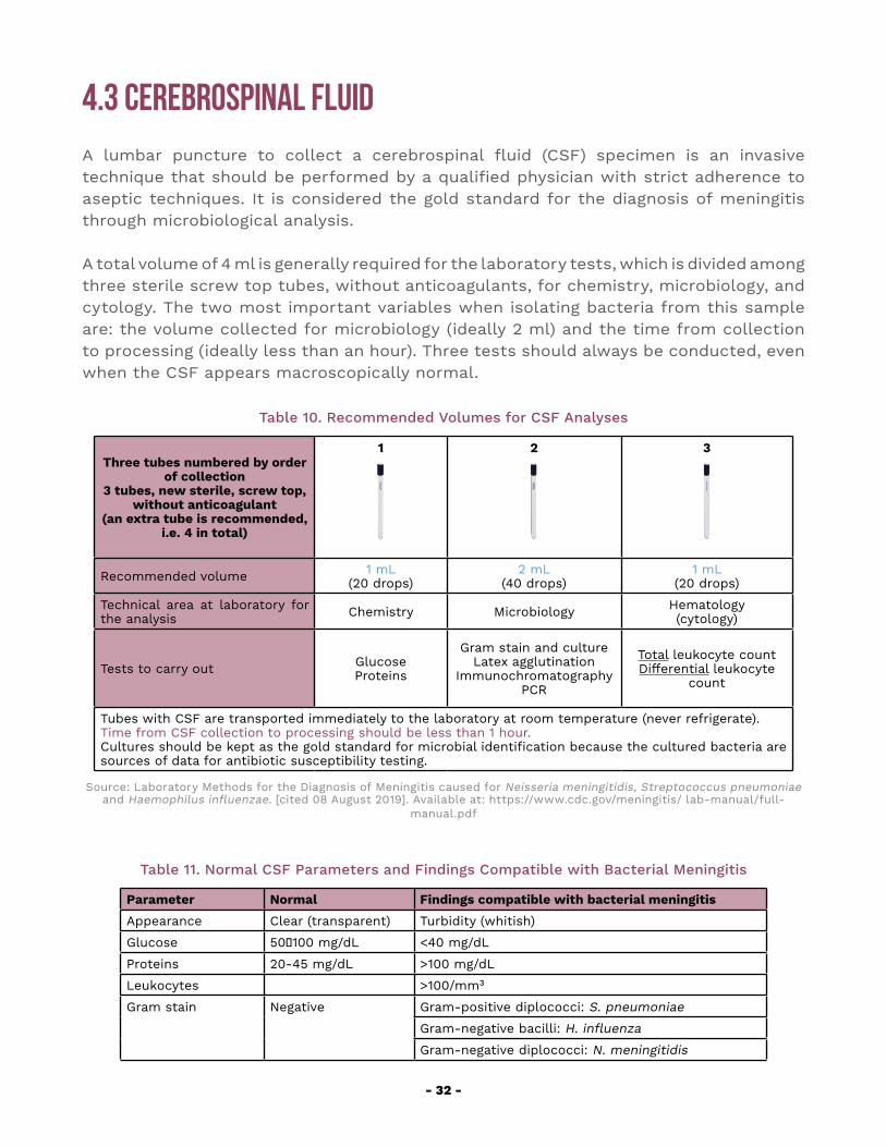
A total volume of 4 ml is generally required for the laboratory tests, which is divided among three sterile screw top tubes, without anticoagulants, for chemistry, microbiology, and cytology. The two most important variables when isolating bacteria from this sample are: the volume collected for microbiology (ideally 2 ml) and the time from collection to processing (ideally less than an hour). Three tests should always be conducted, even when the CSF appears macroscopically normal.
Table 10. Recommended Volumes for CSF Analyses
Three tubes numbered by order of collection
3 tubes, new sterile, screw top, without anticoagulant
(an extra tube is recommended, i.e. 4 in total)
1 2 3
Recommended volume 1 mL(20 drops)
2 mL(40 drops)
1 mL(20 drops)
Technical area at laboratory for the analysis Chemistry Microbiology Hematology
(cytology)
Tests to carry out Glucose Proteins
Gram stain and cultureLatex agglutination
ImmunochromatographyPCR
Total leukocyte countDifferential leukocyte
count
Tubes with CSF are transported immediately to the laboratory at room temperature (never refrigerate). Time from CSF collection to processing should be less than 1 hour.Cultures should be kept as the gold standard for microbial identification because the cultured bacteria are sources of data for antibiotic susceptibility testing.
Source: Laboratory Methods for the Diagnosis of Meningitis caused for Neisseria meningitidis, Streptococcus pneumoniae and Haemophilus influenzae. [cited 08 August 2019]. Available at: https://www.cdc.gov/meningitis/ lab-manual/full-
manual.pdf
Table 11. Normal CSF Parameters and Findings Compatible with Bacterial Meningitis
Parameter Normal Findings compatible with bacterial meningitis
Appearance Clear (transparent) Turbidity (whitish)
Glucose 50‒100 mg/dL <40 mg/dL
Proteins 20-45 mg/dL >100 mg/dL
Leukocytes >100/mm³
Gram stain Negative Gram-positive diplococci: S. pneumoniae
Gram-negative bacilli: H. influenza
Gram-negative diplococci: N. meningitidis
- 33 -
Culture Negative Positive
Source: Laboratory Methods for the Diagnosis of Meningitis caused for Neisseria meningitidis, Streptococcus pneumoniae and Haemophilus influenzae [cited 08 August 2019]. Available at:https://www.cdc.gov/meningitis/lab-manual/ full-manual.pdf
4.3.1 COLLECTING CSF SPECIMENS ◆ The lumbar procedure to collect a sample of cerebrospinal fluid (CSF)
should be carried out by a qualified professional. It should be performed as soon as possible, preferably before establishing the antimicrobial treatment. However, antibiotic therapy should be started as promptly as possible based on the clinician’s criteria. Treatment should never be delayed while waiting for CSF results.
◆ The tubes should be numbered from one to three before collecting the sample. The distribution and volume of CSF in the tubes should follow the recommendations shown in Table 10).
◆ Follow the collection sequence: ◇ The first tube is the most likely of become contaminated with blood in the
case of a traumatic puncture. This should not, therefore, be used for culture (blood inhibits bacterial growth) nor cytology (it would distort total and differential leukocyte counts).
◇ The second tube is less likely to become contaminated than the first, and more likely to reach the recommended volume than the third.
◇ The third tube is least likely to become contaminated but the most likely to fail to attain the recommended volume. In cases where sufficient CSF cannot be collected for the third tube, cytology and biochemistry will be conducted using the first tube. Caution should be exercised when interpreting the leukocyte count in cases of traumatic punctures.
4.3.2 TRANSPORTING CSF SPECIMENS ◆ The three tubes are transported to the laboratory at room temperature.
Never refrigerate them as the microorganisms that cause bacterial meningitis are less viable at temperatures below 18°C.
◆ The three tubes are transported to the laboratory in an upright position in a test tube rack and covered, in accordance with biosafety regulations.
◆ Transportation times of the CSF to the laboratory should be as short as possible because the sooner it is plated on the appropriate culture, the better the possibilities of isolating the causative agent.
◆ CSF tube transport should comply with the triple packaging standard and all samples should be duly marked and labeled.
◆ The tubes should be transported together with the test request sheet and case report form (confirming use of antimicrobial agents 72 hours prior to collection).
4.3.3 RECEPTION OF CSF SPECIMENS BY LABORATORY ◆ Check that data in test request sheet coincide with those on the case
report form and tube.
- 34 -
◆ Enter sample into the laboratory manually or automatically, recording date and time of reception at laboratory.
◆ Deliver immediately to the professional responsible for incubating them.
4.3.4 ANALYZING CSF SPECIMENS ◆ The laboratorian who receives a CSF sample should stop what he or she
is doing and process the sample immediately as this is an urgent sample.
CSF culture
◆ Centrifuge the sample for 15 minutes to concentrate the microorganisms before seeding. This is why immediate transportation to the laboratory is fundamental.
◆ If the sample volume is less than 1 ml, do not centrifuge. ◆ Ideally, the sample should be cultivated and incubated within this first
hour. CSF samples seeded within the first hour of collection are associated with high positivity percentages.
◆ Vigorously mix the CSF sediment with a vortex machine. ◆ Aspire the sediment and inoculate:
◇ 2 drops on a 5% sheep blood agar plate ◇ 2 drops on a chocolate agar plate, and ◇ 2 drops on a separate clean slide for Gram staining
◆ Use a sterile loop to spread the inoculum along the surface of the culture plates to allow isolated colonies to develop.
CSF Gram stain
◆ This is an urgent test so the results should be made available to the clinician as soon as possible.
◆ It is carried out from the sediment remaining in the tube after centrifugation, in accordance with standardized methodology.
◆ Air-dry the slide with the sample before applying the fixation. Use alternative methods such as fixation with alcohol in biosafety cabinets. The slide should be new and very clean.
◆ The result of the Gram stain conditions the type of culture media to be used for the subculturing.
◆ In suspect meningitis cases, the results CSF gram stain would suggest the etiologic agent involved.
CSF examination using immunochromatography, latex particle agglutination and PCR
◆ Homogenize the CSF sample: invert the tube gently two or three times. ◆ Using a micropipette and sterile loop, aspire 250-500 µl of CSF to inoculate
in a cryotube. Store and send to the National Reference Laboratory, maintaining the cold chain.
◆ Using a micropipette and sterile loop, aspire 100-200 µl of CSF to inoculate tube for immunochromatography (Binax Now®).
- 35 -
◆ Centrifuge the CSF sample contained in the first tube at 1,000 × g for 15 minutes.
◆ Using a Pasteur pipette, aspire the supernatant and store in a tube for the latex agglutination test (30 to 50 µl of supernatant is required per microbial agent investigated).
CSF chemistry and cytology tests are carried out immediately on tubes 1 and 3 respectively, together with the microbiology performed in parallel. The results should be sent to the microbiology laboratory so they can be correlated with the CSF Gram stain findings. Both cytochemistry and Gram stain finding should be sent to the physician within the same time period.
4.3.5 DELIVERING CSF ANALYSIS RESULTSAll results of tests carried out on CSF should be reported to clinicians on a timely basis:
◆ The Gram stain and cytochemistry finding should be reported within an hour of collecting the sample.
◆ Blood and CSF diagnostic test (latex agglutination or immunochromatography) results should be reported as soon as possible.
◆ Clinicians and epidemiologists should be notified immediately if S. pneumoniae, H. influenzae or N. meningitidis are identified.
All CSF samples that meet any of the following criteria should be sent to the national reference laboratory by sentinel laboratories:
◆ CSF with turbidity ◆ CSF with a leukocyte count >100/mm3 ◆ CSF with a leukocyte count of 10-100/mm3 and CSF glucose <40 mg/dL
and/or CSF protein >100 mg/dL ◆ CSF with a positive culture: both the sample and bacteria isolate are sent.
4.4 Pleural FluidPleural fluid (PF) is collected in a procedure known as thoracocentesis or pleural tap. This is an invasive procedure, not without risks, so it should only be performed by an experienced physician. It is performed maintaining strict aseptic technique in a setting duly equipped to deal with any complications that could occur during the procedure. Ideally, it should be carried out before initiating antimicrobial therapy.
Diagnostic thoracocentesis: ◆ Aspire 4 ml using a non-heparinized syringe. ◆ Distribute among three tubes for chemistry, microbiology, and cytology,
respectively. ◆ Aspire 1 ml in a heparinized syringe for pH testing and send immediately to
the laboratory under anaerobiosis conditions (i.e. without air, plugging the
- 36 -
opening of the syringe with a rubber stopper).
Therapeutic thoracocentesis (a larger volume may be collected): ◆ It is recommended to send between 15 to 20 mL to the laboratory. ◆ For pediatric patients, it is highly recommended to include a Gram stain
for S. pneumoniae in the basic PF microbiology. Its high sensitivity and specificity would allow identification of this etiologic agent in 75% to 95% of cases.
◆ A PCR for S. pneumoniae, H. influenzae and N. meningitidis should also be included.
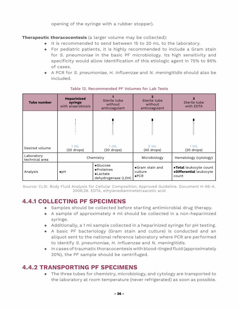
Table 12. Recommended PF Volumes for Lab Tests
Tube number Heparinized
syringe with anaerobiosis
1Sterile tube
without anticoagulant
2Sterile tube
without anticoagulant
3Sterile tube with EDTA
Desired volume 1 mL(20 drops)
1 mL(20 drops)
2 mL(40 drops)
1 mL(20 drops)
Laboratory technical area Chemistry Microbiology Hematology (cytology)
Analysis ◆pH
◆Glucose◆Proteines◆Lactate dehydrogenase (LDH)
◆Gram stain and culture◆PCR
◆Total leukocyte count◆Differential leukocyte count
Source: CLSI. Body Fluid Analysis for Cellular Composition; Approved Guideline. Document H-56-A. 2006;26. EDTA, ethylenediaminetetraacetic acid
4.4.1 COLLECTING PF SPECIMENS ◆ Samples should be collected before starting antimicrobial drug therapy. ◆ A sample of approximately 4 ml should be collected in a non-heparinized
syringe. ◆ Additionally, a 1 ml sample collected in a heparinized syringe for pH testing. ◆ A basic PF bacteriology (Gram stain and culture) is conducted and an
aliquot sent to the national reference laboratory where PCR are performed to identify S. pneumoniae, H. influenzae and N. meningitidis.
◆ In cases of traumatic thoracocentesis with blood-tinged fluid (approximately 20%), the PF sample should be centrifuged.
4.4.2 TRANSPORTING PF SPECIMENS ◆ The three tubes for chemistry, microbiology, and cytology are transported to
the laboratory at room temperature (never refrigerated) as soon as possible.
- 37 -
◆ Maximum transport times should be two hours. ◆ The sample collected in a heparinized syringe for pH testing should be sent
to the laboratory immediately under anaerobiosis (if immediate transfer is not possible, store and transport the sample in ice).
◆ The three tubes are transported to the laboratory in an upright position in a test tube rack with triple packing.
◆ The sample for pH testing should also be transported in accordance with biosafety standards (triple packaging).
◆ They should be transported together with the test request sheet and case report form (confirming the use of antimicrobial agents 72 hours prior to collection).
4.4.3 RECEPTION OF PF SPECIMENS BY LABORATORY ◆ Check that the data on the test request sheet, case report form and the
tubes coincide. ◆ Confirm that the data on the sample coincide with those on the case
report form and laboratory test request form used in the hospital. ◆ Record the date and time of sample collection and its reception at the
laboratory. ◆ Record the number of tubes sent and the sample volume collected in each
tube. ◆ Entre the sample into the IT and/or manual system used by the laboratory,
recording all the data provided. ◆ Deliver the tubes with the PF sample to the professionals responsible for
the bacteriology, chemistry and hematology areas so processing can start immediately.
◆ Enter sample into the laboratory manually or automatically, recording date and time of reception at laboratory, and deliver the hemoculture bottle to the professional responsible for incubating it.
4.4.4 PL CYTOLOGY AND CHEMISTRY ◆ Tubes without anticoagulant are tested for proteins, lactate dehydrogenase
(LDH), and glucose. ◆ The pH determination is carried out on a sample from the heparinized
syringe. ◆ Cytology, for the total and differential leukocyte count, is conducted from
the sample collected in the tube with EDTA anticoagulant.
- 38 -
4.5 General Considerations for CSF and PF Specimens
◆ The bacteriological laboratory should conserve (in a cryo-tube) a 250 to 500 µl aliquot of all CSF/PF specimens from patients under 5 entering the sentinel surveillance system and send them to the national reference laboratory for PCRs to identify H. influenzae, N. meningitidis and S. pneumoniae.
◆ CSF/PF specimens are transported in cryo-tubes, maintaining the cold chain, with a triple packaging system in accordance with international biosafety standards (International Air Transport Association [IATA]).
◆ The temperature at which samples should be stored at the sentinel laboratory and transported to the national reference laboratory is based on how long it will take for them to be delivered to the national reference laboratory:
◇ If the sample will reach the national reference laboratory in less than 72 hours from time of collection: the cryo-tube is stored and transported at 4°C.
◇ If the sample will reach the national reference laboratory between day 3 and day 14 of time of collection: the cryo-tube is stored at -20°C and transported with ice dry.
◇ If the sample will reach the national reference laboratory 14 days after collection: the cryo-tube is stored at -70°C and transported with ice dry.
4.6 S. pneumoniae, H. influenzae and N. meningitidis Identification
The capacity for growth in the culture media used, the morphology of the colonies and Gram staining allow easy presumptive identification of the three etiologic agents most frequently involved in bacterial pneumonia and meningitis.
4.6.1 BACTERIAL ISOLATES OR STRAINSAll S. pneumoniae, H. influenzae and N. meningitidis isolates or strains (or other complex microbes) identified in hemoculture, CSF and PF specimens should be sent to the national reference laboratory for confirmation of isolate identification, antimicrobial susceptibility testing, serogroup/serotype/serosubtype characterization, and quality control.
Isolates must be transported using the most suitable means of transport with correct temperature and climatic conditions to ensure the viability of the microbes. They are always dispatched together with a complete patient case report form. Isolates are transported in tubes containing Amies transport medium with carbon activated, at
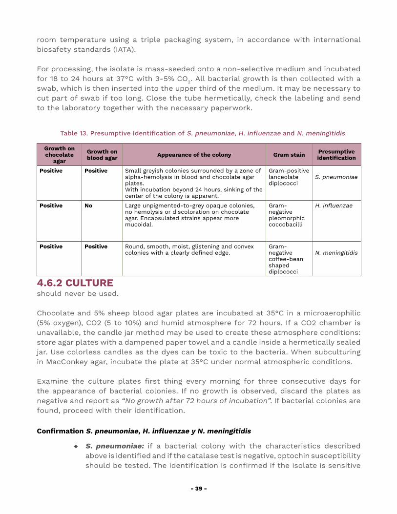
- 39 -
room temperature using a triple packaging system, in accordance with international biosafety standards (IATA).
For processing, the isolate is mass-seeded onto a non-selective medium and incubated for 18 to 24 hours at 37°C with 3-5% CO2. All bacterial growth is then collected with a swab, which is then inserted into the upper third of the medium. It may be necessary to cut part of swab if too long. Close the tube hermetically, check the labeling and send to the laboratory together with the necessary paperwork.
Table 13. Presumptive Identification of S. pneumoniae, H. influenzae and N. meningitidis
Growth on chocolate
agar
Growth onblood agar Appearance of the colony Gram stain Presumptive
identification
Positive Positive Small greyish colonies surrounded by a zone of alpha-hemolysis in blood and chocolate agar plates. With incubation beyond 24 hours, sinking of the center of the colony is apparent.
Gram-positive lanceolate diplococci
S. pneumoniae
Positive No Large unpigmented-to-grey opaque colonies, no hemolysis or discoloration on chocolate agar. Encapsulated strains appear more mucoidal.
Gram-negative pleomorphiccoccobacilli
H. influenzae
Positive Positive Round, smooth, moist, glistening and convex colonies with a clearly defined edge.
Gram-negative coffee-bean shaped diplococci
N. meningitidis
4.6.2 CULTUREshould never be used.
Chocolate and 5% sheep blood agar plates are incubated at 35°C in a microaerophilic (5% oxygen), CO2 (5 to 10%) and humid atmosphere for 72 hours. If a CO2 chamber is unavailable, the candle jar method may be used to create these atmosphere conditions: store agar plates with a dampened paper towel and a candle inside a hermetically sealed jar. Use colorless candles as the dyes can be toxic to the bacteria. When subculturing in MacConkey agar, incubate the plate at 35°C under normal atmospheric conditions.
Examine the culture plates first thing every morning for three consecutive days for the appearance of bacterial colonies. If no growth is observed, discard the plates as negative and report as “No growth after 72 hours of incubation”. If bacterial colonies are found, proceed with their identification.
Confirmation S. pneumoniae, H. influenzae y N. meningitidis
◆ S. pneumoniae: if a bacterial colony with the characteristics described above is identified and if the catalase test is negative, optochin susceptibility should be tested. The identification is confirmed if the isolate is sensitive
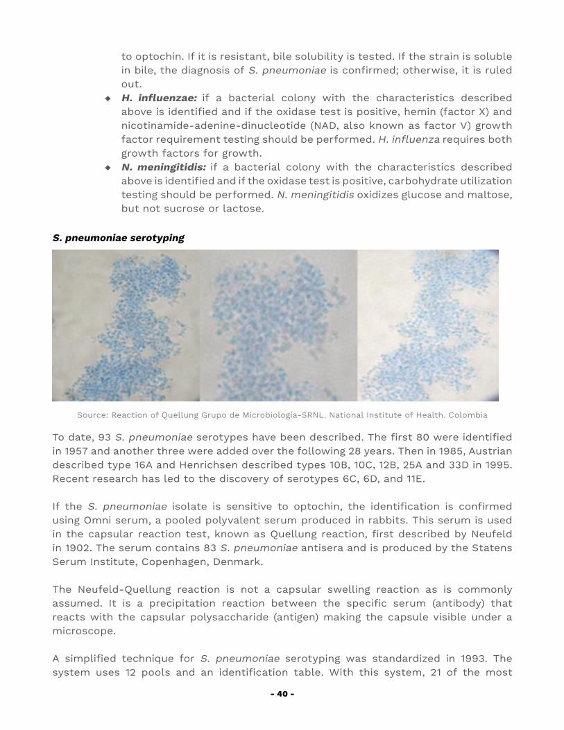
- 40 -
to optochin. If it is resistant, bile solubility is tested. If the strain is soluble in bile, the diagnosis of S. pneumoniae is confirmed; otherwise, it is ruled out.
◆ H. influenzae: if a bacterial colony with the characteristics described above is identified and if the oxidase test is positive, hemin (factor X) and nicotinamide-adenine-dinucleotide (NAD, also known as factor V) growth factor requirement testing should be performed. H. influenza requires both growth factors for growth.
◆ N. meningitidis: if a bacterial colony with the characteristics described above is identified and if the oxidase test is positive, carbohydrate utilization testing should be performed. N. meningitidis oxidizes glucose and maltose, but not sucrose or lactose.
S. pneumoniae serotyping
Source: Reaction of Quellung Grupo de Microbiología-SRNL. National Institute of Health. Colombia
To date, 93 S. pneumoniae serotypes have been described. The first 80 were identified in 1957 and another three were added over the following 28 years. Then in 1985, Austrian described type 16A and Henrichsen described types 10B, 10C, 12B, 25A and 33D in 1995. Recent research has led to the discovery of serotypes 6C, 6D, and 11E.
If the S. pneumoniae isolate is sensitive to optochin, the identification is confirmed using Omni serum, a pooled polyvalent serum produced in rabbits. This serum is used in the capsular reaction test, known as Quellung reaction, first described by Neufeld in 1902. The serum contains 83 S. pneumoniae antisera and is produced by the Statens Serum Institute, Copenhagen, Denmark.
The Neufeld-Quellung reaction is not a capsular swelling reaction as is commonly assumed. It is a precipitation reaction between the specific serum (antibody) that reacts with the capsular polysaccharide (antigen) making the capsule visible under a microscope.
A simplified technique for S. pneumoniae serotyping was standardized in 1993. The system uses 12 pools and an identification table. With this system, 21 of the most
- 41 -
common serotypes or serogroups worldwide can be identified. All these serotypes are included in the S. pneumoniae polysaccharide vaccine (containing 23 serotypes).
In the experience of the National Center for Streptococcus in Alberta, serotype 3 (pool B) has occasionally not reacted with the pool. The mucoid appearance of the isolate is key to its identification. Isolates with this morphology should be examined using serotype 3 antiserum, despite a negative result with pool B, before being classified as a non-typable strain.
H. influenzae serotyping
The capsule is a major virulence factor in many bacterial species, but it also permits serotyping. Encapsulated Haemophilus strains can be identified by serological typing based on the chemical composition of the polysaccharide capsule.
As the capsule is a virulence factor, it is uncommon to find invasive isolates that cannot be serotyped. It is important to remember, however, that the capsule is lost during subculturing and that the capsular structure deteriorates in old culture media. This is why culture serotyping must be carried out within 24 hours and as soon as possible after the primary culture.
Serotyping can be carried out by slide agglutination, capsular swelling (Quellung reaction), coagglutination, immunofluorescence or countercurrent immunoelectrophoresis. Coagglutination and latex agglutination are the serological methods used to detect the capsular polysaccharide directly in CSF, serum, or urine. Coagglutination is based on the capacity of protein A on the surface of Staphylococcus aureus (Cowan strain) to bind to the Fc portion of immunoglobulins allowing visualization of the agglutination, while the latex test uses sensitized latex particles. These tests are especially useful when the culture is negative.
The most commonly used serotyping method in the reference laboratories is slide agglutination.
N. meningitidis serotyping
Like other pathogenic bacterial species, Neisseria meningitidis is capable of expressing capsular polysaccharides and this is strongly correlated with the corresponding infectious diseases. Chemical and structural differences in the capsular polysaccharide allow classification of meningococci into different serogroups, of which only A, B, C, W, Y, and more recently, X, are associated with disease. Strains isolated in asymptomatic carriers rarely express the capsule. Some have even lost the capacity to do so. This means that a large proportion of isolates will not be classifiable (serogroup) using traditional methods.
- 42 -
4.6.3 ANTIMICROBIAL SUSCEPTIBILITY TESTINGEmpiric antibiotic therapy for bacterial pneumonia and meningitis is clearly defined and should be initiated as soon as biological samples are collected. This generally includes a third-generation cephalosporin such as ceftriaxone or cefotaxime. Nevertheless, it is still very important that bacteriology laboratories do their utmost to isolate the bacterial strain and conduct antimicrobial susceptibility testing. The increasing number of S. pneumoniae strains developing resistance to third-generation cephalosporins has prompted several countries to include IV vancomycin4 in their empiric therapy for bacterial meningitis. The antimicrobial susceptibility of H. influenzae and N. meningitidis, on the other hand, is more predictable but sentinel laboratories are still recommended to conduct the tests.
Antibiotic susceptibility testing is defined by the minimum inhibitory concentration (MIC) in a broth microdilution test and/or E-test or disk diffusion methods. Current protocols established by accredited entities such as the Clinical Laboratories Standards Institute (CLSI)5 should be followed. Certified Sensi-Disc™, for example, may be used for the disk diffusion method with specific agar; with the exception S. pneumoniae susceptibility to penicillin and ceftriaxone. In this case, therefore, it is absolutely essential to determine the MIC of the strain for these two antimicrobial drugs as the cut-off values for the interpretation vary depending on the patient’s clinical picture (pneumonia or meningitis). The epsilometer or E-test is recommended in this case.
4.6.4 IMMUNOLOGICAL AND MOLECULAR DIAGNOSTIC METHODS WHO recommends laboratories participating in sentinel epidemiological bacterial meningitis surveillance to include latex agglutination, immunochromatography, and molecular techniques to the basic lab methods used for CSF bacteriological examination (Gram stain and culture).
Immunological methods to detect bacterial antigens should be among the bacteriological tests conducted on all suspect meningitis cases. Its greatest contribution is in cases in which the CSF gram stain is doubtful.
The two most commonly used immunological techniques are the latex agglutination test and immunochromatography. The latter has demonstrated good sensitivity and specificity in the diagnosis of pneumococcal meningitis.
◆ Immunological techniques ◇ Latex agglutination test: used for the diagnosis of bacterial meningitis to
detect the principal bacterial agents. It is performed in the bacteriology lab using the supernatant drawn off following centrifugation of CSF at 1,000 ×
4 Vancomycin, administered slowly as an intravenous infusion every 6 hours, is a nephrotoxic antimicrobial drug. Hence the recommendation to identify as soon as possible if the strain is sensitive or not to third-generation cephalosporins so that administration can be discontinued.5 CLSI. Performance Standards for Antimicrobial Susceptibility Testing; Twenty-Fourth Informational Supplement. CLSI M100-S25. Wayne, PA: Clinical Laboraoty Standards Institute; 2018
- 43 -
g for 15 minutes. The supernatant is then heated to boiling point in a water bath for 1 minute to release the capsular antigen. Special care should be taken to ensure kits are kept refrigerated between 4-8°C so that they are not inactivated before their expiration date.
◇ Immunochromatography (Binax Now®) for S. pneumoniae: an immunological technique designed for direct testing of CSF specimens for S. pneumoniae (bacterial antigen), without prior centrifuging or heating. All CSF specimens from children under 5 years with suspected meningitis should be tested in the case of a doubtful Gram stain result, or if the cytochemistry is compatible with bacterial meningitis but the Gram stain is negative.
◆ Molecular techniques (PCR) The PCR is a molecular biology technique that permits specific nucleic acid sequences to be determined within a few hours, in the case of real-time PCR, or overnight, in the case of conventional PCR. Given its high sensitivity and specificity, it is considered an extremely useful diagnostic tool in identifying the causative agents of meningitis.
There are currently some duly standardized PCR protocols that allow confirmation of several microorganisms. One of those most widely used is that which simultaneously detects S. pneumoniae, H. influenzae and N. meningitidis.
The great advantage of this over culture techniques is that it does not require strains to be live in order to identify them. Its disadvantages include poor accessibility and it does not facilitate antimicrobial susceptibility testing. PCR can also be performed on pleural fluid and blood specimens and enable at least partial characterization of isolates by serogroup and serotype.
4.7 QUALITY CONTROL
Bacteriology laboratories participating in bacterial pneumonia and meningitis sentinel surveillance should guarantee the reliability of their results by executing a quality program that includes:
◆ Internal quality control ◆ External quality control ◆ Indirect quality control ◆ Quality indicators and periodic performance monitoring
4.7.1 INTERNAL QUALITY CONTROL PROGRAM ◆ All the culture media, reagents and diagnostic kits used in bacteriological
diagnosis should be subject to quality control to ensure the reliability of the results obtained.
◆ Materials used to prepare the culture media, as well as reagents or diagnostic kits, must be procured from certified sources and the supplier guarantees
- 44 -
proper conservation, transportation, and storage of these products. Ready-prepared culture media are subject to the same specifications.
◆ The best culture media to isolate S. pneumoniae, N. meningitidis and H. influenzae are trypticase soy agar + 5% sheep blood and chocolate agar. Culture plates should never be prepared with human blood as it contains numerous bactericidal substances that would inhibit bacterial growth.
◆ S. pneumoniae and N. meningitidis grow well in both culture media, while H. influenzae only grows in chocolate agar that contains the two growth factors it needs to grow: hemin (factor X) and NAD (factor V).
◆ Each batch of culture media should be subject to the following quality controls: ◇ Physicochemical ◇ Sterility ◇ Growth: using bacterial reference strains (American Type Culture Collection, ATCC).
◆ These controls should also be carried out in any situations that could ultimately affect the quality of the culture media; for example, power outages.
◆ Each reagent used in the conduct of the diagnostic tests (e.g. catalase, oxidase, optochin, Gram stain, etc.) should be subject to quality control prior to use to ensure the reliability of the results delivered. Reference strains (ATCC) shall be used for this.
◆ Each batch of culture medium used for antimicrobial susceptibility testing should be evaluated using reference strains with known antimicrobial susceptibility values; likewise for antimicrobial susceptibility disks.
◆ The bacteriological laboratory should design its own internal quality control program and evaluate it periodically in compliance with recommendations established by CLSI, as well as maintaining the collection of ATCC strains necessary for quality control in optimal conditions.
4.7.2 RESPONSIBILITIES OF THE LABORATORY NETWORKLaboratories participating in sentinel hospital surveillance should form part of a laboratory network, organized at four levels of complexity:
◆ Sentinel Laboratory (SL) The main responsibilities in this sentinel surveillance are:
◇ Process blood culture, CSF, and PF specimens collected from patients with probable bacterial pneumonia or suspect meningitis using Gram, culture, and biochemistry testing.
◇ Send all S. pneumoniae, H. influenzae and N. meningitidis strains isolated from said sources to the national reference laboratory for confirmation and identification of serotypes and serogroups.
◇ Send CSF and PF specimens from patients with probable bacterial pneumonia and suspect meningitis to the national reference laboratory for PCR analysis.
◇ Participate in an annual external quality control program. ◇ Fulfill laboratory quality indicators defined for sentinel hospital surveillance. ◇ Report the laboratory findings to the attending physician/department and
to the surveillance coordinator. ◇ Participate in the global external performance assessment led by the
United Kingdom National External Quality Assessment Scheme (UKNEQAS) on behalf of PAHO/WHO.
- 45 -
◆ National Reference Laboratory (NRL)The main responsibilities of the national reference laboratory in this sentinel surveillance are:
◇ Confirm bacterial isolates sent from sentinel laboratories. ◇ Characterize isolates through serogroup and serotype analysis. ◇ Conduct antimicrobial susceptibility tests. ◇ Conduct PCR for S. pneumoniae, H. influenzae and N. meningitidis detection
in CSF and PF specimens. ◇ Report laboratory test to SL, the surveillance coordinator, and the regional
laboratory coordinator. ◇ Train SL technical team. ◇ Organize technical supervisory visits to SL and evaluate the laboratory’s
performance. ◇ Send bacterial isolates and CSF/PF specimens to the assigned RRL for
confirmation and external quality control. ◇ Participate in the global external performance assessment led by the
United Kingdom National External Quality Assessment Scheme (UKNEQAS) on behalf of PAHO/WHO.
◆ Regional Reference Laboratory (RRL)The main responsibilities in this sentinel surveillance are:
◇ Confirm and characterize bacterial isolates sent by national reference laboratories.
◇ Conduct PCR for S. pneumoniae, H. influenzae and N. meningitidis detection and typing in CSF and PF specimens sent by national reference laboratories for confirmation or quality control.
◇ Train NRL professionals in countries participating in sentinel hospital surveillance.
◇ Organize technical supervisory visits to NRL and monitor laboratory performance in terms of sentinel hospital surveillance.
◇ Report test results to the NRL and to the regional laboratory coordinator. ◇ Participate in indirect quality control activities by sending previously
analyzed bacterial isolates and CSF/PF specimens and their corresponding results to the World Reference Laboratory (WRL)
◇ Participate in the global external performance assessment led by the United Kingdom National External Quality Assessment Scheme (UKNEQAS) on behalf of PAHO/WHO.
◆ World Reference Laboratory (WRL)For the Region of the Americas, the world or global of reference laboratories are the CDC (Atlanta, United States of America) and UK-NEQAS. Their principal responsibilities with regard to this sentinel surveillance are:
◇ Develop new laboratory tests. ◇ Facilitate technology transfer to RRL participating in the network. ◇ Provide training for professionals at RRL and NRL assigned to the network. ◇ Organize technical supervisory visits to the RRL and monitor performance
of laboratories involved in sentinel hospital surveillance. ◇ Carry out indirect quality control of RRL that have submitted previously analyzed
bacterial isolates and CSF/PF specimens with the corresponding results. ◇ On behalf of WHO, design and execute the global external performance
assessment program for SL, NRL and RRL participating in the network. ◇ Report test results to the RRL, the regional laboratory network coordinator
and global laboratory network coordinator.
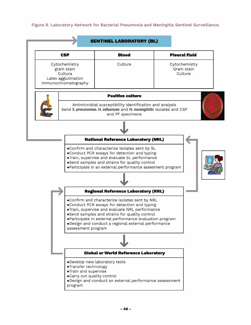
- 46 -
Figure 9. Laboratory Network for Bacterial Pneumonia and Meningitis Sentinel Surveillance.
5. SURVEILLANCE OF BACTERIAL PNEUMONIA AND MENINGITIS
- 48 -
5. SURVEILLANCE OF BACTERIAL PNEUMONIA AND MENINGITIS The purpose of surveillance of vaccine-preventable diseases, such as bacterial pneumonia and meningitis, is to identify the predominant causative agents to inform decision-making on the introduction of new vaccines. It should also measure the impact of the vaccines and monitor the epidemiological behavior of the causative agents. Integrating time, place, and people data with laboratory results is extremely useful.
5.1 Surveillance objectives
The objectives of bacterial pneumonia and meningitis surveillance in children under 5 years in the Region are:
◆ To obtain standardized epidemiological data on these diseases ◆ To identify S. pneumoniae, H. influenzae and N. meningitidis; to characterize
the strains of these agents in circulation as well as changes of serotypes/serogroups as they emerge
◆ To monitor antimicrobial susceptibility patterns of S. pneumoniae, H. influenzae and N. meningitidis
◆ To generate information on the basis of which to introduce new vaccines and monitor their impact
◆ To participate in the Global Sentinel Surveillance Network for Invasive Bacterial Vaccine-Preventable Disease coordinated by WHO
5.2 Surveillance Strategies
5.2.1 TARGET POPULATION SURVEILLANCE The target population for surveillance of bacterial pneumonia and meningitis is children aged under 5 years.
5.2.2 TYPE OF SURVEILLANCESurveillance is conducted through sentinel hospitals. The rationale behind using this kind of surveillance is as follows:
1. Patients under 5 with a presumptive pneumonia diagnosis are frequently hospitalized.
2. Every diagnosed bacterial meningitis case is considered serious and requires hospitalization.
3. Radiology services at sentinel hospitals facilitate diagnosis of probable bacterial pneumonia cases.

- 49 -
4. Specimens are cultured by sentinel hospitals laboratories.5. This type of surveillance makes it possible to target resources, monitor
processes, and generate quality standardized epidemiological data for decision-making.
5.2.3 CRITERIA FOR THE SELECTION OF SENTINEL HOSPITALSThe criteria for the selection of a sentinel hospital are:
◆ The hospital should be a reference hospital for the surveillance target population.
◆ The hospital should be accessible for all socioeconomic groups of the target population.
◆ The hospital should have a radiology service 24 hours a day, seven days a week. The chest X-ray result is one of the criteria for the selection of probable cases.
◆ The hospital should have a bacteriological laboratory capable of isolating Hi, meningococcus and pneumococcus 24 hours a day, seven days a week.
◆ The hospital should have the human, material and logistical resources needed for permanent surveillance.
◆ The hospital should have a strong sense of institutional commitment.
Each country must decide how many hospitals to involve in sentinel surveillance in order to comply with the criteria listed above and the operational capacity for ongoing surveillance monitoring and evaluation. As part of the Global Surveillance Network, these criteria and those listed in Tables 8 and 9 must be met.
Hospitals that cover a clearly defined population, in demographic and geographical terms, can provide data on case incidence.
5.3 Sentinel Hospital-based Bacterial Pneumonia Surveillance
5.3.1 CASE DEFINITIONSFor the purpose of epidemiological surveillance the following case definitions are considered:
Suspect case of pneumoniaEvery patient aged under 5 years hospitalized with a clinical diagnosis of community-acquired pneumonia. A “hospitalized patient” means any patient for whom hospital admission is indicated.Probable case of bacterial pneumoniaEvery suspect case with a chest X-ray showing a pattern compatible with bacterial pneumonia (see section 2.2, Radiological Diagnosis).
- 50 -
Confirmed case of bacterial pneumoniaEvery probable case of bacterial pneumonia in which H. influenzae, S. pneumoniae or another bacterium has been identified in the blood or pleural fluid.Discarded case of bacterial pneumoniaEvery suspect case with a chest X-ray that does not show a radiological pattern compatible with bacterial pneumonia.Inadequately investigated case of pneumonia
◇ Every suspect case without a chest X-ray. With the exception of a suspect case with no chest X-ray but with S. pneumoniae, H. influenzae, N. meningitidis or another bacterium identification, which will be classified as a confirmed case.
◇ Every probable case without hemoculture results.
5.3.2 STEPS OF SENTINEL HOSPITAL-BASED BACTERIAL PNEUMONIA SURVEILLANCE
1. The physician or nurse seeing patients in the emergency room or in an inpatient area will report to the hospital’s head of epidemiology every suspect case of community-acquired pneumonia in children under 5 years where hospital treatment is indicated. He or she will start filling out the case report form (Annex 1).
2. The physician or nurse will confirm details on the vaccination card and note patient’s vaccination status (pneumococcal, Hib, and meningococcal conjugate vaccines) in the case report form. If no Vaccination Card exists, information should be sought from the child’s guardian, and followed up until acquired. This information is of utmost importance for the data analysis and interpretation.
3. The head of epidemiology will enter the case on the IT system and follow up on it. He or she will also assess whether the case meets the criteria for epidemiological surveillance of severe acute respiratory infection (SARI).
4. The physician will order a chest X-ray.5. If the X-ray shows a pattern compatible with bacterial pneumonia, the
physician will classify the case as probable bacterial pneumonia.6. In every probable case of bacterial pneumonia, the physician will take
two blood samples for hemoculture, if possible before beginning antibiotic treatment.
7. If the patient has received antibiotics in the 72 hours before specimen collection, the physician will record this in the case report.
8. In patients requiring thoracocentesis because of pleural effusion, a specimen of pleural fluid will be collected.
9. Specimens will be sent immediately to the hospital laboratory together with the case report.
10. The head of the laboratory will inform the physician and epidemiologist immediately on the test results, the bacteria identified, and the microbial susceptibility tests conducted.
11. The sentinel surveillance team will decide on the final classification of the case.
- 51 -
12. The head of the laboratory will send the isolated H. influenzae and S. pneumoniae strain to the national reference laboratory for characterization.
13. Once the national reference laboratory has reported its results, the head of the laboratory will report the results regarding strain characterization and susceptibility to antimicrobial drugs to the sentinel surveillance team.
14. When the patient is discharged, the head of epidemiology will complete the case report.
15. The head of epidemiology will verify the completeness and consistency of each data entry during the entire process.
16. The sentinel surveillance team, under the coordination of the head of epidemiology, will analyze the information on a monthly basis.
17. The head of epidemiology will send individual and consolidated surveillance data up the next hierarchical level on a monthly basis.
18. The sentinel surveillance team will report on the data and provide and feedback periodically, on a quarterly basis is recommended.
5.3.3 DATA REQUIRED FOR SENTINEL HOSPITAL-BASED BACTERIAL PNEUMONIA SURVEILLANCEData be collected on children aged under 5 years include the following:
A. Number of hospitalizations for any causeB. Number of hospitalizations as a suspect pneumonia caseC. Individual data of suspect pneumonia cases:
◇ Sociodemographic data ◇ Vaccination status ◇ Radiology results ◇ Laboratory data ◇ Patient’s progress ◇ Final classification of case
5.4 Sentinel Hospital-based Bacterial Meningitis Surveillance
5.4.1 CASE DEFINITIONSFor purposes of epidemiological surveillance, cases are to be defined as follows:
Suspect case of meningitisEvery patient aged under 5 years hospitalized with a diagnosis of meningitis.Probable case of bacterial meningitisEvery suspect case in which CSF findings are compatible with bacterial meningitis, i.e., where at least one of the following characteristics is present:
◆ Turbidity ◆ Increased leukocyte count (>100/mm3) ◆ Leukocyte count between 10-100/mm3 and
- 52 -
◇ Elevated protein levels (>100 mg/dL) or ◇ Reduced glucose levels (<40 mg/dL)
Confirmed case of bacterial meningitisEvery suspect case in which S. pneumoniae, H. influenzae, N. meningitidis or another bacterium was identified in CSF or blood. Discarded case of bacterial meningitis
◆ Every suspected case with CSF findings not compatible with bacterial etiology, and in which no bacterium was identified in the CSF or blood specimen.
◆ Every probable case in which a non-bacterial etiologic agent has been identified.
Inadequately investigated case of meningitisEvery suspect case without collection of CSF specimen nor any bacterial agent identified in the hemoculture.
5.4.2 STEPS IN SENTINEL HOSPITAL-BASED BACTERIAL MENINGITIS SURVEILLANCE
1. The physician or nurse seeing patients in the emergency room or in an inpatient area will report to the hospital head of epidemiology every suspect meningitis case contracted in the community in children under 5 years where hospital treatment is indicated. He or she will begin the process of filling out the case report form (Annex 2).
2. The physician or nurse will confirm details on the Vaccination Card and note the patient’s vaccination status (pneumococcal, Hib, and meningococcal conjugate vaccines) in the case report form. If a Vaccination Card does not exist, information should be sought from the child’s guardian, and followed up until acquired. This information is of utmost importance for the data analysis and interpretation.
3. The physician will obtain a CSF specimen and record date and time of collection, as well as characteristics of its appearance in the case report.
4. Two blood samples will be collected for culture and other tests, ideally before starting antibiotic treatment.
5. If the patient received antibiotics in the 72 hours before specimen collection, the physician should record this in the case report.
6. The person collecting the sample should guarantee that it is sent immediately to the hospital laboratory together with the case report. CSF specimens should be processed within an hour of collection.
7. The head of the laboratory will immediately report the results of the CSF Gram stain, cytochemistry and antigen screening to the physician and epidemiologist.
8. The head of epidemiology will enter the case in the IT system and follow up on it.
9. In the event of suspect meningitis caused by Hi or meningococcus, the epidemiologist should initiate epidemiological prevention and control measures.
- 53 -
10. The head of the laboratory will immediately report to the physician and epidemiologist the findings of the hemoculture and antimicrobial susceptibility tests, if applicable.
11. The head of the laboratory will send the isolated Hi, meningococcus, or pneumococcus strain to the national reference laboratory for characterization.
12. Once received from the national reference laboratory, the head of the hospital laboratory will report the findings of the characterization of the strains and antimicrobial susceptibility testing to the physician and epidemiologist.
13. The sentinel surveillance team will decide on the final classification of the case based on the laboratory test results.
14. The sentinel team will decide which results will be taken into account for the final classification of the case, in the case of conflicting results between the different samples or tests.
15. When the patient is discharged, the head of epidemiology will complete the case report including data on the patient’s progress and the final classification of the case.
16. The head of epidemiology will confirm the completeness and consistency of each data entry throughout all stages.
17. The sentinel surveillance team, under the coordination of the head of epidemiology, will analyze the information every month.
18. The head of epidemiology will send individual and consolidated surveillance data up the next hierarchical level each month.
19. The sentinel surveillance team will report on the data and provide and feedback periodically, on a quarterly basis is recommended.
5.4.3 DATA TO BE COLLECTED FOR SENTINEL HOSPITAL-BASED BACTERIAL MENINGITIDES SURVEILLANCEData on children aged under 5 years that should be collected include the following:
A. Number of hospitalizations for any causeB. Number of hospitalizations as a suspect meningitis caseC. Individual data for suspect meningitis case:
◇ Sociodemographic data ◇ Vaccination status ◇ Clinical and laboratory data ◇ Patient’s progress ◇ Final classification of the case
5.5 Data analysis
The purpose of periodic data analysis is to determine the epidemiological disease patterns and to monitor and evaluate the surveillance system. Data should be consolidated and analyzed monthly.

- 54 -
Following the collecting and consolidating bacterial pneumonia (BP) surveillance data, the following are some of the indicators that should be analyzed:
Percentage of hospitalizations for suspect pneumonia in children under 5 years
Number of hospitalizations for suspect pneumonia cases in children under 5 years x 100Total hospitalizations of children under 5 years during the same period
Percentage of suspect pneumonia cases in children under 5 years investigated
Number of suspect pneumonia cases in children under 5 years investigated* x 100Number of hospitalizations with suspect pneumonia in children under 5 years during the same period
Percentage of suspect pneumonia cases in children under 5 years with vaccination records†
Number of suspect pneumonia cases in children under 5 years with vaccination records x 100Number of suspect pneumonia cases investigated* in children under 5 years during the same period
Percentage of suspect pneumonia cases in children under 5 years with appropriate vaccination† for age
Number of suspect pneumonia cases in children under 5 years with appropriate vaccination for age x 100Number of suspect pneumonia cases in children under 5 years investigated* during the same period
Percentage of probable bacterial pneumonia (BP) cases in children under 5 years
Number of probable BP cases in children under 5 × 100Number of suspect pneumonia cases in children under 5 years investigated* during the same period
Percentage of probable BP cases in children under 5 years with specimen for hemoculture
Number of probable BP cases with specimen for hemoculture in children under 5 years × 100Number of probable BP cases in children under 5 years during the same period
Percentage of probable BP cases in children under 5 years with pleural fluid PCR
Number of probable BP cases in children under 5 years with pleural fluid PCR × 100Number of probable BP cases in children under 5 years with pleural fluid specimen during the same period
Percentage of probable BP cases in children under 5 years with bacterial identification
Number of probable BP cases with identification of Hi, Spn, or other bacterium‡ in children under 5 years × 100Number of probable BP cases with blood or PF specimen in children under 5 years during the same period
Percentage of confirmed BP cases in children under 5 years
Number of confirmed BP cases in children under 5 years × 100Number of probable BP cases in children under 5 years during the same period
Percentage of confirmed BP cases in children under 5 years caused by Hi and Spn
Number of confirmed BP cases in children under 5 years caused by each specific bacterium × 100Number of confirmed BP cases in children under 5 years during the same period
Percentage of suspect pneumonia cases in children under 5 years with final classification
Number of suspect pneumonia cases in children under 5 years with final classification × 100Number of suspect pneumonia cases in children under 5 years during the same period
Case-fatality among children under 5 years with BP
Number of cases with probable and confirmed BP final classification in children under 5 years who × 100Number of cases with probable + confirmed BP final classification in children under 5 years in same period
*Suspected cases with chest X-ray and case report.†Vaccination Card and official databases. Seek data on Hib and pneumococcal conjugate vaccines.‡Disregard contaminant bacteria.
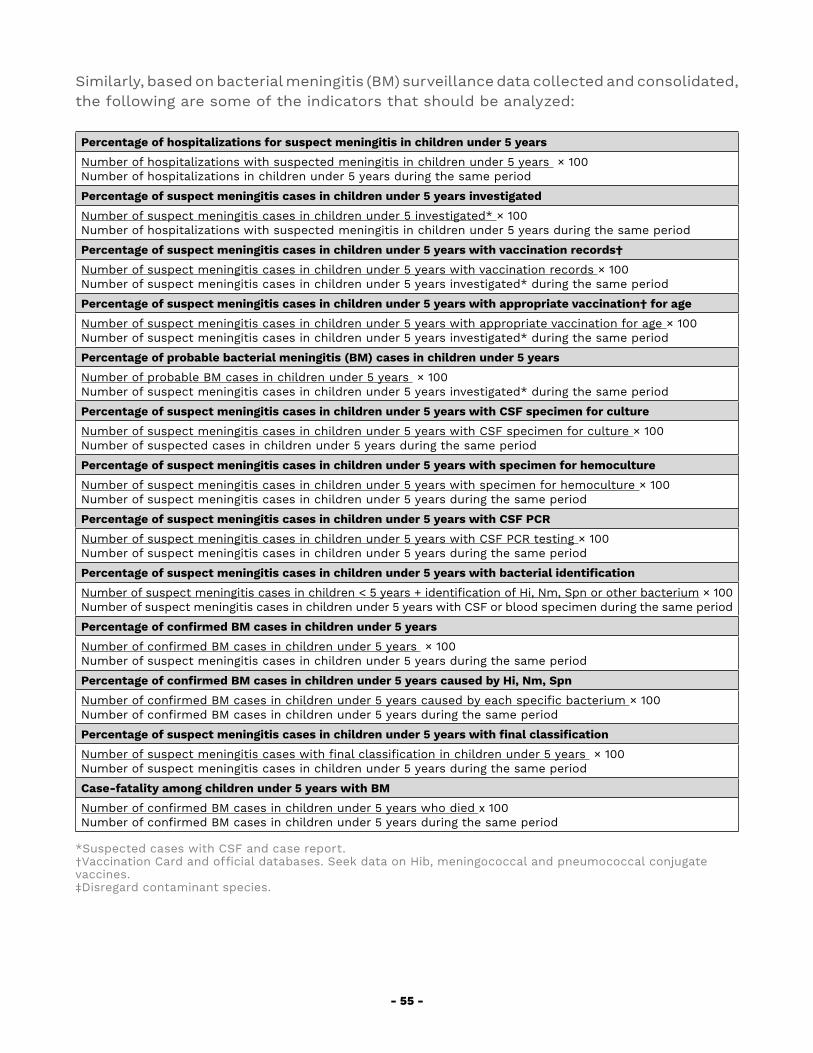
- 55 -
Similarly, based on bacterial meningitis (BM) surveillance data collected and consolidated, the following are some of the indicators that should be analyzed:
Percentage of hospitalizations for suspect meningitis in children under 5 years
Number of hospitalizations with suspected meningitis in children under 5 years × 100Number of hospitalizations in children under 5 years during the same period
Percentage of suspect meningitis cases in children under 5 years investigated
Number of suspect meningitis cases in children under 5 investigated* × 100Number of hospitalizations with suspected meningitis in children under 5 years during the same period
Percentage of suspect meningitis cases in children under 5 years with vaccination records†
Number of suspect meningitis cases in children under 5 years with vaccination records × 100Number of suspect meningitis cases in children under 5 years investigated* during the same period
Percentage of suspect meningitis cases in children under 5 years with appropriate vaccination† for age
Number of suspect meningitis cases in children under 5 years with appropriate vaccination for age × 100Number of suspect meningitis cases in children under 5 years investigated* during the same period
Percentage of probable bacterial meningitis (BM) cases in children under 5 years
Number of probable BM cases in children under 5 years × 100Number of suspect meningitis cases in children under 5 years investigated* during the same period
Percentage of suspect meningitis cases in children under 5 years with CSF specimen for culture
Number of suspect meningitis cases in children under 5 years with CSF specimen for culture × 100Number of suspected cases in children under 5 years during the same period
Percentage of suspect meningitis cases in children under 5 years with specimen for hemoculture
Number of suspect meningitis cases in children under 5 years with specimen for hemoculture × 100Number of suspect meningitis cases in children under 5 years during the same period
Percentage of suspect meningitis cases in children under 5 years with CSF PCR
Number of suspect meningitis cases in children under 5 years with CSF PCR testing × 100Number of suspect meningitis cases in children under 5 years during the same period
Percentage of suspect meningitis cases in children under 5 years with bacterial identification
Number of suspect meningitis cases in children < 5 years + identification of Hi, Nm, Spn or other bacterium × 100Number of suspect meningitis cases in children under 5 years with CSF or blood specimen during the same period
Percentage of confirmed BM cases in children under 5 years
Number of confirmed BM cases in children under 5 years × 100Number of suspect meningitis cases in children under 5 years during the same period
Percentage of confirmed BM cases in children under 5 years caused by Hi, Nm, Spn
Number of confirmed BM cases in children under 5 years caused by each specific bacterium × 100Number of confirmed BM cases in children under 5 years during the same period
Percentage of suspect meningitis cases in children under 5 years with final classification
Number of suspect meningitis cases with final classification in children under 5 years × 100Number of suspect meningitis cases in children under 5 years during the same period
Case-fatality among children under 5 years with BM
Number of confirmed BM cases in children under 5 years who died x 100Number of confirmed BM cases in children under 5 years during the same period
*Suspected cases with CSF and case report. †Vaccination Card and official databases. Seek data on Hib, meningococcal and pneumococcal conjugate vaccines.‡Disregard contaminant species.
- 56 -
The results at each sentinel hospital should then be analyzed and interpreted. Distribution by time, place, and person should be described for suspect, probable and confirmed cases of each disease, with tables, figures, and mapping of cases showing the following:
◆ Date of disease onset and hospital admission ◆ Seasonality patterns ◆ Patient’s age ◆ Place where the cases appeared ◆ Hib, meningococcal and pneumococcal conjugate vaccine status and
vaccination schedule by age ◆ Frequency of microorganisms (serogroups and serotypes) isolated and
antimicrobial susceptibility ◆ Case-fatality by age group and vaccination status.
With sentinel surveillance data, the burden of disease can be estimated and economic studies (cost-benefit, cost-effectiveness) conducted to evaluate the feasibility of introducing new vaccines.
The effectiveness of vaccination programs can be evaluated by analyzing the vaccination history and number of doses administered to patients. The impact of vaccines can be estimated by analyzing trends in hospitalized cases (suspect, probable and confirmed).
Analyzing the serotypes and serogroups of identified bacteria facilitates characterization of circulating strains and changes in these as the emerge.
Additional analyses may be conducted to meet the specific needs that a country may have.
5.6 Information Flow and Reporting Periodicity
When a suspected case is identified, a case report form must be initiated. The case should then be reported to the hospital’s epidemiology department which will be responsible for following up the case and registering individual data.
Each month, the hospital’s epidemiology department will submit to the epidemiology department at the next hierarchical level all individual data as well as supplementary data on the total monthly hospital admissions and total monthly hospitalized suspect pneumonia and meningitis cases. This information should be available at the national level during the first two weeks of the following month.
Data from all the country’s sentinel hospitals will be consolidated at the national level and reported to the PAHO on a quarterly basis, in accordance with schedules described in Table 14.
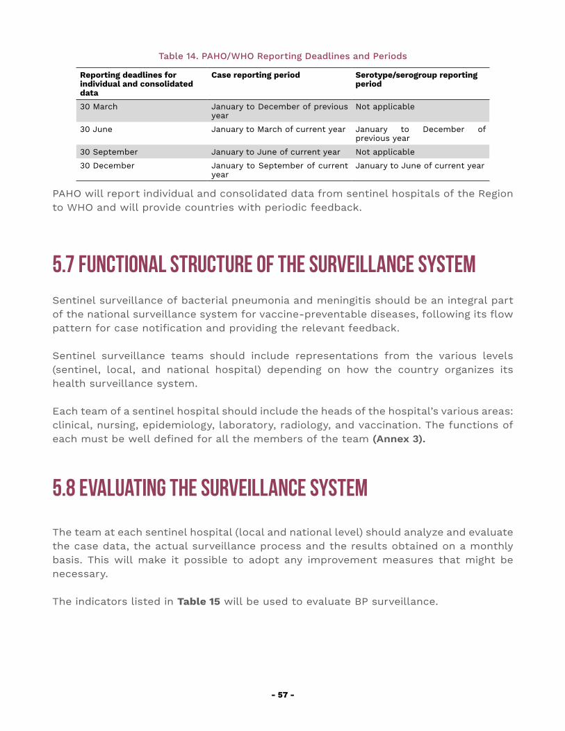
- 57 -
Table 14. PAHO/WHO Reporting Deadlines and Periods
Reporting deadlines for individual and consolidated data
Case reporting period Serotype/serogroup reporting period
30 March January to December of previous year
Not applicable
30 June January to March of current year January to December of previous year
30 September January to June of current year Not applicable
30 December January to September of current year
January to June of current year
PAHO will report individual and consolidated data from sentinel hospitals of the Region to WHO and will provide countries with periodic feedback.
5.7 Functional Structure of the Surveillance SystemSentinel surveillance of bacterial pneumonia and meningitis should be an integral part of the national surveillance system for vaccine-preventable diseases, following its flow pattern for case notification and providing the relevant feedback.
Sentinel surveillance teams should include representations from the various levels (sentinel, local, and national hospital) depending on how the country organizes its health surveillance system.
Each team of a sentinel hospital should include the heads of the hospital’s various areas: clinical, nursing, epidemiology, laboratory, radiology, and vaccination. The functions of each must be well defined for all the members of the team (Annex 3).
5.8 Evaluating the Surveillance System
The team at each sentinel hospital (local and national level) should analyze and evaluate the case data, the actual surveillance process and the results obtained on a monthly basis. This will make it possible to adopt any improvement measures that might be necessary.
The indicators listed in Table 15 will be used to evaluate BP surveillance.
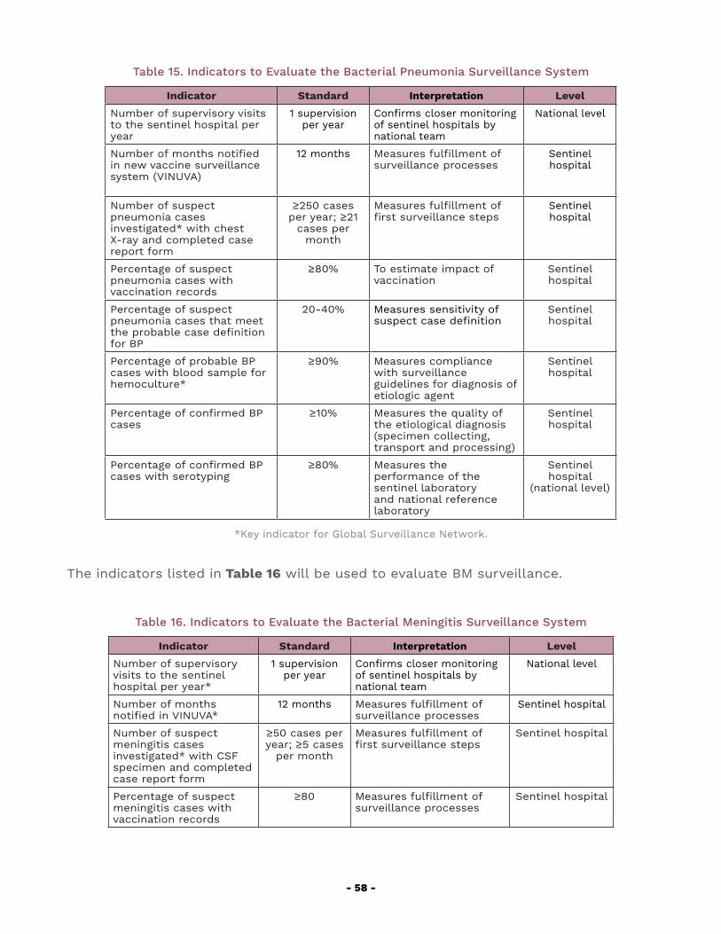
- 58 -
Table 15. Indicators to Evaluate the Bacterial Pneumonia Surveillance System
Indicator Standard Interpretation Level
Number of supervisory visits to the sentinel hospital per year
1 supervision per year
Confirms closer monitoring of sentinel hospitals by national team
National level
Number of months notified in new vaccine surveillance system (VINUVA)
12 months Measures fulfillment of surveillance processes
Sentinel hospital
Number of suspect pneumonia cases investigated* with chest X-ray and completed case report form
≥250 cases per year; ≥21
cases per month
Measures fulfillment of first surveillance steps
Sentinel hospital
Percentage of suspect pneumonia cases with vaccination records
≥80% To estimate impact of vaccination
Sentinel hospital
Percentage of suspect pneumonia cases that meet the probable case definition for BP
20-40% Measures sensitivity of suspect case definition
Sentinel hospital
Percentage of probable BP cases with blood sample for hemoculture*
≥90% Measures compliance with surveillance guidelines for diagnosis of etiologic agent
Sentinel hospital
Percentage of confirmed BP cases
≥10% Measures the quality of the etiological diagnosis (specimen collecting, transport and processing)
Sentinel hospital
Percentage of confirmed BP cases with serotyping
≥80% Measures the performance of the sentinel laboratory and national reference laboratory
Sentinel hospital
(national level)
*Key indicator for Global Surveillance Network.
The indicators listed in Table 16 will be used to evaluate BM surveillance.
Table 16. Indicators to Evaluate the Bacterial Meningitis Surveillance System
Indicator Standard Interpretation Level
Number of supervisory visits to the sentinel hospital per year*
1 supervision per year
Confirms closer monitoring of sentinel hospitals by national team
National level
Number of months notified in VINUVA*
12 months Measures fulfillment of surveillance processes
Sentinel hospital
Number of suspect meningitis cases investigated* with CSF specimen and completed case report form
≥50 cases per year; ≥5 cases
per month
Measures fulfillment of first surveillance steps
Sentinel hospital
Percentage of suspect meningitis cases with vaccination records
≥80 Measures fulfillment of surveillance processes
Sentinel hospital
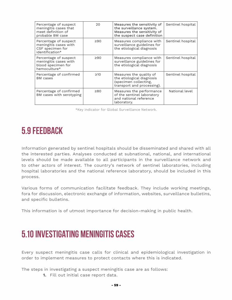
- 59 -
Percentage of suspect meningitis cases that meet definition of probable BM case
20 Measures the sensitivity of the surveillance system. Measures the sensitivity of the suspect case definition
Sentinel hospital
Percentage of suspect meningitis cases with CSF specimen for identification*
≥90 Measures compliance with surveillance guidelines for the etiological diagnosis
Sentinel hospital
Percentage of suspect meningitis cases with blood specimen for hemoculture*
≥90 Measures compliance with surveillance guidelines for the etiological diagnosis
Sentinel hospital
Percentage of confirmed BM cases
≥10 Measures the quality of the etiological diagnosis (specimen collecting, transport and processing).
Sentinel hospital
Percentage of confirmed BM cases with serotyping
≥80 Measures the performance of the sentinel laboratory and national reference laboratory.
National level
*Key indicator for Global Surveillance Network.
5.9 Feedback
Information generated by sentinel hospitals should be disseminated and shared with all the interested parties. Analyses conducted at subnational, national, and international levels should be made available to all participants in the surveillance network and to other actors of interest. The country’s network of sentinel laboratories, including hospital laboratories and the national reference laboratory, should be included in this process.
Various forms of communication facilitate feedback. They include working meetings, fora for discussion, electronic exchange of information, websites, surveillance bulletins, and specific bulletins.
This information is of utmost importance for decision-making in public health.
5.10 Investigating Meningitis Cases
Every suspect meningitis case calls for clinical and epidemiological investigation in order to implement measures to protect contacts where this is indicated.
The steps in investigating a suspect meningitis case are as follows:1. Fill out initial case report data.
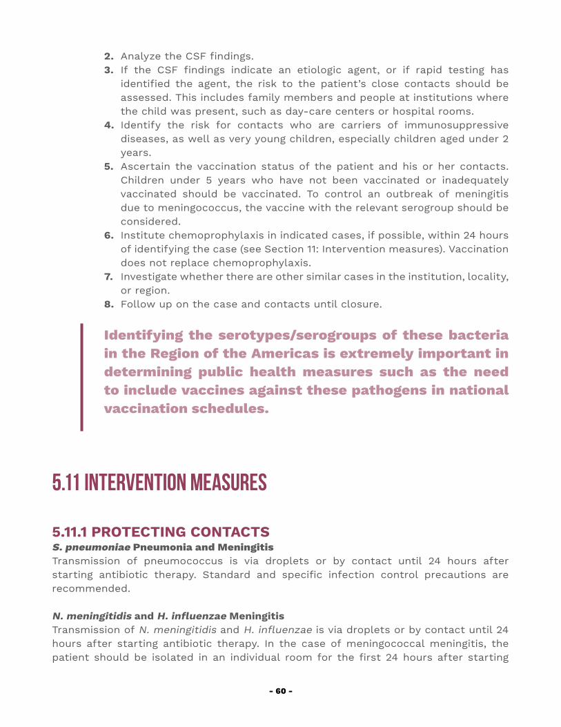
- 60 -
2. Analyze the CSF findings.3. If the CSF findings indicate an etiologic agent, or if rapid testing has
identified the agent, the risk to the patient’s close contacts should be assessed. This includes family members and people at institutions where the child was present, such as day-care centers or hospital rooms.
4. Identify the risk for contacts who are carriers of immunosuppressive diseases, as well as very young children, especially children aged under 2 years.
5. Ascertain the vaccination status of the patient and his or her contacts. Children under 5 years who have not been vaccinated or inadequately vaccinated should be vaccinated. To control an outbreak of meningitis due to meningococcus, the vaccine with the relevant serogroup should be considered.
6. Institute chemoprophylaxis in indicated cases, if possible, within 24 hours of identifying the case (see Section 11: Intervention measures). Vaccination does not replace chemoprophylaxis.
7. Investigate whether there are other similar cases in the institution, locality, or region.
8. Follow up on the case and contacts until closure.
Identifying the serotypes/serogroups of these bacteria in the Region of the Americas is extremely important in determining public health measures such as the need to include vaccines against these pathogens in national vaccination schedules.
5.11 Intervention Measures
5.11.1 PROTECTING CONTACTSS. pneumoniae Pneumonia and MeningitisTransmission of pneumococcus is via droplets or by contact until 24 hours after starting antibiotic therapy. Standard and specific infection control precautions are recommended.
N. meningitidis and H. influenzae MeningitisTransmission of N. meningitidis and H. influenzae is via droplets or by contact until 24 hours after starting antibiotic therapy. In the case of meningococcal meningitis, the patient should be isolated in an individual room for the first 24 hours after starting
- 61 -
antibiotic therapy in addition to other standard and specific infection control precautions. In case of meningitis caused by Hib, standard and specific infection control precautions are required. Thorough monitoring of home and other close contacts, with attention to early signs of disease, is indispensable so as to ensure timely treatment where necessary. For close contacts, chemoprophylaxis is also indicated.
ChemoprophylaxisFor cases of meningitis due to H. influenzae type b and N. meningitidis, prophylactic administration of an effective antimicrobial agent is advised in accordance with protocols established in each country.
VaccinationAll contacts should be vaccinated. Vaccination, however, does not rule out the need for chemoprophylaxis given the time lapse between vaccination and the development of an immune response.
The next chapter covers the vaccines that are available to prevent infections caused by H. influenzae, N. meningitidis and S. pneumoniae.
- 62 -
6. VACCINES
- 63 -
6. VACCINESThere are currently three different types of vaccines available for the treatment of bacterial pneumonia and meningitis: polysaccharide (pneumococcus and meningococcus), conjugate (Hib, meningococcus, and pneumococcus), and recombinant (meningococcus B) vaccines.
There are some major drawbacks to the use of polysaccharide vaccines: they do not induce an immune response in children aged under 2 years, they are not very effective in carriers, protection diminishes within a few years, and they do not generate an immunological memory response.
Conjugate vaccines are inactivated vaccines that have a transport protein attached or conjugated to the polysaccharide of the capsule of the bacterium (which is antigenically active). Various transport proteins are used, including the diphtheria toxoid, the tetanus toxoid, the outer membrane of the meningococcus, and a mutant Corynebacterium diphtheriae protein. This conjugation makes it possible for the immune system of children under 2 to identify the protein and leads to a good and lasting antibody seroconversion as they are capable of inducing secondary humoral response and immunologic memory. Furthermore, it has been shown that these vaccines produce group immunity (herd effect) by diminishing bacterial colonization of the respiratory tract among the vaccinated, thus reducing the transmission to third parties, including adults.
Currently available recombinant meningococcal B vaccines have been shown to produce a good immune response, including in children under one year of age.
6.1 Pneumococcal Vaccines
The 23-valent pneumococcal polysaccharide vaccine (PPV23) is used in many countries of the Region. However, as with other polysaccharide vaccines, it has its limitations.
Conjugate vaccines, which have a transport protein attached or conjugated to the polysaccharide of the capsule of the bacterium, have been shown to have a major impact in reducing invasive and non-invasive pneumococcal diseases.
At present, the majority of countries of the Region of the Americas has included one of the two available conjugate vaccines (PCV10 and PCV13) in their immunization program. The vaccination schedule should be selected based on the country’s epidemiological situation.
All pneumococcal conjugate vaccines contain capsular polysaccharides that are intended
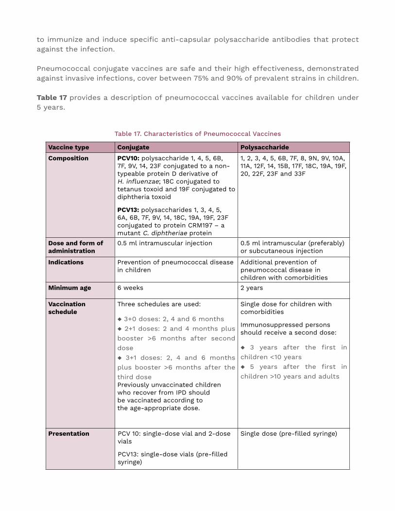
to immunize and induce specific anti-capsular polysaccharide antibodies that protect against the infection.
Pneumococcal conjugate vaccines are safe and their high effectiveness, demonstrated against invasive infections, cover between 75% and 90% of prevalent strains in children.
Table 17 provides a description of pneumococcal vaccines available for children under 5 years.
Table 17. Characteristics of Pneumococcal Vaccines
Vaccine type Conjugate Polysaccharide
Composition PCV10: polysaccharide 1, 4, 5, 6B, 7F, 9V, 14, 23F conjugated to a non-typeable protein D derivative of H. influenzae; 18C conjugated to tetanus toxoid and 19F conjugated to diphtheria toxoid
PCV13: polysaccharides 1, 3, 4, 5, 6A, 6B, 7F, 9V, 14, 18C, 19A, 19F, 23F conjugated to protein CRM197 – a mutant C. diphtheriae protein
1, 2, 3, 4, 5, 6B, 7F, 8, 9N, 9V, 10A, 11A, 12F, 14, 15B, 17F, 18C, 19A, 19F, 20, 22F, 23F and 33F
Dose and form of administration
0.5 ml intramuscular injection 0.5 ml intramuscular (preferably) or subcutaneous injection
Indications Prevention of pneumococcal disease in children
Additional prevention of pneumococcal disease in children with comorbidities
Minimum age 6 weeks 2 years
Vaccination schedule
Three schedules are used:
◆ 3+0 doses: 2, 4 and 6 months ◆ 2+1 doses: 2 and 4 months plus booster >6 months after second dose ◆ 3+1 doses: 2, 4 and 6 months plus booster >6 months after the third dose Previously unvaccinated children who recover from IPD should be vaccinated according to the age-appropriate dose.
Single dose for children with comorbidities
Immunosuppressed persons should receive a second dose:
◆ 3 years after the first in children <10 years ◆ 5 years after the first in children >10 years and adults
Presentation PCV 10: single-dose vial and 2-dose vials
PCV13: single-dose vials (pre-filled syringe)
Single dose (pre-filled syringe)
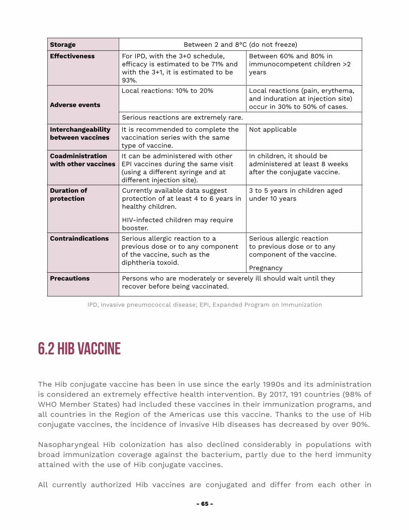
- 65 -
Storage Between 2 and 8°C (do not freeze)
Effectiveness For IPD, with the 3+0 schedule, efficacy is estimated to be 71% and with the 3+1, it is estimated to be 93%.
Between 60% and 80% in immunocompetent children >2 years
Adverse events
Local reactions: 10% to 20% Local reactions (pain, erythema, and induration at injection site) occur in 30% to 50% of cases.
Serious reactions are extremely rare.
Interchangeability between vaccines
It is recommended to complete the vaccination series with the same type of vaccine.
Not applicable
Coadministration with other vaccines
It can be administered with other EPI vaccines during the same visit (using a different syringe and at different injection site).
In children, it should be administered at least 8 weeks after the conjugate vaccine.
Duration of protection
Currently available data suggest protection of at least 4 to 6 years in healthy children.
HIV-infected children may require booster.
3 to 5 years in children aged under 10 years
Contraindications Serious allergic reaction to a previous dose or to any component of the vaccine, such as the diphtheria toxoid.
Serious allergic reaction to previous dose or to any component of the vaccine.
PregnancyPrecautions Persons who are moderately or severely ill should wait until they
recover before being vaccinated.
IPD, invasive pneumococcal disease; EPI, Expanded Program on Immunization
6.2 Hib Vaccine
The Hib conjugate vaccine has been in use since the early 1990s and its administration is considered an extremely effective health intervention. By 2017, 191 countries (98% of WHO Member States) had included these vaccines in their immunization programs, and all countries in the Region of the Americas use this vaccine. Thanks to the use of Hib conjugate vaccines, the incidence of invasive Hib diseases has decreased by over 90%.
Nasopharyngeal Hib colonization has also declined considerably in populations with broad immunization coverage against the bacterium, partly due to the herd immunity attained with the use of Hib conjugate vaccines.
All currently authorized Hib vaccines are conjugated and differ from each other in
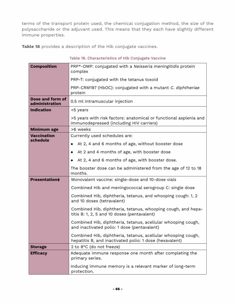
- 66 -
terms of the transport protein used, the chemical conjugation method, the size of the polysaccharide or the adjuvant used. This means that they each have slightly different immune properties.
Table 18 provides a description of the Hib conjugate vaccines.
Table 18. Characteristics of Hib Conjugate Vaccine
Composition PRP*-OMP: conjugated with a Neisseria meningitidis protein complex
PRP-T: conjugated with the tetanus toxoid
PRP-CRM197 (HbOC): conjugated with a mutant C. diphtheriae protein
Dose and form of administration 0.5 ml intramuscular injection
Indication <5 years
>5 years with risk factors: anatomical or functional asplenia and immunodepressed (including HIV carriers)
Minimum age >6 weeks Vaccination schedule
Currently used schedules are:
◆ At 2, 4 and 6 months of age, without booster dose
◆ At 2 and 4 months of age, with booster dose
◆ At 2, 4 and 6 months of age, with booster dose.
The booster dose can be administered from the age of 12 to 18 months.
Presentation‡
Monovalent vaccine: single-dose and 10-dose vials
Combined Hib and meningococcal serogroup C: single dose
Combined Hib, diphtheria, tetanus, and whooping cough: 1, 2 and 10 doses (tetravalent)
Combined Hib, diphtheria, tetanus, whooping cough, and hepa-titis B: 1, 2, 5 and 10 doses (pentavalent)
Combined Hib, diphtheria, tetanus, acellular whooping cough, and inactivated polio: 1 dose (pentavalent)
Combined Hib, diphtheria, tetanus, acellular whooping cough, hepatitis B, and inactivated polio: 1 dose (hexavalent)
Storage 2 to 8°C (do not freeze)Efficacy Adequate immune response one month after completing the
primary series.
Inducing immune memory is a relevant marker of long-term protection.
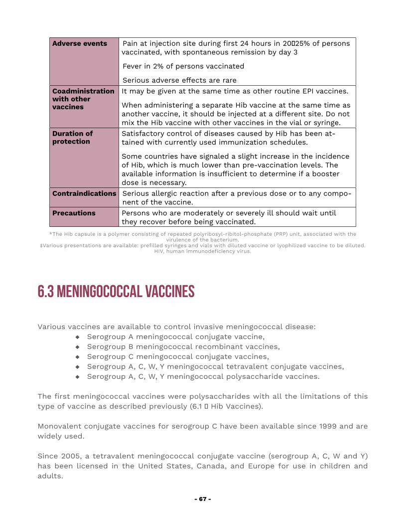
- 67 -
Adverse events Pain at injection site during first 24 hours in 20‒25% of persons vaccinated, with spontaneous remission by day 3
Fever in 2% of persons vaccinated
Serious adverse effects are rare Coadministration with other vaccines
It may be given at the same time as other routine EPI vaccines.
When administering a separate Hib vaccine at the same time as another vaccine, it should be injected at a different site. Do not mix the Hib vaccine with other vaccines in the vial or syringe.
Duration of protection
Satisfactory control of diseases caused by Hib has been at-tained with currently used immunization schedules.
Some countries have signaled a slight increase in the incidence of Hib, which is much lower than pre-vaccination levels. The available information is insufficient to determine if a booster dose is necessary.
Contraindications Serious allergic reaction after a previous dose or to any compo-nent of the vaccine.
Precautions Persons who are moderately or severely ill should wait until they recover before being vaccinated.
*The Hib capsule is a polymer consisting of repeated polyribosyl-ribitol-phosphate (PRP) unit, associated with the virulence of the bacterium.
‡Various presentations are available: prefilled syringes and vials with diluted vaccine or lyophilized vaccine to be diluted.HIV, human immunodeficiency virus.
6.3 Meningococcal Vaccines
Various vaccines are available to control invasive meningococcal disease: ◆ Serogroup A meningococcal conjugate vaccine, ◆ Serogroup B meningococcal recombinant vaccines, ◆ Serogroup C meningococcal conjugate vaccines, ◆ Serogroup A, C, W, Y meningococcal tetravalent conjugate vaccines, ◆ Serogroup A, C, W, Y meningococcal polysaccharide vaccines.
The first meningococcal vaccines were polysaccharides with all the limitations of this type of vaccine as described previously (6.1 ‒ Hib Vaccines).
Monovalent conjugate vaccines for serogroup C have been available since 1999 and are widely used.
Since 2005, a tetravalent meningococcal conjugate vaccine (serogroup A, C, W and Y) has been licensed in the United States, Canada, and Europe for use in children and adults.
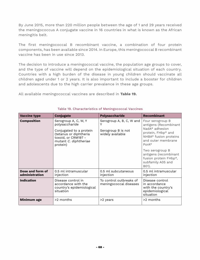
- 68 -
By June 2015, more than 220 million people between the age of 1 and 29 years received the meningococcus A conjugate vaccine in 16 countries in what is known as the African meningitis belt.
The first meningococcal B recombinant vaccine, a combination of four protein components, has been available since 2014. In Europe, this meningococcal B recombinant vaccine has been in use since 2013.
The decision to introduce a meningococcal vaccine, the population age groups to cover, and the type of vaccine will depend on the epidemiological situation of each country. Countries with a high burden of the disease in young children should vaccinate all children aged under 1 or 2 years. It is also important to include a booster for children and adolescents due to the high carrier prevalence in these age groups.
All available meningococcal vaccines are described in Table 19.
Table 19. Characteristics of Meningococcal Vaccines
Vaccine type Conjugate Polysaccharide RecombinantComposition Serogroup A, C, W, Y
polysaccharide
Conjugated to a protein (tetanus or diphtheria toxoid, or CRM197 - mutant C. diphtheriae protein)
Serogroup A, B, C, W and Y
Serogroup B is not widely available
Four serogroup B antigens (Recombinant NadA* adhesion protein, FHbp* and NHBA* fusion proteins and outer membrane PorA*
Two serogroup B antigens (recombinant fusion protein FHbp*, subfamily A05 and B01).
Dose and form of administration
0.5 ml intramuscular injection
0.5 ml subcutaneous injection
0.5 ml intramuscular injection
Indication Disease control in accordance with the country’s epidemiological situation
To control outbreaks of meningococcal diseases
Disease control in accordance with the country’s epidemiological situation
Minimum age >2 months >2 years >2 months

- 69 -
Vaccine type Conjugate Polysaccharide RecombinantPresentation Monovalent serogroup A:
10-dose vials
Monovalent serogroup C: single-dose prefilled syringe; single-dose vials
Combined Hib + serogroup C: single-dose vial and 10-dose vials
Tetravalent serogroups A, C, W and Y: single-dose vials
Monovalent serogroup B (limited availability)
Bivalent serogroups A and C: single-, 10- and 50-dose vials.
Trivalent serogroups A, C and W: single-, 10- and 50-dose vials
Tetravalent serogroups A, C, W, Y: 10-dose vials
Monovalent serogroup B: single-dose prefilled syringe
Vaccination schedule
Two doses from the age of 2 months (2 months apart, minimum one month); booster >1 year old
Adolescents and adults: a single dose.
Vaccine combined with Hib: 3 doses in children aged 2, 4, 6 months and booster at 12-15 months
Single dose 3 doses at 2-month intervals (minimum one) from the age of 2 months; booster >1 year old;
If first dose is given to children over 6 months old only 2 doses are required plus a booster when >1 year old;
In children >1 year, adolescents and adults, two doses at two-month intervals or, depending on the product, three doses with a two-month interval between the first and second dose and four months between the second and third.
Storage 2 to 8°C (do not freeze)
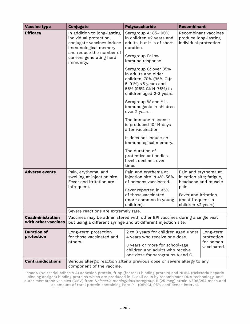
- 70 -
Vaccine type Conjugate Polysaccharide RecombinantEfficacy In addition to long-lasting
individual protection, conjugate vaccines induce immunological memory and reduce the number of carriers generating herd immunity.
Serogroup A: 85-100% in children >2 years and adults, but it is of short-duration.
Serogroup B: low immune response
Serogroup C: over 85% in adults and older children, 70% (95% CI‡: 5-91%) <5 years and 55% (95% CI:14-76%) in children aged 2-3 years.
Serogroup W and Y is immunogenic in children over 2 years.
The immune response is produced 10-14 days after vaccination.
It does not induce an immunological memory.
The duration of protective antibodies levels declines over time.
Recombinant vaccines produce long-lasting individual protection.
Adverse events
Pain, erythema, and swelling at injection site. Fever and irritation are infrequent.
Pain and erythema at injection site in 4%-56% of persons vaccinated.
Fever reported in <5% of those vaccinated (more common in young children).
Pain and erythema at injection site; fatigue, headache and muscle pain.
Fever and irritation (most frequent in children <2 years)
Severe reactions are extremely rare.Coadministration with other vaccines
Vaccines may be administered with other EPI vaccines during a single visit but using a different syringe and at different injection site.
Duration of protection
Long-term protection for those vaccinated and others.
2 to 3 years for children aged under 4 years who receive one dose.
3 years or more for school-age children and adults who receive one dose for serogroups A and C.
Long-term protection for person vaccinated.
Contraindications Serious allergic reaction after a previous dose or severe allergy to any component of the vaccine.
*NadA (Neisserial adhesin A) adhesion protein, fHbp (factor H binding protein) and NHBA (Neisseria heparin binding antigen) binding proteins which are produced in E. coli cells by recombinant DNA technology, and
outer membrane vesicles (OMV) from Neisseria meningitidis serogroup B (25 mcg) strain NZ98/254 measured as amount of total protein containing PorA P1. ‡95%CI, 95% confidence interval.
- 71 -
GLOSARYAir bronchogram: a radiolucent image (black) consisting of branching lines. It results from the contrast between the air content of a bronchus and the dense image produced by alveolar infiltrates surrounding it.
Alveolar infiltrate: dense pulmonary infiltrate of a homogeneous spongy or cottony appearance, indicating that there is fluid (pus, edema) in the alveolar air space.
Alveolus: small air spaces where the exchange of O2 and CO2 gases occurs.
Antiseptic: a substance applied to the skin and mucous membranes to inhibit microbial growth.
Atelectasis: loss of lung volume due to absorption of air in the lung distal to an obstruction of the airway (e.g. mucus plug). The pulmonary tissue collapses in a fan-like shape, and the X-ray shows a dense band, usually triangular with the vertex pointing towards the hilar area.
Automated hemoculture system: hemoculture bottles incubated in a system that automatically detects bacterial growth.
Bacteremia: temporary presence of bacteria in the blood.
Blind subculture: transfer of a blood culture to a 5% sheep blood and chocolate agar plate when there is NO macroscopic evidence of microbial growth. This is recommended for manual blood culture systems (not automated) in order to increase possible isolation of bacteria that undergo rapid autolysis as happens in the case of pneumococci.
Chlorhexidine-alcohol: a 2% solution of chlorhexidine in alcohol used for skin antisepsis.
Condensation: confluent alveolar infiltrate, dense, usually uniform or cottony in appearance, that compromises a complete pulmonary lobe, segment, or part of a segment, that usually contains air bronchograms and is accompanied by silhouette sign (see below). Sometimes associated with pleural effusion.
Contaminated hemoculture: the isolation of a microorganism introduced into the hemoculture bottle at the time of collecting the blood sample or during its processing, and not found in the blood of the patient.
Costophrenic angle: The angular space formed by the diaphragm and ribs on both sides of the thorax. When free of fluids, it can be seen in X-rays.
Hemoculture series: two to three temporally related blood cultures collected from
- 72 -
different venipunctures sites.
Hemoculture set: is the combination of bottles into which a single blood specimen collected for culture is inoculated. Sets may include one aerobic and one anaerobic bottle, or two aerobic bottles. In some cases, only one aerobic bottle (children weighing less than 13 kg) may be used.
Hemoculture: a blood sample obtained through venipuncture and inoculated in one or more bottles containing culture broth.
Hypoacusis: loss or reduction of hearing in one or both ears.
Inadequate blood volume: the amount of blood collected is less than 80% volume of blood recommended for a laboratory test.
Infiltrate: any pathological density of the lung fields appearing in the chest X-ray.
Interstice: pulmonary tissue located outside the alveoli and bronchia. It includes the supporting tissue, blood vessels, and lymph vessels.
Interstitial infiltrate: linear or reticular densities on X-ray corresponding to vascular and bronchial structures and reflecting processes involving the interstitial structures of the lung. It is usually diffuse giving the chest X-ray a “dirty lung” appearance.
Manual hemoculture system: hemoculture bottles incubated in an incubator with special mechanisms to detect bacterial growth, which must be inspected visually.
Nodding: movement accompanying inspiration that indicates the use of accessory muscles in cases of severe difficulty breathing.
Povidone-iodine: an aqueous iodine solution with polyvinylpyrrolidone applied topically to the skin as an antiseptic. It is available as a solution or ointment.
Septicemia or sepsis: infection of the blood flow, generally from a very large quantity of bacteria entering the bloodstream and not eliminated by the white blood cells. It is associated with severe clinical symptoms and can be fatal (septic shock).
Silhouette sign: blurring of an edge or normally well-defined contour. For example, the opacity that appears with pneumonia makes it difficult to see the silhouette of the lung, or the borders of neighboring structures such as the heart, diaphragm, etc.
Subculture: transfer of a blood culture, or culture medium with CSF, PF, or other specimens, from a sterile well to another plate (e.g. 5% sheep blood or chocolate agar plate) when macroscopic evidence shows microbial growth, or when a positivity alarm is given in the case of automated systems. Subcultures should always be carried out
- 73 -
together with Gram staining.
Tachypnea: accelerated respiratory rate, which in infants under 2 months is over 60 breaths/minute, in children between 2 and 11 months is over 50 breaths/minute, and in children from 12 months to 5 years over 40 breaths/minute.
Terminal subculture: transfer of a blood culture to a 5% sheep blood and chocolate agar plate at the end of the incubation period, before delivering a negative result. This is recommended for manual blood culture systems and unnecessary for automated systems.
Tincture of iodine: a 2% solution of iodine in alcohol and water used on the skin as an antiseptic.
- 74 -
BIBLIOGRAPHY
Alamadi YM, Aldeyab MA, McElnay J.C et al. Clinical and economic impact of contaminated blood cultures within the hospital setting. J Hosp Infect. 2011;77(3):233-236.
Baquero-Artigao F, Hernández-Sampelayo T, Navarro ML. Meningitis bacteriana. An Pediatr Contin. 2007;5:22-29.
Baraff LJ, Lee SI, Schriger DL. Outcomes of bacterial meningitis in children: a metaanalysis. Pediatr Infect Dis J. 1993;12:392.
Barenfanger J, Drake C, Lawhorn J, et al. Comparison of chlorhexidine and tinture of iodine for skin antisepsis in preparation for blood sample collection. J Clin Microbiol. 2004;42:2216-2217.
Baron EJ, Weinstein MP, Dunne WM, Yagupski P, Welch DF, Wilson DM. Cumitech 1C: Blood Cultures IV. Washington, DC: ASM Press; 2005.
Baron J. Transport clinical samples. In: Murray PR, ed. Manual of Clinical Microbiology 11th Ed. Washington DC: ASM; 2015.
Bourbeau P, Riley J, Heiter BJ et al. Use of the Bact Alert blood culture system for culture of sterile body fluid other than blood. J Clin Microbiol. 1998;36(11):3273-3277.
Centers for Disease Control and Prevention. Epidemiology and prevention of vaccinepreventable diseases. Hamborsky J, Kroger A, Wolfe S (eds.) 13th ed. Washington D.C. Public Health Foundation; 2015.
Clinical and Laboratory Standards Institute. 2006. Body Fluid Analysis for Cellular Composition: Approved Guideline. CLSI document H56-A. Wayne, PA: CLSI; 2006.
Clinical and Laboratory Standards Institute. 2007. Principles and Procedures for Blood Cultures; Approved Guideline 6 Ed. Wayne, PA: CLSI document M47-A; 2007.
Clinical and Laboratory Standards Institute. 2012. Collection, Transport, Preparation, and Storage of Specimen for Molecular Methods. MM13-A. 2012;25(31).
Cockerill FR III, Wilson JW, Vetter EA et al. Optimal testing parameters for blood cultures. Clin Infect Dis 2004; 38:1724-1730.
de Cueto M, Pascual A. El hemocultivo pediátrico, indicaciones y técnica. An Pediatr Contin. 2007;5(5):279-282.
de Oliveira LH, Camacho LA, Coutinho ES, Martinez-Silveira MS, Carvalho AF, Ruiz-Matus
- 75 -
C et al. Impact and effectiveness of 10 and 13-valent pneumococcal conjugate vaccines on hospitalization and mortality in children aged less than 5 years in Latin American countries: a systematic review. PLoS One. 2016;11(12):e0166736.
de Oliveira LH, Trumbo SP, Ruiz Matus C, Sanwogou NJ, Toscano CM. Pneumococcal conjugate vaccine introduction in Latin America and the Caribbean: progress and lessons learned. Expert Rev Vaccines. 2016;15(10):1295-1304.
Deiros L, Baquero- Artigao F, Garcia-Miguel N. Derrame pleural paraneumónico: revisión de 11 años. An Pediatr. 2006;64:40-45.
Falguera M, López A, Nogues JM et al. Evaluation of the polymerase chain reaction method for detection of Streptococcus pneumoniae DNA in pleural fluid samples. Chest. 2002;122:2212-2216.
Gray J, Gossain S, Morris K. Three years survey of bacteremia and fungemia in a pediatric intensive care unit. Pediatr Infect Dis J. 2001;20:416-421.
Hall KK, Lyman JA. Updated review of blood culture contamination. Clin Microbiol Rev. 2006; 19:788-802.
Heymann DL (ed.). El control de las enfermedades transmisibles. 20ª ed. Washington, D.C.: OPS; 2017.
Isaacman DJ, Karasic RB, Reynolds LA et al. Effect of number of blood cultures and volume of blood on detection of bacteremia in children. J Pediatr. 1996;128:190-195.
Kellog JA, Manzella JP, Bankert DA. Frecuency of low-level bacteremia in children from birth to fifteen years of age. J Clin Microbiol. 2000;38:2181-2185.
Kellogg JA, Ferrentino FL, Goodstein MH et al. Frequency of low-level bacteremia in infants from birth to two months of age. Pediatr Infect Dis J. 1997;16:381-385.
Kellogg JA, Ferrentino FL, Liss J, et al. Justification and implementation of a policy requiring two blood cultures when one is ordered. Lab Med. 1994;25:323-330.
Laboratory Methods for the Diagnosis of Meningitis Caused by Neisseria meningitidis, Streptococcus pneumoniae, and Haemophilus influenzae. WHO Manual, Second Edition, 2011.
Li J, Plorde J, Carlson L. Effects of volume and periodicity on blood cultures. J Clin Microbiol. 1994;32:2829-2831.
Little JR, Murray PR, Traynor PS et al. A randomized trial of povidone-iodine compared with iodine tincture for venipuncture site disinfection: effect on rates of blood culture
- 76 -
contamination. Am J Med. 1999;107:119-125.
Maiwald M, Chan ES. The forgotten role of alcohol: a systematic review and metaanalysis of the clinical efficacy and perceived role of chlorhexidine in skin antisepsis. PLoS One. 2012;7(9):e44277.
Mayoral C, Norona MR, Baroni R et al. Evaluation of a nested –PCR assay for Streptococcus pneumoniae detection in pediatric patients with community-acquired pneumonia. Rev Argent Microbiol. 2005;37:184-188.
McGowan KL, Foster JA, Coffin SE. Outpatient pediatric blood cultures: time to positivity. Pediatrics. 2000;106:251-255.
Menzies SM, Rahman N, Wrightson J et al. Blood culture bottle culture of pleural fluid in pleural infection. Thorax. 2011;66:658-662.
Mermel LA, Allon M, Bouza E et al. Clinical practice guidelines for the diagnosis and management of intravascular catheter related infection. 2009 update by the infectious Diseases Society of America. Clin Infect Dis. 2009;49:1-45.
Mimoz O, Karim A, Mercat A et al. Chlorhexidine compared with povidone-iodine as skin preparation before blood culture: a randomized controlled trial. Ann Intern Med. 1999; 131:834-837.
Ostensen H, Pettersson H, eds. The WHO manual of diagnostic imaging: radiographic anatomy and interpretation of the chest and the pulmonary system. Singapore: World Health Organization in collaboration with International Radiology Society: 2006. Available at: https://apps.who.int/iris/bitstream/handle/10665/43293/9241546778_eng.pdf?sequence=1 [consulted 22 August 2019].
Pan American Health Organization. Epidemiological Surveillance of Healthcare-Associated Infections. Washington, D.C.: PAHO, 2010. Available at: https://www.paho.org/hq/dmdocuments/2011/ENG_Modulo_I_final.pdf.
Pan American Health Organization. Diagnóstico y tratamiento de enfermedades prevalentes graves de la infancia (0-5 años). 2. Ed. Washington D.C.: PAHO/PALTEX; 2004.
Pan American Health Organization. Final report of the XXIII TAG Meeting, held in Varadero, Cuba, 1-3 July 2015. Available at: https://www.paho.org/hq/index.php?option=com_content&view=article&id=1862:reports-technical-advisory-group-vaccine-preventable-diseases&Itemid=39430&lang=en [consulted: 21 August 2019].
Pan American Health Organization. Informe Regional de SIREVA II, 2013: Datos por país y por grupos de edad sobre las características de los aislamientos de Streptococcus pneumoniae, Haemophilus influenzae y Neisseria meningitidis, en procesos invasivos
- 77 -
bacterianos. Washington, D.C.: PAHO; 2016.
Pan American Health Organization. Informe Regional de SIREVA II: Datos por país y por grupos de edad sobre las características de los aislamientos de Streptococcus pneumoniae, Haemophilus influenzae y Neisseria meningitidis, en procesos invasores, 2000-2005. Washington, D.C.: PAHO; 2007.
Pan American Health Organization. Procedimientos para el diagnóstico de Neumonías y Meningitis Bacterianas y la caracterización de cepas de Streptococcus pneumoniae y Haemophilus influenzae, SIREVA II Grupo Microbiologia, Instituto Nacional de Salud Bogotá – Colombia, 2012
Pitetti RD, Choi S. Utility of blood cultures in febrile children with UTI. Am Journal Emerg Med. 2002;20:271-274.
Plotkin SA, Orenstein WA, Offit PA, eds. Vaccines. 7th ed. Philadelphia: Elsevier: 2017. Proehl JA, Leviner S, Bradford JY, Storer A, et al. Clinical practice guideline: prevention of blood culture contamination. Emergency Nurses Association. 2012:1-12.
Reimer LG. Procedures for the storage for microorganisms. In: Murray PR, Baron EJ, Jorgensen JH et al. Manual of Clinical Microbiology (8th ed), Washington DC: ASM Press; 2003:67-73.
Samra Z, Shmuely H, Nahum E, et al. Use of the NOW Streptococcus pneumoniae urinary antigen test in cerebrospinal fluid for rapid diagnosis of pneumococcal meningitis. Diagn Microbiol Infect Dis. 2003;45(4):237-240.
Schelonka RL, Chai MK, Yoder BA, et al. Volume of blood required to detect common neonatal pathogens. J. Pediatr. 1996 ;129 :275-278.
Sevjar JJ, Johnson D, Popovic T, et al. Assessing the risk of laboratory –acquired meningococcal disease. J Clin Microbiol. 2005;43:4811-4813.
Stonecypher K. Going around in circles: Is this the best practice for preparing the skin? Crit Care Nurse. 2009;32:94-98.
Thomson RB, Miller M. Specimen collection, transport and processing: bacteriology. In: Murray PR, Barón EJ, Jorgensen JH, Pfaller MA, Yolken RH, editores. Manual of Clinical Microbiology. 8th ed. Washington DC.: American Society for Microbiology; 2003:286-331.
Wahl B, O’Brian KL, Greenbaum A et al. Burden of Streptococcus pneumoniae and Haemophilus influenza type b disease in the era of conjugate vaccines: global, regional, and national estimates for 200-2015. The Lancet 2018; 6: e744-e757.
Walker CL, Rudan I, Liu L, Nair H, Theodoratou E, Bhutta ZA, et al. Global burden of
- 78 -
childhood pneumonia and diarrhoea. The Lancet 2013;381(9875):1405-1416.
Wiswell TE, Hachey WE. Multiple site blood cultures in the initial evaluation for neonatal sepsis during the first week of life. Pediatr Infect Dis J. 1991;10:365-369.
World Health Organization. Changing epidemiology of pneumococcal serotypes after introduction of conjugate vaccine: WER. 2010;85:425-436.
World Health Organization. Haemophilus influenzae type b (Hib) Vaccination Position Paper, July 2013. WER. 2013;88(39):413-426.
World Health Organization. Meningococcal vaccines: WHO position paper, November 2011. WER. 2011;86(47):521-539.
World Health Organization. Pneumococcal vaccines WHO position paper, February 2019. WER. 2019; 94(8):85-104.
World Health Organization. Vaccine-Preventable Diseases Surveillance Standards. Haemophilus influenzae. Last updated: September 5, 2018. [consulted: 21 August 2019].
World Health Organization. Vaccine-Preventable Diseases Surveillance Standards. Meningococcus. Last updated: 5 September 2018. [consulted: 21 August 2019].
World Health Organization. Vaccine-Preventable Diseases Surveillance Standards. Pneumococcus. Last updated: 5 September 2018. [consulted: 21 August 2019].
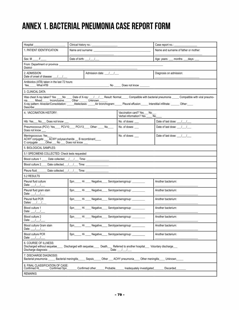
- 79 -
ANNEX 1. BACTERIAL PNEUMONIA CASE REPORT FORM
Hospital ______________________ Clinical history no.: __________________ Case report no.: ________________1. PATIENT IDENTIFICATION Name and surname: ________________________________________
_______________________________Name and surname of father or mother:_________________________________
Sex: M ____ F____ Date of birth: ___/___/___ Age: years: ___ months: ___days: ___From: Department or province _________________________________________________________________________ District: _________________________________________________________________________2. ADMISSION Date of onset of disease: ___/___/___
Admission date: ___/___/___ Diagnosis on admission: _______________________________
Antibiotics (ATB) taken in the last 72 hours: Yes_____ What ATB: __________________________________________ No _____ Does not know _______
3. CLINICAL DATAWas chest X-ray taken? Yes ____No ____ Date of X-ray: ___/___/___ Result: Normal_____ Compatible with bacterial pneumonia _____ Compatible with viral pneumo-nia_____ Mixed_____ Inconclusive_____ Other ______ Unknown______ X-ray pattern: Alveolar/Consolidation: _____Atelectasis: _____Air bronchogram:_____ Pleural effusion:_____ Interstitial infiltrate: ______ Other:____ Describe:____________________________________________4. VACCINATION HISTORY: Vaccination card? Yes___ No___
Verbal information? Yes ____ No____Hib: Yes___ No___ Does not know ___ No. of doses: ___ Date of last dose: ___/___/___ Pneumococcus (PCV): Yes____ PCV10____ PCV13____ Other: ____ No____ Does not know ___
No. of doses: ___ Date of last dose: ___/___/___
Meningococcus: Yes___ ACWY conjugate ___ ACWY polysaccharide __ B recombinant____ C conjugate _____Other___ No ___ Does not know ___
No. of doses: ___ Date of last dose: ___/___/___
5. BIOLOGICAL SAMPLES:
5.1 SPECIMENS COLLECTED: Check tests requestedBlood culture 1 ____ Date collected___/___/___ Time: ________________Blood culture 2____ Date collected___/___/___ Time: ________________
Pleura fluid_______ Date collected___/___/___ Time: ________________5.2 RESULTSPleural fluid cultureDate: ___/___/___
Spn_____ Hi ____ Negative____ Serotype/serogroup: _________ Another bacterium:______________________________
Pleural fluid gram stainDate: ___/___/___
Spn_____ Hi ____ Negative____ Serotype/serogroup: _________ Another bacterium:______________________________
Pleural fluid PCRDates: ___/___/___
Spn_____ Hi ____ Negative____ Serotype/serogroup: _________ Another bacterium:______________________________
Blood culture 1 Date: ___/___/___
Spn_____ Hi ____ Negative____ Serotype/serogroup: _________ Another bacterium:______________________________
Blood culture 2 Date: ___/___/___
Spn_____ Hi ____ Negative____ Serotype/serogroup: _________ Another bacterium:______________________________
Blood culture Gram stain Date: ___/___/___
Spn_____ Hi ____ Negative____ Serotype/serogroup: _________ Another bacterium:______________________________
Blood culture PCR Date: ___/___/___
Spn_____ Hi ____ Negative____ Serotype/serogroup: _________ Another bacterium:______________________________
6. COURSE OF ILLNESS:Discharged without sequelae____ Discharged with sequelae____ Death___ Referred to another hospital___ Voluntary discharge___ Discharge diagnosis: ___________________________________________ Date: ___/___/___
7. DISCHARGE DIAGNOSIS:Bacterial pneumonia _____ Bacterial meningitis_____ Sepsis_____ Other ___ ACHY pneumonia____ Other meningitis____ Unknown_____
8. FINAL CLASSIFICATION OF CASE: Confirmed Hi_______ Confirmed Spn_______ Confirmed other______ Probable______ Inadequately investigated_______ Discarded_______REMARKS
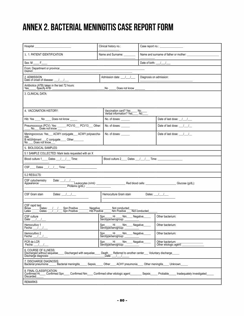
- 80 -
ANNEX 2. BACTERIAL MENINGITIS CASE REPORT FORM
Hospital _________________________ Clinical history no.: ______________
Case report no.: ________________
1. 1. PATIENT IDENTIFICATION Name and Surname: ________ _________________________
Name and surname of father or mother: ______________________ __________
Sex: M ____ F____ Date of birth: ___/___/___From: Department or province:__________________________________________________________ District:____________________________________________________________________
2. ADMISSION Date of onset of disease: ___/___/___
Admission date: ___/___/___ Diagnosis on admission: ____________________________________
Antibiotics (ATB) taken in the last 72 hours: Yes_____ Specify ATB: ____________________________________No _____ Does not know _______3. CLINICAL DATA:
4. VACCINATION HISTORY: Vaccination card? Yes ____ No____Verbal information? Yes____ No____
Hib: Yes ____ No ____ Does not know _____ No. of doses: ______ Date of last dose: __/___/___
Pneumococcus (PCV): Yes: ______ PCV10___ PCV13___ Other: ____ No___ Does not know ___
No. of doses: ______ Date of last dose: ___/___/__
Meningococcus: Yes___ ACWY conjugate___ ACWY polysaccha-ride___ B recombinant ___C conjugate____ Other_______No ___ Does not know___
No. of doses: ______ Date of last dose: ___/___/__
5. BIOLOGICAL SAMPLES:5.1 SAMPLE COLLECTED: Mark tests requested with an XBlood culture 1____ Dates ___/___/___ Time: _________________
Blood culture 2____ Dates ___/___/___ Time: _________________
CSF____ Dates ___/___/___ Time: _____________________
5.2 RESULTS:CSF cytochemistry Date: ___/___/___Appearance: _______________________ Leukocytes (n/ml): ____________________ Red blood cells: _____________________ Glucose (g/dL): _____________________________ Proteins (g/dL): _________________
CSF Gram stain Dates: ___/___/_______________________________________________
Hemoculture Gram stain Dates: ___/___/_____________________________________________________
CSF rapid test Binax _____ Dates: ___/___/___ Spn Positive _______ Negative________ Not conducted______Latex _____ Dates: ___/___/___ Spn Positive _______ Hib Positive _____ Nm Positive ___ Not conducted______ CSF cultureDate: ___/___/___
Spn_____ Hi ____ Nm____ Negative_____ Serotype/serogroup: __________
Other bacterium:
Hemocultivo 1Fecha: ___/___/___
Spn_____ Hi ____ Nm____ Negative_____ Serotype/serogroup: __________
Other bacterium:
Hemocultivo 2Fecha: ___/___/___
Spn_____ Hi ____ Nm____ Negative_____ Serotype/serogroup: __________
Other bacterium:
PCR de LCR Fecha: ___/___/___
Spn_____ Hi ____ Nm____ Negative_____ Serotype/serogroup: __________
Other bacterium: __________________Other etiologic agent: _______________
6. COURSE OF ILLNESS:Discharged without sequelae____ Discharged with sequelae____ Death___ Referred to another center___ Voluntary discharge_____ Discharge diagnosis: ______________________________________Date: ___/___/_____7. DISCHARGE DIAGNOSIS:Bacterial pneumonia _____ Bacterial meningitis_____ Sepsis_____ Other____ ACHY pneumonia____ Other meningitis____ Unknown_____
8. FINAL CLASSIFICATION:Confirmed Hi____ Confirmed Spn____ Confirmed Nm____ Confirmed other etiologic agent________ Sepsis_____ Probable_____ Inadequately investigated_____ Discarded_____REMARKS
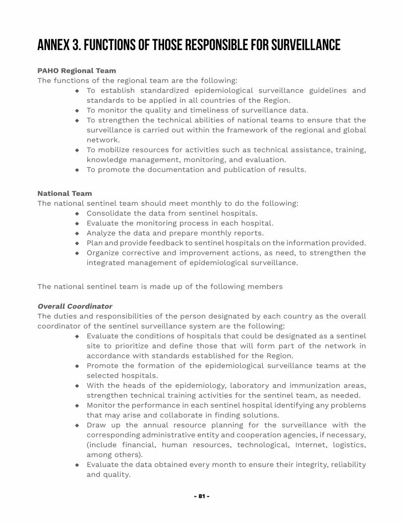
- 81 -
ANNEX 3. FUNCTIONS OF THOSE RESPONSIBLE FOR SURVEILLANCE
PAHO Regional Team The functions of the regional team are the following:
◆ To establish standardized epidemiological surveillance guidelines and standards to be applied in all countries of the Region.
◆ To monitor the quality and timeliness of surveillance data. ◆ To strengthen the technical abilities of national teams to ensure that the
surveillance is carried out within the framework of the regional and global network.
◆ To mobilize resources for activities such as technical assistance, training, knowledge management, monitoring, and evaluation.
◆ To promote the documentation and publication of results.
National TeamThe national sentinel team should meet monthly to do the following:
◆ Consolidate the data from sentinel hospitals. ◆ Evaluate the monitoring process in each hospital. ◆ Analyze the data and prepare monthly reports. ◆ Plan and provide feedback to sentinel hospitals on the information provided. ◆ Organize corrective and improvement actions, as need, to strengthen the
integrated management of epidemiological surveillance.
The national sentinel team is made up of the following members
Overall CoordinatorThe duties and responsibilities of the person designated by each country as the overall coordinator of the sentinel surveillance system are the following:
◆ Evaluate the conditions of hospitals that could be designated as a sentinel site to prioritize and define those that will form part of the network in accordance with standards established for the Region.
◆ Promote the formation of the epidemiological surveillance teams at the selected hospitals.
◆ With the heads of the epidemiology, laboratory and immunization areas, strengthen technical training activities for the sentinel team, as needed.
◆ Monitor the performance in each sentinel hospital identifying any problems that may arise and collaborate in finding solutions.
◆ Draw up the annual resource planning for the surveillance with the corresponding administrative entity and cooperation agencies, if necessary, (include financial, human resources, technological, Internet, logistics, among others).
◆ Evaluate the data obtained every month to ensure their integrity, reliability and quality.
- 82 -
◆ Prepare the national report in collaboration with staff responsible for epidemiology, the laboratory and the immunization program.
◆ Disseminate the report widely to the various areas of the Ministry of health, related entities, and sentinel hospitals.
◆ Promote decision-making based on epidemiologic surveillance findings. ◆ Disseminate the information periodically through the regional and global
network.
Epidemiology CoordinatorThe epidemiology coordinator will perform the following functions:
◆ Monitor the resources available to ensure uninterrupted sentinel surveillance.
◆ Supervise the implementation of surveillance standards. ◆ With the national coordinator, promote training and awareness activities
to advance the technical abilities of sentinel hospital teams, as needed. ◆ Monitor and evaluate the quality of the data on a permanent basis. ◆ Coordinate and ensure that the country’s sentinel hospital network receives
proper and data on laboratory findings, vaccines, hospital statistics and any other information that may be required for the surveillance.
◆ Ensure the consolidation and analysis of information generated in all sentinel hospitals in the country.
◆ With the overall coordinator and the laboratory, epidemiological surveillance and immunization teams, timely prepare and send the national report.
National Reference Laboratory
◆ Be the national reference for the laboratory diagnosis of the diseases under epidemiological surveillance.
◆ Together with the epidemiologist, make sure that laboratory supplies are continually available to ensure uninterrupted surveillance.
◆ Evaluate the data obtained together with the other surveillance coordinators.
◆ With the epidemiology coordinator, promote national training and awareness activities to increase the technical skills of sentinel hospital teams, as needed.
◆ Act in coordination with sentinel hospital laboratories to ensure that isolated strains are properly forwarded to the next level.
◆ Perform quality control for sentinel hospital laboratories that process specimens.
◆ Perform serotyping of cultured strains and determine the minimum inhibitory concentrations (MICs).
◆ Report the results of these tests to the sentinel laboratory as soon as they are available and within the defined times.
◆ Enter the laboratory results in the IT system of the sentinel surveillance network.
◆ Participate in the preparation of the monthly report.

- 83 -
◆ Confirmation, storage, and maintenance of bacterial isolates sent from sentinel laboratories (SL).
◆ Conduct antimicrobial susceptibility tests. ◆ Characterize the isolates by analyzing the serogroup and serotype. ◆ Perform PCR tests for the identification of S. pneumoniae, H. influenzae,
and N. meningitidis in CSF and PF specimens sent from SL. ◆ Send the results promptly to the SL coordinator and to other members of
the health team. ◆ Train, supervise and evaluate the performance of the SL. ◆ Send to the Regional Reference Laboratory (RRL) all S. pneumoniae,
H. influenzae, and N. meningitidis strains pending serotyping or with incomplete serotyping.
◆ Participate in a “Direct External Quality Control Program” (panel). ◆ Send strains and CSF/PF specimens to the RRL for indirect quality control
(evaluate level of concordance). ◆ Fulfill specific performance indicators for laboratories participating in
sentinel surveillance. ◆ Report the results to the country’s Ministry of Health and PAHO Office
according to the periodicity and formats established for sentinel surveillance.
◆ Receive training and educational supervisory visits on technical subjects related to sentinel surveillance.
SENTINEL HOSPITAL TEAMEach hospital team should include the person in charge of the hospital’s clinical and nursing departments, the head of the local laboratory, the person in charge of vaccination, an epidemiologist in charge of the information, and, where possible, a radiologist. It should also have a coordinator (it is suggested that the epidemiologist assume this role).
Each hospital team should meet monthly to analyze the cases identified by the surveillance, and to do the following:
◆ Discuss the weaknesses and progress of the surveillance process. ◆ Answer questions. ◆ Propose adjustments. ◆ Evaluate data and make decisions on action. ◆ Prepare the monthly report, deliver it on time, and ensure its dissemination. ◆ Program corrective and improvement action, as needed, to strengthen the
integrated management of the surveillance.
The sentinel hospital team includes the following members:
Clinical CoordinatorThe person in charge of the clinical area will perform the following functions:
◆ Ensure identification of cases based on established surveillance standards.
- 84 -
◆ With the nursing coordinator, ensure proper and timely data collection in the emergency room, inpatient areas or other departments.
◆ Supervise the participation of clinical personnel. ◆ Follow up on the cases identified up to time of discharge, complete the
case report form, and enter in database. ◆ Collaborate in the data analysis and in preparing the monthly report. ◆ Work with the epidemiology, laboratory, radiology, and nursing coordinators
to train the various shifts of the hospital team participating in sentinel surveillance.
Nursing CoordinatorThe nursing coordinator will perform the following functions:
◆ Together with the clinical coordinator, follow up on cases identified. ◆ Ensure proper and timely collection of laboratory specimens and the
taking of chest X-rays and other medical tests required to guarantee correct functioning of the surveillance activity. This also includes seeking information on patient’s vaccination status according to his or her vaccination card, as far as possible.
◆ With the clinical coordinator, ensure that data collected in the emergency room, inpatient areas or other departments are obtained in a timely manner to complete the case report forms.
◆ Follow up on the cases identified, help compile and enter the information needed to complete the case report and database.
◆ Work with clinical, epidemiology, laboratory and radiology coordinators to train the various shifts of the hospital team participating in sentinel surveillance.
◆ Supervise the participation of nursing personnel, including substitutes or students/internists on shift work, to ensure compliance with the required sentinel surveillance procedures.
◆ Collaborate in the data analysis and management and in preparing the monthly report.
Epidemiology CoordinatorThe epidemiology coordinator will perform the following functions:
◆ Supervise the implementation of sentinel surveillance standards. ◆ Coordinate the activities of the sentinel team. ◆ Work with clinical, laboratory, radiology, nursing, and vaccination
coordinators to train the various shifts of the hospital team participating in surveillance.
◆ Identify cases in the emergency room, inpatient areas or other departments that are eligible cases for sentinel surveillance.
◆ Collect the information generated in the clinical (hospital records), vaccination and laboratory areas to complete the data in the case report form and enter data in a database.
◆ Implement data quality monitoring strategies and provide immediate
- 85 -
feedback to those generating the data. ◆ Permanently monitor rotation of staff involved in surveillance activities
and ensure timely induction and training activities for all new personnel. ◆ On the first day of every month, consolidate the case data entered on the
IT system and confirm its completeness and quality. ◆ Conduct monthly data analyses, including an assessment of the surveillance
indicators. ◆ Send the surveillance data up the hierarchical chain (local-regional-
national) according to the periodicity established in the country using the specific databases of this surveillance.
◆ With the clinical, nursing, laboratory and vaccination coordinators, prepare a monthly report.
◆ Send the report to: ◇ The hospital director ◇ The hospital’s technical team (clinical, nursing, laboratory and vaccination) ◇ The overall coordinator at the next hierarchical level of the bacterial pneumonia
and meningitis epidemiological surveillance system.
Sentinel Hospital Laboratory CoordinatorThe duties and responsibilities of the laboratory coordinator are the following:
◆ Ensure sufficient resources for the correct functioning of the laboratory in terms of surveillance activities throughout the year.
◆ Work with clinical, radiology, nursing, epidemiology and vaccination coordinators to train the various shifts of the hospital team participating in surveillance.
◆ Receive and store blood, pleural fluid and CSF specimens in accordance with established surveillance standards. Ensure proper storage of an aliquot of each of the specimens that were processed.
◆ Confirm that all data on the laboratory specimens of identified cases are complete and meet the standards required for timely transportation and processing.
◆ Perform diagnostic tests on a timely basis and report test findings to the clinical and epidemiology areas. Enter laboratory data in the surveillance system.
◆ Whenever a Hi, meningococcus, or pneumococcus strain is isolated, send the positive strains to the national reference laboratory for serotyping and determination of the MIC.
◆ Ensure proper and timely transport of isolated strains to the reference laboratory.
◆ Collaborate in the data analysis and in preparing the monthly surveillance report.
◆ With the laboratory team, collaborate in the activities defined to evaluate performance.
◆ Process hemoculture, CSF, and PF specimens of probable bacterial pneumonia and suspect meningitis cases in children under 5 years.
◆ If the laboratory does not have serotyping facilities, send all S. pneumoniae,
- 86 -
H. influenzae, and N. meningitidis strains isolated to the national reference laboratory.
◆ Send an aliquot of all CSF and PF specimens of patients subject to sentinel surveillance to the national reference laboratory for PCR testing.
◆ Participate in an external quality control program. ◆ Fulfill specific laboratory performance indicators for sentinel surveillance. ◆ Report the results to the Ministry of Health and PAHO Office according to
the periodicity and formats established for sentinel surveillance. ◆ Receive training and supervisory visits on matters related to sentinel
surveillance.
Training and Knowledge Management ◆ All healthcare personnel participating in sentinel surveillance at the
different hierarchical and operational levels of the health system should receive training and knowledge management activities, with special attention to personnel at hospitals selected for the country’s sentinel surveillance.
◆ Facilitating contact and communication with personnel at sentinel hospitals while providing constant support is essential in order to foster commitment and participatory attitudes, and to achieve goals and targets.
◆ Promoting sentinel surveillance results at various levels (e.g., hospital’s technical council, clinical and nursing meetings, laboratory and immunization meetings, among others) will highlight the importance and utility of the information generated by this surveillance and, ultimately, contribute to decision-making.
Facilitating contact and communication with personnel at sentinel hospitals while providing constant support is essential in order to foster commitment and participatory attitudes, and to achieve goals and targets.
Promoting sentinel surveillance results at various levels (e.g. hospital’s technical council, clinical and nursing meetings, laboratory and immunization meetings, among others,) will highlight the importance and utility of the information generated by this surveillance and, ultimately, contribute to decision-making.
The field guide on Surveillance of Bacterial Pneumonia and Meningitis in Children Aged Under 5 Years has become an important reference manual for health professionals in the Region of the Americas involved in epidemiological surveillance. It provides information on diseases, principal etiologic agents, available vaccines, laboratory procedures, and surveillance activities to detect and monitor cases, as well as data analysis to generate relevant information. This second edition describes some new developments as well as updating procedures to reflect advances in molecular testing for laboratory diagnoses and the availability of new vaccines.
525 Twenty-third Street, NW Washington, D.C., 20037United States of AmericaTel.: +1 (202) 974-3000 www.paho.org