Embed Size (px)
Citation preview
Conjoined Twins: A Worldwide Collaborative Epidemiological Study of the International Clearinghouse for Birth Defects Surveillance and Research
OSVALDO M. MUTCHINICK1,*, LEONORA LUNA-MUÑOZ1, EMMANUELLE AMAR2, MARIAN K. BAKKER3, MAURIZIO CLEMENTI4, GUIDO COCCHI5, MARIA DA GRAÇA DUTRA6,7, MARCIA L. FELDKAMP8,9, DANIELLE LANDAU10, EMANUELE LEONCINI11, ZHU LI12, BRIAN LOWRY13, LISA K. MARENGO14, MARÍA-LUISA MARTÍNEZ-FRÍAS15,16,17, PIERPAOLO MASTROIACOVO11, JULIA MÉTNEKI18, MARGERY MORGAN19, ANNA PIERINI20, ANKE RISSMAN21, ANNUKKA RITVANEN22, GIOACCHINO SCARANO23, CSABA SIFFEL24, ELENA SZABOVA25, and JAZMÍN ARTEAGA-VÁZQUEZ1
1Instituto Nacional de Ciencias Médicas y Nutrición “Salvador Zubirán”, Departamento de Genética, RYVEMCE (Registro y Vigilancia Epidemiológica de Malformaciones Congénitas), México City, Mexico 2Rhone-Alps Registry of Birth Defects REMERA, Lyon, France 3Eurocat Northern Netherlands, Department of Genetics, University Medical Center Groningen, Groningen, The Netherlands 4Clinical Genetics Unit, Department of Pediatrics, University of Padua, Padua, Italy 5IMER Registry, Department of Pediatrics, Bologna University, Bologna, Italy 6INAGEMP (Instituto Nacional de Genética Médica Populacional) Rio de Janeiro, Brazil 7ECLAMC (Estudio Colaborativo Latino Americano de Malformaciones Congénitas) at Instituto Oswaldo Cruz, Fundação Oswaldo Cruz, Rio de Janeiro, Brazil 8Department of Medical Genetics, University of Utah Health Sciences Center, Salt Lake City, Utah 9Utah Birth Defect Network, Utah Department of Health, Salt Lake City, Utah 10Department of Neonatology, Soroka University Medical Center, Beer-Sheba, Israel 11Centre of the International Clearinghouse for Birth Defects Surveillance and Research, Rome, Italy 12National Center for Maternal and Infant Health, Peking University Health Science Center, Beijing, Peoples Republic of China 13Alberta Congenital Anomalies Surveillance System, Alberta Health & Wellness, Calgary, Alberta, Canada 14Birth Defects Epidemiology and Surveillance Branch, Texas Department of State Health Services, Texas 15ECEMC (Spanish Collaborative Study of Congenital Malformations), Centro de Investigación sobre Anomalías Congénitas (CIAC), Instituto de Salud Carlos III (ISCIII), Madrid, Spain 16CIBER de Enfermedades Raras (CIBERER) (Centre for Biomedical Research on Rare Diseases), Madrid, Spain 17Faculty of Medicine, Department of Pharmacology, Universidad Complutense de Madrid. Madrid. Spain 18Department of Hungarian Congenital Abnormality Registry and Surveillance, National Center for Healthcare Audit and Inspection, Budapest, Hungary 19CARIS, the Congenital Anomaly Register for Wales, Singleton Hospital, Swansea, UK 20Tuscany Registry of Congenital
© 2011 Wiley Periodicals, Inc.*Correspondence to: Osvaldo M. Mutchinick, Department of Genetics, Instituto Nacional de Ciencias Médicas y Nutrición “Salvador Zubirán,” Vasco de Quiroga 15, Sección XVI, Delegación Tlalpan, 14000 México, D.F., Mexico. [email protected].
Disclaimer: The findings and conclusions in this report are those of the authors and do not necessarily represent the official position of the Centers for Disease Control and Prevention.
HHS Public AccessAuthor manuscriptAm J Med Genet C Semin Med Genet. Author manuscript; available in PMC 2015 June 05.
Published in final edited form as:Am J Med Genet C Semin Med Genet. 2011 November 15; 0(4): 274–287. doi:10.1002/ajmg.c.30321.
Author M
anuscriptA
uthor Manuscript
Author M
anuscriptA
uthor Manuscript
Defects (RTDC), Epidemiology Unit, CNR, Institute of Clinical Fisiology, Pisa, Italy 21Malformation Monitoring Centre Saxony-Anhalt, Germany 22The Finnish Register of Congenital Malformations, National Institute for Health and Welfare, THL, Helsinki, Finland 23Birth Defects Campania Registry, Medical Genetics Department, General Hospital “G. Rummo” Benevento, Italy 24Metropolitan Atlanta Congenital Defects Program, National Center on Birth Defects and Developmental Disabilities, Centers for Disease Control and Prevention, Atlanta, Georgia 25Slovak Teratologic Information Centre, Slovak Medical University, Bratislava, Slovak Republic
Abstract
Conjoined twins (CT) are a very rare developmental accident of uncertain etiology. Prevalence has
been previously estimated to be 1 in 50,000 to 1 in 100,000 births. The process by which
monozygotic twins do not fully separate but form CT is not well understood. The purpose of the
present study was to analyze diverse epidemiological aspects of CT, including the different
variables listed in the Introduction Section of this issue of the Journal. The study was made
possible using the International Clearinghouse for Birth Defects Surveillance and Research
(ICBDSR) structure. This multicenter worldwide research includes the largest sample of CT ever
studied. A total of 383 carefully reviewed sets of CT obtained from 26,138,837 births reported by
21 Clearinghouse Surveillance Programs (SP) were included in the analysis. Total prevalence was
1.47 per 100,000 births (95% CI: 1.32–1.62). Salient findings including an evident variation in
prevalence among SPs: a marked variation in the type of pregnancy outcome, a similarity in the
proportion of CT types among programs: a significant female predominance in CT: particularly of
the thoracopagus type and a significant male predominance in parapagus and parasitic types:
significant differences in prevalence by ethnicity and an apparent increasing prevalence trend in
South American countries. No genetic, environmental or demographic significant associated
factors were identified. Further work in epidemiology and molecular research is necessary to
understand the etiology and pathogenesis involved in the development of this fascinating
phenomenon of nature.
Keywords
conjoined twins; epidemiology; multicentric study; ICBDSR
INTRODUCTION
Conjoined twins (CT) are a rare embryologic developmental accident of uncertain etiology.
Prevalence, although variable, has been estimated to be 1 in 50,000 to 1 in 100,000 births
[Hanson, 1975; Källén and Rybo, 1978; Edmonds and Layde, 1982; Viljoen et al., 1983;
Castilla et al., 1988; ICBDMS, 1991; Rees et al., 1993; Martínez-Frías et al., 2009]. CT is
not restricted to humans; it has been reported in fish, reptiles, birds, primates, and other
mammals [Levin et al., 1996; Canfield et al., 2000]. The first aspect to consider is as stated
by Weber and Sebire [2010] that “CT is itself a malformation and is associated with
secondary changes related to abnormal conjoined organs and superimposed effects of
abnormal hemodynamics.” Proposed mechanisms of the defect cannot explain the alterations
MUTCHINICK et al. Page 2
Am J Med Genet C Semin Med Genet. Author manuscript; available in PMC 2015 June 05.
Author M
anuscriptA
uthor Manuscript
Author M
anuscriptA
uthor Manuscript
in the normal developmental process, by which a pair of monozygotic (MZ) twins do not
fully separate from each other and continue their normal embryologic development.
Historical Background
Ancient citations of CT exist from quotations in different cultures such as in early pre-
Colombian ceramics of the Moche Peruvian civilization [Berrin and Larco, 1997], to more
formally scientific documented cases. Probably one of the first cases documented was a pair
of rachipagus CT born in the Isle-Brewers, England, joined at the back from the middle
chest to near the lumbar region [Bondeson, 1993]. Another very interesting pair of CT and
one of the earliest and well-documented cases were the girls known as Mary and Eliza
Chulkhurst who were joined at the hip (pygopagus). They were born in year 1100 in the
town of Biddenden, County of Kent, England, and died in 1134 [Ballantyne, 1895].
Although controversy existed regarding the true existence of these CT, because of their
generosity to the local church, every Easter Sunday small cakes with the twins’ images were
distributed to the poor in their honor for centuries [Bondeson, 1992]. Other cases of
pygopagus CT described were the Hungarian Helena and Judith sisters (1701–1723) and the
Rosa and Josepha Blazek twins (1879–1922) that were born in Skreychov, Bohemia, now
the Czech Republic [Guttmacher, 1967]. Rosa supposedly gave birth to a male child in 1910.
To our knowledge this is the only example of a female CTwho had a healthy child. One of
the most famous sets of CT was Eng and Chang Bunker, who were born in the Kingdom of
Siam (now Thailand) in 1811 and died in 1874 in North Carolina, USA. The term “Siamese
twins” was coined as a reference to them. They became famous while working in an
international circus. They were considered xiphopagus (thoraco-pagus) as they were joined
at the lower thorax by soft tissue and shared a common liver. They married sisters, fathered
21 children, and were one of the longest living CT at 63 years [Bondeson, 1992]. One of the
first cases of CT reported in the Spanish medical literature was an asymmetric type born in
the city of Durango, México in 1868 [Rodríguez, 1870].
A fascinating book published at the end of the 19th century, Anomalies and Curiosities of
Medicine, contained an encyclopedic collection of rare and extraordinary cases. In chapter V
under the title of Major Terata, a large number of rare birth defects are quoted as
monstrosities, including a collection of CT. In the same chapter, the authors made reference
to Ambroise Paré (1510–1590), a famous barber-surgeon who described the several types of
CTas they are currently classified [Gould and Pyle, 1896].
The Uncertain Embryology of Conjoined Twins
MZ twins originate from the division and separation of a single early embryo. Depending on
the completeness of the inner cell mass division of the blastocyst in the early stages of
human development, MZ twins can be dichor-ionic diamniotic (30–40%), monochor-ionic
diamniotic (60–70%), and very much less common, monochorionic monoamniotic MZ
twins. It is assumed that the last type evolve from the partition of the embryonic axis into
two parallel ones, giving origin to the monoammiotic monochorionic type of MZ twins
[Kaufman, 2004; Sadler, 2010]. This type of placentation is characteristic of CT. Currently,
it is accepted that CToriginate from a failure in the development of primitive structures at
later stages of development, that is, Carnegie stage 6 (days 12–15), or the primitive streak
MUTCHINICK et al. Page 3
Am J Med Genet C Semin Med Genet. Author manuscript; available in PMC 2015 June 05.
Author M
anuscriptA
uthor Manuscript
Author M
anuscriptA
uthor Manuscript
stage of human development [Levin et al., 1996; Kaufman, 2004; Sadler, 2010]. However,
the exact mechanisms of CT remain obscure.
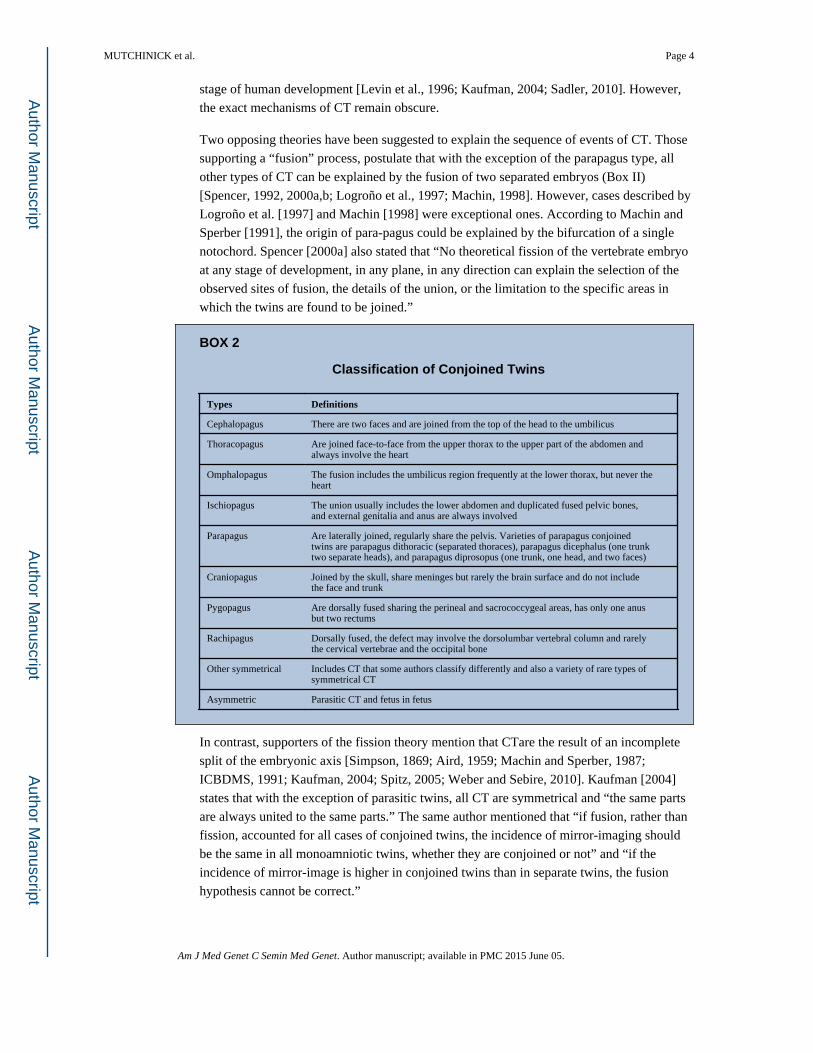
Two opposing theories have been suggested to explain the sequence of events of CT. Those
supporting a “fusion” process, postulate that with the exception of the parapagus type, all
other types of CT can be explained by the fusion of two separated embryos (Box II)
[Spencer, 1992, 2000a,b; Logroño et al., 1997; Machin, 1998]. However, cases described by
Logroño et al. [1997] and Machin [1998] were exceptional ones. According to Machin and
Sperber [1991], the origin of para-pagus could be explained by the bifurcation of a single
notochord. Spencer [2000a] also stated that “No theoretical fission of the vertebrate embryo
at any stage of development, in any plane, in any direction can explain the selection of the
observed sites of fusion, the details of the union, or the limitation to the specific areas in
which the twins are found to be joined.”
BOX 2
Classification of Conjoined Twins
Types Definitions
Cephalopagus There are two faces and are joined from the top of the head to the umbilicus
Thoracopagus Are joined face-to-face from the upper thorax to the upper part of the abdomen and always involve the heart
Omphalopagus The fusion includes the umbilicus region frequently at the lower thorax, but never the heart
Ischiopagus The union usually includes the lower abdomen and duplicated fused pelvic bones, and external genitalia and anus are always involved
Parapagus Are laterally joined, regularly share the pelvis. Varieties of parapagus conjoined twins are parapagus dithoracic (separated thoraces), parapagus dicephalus (one trunk two separate heads), and parapagus diprosopus (one trunk, one head, and two faces)
Craniopagus Joined by the skull, share meninges but rarely the brain surface and do not include the face and trunk
Pygopagus Are dorsally fused sharing the perineal and sacrococcygeal areas, has only one anus but two rectums
Rachipagus Dorsally fused, the defect may involve the dorsolumbar vertebral column and rarely the cervical vertebrae and the occipital bone
Other symmetrical Includes CT that some authors classify differently and also a variety of rare types of symmetrical CT
Asymmetric Parasitic CT and fetus in fetus
In contrast, supporters of the fission theory mention that CTare the result of an incomplete
split of the embryonic axis [Simpson, 1869; Aird, 1959; Machin and Sperber, 1987;
ICBDMS, 1991; Kaufman, 2004; Spitz, 2005; Weber and Sebire, 2010]. Kaufman [2004]
states that with the exception of parasitic twins, all CT are symmetrical and “the same parts
are always united to the same parts.” The same author mentioned that “if fusion, rather than
fission, accounted for all cases of conjoined twins, the incidence of mirror-imaging should
be the same in all monoamniotic twins, whether they are conjoined or not” and “if the
incidence of mirror-image is higher in conjoined twins than in separate twins, the fusion
hypothesis cannot be correct.”
MUTCHINICK et al. Page 4
Am J Med Genet C Semin Med Genet. Author manuscript; available in PMC 2015 June 05.
Author M
anuscriptA
uthor Manuscript
Author M
anuscriptA
uthor Manuscript
GENETICS AND OTHER RISK FACTORS ASSOCIATED TO CONJOINED
TWINS
There is no record in the literature of familial aggregation of CT, nor for preferential
associations with other unrelated anomalies. As for the former, an example is the large
multigenerational kindred, descendants from the famous Eng and Chang Bunker CT. Among
1,500 descendants of both of them, several pairs of twins including MZ twins were born, but
no other CTwere recorded [Newman, 2006]. In the Online Mendelian Inheritance in Man
(OMIM 164750) only one instance of CT concordant for omphalocele is reported, which
constitutes a related defect to the omphalopagus type of CT [Bugge, 2010].
A report by Rosa et al. [1987] mentioned the exposure during pregnancy to griseofulvin, an
antifungal medication, was noted in two sets of CT in humans, but this was not further
confirmed by Knudsen [1987] and Métneki and Czeizel [1987]. Griseofulvin crosses the
placental barrier and is recognized as a human teratogen. In a population-based study on
22,843 pregnancy outcomes with birth defects and 38,151 controls, the authors reported that
a 0.03% and 0.06% of cases and controls mothers were treated during pregnancy with this
drug. CT was observed in 55 pregnancies; however none of the mothers of the CT were
exposed to griseofulvin [Czeizel et al., 2004]. Some proteins such as activin, nodal, and
Sonic hedgehog have been associated with laterality defects in chicken CT, but not in
humans [Levin et al., 1996]. Recently, it has been reported [Wertelecki, 2010] that chronic
low-dose radiation exposure could favor the occurrence of twinning and the prevalence of
CT. The analysis of approximately 100,000 births born between 2000 and 2006 in the area
of Rivne, close to Chernobyl, Ukraine, showed an apparent cluster of CT (5 in 96,438
births). However, numbers were too small to reach conclusions.
EPIDEMIOLOGY OF CONJOINED TWINS
As mentioned above, worldwide prevalence of CT, although variable, has been estimated to
be 1 in 50,000 pregnancies, but approximately 1 in 200,000 live-births (LB) [Spitz, 2005].
However, some studies reported prevalences as high as 1 in 2,800 LB in India [Mudaliar,
1930], to as low as 1 in 200,000 LBs in the USA [Bender, 1967].
Prevalence of conjoined twins observed in diverse populations studied: 1930–2010
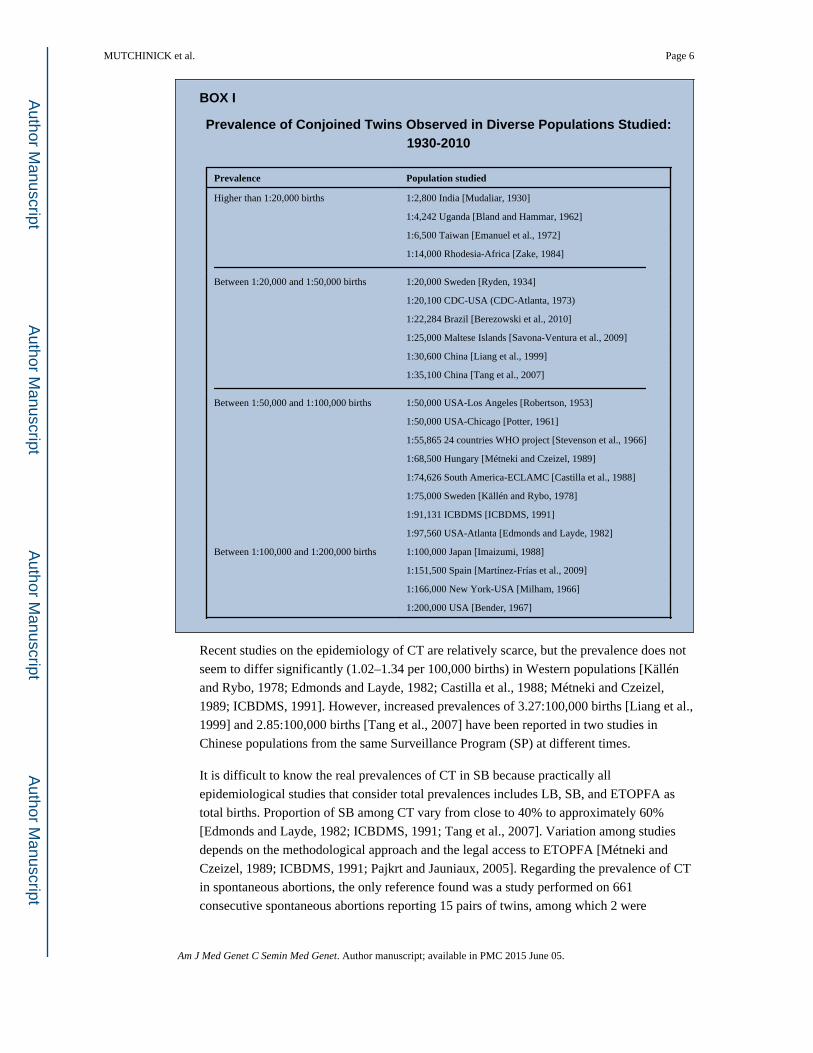
Prevalence of symmetrical CT can be assessed in four categories: (i) higher than 1:20,000
births; (ii) between 1:20,000 and 1:50,000 births; (iii) between 1:50,000 and 1:100,000
births; and (iiii) between 1:100,000 and 1:200,000 births (Box I). The marked differences
reported could be attributed to the population size monitored, and inclusion or not of
stillbirths (SB), spontaneous abortions, and elective termination of pregnancy for fetal
anomaly (ETOPFA). Significant under-registration of non-liveborn prenatal or perinatal
cases in any of the four categories may explain, in part, the differences observed among
populations studied [Hanson, 1975; Källén and Rybo, 1978; Liang et al., 1999; Tang et al.,
2007] (Box I).
MUTCHINICK et al. Page 5
Am J Med Genet C Semin Med Genet. Author manuscript; available in PMC 2015 June 05.
Author M
anuscriptA
uthor Manuscript
Author M
anuscriptA
uthor Manuscript
BOX I
Prevalence of Conjoined Twins Observed in Diverse Populations Studied: 1930-2010
Prevalence Population studied
Higher than 1:20,000 births 1:2,800 India [Mudaliar, 1930]
1:4,242 Uganda [Bland and Hammar, 1962]
1:6,500 Taiwan [Emanuel et al., 1972]
1:14,000 Rhodesia-Africa [Zake, 1984]
Between 1:20,000 and 1:50,000 births 1:20,000 Sweden [Ryden, 1934]
1:20,100 CDC-USA (CDC-Atlanta, 1973)
1:22,284 Brazil [Berezowski et al., 2010]
1:25,000 Maltese Islands [Savona-Ventura et al., 2009]
1:30,600 China [Liang et al., 1999]
1:35,100 China [Tang et al., 2007]
Between 1:50,000 and 1:100,000 births 1:50,000 USA-Los Angeles [Robertson, 1953]
1:50,000 USA-Chicago [Potter, 1961]
1:55,865 24 countries WHO project [Stevenson et al., 1966]
1:68,500 Hungary [Métneki and Czeizel, 1989]
1:74,626 South America-ECLAMC [Castilla et al., 1988]
1:75,000 Sweden [Källén and Rybo, 1978]
1:91,131 ICBDMS [ICBDMS, 1991]
1:97,560 USA-Atlanta [Edmonds and Layde, 1982]
Between 1:100,000 and 1:200,000 births 1:100,000 Japan [Imaizumi, 1988]
1:151,500 Spain [Martínez-Frías et al., 2009]
1:166,000 New York-USA [Milham, 1966]
1:200,000 USA [Bender, 1967]
Recent studies on the epidemiology of CT are relatively scarce, but the prevalence does not
seem to differ significantly (1.02–1.34 per 100,000 births) in Western populations [Källén
and Rybo, 1978; Edmonds and Layde, 1982; Castilla et al., 1988; Métneki and Czeizel,
1989; ICBDMS, 1991]. However, increased prevalences of 3.27:100,000 births [Liang et al.,
1999] and 2.85:100,000 births [Tang et al., 2007] have been reported in two studies in
Chinese populations from the same Surveillance Program (SP) at different times.
It is difficult to know the real prevalences of CT in SB because practically all
epidemiological studies that consider total prevalences includes LB, SB, and ETOPFA as
total births. Proportion of SB among CT vary from close to 40% to approximately 60%
[Edmonds and Layde, 1982; ICBDMS, 1991; Tang et al., 2007]. Variation among studies
depends on the methodological approach and the legal access to ETOPFA [Métneki and
Czeizel, 1989; ICBDMS, 1991; Pajkrt and Jauniaux, 2005]. Regarding the prevalence of CT
in spontaneous abortions, the only reference found was a study performed on 661
consecutive spontaneous abortions reporting 15 pairs of twins, among which 2 were
MUTCHINICK et al. Page 6
Am J Med Genet C Semin Med Genet. Author manuscript; available in PMC 2015 June 05.
Author M
anuscriptA
uthor Manuscript
Author M
anuscriptA
uthor Manuscript
conjoined. These data allow an estimate of the prevalence of CT of 3.03 per 1,000 (95% CI:
0.40–10.89) in spontaneous abortions [Uchida et al., 1983].
Although in one epidemiologic study [Castilla et al., 1988] predominance of females was
not observed, many other studies have shown a 1.5–2.5 predominance of female sex over
male sex [Edmonds and Layde, 1982; Imaizumi, 1988; Métneki and Czeizel, 1989;
ICBDMS, 1991; Tang et al., 2007; Martínez-Frías et al., 2009].
Asymmetric or parasitic CT is another fairly rare atypical presentation of MZ twins, where
one of them is significantly underdeveloped and considered parasitic from the other, often
unaffected. Parasitic twins occur when one embryo of a pair of MZ twins starts to develop,
but the pair does not fully separate, and one embryo’s development prevails over the other.
Rather than conjoined, it is considered parasitic because it is incompletely formed or wholly
dependent on the body functions of the complete fetus. Prevalence has been estimated to be
approximately 20 times less frequent than the prevalence of the symmetrical types
[Edmonds and Layde, 1982; ICBDMS, 1991]. Another type of twins considered by some
authors as parasitic is the fetus in fetus. However, this developmental anomaly is considered
by others as a different parasitic twin fetus growing within its host twin very early in a MZ
pregnancy, where one fetus grows around the other. The internal twin survival depends on
the survival of its host twin [Aquino et al., 1997; Arlikar et al., 2009; Sharma et al., 2010].
Survival of CT is precarious, most dying during the very early perinatal period or as the
result of surgical separation. Survival mainly depends on the type of CT, the sharing of
organs, and timely and appropriate surgical or non-surgical treatment. Options for therapy
include emergency or planned separation if appropriate [Bland and Hammar, 1962; Hoyle,
1990; Kingston et al., 2001; Spitz, 2003].
The purpose of the present study was to identify the main epidemiological characteristics
associated to this very rare defect. Variables considered in the analysis are described in
detail in introductory article of this issue [Castilla and Mastroiacovo, 2011].
MATERIALS AND METHODS
Population Studied
The sample of CT was obtained from 21 worldwide SPs who are all members of the
International Clearinghouse for Birth Defects Surveillance and Research (ICBDSR). Each
had agreed to participate in the analysis of the epidemiology of this very rare defect.
Programs were asked to provide re-identified case records following a common protocol,
with information on phenotype, genetic testing and selected demographic and prenatal
information. Data were submitted according to a designed Excel database to obtain more
uniform information from each participating SP. The time in years covered and information
sent by each SP was variable, although all covered a minimum of 5 years period of time of
epidemiological surveillance of birth defects. Some covered more than 30 years. Information
regarding the individual characteristics of each SP is described in introductory article of this
issue [Castilla and Mastroiacovo, 2011]. Submitted data were reviewed by two of the
authors (J.A.V. and L.L.M.) and the principal investigator (O.M.M.) to identify the cases,
MUTCHINICK et al. Page 7
Am J Med Genet C Semin Med Genet. Author manuscript; available in PMC 2015 June 05.
Author M
anuscriptA
uthor Manuscript
Author M
anuscriptA
uthor Manuscript
confirm the diagnosis, classify the CT, and decide upon the inclusion or exclusion in the
sample. From a total of 402 CT pairs reported, 15 were excluded because they were included
twice and 4 had a wrong diagnosis. This resulted in a total of 383 sets of CT born among a
total of 26,138,837 births. Statistical analysis included the chi-squared test and Fisher’s
exact test to compare proportions, the chi-squared test for trends for the analysis of time
trends, the Poisson test to estimate exact 95% confidence intervals (CI) and the cumulative
Poisson P-values for comparisons of total prevalence between programs. More detailed
information is provided in the introductory article of this issue [Castilla and Mastroiacovo,
2011].
Classification of Conjoined Twins
According to the site of union, symmetrical CT are classified in different manners, including
diverse wide-ranging classifications [ICBDMS, 1991; Phelan and Hall, 2006] and simplified
commonly used ones [Edmonds and Layde, 1982; Métneki and Czeizel, 1989; Spencer,
1996, 2000a,b; Kingston et al., 2001; Kaufman, 2004]. We decided to adopt the
classification exhibited in Box II. Eight well-defined types are listed for symmetrical CT,
one for very rare types of CT, and an extra category for asymmetrical types. Some
classifications includes more types resulting from the extension of the junction, although not
all authors accept combined types such as cephalo-thoracopagus or thoracoomphalopagus,
arguing that practically cephalopagus always includes part of the thorax and thoracopagus
includes part of the abdomen [Spencer, 1996, 2000a,b; Kingston et al., 2001; Kaufman,
2004]. The classification chosen for our analyses (Box II) is the one that fits our data well
and permits comparisons with previously reported data.
Classification of Unrelated Congenital Anomalies
Only those major congenital anomalies not related to the site of union of the CT and those
cases in which the defects were clearly described were included in the analysis, independent
of the occurrence of the anomalies in one or both twins. Malformations were grouped by
developing system. If the same malformation occurred in each twin of a CT pair, it was
counted only once. Proportion of each type of congenital anomaly was estimated among the
total number of malformations.
RESULTS
Total Prevalence and Prevalence by Surveillance Program
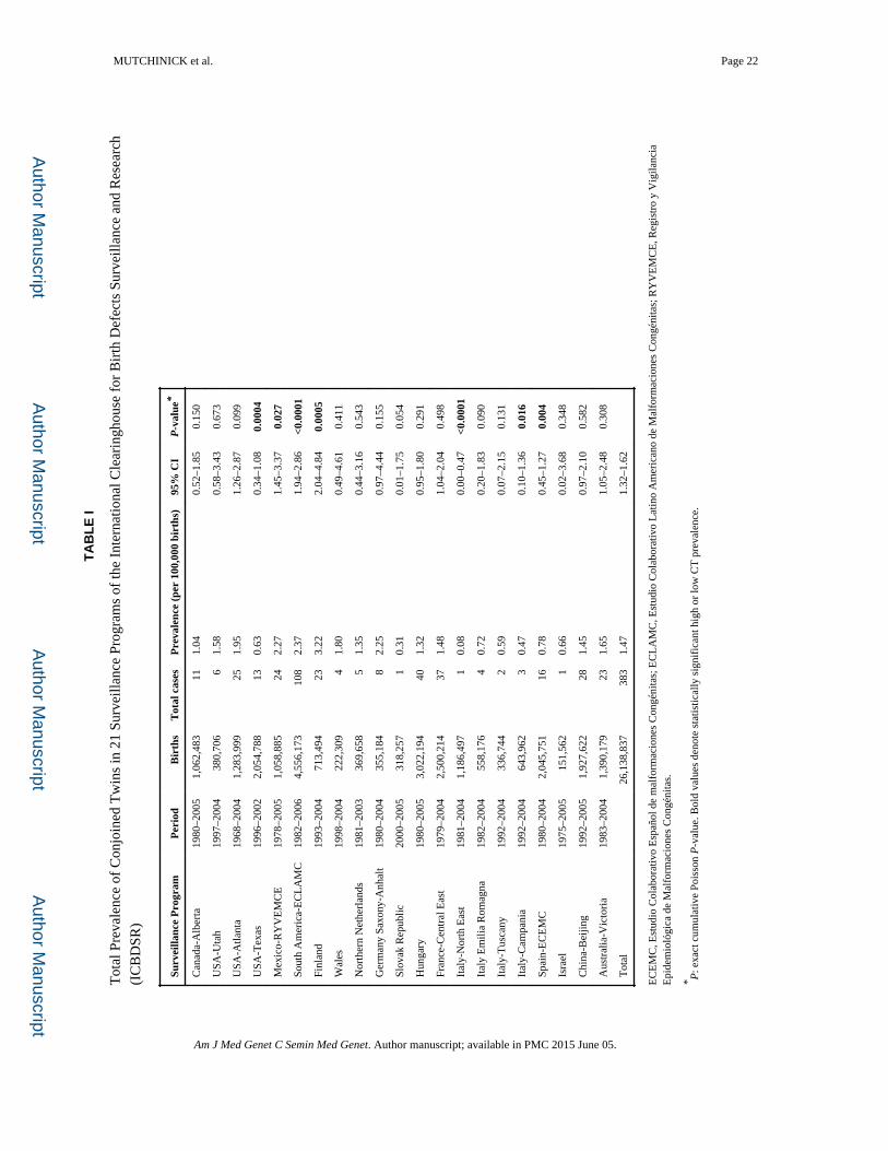
Total prevalence of CT was 1.47 (95% CI: 1.32–1.62) per 100,000 births (Table I).
Prevalences show a marked variation among SPs, from as high as 3.22 (95% CI: 2.04–4.84)
per 100,000 births in the Finland SP to as low as less than 0.08 per 100,000 births in the
Italy-North East program. Besides Finland, three other SPs have prevalence over 2 per
100,000 births: South America-ECLAMC, México-RYVEMCE, and Germany Saxony-
Anhalt, in decreasing order. Nine other SPs, USA-Atlanta, Wales, Australia-Victoria, USA-
Utah France-Central East, China-Beijing, Northern Netherlands, Hungary, and Canada-
Alberta showed a prevalence of more than 1 but less than 2 per 100,000 births; and the
remaining eight programs, Spain-ECEMC, Italy-Emilia Romagna, Israel, USA-Texas, Italy-
Tuscany, Italy-Campania, Slovak Republic, and Italy-North East, reported a prevalence
MUTCHINICK et al. Page 8
Am J Med Genet C Semin Med Genet. Author manuscript; available in PMC 2015 June 05.
Author M
anuscriptA
uthor Manuscript
Author M
anuscriptA
uthor Manuscript
lower than 1 per 100,000 births. As shown in Table I, the prevalence reported by only 7 of
the 21 participating SPs differed significantly from the total prevalence; Finland, South
America-ECLAMC, and Mexico-RYVEMCE SPs had a statistically significant high
prevalence, and Italy-North East, Italy-Campania, Spain-ECEMC, and USA-Texas SPs had
a statistically significant low prevalence. Prevalence per 100,000 births and 95% CI for each
of the 21 participating SPs is presented in Figure 1 by decreasing prevalence.
Pregnancy Outcome of Conjoined Twins by Surveillance Program
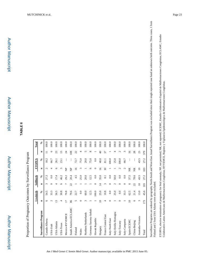
Table II shows the proportion of cases delivered as LB, SB, or ETOPFA. The largest
proportion corresponded to LB sets of CT (45.6%), contrary to the literature reporting a
higher proportion of SB among CT [Edmonds and Layde, 1982; ICBDMS, 1991]. The total
proportion of SB cases and ETOPFA was identical, at 27.2% each. However, when
considering only the 16 programs where termination for fetal anomaly is available the
proportion of ETOPFA is 50.7% (103/203). There are some SPs with a very high prevalence
of ETOPFA for CT such as France, Finland, and Germany Saxony-Anhalt. There are other
programs like Mexico-RYVEMCE and South American-ECLAMC in countries in which
ETOPFA is not permitted and do not offer termination of pregnancy for fetal anomaly.
However, in these SPs as well as in programs where termination is permitted (China-
Beijing, USA-Atlanta, and Spain-ECEMC), the prevalence of CT is also higher in live born
infants.
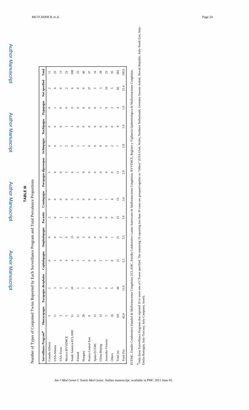
Proportion of the Different Types of Conjoined Twins by Surveillance Program
The proportion of the different types of CT by SP (Table III) is presented according to the
classification scheme described in Box II. Proportions of the total number of cases include
82 cases (21.4%) in which the type of CTwas not specified. The different types of CT are
displayed by decreasing prevalence. Thoracopagus CT, that also includes
thoracoomphalopagus CT, represent the largest number of cases reported (42.0%). The
second most common CT type was parapagus dicephalus (11.5%). The remaining most
common types were craniopagus and omphalopagus with 5.5% each. Other CT types such as
parapagus diprosopus, ischiopagus, rachipagus, and pygopagus were observed in less than
3% of the cases, with the last two being the rarest types (1.0%). Parasitic CT were observed
in 3.9% of all specified CT reported.
Interestingly, a similar pattern of proportions of cases was observed in all participating
programs, except for the omphalopagus type. For this type, 15 (71.4%) of the 21 cases were
reported by the South America-ECLAMC SP, the rest by the Hungarian, USA-Texas, and
USA-Utah registries.
Sex Ratio by Type of Conjoined Twins
Findings on the sex ratio were similar to previous studies, which reported a predominance of
female cases. However, in the present sample the number of female cases was more than
twice as frequent as that of males (218 females and 108 males; 20 CT cases were of
indeterminate sex and 37 of sex not specified). Prevalence at birth of CTwas 56.9% for
females and 28.2% for males (Table IV). Using an estimate of the number of males and
females from the total of 26,138,837 births reported, a significant statistical difference was
MUTCHINICK et al. Page 9
Am J Med Genet C Semin Med Genet. Author manuscript; available in PMC 2015 June 05.
Author M
anuscriptA
uthor Manuscript
Author M
anuscriptA
uthor Manuscript

observed (P <0.01; OR =2.23; 95% CI: 1.76–2.83). Although, omphalopagus CT was four
times more frequent in females than in males, the difference was not statistically significant.
Female predominance was significantly higher for the thoracopagus CT type (P <0.01; OR
=3.27; 95% CI: 1.83–5.99). Para-pagus as a whole (P <0.01; OR =2.39; 95% CI: 1.21–4.71),
parapagus dice-phalus alone (P <0.05; OR =2.23; 95% CI: 1.06–4.68), and parasitic (P
<0.01; OR =5.32; 95% CI: 1.42–24.22) CT types showed a significantly higher prevalence
in males. No significant differences were observed for the rest of the CT types analyzed.
Indeterminate sex was found in a small proportion (5.2%) of cases (Table IV).
Unrelated Congenital Anomalies Associated by Type of Conjoined Twin
Cases were carefully reviewed for unrelated associated anomalies (Table V), although in
some cases and types of CT it was difficult to determine with certainty whether other defects
present were unrelated malformations. Associated congenital anomalies were observed in
115 pairs of 182 CT sets in which the information was available (63.2%). However, the
malformation was adequately described in only 73 cases. In these 73 cases, 111
malformations were described, but detailed information of whether just one or both twins
had the malformations was not noted. The malformations more frequently reported were
those affecting the genitourinary tract (19.8%), the central nervous system (18.9%),
comprising neural tube defects (9.9%), hydrocephalus (3.6%), microphthalmia (0.9%), and
other central nervous system defects (4.5%), and the musculoskeletal system (12.6%).
Combining musculoskeletal anomalies with limb deficiency defects and polydactyly, the
proportion of limb anomalies increases to 20.7%. Other frequent malformations reported
were gastrointestinal atresias (9.9%) and facial clefts (9.9%).
Ethnicity and Conjoined Twins
Conjoined twins were also stratified by ethnicity in four categories: Anglo-Saxon/Caucasian,
Chinese, Latin American, and Latin European. Prevalence was higher in the Latin American
ethnic group. Statistical differences were observed when compared to the Anglo-Saxon/
Caucasian (P <0.01; OR =1.51, 95% CI: 1.20–1.91), Chinese (P =0.02: OR =1.65, 95% CI:
1.06–2.49), and Latin European (P <0.01; OR =2.50, 95% CI: 1.84–3.40) ethnic groups.
Other comparisons were not statistically significant.
Total Prevalence Time Trends
Time trends are presented in Figure 2, with the data analyzed in three different ways: (i) all
participating SPs of the ICBDSR; (ii) South America-ECLAMC alone; and (iii) ICBDSR
SPs excluding South America-ECLAMC. As shown in Figure 2, a slight steadily increasing
but statistically non-significant trend in the prevalence of CT is present in the South
America-ECLAMC SP for years 1987–2006, that differ from the decreasing prevalence
trend observed in the ICBDSR excluding South America-ECLAMC.
Genetic, Demographic, and Environmental Risk Factors
Analyses were performed to evaluate for associations with genetic factors such as
consanguinity, familial aggregation, and cytogenetic studies; demographic characteristics
such as maternal age and maternal education; reproductive data, including gestational age,
MUTCHINICK et al. Page 10
Am J Med Genet C Semin Med Genet. Author manuscript; available in PMC 2015 June 05.
Author M
anuscriptA
uthor Manuscript
Author M
anuscriptA
uthor Manuscript
birth order, birth weight, spontaneous previous abortions; and environmental factors such as
maternal exposures and diseases during pregnancy. No associations were observed between
the mentioned genetic factors and CT. No familial cases were observed, consanguinity was
reported in 5 of 209 CT cases (2.38%; 95% CI: 0.78–5.49) and all chromosome studies
performed (12.1%) were normal.
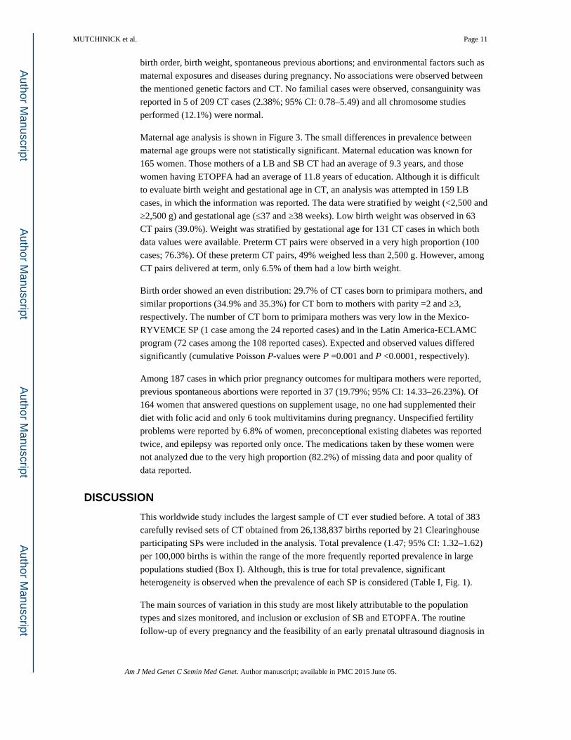
Maternal age analysis is shown in Figure 3. The small differences in prevalence between
maternal age groups were not statistically significant. Maternal education was known for
165 women. Those mothers of a LB and SB CT had an average of 9.3 years, and those
women having ETOPFA had an average of 11.8 years of education. Although it is difficult
to evaluate birth weight and gestational age in CT, an analysis was attempted in 159 LB
cases, in which the information was reported. The data were stratified by weight (<2,500 and
≥2,500 g) and gestational age (≤37 and ≥38 weeks). Low birth weight was observed in 63
CT pairs (39.0%). Weight was stratified by gestational age for 131 CT cases in which both
data values were available. Preterm CT pairs were observed in a very high proportion (100
cases; 76.3%). Of these preterm CT pairs, 49% weighed less than 2,500 g. However, among
CT pairs delivered at term, only 6.5% of them had a low birth weight.
Birth order showed an even distribution: 29.7% of CT cases born to primipara mothers, and
similar proportions (34.9% and 35.3%) for CT born to mothers with parity =2 and ≥3,
respectively. The number of CT born to primipara mothers was very low in the Mexico-
RYVEMCE SP (1 case among the 24 reported cases) and in the Latin America-ECLAMC
program (72 cases among the 108 reported cases). Expected and observed values differed
significantly (cumulative Poisson P-values were P =0.001 and P <0.0001, respectively).
Among 187 cases in which prior pregnancy outcomes for multipara mothers were reported,
previous spontaneous abortions were reported in 37 (19.79%; 95% CI: 14.33–26.23%). Of
164 women that answered questions on supplement usage, no one had supplemented their
diet with folic acid and only 6 took multivitamins during pregnancy. Unspecified fertility
problems were reported by 6.8% of women, preconceptional existing diabetes was reported
twice, and epilepsy was reported only once. The medications taken by these women were
not analyzed due to the very high proportion (82.2%) of missing data and poor quality of
data reported.
DISCUSSION
This worldwide study includes the largest sample of CT ever studied before. A total of 383
carefully revised sets of CT obtained from 26,138,837 births reported by 21 Clearinghouse
participating SPs were included in the analysis. Total prevalence (1.47; 95% CI: 1.32–1.62)
per 100,000 births is within the range of the more frequently reported prevalence in large
populations studied (Box I). Although, this is true for total prevalence, significant
heterogeneity is observed when the prevalence of each SP is considered (Table I, Fig. 1).
The main sources of variation in this study are most likely attributable to the population
types and sizes monitored, and inclusion or exclusion of SB and ETOPFA. The routine
follow-up of every pregnancy and the feasibility of an early prenatal ultrasound diagnosis in
MUTCHINICK et al. Page 11
Am J Med Genet C Semin Med Genet. Author manuscript; available in PMC 2015 June 05.
Author M
anuscriptA
uthor Manuscript
Author M
anuscriptA
uthor Manuscript
some cases may have introduced an underascertainment due to unreported ETOPFA. As
reported by Martínez-Frías et al. [2009] in Spain, termination for fetal anomaly has been
offered by the public health system for many years, but it does not have for a system for
including cases with ETOPFA in its registry program. As a result, current prevalence of CT
in this SP decreased considerably to 0.68 per 100,000 births. This probably accounts for the
differences observed within and between the four depicted categories when prevalence
results were described (Table I). The underreporting is also a dilemma when present results
are compared with previous reports [Hanson, 1975; Källén and Rybo, 1978; Edmonds and
Layde, 1982; Viljoen et al., 1983; Castilla et al., 1988; ICBDMS, 1991; Liang et al., 1999;
Tang et al., 2007] and other studies listed in Box II.
Pregnancy outcomes, LB (45.6%), SB (27.2%), and ETOPFA (27.2%) are shown in Table
II. The proportion of CT described in each of these categories depends in part on the health
services characteristics and registration systems in the communities served by the SP, as
well as the availability of legal ETOPFA. This perhaps could account for the low proportion
of SB reported in the present study in comparison with other surveys carried out when
ETOPFA was not available [Edmonds and Layde, 1982; Métneki and Czeizel, 1989;
ICBDMS, 1991]. The proportion of CT resulting in ETOPFA varies from zero in Mexico-
RYVEMCE and South America-ECLAMC where ETOPFA is not legal to as much as
81.1% in France Central East where elective termination of pregnancy is a common practice
(Table II).
The observed prevalence of types of CT (Table III) shows a predominance (42.0%) of the
thoracopagus type as reported in previous epidemiological studies with similar proportions:
49.3% [Edmonds and Layde, 1982], 43.5% [Castilla et al., 1988], 43.6% [Métneki and
Czeizel, 1989], and 39.1% [ICBDMS, 1991]. It is not clear whether the high proportion of
omphalopagus cases (71.4%) reported by the South America-ECLAMC program is a real
cluster or an anomaly. Perhaps one way to evaluate this would be to look at the hospitals of
origin of the 15 reported cases, mindful of the fact that the South America-ECLAMC SP
collects data from 10 different countries and from close to 100 hospitals. Except for an
uncommon high proportion of para-pagus dicephalus and diprosopus CT in the Mexican SP
(33.3%), other types of reported CT showed a similar distribution among the 21
participating Clearinghouse SPs (Table III).
The predominance of female sex observed in the present study has been previously reported
[Edmonds and Layde, 1982; Imaizumi, 1988; Métneki and Czeizel, 1989; ICBDMS, 1991;
Tang et al., 2007; Martínez-Frías et al., 2009]. Considering the entire CT sample, prevalence
was more than twice (2.02) as frequent in females as in males. Changes in prevalence by sex
may depend on sample size and chance. In a previous study of the South American SP,
Castilla et al. [1988] reported a very similar number of male and female CT, but in the data
reported by the South American SP for the present study an overt female predominance of
2.09 was observed (69 females and 33 males, data not shown). Another interesting finding
was the contrasting and significant differences observed when prevalence by sex is analyzed
by type of CT. Thoracopagus type is almost four times more frequent in females than in
males (P <0.01; OR 3.13; 95% CI: 1.76–5.63). However, parapagus and parasitic types were
significantly more frequent in males than in females, P =0.01 and P <0.01, respectively
MUTCHINICK et al. Page 12
Am J Med Genet C Semin Med Genet. Author manuscript; available in PMC 2015 June 05.
Author M
anuscriptA
uthor Manuscript
Author M
anuscriptA
uthor Manuscript
(Table IV). No significant differences were observed for the rest of the CT types analyzed.
Detailed similar information of this type of data (Table IV) was reported previously
[ICBDMS, 1991], although proportions reported were different from the ones reported
herein, and no comments were made regarding statistical analysis. No explanation is
proposed for the sex differences but future studies could look at these in the CT outcomes in
the SB and ETOPFA groups. More research is needed to explore the relation and severity of
types of CT with sex to identify a possible biased selection in utero of some CT types that
might explain the observed differences.
The proportion of associated malformations unrelated to the site of union of the twins has
been reported in other studies [Edmonds and Layde, 1982; Métneki and Czeizel, 1989;
ICBDMS, 1991], and was similar to the 63% observed in the present study. However, only
two studies [Métneki and Czeizel, 1989; ICBDMS, 1991] stratified the associated
malformations according to the CT type. Neural tube defects were observed in more than
15% of parapagus dicephalus and diprosopus types. Genitourinary anomalies were recorded
in more than 15% of thoracopagus, omphalopagus, ischiopagus, and pygo-pagus types, and
musculoskeletal defects in more than 15% of thoracopagus, parapagus dicephalus,
craniopagus, and ischiopagus types. Oral clefts and gastrointestinal atresias were evenly
distributed (Table V). The reported proportion of associated anomalies does not differ
significantly from those reported by Métneki and Czeizel [1989], and the ICBDMS [1991].
Although, some anomalies occurred more frequently with certain CT types, numbers are still
too small to suggest specific associations and could merely represent spurious associations.
Ethnicity analysis showed that the prevalence of CT is significantly higher in Latin
American SPs than in Anglo-Saxon/Caucasian, Chinese and Latin European programs
(Table I). A possible explanation of these findings could be the under-registration of CT
pregnancies undergoing ETOPFA in some SPs, but particularly in the Latin European SP.
This observation agrees with the proportion of prenatal diagnosis before 20 weeks of
gestation and the high proportion of ETOPFA in European SPs (Table II). This could also
explain the significantly higher prevalence in Latin American SPs than in the one of China,
where ETOPFA was not described in the 28 CT cases reported, although elective
termination of pregnancy is permitted but not recorded.
Although several references to clusters of CT have been observed [Hanson, 1975; Källén
and Rybo, 1978; Mabogunje and Lawrie, 1980; Viljoen et al., 1983; Zake, 1984; Rees et al.,
1993; Savona-Ventura et al., 2009] a clear explanation has not been found. Different
explanations bias in the results have been considered, particularly with respect to population
types and sizes surveyed, and hospital records not contributing to the regional or national
SPs or a multicentric hospital-based surveillance system. Retrospective versus prospective
CT sample collection and the lack of a population reference of total births born during the
same period of time of diagnosis could also be sources of bias.
When prevalence data were analyzed in a time trend, the South America-ECLAMC SP,
exclusively for years 1987–2006, showed an increasing but statistically non-significant
prevalence trend for CT (χ for trend: 2.83; P =0.09). Although not significant, it would be
important to monitor the increasing prevalence trend in this SP.
MUTCHINICK et al. Page 13
Am J Med Genet C Semin Med Genet. Author manuscript; available in PMC 2015 June 05.
Author M
anuscriptA
uthor Manuscript
Author M
anuscriptA
uthor Manuscript
Genetic and environmental risk factors such as consanguinity, familial aggregation,
cytogenetic studies, and maternal exposures, and acute and chronic diseases during
pregnancy did not reveal any association with the occurrence of CT, in general, or for any
particular type of CT. No familial cases were reported and all karyotypes performed were
normal. Regarding reproductive characteristic patterns, maternal age does not show any
significant trend or association with CT (Fig. 3). However, other related variables, such as
gestational age, birth weight, birth order, previous spontaneous abortions, and maternal
education showed certain peculiar relationships. A high proportion of preterm deliveries
(76.3%) occurred among CT cases; however, interestingly low birth weight varied by
gestational age (49.0% in pregnancies ≤37 weeks and 6.5% in pregnancies of >37 weeks).
The significant difference in birth order in the Mexico RYVEMCE and the Latin America
ECLAMC SPs is an unexpected finding compared with other SPs that reported information
on birth order in more than 20 CT pairs. Previous spontaneous abortions were reported in a
high proportion (19.8%) of CT mothers. These figures are higher than that usually reported
in most healthy populations studied.
Even though success in surgical separation of CT pairs has improved, surgical separation is
still a major challenge. The procedure requires a multi-disciplinary team, accurate imaging
to assess organ sharing, and a consideration of aspects related to survival and ethics in each
case. Experiences of dedicated groups in surgical separation, clinical supervision, and
support directives for parents and patients are available from the literature [Bland and
Hammar, 1962; Hoyle, 1990; Spitz, 1996; Kingston et al., 2001; Pearn, 2001; Spitz and
Kiely, 2002; Spitz, 2003, 2005; Arkinson, 2004; Pajkrt and Jauniaux, 2005; Votteler and
Lipsky, 2005; Arlikar et al., 2009; Sharma et al., 2010; Weber and Sebire, 2010].
Review of variable total prevalence, variable prevalence by occurrence type, predominance
in females, a higher prevalence in some ethnic groups such as Blacks [Edmonds and Layde,
1982] and Chinese [Liang et al., 1999; Tang et al., 2007], geographic variation, socio-
demographic variables, genetic and environmental factors, has not helped to advance the
understanding of the altered mechanisms that interrupt the normal separation of an inner cell
mass leading to the occurrence of a set of CT. Considering the fission theory as currently
accepted [ICBDMS, 1991; Kaufman, 2004; Spitz, 2005; Sadler, 2010; Weber and Sebire,
2010], a simple molecular disorder at a deep cellular level distorting cell adhesion or
apoptosis in a very early stage of embryogenesis could be involved in a etiology of CT that
involves incomplete split of the inner mass cell.
This is the largest international collaborative study of CT to date. Salient findings were a
notable variation in prevalence among SPs; a marked variation in the type of pregnancy
outcome; a significant female predominance in CT, particularly of the thoracopagus type,
and a significant male predominance in parapagus and parasitic types; significant differences
in prevalence by ethnicity; and apparent non-significant increasing prevalence trend in South
American countries. Further work in epidemiology and molecular research is needed to
elucidate the etiologic processes involved and associated risk factors for the development of
this fascinating phenomenon from nature.
MUTCHINICK et al. Page 14
Am J Med Genet C Semin Med Genet. Author manuscript; available in PMC 2015 June 05.
Author M
anuscriptA
uthor Manuscript
Author M
anuscriptA
uthor Manuscript
Acknowledgments
The authors are grateful to each Surveillance Program’s Staff and Members for their work in collecting case data and submission to the ICBDSR Centre. The work conducted at the ICBDSR Centre was supported by the Centers for Disease Control and Prevention (1U50DD000524-02). Grant sponsor for South America ECLAMC: MCT/ CNPq, Brazil; Grant numbers: 573993/ 2008-4, 476978/2008-4, 554755/ 2009-2; 306750/2009-0; 402045/ 2010-6. This work was in part supported by Instituto de Salud Carlos III (ISCIII, Ministry of Science and Innovation) of Spain, and the Fundación 1000 sobre Defectos Congénitos, of Spain. CIBERER is an initiative of ISCIII. Components of ECEMC’s Peripheral Group are gratefully acknowledged. The Tuscany Registry of Birth Defects is funded by the “Direzione Generale Diritti di cittadinanza e Coesione sociale—Regione Toscana.” Public Health Genetics, Murdoch Childrens Research Institute and Department of Paediatrics, University of Melbourne, Parkville, Victoria, Australia, formerly Victorian Birth Defects Register.
Grant sponsor: Centers for Disease Control and Prevention; Grant number: 1U50DD000524-02; Grant sponsor: South America ECLAMC: MCT/ CNPq, Brazil; Grant numbers: 573993/2008-4, 476978/2008-4, 554755/2009-2, 306750/2009-0, 402045/2010-6; Grant sponsor: Instituto de Salud Carlos III (ISCIII, Ministry of Science and Innovation) of Spain; Grant sponsor: Fundación 1000 sobre Defectos Congénitos, of Spain; Grant sponsor: The Tuscany Registry of Birth Defects is funded by the “Direzione Generale Diritti di cittadinanza e Coesione sociale—Regione Toscana”
References
Aird I. Conjoined twins: Further observations. Br Med J. 1959; 1(5133):1313–1315. [PubMed: 13651722]
Aquino DB, Timmons C, Burns D, Lowichik A. Craniopagus parasiticus: A case illustrating its relationship to craniopagus conjoined twinning. Pediatr Pathol Lab Med. 1997; 17(6):939–944. [PubMed: 9353833]
Atkinson L. Ethics and conjoined twins. Childs Nerv Syst. 2004; 20:504–507. [PubMed: 15258819]
Arlikar J, Mane S, Dhende N, Sanghavi Y, Valand A, Butale P. Fetus in fetus: Two case reports and review of literature. Pediatr Surg Int. 2009; 25(3):289–292. [PubMed: 19184054]
Ballantyne JW. The Biddenden Maids: The medieval pygopagus. Teratologia. 1895; 2:268–274.
Bender C. Studies on symmetrically conjoined twins. J Pediatr. 1967; 70(6):1010–1011. [PubMed: 6027285]
Berezowski AT, Duarte G, Rodrigues R, de Carvalho Cavalli R, de Olivera Cardoso dos Santos R, de Moraes Villela de Andrade Vicente YA, Galli Sorita Tazima M, de F. Conjoined twins: An experience of a tertiary hospital in Southeast Brazil. Rev Bras Ginecol Obstet. 2010; 32(2):61–65. [PubMed: 20305942]
Berrin, K.; Larco, M. The spirit of ancient Peru: treasures from the Museo Arqueoló-gico Rafael Larco Herrera. New York: Thames and Hudson; 1997. p. 216
Bland KG, Hammar B. Xiphopagus twins. Report of obstetric and surgical management of a case. Cent Afr J Med. 1962; 8:371–375. [PubMed: 13971497]
Bondeson J. The Biddenden Maids: A curious chapter in the history of conjoined twins. J R Soc Med. 1992; 85(4):217–221. [PubMed: 1433064]
Bondeson J. The Isle-Brewers conjoined twins of 1680. J R Soc Med. 1993; 86(2):106–109. [PubMed: 8433291]
Bugge M. Twins with omphalocele in Denmark (1970–1989). Am J Med Genet Part A. 2010; 152A(8):2048–2052. [PubMed: 20635337]
Canfield D, Brignolo L, Peterson PE, Hendrickx AG. Conjoined twins in a rhesus monkey (Macaca mulatta). J Med Primatol. 2000; 29(6):427–430. [PubMed: 11168835]
Castilla EE, Mastroiacovo P. Very rare defects: what can we learn?. 2011 This issue.
Castilla EE, López-Camelo JS, Orioli IM, Sánchez O, Paz JE. The epidemiology of conjoined twins in Latin America. Acta Genet Med Gemellol (Roma). 1988; 37(2):111–118. [PubMed: 3239351]
Czeizel AE, Metneki J, Kazy Z, Puho E. A population-based case–control study of oral griseofulvin treatment during pregnancy. Acta Obstet Gynecol Scand. 2004; 83(9):827–831. [PubMed: 15315593]
MUTCHINICK et al. Page 15
Am J Med Genet C Semin Med Genet. Author manuscript; available in PMC 2015 June 05.
Author M
anuscriptA
uthor Manuscript
Author M
anuscriptA
uthor Manuscript
Edmonds LD, Layde PM. Conjoined twins in the United States, 1970–1977. Teratology. 1982; 25(3):301–308. [PubMed: 7112433]
Emanuel I, Huang SW, Gutman LT, Yu FC, Lin CC. The incidence of congenital malformations in a Chinese population: The Taipei Collaborative Study. Teratology. 1972; 5(2):159–169. [PubMed: 4554141]
Gould, GM.; Pyle, WL. Major terata. In: Gould, GM.; Pyle, WL., editors. Anomalies and curiosities of medicine. Philadelphia: W.B. Saunders; 1896. p. 162-213.
Guttmacher, AF. Biographical notes on some famous conjoined twins. Bergsma, D., editor. Vol. III. New York: National Foundation-March of Dimes; 1967. p. 10-17.Birth defects original Article series
Hanson JW. Letter: Incidence of conjoined twinning. Lancet. 1975; 2(7947):1257. [PubMed: 53741]
Hoyle RM. Surgical separation of conjoined twins. Surg Gynecol Obstet. 1990; 170(6):549–562. [PubMed: 2188386]
ICBDMS. Conjoined twins—An epidemiological study based on 312 cases. The International Clearinghouse for Birth Defects Monitoring Systems. Acta Genet Med Gemellol (Roma). 1991; 40(3–4):325–335. [PubMed: 1821509]
Imaizumi Y. Conjoined twins in Japan, 1979–1985. Acta Genet Med Gemellol (Roma). 1988; 37(3–4):339–345. [PubMed: 3254024]
Källén B, Rybo G. Conjoined twinning in Sweden. Acta Obstet Gynecol Scand. 1978; 57(3):257–259. [PubMed: 665171]
Kaufman MH. The embryology of conjoined twins. Childs Nerv Syst. 2004; 20(8–9):508–525. [PubMed: 15278382]
Kingston CA, McHugh K, Kumaradevan J, Kiely EM, Spitz L. Imaging in the preoperative assessment of conjoined twins. Radiographics. 2001; 21(5):1187–1208. [PubMed: 11553825]
Knudsen LB. No association between griseofulvin and conjoined twinning. Lancet. 1987; 2(8567):1097. [PubMed: 2890014]
Levin M, Roberts DJ, Holmes LB, Tabin C. Laterality defects in conjoined twins. Nature. 1996; 384(6607):321. [PubMed: 8934513]
Liang J, Xu CI, Wang Y. Epidemiological survey of conjoined twins in China. Hua Xi Yi Ke Da Xue Xue Bao. 1999; 30(1):56–58. [PubMed: 12205925]
Logroño R, Garcia-Lithgow C, Harris C, Kent M, Meisner L. Heteropagus conjoined twins due to fusion of two embryos: Report and review. Am J Med Genet. 1997; 73(3):239–243. [PubMed: 9415676]
Mabogunje OA, Lawrie JH. Conjoined twins in West Africa. Arch Dis Child. 1980; 55(8):626–630. [PubMed: 7192076]
Machin GA. Heteropagus conjoined twins due to fusion of two embryos. Am J Med Genet. 1998; 78(4):388–389. [PubMed: 9714447]
Machin GA, Sperber GH. Lessons from conjoined twins. Am J Med Genet. 1987; 28(1):89–97. [PubMed: 3674121]
Machin GA, Sperber GH. Comments on “Unique anomalies in cephalothoracopagus janiceps conjoined twins with implications for multiple mechanisms in the abnormal embryogenesis. Teratology. 1991; 44(5):481–483. [PubMed: 1771590]
Martínez-Frías ML, Bermejo E, Mendioroz J, Rodríguez-Pinilla E, Blanco M, Egüés J, Félix V, García A, Huertas H, Nieto C, López JA, López S, Paisán L, Rosa A, Vázquez MS. Epidemiological and clinical analysis of a consecutive series of conjoined twins in Spain. J Pediatr Surg. 2009; 44(4):811–820. [PubMed: 19361646]
Métneki J, Czeizel A. Griseofulvin teratology. Lancet. 1987; 1(8540):1042. [PubMed: 2883385]
Métneki J, Czeizel A. Conjoined twins in Hungary, 1970–1986. Acta Genet Med Gemellol (Roma). 1989; 38(3–4):285–299. [PubMed: 2631499]
Milham S. Symmetrical conjoined twins: An analysis of the birth records of twenty-two sets. J Pediatr. 1966; 69(4):643–647. [PubMed: 5921342]
Mudaliar AL. Double monsters: A study of their circulatory system and some other anatomical abnormalities and the complications in labor. BJOG. 1930; 37(4):753–768.
MUTCHINICK et al. Page 16
Am J Med Genet C Semin Med Genet. Author manuscript; available in PMC 2015 June 05.
Author M
anuscriptA
uthor Manuscript
Author M
anuscriptA
uthor Manuscript
Newman C. Together forever. Natl Geogr. 2006; 209(6):148–152.
Pajkrt E, Jauniaux E. First-trimester diagnosis of conjoined twins. Prenat Diagn. 2005; 25(9):820–826. [PubMed: 16170847]
Pearn J. Bioethical issues in caring for conjoined twins and their parents. Lancet. 2001; 357:1968–1971. [PubMed: 11425439]
Phelan, MC.; Hall, JG. Twins. In: Stevenson, RE.; Hall, JG., editors. Human malformations and related anomalies. 2. New York: Oxford University Press; 2006. p. 1377-1495.
Potter, EL. Pathology of the fetus and infant. Chicago: Chicago Year Book Medical Publishers; 1961. p. 217
Rees AE, Vujanic GM, Williams WM. Epidemic of conjoined twins in Cardiff. Br J Obstet Gynaecol. 1993; 100(4):388–391. [PubMed: 8494843]
Robertson EG. Craniopagus parietalia. Arch Neurol Psychiatry. 1953; 70:189–205. [PubMed: 13064881]
Rodríguez JM. Descripción de un monstruo humano cuádruple, nacido en Durango el año de 1868. Gac Med Mex V. 1870:18–31.
Rosa FW, Hernandez C, Carlo WA. Griseofulvin teratology, including two thoracopagus conjoined twins. Lancet. 1987; 1(8525):171. [PubMed: 2880014]
Ryden AL. Kasuistischur beitrog zur kenntris der gerburtron thoracapagen. Zbl Gynak. 1934; 58:972–975.
Sadler, TW. Third month to birth: The fetus and placenta. In: Sadler, TW., editor. Langman’s Medical Embryology. 11. Philadelphia: Lippincott Williams & Wilkins; 2010. p. 91-111.
Savona-Ventura C, Grima S, Buttigieg GG. Conjoint twinning in the Maltese Islands. J Obstet Gynaecol. 2009; 29(7):599–604. [PubMed: 19757262]
Sharma G, Mobin SS, Lypka M, Urata M. Heteropagus (parasitic) twins: A review. J Pediatr Surg. 2010; 45(12):2454–2463. [PubMed: 21129567]
Simpson JY. A lecture on the Siamese and other viable United twins. Br Med J. 1869; 1(428):229–233. [PubMed: 20780450]
Spencer R. Conjoined twins: Theoretical embryologic basis. Teratology. 1992; 45(6):591–602. [PubMed: 1412053]
Spencer R. Anatomic description of conjoined twins: A plea for standardized terminology. J Pediatr Surg. 1996; 31(7):941–944. [PubMed: 8811563]
Spencer R. Theoretical and analytical embryology of conjoined twins: Part I: Embryogenesis. Clin Anat. 2000a; 13(1):36–53. [PubMed: 10617886]
Spencer R. Theoretical and analytical embryology of conjoined twins: Part II: Adjustments to union. Clin Anat. 2000b; 13(2):97–120. [PubMed: 10679855]
Spitz L. Conjoined twins. Br J Surg. 1996; 83(8):1028–1030. [PubMed: 8869298]
Spitz L. Surgery for conjoined twins. Ann R Coll Surg Engl. 2003; 85(4):230–235. [PubMed: 12855023]
Spitz L. Conjoined twins. Prenat Diagn. 2005; 25(9):814–819. [PubMed: 16170846]
Spitz L, Kiely EM. Experience in the management of conjoined twins. Br J Surg. 2002; 89(9):1188–1192. [PubMed: 12190687]
Stevenson AD, Johnston HA, Stewart MIP, et al. Congenital malformations. Bull World Health Org. 1966; 34(Suppl):9–127. [PubMed: 5296840]
Tang Y, Zhu J, Zhou GX, Dai L, Wang YP, Liang J. An epidemiological study on conjoined twins in China, from 1996 to 2004. Zhonghua Yu Fang Yi Xue Za Zhi. 2007; 41(Suppl):146–149. [PubMed: 17767883]
Uchida, Ia; Freeman, VCP.; Gedeon, M.; Goldmaker, J. Twining rate in spontaneous abortions. Am J Hum Genet. 1983; 35:987–993. [PubMed: 6614011]
Viljoen DL, Nelson MM, Beighton P. The epidemiology of conjoined twinning in Southern Africa. Clin Genet. 1983; 24(1):15–21. [PubMed: 6616941]
Votteler TP, Lipsky K. Long-term results of 10 conjoined twin separations. J Pediatr Surg. 2005; 40(4):618–629. [PubMed: 15852268]
MUTCHINICK et al. Page 17
Am J Med Genet C Semin Med Genet. Author manuscript; available in PMC 2015 June 05.
Author M
anuscriptA
uthor Manuscript
Author M
anuscriptA
uthor Manuscript
Weber MA, Sebire NJ. Genetics and developmental pathology of twinning. Semin Fetal Neonatal Med. 2010; 15(6):313–318. [PubMed: 20663725]
Wertelecki W. Malformations in a Chornobil-impacted region. Pediatrics. 2010; 125(4):e836–e843. [PubMed: 20308207]
Zake EZ. Case reports of 16 sets of conjoined twins from a Uganda hospital. Acta Genet Med Gemellol (Roma). 1984; 33(1):75–80. [PubMed: 6741422]
MUTCHINICK et al. Page 18
Am J Med Genet C Semin Med Genet. Author manuscript; available in PMC 2015 June 05.
Author M
anuscriptA
uthor Manuscript
Author M
anuscriptA
uthor Manuscript

Figure 1. Total prevalence per 100,000 births (bar) and 95% confidence interval (line) by Surveillance
Programs of conjoined twins in 21 Surveillance Programs of the International Clearinghouse
for Birth Defects Surveillance and Research (ICBDSR).
MUTCHINICK et al. Page 19
Am J Med Genet C Semin Med Genet. Author manuscript; available in PMC 2015 June 05.
Author M
anuscriptA
uthor Manuscript
Author M
anuscriptA
uthor Manuscript
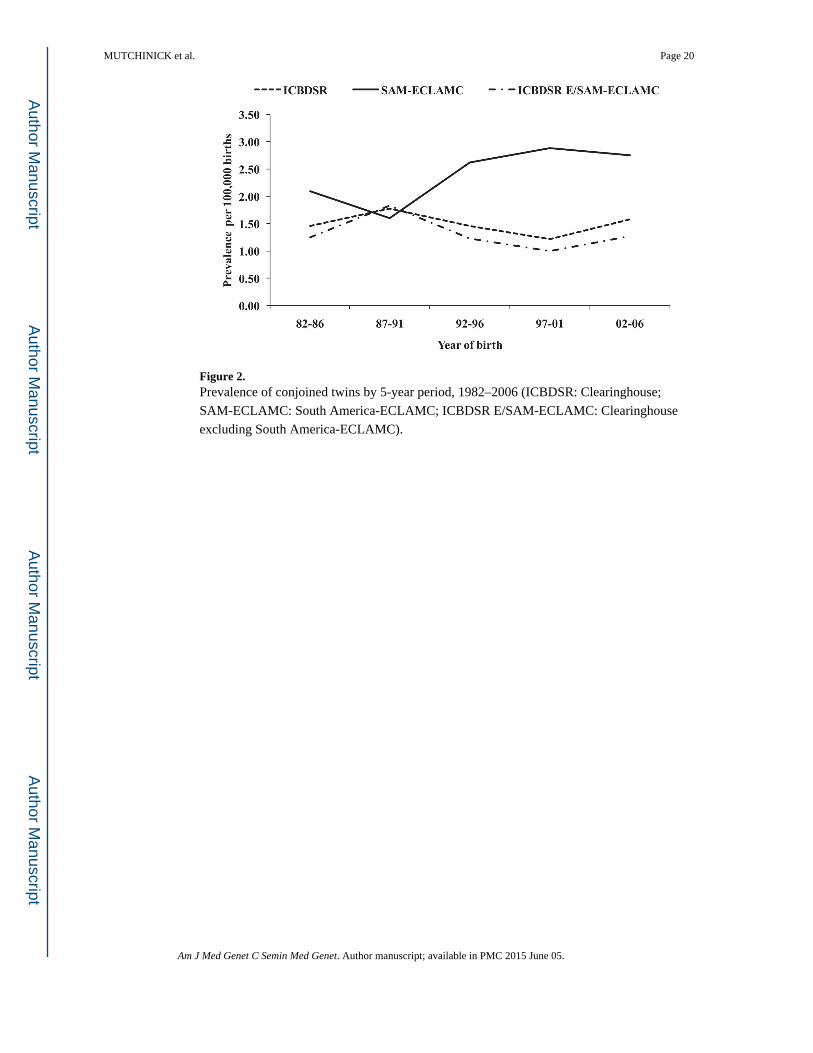
Figure 2. Prevalence of conjoined twins by 5-year period, 1982–2006 (ICBDSR: Clearinghouse;
SAM-ECLAMC: South America-ECLAMC; ICBDSR E/SAM-ECLAMC: Clearinghouse
excluding South America-ECLAMC).
MUTCHINICK et al. Page 20
Am J Med Genet C Semin Med Genet. Author manuscript; available in PMC 2015 June 05.
Author M
anuscriptA
uthor Manuscript
Author M
anuscriptA
uthor Manuscript
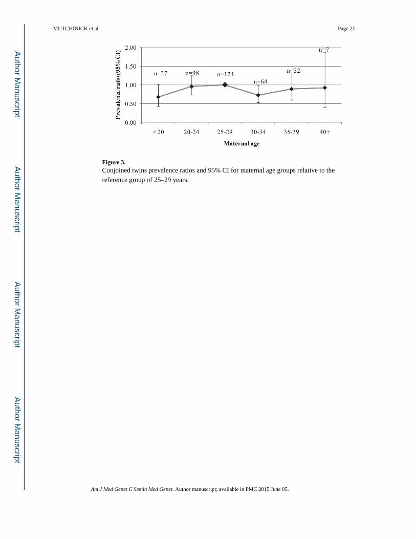
Figure 3. Conjoined twins prevalence ratios and 95% CI for maternal age groups relative to the
reference group of 25–29 years.
MUTCHINICK et al. Page 21
Am J Med Genet C Semin Med Genet. Author manuscript; available in PMC 2015 June 05.
Author M
anuscriptA
uthor Manuscript
Author M
anuscriptA
uthor Manuscript
Author M
anuscriptA
uthor Manuscript
Author M
anuscriptA
uthor Manuscript
MUTCHINICK et al. Page 22
TA
BL
E I
Tot
al P
reva
lenc
e of
Con
join
ed T
win
s in
21
Surv
eilla
nce
Prog
ram
s of
the
Inte
rnat
iona
l Cle
arin
ghou
se f
or B
irth
Def
ects
Sur
veill
ance
and
Res
earc
h
(IC
BD
SR)
Surv
eilla
nce
Pro
gram
Per
iod
Bir
ths
Tot
al c
ases
Pre
vale
nce
(per
100
,000
bir
ths)
95%
CI
P-v
alue
*
Can
ada-
Alb
erta
1980
–200
51,
062,
483
111.
040.
52–1
.85
0.15
0
USA
-Uta
h19
97–2
004
380,
706
61.
580.
58–3
.43
0.67
3
USA
-Atla
nta
1968
–200
41,
283,
999
251.
951.
26–2
.87
0.09
9
USA
-Tex
as19
96–2
002
2,05
4,78
813
0.63
0.34
–1.0
80.
0004
Mex
ico-
RY
VE
MC
E19
78–2
005
1,05
8,88
524
2.27
1.45
–3.3
70.
027
Sout
h A
mer
ica-
EC
LA
MC
1982
–200
64,
556,
173
108
2.37
1.94
–2.8
6<0
.000
1
Finl
and
1993
–200
471
3,49
423
3.22
2.04
–4.8
40.
0005
Wal
es19
98–2
004
222,
309
41.
800.
49–4
.61
0.41
1
Nor
ther
n N
ethe
rlan
ds19
81–2
003
369,
658
51.
350.
44–3
.16
0.54
3
Ger
man
y Sa
xony
-Anh
alt
1980
–200
435
5,18
48
2.25
0.97
–4.4
40.
155
Slov
ak R
epub
lic20
00–2
005
318,
257
10.
310.
01–1
.75
0.05
4
Hun
gary
1980
–200
53,
022,
194
401.
320.
95–1
.80
0.29
1
Fran
ce-C
entr
al E
ast
1979
–200
42,
500,
214
371.
481.
04–2
.04
0.49
8
Ital
y-N
orth
Eas
t19
81–2
004
1,18
6,49
71
0.08
0.00
–0.4
7<0
.000
1
Ital
y E
mili
a R
omag
na19
82–2
004
558,
176
40.
720.
20–1
.83
0.09
0
Ital
y-T
usca
ny19
92–2
004
336,
744
20.
590.
07–2
.15
0.13
1
Ital
y-C
ampa
nia
1992
–200
464
3,96
23
0.47
0.10
–1.3
60.
016
Spai
n-E
CE
MC
1980
–200
42,
045,
751
160.
780.
45–1
.27
0.00
4
Isra
el19
75–2
005
151,
562
10.
660.
02–3
.68
0.34
8
Chi
na-B
eijin
g19
92–2
005
1,92
7,62
228
1.45
0.97
–2.1
00.
582
Aus
tral
ia-V
icto
ria
1983
–200
41,
390,
179
231.
651.
05–2
.48
0.30
8
Tot
al26
,138
,837
383
1.47
1.32
–1.6
2
EC
EM
C, E
stud
io C
olab
orat
ivo
Esp
añol
de
mal
form
acio
nes
Con
géni
tas;
EC
LA
MC
, Est
udio
Col
abor
ativ
o L
atin
o A
mer
ican
o de
Mal
form
acio
nes
Con
géni
tas;
RY
VE
MC
E, R
egis
tro
y V
igila
ncia
E
pide
mio
lógi
ca d
e M
alfo
rmac
ione
s C
ongé
nita
s.
* P: e
xact
cum
ulat
ive
Pois
son
P-v
alue
. Bol
d va
lues
den
ote
stat
istic
ally
sig
nifi
cant
hig
h or
low
CT
pre
vale
nce.
Am J Med Genet C Semin Med Genet. Author manuscript; available in PMC 2015 June 05.
Author M
anuscriptA
uthor Manuscript
Author M
anuscriptA
uthor Manuscript
MUTCHINICK et al. Page 23
TA
BL
E II
Prop
ortio
n of
Pre
gnan
cy O
utco
mes
by
Surv
eilla
nce
Prog
ram
Liv
ebir
thSt
illbi
rth
ET
OP
FA
Tot
al
Surv
eilla
nce
Pro
gram
n%
n%
n%
n%
Can
ada-
Alb
erta
654
.53
27.3
218
.211
100.
0
USA
-Uta
h2
33.3
00.
04
66.7
610
0.0
USA
-Atla
nta
1356
.54
17.4
626
.123
100.
0
USA
-Tex
as4
30.8
646
.23
23.1
1310
0.0
Mex
ico-
RY
VE
MC
E17
70.8
729
.2N
P—
2410
0.0
Sout
h A
mer
ica-
EC
LA
MC
8679
.622
20.4
NP
—10
810
0.0
Finl
and
522
.71
4.5
1672
.722
100.
0
Wal
es0
0.0
125
.03
75.0
410
0.0
Nor
ther
n N
ethe
rlan
ds3
60.0
120
.01
20.0
510
0.0
Ger
man
y Sa
xony
-Anh
alt
112
.51
12.5
675
.08
100.
0
Slov
ak R
epub
lic1
100.
00
0.0
00.
01
100.
0
Hun
gary
1025
.014
35.0
1640
.040
100.
0
Fran
ce-C
entr
al E
ast
410
.83
8.1
3081
.137
100.
0
Ital
y-N
orth
Eas
t0
0.0
00.
01
100.
01
100.
0
Ital
y-E
mili
a R
omag
na1
25.0
250
.01
25.0
410
0.0
Ital
y-T
usca
ny0
0.0
00.
02
100.
02
100.
0
Ital
y-C
ampa
nia
133
.30
0.0
266
.73
100.
0
Spai
n-E
CE
MC
1062
.56
37.5
NR
—16
100.
0
Chi
na-B
eijin
g6
21.4
2278
.6N
R—
2810
0.0
Aus
tral
ia-V
icto
ria
313
.010
43.5
1043
.523
100.
0
Tot
al17
345
.610
327
.210
327
.237
910
0.0
Surv
eilla
nce
Prog
ram
s ar
e or
dere
d by
geo
grap
hy N
orth
-Sou
th a
nd W
est-
Eas
t. Is
rael
Sur
veill
ance
Pro
gram
was
exc
lude
d si
nce
thei
r si
ngle
rep
orte
d ca
se h
and
an u
nkno
wn
birt
h ou
tcom
e. T
hree
cas
es, 1
fro
m
Finl
and
and
2 fr
om U
SA A
tlant
a w
ere
also
exc
lude
d.
ET
OPF
A, e
lect
ive
term
inat
ion
of p
regn
ancy
for
fet
al a
nom
aly;
NP,
not
per
mitt
ed; N
R, n
ot r
epor
ted;
EC
EM
C, E
stud
io C
olab
orat
ivo
Esp
añol
de
Mal
form
acio
nes
Con
géni
tas;
EC
LA
MC
, Est
udio
C
olab
orat
ivo
Lat
ino
Am
eric
ano
de M
alfo
rmac
ione
s C
ongé
nita
s; R
YV
EM
CE
, Reg
istr
o y
Vig
ilanc
ia E
pide
mio
lógi
ca d
e M
alfo
rmac
ione
s C
ongé
nita
s.
Am J Med Genet C Semin Med Genet. Author manuscript; available in PMC 2015 June 05.
Author M
anuscriptA
uthor Manuscript
Author M
anuscriptA
uthor Manuscript
MUTCHINICK et al. Page 24
TA
BL
E II
I
Num
ber
of T
ypes
of
Con
join
ed T
win
s R
epor
ted
by E
ach
Surv
eilla
nce
Prog
ram
and
Tot
al P
reva
lenc
e Pr
opor
tions
Surv
eilla
nce
Pro
gram
aT
hora
copa
gus
Par
apag
us d
icep
halu
sC
epha
lopa
gus
Om
phal
opag
usP
aras
itic
Cra
niop
agus
Par
apag
us d
ipro
sopu
sIs
chio
pagu
sR
achi
pagu
sP
ygop
agus
Not
spe
cifi
edT
otal
Can
ada-
Alb
erta
51
00
20
10
00
211
USA
-Atla
nta
113
10
00
00
01
925
USA
-Tex
as3
33
21
00
10
00
13
Mex
ico-
RY
VE
MC
E5
54
02
03
21
02
24
Sout
h A
mer
ica-
EC
LA
MC
5110
415
85
60
30
610
8
Finl
and
111
50
22
11
00
023
Hun
gary
287
13
00
01
00
040
Fran
ce-C
entr
al E
ast
00
00
00
00
00
3737
Spai
n-E
CE
MC
122
00
00
00
00
216
Chi
na-B
eijin
g14
72
00
10
00
13
28
Aus
tral
ia-V
icto
ria
50
10
01
00
00
1623
Oth
ers
165
01
04
02
02
535
Tot
al (
n)16
144
2121
1513
117
44
8238
3
Tot
al (
%)
42.0
11.6
5.5
5.5
3.9
3.4
2.9
1.8
1.0
1.0
21.4
100.
0
EC
EM
C, E
stud
io C
olab
orat
ivo
Esp
añol
de
Mal
form
acio
nes
Con
géni
tas;
EC
LA
MC
, Est
udio
Col
abor
ativ
o L
atin
o A
mer
ican
o de
Mal
form
acio
nes
Con
géni
tas;
RY
VE
MC
E, R
egis
tro
y V
igila
ncia
Epi
dem
ioló
gica
de
Mal
form
acio
nes
Con
géni
tas.
a Onl
y th
ose
Surv
eilla
nce
Prog
ram
s th
at r
epor
ted
10 o
r m
ore
sets
of
CT
wer
e sp
ecif
ied.
The
rem
aini
ng 1
0 re
port
ing
less
than
10
case
s ar
e gr
oupe
d to
geth
er a
s “O
ther
s” (
USA
-Uta
h, W
ales
, Nor
ther
n N
ethe
rlan
ds, G
erm
any
Saxo
ny-A
nhal
t, Sl
ovak
Rep
ublic
, Ita
ly-N
orth
Eas
t, It
aly-
Em
ilia
Rom
agna
, Ita
ly-T
usca
ny, I
taly
-Cam
pani
a, I
srae
l).
Am J Med Genet C Semin Med Genet. Author manuscript; available in PMC 2015 June 05.
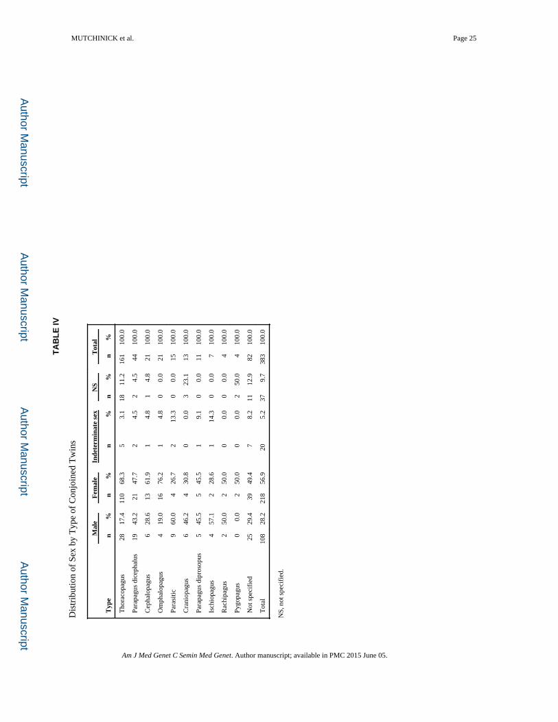
Author M
anuscriptA
uthor Manuscript
Author M
anuscriptA
uthor Manuscript
MUTCHINICK et al. Page 25
TA
BL
E IV
Dis
trib
utio
n of
Sex
by
Typ
e of
Con
join
ed T
win
s
Mal
eF
emal
eIn
dete
rmin
ate
sex
NS
Tot
al
Typ
en
%n
%n
%n
%n
%
Tho
raco
pagu
s28
17.4
110
68.3
53.
118
11.2
161
100.
0
Para
pagu
s di
ceph
alus
1943
.221
47.7
24.
52
4.5
4410
0.0
Cep
halo
pagu
s6
28.6
1361
.91
4.8
14.
821
100.
0
Om
phal
opag
us4
19.0
1676
.21
4.8
00.
021
100.
0
Para
sitic
960
.04
26.7
213
.30
0.0
1510
0.0
Cra
niop
agus
646
.24
30.8
00.
03
23.1
1310
0.0
Para
pagu
s di
pros
opus
545
.55
45.5
19.
10
0.0
1110
0.0
Isch
iopa
gus
457
.12
28.6
114
.30
0.0
710
0.0
Rac
hipa
gus
250
.02
50.0
00.
00
0.0
410
0.0
Pygo
pagu
s0
0.0
250
.00
0.0
250
.04
100.
0
Not
spe
cifi
ed25
29.4
3949
.47
8.2
1112
.982
100.
0
Tot
al10
828
.221
856
.920
5.2
379.
738
310
0.0
NS,
not
spe
cifi
ed.
Am J Med Genet C Semin Med Genet. Author manuscript; available in PMC 2015 June 05.

Author M
anuscriptA
uthor Manuscript
Author M
anuscriptA
uthor Manuscript
MUTCHINICK et al. Page 26
TA
BL
E V
Num
ber
and
Prop
ortio
n of
Maj
or C
onge
nita
l Mal
form
atio
ns U
nrel
ated
to th
e Si
te o
f U
nion
Obs
erve
d in
73
Sets
of
Con
join
ed T
win
s
Mal
form
atio
nT
hora
copa
gus
Par
apag
us D
icep
halu
sO
mph
alop
agus
Par
asit
icC
rani
opag
usP
arap
agus
Dip
roso
pus
Cep
halo
pagu
sIs
chio
pagu
sP
ygop
agus
Tot
al%
Neu
ral t
ube
defe
cts
43
00
03
00
111
9.91
Hyd
roce
phal
y3
00
00
00
01
43.
60
Oth
er c
entr
al n
ervo
us s
yste
m d
efec
ts1
22
00
00
00
54.
50
Mic
ro-o
phth
alm
ia1
00
00
00
00
10.
90
Faci
al c
left
s5
12
00
21
00
119.
91
Con
geni
tal h
eart
def
ects
01
21
00
01
27
6.31
Gas
troi
ntes
tinal
atr
esia
s1
32
11
11
01
119.
91
Abd
omin
al w
all d
efec
ts0
00
11
00
20
43.
60
Gen
itour
inar
y de
fect
s6
15
11
11
24
2219
.82
Mus
culo
skel
etal
ano
mal
ies
63
20
10
02
014
12.6
1
Lim
b re
duct
ion
anom
alie
s5
00
11
00
00
76.
31
Poly
dact
yly/
synd
acty
ly0
20
00
00
00
21.
80
Oth
er5
22
20
10
00
1210
.81
Tot
al37
1817
75
83
79
111
100.
00
Am J Med Genet C Semin Med Genet. Author manuscript; available in PMC 2015 June 05.


![National and Regional Summary of Select Surveillance ... and Regional Summary of Select Surveillance Components HHS ... number positive for influenza by virus type. ... [138]: 126](https://img.pdfslide.us/doc/110x75/5ae677477f8b9a29048d8403/national-and-regional-summary-of-select-surveillance-and-regional-summary-of.jpg)