Embed Size (px)
Citation preview
/ J of IMAB. 2013, vol. 19, issue 4/ http://www.journal-imab-bg.org 407
SURGICAL TREATMENT AND RECONSTRUCTION
FOR CENTRAL GIANT CELL GRANULOMA OF
MANDIBLE- case report and literature review
Elitsa G. Deliverska, Hristo D. Stoyanov.Department of Oral and Maxillofacial surgery, Faculty of dental medicine, MedicalUniversity, Sofia, Bulgaria
Journal of IMAB - Annual Proceeding (Scientific Papers) 2013, vol. 19, issue 4ISSN: 1312-773X (Online)
ABSTRACT:Introduction: Central giant cell granuloma (CGCG)
is a benign aggressive destructive osteolytic lesion ofosteoclastic origin. The central giant cell granuloma is oftenfound in the mandible, anterior to the first molars. It mostcommonly occurs in patients under the age of 30, with a clearfemale prevalence
Purpose: To present a case of CGCG of the lower jawin Department of Oral and maxillofacial surgery, UniversityHospital ‘St Anna’. Although en bloc resection provides thelowest recurrence rate, only a few single case reports describethe use of this technique followed by reconstruction withautogenous bone grafts.
Material and methods: The medical history of a 28years patient with a large central giant cell granuloma in themandible. Biopsy specimen taken from the lesion showedCGCG followed by curettage with peripheral ostectomy withpreservation of the continuity of the mandible.
Result: At the 1-year clinical and radiological followup there was no sign of recurrence.
Conclusion: After complete healing of the graft,prosthetic rehabilitation with implants will be perfomed. Thisallows the best functional and aesthetic results.
Key words: central giant cell granuloma, autogenousbone grafts, reconstruction
INTRODUCTION:The central giant-cell reparative granuloma(CGCRG)
has been defined as a localized benign but sometimesaggressive osteolytic proliferation consisting of fibrous tissuewith hemorrhage and hemosiderin deposits, presence ofosteoclast-like giant cells and reactive bone formation.CGCRG has been first described by Jaffe in 1953 [5] andaccounts for approximately 7% of all benign tumours of thejaws.[6] It is usually appears as solitary, multilocular,radiolucencies, located in the mandible ( anterior to the firstmolars) and maxilla. It occurs at least twice as often in themandible than in the maxilla. CGCRG most commonlyoccurs in patients under the age of 30, with a clear femaleprevalence.[6] The aetiology of giant cell granuloma is
undefined; some describe it as an inflammatory proliferation,some lesions behave as a neoplastic process in an aggressivefashion. Jaffe considered this tumour as a locally reparativereaction of the bone due to inflammation, local trauma orhaemorrhage. [5] In the literature there is little evidence ofany local reparative process.
The clinical behaviour of CGCG ranges from a slowgrowing asymptomatic swelling to an aggressive lesion thatpresents pain, local bone destruction, root resorption or toothdisplacement .Currently, clinical signs and symptoms,radiological features and histological features are the maincriteria to differentiate between non-aggressive (indolent) andaggressive lesions.
Aggressive lesions are characterized by one or moreof the following features: pain, paraesthesia, root resorption,rapid growth, cortical perforation, and a high recurrence rateafter surgical curettage- between 37.5% and 70% [4, 7, 10]and are mostly found in younger patients [2] are larger (over5 cm)[1]. These aggressive type or recurrent lesions requirewide en-bloc resection that leads to major defects in the jawsthat can alter the facial contours [2, 3, 8] and necessitatemajor reconstruction. Some surgeons use autogenous bonegrafts or vascularized fibula free flap for reconstruction ofextensive CGCG.[3, 8] Histologically there is no strictcriterion to differentiate between aggressive and non-aggressive lesions, however the number and volume of giantcells versus other components of the lesion might give anindication on its clinical behaviour.[1, 2, 6, 10]
Non-aggressive lesions are usually slow growing,symptom free and the treatment includes conservativesurgical procedures.
Although the majority of cases were asymptomatic,the most common feature was a painless smooth swelling inthe face or in the oral cavity. The lesion does not invade theperineural sheets so paresthesia is not usually observed inthese patients. The other symptoms and signs are facialasymmetry, impaired nasal breathing, loosing or displacementof teeth, and pathologic fracture.[6, 9]
CGCRGs usually present as an expansile radiolucency(87.5%) in X-ray films, but radiologic features vary from ill-defined destructive lesions to a well-defined, multilocular or
http://dx.doi.org/10.5272/jimab.2013194.407
408 http://www.journal-imab-bg.org / J of IMAB. 2013, vol. 19, issue 4/
unilocular appearance, with root resorption in 13.5% of thelesions and displacement of teeth in 18.0%.
Histologically, multinucleated giant cells, in a cellularvascular stroma, and often-new bone formation aredemonstrated. The osteoclast-like giant cells have a patchydistribution and are usually associated with areas ofhemorrhage. Ultrastructurally, the proliferating cells includespindle- shaped fibroblasts, myofibroblasts, and inflammatorymononuclear cells.[2, 6]
Differential diagnosis should be considered:Aneurysmal bone cyst, chondroblastoma, osteoblasto-clastoma, ameloblastoma, fibroma non-ossificans, hyper-parathyroidism, odontogenic cysts etc.
The treatment of CGCG of the jaws is performedaccording to the following factors: aggressive versus non-aggressive behaviour, location, size and radiographicappearance. Surgical options range from large (en blocresection) to more conservative approaches (curettage).
The traditional therapy of CGCRG has been localcurettage( this has been associated with a high success rate-80%), peripheral osteotomy, excision if needed reconstructionby using an autologous bone graft.[2, 6]
Surgical treatment of CGCRGs can be associated withrecurrence and serious facial mutilation and loss of teeth andtooth germs. To avoid such disadvantages, a number ofalternative nonsurgical herapies including interferon alpha-2a, calcitonin and intralesional corticosteroid injection havebeen advocated for the management of CGCRG . Non-surgical treatment of CGCRG is probably a good treatmentoption for small slowly enlarging lesions. Successfultreatment of painful, large, and rapidly growing lesions ismore likely achieved by surgical removal. In the literature,recurrence rates vary between 11% and 35%.[6]
CASE REPORTWe present 26 years old male with histopathologic
examination of the lesion reported as ‘giant cell reparativegranuloma’ of the mandible. On clinical examination thepatient was without subjective complaints. Biopsy specimentaken from the lesion showed CGCG.
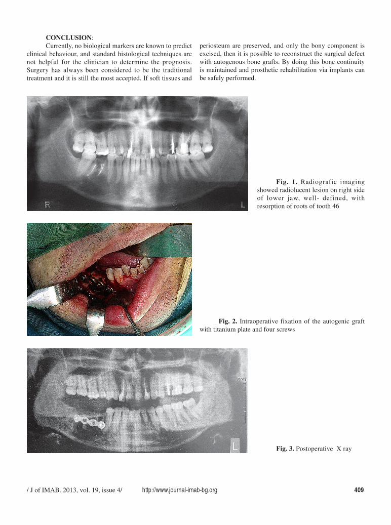
Radiografic imaging showed radiolucent lesion onright side of lower jaw, well- defined, with resorption of rootsof tooth 46, unilocular in appearance (Fig. 1). The patient wasoperated under general anaesthesia. The tumour mass wasremoved through an intraoral approach and curettage withperipheral ostectomy with preservation of the continuity ofthe mandible with at least a 5 mm margin was performed.Teeth 45 and 47 were extracted. The inferior alveolar nervewas preserved. Immediate reconstruction was carried out forthis case with with autogenous bone graft from mental region(Fig. 2). The bone graft was fixed with four screws. (Fig. 3)No complication was observed in terms of loss of teeth,wound dehiscence, infection of the surgical site, graftincorporation, fracture or loss of plates and screws and
necrosis of bone segments. Prosthetic rehabilitation withimplants will be perfomed.
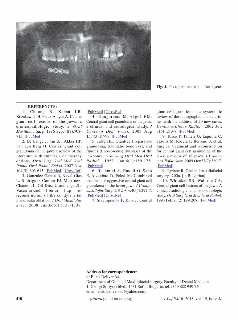
RESULT:At the 1-year clinical and radiological follow up there
was no sign of recurrence. The postoperative defect is fullyreconstructed. (Fig. 4)
DISCUSSION:The true nature of CGCG remains speculative and
considerable controversy exists in the literature. Normally,it is not considered an odontogenic lesion.[8] It has
been suggested that it might be an inflammatory lesion, areactive lesion, a true tumour, or an endocrine lesion.[5, 8]One hypothesis suggests that CGCG belongs to the spectrumof mesenchymal proliferative vascular primary jaw lesions.[8]CGCG occurs predominantly in children or young adults,with approximately 75% of cases presenting before 30 yearsof age(our patient is 26 years of age), however it really canoccur at any age. Females are affected more frequently thanmales, with a ratio of 2:1 [8] and more than 70% of CGCGsoccur in the mandible and less than 30% in the maxilla witha preference for the anterior portions of both bones.[8] Dueto the special anatomical characteristics of the maxilla,presentation, diagnosis, progress, management, and prognosisof maxillary CGCG are different from that of mandibularlesions: the cancellous nature of the maxilla and its thincortical plates allow the lesion to expand much earlier thanin the mandible.[8] The radiographic features of maxillaryCGCGs are variable and may be confused with those of otherlesions. They have been described as ranging from aunilocular to a multilocular radiolucent appearance with well-or illdefined borders.[10]
In the present case of CGCGs, there was amultilocular, radiolucent non perforating lesion which do notinvolved the cortical bone. Different authors [10] haveclassified CGCG into two types, based on clinical andradiographic features. The first is non-aggressive CGCG,which is characterized by a slow, almost asymptomaticgrowth that does not perforate the cortical bone or induce rootresorption and has a low tendency to recur. The second isaggressive CGCG, which is characterized by pain, rapidgrowth, expansion, and perforation of the cortical bone,radicular resorption and a high tendency to recur. Theaggressive lesions are mostly found in younger patients.[2]Aggressive lesions were also larger in size and from thehistological point of view they showed a larger fractionalsurface area occupied by giant cells. In aggressive lesions alsocan be found a higher number of giant cells. The most reliablefactors which relate to an increased risk of recurrence includeclinical activity of lesions (72% of recurrence in theaggressive forms, 3% of recurrence in the non-aggressiveforms), young age, presence of perforation of cortical boneand tumour size.[8]
/ J of IMAB. 2013, vol. 19, issue 4/ http://www.journal-imab-bg.org 409
CONCLUSION:Currently, no biological markers are known to predict
clinical behaviour, and standard histological techniques arenot helpful for the clinician to determine the prognosis.Surgery has always been considered to be the traditionaltreatment and it is still the most accepted. If soft tissues and
periosteum are preserved, and only the bony component isexcised, then it is possible to reconstruct the surgical defectwith autogenous bone grafts. By doing this bone continuityis maintained and prosthetic rehabilitation via implants canbe safely performed.
Fig. 1. Radiografic imagingshowed radiolucent lesion on right sideof lower jaw, well- defined, withresorption of roots of tooth 46
Fig. 2. Intraoperative fixation of the autogenic graftwith titanium plate and four screws
Fig. 3. Postoperative X ray
410 http://www.journal-imab-bg.org / J of IMAB. 2013, vol. 19, issue 4/
1. Chuong R, Kaban LB,Kozakewich H, Perez-Atayde A. Centralgiant cell lesions of the jaws: aclinicopathologic study. J OralMaxillofac Surg. 1986 Sep;44(9):708-713, [PubMed]
2. De Lange J, van den Akker HP,van den Berg H. Central giant cellgranuloma of the jaw: a review of theliterature with emphasis on therapyoptions. Oral Surg Oral Med OralPathol Oral Radiol Endod. 2007 Nov104(5): 603-615. [PubMed] [CrossRef]
3. Gonzalez-Garcia R, Naval-GiasL, Rodriguez-Campo FJ, Martinez-Chacon JL, Gil-Diez Usandizaga JL.Vascularized fibular flap forreconstruction of the condyle aftermandibular ablation. J Oral MaxillofacSurg. 2008 Jun;66(6):1133-1137.
[PubMed] [CrossRef]4. Gungormus M, Akgul HM.
Central giant cell granuloma of the jaws:a clinical and radiological study. JContemp Dent Pract. 2003 Aug15;4(3):87-97. [PubMed]
5. Jaffe HL. Giant-cell reparativegranuloma, traumatic bone cyst, andfibrous (fibro-oseous) dysplasia of thejawbones. Oral Surg Oral Med OralPathol. 1953 Jan;6(1):159-175.[PubMed]
6. Rachmiel A, Emodi O, SaboE, Aizenbud D, Peled M. Combinedtreatment of aggressive central giant cellgranuloma in the lower jaw. J Cranio-maxillofac Surg. 2012 Apr;40(3):292-7.[PubMed] [CrossRef]
7. Stavropoulos F, Katz J. Central
giant cell granulomas: a systematicreview of the radiographic characteris-tics with the addition of 20 new cases.Dentomaxillofac Radiol. 2002 Jul;31(4):213-7. [PubMed]
8. Tosco P, Tanteri G, Iaquinta C,Fasolis M, Roccia F, Berrone S, et al.Surgical treatment and reconstructionfor central giant cell granuloma of thejaws: a review of 18 cases. J Cranio-maxillofac Surg. 2009 Oct;37(7):380-7.[PubMed]
9. Ugrinov R. Oral and maxillofacialsurgery, 2006. [in Bulgarian]
10. Whitaker SB, Waldron CA.Central giant cell lesions of the jaws. Aclinical, radiologic, and histopathologicstudy. Oral Surg Oral Med Oral Pathol.1993 Feb;75(2):199-208. [PubMed]
REFERENCES:
Fig. 4. Postoperative result after 1 year.
Address for correspondence:dr Elitsa Deliverska,Department of Oral and Maxillofacial surgery, Faculty of Dental Medicine,1, Georgi Sofiyski blvd., 1431 Sofia, Bulgaria; tel.+359 888 949 740;email: [email protected],