Embed Size (px)
Citation preview
SURGICAL SITE INFECTIONS AMONG
PATIENTS UNDERWENT CLEAN AND CLEAN-
CONTAMINATED SURGERY IN HOSPITAL
UNIVERSITI SAINS MALAYSIA: RISK FACTORS,
MICROBIOLOGICAL AND Staphylococcus aureus
MOLECULAR PROFILE
By
WONG JUN LEONG
Thesis submitted in fulfilment of the requirements
for the degree of
Master of Science
DECEMBER 2015
ii
ACKNOWLEDGEMENTS
I would like to express my sincere appreciation to my supervisor Associate
Professor Dr. Siti Asma’ binti Hassan for allowing me to join her research group
and also for her continuous encouragement, patience, and guidance over the
years. She has enlightened me how to conduct research experiments and write
research papers. In addition, I am grateful to my co-supervisor, Professor Dr.
Habsah binti Hasan, Professor Dr. Mohamad Ziyadi bin Hj Ghazali and Dr. Zaidi
bin Zakaria, and biostatistician Dr. Siti Azrin binti Ab Hamid for their constant
advice and problem solving assistance that making me able to complete my study
well.
I want to thank my colleagues Chan Shiao Ee, Engku Ibrahim Syubli bin
Engku Safruddin, Engku Nur Syafirah binti Engku Abd Rahman, Fitrien binti
Husin, Low Kim Fatt, Mohd Fazli bin Ismail, Muhammad Azharuddin bin Azali,
Muhammad Lukman bin Yahya, Nik Zuraina binti Nik Mohd Noor, Nur Adila binti
Zakaria, Nur Adlina binti Zainuddin, Nur Amalina binti Khazani, Nur Izzah
Farakhin binti Ayub, Nur Izzati binti Hamdan, Nurul Najian binti Aminuddin Baki,
Siti Nurain binti Osman, Tengku Ahmad Akram bin Tengku Mohd Ariffin, Yasmin
Kahirani binti Muhammad Ismadi and others for their support, enthusiasm, and
friendship that have helped me through many failed experiments. I also gratefully
acknowledge all lecturers, administrative officers, Amanina binti Aminuddin,
Fadzilah binti Hj Ahmad as well as other Medical Lab Technologists, from the
Department of Medical Microbiology and Parasitology, Universiti Sains Malaysia
(USM) for their dedication in helping me and answering all of my numerous
questions.
iii
I would also like to thank the research funding support received in the form
of a Short Term Grant (304.PPSP.61312112) from USM. In addition, support from
Pahang State Foundation in the form of an education loan is gratefully
acknowledged.
Last but not least, my deep appreciation goes to my parents (Wong Chai
Sing, Lau Poh Chian), siblings (Jun Yen, Jin Yee and Jun Chau), Lim Zhe Xin
and family who have influenced me the most and were always to back up and
encourage me. They have been my source of strength. I dedicate this thesis to
them.
iv
TABLE OF CONTENTS
ACKNOWLEDGEMENTS .................................................................................. II
TABLE OF CONTENTS .................................................................................... IV
LIST OF TABLES .............................................................................................. IX
LIST OF FIGURES ........................................................................................... XII
LIST OF SYMBOLS AND ABBREVIATIONS .................................................. XIV
ABSTRAK ....................................................................................................... XVI
ABSTRACT ..................................................................................................... XIX
CHAPTER 1 ....................................................................................................... 1
INTRODUCTION ................................................................................................ 1
CHAPTER 2 ....................................................................................................... 5
LITERATURE REVIEW ...................................................................................... 5
2.1 Surgical Site Infections (SSI) .................................................................... 5
2.1.1 Signs of SSI ........................................................................................ 6
2.1.2 Sites of SSI ......................................................................................... 7
2.1.3 Class of SSI ...................................................................................... 10
2.1.4 Risk Factors for SSI .......................................................................... 11
2.1.5 Prevention Bundles........................................................................... 12
2.1.5.1 Preoperative Care Bundles ............................................................ 13
2.1.5.1 (i) Optimize patient’s risk factors ................................................ 13
2.1.5.1 (ii) Nasal Screening for Methicillin-resistance Staphylococcus aureus .................................................................................................... 13
2.1.5.1 (iii) Antimicrobial Prophylaxis ...................................................... 14
2.1.5.1 (iv) Hair Removal ........................................................................ 17
2.1.5.1 (v) Other preoperative measures ................................................ 17
2.1.5.2 Intraoperative Care Bundles .......................................................... 18
2.1.5.2 (i) Operating Room Environment ................................................ 18
2.1.5.2 (ii) Skin Preparation .................................................................... 18
2.1.5.2 (iii) Maintaining Patient’s Body Temperature and Homeostasis . 18
2.1.5.2 (iv) Other intraoperative measures ............................................. 19
2.1.5.3 Postoperative Care Bundles .......................................................... 19
2.1.5.3 (i) Wound Care ........................................................................... 19
2.1.5.4 Treatment of SSI ............................................................................ 19
v
2.2 Staphylococcus aureus ........................................................................... 21
2.2.1 Pathogenicity .................................................................................... 22
2.2.2 Virulent factors .................................................................................. 23
2.2.3 Infections .......................................................................................... 26
2.2.4 Laboratory Detection ........................................................................ 27
2.2.5 Treatment ......................................................................................... 28
2.2.6 Antibiotic Resistance ........................................................................ 28
2.2.7 Methicillin-resistance Staphylococcus aureus (MRSA) ..................... 28
CHAPTER 3 ..................................................................................................... 30
General objective of study ............................................................................ 30
Specific objective of study ............................................................................. 30
CHAPTER 4 ..................................................................................................... 31
MATERIALS AND METHODS ......................................................................... 31
4.1 Study design ........................................................................................... 31
4.2 Study area .............................................................................................. 31
4.3 Study period ............................................................................................ 31
4.4 Study population and samples ................................................................ 31
4.4.1 Target population .............................................................................. 31
4.4.2 Source population ............................................................................. 32
4.4.3 Study population ............................................................................... 32
4.4.4 Sampling frame ................................................................................ 32
4.4.5 Sample size calculation .................................................................... 33
4.4.6 Sampling method .............................................................................. 35
4.5 Research and measurement tools .......................................................... 36
4.6 Method of data collection ........................................................................ 39
4.6.1 Pre-operative .................................................................................... 39
4.6.2 Intra-operative .................................................................................. 39
4.6.3 Post-operative .................................................................................. 40
4.6.4 Nasal swab ....................................................................................... 40
4.6.5 Wound inspection ............................................................................. 40
4.6.6 Follow up .......................................................................................... 41
4.6.7 Infected wound ................................................................................. 41
4.6.7.1 Tissue sample ............................................................................ 41
vi
4.6.7.2 Wound swab ............................................................................... 42
4.6.8 Discharged from the study ................................................................ 42
4.7 Microbiological processing and sampling method ................................... 42
4.7.1 Nasal swab ....................................................................................... 42
4.7.2 Wound tissue and swab .................................................................... 43
4.7.2.1 Day one ...................................................................................... 43
4.7.2.2 Day two ...................................................................................... 44
4.7.2.3 Day three .................................................................................... 44
4.7.2.4 Day four ...................................................................................... 45
4.7.3 Gram staining method ...................................................................... 45
4.7.4 Catalase test by slide method ........................................................... 46
4.7.5 Tube coagulase test ......................................................................... 46
4.7.6 Oxidase test ...................................................................................... 47
4.7.7 Vitek 2 identification method ............................................................. 48
4.7.8 Antimicrobial susceptibility test by Kirby-Bauer method .................... 49
4.8 Molecular Methods .................................................................................. 52
4.8.1 Preparation of Samples for Polymerase Chain Reaction (PCR) Analysis ..................................................................................................... 52
4.8.1.1 Extraction of Deoxyribonucleic acid (DNA) ................................. 52
4.8.1.2 Preparation of Primer ................................................................. 52
4.8.1.3 PCR reagent ............................................................................... 55
4.8.1.4 Mixture of PCR reagent and DNA templates .............................. 55
4.8.1.5 PCR amplification ....................................................................... 56
4.8.2 PCR analysis .................................................................................... 57
4.8.2.1 Electrophoresis by agarose gel .................................................. 57
4.8.2.2 Visualize the DNA band.............................................................. 58
4.8.2.3 DNA sequencing of PCR products ............................................. 58
4.9 Review of patients’ medical record ......................................................... 59
4.10 Statistical analysis ................................................................................. 59
4.11 Ethical approval .................................................................................... 59
CHAPTER 5 ..................................................................................................... 60
RESULTS......................................................................................................... 60
5.1 Incidence of SSI ...................................................................................... 61
5.2 Sociodemographic profile ....................................................................... 63
vii
5.3 Microbiological profile and antibiotic sensitivity pattern ........................... 70
5.4 Antimicrobial prophylaxis ........................................................................ 74
5.5 Molecular characterization of S. aureus strain isolated ........................... 77
CHAPTER 6 ..................................................................................................... 85
DISCUSSION ................................................................................................... 85
6.1 Introduction ............................................................................................. 85
6.2 Demographic of the Study ...................................................................... 86
6.3 Incidence of Surgical Site Infection ......................................................... 86
6.4 Risk Factors for Surgical Site Infection ................................................... 88
6.5 Microbiological Profile for Surgical Site Infection .................................... 90
6.6 Antimicrobial Sensitivity Pattern of Causative Microorganism ................ 91
6.7 Antimicrobial Prophylactic Agent............................................................. 92
6.8 Nasal Screening of S. aureus and MRSA carriage ................................. 94
6.9 Molecular Characterization of Staphylococcus aureus ........................... 94
CHAPTER 7 ..................................................................................................... 97
CONCLUSION ................................................................................................. 97
LIMITATION OF STUDY .................................................................................. 98
RESEARCH RECOMMENDATIONS ............................................................... 99
Funding sources ......................................................................................... 100
Competing interest ...................................................................................... 100
REFERENCES ............................................................................................... 101
APPENDICES .................................................................................................... 1
Appendices A - Patient Information................................................................. 1
Appendices B - Pre-Operation Checklist ......................................................... 2
Appendices C - Intra-Operation Checklist ....................................................... 5
Appendices D - Intra-Operation Checklist ....................................................... 6
Appendices E - Post-Operation Checklist ....................................................... 7
Appendices F - Follow Up During Suture To Open (Sto) Checklist ................. 8
Appendices G - Follow Up On Third Week Post-Operation Checklist ............. 9
Appendices H - Follow Up On Day 30 Post-Operation Checklist .................. 10
Appendices I - Questionare .......................................................................... 11
Appendices J - Gantt Chart of Research Activities: ...................................... 12
Appendices K – Ethical Approval (USMKK/PPP/JEPem [261.3.(5)] ............. 14
viii
Appendices L – Approval from Hospital USM ............................................... 16
Appendices M – Approval from Department of Surgery ................................ 17
Appendices N - Sequences of the isolated microorganism from the study. .. 18
LIST OF PUBLICATIONS AND PRESENTATIONS ......................................... 24
ix
LIST OF TABLES
Table Page
Table 2.1 Criteria for defining a SSI 8
Table 2.2 The CDC criteria were used to classify and define the
types of surgical wounds
10
Table 2.3 Risk factors that affect the outcome of surgery 11
Table 2.4 Antimicrobial prophylaxis agent recommended by
Minister of Health, Malaysia
16
Table 2.5 List of some virulent factors of S. aureus 24
Table 4.1 Inclusions and exclusions criteria 32
Table 4.2 Details of antimicrobial susceptibility test disc used 36
Table 4.3 Details of consumable used 37
Table 4.4 Details of laboratory equipment used 38
Table 4.5 Staphylococcus sp. antimicrobial susceptibility test
panel
49
Table 4.6 Enterobacteriaceae sp. antimicrobial susceptibility test
panel
50
Table 4.7 ESBL screening test panel 51
Table 4.8 Pseudomonas aeruginosa antimicrobial susceptibility
test panel
51
x
Table 4.9 Streptococcus ssp antimicrobial susceptibility test panel 51
Table 4.10 Primers used in this study 54
Table 4.11 Composition of DreamTaq Green PCR Master Mix (2X) 55
Table 4.12 Mixture of PCR reagents and DNA templates 56
Table 4.13 Thermal cycling conditions 57
Table 5.1 Demographic detail of patients 64
Table 5.2 Demographic detail of patients underwent elective clean
surgery in Hospital USM, by Fisher's exact test
65
Table 5.3 Demographic detail of patients underwent elective
clean-contaminated surgery in Hospital USM, by
Fisher's exact test
66
Table 5.4 Associated factors of SSI in patients by Simple Logistic
Regression
67
Table 5.5 Associated factors of SSI in patients by Multiple Logistic
Regression
68
Table 5.6 Microbiological profile of microorganism isolated from
SSI patients underwent clean and clean-contaminated
surgery
70
Table 5.7 Percentage of susceptibility pattern of Staphylococcus
sp.
71
xi
Table 5.8 Percentage of susceptibility pattern of
Enterobacteriaceae
72
Table 5.9 Percentage of susceptibility pattern of Pseudomonas
aeruginosa
73
Table 5.10 Percentage of susceptibility pattern of Streptococcus
agalactiae (Group B)
73
Table 5.11 Antimicrobial prophylaxis according to the class of
surgery
74
Table 5.12 List of microorganism that underwent PCR analysis 78
Table 5.13 List of genes detected among S. aureus and MRSA 84
xii
LIST OF FIGURES
Figure Page
Figure 2.1 Cross-section of abdominal wall according to the CDC
classifications of SSI
9
Figure 5.1 Incidence rate of SSI in Hospital USM 61
Figure 5.2 Type of SSI in Hospital USM 62
Figure 5.3 Nasal screening of patients 69
Figure 5.4 Antimicrobial prophylaxis according to the type of
surgery
76
Figure 5.5
A&B
Electrophoresis of universal PCR products on 1%
agarose gel
79
Figure 5.6 Multiplex PCR analysis for MRSA sample 80
Figure 5.7 Electrophoresis of TSST-1 PCR products on 1%
agarose gel
81
Figure 5.8 Electrophoresis of PVL PCR products on 1% agarose
gel
81
Figure 5.9 Electrophoresis of cna PCR products on 1% agarose
gel
82
Figure 5.10 Electrophoresis of hlg PCR products on 1% agarose gel 82
Figure 5.11 Electrophoresis of icaA PCR products on 1% agarose
gel
83
xiii
Figure 5.12 Electrophoresis of SdrE PCR products on 1% agarose
gel
83
xiv
LIST OF SYMBOLS AND ABBREVIATIONS
°C ………………………………………..………………………… Degree Celcius
BLAST ……………………………………… Basic Local Alignment Search Tool
BMI …………………………………..………………………….. Body Mass Index
CABG …………………………………………….. Coronary Artery Bypass Graft
CDC ………………………………. Centers for Disease Control and Prevention
cna ……………………………………………………………… Collagen adhesins
dATP ……………………………………………… Deoxyadenosine triphosphate
dCTP …………………………………………………. Deoxycytidine triphosphate
dGTP ……………………………………………… Deoxyguanosine triphosphate
DM ……………………………………………………………..… Diabetes Mellitus
DNA ………………………………………………………… Deoxyribonucleic acid
dTTP ……………………………………………….. Deoxythymidine triphosphate
ESBL ……………………………………….. Extended spectrum beta-lactamase
HCAI …………………………………………….. Healthcare associated infection
hlg …………………………………………………………….. Gamma haemolysin
IBM ……………………………… International Business Machines Corporation
ica ………………………………………… Polysaccharide intercellular adhesins
ILTKP …………………………… Infeksi Luka di Tapak Kawasan Pembedahan
xv
MIC ……………………………………………… Minimal inhibitory concentration
MRSA …………………………… Methicillin-resistance Staphylococcus aureus
MSSA ………………………..……. Methicillin-sensitive Staphylococcus aureus
NCBI ……………………………. National Center for Biotechnology Information
NICE …………………... The National Institute for Health and Care Excellence
PCR …………………………………………………. Polymerase Chain Reaction
PVL ………………………………………………….. Panton-Valentine leukocidin
S. aureus …………………………………………………. Staphylococcus aureus
SdrE …………………………………………………………….. Putative adhesins
SPSS ……………………….…. Software Package used for Statistical Analysis
SSI ………………………………………………………….. Surgical Site Infection
TBE …………………………………………………………….. Tris-Biorate-EDTA
TSS …………………………….…………………………. Toxic Shock Syndrome
TSST-1 ………………………………………….. Toxic Shock Syndrome Toxin 1
USM …………………………………………………….. Universiti Sains Malaysia
xvi
JANGKITAN PADA KAWASAN TAPAK PEMBEDAHAN DALAM
KALANGAN PESAKIT YANG MENJALANI PEMBEDAHAN BERSIH DAN
PEMBEDAHAN BERSIH TERCEMAR DI HOSPITAL UNIVERSITI SAINS
MALAYSIA: FAKTOR RISIKO, PROFIL Staphylococcus aureus DAN
PROFIL MIKROBIOLOGI
ABSTRAK
Infeksi luka di tapak kawasan pembedahan (ILTKP) adalah diantara
masalah kesihatan yang kerap dialami oleh pesakit yang menjalani pembedahan
di hospital. Kejadian ini dipantau secara rutin kerana ia melibatkan peningkatan
kadar mortaliti, morbiditi, dan juga kos rawatan. Jangkitan ini biasanya
disebabkan oleh mikroorganisma seperti Staphylococcus aureus, Streptococcus
spp, Enterococcus spp, dan Pseudomonas aeruginosa. Kenalpasti jenis
mikroorganisma penyebab serta corak kepekaan antimikrobial amat membantu
dalam pelan rawatan. Oleh itu, tujuan kajian ini adalah untuk menentukan kadar
dan faktor risiko ILTKP serta mengenal pasti agen penyebab dan corak kepekaan
antimikrobialnya.
Prospektif kajian kohort ini dijalankan dari Jun 2013 hingga Julai 2014 di
Hospital Universiti Sains Malaysia. Tujuh puluh dua orang pesakit yang menjalani
pembedahan bersih dan bersih tercemar telah bersetuju untuk menyertai kajian
ini dan mereka dipantau sama ada terdapat sebarang tanda-tanda ILTKP selama
30 hari selepas pembedahan. Pemeriksaan calitan hidung untuk mengesan
Staphylococcus aureus dan Methicillin-resistance Staphylococcus aureus juga
telah dijalankan sebelum pembedahan. Sampel tisu atau swab luka telah diambil
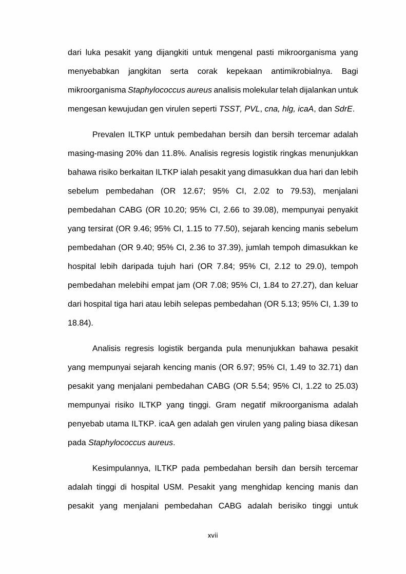
xvii
dari luka pesakit yang dijangkiti untuk mengenal pasti mikroorganisma yang
menyebabkan jangkitan serta corak kepekaan antimikrobialnya. Bagi
mikroorganisma Staphylococcus aureus analisis molekular telah dijalankan untuk
mengesan kewujudan gen virulen seperti TSST, PVL, cna, hlg, icaA, dan SdrE.
Prevalen ILTKP untuk pembedahan bersih dan bersih tercemar adalah
masing-masing 20% dan 11.8%. Analisis regresis logistik ringkas menunjukkan
bahawa risiko berkaitan ILTKP ialah pesakit yang dimasukkan dua hari dan lebih
sebelum pembedahan (OR 12.67; 95% CI, 2.02 to 79.53), menjalani
pembedahan CABG (OR 10.20; 95% CI, 2.66 to 39.08), mempunyai penyakit
yang tersirat (OR 9.46; 95% CI, 1.15 to 77.50), sejarah kencing manis sebelum
pembedahan (OR 9.40; 95% CI, 2.36 to 37.39), jumlah tempoh dimasukkan ke
hospital lebih daripada tujuh hari (OR 7.84; 95% CI, 2.12 to 29.0), tempoh
pembedahan melebihi empat jam (OR 7.08; 95% CI, 1.84 to 27.27), dan keluar
dari hospital tiga hari atau lebih selepas pembedahan (OR 5.13; 95% CI, 1.39 to
18.84).
Analisis regresis logistik berganda pula menunjukkan bahawa pesakit
yang mempunyai sejarah kencing manis (OR 6.97; 95% CI, 1.49 to 32.71) dan
pesakit yang menjalani pembedahan CABG (OR 5.54; 95% CI, 1.22 to 25.03)
mempunyai risiko ILTKP yang tinggi. Gram negatif mikroorganisma adalah
penyebab utama ILTKP. icaA gen adalah gen virulen yang paling biasa dikesan
pada Staphylococcus aureus.
Kesimpulannya, ILTKP pada pembedahan bersih dan bersih tercemar
adalah tinggi di hospital USM. Pesakit yang menghidap kencing manis dan
pesakit yang menjalani pembedahan CABG adalah berisiko tinggi untuk

xviii
mendapat ILTKP. Mikroorganisma Gram negatif adalah lebih kerap menjangkiti
pesakit berbanding Gram positif dan semua mikroorganisma ini adalah dalam
kumpulan yang peka kepada antibiotik.
xix
SURGICAL SITE INFECTIONS AMONG PATIENTS UNDERWENT CLEAN
AND CLEAN-CONTAMINATED SURGERY IN HOSPITAL UNIVERSITI SAINS
MALAYSIA: RISK FACTORS, MICROBIOLOGICAL AND Staphylococcus
aureus MOLECULAR PROFILE
ABSTRACT
Surgical site infections (SSI) are among the most commonly encountered
healthcare associated infection. The incidence were closely been monitored as it
is associated with considerable morbidity and mortality. The common aetiological
agents responsible for the infection include Staphylococcus aureus,
Streptococcus spp, Enterococcus spp, and Pseudomonas aeruginosa. The
identification of the causative agents as well as their antimicrobial sensitivity
pattern helps in the treatment plan. Therefore the aims of this study were to
determine the incidence and risk factors of SSI as well as to identify the causative
microorganisms and their sensitivity profile.
This prospective cohort study was conducted from June 2013 until July
2014 at Hospital Universiti Sains Malaysia. Seventy-two patients underwent
clean and clean-contaminated surgeries were consented preoperatively and
strictly followed up for any signs of SSI for duration of 30 days post operation.
Nasal screening for Staphylococcus aureus and Methicillin-resistance
Staphylococcus aureus was carried out preoperatively. Tissue samples or wound
swab from infected patients were taken for microbial identification and its
sensitivity pattern. Staphylococcus aureus strain isolated were proceed to
xx
polymerase chain reaction analysis to detect the virulence genes (TSST, PVL,
cna, hlg, icaA, and SdrE).
The overall incidence rate of SSI was 18.1% specifically for clean and
clean-contaminated surgeries are 20% and 11.8%, respectively. Significant risk
associated with SSI by simple logistic regression analysis included patients
admitted two days or more prior to surgery (OR 12.67; 95% CI, 2.02 to 79.53),
underwent CABG surgery (OR 10.20; 95% CI, 2.66 to 39.08), underlying
diseases (OR 9.46; 95% CI, 1.15 to 77.50), history of diabetes mellitus (DM) prior
to the surgery (OR 9.40; 95% CI, 2.36 to 37.39), total hospitalization period more
than seven days (OR 7.84; 95% CI, 2.12 to 29.0), duration of surgery more than
four hours (OR 7.08; 95% CI, 1.84 to 27.27), and discharged home three days or
longer after surgery (OR 5.13; 95% CI, 1.39 to 18.84).
Multiple logistic regression method demonstrated that the patients who
have history of DM (OR 6.97; 95% CI, 1.49 to 32.71) and underwent CABG
surgery (OR 5.54; 95% CI, 1.22 to 25.03) had significant risks of SSI. Gram
negative microorganism was the leading causative microorganism and in
Staphylococcus aureus strains, icaA gene was the most common virulence gene
detected.
In conclusion, SSI among clean and clean-contaminated surgeries are
high in our setting. DM and patients underwent CABG operation are at high risk
to get SSI. Gram negative microorganisms are common as compare to Gram
positive, however they are all sensitive strains.
1
CHAPTER 1
INTRODUCTION
Surgery is one of the major and important branches of medicine that
perform a surgical procedure to the human body for diagnostic, treatment,
prevention as well as palliative purpose. Microorganisms have an opportunity to
invade when a part of the body is operated during surgical procedure and causing
a post-operative infection. Such infection occurred post-operatively and was
referred as surgical site infection (SSI).
The previous term ‘Surgical Wound Infection’ was replaced by the term
‘SSI’ in year 1992 (Horan et al., 1992). SSI is defined as an infection that occurs
at site of incision, or any anatomical parts that was either opened or manipulated
during the procedure that occurs within 30 days after surgery, or within one year
if involve implantation (Horan et al., 1992). However, based on the Centers for
Disease Control and Prevention (CDC) criteria, stitch abscess was not
considered as SSI because stitch abscess is considered the minimal
inflammation and discharge limited only to the point of suture penetration (Horan
et al., 1992).
SSI remain as the most common Healthcare-associated infection (HCAI)
especially in patients who underwent surgery (Dionigi et al., 2001). According to
a surveillance done in the United States acute hospital in year 2011, SSI made
up of 21.8% of inpatient infections (Magill et al., 2014).
2
Studies reported that prevalence rate of SSI were range between 1.8%
and 20.3%, depends on the types of surgery (Dimick et al., 2004; Khan et al.,
2006; Emmanuel et al., 2012). Epidemiology studies reported that prevalence of
SSI were 1.9% in United States, 0.6% in Scotland, 5-10% in Australia and Japan,
and 12.9% in University of Malaya Medical Centre (Hughes et al., 2005; Coleman
et al., 2010; Scottish Surveillance of Healthcare Associated Infection Programme.,
2010; Tan et al., 2010; Mu et al., 2011).
Microorganisms responsible for the development of SSI varies.
Staphylococcus aureus (S. aureus) was reported as one of the leading causative
agent for SSI (A. report from the NNIS System., 2004; Magill et al., 2014). But in
the past decade, Methicillin-resistance Staphylococcus aureus (MRSA) was
responsible for most of the SSI and it becomes a challenge for medical
practitioner due to emergence and spread of resistance to a wide range of
antibiotics (Klein et al., 2007). Streptococcus sp. is the second most common
pathogen for SSI, followed by Enterococcus sp. and Pseudomonas aeruginosa
(Weigelt et al., 2009).
SSI often led to substantial morbidity and mortality in some patients.
Several studies reported that mortality rate of SSI were between 3% and 25.3%
(Kathryn B. Kirkland et al., 1999; Awad, 2012; Horasan et al., 2013). A study done
in Duke University Medical Center and Durham Regional Hospital had reported
mortality rate of SSI patient with MSSA was 6.7% and MRSA was 20.7%, MRSA
infected patients are 3.4 times at higher risk to die in 90-day post operation
(Engemann et al., 2003).
3
Besides that, the duration of hospital stay of SSI patient are also longer
than patients that did not develop any complication by two to 36 days, depending
on types of surgical intervention (Kathryn B. Kirkland et al., 1999; Dimick et al.,
2004; Broex et al., 2009; Jenks et al., 2014). Prolonged hospital stay will require
more resources, included additional diagnostic tests, and therapeutic use of
antibiotics. It also reduces the availability of beds for other patients as well as
human resources. As a consequences, this not only increase the financial burden
of patients’ family but also burden the healthcare system and economic.
A study done in the year 2009 has found that treating a SSI patient
required as high as 10,523 British pound (Tanner et al.), which include extended
hospital charges, medicine as well as additional diagnostic tests. Many factors
can affect the postoperative outcome of the patients such as patient-related (age,
underlying disease, tobacco use, diabetic mellitus status, obesity) and procedure-
related (method of skin preparation, selection of prophylactic antibiotics, duration
of operation, surgeon experience) (Arabshahi and Koohpayezade, 2006; Utsumi
et al., 2010; Hafez et al., 2012). Identification of modifiable risk factors for SSI
helps in developing the prevention strategies on reducing SSI, especially in
limited resources healthcare facilities.
SSI is a preventable surgical complication. Many SSI can be prevented if
SSI care bundle has been applied properly. The National Institute for Health and
Care Excellence (NICE) had published a guideline on prevention and treatment
of SSI in year 2008 (Leaper et al., 2008). This guideline gives details of the
methods on how to prevent SSI in pre-, intra- and postoperative phase.
4
Most of the time, our local data consist only those who acquired the
infection during hospitalization period, but SSI can develop within 30 days after
surgery. Lack of close monitoring system which failed to capture the SSI cases
from the patients that seek treatment from private clinical or other healthcare
institutional lead to under reporting the incidence rate. Knowing the incidence is
crucial to develop infection control policy for SSI. Furthermore, a cohort
prospective study will enable us to do elaborate profiles of the patient who
develop SSI and identify the problems with regards to preoperative preparation,
during operation, post-operative wound care and co morbid illness associated
with SSI.
Peacock et al. (2002) demonstrated virulent genes play a role in
determining invasive disease of S. aureus as well as its effect was accumulative
and increase the risk of disease. Among the S. aureus virulence genes, collagen
adhesins (cna), polysaccharide intercellular adhesins (ica), gamma haemolysin
(hlg), and putative adhesins (SdrE) are the most common isolated virulence gene
in invasive isolates (Peacock et al., 2002). By studying the associated virulence
genes in S. aureus can help in demonstrating their involvement in the occurrence
of SSI.
Therefore this study set up to determine the incidence and quantify the risk
factors of SSI. This is an essential step in order to develop the preventive strategy
to reduce the occurrence of SSI. In addition, identify the causative agent,
especially S. aureus and MRSA strain by using molecular method is not
necessary for treatment in SSI but can determine the evolution of the properties
of S. aureus and MRSA as well as provides useful information for characterization
of S. aureus and MRSA strain.
5
CHAPTER 2
LITERATURE REVIEW
2.1 Surgical Site Infections (SSI)
SSI is an infection that occurs at site of incision or any anatomical parts
that was either opened or manipulated during surgical procedure. It can occur
within 30 days after surgical procedure or within one year if involve implantation.
Complication of a SSI may range from spontaneously wound pus discharge and
inflammation to life-threatening, such as a sternal wound infection after CABG
surgery.
SSI may caused by contamination of an incision with microorganisms
either from the instruments, environment or patient's own body during surgery.
However, SSI caused by microorganisms from an outside source following
surgery is less common. SSI can badly effect on patient’s quality of life and
associated with considerable morbidity and extended hospital stay.
In addition, prolonged hospital stay will require more resources, included
additional diagnostic tests, and therapeutic use of antibiotics. As a consequences,
this not only increase the financial burden of patients’ family but also burden the
healthcare system and economic. Besides that, patients in primary care are
allowed to discharge home earlier on the day or following day case as well as
fast-track surgery are associated in increased numbers of infections.
6
2.1.1 Signs of SSI
According to Leaper et al. (2008), the common signs and symptoms
presence in surgical site infections are:
1. Moderate to high grade fever (a low grade fever on the first two days is
common due to physiological respond following surgery).
2. Foul smelling drainage or pus from the wound. It can be bloody, greenish,
whitish, yellowish or mixed colours. The drainage may be foamy or thick.
3. Swelling of the wound, sometimes can feel hardening as the tissue
underneath are inflamed.
4. Warm feeling on the skin around the wound.
5. Redness of the surrounding skin around the wound, sometimes may even
feel warm.
6. Pain or tenderness around the wound. Normally following operation, the
pain is steadily and slowly diminished during healing process, but if the
pain increases for no reason, probably there is an infection developing in
the wound.
The above symptoms present on the first 48-72 hours are usually normal
physiological response following surgery due to the healing process, but if it
becomes severe and worsen or delayed healing, then infection should be
suspected. A more specific clinical diagnostic criteria based on signs and
symptoms is used to define an SSI according to site of infection can be found on
Table 2.1 (Horan et al., 1992).

7
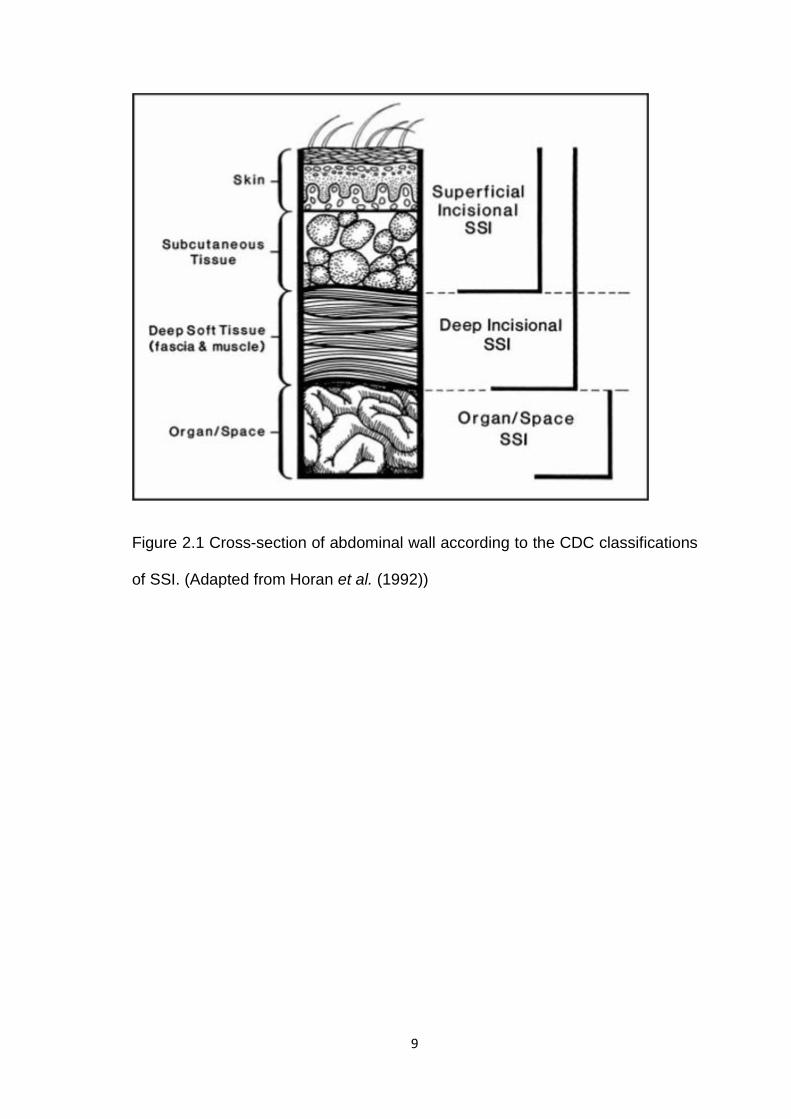
2.1.2 Sites of SSI
Based on the site of infection, SSI can be classified into three categories,
which are superficial, deep, and organ or space. Superficial SSI occurred at the
epidermis, dermis and subcutaneous fat tissue layer; deep SSI occurred at the
fascia and muscle under the subcutaneous fat tissue layer; and organ or space
SSI occurred at the organ or space within the body (Figure 2.1).
Any infections that involve more than two layers will be classified into the
deeper site. For example infection that involved superficial and deep layer is
classified as deep SSI.
8
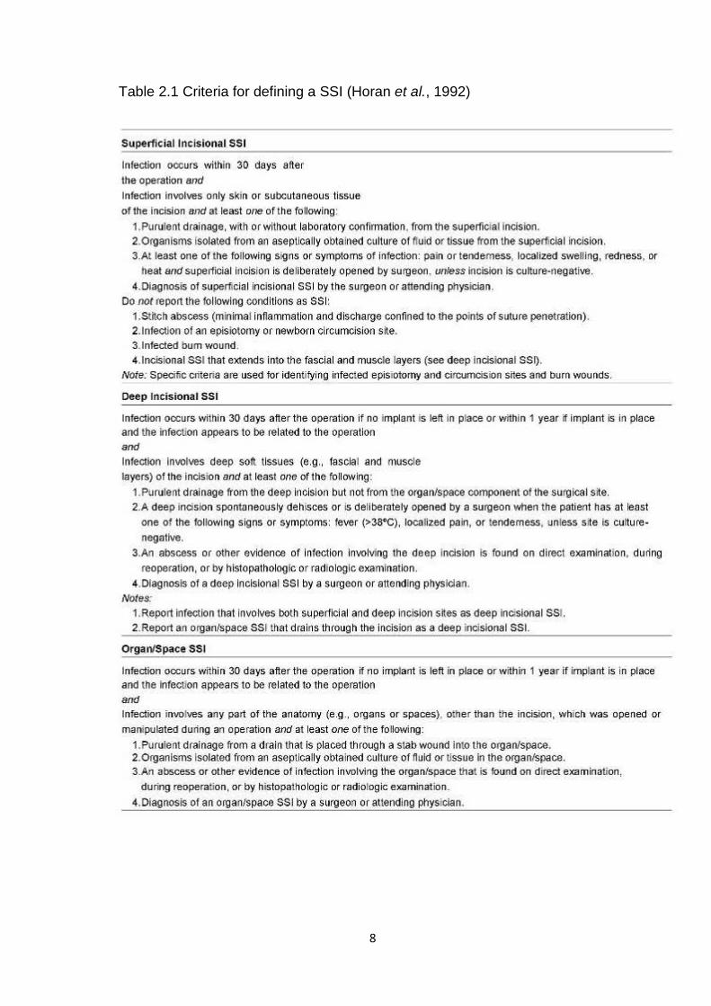
Table 2.1 Criteria for defining a SSI (Horan et al., 1992)
Superficial lncisional SSt
Infection occurs within 30 days after the operation and Infection involves only skin or subctJtaneous tissue of the incision and at least one of the following:
1. Purulent drainage, w~h or without laboratory confirmation . from the superficial Incision. 2.0rganlsms isolated from an aseptically obtained culture of nuld or tissue from the superficial Incision. 3.At least one of the following signs or symptoms of infection: pain or tenderness, localized swelling, redness. or
heat and superticial incision is deliberately opened by surgeon, unless incision is culture-negative. 4. Diagnosis of superficial incisional SSI by the surgeon or attending physician.
Do not repon the following conditions as SSt: 1.Stitch abscess (minimal Inflammation and discharge confined to the poln1s of suture penetration). 2.1nfecUon of an episiotomy or newborn circumcision site. 3.1nfected bum wound. 4.1ncisional SSt that el<lends into the fascial and muscle layers (see deep incisional SSI).
Note: specific c1iteria afe used for identifying infected episiotomy and circumcision sites and burn wounds.
Deep tncisionat SSt
Infection occurs within 30 days after the operation if no implant is left in place or within 1 year if implant is in place and the inrectioo appears to be related to the operation and Infection involves deep sofi tissues (e.g .. fascial and muscle layers) of the incision and at least one of the foiiOINing:
1. Purulent drainage from tile deep Incision but not from the organ/space component of the surgical s~e. 2.A deep incision spontaneously dehisces or is deliberately opened by a surgeon \'ihen the patient has at least
one of the following signs or symptoms: fever {>38°C), localized pain, or tendemess, unless site is cutture
negative. 3.An abscess or other evidence of infection involving the deep incision is found on direct examination, during
reoperation, or by histopathologic or radiologic examination. 4. Diagnosis of a deep lnclslonat SSt by a surgeon or attending physician.
Notes: 1.Report infection that involves both superticial and deep incision s~es as deep incisional SSt. 2. Repon an organ/space SSI that drains through the incision as a deep incisional SSt.
Organ!Space SSt
Infection oocurs within 30 days after the operation if no implant is left in place or within 1 year if implant is in place and the infection appears to be related to the operation 8tJd Infection involves any part of the anatomy {e.g., organs or spaces). other than the incision, which was opened or manipulated during an operation and at least one of the following:
1. Purulent drainage from a drain that Is placed through a stab wound Into the organ/space. 2.0rganlsms Isolated from an aseptically obtained culture of nuld or Ussue In the organ/space. 3.An abscess or other evidence of infection involving the organfspace that is found on direct examination,
during reoperation, or by histopathologic or radiologic examination.
4. Diagnosis of an organ/space sst by a surgeon or attending physician.
9
Figure 2.1 Cross-section of abdominal wall according to the CDC classifications
of SSI. (Adapted from Horan et al. (1992))
10
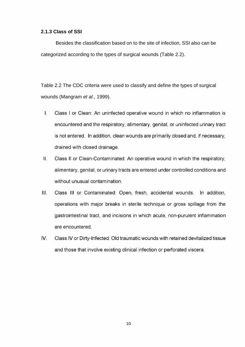
2.1.3 Class of SSI
Besides the classification based on to the site of infection, SSI also can be
categorized according to the types of surgical wounds (Table 2.2).
Table 2.2 The CDC criteria were used to classify and define the types of surgical
wounds (Mangram et al., 1999).
11
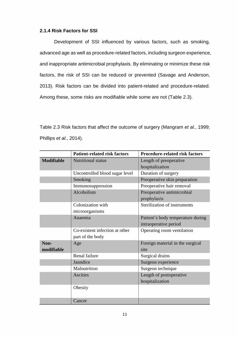
2.1.4 Risk Factors for SSI
Development of SSI influenced by various factors, such as smoking,
advanced age as well as procedure-related factors, including surgeon experience,
and inappropriate antimicrobial prophylaxis. By eliminating or minimize these risk
factors, the risk of SSI can be reduced or prevented (Savage and Anderson,
2013). Risk factors can be divided into patient-related and procedure-related.
Among these, some risks are modifiable while some are not (Table 2.3).
Table 2.3 Risk factors that affect the outcome of surgery (Mangram et al., 1999;
Phillips et al., 2014).
Patient-related risk factors Procedure-related risk factors Modifiable Nutritional status Length of preoperative
hospitalization Uncontrolled blood sugar level Duration of surgery Smoking Preoperative skin preparation Immunosuppression Preoperative hair removal Alcoholism Preoperative antimicrobial
prophylaxis Colonization with
microorganisms Sterilization of instruments
Anaemia Patient’s body temperature during intraoperative period
Co-existent infection at other part of the body
Operating room ventilation
Non-modifiable
Age Foreign material in the surgical site
Renal failure Surgical drains Jaundice Surgeon experience Malnutrition Surgeon technique Ascities Length of postoperative
hospitalization Obesity
Cancer
12
Modifiable risk factors such as patient’s nutritional status, blood sugar level,
anaemia, and smoking status can be modified and eliminated. For example
patient with hyperglycaemia can be treated and normalize the blood sugar level
prior to operation. While non-modifiable risk factors such as age, renal failure and
malnutrition cannot be restored to normal.
Although not all risk factors are modifiable, but optimizing modifiable risk
factors before surgery can reduce the risk of SSIs (Savage and Anderson, 2013).
However, this only applied on the elective surgery. For emergency operation, due
to the urgency and short of time to optimize the modifiable risk factors, not all
modifiable risk cannot be treated prior the operation, and emergency operation
itself also as a risk factor that contribute to SSI (Neumayer et al., 2007).
2.1.5 Prevention Bundles
SSI is a preventable surgical complication. Strictly follow the infection
control measures during pre-, intra- as well as post-operation were able to reduce
and prevent the occurrence of SSI. One of the prevention strategies is the
concept of care bundles. Majority of SSIs can be prevented by implement care
bundles (Herruzo Cabrera, 2010; Miyahara et al., 2014; Phillips et al., 2014).
According to the NICE guideline, care bundles are divided into three phases,
which are preoperative, intraoperative and postoperative phases (Leaper et al.,
2008). Each phase consists of several measures to minimize the risk of SSI, and
all of this known as care bundle.
13
2.1.5.1 Preoperative Care Bundles
2.1.5.1 (i) Optimize patient’s risk factors
Risk factors such as gender, age, duration of surgery, anaemia, cigarette
smoking, diabetes mellitus and obesity were evaluated. Their association and
influence on occurrence of SSI was studied as well. However, risk factors such
as gender and age are not modifiable. Therefore focusing on modifiable risk
factors (Table 2.3) such as smoking cessation, weight reduction, diabetic control,
reduce or stop alcoholic consumption, and coexisting infection are essential and
they should be optimized and treated prior to any elective operation (Neumayer
et al., 2007; Ministry of Health Malaysia., 2010).
2.1.5.1 (ii) Nasal Screening for Methicillin-resistance Staphylococcus
aureus
Many previous studies revealed that S. aureus was the commonest
isolated pathogen involved in SSI, it accounts up to 46% of Methicillin-sensitive
and Methicillin-resistance Staphylococcus aureus (MRSA) (Owens and Stoessel,
2008; Weigelt et al., 2009; Kang et al., 2012; Takesue et al., 2012).
Epidemiologic studies have shown that majority of SSI are originated from
patients itself, such as endogenous flora and nasal colonization.
S. aureus is a pathogenic microorganism, however it colonized in human
skin flora as transient and resident flora in 20% of normal healthy adult
(Kluytmans et al., 1997). It commonly colonized on skin, axilla, perineal area,
groin, and anterior nares (Friedrich et al., 2006). NICE guidelines (2008)
14
recommends nasal decolonization should only target to patients who are nasally
colonised with MRSA instead of all S. aureus. However, a few studies have
shown that nasal decolonization for both Methicillin-sensitive S. aureus (MSSA)
and MRSA significantly reduce the incidence of SSI (Bode et al., 2010; Rao et al.,
2011).
MRSA decolonization protocol are very depends on the health care
institutions and hospitals. Based on the latest MRSA decolonization protocol from
Hospital USM, MRSA nasal carriage should be treated with either Mupirocin 2%
ointment three times a daily for five days or Chlorhexidine 1% cream three times
daily for seven days (Hospital Universiti Sains Malaysia., 2012).
2.1.5.1 (iii) Antimicrobial Prophylaxis
Antimicrobial prophylaxis is the use of antibiotics in order to prevent the
occurrence of infection (Wendy, 2005). Antimicrobial agent used for prophylaxis
should be actively against and cover the most common and most likely cause of
infection during and after the procedure (Ministry of Health Malaysia., 2010). The
selection of antibiotics to be used as prophylaxis must ideally based on the
antibiogram pattern of the particular institution or hospital, therefore the choice of
antimicrobial agent would not be same in every hospital, nation and region.
NICE guidelines recommends a single dose of intravenous antimicrobial
prophylaxis to be given to clean surgery that involved implant or prosthetic
placement; clean-contaminated surgery; contaminated surgery and dirty-infected
operation within 60 minutes prior to the incision. Repeat dose should be given
when the duration of operation is longer than the half-life of the antimicrobial
15
agent (Leaper et al., 2008; Phillips et al., 2014). Benefit and efficacy on
antimicrobial prophylaxis is well established and a study conducted by Mazaki
(2014) also found that there were significant reduction of incidence of SSI when
patients were given antimicrobial prophylaxis.
According to the National Antibiotic Guideline (2008) published by Ministry
of Health, Malaysia, it recommends antimicrobial prophylaxis to be given as soon
as the patient has been stabilized after induction (Table 2.4). Continuing
antimicrobial prophylactic until removal of surgical drain is not recommended.
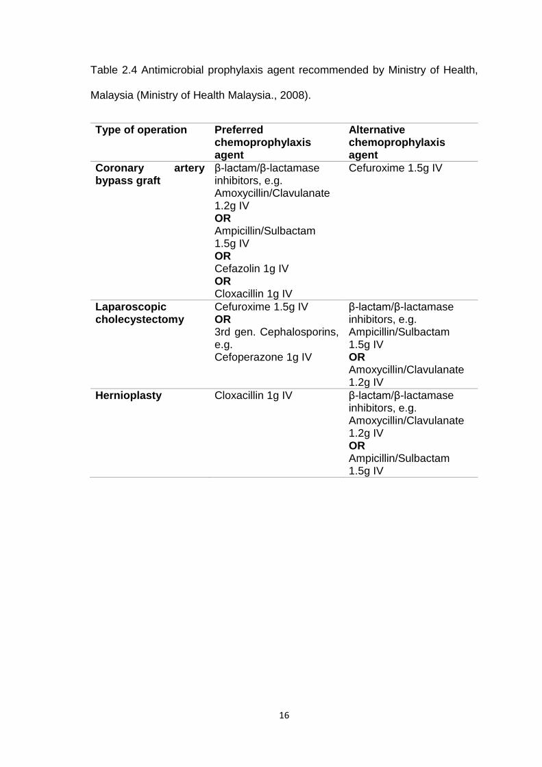
16
Table 2.4 Antimicrobial prophylaxis agent recommended by Ministry of Health,
Malaysia (Ministry of Health Malaysia., 2008).
Type of operation Preferred chemoprophylaxis agent
Alternative chemoprophylaxis agent
Coronary artery bypass graft
β-lactam/β-lactamase inhibitors, e.g. Amoxycillin/Clavulanate 1.2g IV OR Ampicillin/Sulbactam 1.5g IV OR Cefazolin 1g IV OR Cloxacillin 1g IV
Cefuroxime 1.5g IV
Laparoscopic cholecystectomy
Cefuroxime 1.5g IV OR 3rd gen. Cephalosporins, e.g. Cefoperazone 1g IV
β-lactam/β-lactamase inhibitors, e.g. Ampicillin/Sulbactam 1.5g IV OR Amoxycillin/Clavulanate 1.2g IV
Hernioplasty Cloxacillin 1g IV β-lactam/β-lactamase inhibitors, e.g. Amoxycillin/Clavulanate 1.2g IV OR Ampicillin/Sulbactam 1.5g IV
17
2.1.5.1 (iv) Hair Removal
Removal of hair during routine operation is not recommended, unless that
area interfere the incision site (National Institute for Health and Care Excellence.,
2008). Hair removal can be done in such case, if it’s necessary, and it’s preferable
to use electronic clippers instead of razor or shaving to reduce the injury on skin
that can lead to colonization or infection (National Institute for Health and Care
Excellence., 2008; Phillips et al., 2014). Infection control guideline published by
Minister of Health, Malaysia (2010) recommend hair removal should be done just
before operation.
2.1.5.1 (v) Other preoperative measures
Besides that, NICE guideline also recommends patient to shower or bath
preoperatively, wear antiseptic impregnated clothes and to avoid routine
mechanical bowel preparation to reduce SSI (Leaper et al., 2008; Savage and
Anderson, 2013).
18
2.1.5.2 Intraoperative Care Bundles
2.1.5.2 (i) Operating Room Environment
Operating room must be clean from any soil or contamination of any body
fluids, such as blood. Air entered operating room must be filtered and maintained
at least 15 to 20 air changes hourly. Operating room must also under positive
pressure, temperature optimized at around 21°C and humidity should be
maintained around 40-60% (Owens and Stoessel, 2008; Ministry of Health
Malaysia., 2010; Phillips et al., 2014).
2.1.5.2 (ii) Skin Preparation
Skin should be sterilized just before incision by using antiseptics. Common
antiseptics used for operation is either povidone-iodine or chlorhexidine alcohol
aqueous. Studies compared the efficacy of povidone-iodine or chlorhexidine
alcohol to sterilize the skin shown that chlorhexidine alcohol offered significant
protection and more advance than povidone-iodine (Macias et al.; Darouiche et
al., 2010; Noorani et al., 2010; Banjong et al., 2011).
2.1.5.2 (iii) Maintaining Patient’s Body Temperature and Homeostasis
Maintaining patient’s body temperature, oxygenation, perfusion and
homeostasis during intraoperative period are important to reduce the chances of
SSI. Patient who was hypothermia during intraoperative period required longer
time in wound healing and increase occurrence of SSI (Kurz et al., 1996). Studies
also found that hypothermia patients stayed longer during postoperative
hospitalization period (Kurz et al., 1996; Harper et al., 2003).
19
2.1.5.2 (iv) Other intraoperative measures
Surgeons, anaesthesiologists, nurses as well as other operating team
members that involves in operation must strictly follow WHO (2009) Hand
Hygiene Guideline when decontaminate their hands. Operating team members
must also wear sterile gowns, gloves, facemasks, and caps during the operation
to minimize the SSI due to transmission of potential pathogens (Leaper et al.,
2008; Owens and Stoessel, 2008). Besides that, surgeon experience and skill
also greatly affect the outcome (Phillips et al., 2014).
2.1.5.3 Postoperative Care Bundles
2.1.5.3 (i) Wound Care
Unless dressing soaked, otherwise sterilize dressing on primarily closed
wound should be protected 24 to 48 hours postoperatively. If inspection, changing,
or removing of dressing are necessary during first 48 hours, use an aseptic
technique and strictly compiled to WHO Hand Hygiene Guideline (Leaper et al.,
2008; Owens and Stoessel, 2008; World Health Organization., 2009; Ministry of
Health Malaysia., 2010; Phillips et al., 2014).
2.1.5.4 Treatment of SSI
Treatment of SSI is basically based on the site of infection. Once SSI was
diagnosed by surgeon or physician, early empiric antimicrobial agent is important
to eliminate the pathogen. For superficial SSI and pus discharge, wound
cleansing is the first step to remove the pus and disinfect the surrounding tissue.
20
In deep SSI, the pus may locate beneath the superficial tissue. In such
condition, drainage of pus and fluid from the infected site is adequate while in
severe case, debridement of the wound and an affected tissue is necessary.
Occasionally, patient is required to be hospitalized and re-operate the wound.
Organ or space SSIs are often diagnosed by signs of infection and
instrumental examination. Drainage of pus are done under the guidance of
ultrasonography or computed tomography examination. If it’s not possible, re-
operate would be the only choice of treatment.
Debrided tissue, aspirated pus or pus swab should be quickly transport to
laboratory for identification of pathogenic microorganism as well as antimicrobial
sensitivity test to find out the most suitable antimicrobial agent.
21
2.2 Staphylococcus aureus
Staphylococcus was first identified by a Scottish surgeon, Sir Alexander
Ogston, in pus from a surgical abscess in a knee joint in Aberdeen, United
Kingdom in year 1880. He proposed the name ‘Staphylococci’ based on its shape
and morphology (Humphreys, 2012). Staphylococci are resistant to high salt
concentration as well as dry conditions, therefore it can survive for long periods
in the environment such as the skin and upper respiratory tract of human and
animals.
Staphylococcus aureus (S. aureus) is the major pathogen in the genius
Staphylococcus, due to its ability to infect human and animals. It is differentiated
from other species by its ability to clot blood plasma via the action of enzyme
coagulase (Humphreys, 2012).
List below is the hierarchy taxonomic for S. aureus (UniProt., 2014).
Domain: Bacteria;
Phylum: Firmicutes;
Class: Bacilli;
Order: Bacillales;
Family: Staphylococcaceae;
Genus: Staphylococcus;
Species: Staphylococcus aureus
22
S. aureus are grouped under genus Staphylococcus within family
Staphylococcaceae. The microorganism is Gram-positive, round shape,
arranged in grape-like cluster, about 1µm in diameter, non-sporing, non-motile,
usually non-capsulated, produce enzyme coagulase, and ferment mannitol
(Chapman, 1945; Humphreys, 2012). Therefore it produces positive reaction in
mannitol salt agar and changes the colour from red/pink to yellow. This
microorganism can grow on most types of media, including Mueller Hinton agar,
blood agar, DNase agar, Tryptic Soy Agar, and Mannitol Salt agar (MSA). Its
colonies are circular, smooth and shiny surface, and often pigmented.
It differs from other microorganism by mean of the characteristic
appearance on Mannitol Salt agar and DNase agar. On Mannitol Salt agar, its
yellow appearance due to the ability to ferment mannitol while on DNase agar, a
clear zone surround the colonies can be seen when added a few drop of
hydrochloride acid. S. aureus was then confirmed by tube coagulase test to differ
from other coagulase-negative Staphylococci.
2.2.1 Pathogenicity
S. aureus is present in the nose of 21.5 - 30% of healthy individual and
may be found on the skin as well (Fishbain et al., 2003; Davis et al., 2004; Alex,
2007; Humphreys, 2012).
It is an opportunistic pathogen that can cause a numerous diseases, from
minor skin infections to life threatening diseases, such as impetigo, scalded skin
syndrome, abscesses, osteomyelitis, pneumonia, meningitis, endocarditis, toxic
23
shock syndrome (TSS), sepsis as well as surgical site infection. It also one of the
major causative organisms in surgical site infection.
Our body consists of three defence mechanism to protect us from S.
aureus invasion. Primary and the most frontier barrier consists of our skin layer,
and physiological factors; secondary barrier is our cellular factors and
inflammatory response, which consist of phagocytes, neutrophils, basophils,
interleukin and other inflammatory markers; tertiary which is the final barrier
comprises active immune response provided by lymphocytes, such as
immunoglobulin (Hattie, 2009).
By the help of virulent factors, S. aureus can invade and against our
defence barrier and cause infections.
2.2.2 Virulent factors
S. aureus contain numbers of cell-associated and extracellular factors to
assist it to overcome the human defend mechanism, and to colonized, invade as
well as survive in the human body (Table 2.5). Although not all the role of every
factors are well studied, but it’s likely that they are responsible for enabling the S.
aureus to bind and to resist and survive from the intracellular killing by phagocytes
and bactericidal activities from humoral factors (Humphreys, 2012).
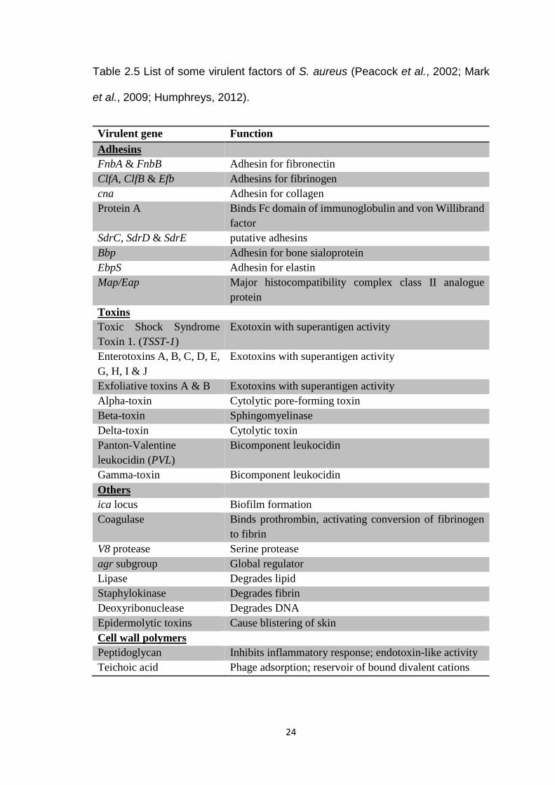
24
Table 2.5 List of some virulent factors of S. aureus (Peacock et al., 2002; Mark
et al., 2009; Humphreys, 2012).
Virulent gene Function Adhesins FnbA & FnbB Adhesin for fibronectin ClfA, ClfB & Efb Adhesins for fibrinogen cna Adhesin for collagen Protein A Binds Fc domain of immunoglobulin and von Willibrand
factor SdrC, SdrD & SdrE putative adhesins Bbp Adhesin for bone sialoprotein EbpS Adhesin for elastin Map/Eap Major histocompatibility complex class II analogue
protein Toxins Toxic Shock Syndrome Toxin 1. (TSST-1)
Exotoxin with superantigen activity
Enterotoxins A, B, C, D, E, G, H, I & J
Exotoxins with superantigen activity
Exfoliative toxins A & B Exotoxins with superantigen activity Alpha-toxin Cytolytic pore-forming toxin Beta-toxin Sphingomyelinase Delta-toxin Cytolytic toxin Panton-Valentine leukocidin (PVL)
Bicomponent leukocidin
Gamma-toxin Bicomponent leukocidin Others ica locus Biofilm formation Coagulase Binds prothrombin, activating conversion of fibrinogen
to fibrin V8 protease Serine protease agr subgroup Global regulator Lipase Degrades lipid Staphylokinase Degrades fibrin Deoxyribonuclease Degrades DNA Epidermolytic toxins Cause blistering of skin Cell wall polymers Peptidoglycan Inhibits inflammatory response; endotoxin-like activity Teichoic acid Phage adsorption; reservoir of bound divalent cations