Embed Size (px)
Citation preview
This document is downloaded at: 2019-04-03T17:54:48Z
Title Surgical Consideration of Esophagectomy Combined withTracheobronchoplastic Procedure for the Treatment of Esophageal Cancer
Author(s)
Tomita, Masao; Ayabe, Hiroyoshi; Kawahara, Katsunobu; Azuma, Koji;Ishikawa, Hiroshi; Oh, Shimei; Kawabuchi, Takaaki; Sato, Tetsuya;Shibata, Ryuichiro; Nakao, Haruhiko; Hara, Shinsuke; Hashimoto, Satoru;Shiraishi, Enju; Furukawa, Taizo; Uchida, Yuzo
Citation Acta Medica Nagasakiensia. 1986, 31(1-4), p.143-151
Issue Date 1986-10
URL http://hdl.handle.net/10069/17481
Right
NAOSITE: Nagasaki University's Academic Output SITE
http://naosite.lb.nagasaki-u.ac.jp

Acta Med. Nagasaki 31: 143-151
Surgical Consideration of Esophagectomy Combined
with Tracheobronchoplastic Procedure for the Treatment of
Esophageal Cancer
Masao TOMITA, Hiroyoshi AYABE, Katsunobu KAWAHARA,
Koji AZUMA, Hiroshi ISHIKAWA, Shimei OH,
Takaaki KAWABUCHI, Tetsuya SATO, Ryuichiro SHIBATA,
Haruhiko NAKAO, Shinsuke HARA, Satoru HASHIMOTO,
Enju SHIRAISHI, Taizo FURUKAWA, Yuzo UCHIDA*
1st Department of Surgery, Nagasaki University School of
Medicine.
* 2nd Department of Surgery, Oita Medical Collage.
Reprint requests to : Masao Tomita, 1st Department of
Surgery, Nagasaki University School of Medicine, 7-1
Sakamoto-machi, Nagasaki, Japan.
Received for publication, June 4, 1986
Four cases who underwent a resection of the esophagus in combination with tracheo- bronchoplasty were reported in evaluating an operative efficacy as well as in overcoming a
surgically bothersome problem. 1) Obliquely cutting edges of the bronchial stumps, which can adjust to fit the anastomosed
bronchi, are a weak point of their approximations because of loss of the supporting ability of the cartilages as indicated in case I.
2) Combined resection of the esophagus with the bronchus seems to reduce the blood flow in the bronchial anastomotic site. One must account into consideration that bronchial
wrapping with the pleura may inhibit a new development of vascular networks in the bronchial anastomosis as shown in case 2.
3) Irradiation of as large 60 Gy interferes with the healing process due to damages of severely fibrous changes with a finding of vasculitis to the bronchial wall as presented
in case 3. 4) Inevitable truncal vagotomy by a resection of the esophagus leads at times to bile stasis,
followed by cholecystitis and bile-transudation peritonitis, reflecting serious operative
富田 正雄,綾 部 公酪,川 原 克信,吾 妻 康次,石 川 啓,王 志明,川 渕 孝明
佐藤 哲也,柴 田隆一郎,中 尾 治彦,原 信介,橋 本 哲,白 石 円樹,古 川 泰蔵
内田 雄三*
143
144 M. TOMITA ET AL.
insult as exhibited in case 4.
INTRODUCTION
Great strides in the treatment of esophageal cancer have been achieved with
advanced in diagnostic techniques, and pre-and postoperative cares.
Most of esophageal cancer patients are impossible to allow food and/or fluid intakes
by mouth. Therefore, it is taken into account that only a desperate surgery warrants
palliation and allowance of oral intake of food as far as possible, even if a surgical efficacy
may last for a short period of time and loose their radical nature of surgery.
The esophagus is anatomically specific of a composition of the adventitia on its
outer, which is voiding the serosa. Consequently the esophagus is susceptible to cancer
invasion, which extend easily outwards across its wall. In advanced cases, therefore,
combined resections with the involved adjacent organs such as the bronchus, Iung and
descending aorta have become necessary to ensure their radical natures if it were not for
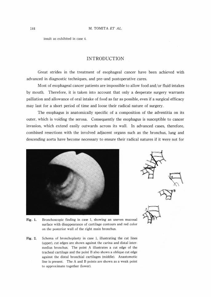
Fig. 1.
Fig. 2.
Bronchoscopic finding in case 1, showing an uneven mucosal
surface with disappearance of cartilage contours and red color
on the posterior wall of the right main bronchus.
Schema of bronchoplasty in case 1, illustrating the cut lines
(upper), cut edges are shown against the carina and distal inter-
medius bronchus. The point A illustrates a cut edge of the
tracheal cartilage and the point B also shows a oblique cut edge
against the distal bronchial cartilages (middle). Anastomotic
line is present. The A and B points are shown as a weak point
to approximate together (Iower).
~~"

ESOPHAGECTOMY WITH TRACHEOBRONCHOPLASTY 145
a greater degree.
The combined resection with the trachea and bronchus for the treatment of car-
cinoma of the esophagus is a matter of great concern as to whether radical nature of
surgery will be enhanced or not. So far little information is available with regard to
surgical safegards. A bothersome problem in surgery was disscussed to achieve an
operative security of t.he basis of our clinical experiences.
PATIENTS
Case I : A 65-year old Japanese woman complained of dysphagia with a spiral
defect, which was located in the upper third to middle of the thoracic esophagus on the
f luoroesophagogram.
Bronchoscopy preoperatively performed revealed an uneven mucosal surface on the
right main bronchus with red color (Fig. 1). At surger~, it is noticed that cancer invasion
from the esophagus reached the point extending from the posterior wall of the right main
bronchus to the lateral wall of the lower trachea. The right main bronchus, measuring 3 x
4cm, was resected including a part of the lateral wall of the lower trachea.
The bronchial edges were sutured with use of 3-0 Dexon. On the first day of
surgery, air leak accidentally occurred.
The 2nd thoracotomy on that day identified a small air leak from the upper edge of
the suture line. Additional two stitches were sufficient for a repair of air leakage. On the
3rd day of surgery, air leak took place again. At the 3rd thoracotomy performed on that
day the origin of air leak was confirmed to be an opposite edge of the suture line to that
prevdiusly sutured and necessited three stiches to achieve a complete repair. Repeated air
leaks occurring in early postoperative period in this case alert us to hinderance of healing
on the suture line which has the jeopardy of the impaired blood flow and is more susceptible
to tension due to losing the continuity of the cartilages in oblique cut edges as shown in Fig.
2. The patient's condition, thereafter, was stable until she died 357 days after surgery of
pneumonia.
Case 2 : A 68-year old Japanese man complained of dysphagia with spiral shadow
defect on the fluoroesophagogram in the upper third and middle of the esophagus (Fig. 3).
Preoperative bronchoscopy revealed a deformity of the left main bronchus and also CT
scan showed a stenosis to some extent in both the main bronchi (Fig. 4). At the time of
performing a thoracotomy, it was noted that cancer infiltration on the left bronchus was
146 M. TOMITA ET AL.
ru
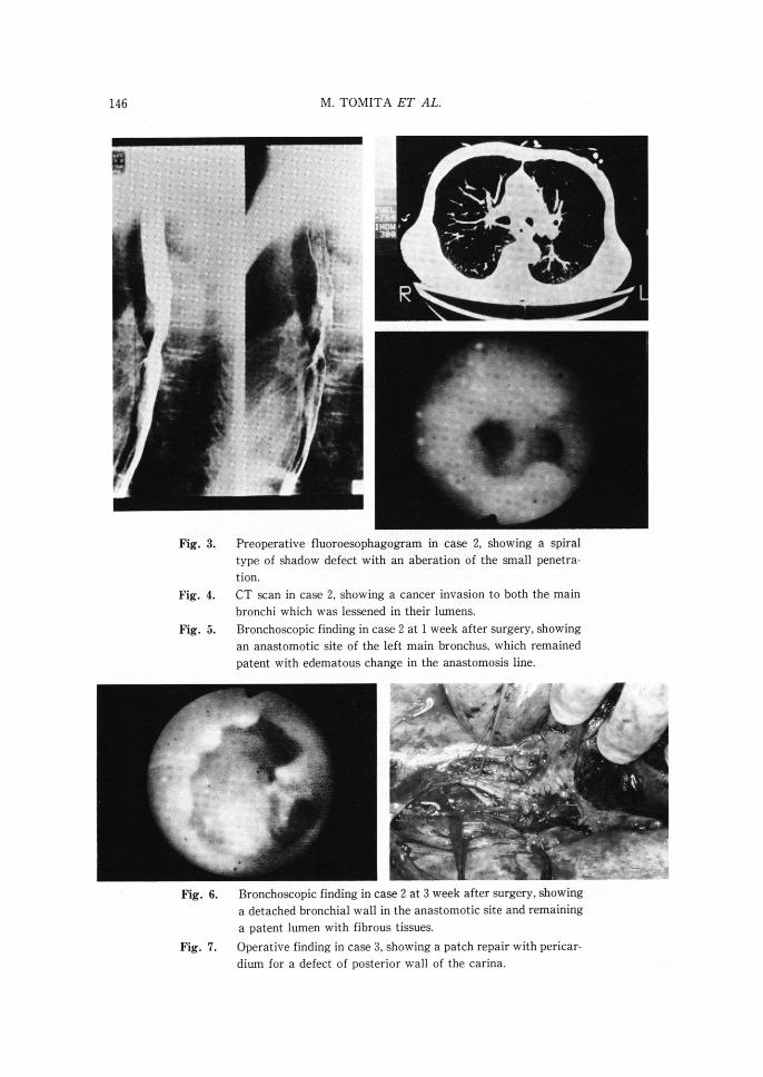
Fig. 3.
Fig. 4.
Fig. 5.
Preoperative fluoroesophagogram in case 2, showing a spiral
type of shadow defect with an aberation of the small penetra-
tion.
CT scan in case 2, showing a cancer invasion to both the main
bronchi which was lessened in their lumens.
Bronchoscopic finding in case 2 at I week after surgery, showing
an anastomotic site of the left main bronchus, which remained
patent with edematous change in the anastomosis line.
Fig. 6.
Fig. 7.
~~:$s,~~,*~'** ~~~~{~:
~;~~~~# .'s e~ "
Bronchoscopic finding in case 2 at 3 week after surgery, showing
a detached bronchial wall in the anastomotic site and remaining
a patent lumen with fibrous tissues.
Operative finding in case 3, showing a patch repair with pericar-
dium for a defect of posterior wall of the carina.
ESOPHAGECTOMY WITH TRACHEOBRONCHOPLASTY 147
extending from its begining to 3cm distal to its attachment. After performing a resection
of a 3cm left main bronchus from the carina and a mobilization of the right lung by dividihg
the pulmonary ligament and skeletonizing the hilar vessels and the main bronchus.
The bronchial edges after a resection of the left main bronchus were sutured with
use of 3-0 Vicryl without tension, adding a procedure of pleural bronchial wrapping
At I week after surgery the bronchoscopy revealed that healing at the anastomotic
site was satisfactory except for edema in the anastomotic site (Fig. 5). An anemic and
edematous mucosal membrane was visualized.
In contrast, at 3 weeks the bronchial anastomosis was disrupted and the fibrous
tissues surrounded to maintain a continuity of the lumen between the separated edges of
the left bronchus (Fig. 6).
Surgery to reanastomose was not predicted until pneumonia on the left would be
cured. During a waiting period for improving a lung shadow of pneumonia, violent
hemoptysis unexpectedly occurred to entain his death on day 45.
Case 3 : A 69-year old Japanese woman received a 60 Gy irradiation therapy for the
treatment of esophageal cancer. At that time, the fluoroesophagogram demontrated a
small penetrating lesion with mild fever of 37.5'C in body temperature. Surgery was
indicated for fear of the ensuing perforation and/or bleeding. At thoracotomy, the
esophagus adjacent to the tracheal carina was firmly adherent to the trachea, suggesting
a presence of the inflammatory and cancer-infiltrating lesions around the penetrating
fistula. The operative steps to ablate the esophagus resulted in 53cm defect of the
membranous portion of the trachea. A defect was repaired with free pericardial patch
graft (Fig. 7). Further esophageal resection approach was abondoned on account of an
technical difficulty due to tight adhesion.
Postoperative pneumonia worsened her condition so that she died on day 7.
Autopsy showed that the pericardial patch was partly detached from the tracheal wall and
also histologic irradiation damage to the bronchial wall, which was composed of severely
fibrous changes with vasculitis (Fig. 8), had become evident.
It is defined that irradiation damage to the walls of the trachea and bronchus may
well result in worsened healing process in the suturing lines.
Case 4 : A 64-year old Japanese man complained of dysphagia and hematemesis
with spiral shadow defect on the fluoroesophagogram in the upper third to middle portion
of the esophagus.
Preoperative bronchoscopy revealed a reddish mucosal change and a circumferential
disappearance of cartilage contours in a 3cm left main bronchus from 1.0cm distal to the
148 M. TOMITA ET AL.
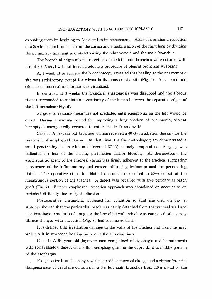
Fig. 8.
Fig. 9.
Histologic finding of the tracheal wall at autopsy in case 3,
showing a fibrous change with vasculitis of a 6000R irradiation
damage to the tracheal wall.
CT scan in case 4 showing a cancer invasion to the carina and
the descending aorta.
carina to just proximal to the attachment of the right upper bronchus.
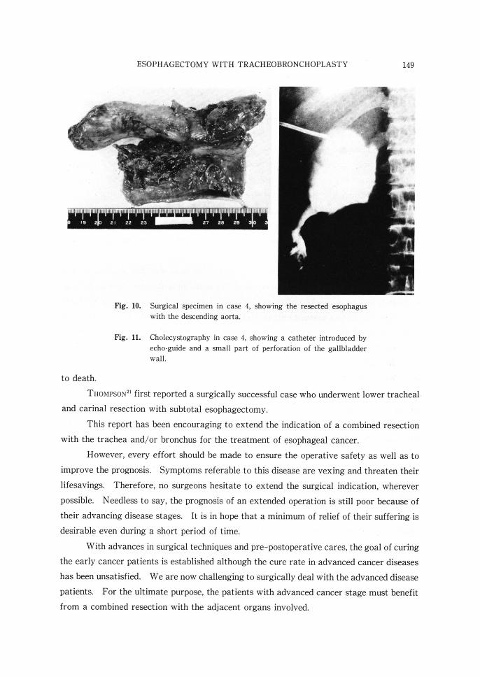
Preoperative CT scan also indicated that there was a cancer infiltration lying in the
medial half the circumferencial wall of the descending aorta (Fig. 9). He underwent a
combined resection of the esophagus with the circumference of a 9cm decending aorta from
a point caudal to the attachment of the left subclavian artery (Fig. 10) and the left main
bronchus of 3 cartilaginous rings (1.5cm) by using a temporary external bypass with a 12lnm
diameter cooley graft between the left subclavian artery and distal descending aorta just
above the diaphragma via left thoracotomy. Immediately after surgery, postoperative
course was uneventful, but on day 3 he complained of abdominal distension and the
gallbladder has become palpated. The sign of bile-transudation peritonitis due to bile
stasis, followed by cholecystitis was evident.
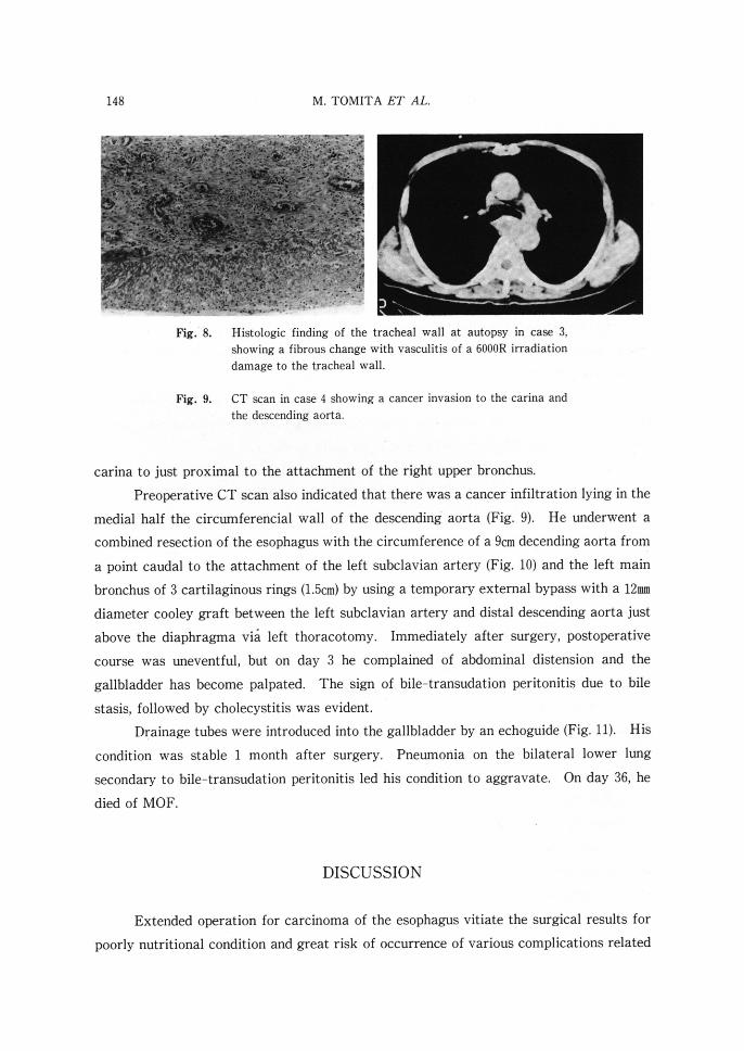
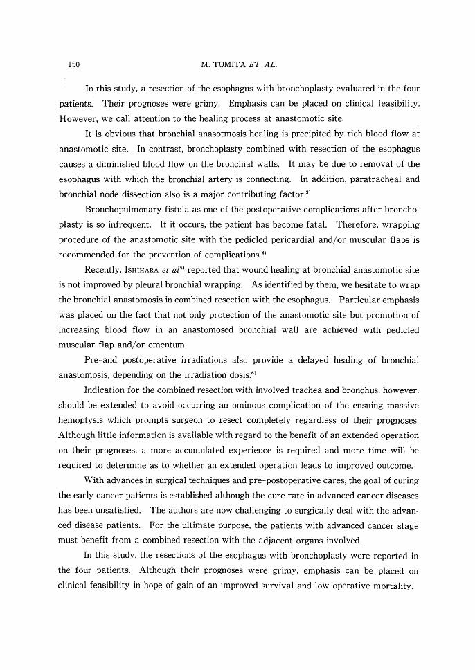
Drainage tubes were introduced into the gallbladder by an echoguide (Fig. 11). His
condition was stable I month after surgery. Pneumonia on the bilateral lower lung
secondary to bile-transudation peritonitis led his condition to aggravate. On day 36, he
died of MOF.
DISCUSSION
Extended operation for carcinoma of the esophagus vitiate the surgical results for
poorly nutritional condition and great risk of occurrence of various complications related
ESOPHAGECTOMY WITH TRACHEOBRONCHOPLASTY 149
~~;;'"・・~~~~~*~'~~,/~;~i;~~:'"
"**S~...**~f**'**~
Fig. lO.
Fig. 11.
Surgical specimen in case 4, showing the resected esophagus
with the descending aorta.
Cholecystography in case 4, showing a catheter introduced by
echo-guide and a small part of perforation of the gallbladder
wall.
s
to death.
THOMPSoN2) first reported a surgically successful case who underwent lower tracheal
and carinal resection with subtotal esophagectomy.
This report has been encouraging to extend the indication of a combined resection
with the trachea and/or bronchus for the treatment of esophageal cancer.
However, every effort should be made to ensure the operative safety as well as to
improve the proguosis. Symptoms referable to this disease are vexing and threaten their
lifesavings. Therefore, no surgeons hesitate to extend the surgical indication, wherever
possible. Needless to say, the prognosis of an extended operation is still poor because of
their advancing disease stages. It is in hope that a minimum of relief of their suffering is
desirable even during a short period of time.
With advances in surgical techniques and pre-postoperative cares, the goal of curing
the early cancer patients is established although the cure rate in advanced cancer diseases
has been unsatisfied. We are now challenging to surgically deal with the advanced disease
patients. For the ultimate purpose, the patients with advanced cancer stage must benefit
from a combined resection with the adjacent organs involved.
150 M. TOMITA ET AL.
In this study, a resection of the esophagus with bronchoplasty evaluated in the four
patients. Their proguoses were grimy. Emphasis can be placed on clinical feasibility.
However, we call attention to the healing process at anastomotic site.
It is obvious that bronchial anasotmosis healing is precipited by rich blood flow at
anastomotic site. In contrast, bronchoplasty combined with resec_tion of the esophagus
causes a diminished blood flow on the bronchial walls. It may be due to removal of the
esophagus with which the bronchial artery is connecting. In addition, paratracheal and
bronchial node dissection also is a major contributing factor.3)
Bronchopulmonary fistula as one of the postoperative complications after broncho-
plasty is so infrequent. If it occurs, the patient has become fatal. Therefore, wrapping
procedure of the anastomotic site with the pedicled pericardial and/or muscular flaps is
recommended for the prevention of complications.4)
Recently, ISHIHARA et al5) reported that wound healing at bronchial anastomotic site
is not improved by pleural bronchial wrapping. As identified by them, we hesitate to wrap
the bronchial anastomosis in combined resection with the esophagus. Particular emphasis
was placed on the fact that not only protection of the anastomotic site but promotion of
increasing blood flow in an anastomosed bronchial wall are achieved with pedicled
muscular flap and/or omentum.
Pre-and postoperative irradiations also provide a delayed healing of bronchial
anastomosis, depending on the irradiation dosis.6)
Indication for the combined resection with involved trachea and bronchus, however,
should be extended to avoid occurring an ominous complication of the ensuing massive
hemoptysis which prompts surgeon to resect completely regardless of their prognoses.
Although little information is available with regard to the benefit of an extended operation
on their prognoses, a more accumulated experience is required and more time will be
required to determine as to whether an extended operation leads to improved outcome.
With advances in surgical techniques and pre-postoperative cares, the goal of curing
the early cancer patients is established although the cure rate in advanced cancer diseases
has been unsatisfied. The authors are now challenging to surgically deal with the advan-
ced disease patients. For the ultimate purpose, the patients with advanced cancer stage
must benefit from a combined resection with the adjacent organs involved.
In this study, the resections of the esophagus with bronchoplasty were reported in
the four patients. Although their prognoses were grimy, emphasis can be placed on
clinical feasibility in hope of gain of an improved survival and low operative mortality.

ESOPHAGECTOMY WITH TRACHEOBRONCHOPLASTY 151
REFERENCES
l ) AKIYAMA, H., TSURUMARU, M., KAWAMURA, T., ONO, Y. : Principles of surgical treatment
for carcinoma of the esophagus : Analysis of lymph node involvement. Ann Surg 1 94 :
438-446, 1981.
2 ) THOMPSON, D. T. : Lower tracheal and carinal resection associated with subtotal oeso-
phagectomy for carcinoma of oesophagus involving trachea. Thorax 28: 257-262,
1937.
3 ) TOMITA, M. et al. : An experimental evaluation of tracheal blood flow with special
reference to operative procedure of tracheal mobilization. Aorta Medica Nagasakiensia
25 : 83-89, 1980.
4 ) KAWAHARA, H. et al. : Intrathoracic application of latissimus dorsi muscle flap for
exteneded radical esophagectomy. JJSA 85 : 300-306, 1984.
5 ) ISHIHARA, T. et al. : Does pleural bronchial wrapping improve wound healing in right
sleeve lobectomy ? J. Thorac. Cariovasc. Surg. 89 : 665-672, 1985.
6 ) SHESOL, B. F. and CLARKE, T. K. : Intrathoracic application of the latissmus dorsi
musculocutaneous flap. Plast. Reconstruct. Surg. 66 : 842-849, 1980.
7 ) TSUBOTA, N. et al. : The effects of preoperative irradiation on primary tracheal anas-
tomosis. Ann. Thoracic Surg. 20 : 52-160, 1975.