Embed Size (px)
Citation preview
Surgical Approaches for Fractures and Injuries of the
Pelvic Ring
Mara L. Schenker, MD Emory University / Grady Hospital
Created by Steven A. Olson, MD in 2004 and Kyle Dickson, MD in 2004 First revised by Rafael Neiman, MD and Sean Nork in 2007
Second Revision by James C. Krieg, MD in 2009 and Sean Nork, MD in 2010 Third Revision by Mara Schenker and Clifford Jones in 2016
Goals • Indications: non-operative vs. operative • Acute management • Definitive management
– Surgical approaches – Reduction – Fixation
Pelvic Ring Stability • Biomechanical stability = ability to support a
physiological load, and is dependent on an intact posterior sacroiliac ligamentous complex for load transfer from axial to appendicular skeleton
Pelvic Ring Instability
• Biomechanical Instability – Posterior ring injuries frequently lead to instability – Anterior ring injuries MAY lead to instability, and
are often associated with posterior ring injuries – Tile Classification based on instability – May lead to hemodynamic instability – If untreated, may lead to permanent disability from
pelvis mobility
Assessing Stability
• Is there deformity? • Is there posterior ring involvement? • Are there associated injuries? • Is there displacement with stress
examination?
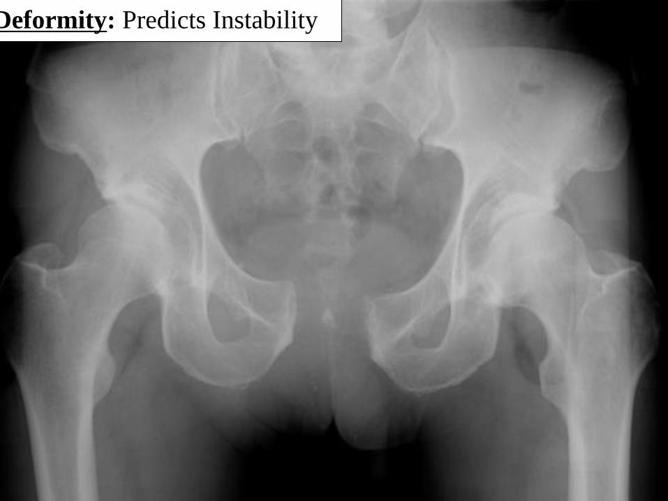
Deformity: Predicts Instability
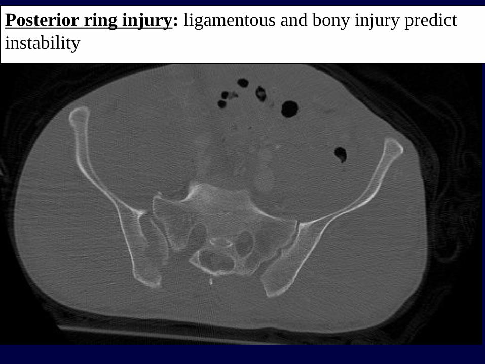
Posterior ring injury: ligamentous and bony injury predict instability
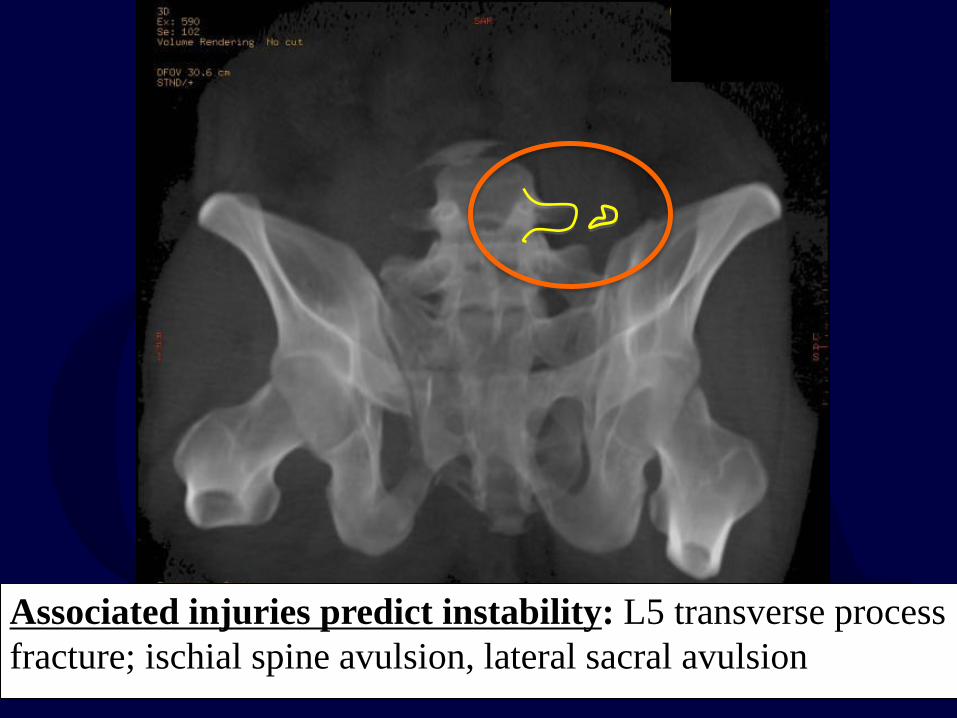
Associated injuries predict instability: L5 transverse process fracture; ischial spine avulsion, lateral sacral avulsion
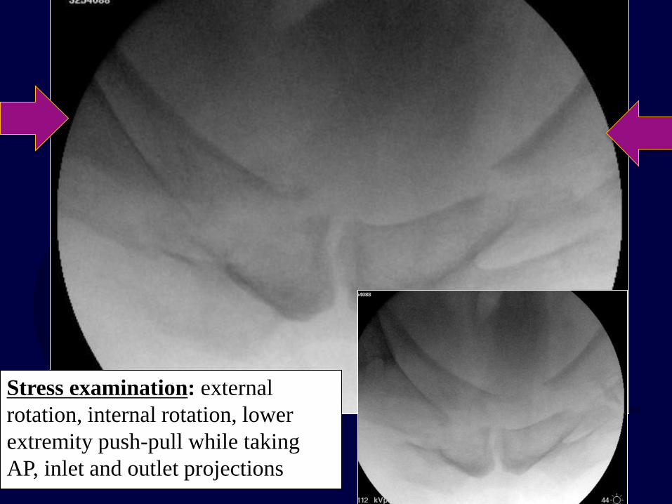
Stress examination: external rotation, internal rotation, lower extremity push-pull while taking AP, inlet and outlet projections
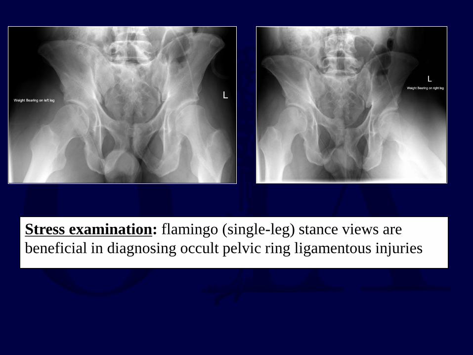
Stress examination: flamingo (single-leg) stance views are beneficial in diagnosing occult pelvic ring ligamentous injuries

Describing Instability
• Refer to previous lecture on Classification • Tile Classification
– A stable – B partially stable – C unstable

Operative Indications • Resuscitation • Mobilization
– Just as stabilizing long bones helps in mobilization of polytrauma patients
• Preventing long term functional impairment – Malunion can affect function (bladder,
dyspareunia, sitting imbalance, leg length inequality, mechanical low back pain) and quality of life
Non-Operative Indications
• Lateral impaction type injuries, without cephalad displacement or excessive hemipelvis rotation
• Pubic symphyseal widening < 2.5 cm – Without associated SI injury – Assuming no motion with stress or mobilization – This number is not absolute, so other evidence of
instability (like SI injury) must be ruled out
Non-Operative Treatment: TILE A
• Stable injuries can generally WBAT • Serial radiographs • Displacement requires reassessment of
stability and consideration given to operative treatment
• Partially stable injuries can be treated non-operatively if deformity is minimal
• Weight bearing should be restricted (TTWB) on side of posterior ring injury
• Serial radiographs • Displacement requires reassessment of
stability and consideration given to operative treatment
Non-Operative Treatment: TILE B

ACUTE MANAGEMENT Resuscitation, Containment, Angiography
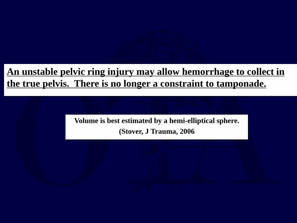
An unstable pelvic ring injury may allow hemorrhage to collect in the true pelvis. There is no longer a constraint to tamponade.
Volume is best estimated by a hemi-elliptical sphere. (Stover, J Trauma, 2006
ATLS Protocol
Airway maintenance with cervical spine protection
Breathing and ventilation Circulation with hemorrhage control Disability: Neurologic status Exposure/environment control: undress
patient but prevent hypothemia
Physical Examination
• Open wounds • Degloving injuries • Blood at the urethral meatus • Perineal and scrotal ecchymosis • Neurologic deficiency
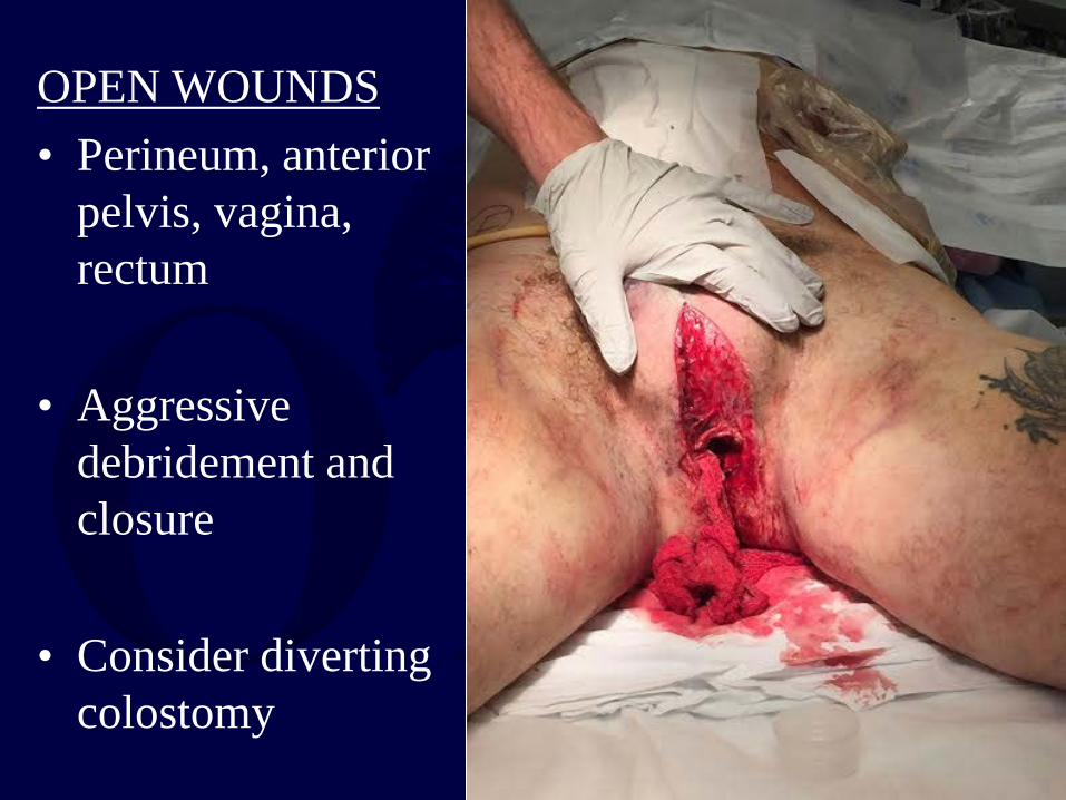
OPEN WOUNDS • Perineum, anterior
pelvis, vagina, rectum
• Aggressive debridement and closure
• Consider diverting colostomy
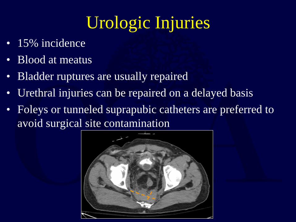
Urologic Injuries • 15% incidence • Blood at meatus • Bladder ruptures are usually repaired • Urethral injuries can be repaired on a delayed basis • Foleys or tunneled suprapubic catheters are preferred to
avoid surgical site contamination
Hemorrhage Management
• AP pelvis with an understanding of the mechanism of injury helps determine whether the pelvis is a source of bleeding in the hemodynamically unstable patient
– APC injuries have increased need for blood
transfusion (Burgess J Trauma 1990)
Methods of Hemorrhage Control
• Pelvic containment – Binder – Sheet – External fixation
• Angiography • Laparotomy, with or without packing
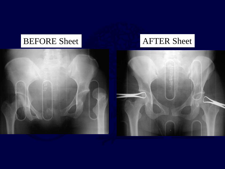
BEFORE Sheet AFTER Sheet
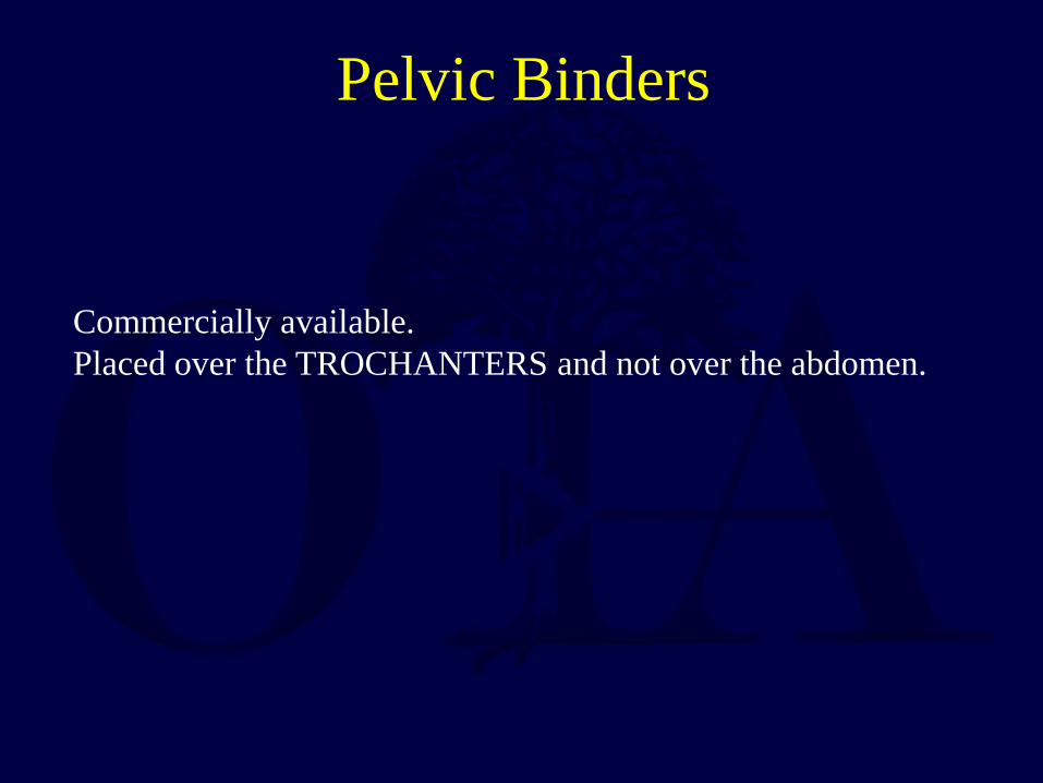
Commercially available. Placed over the TROCHANTERS and not over the abdomen.
Pelvic Binders
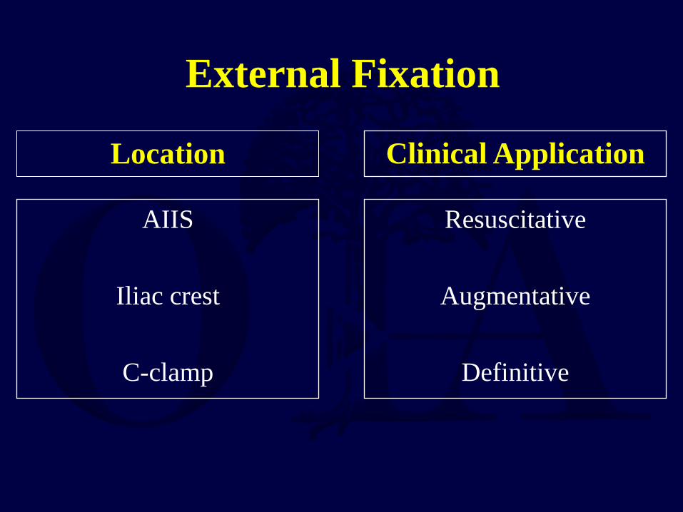
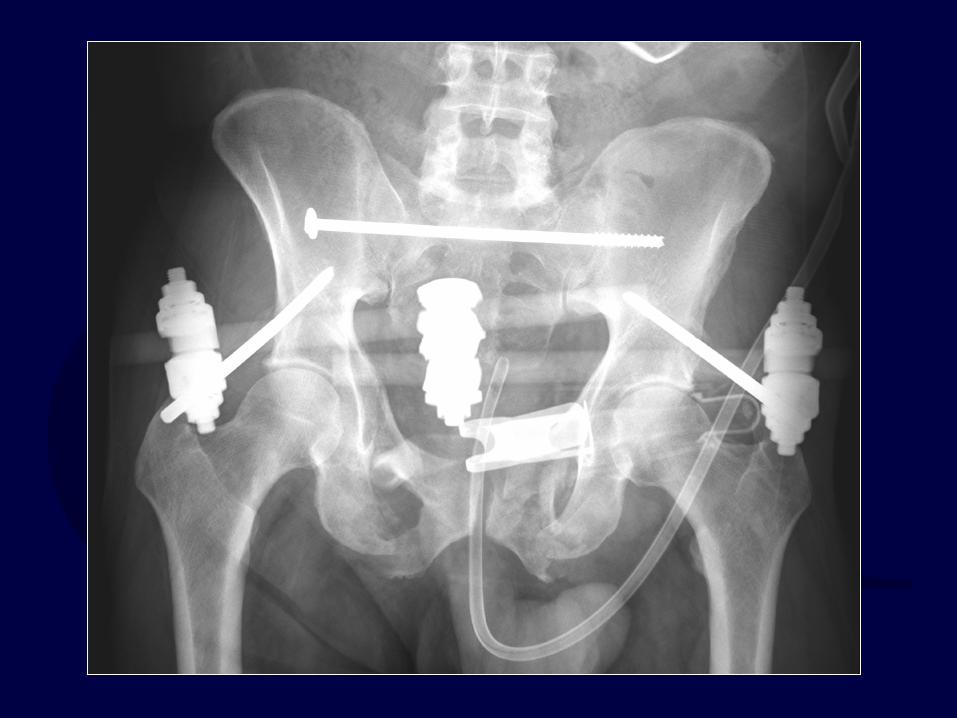
External Fixation
Location
AIIS
Iliac crest
C-clamp
Clinical Application
Resuscitative
Augmentative
Definitive
External Fixation: AIIS frames
• Advantages: – Thought to be biomechanically superior to crest
frames – Patients can sit
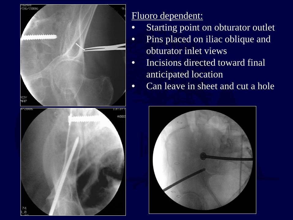
Fluoro dependent: • Starting point on obturator outlet • Pins placed on iliac oblique and
obturator inlet views • Incisions directed toward final
anticipated location • Can leave in sheet and cut a hole
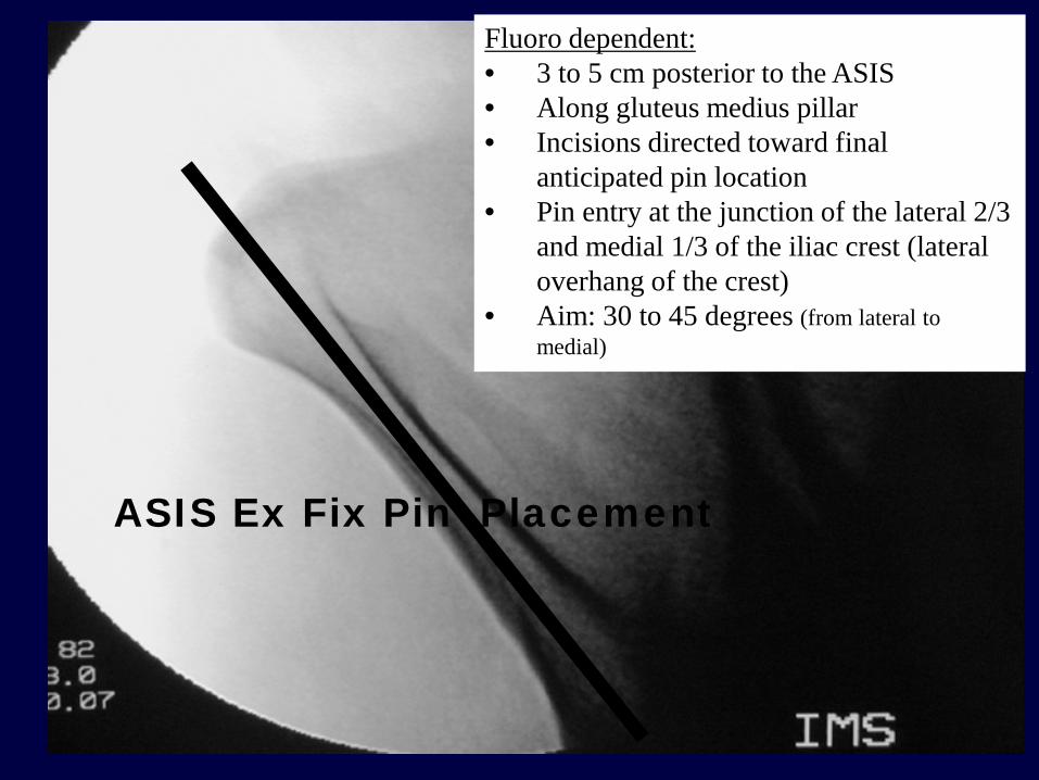
Fluoro dependent: • 3 to 5 cm posterior to the ASIS • Along gluteus medius pillar • Incisions directed toward final
anticipated pin location • Pin entry at the junction of the lateral 2/3
and medial 1/3 of the iliac crest (lateral overhang of the crest)
• Aim: 30 to 45 degrees (from lateral to medial)
ASIS Ex Fix Pin Placement
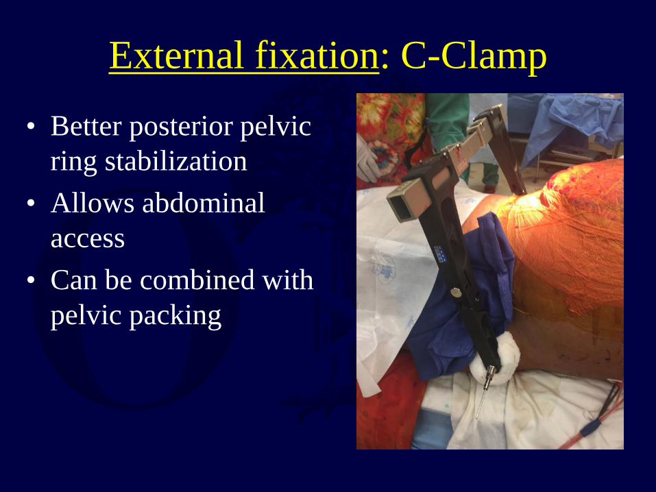
External fixation: C-Clamp
• Better posterior pelvic ring stabilization
• Allows abdominal access
• Can be combined with pelvic packing
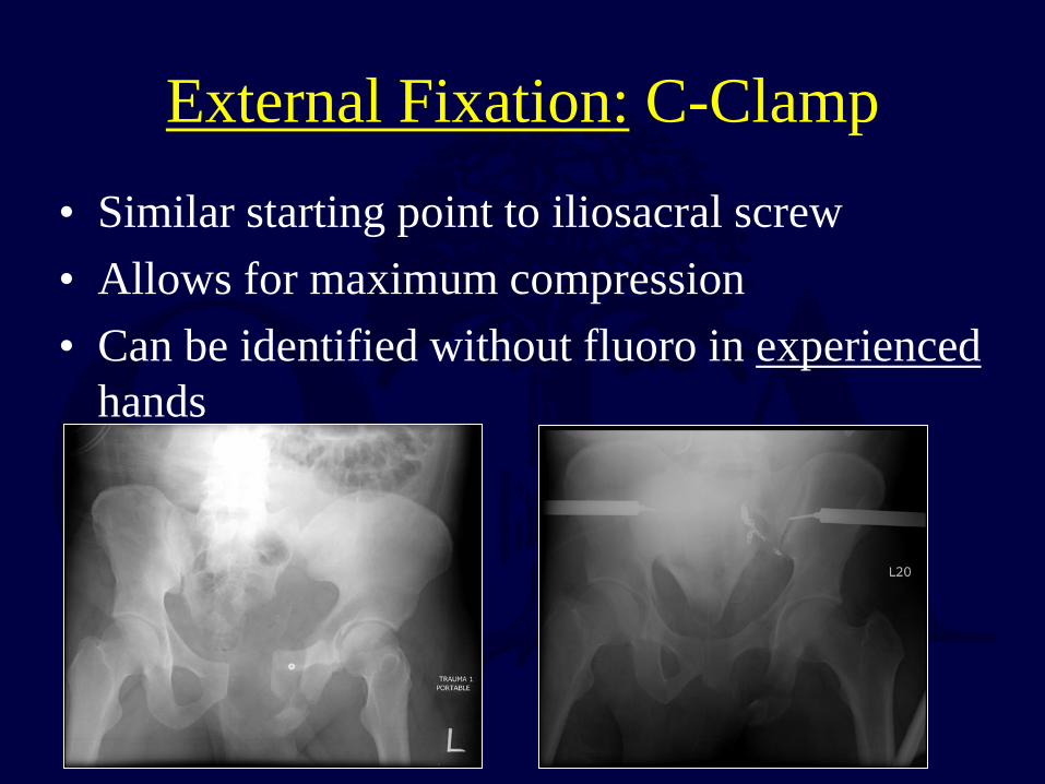
External Fixation: C-Clamp • Similar starting point to iliosacral screw • Allows for maximum compression • Can be identified without fluoro in experienced
hands
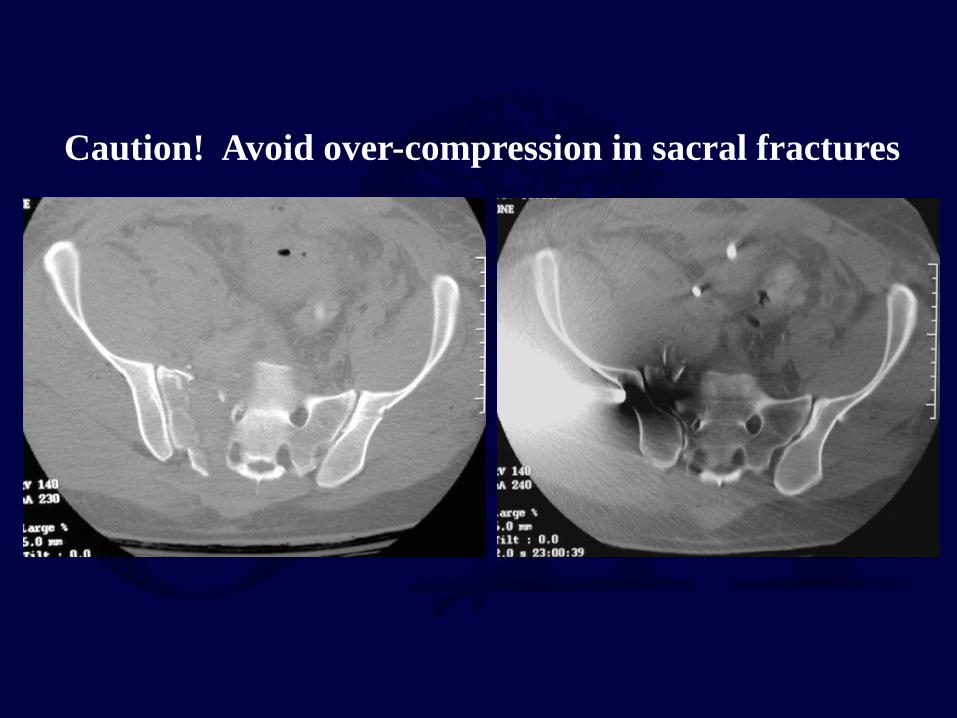
Caution! Avoid over-compression in sacral fractures

Pelvic Packing
• Direct retroperitoneal packing, Pfannenstiel approach • Combine with mechanical stabilization (internal versus external) • May decrease need for transfusion, make angiography more
efficient, and decrease mortality; requires additional OR trips and may increase incidence of abdominal compartment syndrome
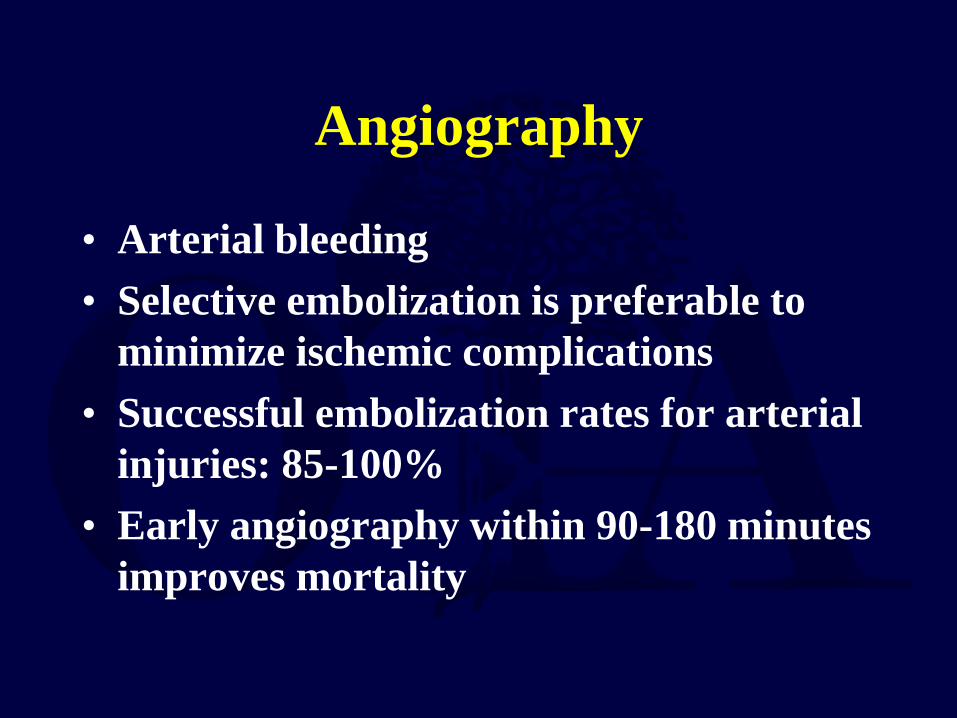
Angiography
• Arterial bleeding • Selective embolization is preferable to
minimize ischemic complications • Successful embolization rates for arterial
injuries: 85-100% • Early angiography within 90-180 minutes
improves mortality
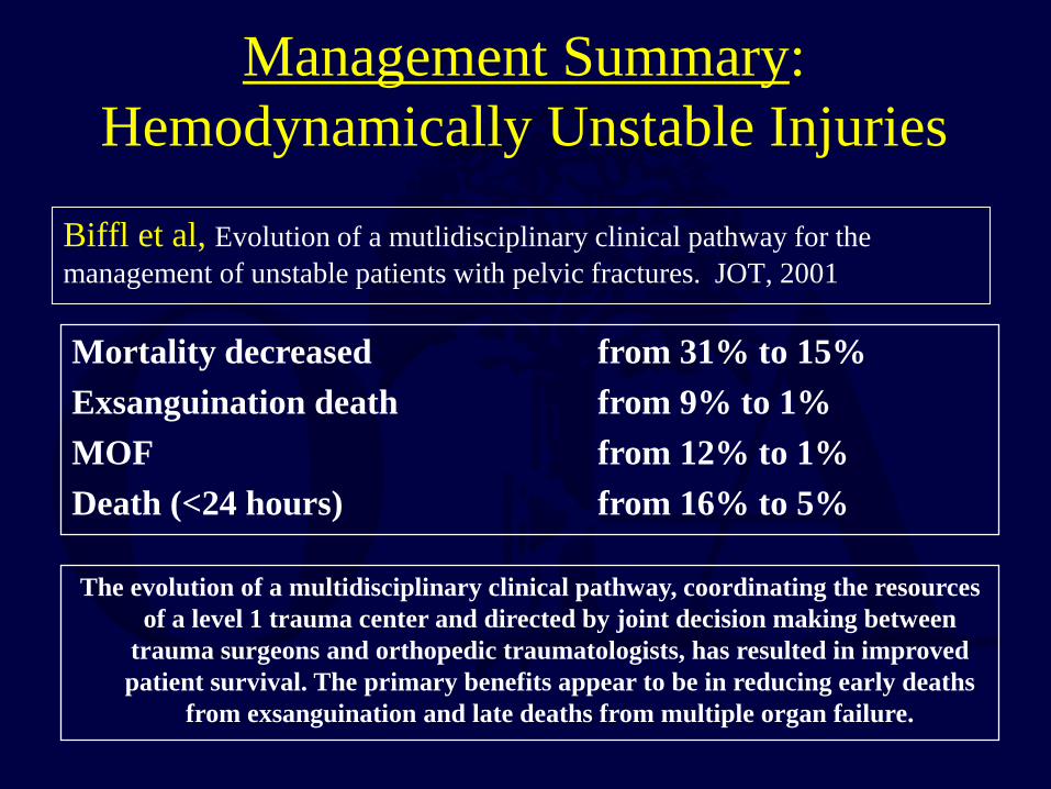
Management Summary: Hemodynamically Unstable Injuries
Biffl et al, Evolution of a mutlidisciplinary clinical pathway for the management of unstable patients with pelvic fractures. JOT, 2001
5 elements: Immediate trauma surgeon availability (+ Ortho!) Early simultaneous blood and coagulation products Prompt diagnosis & treatment of life threatening injuries Stabilization of the pelvic girdle Timely pelvic angiography and embolization Changes: Patients more severely injured (52% vs 35% SBP < 90) DPL phased out for U/S Pelvic binders and C-clamps replaced traditional ex fix
Biffl et al, Evolution of a mutlidisciplinary clinical pathway for the management of unstable patients with pelvic fractures. JOT, 2001
Mortality decreased from 31% to 15% Exsanguination death from 9% to 1% MOF from 12% to 1% Death (<24 hours) from 16% to 5%
The evolution of a multidisciplinary clinical pathway, coordinating the resources of a level 1 trauma center and directed by joint decision making between
trauma surgeons and orthopedic traumatologists, has resulted in improved patient survival. The primary benefits appear to be in reducing early deaths
from exsanguination and late deaths from multiple organ failure.
Management Summary: Hemodynamically Unstable Injuries
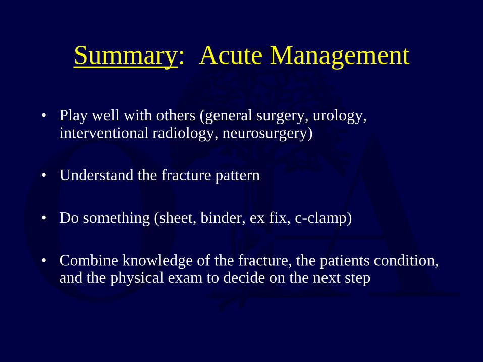
Summary: Acute Management
• Play well with others (general surgery, urology, interventional radiology, neurosurgery)
• Understand the fracture pattern
• Do something (sheet, binder, ex fix, c-clamp)
• Combine knowledge of the fracture, the patients condition, and the physical exam to decide on the next step

DEFINITIVE MANAGEMENT Approaches, Reduction, Fixation
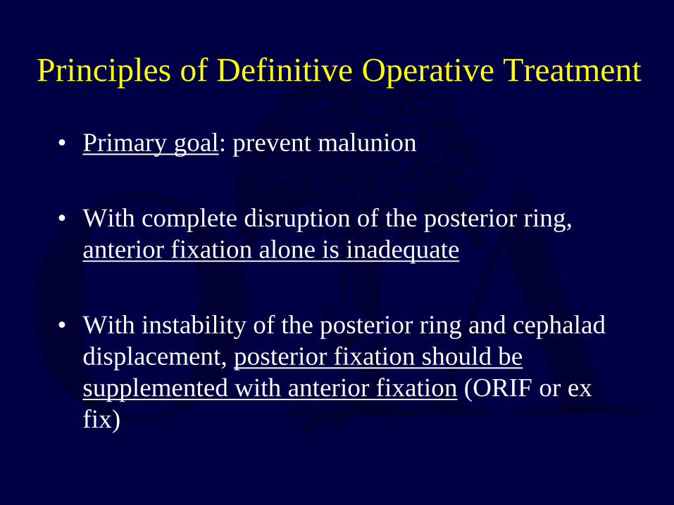
Principles of Definitive Operative Treatment
• Primary goal: prevent malunion
• With complete disruption of the posterior ring, anterior fixation alone is inadequate
• With instability of the posterior ring and cephalad displacement, posterior fixation should be supplemented with anterior fixation (ORIF or ex fix)
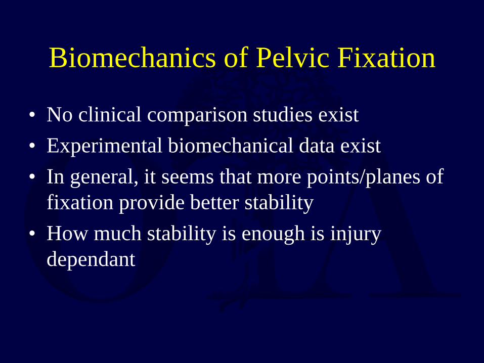
Biomechanics of Pelvic Fixation
• No clinical comparison studies exist • Experimental biomechanical data exist • In general, it seems that more points/planes of
fixation provide better stability • How much stability is enough is injury
dependant
Preoperative Planning • Consider patient-related factors: resuscitation,
coordination of care (trauma surgeon, intensivist, neurosurgery, urology, gynecology), examine soft tissues, is it safe to position prone if needed?
• Consider timing: reduction may be easier
(particularly for percutaneous fixation) in first 24-48 hours; risk of “second hit” in days 2-5 (particularly for open surgery)
Preoperative Planning
• Intraoperative imaging – Radiolucent table – Fluoroscopy – Radiologic Technician and Surgeon understand C-
arm views necessary
• Reduction tools – Traction – Pelvic manipulator (e.g. femoral distractor) – Specialized clamps
Preoperative Planning
• Implants needed – Extra-long screws – Cannulated screws, often extra-long with
appropriate instruments – Specialized plates for contourability
(reconstruction plates) – External fixation
Surgical Approach: Anterior Pelvic Ring
• Pfannenstiel approach – Exposure of symphysis pubis and pubic bones – Longitudinal incision along the fascia of the linea alba – Elevate rectus subperiosteally, protect the bladder with a
malleable retractor
Kain, Tornetta Op Tech Orthop
• Stoppa extension – Exposes symphysis to SI joint along pelvic brim – Care taken laterally, as the corona mortis tends to be 6
cm lateral to the pubic symphysis (anastamosis between obturator and external iliac vessels)
Surgical Approach: Anterior Pelvic Ring
Surgical Approach: Posterior Pelvic Ring
• Lateral window of the ilioinguinal approach – Exposure of sacroiliac joint anteriorly – Avoid injury to the L5 nerve root with retractor placement
anteriorly along the sacrum
• Paramedian approach – Exposure of sacrum and posterior ilium – Sacral fractures – Iliac fracture dislocations of the SI joint (crescent
fracture) – Allows simultaneous reduction and lumbopelvic
fixation when necessary
Surgical Approach: Posterior Pelvic Ring
Reduction and Fixation: Symphysis
Kain, Tornetta Op Tech Orthop
• Weber clamp placed through drill holes anteriorly
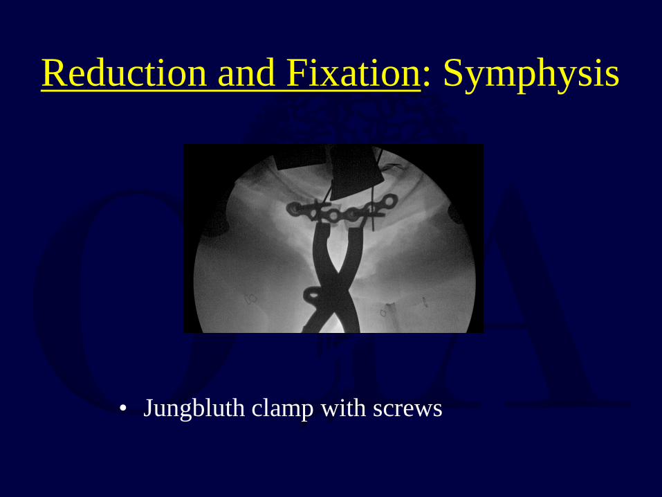
• Jungbluth clamp with screws
Reduction and Fixation: Symphysis
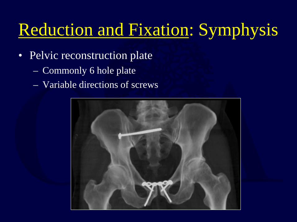
• Pelvic reconstruction plate – Commonly 6 hole plate – Variable directions of screws
Reduction and Fixation: Symphysis
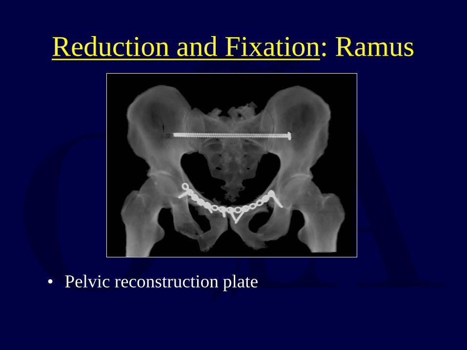
• Pelvic reconstruction plate
Reduction and Fixation: Ramus
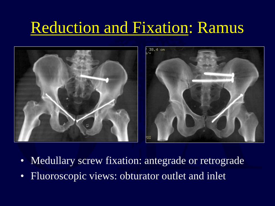
Reduction and Fixation: Ramus
• Medullary screw fixation: antegrade or retrograde • Fluoroscopic views: obturator outlet and inlet
Biomechanics of Pelvic Fixation: Anterior Fixation
• Anterior plating superior to external fixation in internal/external rotation
• Neither technique very effective at control of vertical displacement
• Anterior fixation can “protect” posterior fixation from failure
Biomechanics of Pelvic Fixation: Anterior Fixation
• Two hole symphyseal plate inadequate • Retrograde pubic screw higher failure rate than
antegrade
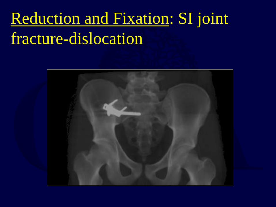
Reduction and Fixation: SI joint fracture-dislocation
• Jungbluth clamp • Anterior provisional or
definitive plating
Reduction and Fixation: SI joint fracture-dislocation
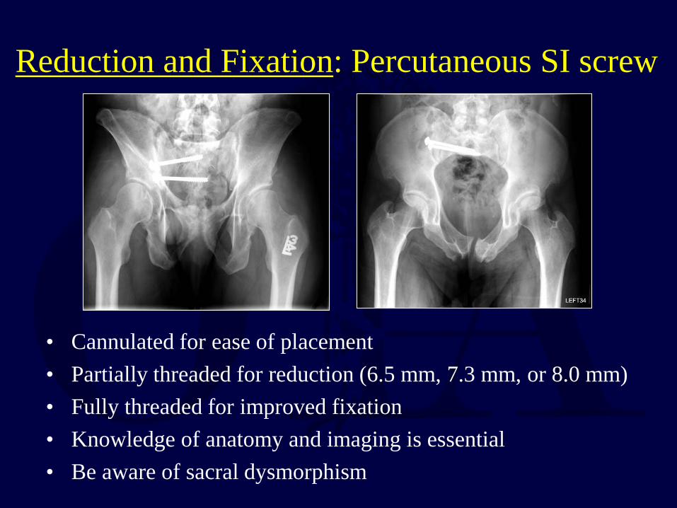
• Cannulated for ease of placement • Partially threaded for reduction (6.5 mm, 7.3 mm, or 8.0 mm) • Fully threaded for improved fixation • Knowledge of anatomy and imaging is essential • Be aware of sacral dysmorphism
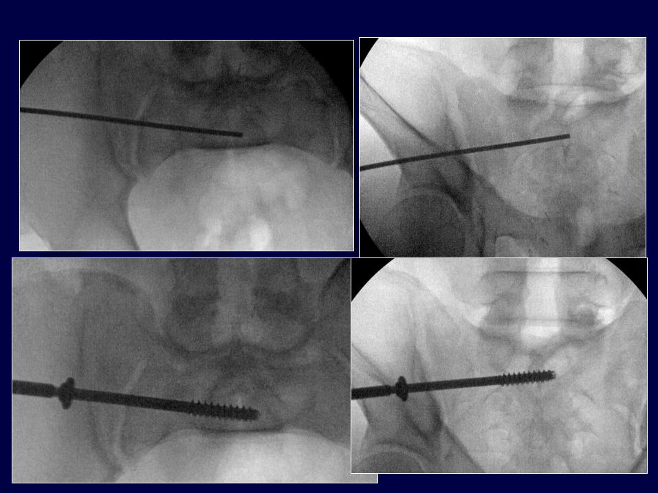
Reduction and Fixation: Percutaneous SI screw
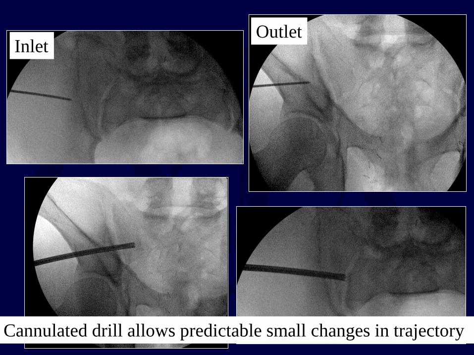
Inlet Outlet
Cannulated drill allows predictable small changes in trajectory
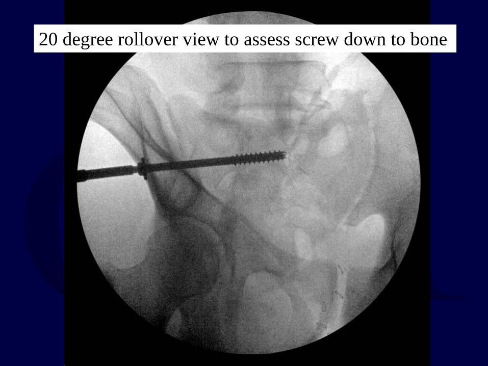
20 degree rollover view to assess screw down to bone
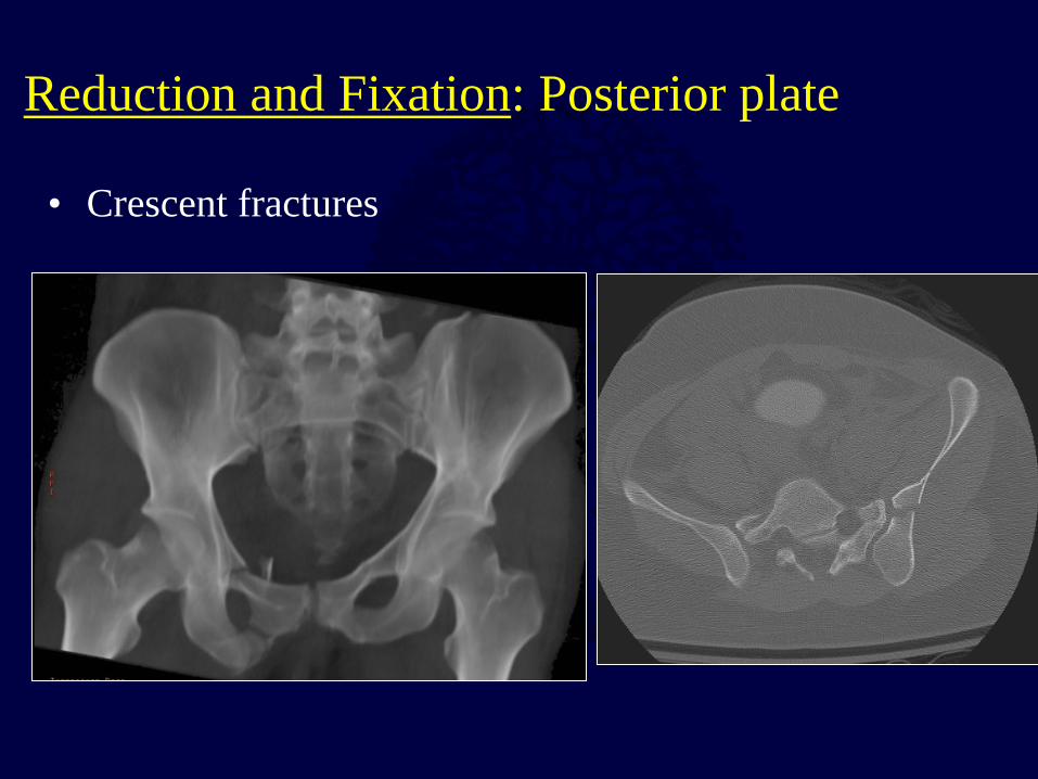
• Crescent fractures
Reduction and Fixation: Posterior plate
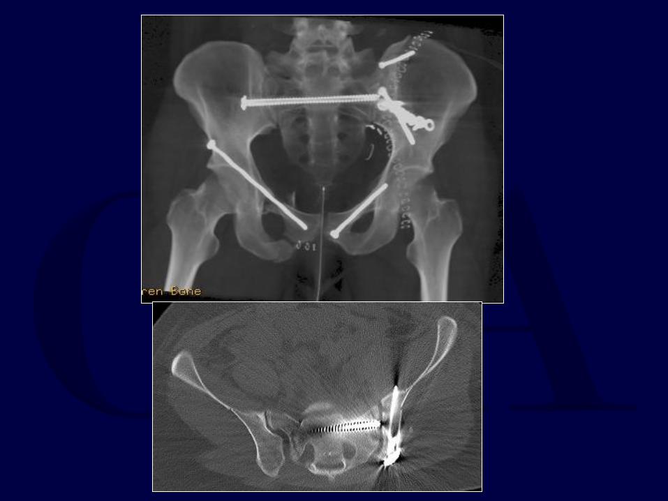
• Indirect reduction – Anterior ring reduction – Traction – Distractor
Reduction and Fixation: Sacral Fracture
• Direct reduction – Posterior exposure – Clamp application (Pointed Weber clamps)
– Can decompress as well if needed – Can perform lumbopelvic fixation if needed
Reduction and Fixation: Sacral Fracture
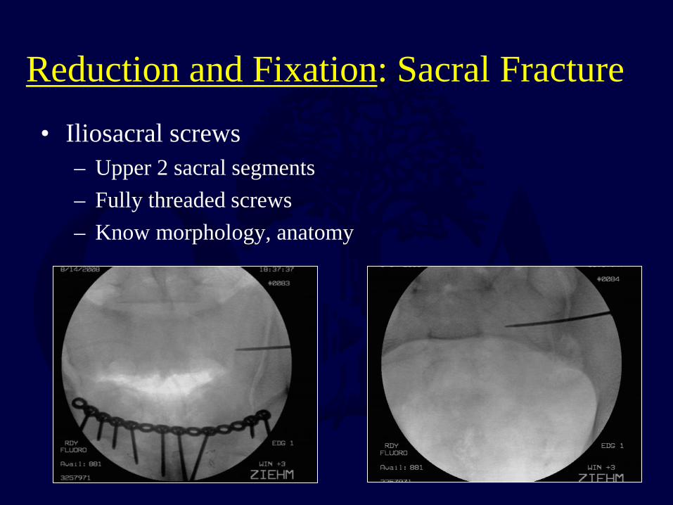
• Iliosacral screws – Upper 2 sacral segments – Fully threaded screws – Know morphology, anatomy
Reduction and Fixation: Sacral Fracture
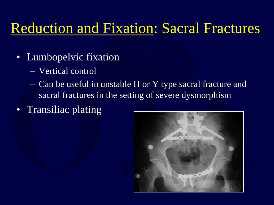
• Lumbopelvic fixation – Vertical control – Can be useful in unstable H or Y type sacral fracture and
sacral fractures in the setting of severe dysmorphism • Transiliac plating
Reduction and Fixation: Sacral Fractures

Biomechanics of Pelvic Fixation: Posterior Fixation
• Options include single SI screw, multiple SI screws, double plating of SI joint, transiliac plate of sacral fracture, or plate plus SI screw for sacral fracture or SI dislocation
• Any of the above are more stable than single SI screw in unstable injuries
Biomechanics of Pelvic Fixation: Posterior Fixation
• Lumbopelvic fixation – Lumbopelvic dissociation (unstable Y, H, or U
type sacral fractures) – Sacral fractures with significant instability – Can provide axial (vertical) stability that is not as
dependant on fracture reduction/stability
Post-Operative Protocol • Mobilize when systemic and physiologic
status allow • Any complete disruption of posterior ring
should be immobilized with touch-down weightbearing for 10 to 12 weeks
• Incomplete posterior ring disruptions can typically be allowed full weightbearing as tolerated
Outcomes
• Pain common • Improvement occurs for at least a year in most
patients • Neurologic injury most common predictor of
poor outcome

Outcomes
• SI dislocations have poor tolerance for residual displacement
• Sacral fractures have more tolerance for displacement, but parameters poorly understood
• Injury Severity Score and fracture type do not correlate with functional outcome
Conclusions: Pelvic Ring Injury
• Complex constellation of injuries • Treatment based on comprehensive
understanding of potential pelvic ring instability, displacement, and associated injuries
• Surgical techniques for reduction and stabilization continue to evolve

• For questions or comments, please send to [email protected]