Embed Size (px)
Citation preview
Surgical andNonsurgicalTreatments ofthe Nasal Valves
Judy Lee, MD, W. Matthew White, MD, Minas Constantinides, MD, FACS*
KEYWORDS
� Functional rhinoplasty � Internal nasal valve� External nasal valve � Nasal obstruction � Spreader grafts� Batten grafts � Alar strut grafts
Nasal obstruction is known to be associated with a major decrease in disease-specificquality of life, and nasal valve dysfunction can play a considerable role in nasal airflowobstruction.1 Diagnosis and treatment of nasal valve dysfunction requires a thoroughunderstanding of normal anatomy and function and pathophysiology of commonabnormalities to properly treat the exact source of dysfunction.
ANATOMY
First described by Mink2 in 1903, the term ‘‘nasal valve’’ has been used to describe themain site of nasal resistance, which he initially described as a ‘‘slit-like opening’’placed at the junction between the upper lateral cartilages (ULC) and the lower lateralcartilages (LLC). Historically, the internal valve has been defined as the area betweenthe caudal end of the ULC and the cartilaginous septum. This angle is typically 10 to15 degrees in the Caucasian nose, and more obtuse in the African American or Asiannose. A more contemporary, three-dimensional description includes the ULC superi-orly, cartilaginous septum medially, head of the inferior turbinate posteriorly, nasalfloor inferiorly, and nasal alar and bony pyriform aperture laterally in the nasal valvearea.3 Today, the nasal valve is further divided into an internal and external compo-nent. The external nasal valve is described as the cross-sectional area caudal to theinternal valve under the alar lobule, bounded superolaterally by the caudal edge ofthe ULC, laterally by the nasal alar and ligamentous attachment of the lateral crus,medially by the caudal septum and columella, and inferiorly by the nasal sill. The
Division of Facial Plastic and Reconstructive Surgery, Department of Otolaryngology, Head andNeck Surgery, New York University School of Medicine, 530 First Avenue, Suite 7U, New York,NY 10016, USA* Corresponding author.E-mail address: [email protected] (M. Constantinides).
Otolaryngol Clin N Am 42 (2009) 495–511doi:10.1016/j.otc.2009.03.010 oto.theclinics.com0030-6665/09/$ – see front matter ª 2009 Elsevier Inc. All rights reserved.
Lee et al496
primary muscles responsible for maintaining the patency of the external nasal valveinclude the nasalis and dilator naris muscles.
A contemporary classification system of the internal nasal valve has been describedby Miman and colleagues,4 using endoscopic evaluation to describe various valvecharacteristics, including convex, concave, sharp angle, blunt angle, twisted caudalborder, and angle occupied by the septal body. They found that the internal nasal-valve angle occupied by the septal body was found to have increased nasalresistances compared with the sharp-angled internal nasal-valve type. The authorsdesignated classification groups according to either the upper cartilage’s caudalborder status (convex, concave, or twisted), or the angle status (blunt, sharp, or occu-pied by the septal body).
PHYSIOLOGY
The cross-sectional area of the nasal valve is between 55 to 83 mm2 and is the mainsite of greatest nasal resistance. It functions as the primary regulator of airflow andresistance, providing the sensation of normal airway patency. As described by Pois-euille’s law, nasal resistance is inversely proportional to the radius of the nasalpassages raised to the fourth power (resistance 5 [viscosity * length]/radius4). Smallchanges in the cross-sectional area of the nasal valve produce exponential effectson airflow and resistance.
The nasal valve functions as a Starling resistor, which consists of a semirigid tubewith a collapsible segment anteriorly, and collapses with forceful inspiration to limitairflow. As described by the Bernoulli principle, the degree of lateral sidewall collapsedepends on the intrinsic stability of the valve and on the transmural pressure changesduring normal and forceful inspiration. As flow increases through a fixed space orvolume, pressure in that fixed space decreases. As airflow velocity increases, thepressure inside the nasal valve decreases relative to atmospheric pressure, thusincreasing the transmural pressure difference. As this transmural difference increases,the likelihood of nasal valve collapse increases. This may be a protective mechanismto prevent large volumes of unheated and unhumidified air from reaching the lowerrespiratory tract. In individuals with either acquired or congenital valve collapse, thismechanism functions at a transmural pressure that is too low and can lead to prema-ture collapse and difficulty with nasal breathing. Partial collapse of the ULC normallyoccurs at a respiratory flow rate of 30 L/min, preventing further increases in intranasalpressure from increasing flow.
Nasal valve obstruction can be further divided into static and dynamic dysfunction.Static dysfunction is caused by continuous obstruction at the level of the nasal valvebecause of structural and skeletal deformities, such as inferior turbinate hypertrophy,deviated nasal septum, cicatricle stenosis, or medially displaced ULC. Static dysfunc-tion requires more intranasal pressure to generate a given amount of nasal airflow.Dynamic dysfunction, in contrast, is caused by collapsible or deficient structuralsupport of the nasal sidewall, including the cartilaginous, fibrofatty, and muscularcomponents, resulting in collapse of the nasal valve at low transmural pressures.
ETIOLOGIES
As described by Kern and Wang,5 the etiologies of nasal valve dysfunction can beclassified as mucocutaneous or structural/skeletal abnormalities. Conditions thatcan cause mucosal inflammation and edema, contributing to nasal valve obstruction,include sinusitis, nasal polyposis, and all forms of rhinitis ranging from allergic to vaso-motor to infectious. Structural or skeletal causes of nasal valve obstruction include any
Nasal Valve Treatments 497
deformities of individual components of the nasal valve complex. These may includethe nasal septum, upper and lower lateral cartilages, fibrofatty sidewall tissue, pyri-form aperture, and floor of nose.
Static structural deformities of the internal nasal valve can be caused by inferome-dially displaced ULC, narrowed pyriform aperture, scarring at the intercartilaginousjunction, deviated nasal septum, and inferior turbinate hypertrophy. Dynamic defor-mities are often secondary to destabilization of the septum and LLC, resulting inULC collapse. Static abnormalities of the external nasal valve can be caused by tipptosis, cicatricle stenosis, or caudal septal deviations, whereas dynamic deformitiesinclude musculature deficiencies and either primary or postoperative LLCweaknesses.
Previous nasal surgeries, namely reduction rhinoplasties, can contribute signifi-cantly to nasal valve obstruction. Grymer6 showed that the cross-sectional area atthe nasal valve decreased by 25% and the pyriform aperture by 11% to 13% usingacoustic rhinometry after reduction rhinoplasty. A recent retrospective review of 53subjects by Khosh and colleagues7 showed that previous rhinoplasty was the causeof nasal valve obstruction in 79% of subjects, followed by nasal trauma (15%) andcongenital anomaly (6%).
Several rhinoplasty techniques can contribute to postrhinoplasty nasal valvedysfunction. Overaggressive dorsal hump reductions that destabilize the ULC, andsurgical over-resections of the LLC, may lead to collapse of the nasal sidewall. Scrollrelease with knuckling may also occur with overaggressive cephalic trims of the LLCand caudal trims of the ULC. Bossa formation at the nasal tip can occur with scrollrelease, tip-graft migration, or excessive postoperative scarring, especially in patientswith preexisting bifidity or stiff LLC, all of which can lead to nasal valve obstructionpostrhinoplasty.
Sheen described that with resection of the middle vault roof, the flaccid ULC, oncedisarticulated from the nasal septum, tends to fall inferomedially toward the nasalseptum. This results in a narrowed middle vault characteristically described as theinverted-V deformity.8 This may lead to dynamic and static collapse of the ULCcaused by their disarticulation from the septum medially, decreasing nasal valveareas, and more readily allowing dynamic collapse with inspiration. Traumaticdisplacement of the nasal bones, ULC, LLC, or nasal septum is a leading cause ofacquired nasal valve dysfunction. When nasal fractures are being repaired, mobilizingand correcting the nasal bones and the attached cephalic border of the ULCs shouldbe accomplished before correction of the internal nasal valve.9
Other causes of nasal valve dysfunction include tip ptosis, cicatricial stenosis, facialparalysis, and paradoxical lateral crura.9 Tip ptosis can be from excess soft-tissuebulk causing narrowing of the nasal vestibule or structural ptosis secondary to saddlenose deformity or weakened LLC medial crura postrhinoplasty. Cicatricial stenosis isan uncommon cause of external nasal valve obstruction and is usually iatrogenic.Facial paralysis can result in collapse of the nasal sidewall caused by loss of musculartone of the dilator naris and nasalis muscles. Paradoxical lateral crura describes a rarephenomenon where the LLC lack normal external convexity in the lateral crura. Theseabnormal cartilages may project into the nasal vestibule causing static obstructionand dynamic obstruction with decreased resistance to collapse during inspiration.
EVALUATION OF THE PATIENT
When evaluating a patient for nasal obstruction, a thorough history and systematicphysical examination is taken to determine appropriate management. Once the
Lee et al498
source of nasal obstruction is determined and is amenable to surgery, there are threeareas of the nose that are typically involved that require evaluation: the medial nasalwall, the lateral wall, and the nasal valves. Constantinides, Galli, and Miller describe10
a simple, systematic method of patient evaluation examining these three areas so thatsurgical treatment can be modified to address the specific anatomic deformity. Preop-erative evaluation includes a detailed intranasal examination with a nasal speculumand nasal endoscopy. The Cottle maneuver has been well described in the evaluationof nasal obstruction, where the cheek and lateral nostril are displaced laterally toassess for improved nasal airflow. A modified Cottle maneuver using a small earcurette that examines two separate areas of nasal support, lower lateral cartilageand upper lateral cartilage, can be performed to assess specific deficiencies.
First, the patient is asked to rate their breathing on a 0- to 10-scale, with 0 indicatingcomplete nasal obstruction, and 10 indicating clear inspiration. Each side is ratedindependently, with the side that is not being rated gently occluded. Then, the earcurette is used to elevate the LLC and then the ULC, just enough to mimic the supportthat is expected with surgical grafting. At each level of support, the patient is askedagain to rate the breathing on the same 0- to 10-scale. Improved nasal airflow withLLC support suggests that the external nasal valve needs grafting. Improved nasalpatency with ULC support suggests a need for internal nasal valve correction. Thismaneuver should be done before and after decongestant therapy. These examinationfindings help guide the proper management of nasal valve dysfunction.
NONSURGICALTREATMENTS OF NASALVALVE OBSTRUCTION
Nonsurgical and medical interventions for the treatment of nasal valve dysfunction areappropriate for many patients with mild or mucosal etiologies for their dysfunction.Patients with mild-structural dysfunction or those that are poor surgical candidatesmay find relief with commercial nasal valve dilators, such as Breathe-Right strips(CNS Inc., Minneapolis, Minnesota). A newer nonsurgical technique described byNyte11 for correcting nasal valve collapse is a spreader graft like injection with calciumhydroxylapatite (Radiesse, BioForm Medical, Franksville, Wisconsin) into the submu-coperichondrial or submucosal plane at points on the ULC and at the junction betweenthe dorsal septum and ULC. This may lateralize the ULC, making it less likely tocollapse with inspiration. The author notes successful spreader graft injection in 23subjects to date, with minimal adverse effects with follow-up ranging from 3 to10 months, although percentages are not provided. All patients reported subjectiveimprovement in nasal patency or alleviation of snoring.
Patients with symptoms that improve significantly with nasal-decongestant therapyor those associated with inflammatory or infectious processes, should be treatedmedically, at least initially, but may require surgical intervention for refractory cases.A recent retrospective review by Inanli examining 45 subjects who underwent con-current functional endoscopic sinus surgery and rhinoplasty demonstrated thatcombined surgery may be done safely without major complication, may be morecost-effective, and yield pleasing aesthetic and functional outcomes.12
SURGICALTREATMENTS OF NASALVALVE DYSFUNCTION
If a patient has exhausted medical management and the site of obstruction is identi-fied, a surgical treatment plan specific to the dysfunctional element is determined.Nasal septal deviations and inferior turbinate hypertrophy can significantly contributeto obstruction of the nasal valve complex and should be addressed at the time ofsurgery, either alone or in conjunction with additional nasal surgery.9 Many authors
Nasal Valve Treatments 499
will agree that septoplasty for anterior septal deviation is beneficial. Hypertrophic infe-rior turbinates can be reduced in multiple ways, including submucous resection, KTPlaser, coblation, and radiofrequency ablation, with or without outfracturing.
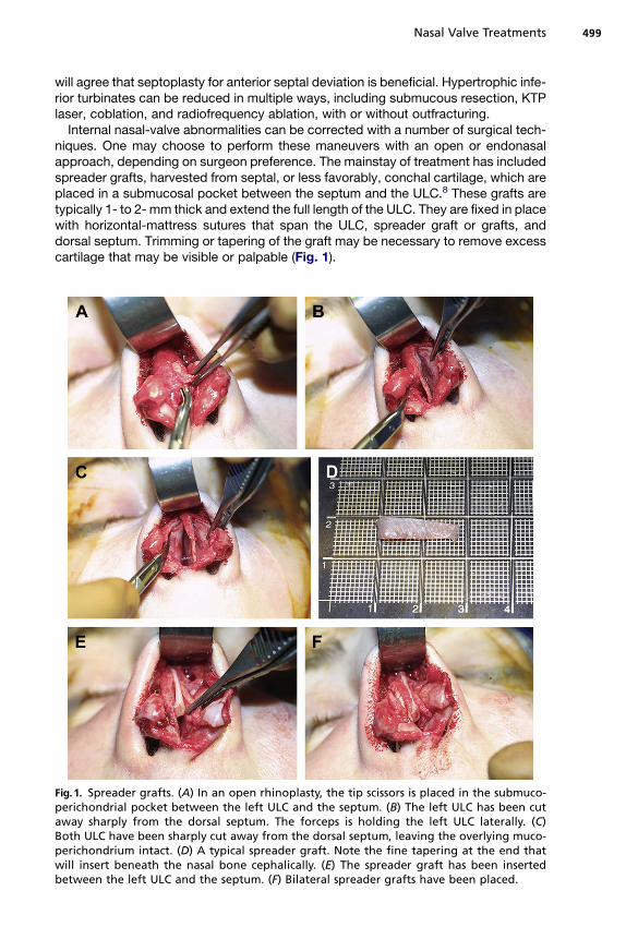
Internal nasal-valve abnormalities can be corrected with a number of surgical tech-niques. One may choose to perform these maneuvers with an open or endonasalapproach, depending on surgeon preference. The mainstay of treatment has includedspreader grafts, harvested from septal, or less favorably, conchal cartilage, which areplaced in a submucosal pocket between the septum and the ULC.8 These grafts aretypically 1- to 2- mm thick and extend the full length of the ULC. They are fixed in placewith horizontal-mattress sutures that span the ULC, spreader graft or grafts, anddorsal septum. Trimming or tapering of the graft may be necessary to remove excesscartilage that may be visible or palpable (Fig. 1).
Fig.1. Spreader grafts. (A) In an open rhinoplasty, the tip scissors is placed in the submuco-perichondrial pocket between the left ULC and the septum. (B) The left ULC has been cutaway sharply from the dorsal septum. The forceps is holding the left ULC laterally. (C)Both ULC have been sharply cut away from the dorsal septum, leaving the overlying muco-perichondrium intact. (D) A typical spreader graft. Note the fine tapering at the end thatwill insert beneath the nasal bone cephalically. (E) The spreader graft has been insertedbetween the left ULC and the septum. (F) Bilateral spreader grafts have been placed.
Lee et al500
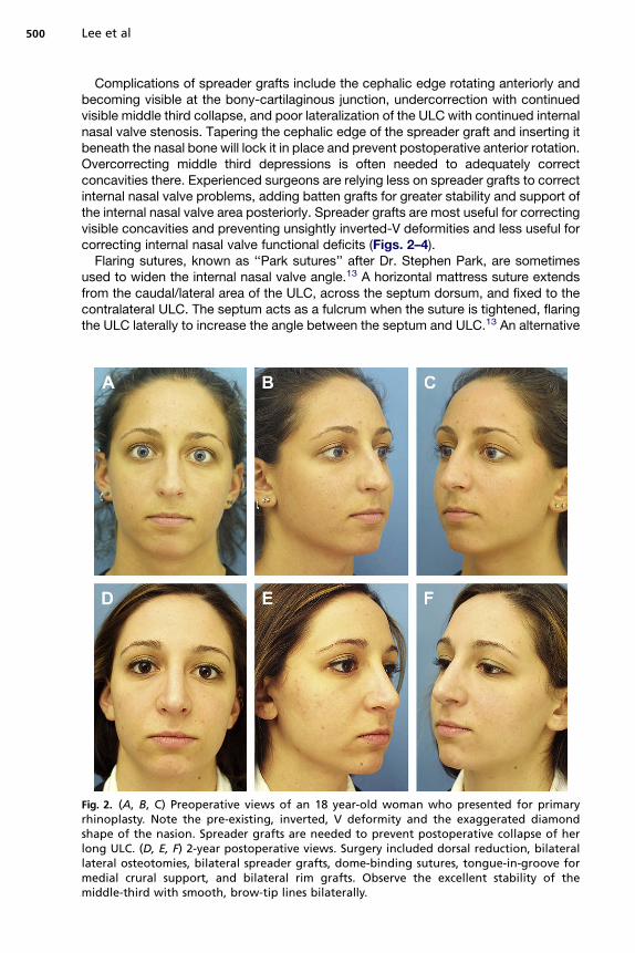
Complications of spreader grafts include the cephalic edge rotating anteriorly andbecoming visible at the bony-cartilaginous junction, undercorrection with continuedvisible middle third collapse, and poor lateralization of the ULC with continued internalnasal valve stenosis. Tapering the cephalic edge of the spreader graft and inserting itbeneath the nasal bone will lock it in place and prevent postoperative anterior rotation.Overcorrecting middle third depressions is often needed to adequately correctconcavities there. Experienced surgeons are relying less on spreader grafts to correctinternal nasal valve problems, adding batten grafts for greater stability and support ofthe internal nasal valve area posteriorly. Spreader grafts are most useful for correctingvisible concavities and preventing unsightly inverted-V deformities and less useful forcorrecting internal nasal valve functional deficits (Figs. 2–4).
Flaring sutures, known as ‘‘Park sutures’’ after Dr. Stephen Park, are sometimesused to widen the internal nasal valve angle.13 A horizontal mattress suture extendsfrom the caudal/lateral area of the ULC, across the septum dorsum, and fixed to thecontralateral ULC. The septum acts as a fulcrum when the suture is tightened, flaringthe ULC laterally to increase the angle between the septum and ULC.13 An alternative
Fig. 2. (A, B, C) Preoperative views of an 18 year-old woman who presented for primaryrhinoplasty. Note the pre-existing, inverted, V deformity and the exaggerated diamondshape of the nasion. Spreader grafts are needed to prevent postoperative collapse of herlong ULC. (D, E, F) 2-year postoperative views. Surgery included dorsal reduction, bilaterallateral osteotomies, bilateral spreader grafts, dome-binding sutures, tongue-in-groove formedial crural support, and bilateral rim grafts. Observe the excellent stability of themiddle-third with smooth, brow-tip lines bilaterally.
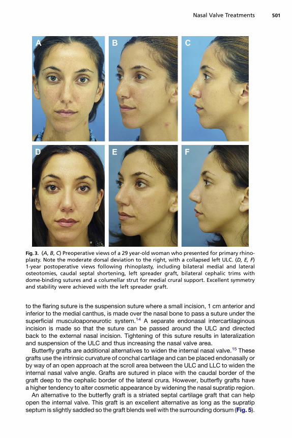
Fig. 3. (A, B, C) Preoperative views of a 29 year-old woman who presented for primary rhino-plasty. Note the moderate dorsal deviation to the right, with a collapsed left ULC. (D, E, F)1-year postoperative views following rhinoplasty, including bilateral medial and lateralosteotomies, caudal septal shortening, left spreader graft, bilateral cephalic trims withdome-binding sutures and a columellar strut for medial crural support. Excellent symmetryand stability were achieved with the left spreader graft.
Nasal Valve Treatments 501
to the flaring suture is the suspension suture where a small incision, 1 cm anterior andinferior to the medial canthus, is made over the nasal bone to pass a suture under thesuperficial musculoaponeurotic system.14 A separate endonasal intercartilaginousincision is made so that the suture can be passed around the ULC and directedback to the external nasal incision. Tightening of this suture results in lateralizationand suspension of the ULC and thus increasing the nasal valve area.
Butterfly grafts are additional alternatives to widen the internal nasal valve.15 Thesegrafts use the intrinsic curvature of conchal cartilage and can be placed endonasally orby way of an open approach at the scroll area between the ULC and LLC to widen theinternal nasal valve angle. Grafts are sutured in place with the caudal border of thegraft deep to the cephalic border of the lateral crura. However, butterfly grafts havea higher tendency to alter cosmetic appearance by widening the nasal supratip region.
An alternative to the butterfly graft is a striated septal cartilage graft that can helpopen the internal valve. This graft is an excellent alternative as long as the supratipseptum is slightly saddled so the graft blends well with the surrounding dorsum (Fig. 5).
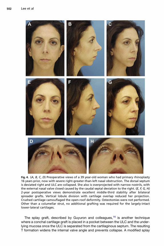
Fig. 4. (A, B, C, D) Preoperative views of a 39 year-old woman who had primary rhinoplasty16 years prior, now with severe right-greater-than-left nasal obstruction. The dorsal septumis deviated right and ULC are collapsed. She also is overprojected with narrow nostrils, withthe external nasal valve closed caused by the caudal septal deviation to the right. (E, F, G, H)2-year postoperative views demonstrate excellent middle-third stability after bilateralspreader grafts. Vertical lobule division with cartilage overlap reduced her projection.Crushed cartilage camouflaged the open-roof deformity. Osteotomies were not performed.Other than a columellar strut, no additional grafting was required for the largely-intactlower-lateral cartilages.
Lee et al502
The splay graft, described by Guyuron and colleagues,16 is another techniquewhere a conchal cartilage graft is placed in a pocket between the ULC and the under-lying mucosa once the ULC is separated from the cartilaginous septum. The resultingT formation widens the internal valve angle and prevents collapse. A modified splay
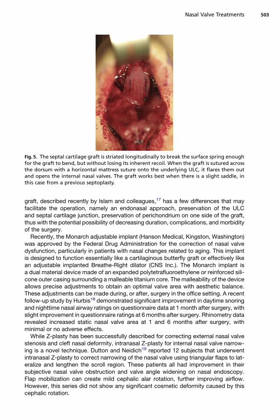
Fig. 5. The septal cartilage graft is striated longitudinally to break the surface spring enoughfor the graft to bend, but without losing its inherent recoil. When the graft is sutured acrossthe dorsum with a horizontal mattress suture onto the underlying ULC, it flares them outand opens the internal nasal valves. The graft works best when there is a slight saddle, inthis case from a previous septoplasty.
Nasal Valve Treatments 503
graft, described recently by Islam and colleagues,17 has a few differences that mayfacilitate the operation, namely an endonasal approach, preservation of the ULCand septal cartilage junction, preservation of perichondrium on one side of the graft,thus with the potential possibility of decreasing duration, complications, and morbidityof the surgery.
Recently, the Monarch adjustable implant (Hanson Medical, Kingston, Washington)was approved by the Federal Drug Administration for the correction of nasal valvedysfunction, particularly in patients with nasal changes related to aging. This implantis designed to function essentially like a cartilaginous butterfly graft or effectively likean adjustable implanted Breathe-Right dilator (CNS Inc.). The Monarch implant isa dual material device made of an expanded polytetrafluoroethylene or reinforced sili-cone outer casing surrounding a malleable titanium core. The malleability of the deviceallows precise adjustments to obtain an optimal valve area with aesthetic balance.These adjustments can be made during, or after, surgery in the office setting. A recentfollow-up study by Hurbis18 demonstrated significant improvement in daytime snoringand nighttime nasal airway ratings on questionnaire data at 1 month after surgery, withslight improvement in questionnaire ratings at 6 months after surgery. Rhinometry datarevealed increased static nasal valve area at 1 and 6 months after surgery, withminimal or no adverse effects.
While Z-plasty has been successfully described for correcting external nasal valvestenosis and cleft nasal deformity, intranasal Z-plasty for internal nasal valve narrow-ing is a novel technique. Dutton and Neidich19 reported 12 subjects that underwentintranasal Z-plasty to correct narrowing of the nasal valve using triangular flaps to lat-eralize and lengthen the scroll region. These patients all had improvement in theirsubjective nasal valve obstruction and valve angle widening on nasal endoscopy.Flap mobilization can create mild cephalic alar rotation, further improving airflow.However, this series did not show any significant cosmetic deformity caused by thiscephalic rotation.
Lee et al504
SURGICALMANAGEMENT OF THE EXTERNAL NASALVALVE
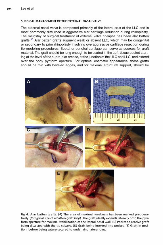
The external nasal valve is composed primarily of the lateral crus of the LLC and ismost commonly disturbed in aggressive alar cartilage reduction during rhinoplasty.The mainstay of surgical treatment of external valve collapse has been alar battengrafts.13 Alar batten grafts augment weak or absent LLC, which may be congenitalor secondary to prior rhinoplasty involving overaggressive cartilage resection duringtip-modeling procedures. Septal or conchal cartilage can serve as sources for graftmaterial. The graft should be long enough to be seated in the soft-tissue pocket start-ing at the level of the supra-alar crease, at the junction of the ULC and LLC, and extendover the bony pyriform aperture. For optimal cosmetic appearance, these graftsshould be thin with beveled edges, and for maximal structural support, should be
Fig. 6. Alar batten grafts. (A) The area of maximal weakness has been marked preopera-tively. (B) Typical size of a batten graft (top). The graft ideally extends laterally onto the pyri-form aperture for maximal stabilization of the lateral-nasal wall. (C) Pocket to receive graftbeing dissected with the tip scissors. (D) Graft being inserted into pocket. (E) Graft in posi-tion, before being suture-secured to underlying lateral crus.
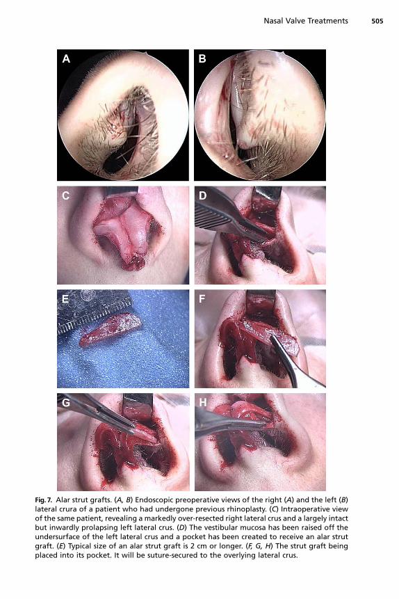
Fig. 7. Alar strut grafts. (A, B) Endoscopic preoperative views of the right (A) and the left (B)lateral crura of a patient who had undergone previous rhinoplasty. (C) Intraoperative viewof the same patient, revealing a markedly over-resected right lateral crus and a largely intactbut inwardly prolapsing left lateral crus. (D) The vestibular mucosa has been raised off theundersurface of the left lateral crus and a pocket has been created to receive an alar strutgraft. (E) Typical size of an alar strut graft is 2 cm or longer. (F, G, H) The strut graft beingplaced into its pocket. It will be suture-secured to the overlying lateral crus.
Nasal Valve Treatments 505
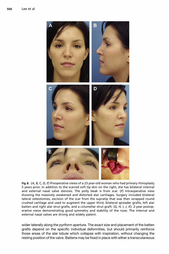
Fig. 8. (A, B, C, D, E) Preoperative views of a 23 year-old woman who had primary rhinoplasty5 years prior. In addition to the scarred soft tip skin on the right, she has bilateral internaland external nasal valve stenosis. The polly beak is from scar. (F) Intraoperative viewshowing the massively weakened and distorted alar cartilages. Surgery included bilaterallateral osteotomies, excision of the scar from the supratip that was then wrapped roundcrushed cartilage and used to augment the upper third, bilateral spreader grafts, left alarbatten and right alar strut grafts, and a columellar strut graft. (G, H, I, J, K). 2-year postop-erative views demonstrating good symmetry and stability of the nose. The internal andexternal nasal valves are strong and widely patent.
Lee et al506
wider laterally along the pyriform aperture. The exact size and placement of the battengrafts depend on the specific individual deformities, but should primarily reinforcethose areas of the alar lobule which collapse with inspiration, without changing theresting position of the valve. Battens may be fixed in place with either a transcutaneous
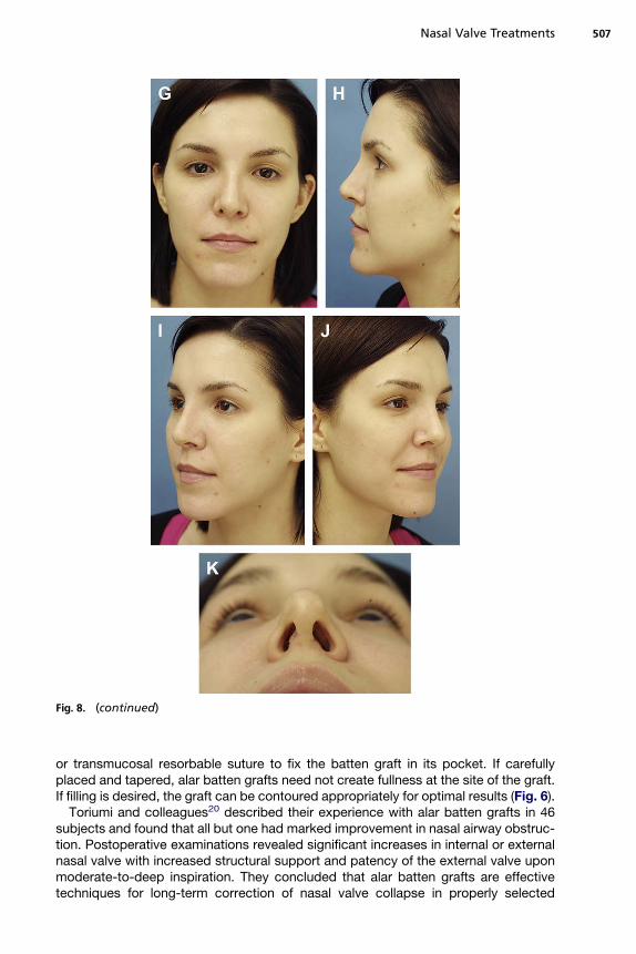
Fig. 8. (continued)
Nasal Valve Treatments 507
or transmucosal resorbable suture to fix the batten graft in its pocket. If carefullyplaced and tapered, alar batten grafts need not create fullness at the site of the graft.If filling is desired, the graft can be contoured appropriately for optimal results (Fig. 6).
Toriumi and colleagues20 described their experience with alar batten grafts in 46subjects and found that all but one had marked improvement in nasal airway obstruc-tion. Postoperative examinations revealed significant increases in internal or externalnasal valve with increased structural support and patency of the external valve uponmoderate-to-deep inspiration. They concluded that alar batten grafts are effectivetechniques for long-term correction of nasal valve collapse in properly selected
Lee et al508
patients without intranasal scarring, loss of vestibular skin, or excessive narrowing ofthe pyriform aperture.
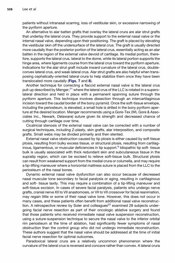
An alternative to alar batten grafts that overlay the lateral crura are alar strut graftsthat underlay the lateral crura. They provide support to the external nasal valve or theinternal nasal valve, depending upon their positioning. The graft is placed by elevatingthe vestibular skin off the undersurface of the lateral crus. The graft is usually directedmore caudally than the posterior portion of the lateral crus, essentially acting as an alarbatten in the region of the external valve devoid of cartilage. Its medial portion, there-fore, supports the lateral crus, lateral to the dome, while its lateral portion supports thehinge area, where ligaments course from the lateral crus toward the pyriform aperture.Indications for the alar strut graft include inward curvature of the lateral crus, double-convex lateral crus, and weak lateral crus. Alar strut grafts are also helpful when trans-posing cephalically-oriented lateral crura to help stabilize them once they have beentranslocated more caudally (Figs. 7 and 8).
Another technique for correcting a flaccid external nasal valve is the lateral cruspull-up described by Menger,21 where the lateral crus of the LLC is rotated in a supero-lateral direction and held in place with a permanent spanning suture through thepyriform aperture. This technique involves dissection through an intercartilaginousincision toward the caudal border of the bony pyramid. Once the soft-tissue envelope,including the periosteum, is elevated, a small hole is drilled in the bony pyriform aper-ture at the desired location. Menger recommends using a Gore-Tex (WL Gore & Asso-ciates Inc., Newark, Delaware) suture given its strength and decreased chance ofcutting through cartilage over time.
Cicatricial stenosis of the external nasal valve can be corrected with a number ofsurgical techniques, including Z-plasty, skin grafts, alar interposition, and compositegrafts. Small webs may be divided primarily and then stented.
External nasal valve obstruction caused by tip ptosis is either caused by soft tissueptosis, resulting from bulky excess tissue, or structural ptosis, resulting from cartilag-inous, ligamentous, or muscular deficiencies in tip support.9 Idiopathic tip soft- tissuebulk is usually associated with thick redundant skin and subcutaneous tissue in thesupratip region, which can be excised to relieve soft-tissue bulk. Structural ptosiscan result from weakened support from the medial crura or columella, and may requirea tip-lifting maneuver where a horizontal mattress suture is placed from the LLC to theperiosteum of the nasal bones.
Dynamic external nasal valve dysfunction can also occur because of decreasednasal muscular tone secondary to facial paralysis or aging, resulting in cartilaginousand soft- tissue laxity. This may require a combination of a tip-lifting maneuver andsoft-tissue excision. In cases of severe facial paralysis, patients who undergo nervegrafts, cranial nerve XII to VII anastomosis, or VII to VII crossover for facial reanimation,may regain little or some of their nasal valve tone. However, this does not occur inmany cases, and these patients often benefit from additional nasal valve reconstruc-tion. A retrospective review by Soler and colleagues22 examined 28 subjects under-going facial nerve resection as part of their oncologic ablative surgery and foundthat those patients who received immediate nasal valve suspension reconstruction,using a suture-suspension technique to secure the nasal valve to the inferior orbitalrim periosteum at the time of ablation, had significantly fewer symptoms of nasalobstruction than the control group who did not undergo immediate reconstruction.These authors suggest that the nasal valve should be addressed at the time of initialfacial nerve resection for optimal outcomes.
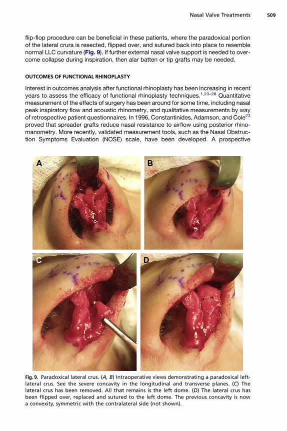
Paradoxical lateral crura are a relatively uncommon phenomenon where thecurvature of the lateral crus is reversed and concave rather than convex. A lateral crura
Nasal Valve Treatments 509
flip-flop procedure can be beneficial in these patients, where the paradoxical portionof the lateral crura is resected, flipped over, and sutured back into place to resemblenormal LLC curvature (Fig. 9). If further external nasal valve support is needed to over-come collapse during inspiration, then alar batten or tip grafts may be needed.
OUTCOMES OF FUNCTIONAL RHINOPLASTY
Interest in outcomes analysis after functional rhinoplasty has been increasing in recentyears to assess the efficacy of functional rhinoplasty techniques.1,23–28 Quantitativemeasurement of the effects of surgery has been around for some time, including nasalpeak inspiratory flow and acoustic rhinometry, and qualitative measurements by wayof retrospective patient questionnaires. In 1996, Constantinides, Adamson, and Cole23
proved that spreader grafts reduce nasal resistance to airflow using posterior rhino-manometry. More recently, validated measurement tools, such as the Nasal Obstruc-tion Symptoms Evaluation (NOSE) scale, have been developed. A prospective
Fig. 9. Paradoxical lateral crus. (A, B) Intraoperative views demonstrating a paradoxical left-lateral crus. See the severe concavity in the longitudinal and transverse planes. (C) Thelateral crus has been removed. All that remains is the left dome. (D) The lateral crus hasbeen flipped over, replaced and sutured to the left dome. The previous concavity is nowa convexity, symmetric with the contralateral side (not shown).
Lee et al510
observational outcomes study on subjects who underwent functional rhinoplastyexamined preoperative and postoperative NOSE scores and found that all subjectsdemonstrated significant airway improvement in all subcategories based on thespecific procedure that was performed.25 Additionally, a large recent meta-analysisby Rhee and colleagues24 reviewed the efficacy of functional rhinoplasty techniquesfor the treatment of nasal valve dysfunction over the past 25 years found that thereis substantial level 4 evidence (case series) to support the efficacy of current functionalrhinoplasty techniques. The authors suggest a need for increased use of comparisoncohorts and standardized objective outcome measures to strengthen evidence sup-porting what is already recognized clinically.
SUMMARY
Treatment of the nasal valve, both surgical and nonsurgical, continues to improve withbetter understanding of nasal anatomy and physiology and advancements intechnology.
While spreader grafts and alar batten grafts have been the mainstay of treatment ofnasal valve dysfunction for quite some time, newer techniques are emerging and gain-ing popularity among surgeons. Areas for future improvements include better use ofstandardized-outcome assessments to evaluate efficacy of specific surgical proce-dures in repairing nasal valve abnormalities. Nasal anatomy and physiology is uniqueacross individuals, and nasal valve dysfunction should always be treated witha tailored regimen specific to the individual deformities.
REFERENCES
1. Rhee J, Poetker D, Smith TL, et al. Nasal valve surgery improves disease specificquality of life. Laryngoscope 2005;115:437–40.
2. Mink PJ. Le nez comme voie respiratorie. Presse Otolaryngol (Belg) 1903;481–96[French].
3. Constantian MB, Brian CR. The relative importance of septal and nasal valvularsurgery in correcting airway obstruction in primary and secondary rhinoplasty.Plast Reconstr Surg 1996;98(1):38–54.
4. Miman MC, Deliktas H, Ozturan O, et al. Internal nasal valve: revisited with objec-tive facts. Otolaryngol Head Neck Surg 2006;134(1):41–7.
5. Kern EB, Wang TD. Nasal valve surgery. In: Daniel RK, Regnault P, Goldwyn RM,editors. Aesthetic plastic surgery: rhinoplasty. London: Little, Brown and Co.;1993. p. 613–30.
6. Grymer LF. Reduction rhinoplasty and nasal patency: change in the cross-sectional area of the nose evaluated by acoustic rhinometry. Laryngoscope1995;105:429–31.
7. Khosh JM, Jen A, Honrado C, et al. Nasal valve reconstruction: experience in 53consecutive patients. Arch Facial Plast Surg 2004;6:167–71.
8. Sheen JH. Spreader graft: a method of reconstructing the roof of the middle nasalvault following rhinoplasty. Plast Reconstr Surg 1984;73(2):230–7.
9. Schlosser RJ, Park SS. Functional rhinoplasty. Otolaryngol Clin North Am 1999;32(1):37–51.
10. Constantinides MS, Galli SK, Miller PJ. A simple and reliable method of patientevaluation in the surgical treatment of nasal obstruction. Ear Nose Throat J2002;81(10):734–7.
11. Nyte CP. Hyaluronic acid spreader-graft injection for internal nasal valve collapse.Ear Nose Throat J 2007;86(5):272–3.
Nasal Valve Treatments 511
12. Inanli S, Sari M, Yazici MZ. The results of concurrent functional endoscopic sinussurgery and rhinoplasty. J Craniofac Surg 2008;19(3):701–4.
13. Ballert JA, Park SS. Functional rhinoplasty: treatment of the dysfunctional nasalsidewall. Facial Plast Surg 2006;22(1):49–54.
14. Nuara MJ, Mobley SR. Nasal valve suspension revisited. Laryngoscope 2007;117:2100–6.
15. Clark JM, Cook TA. The ‘butterfly’ graft in functional secondary rhinoplasty. Laryn-goscope 2002;112:1917–25.
16. Guyuron B, Michelow BJ, Englebardt C. Upper lateral splay graft. Plast ReconstrSurg 1998;102:2169–77.
17. Islam A, Arslan N, Felek SA, et al. Reconstruction of the internal nasal valve:modified splay graft technique with endonasal approach. Laryngoscope 2008;118:1739–43.
18. Hurbis CG. A follow-up study of the Monarch adjustable implant for correction ofnasal valve dysfunction. Arch Facial Plast Surg 2008;10(2):142–3.
19. Dutton JM, Neidich MJ. Intranasal Z-plasty for internal nasal valve collapse. ArchFacial Plast Surg 2008;10(3):164–8.
20. Toriumi DM, Josen J, Weinberger M, et al. Use of alar batten grafts for correctionof nasal valve collapse. Arch Otolaryngol Head Neck Surg 1997;123(8):802–8.
21. Menger DJ. Lateral crus pull-up: a method for collapse of the external nasalvalve. Arch Facial Plast Surg 2006;8(5):333–7.
22. Soler ZM, Rosenthal E, Wax MK. Immediate nasal valve reconstruction after facialnerve resection. Arch Facial Plast Surg 2008;10(5):312–5.
23. Constantinides MS, Adamson PA, Cole P. The long term effects of open cosmeticseptorhinoplasty on nasal air flow. Arch Otolaryngol Head Neck Surg 1996;122:41–5.
24. Rhee JS, Arganbright JM, McMullin BT, et al. Evidence supporting functionalrhinoplasty or nasal valve repair: a 25-year systematic review. OtolaryngolHead Neck Surg 2008;139(1):10–20.
25. Most SP. Analysis of outcomes after functional rhinoplasty using a disease-specific quality-of-life instrument. Arch Facial Plast Surg 2006;8(5):306–9.
26. Costa F, Robiony M, Salvo I, et al. Simultaneous functional endoscopic sinussurgery and esthetic rhinoplasty in orthognathic patients. J Oral MaxillofacSurg 2008;66(7):1370–7.
27. Faris C, Koury E, Kothari P, et al. Functional rhinoplasty with batten and spreadergrafts for correction of internal nasal valve incompetence. Rhinology 2006;44(2):114–7.
28. Fischer H, Gubisch W. Nasal valves – importance and surgical procedures.Facial Plast Surg 2006;22:266–80.


![Implant Science formation via parathyroid hormone … · 2020. 4. 28. · MRONJ after surgical [6-8] and nonsurgical treatments [9,10] have been reported. However, MRONJ still adversely](https://img.pdfslide.us/doc/110x75/5fc176e90cc45504ea2c7f83/implant-science-formation-via-parathyroid-hormone-2020-4-28-mronj-after-surgical.jpg)