Embed Size (px)
Citation preview

The Acute Abdomen

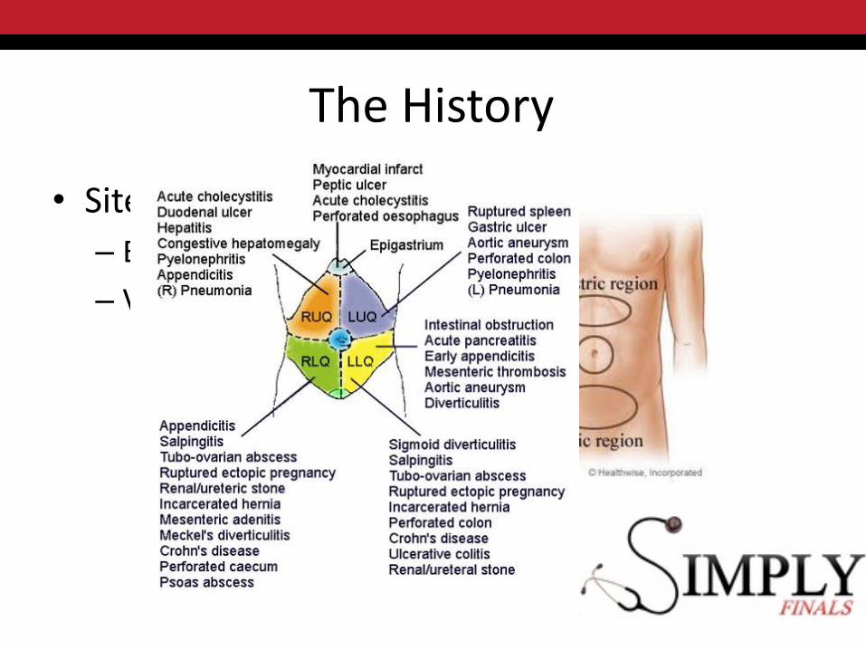
The History
• Site
– Embryology
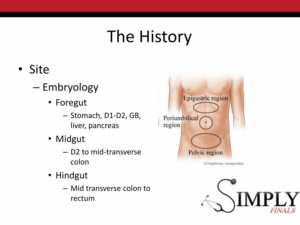
The History
• Site
– Embryology
• Foregut– Stomach, D1-D2, GB,
liver, pancreas
• Midgut– D2 to mid-transverse
colon
• Hindgut– Mid transverse colon to
rectum
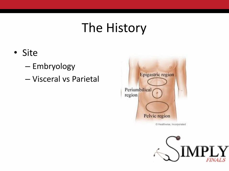
The History
• Site
– Embryology
– Visceral vs Parietal
The History
• Site
– Embryology
– Visceral vs Parietal
The History
• Onset
– Sudden – think of perforation/embolus
– Inflammatory – more gradual
The History
• Character
– Visceral vs parietal
– Constant think inflammatory
– Intermittent think mechanical
• Colic ≈ obstruction of a hollow viscus
The History
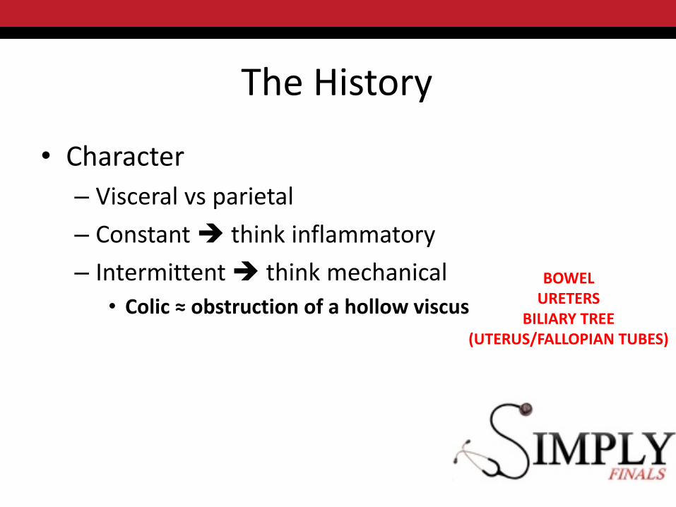
• Character
– Visceral vs parietal
– Constant think inflammatory
– Intermittent think mechanical
• Colic ≈ obstruction of a hollow viscus
BOWELURETERS
BILIARY TREE(UTERUS/FALLOPIAN TUBES)
The History
• Radiation
– Back – retroperitoneum
– Shoulder tip – diaphragm
– Loin to groin – renal tract/aorta
The History
• Associated features
– ‘Abdominal pain in isolation rarely indicates severe pathology…’
– Appetite
– Vomiting
– Bowel habit
– Urinary
– Gynae
• LMP
The History
• Timing/Duration
The History
• Exacerbating/relieving
– MOVEMENT
– Position
– Morphine…

The History
• Severity….
– Trend can be useful
• Same pain before???
The Acute Abdomen - History
• Inflammatory vs mechanical
• Associated features
• Previous episodes
• RISK FACTORS
The Acute Abdomen - History
• Peritonism vs Colic
• Associated features
• Previous episodes
• RISK FACTORS

• 44yr Male
• PC: Abdominal pain
• HPC:
• Acute onset of epigastric and periumbilical pain
• Associated sweating, mild breathlessness and vomiting
Case 1
The Case
• Sudden onset circa 3am
• Constant
• Non radiating
• Worse on movement
• No previous episodes
The Case
• PMH
– T2DM
– HTN
• No surgical history
• DH
– Antihypertensive
– NKDA
The Case
• PMH
– T2DM
– HTN
• No surgical history
• DH
– Antihypertensive
– NKDA
– OTC Ibuprofen for back pain
The Case
• SH
– Smokes 10/day
– Not much booze
– Lorry driver

Examination
Examination
• ABCDE...
Examination
• A
– Can they talk?
Examination
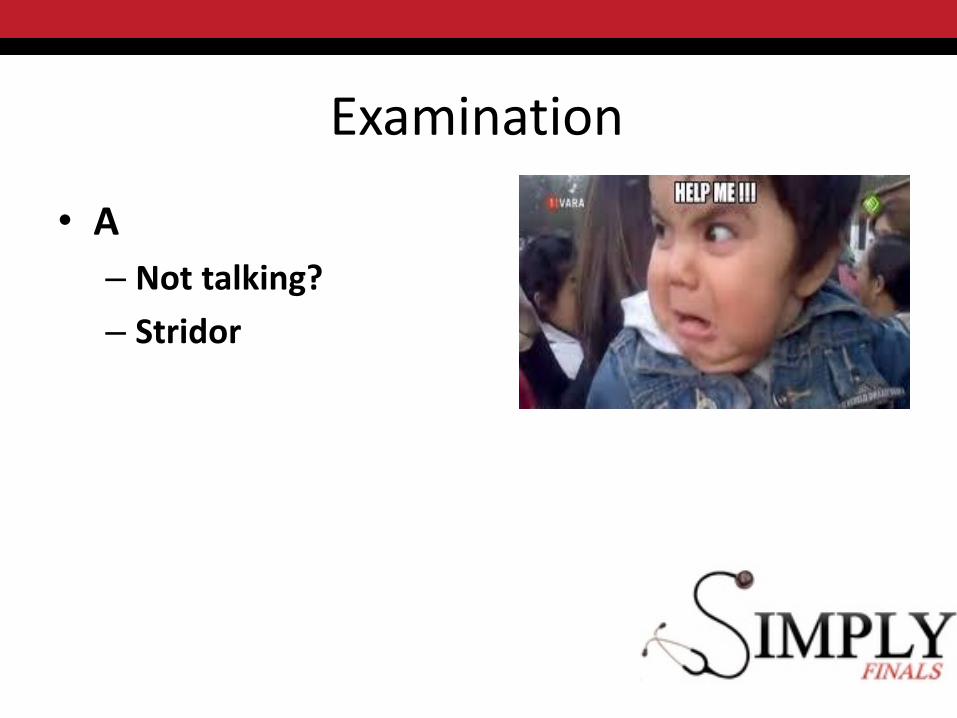
• A
– Not talking?
– Stridor
Examination
• A
• B
– RESPIRATORY RATE
– Saturation
– (Auscultation)
Examination
• A
• B
• C
– HR
– BP
– Volume status
– PERFUSION
Examination
• A
• B
• C
– PERFUSION
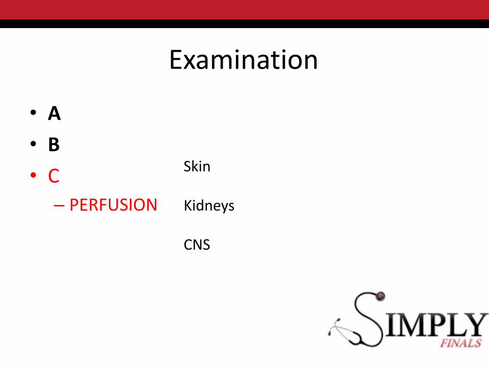
Examination
• A
• B
• C
– PERFUSION
Skin
Kidneys
CNS
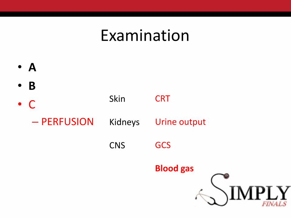
Examination
• A
• B
• C
– PERFUSION
Skin
Kidneys
CNS
CRT
Urine output
GCS
Blood gas
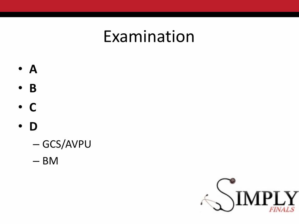
Examination
• A
• B
• C
• D
– GCS/AVPU
– BM
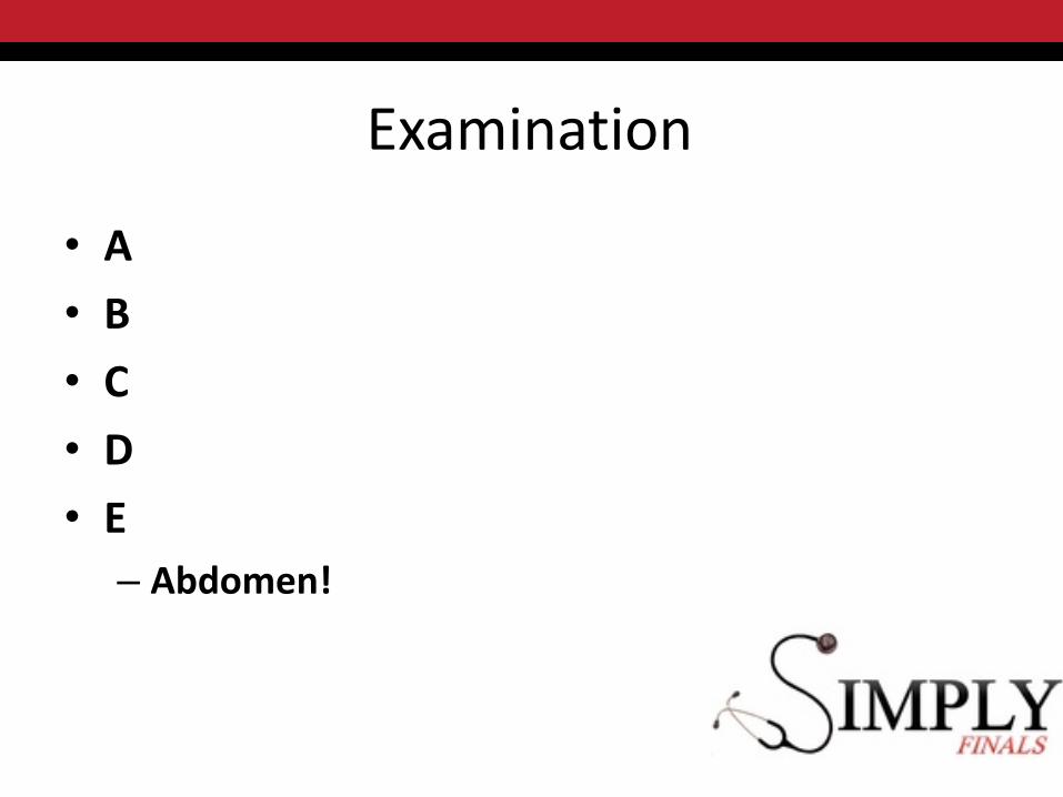
Examination
• A
• B
• C
• D
• E
– Abdomen!
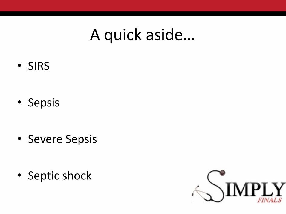
A quick aside…
• SIRS
• Sepsis
• Severe Sepsis
• Septic shock

A quick aside…
• SIRS
• Sepsis
– SIRS + infection
• Severe Sepsis
– Sepsis + organ dysfunction
• Septic shock
– Sepsis + hypotension (despite fluid)
HR >90RR >20
Temp >38 or <36WCC >12 or <4
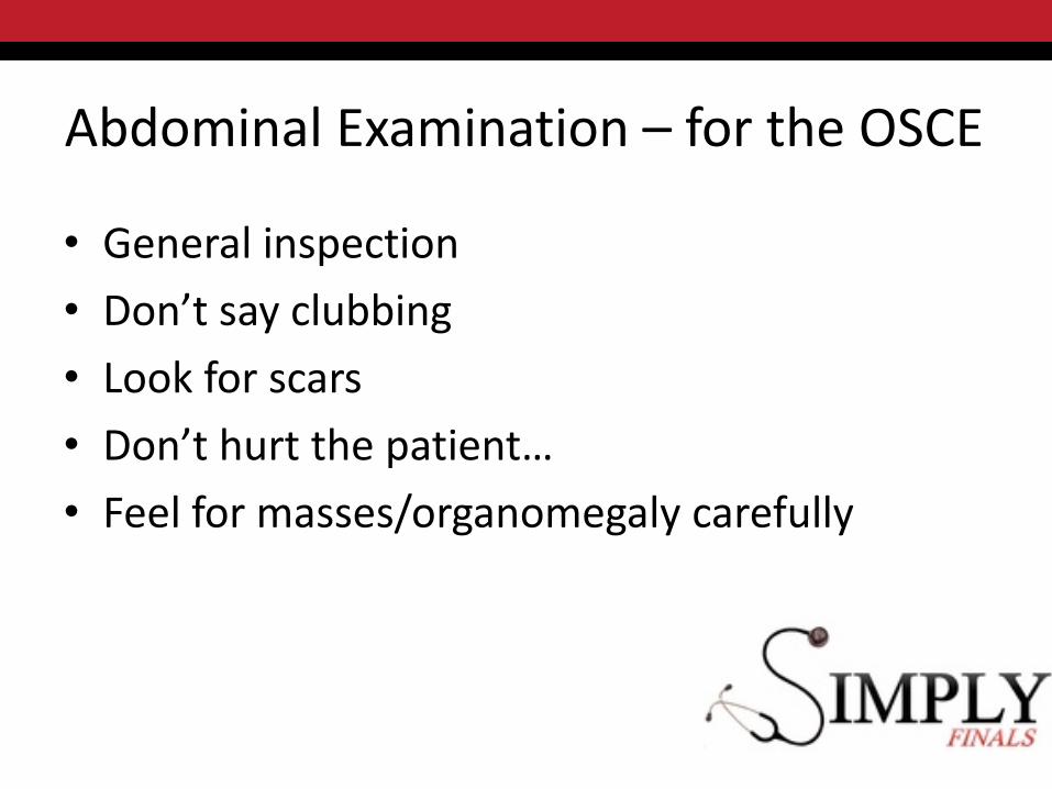
Abdominal Examination – for the OSCE
• General inspection
• Don’t say clubbing
• Look for scars
• Don’t hurt the patient…
• Feel for masses/organomegaly carefully

SpleenLiver
Pseudocyst
AAARenal transplant
Ovarian Cyst
Bladder/uterus
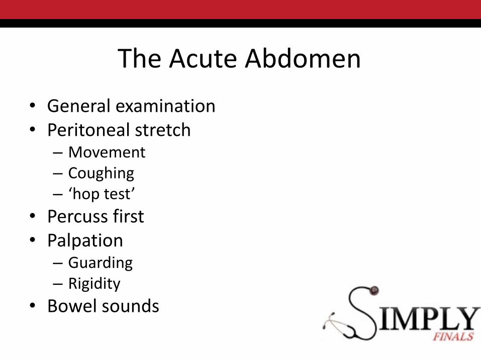
The Acute Abdomen
• General examination• Peritoneal stretch
– Movement– Coughing– ‘hop test’
• Percuss first• Palpation
– Guarding– Rigidity
• Bowel sounds
The Acute Abdomen
• General examination• Peritoneal stretch
– Movement– Coughing– ‘hop test’
• Percuss first• Palpation
– Guarding– Rigidity
• Bowel sounds
Is the patient peritonitic?

Anything else?
Anything else?
• Hernial orifices
• External genitalia
• PR
The Case
• A– SM
• B– RR 24/min– SaO2 95%
• C– CRT 2secs
– HR 110/min
– BP 160/90
• D– Alert
• E– T 37.4
– Abdomen• Tender epigastrium and
RUQ
• Guarding
• Percussion tenderness

Thoughts?

Investigations
• Bloods
• Imaging
• Other

Think about ‘non-surgical’ causes
‘Medical’ causes of the acute abdomen
• Epigastric pain - Acute MI
• RUQ/LUQ – LRTI
• Suprapubic/loin – UTI
• Gastroenteritis
• DKA
• etc etc
Investigations
• Bloods
– ABG
– FBC
– U+E
– (LFT)
– Amylase
– (CRP)
– Glucose
– Clotting
– G+S
• Imaging
– Erect CXR
– USS Abdo
– CT
• Other
– ECG
– (Urinalysis)
Investigations
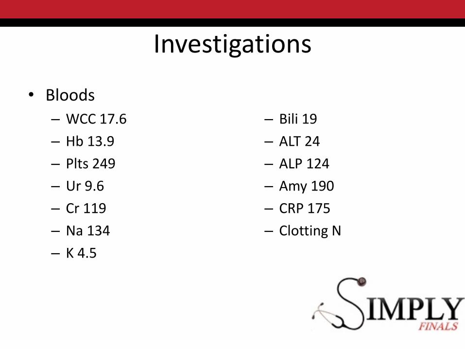
• Bloods
– WCC 17.6
– Hb 13.9
– Plts 249
– Ur 9.6
– Cr 119
– Na 134
– K 4.5
– Bili 19
– ALT 24
– ALP 124
– Amy 190
– CRP 175
– Clotting N


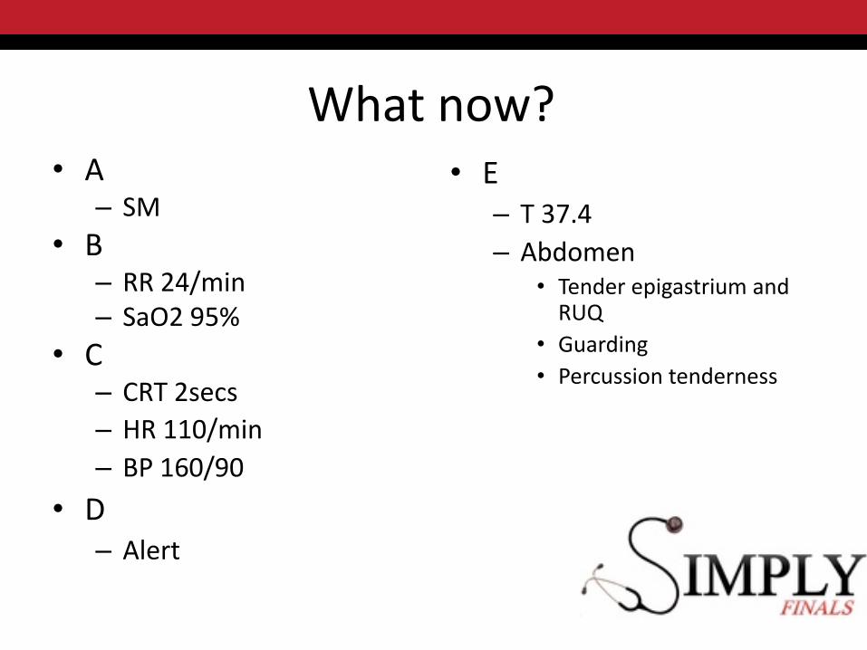
What now?• A
– SM
• B– RR 24/min– SaO2 95%
• C– CRT 2secs
– HR 110/min
– BP 160/90
• D– Alert
• E– T 37.4
– Abdomen• Tender epigastrium and
RUQ
• Guarding
• Percussion tenderness
Management
• ABCDE
• Oxygen…
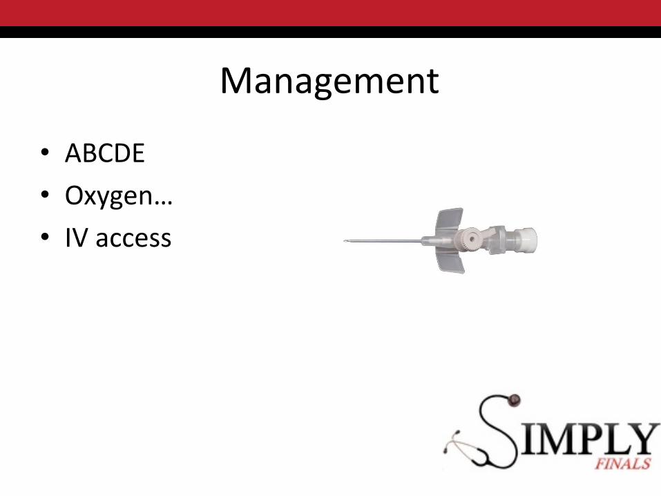
Management
• ABCDE
• Oxygen…
• IV access
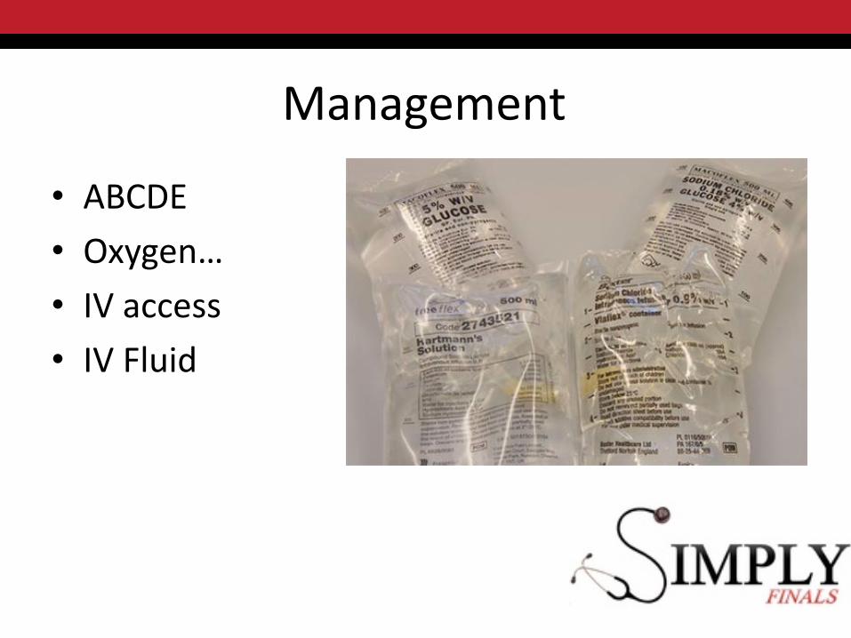
Management
• ABCDE
• Oxygen…
• IV access
• IV Fluid
Management
• ABCDE
• Oxygen…
• IV access
• IV Fluid
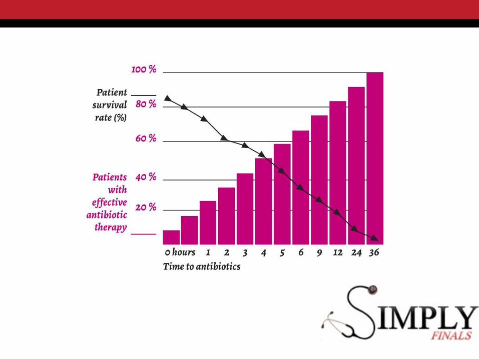
• Antibiotics
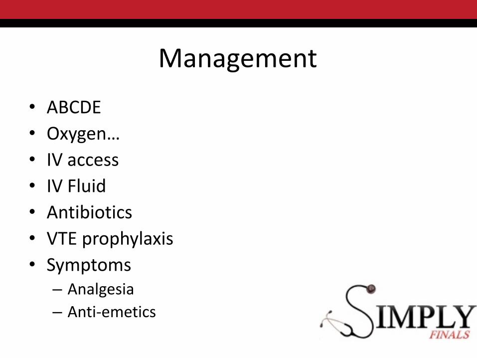
Management
• ABCDE
• Oxygen…
• IV access
• IV Fluid
• Antibiotics
• VTE prophylaxis
• Symptoms– Analgesia
– Anti-emetics
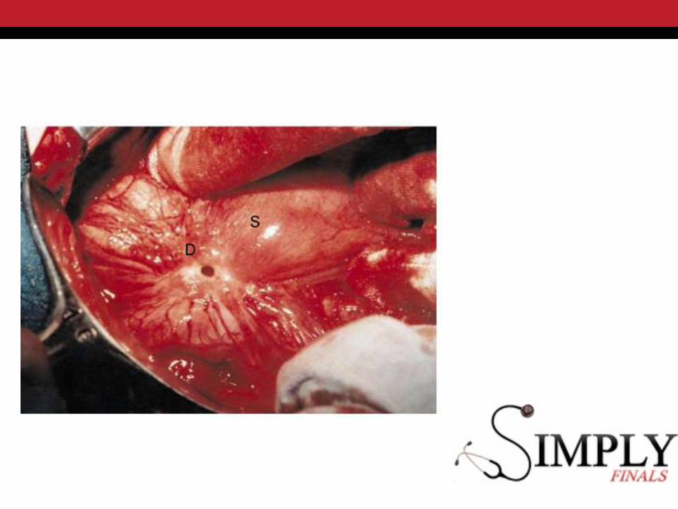
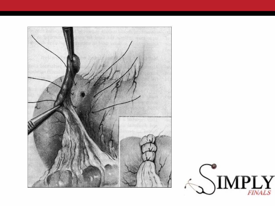
Perforated Viscus
Upper GI Lower GI
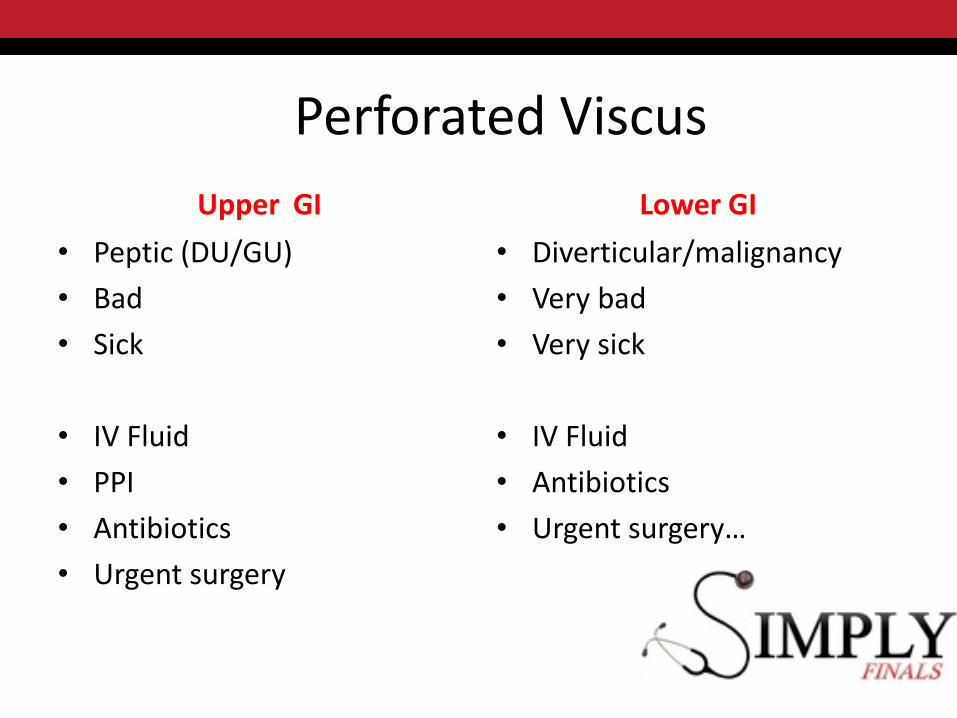
Perforated Viscus
Upper GI
• Peptic (DU/GU)
• Bad
• Sick
• IV Fluid
• PPI
• Antibiotics
• Urgent surgery
Lower GI
• Diverticular/malignancy
• Very bad
• Very sick
• IV Fluid
• Antibiotics
• Urgent surgery…
Perforation– Upper vs Lower
• Age
• Risk factors
– PUD
• NSAIDS
• Smoking
• Location of pain
• Clinical findings
• Radiology
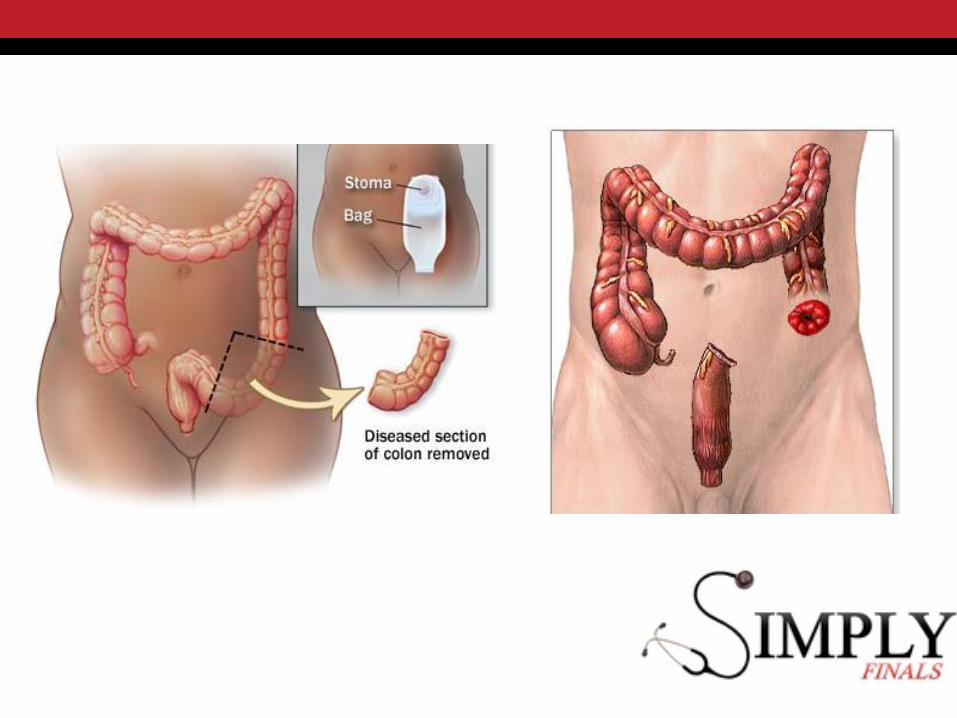
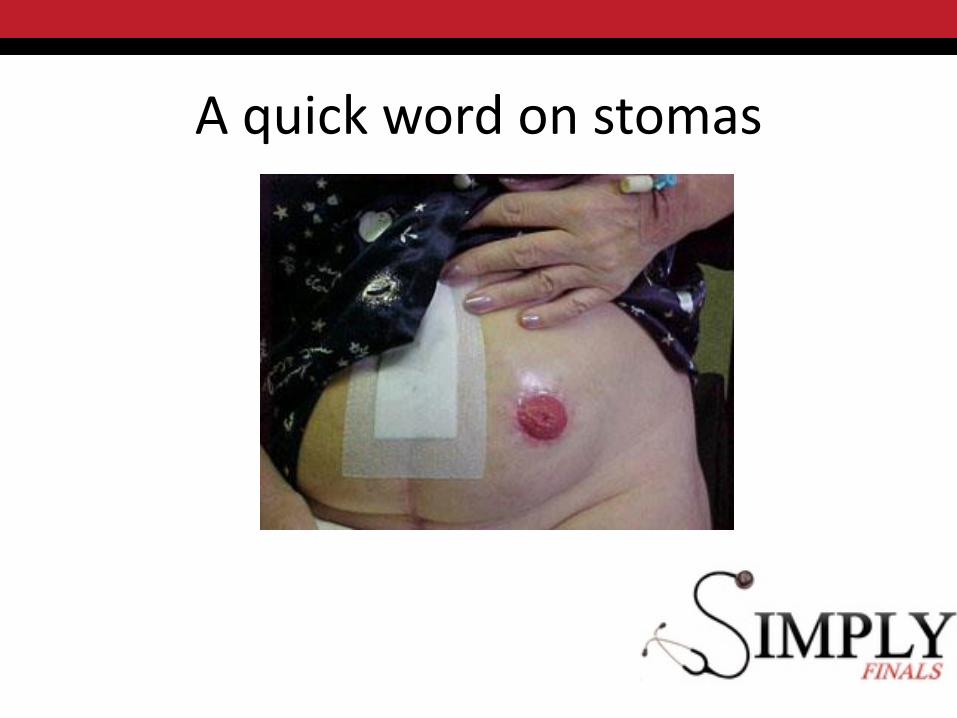
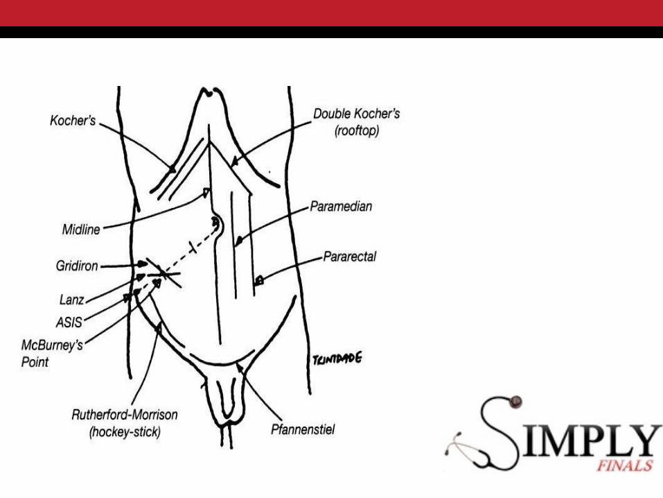
A quick word on stomas

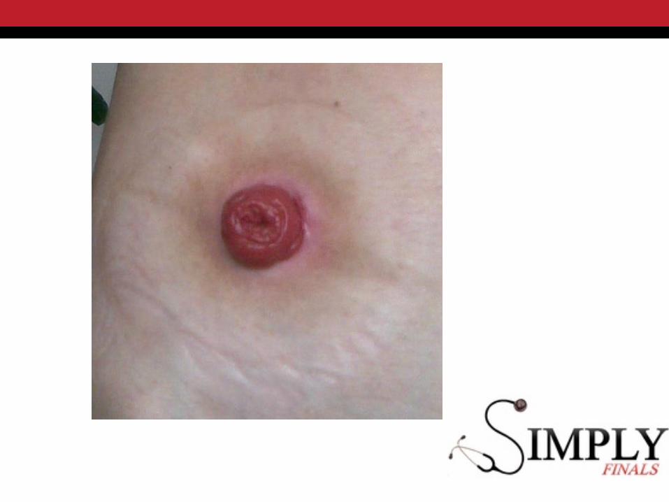
Examining a stoma
• Location
• Size
• Output
• Number of lumens
– End
– Loop
• Skin condition
Types of Stoma• Colostomy
– Usually LIF/RUQ
– Flush with skin
– Larger lumen
– Faecal output
– Common operations:• Hartmann’s
• Abdominoperineal resection
• Decompression (inoperable distal malignancy)
• Urostomy (ileal conduit)
• Ileostomy– Usually RIF
– Spouted
– Smaller lumen
– Liquid output initially
– Common operations:• Loop
– After anterior resection
• End– After total/subtotal colectomy
Complications of Stomas
• Early
– High output
– Ischaemia
– Retraction
• Late
– Prolapse
– Parastomal hernia
– Psychosocial

Case 2• 78F
• Under medics with chest infection
• PMH Dementia, COPD
• Two days of
– Profuse vomiting
– Central abdominal pain
Case 2
• Profuse vomiting
• Central abdominal pain
– ‘comes and goes in waves’
• BNO for 5/7
• No flatus
On examination
• A– SM
• B– RR 24/min
– SaO2 92%
– Coarse crackles Rt base
• C– Cool peripheries
– HR 110
– BRP 120/75
• D– Confused
• E– T 37.2
– Abdomen• Distended
• Generally tender
• Not peritonitic
• BS active
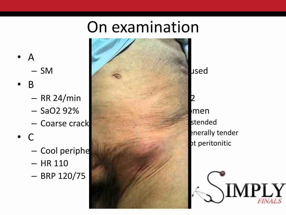
On examination
• A– SM
• B– RR 24/min
– SaO2 92%
– Coarse crackles Rt base
• C– Cool peripheries
– HR 110
– BRP 120/75
• D– Confused
• E– T 37.2
– Abdomen• Distended
• Generally tender
• Not peritonitic

What next?
Investigations
• Bloods
– FBC
– U+E
– (CRP)
– Clotting
– G&S
– ABG
• Imaging
– AXR/CXR
– CT?
• Other
– ECG
– Urine
Investigations
• Hb 12.4
• WCC 16.4
• Plts 252
• Urea 12.4
• Creat 110
• Clotting N
• ABG (on 4L)
– pH 7.33
– PCO2 4.0
– PO2 10.2
– BE -5.8
– Lac 2.9

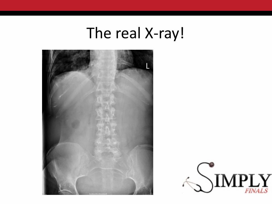
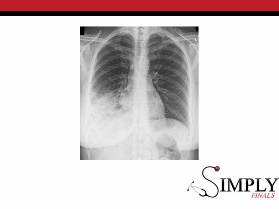
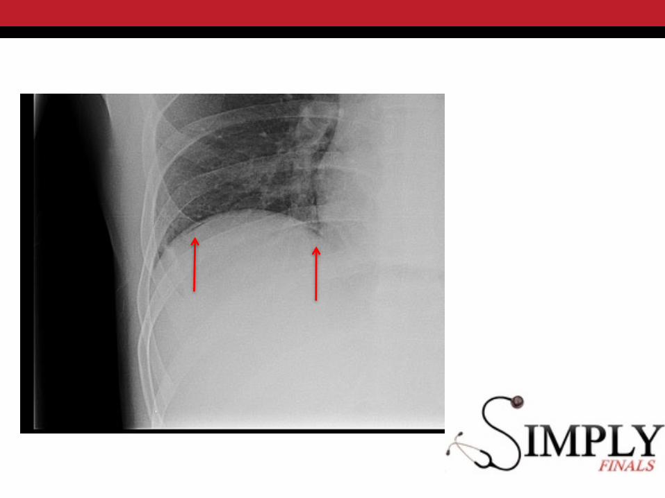
The real X-ray!
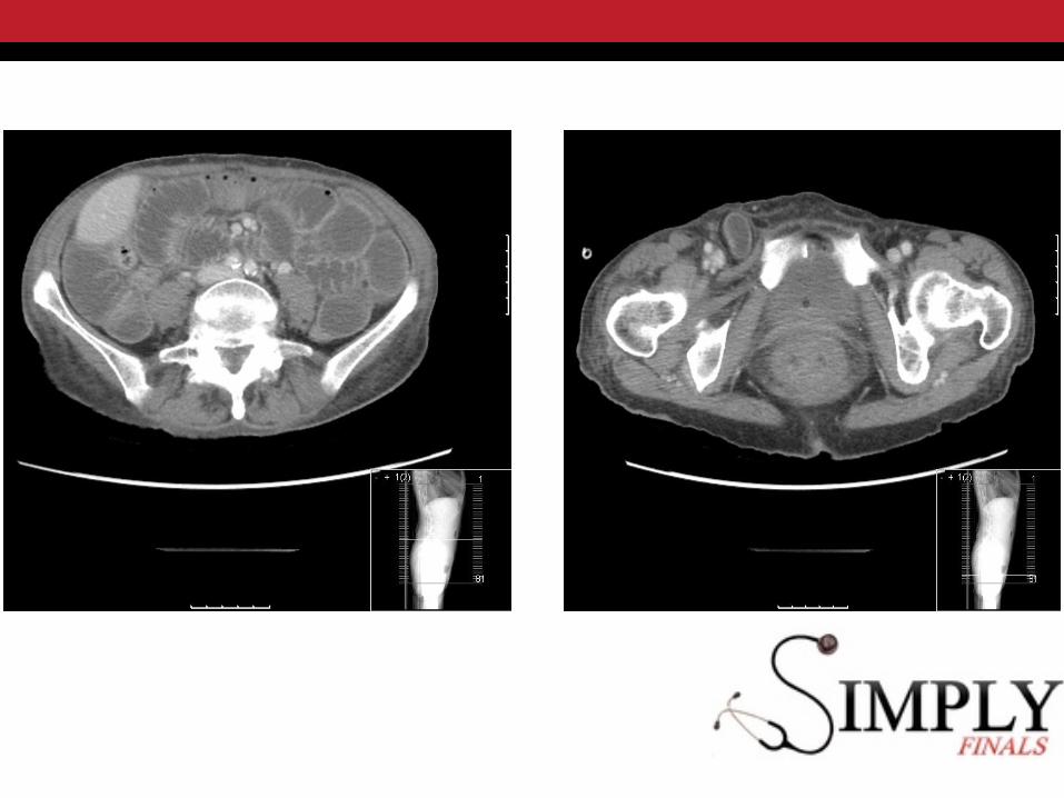

What next?
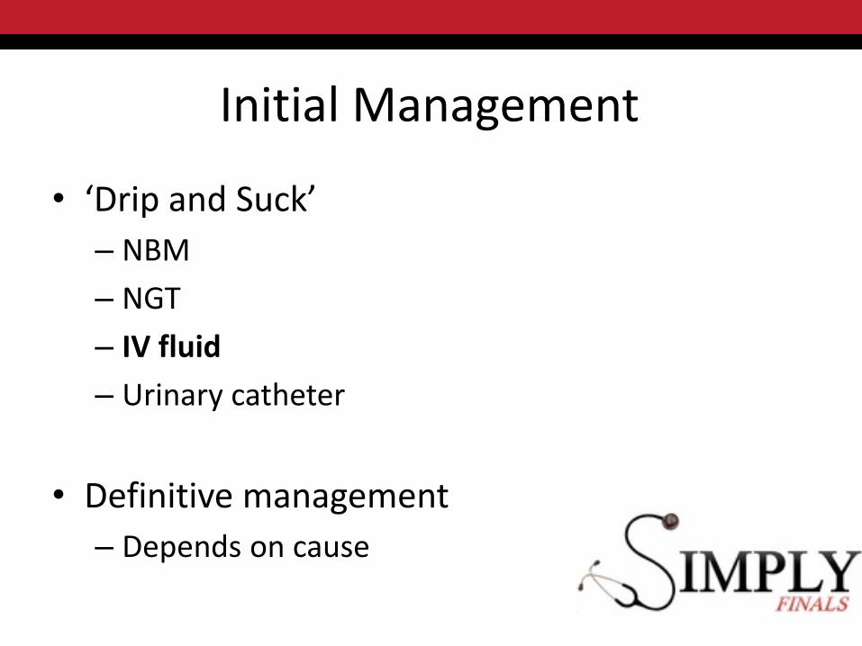
Initial Management
• ‘Drip and Suck’
– NBM
– NGT
– IV fluid
– Urinary catheter
• Definitive management
– Depends on cause
Bowel Obstruction – 4 symptoms
• Vomiting
• Colic
• Distension
• Constipation/obstipation

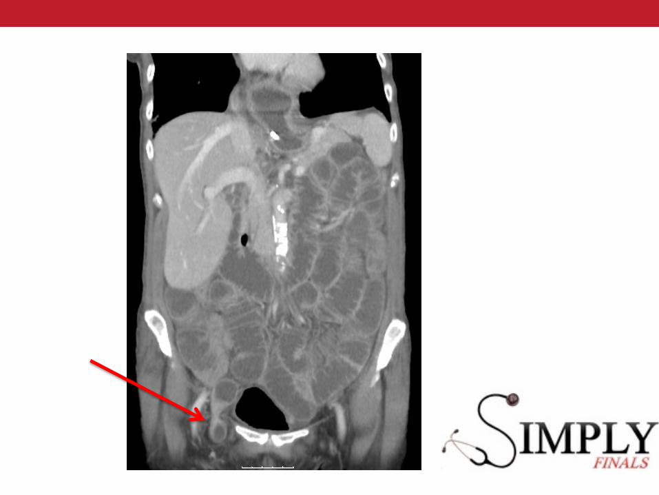
Bowel Obstruction – Aetiology
SBO
• Adhesions
• Hernias
• Crohn’s
LBO
• Cancer
• Volvulus
• Diverticular stricture
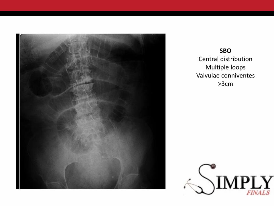
SBOCentral distribution
Multiple loopsValvulae conniventes
>3cm
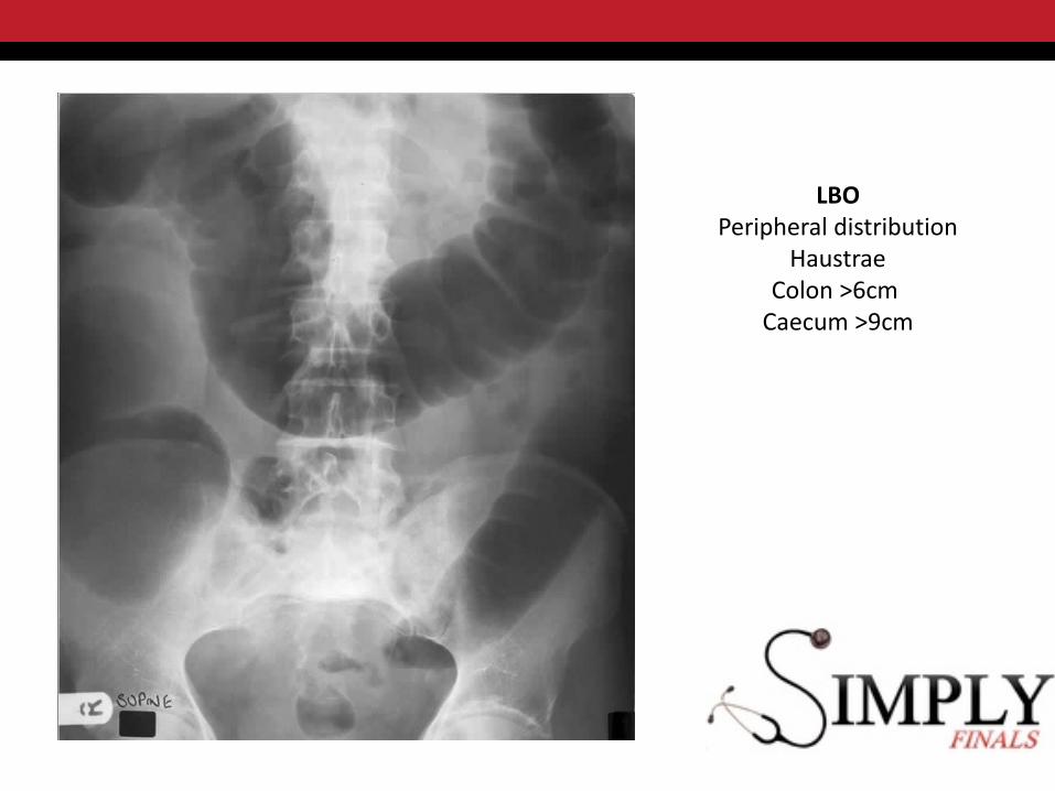
LBOPeripheral distribution
HaustraeColon >6cm
Caecum >9cm
Definitive Management
• SBO
– Adhesional
• Trial of conservative management
• UNLESS signs of ischaemia/peritonism
– Hernia
• Surgery required
Definitive Management
• LBO
– CT usually helpful
– Volvulus
• Sigmoidoscopy decompression
• UNLESS signs of ischaemia
– Cancer
• Laparotomy + resection
• Urgency depends on competence of ileocaecal valve
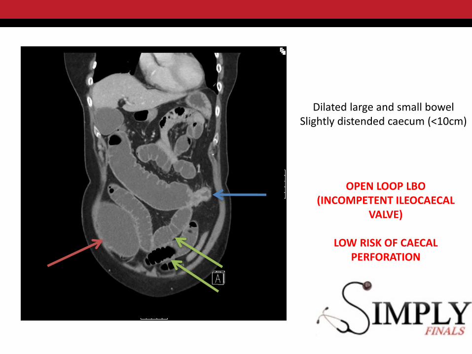
Dilated large and small bowelSlightly distended caecum (<10cm)
OPEN LOOP LBO(INCOMPETENT ILEOCAECAL
VALVE)
LOW RISK OF CAECAL PERFORATION
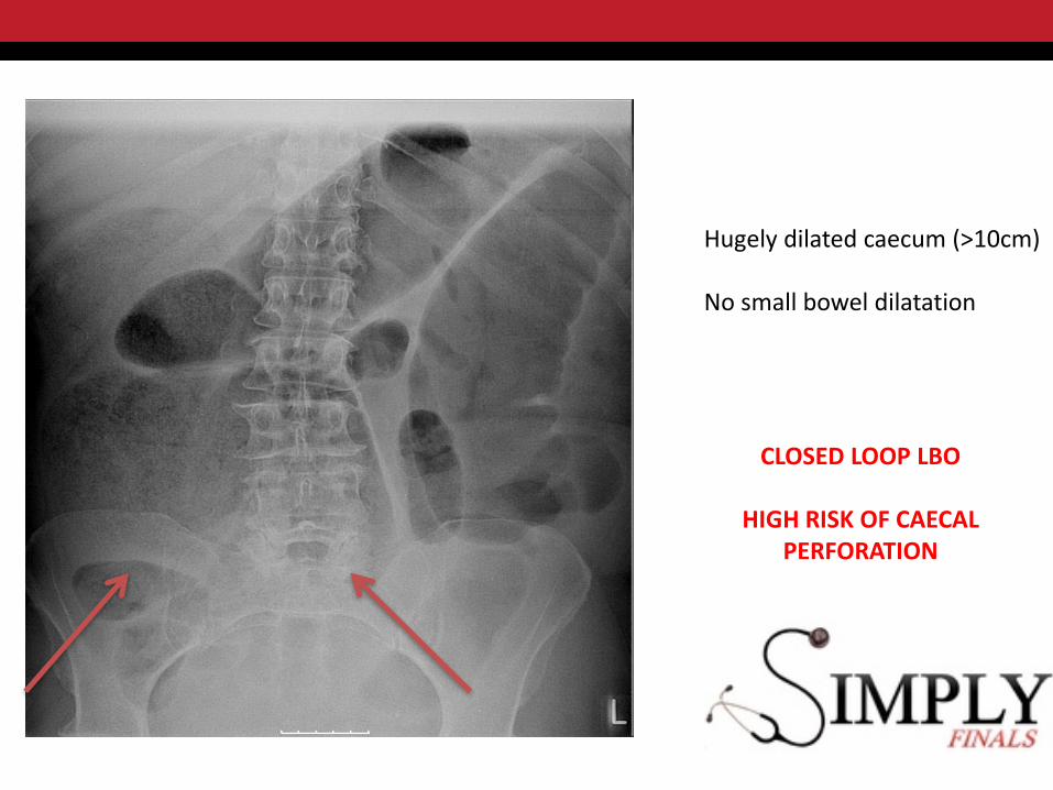
Hugely dilated caecum (>10cm)
No small bowel dilatation
CLOSED LOOP LBO
HIGH RISK OF CAECAL PERFORATION
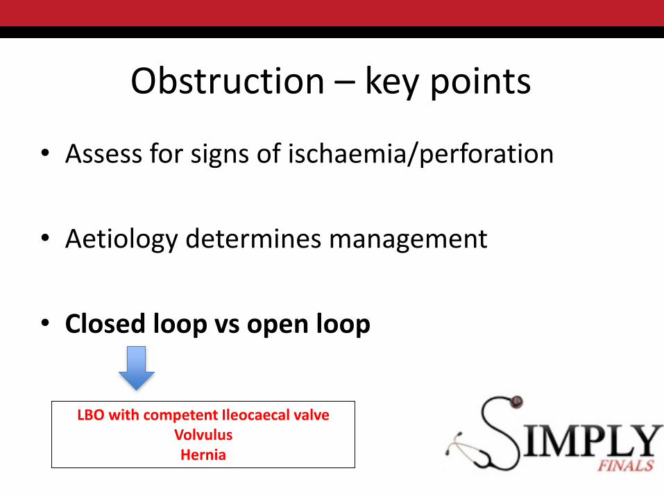
Obstruction – key points
• Assess for signs of ischaemia/perforation
• Aetiology determines management
• Closed loop vs open loop
Obstruction – key points
• Assess for signs of ischaemia/perforation
• Aetiology determines management
• Closed loop vs open loop
LBO with competent Ileocaecal valveVolvulusHernia

Any questions?
Hernias
• Protrusion of part or all of a structure through another structure and ending up in the wrong anatomical location
Examining a Hernia
• Question 1 - Is there a hernia?
– Start with patient standing
– Inspect first
– Palpate for cough impulse
• Question 2 – Is it reducible?
– Ask patient to reduce
– If difficult – lie flat
Examining a Hernia
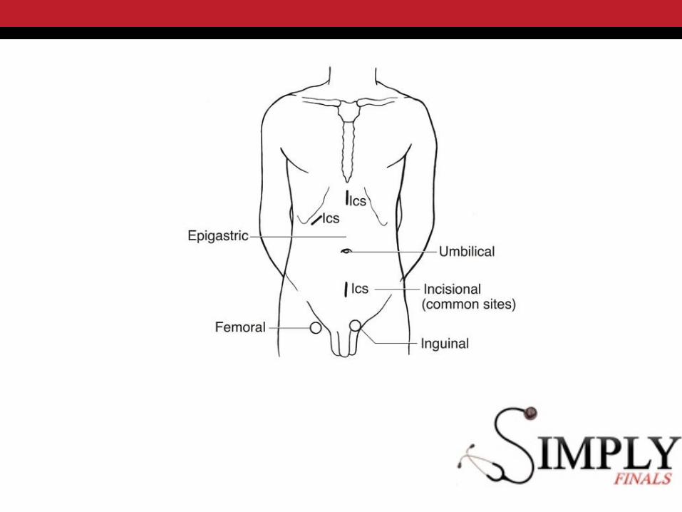
• Question 3 – What type of hernia is it?– Find the pubic tubercle
– Reduce the hernia
– *Cough*
• Below and lateral to PT = Femoral hernia
• Above (and medial) to PT = Inguinal Hernia
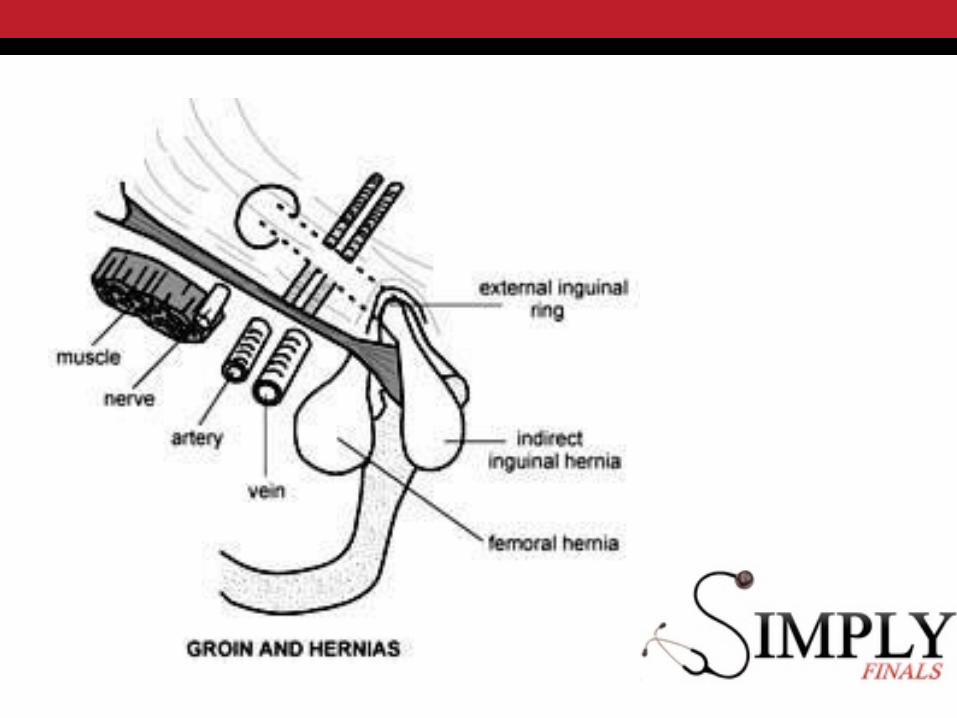
• Question 4 – Inguinal only – Direct or Indirect?– Reduce hernia
– Occlude Deep Ring
– *Cough*
• Hernia protrudes = DIRECT
• Hernia doesn’t appear = INDIRECT
Hernias – Key Points
• Inguinal hernia
– Indirect• More common
• moderate strangulation risk
– Direct• Less common
• Scrotal extension
• low strangulation risk
– M>F
• Femoral hernia
– High risk of strangulation
– F>M
Hernias
• Complications
– Incarceration
– Obstruction
– Strangulation

Any Questions?
Case 3
• 21F
• PC
– RIF pain for 2/7
– No vomiting
– Not hungry
– Loose stool for 1/7
On Examination
• A – SM
• B– RR 18/min– SaO2 100%– Equal A/E
• C– Dry tongue– HR 90bpm– BP 140/80
• D– Alert
• E– T 37.5– Abdomen
• Tender RIF• Guarding

What next?
• Additional questions?
• Possible diagnoses?
• Any investigations?
Investigations
• Urine – bHCG Negative
– leuk ++ Nit –ve
• Bloods– WCC 14.2
– CRP 112
– U+E/LFT N
– VBG N
Investigations
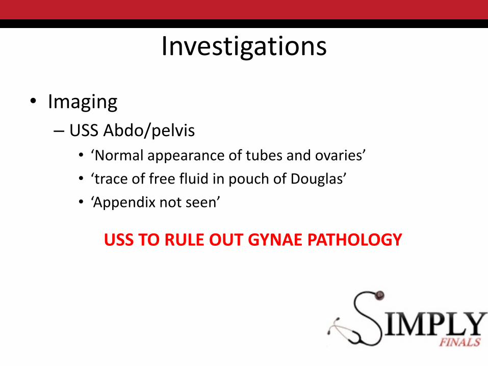
• Imaging
– USS Abdo/pelvis
• ‘Normal appearance of tubes and ovaries’
• ‘trace of free fluid in pouch of Douglas’
• ‘Appendix not seen’
Investigations
• Imaging
– USS Abdo/pelvis
• ‘Normal appearance of tubes and ovaries’
• ‘trace of free fluid in pouch of Douglas’
• ‘Appendix not seen’
USS TO RULE OUT GYNAE PATHOLOGY
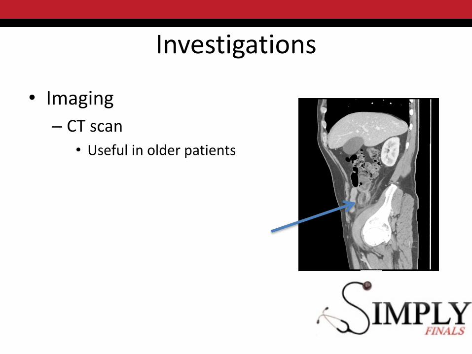
Investigations
• Imaging
– CT scan
• Useful in older patients
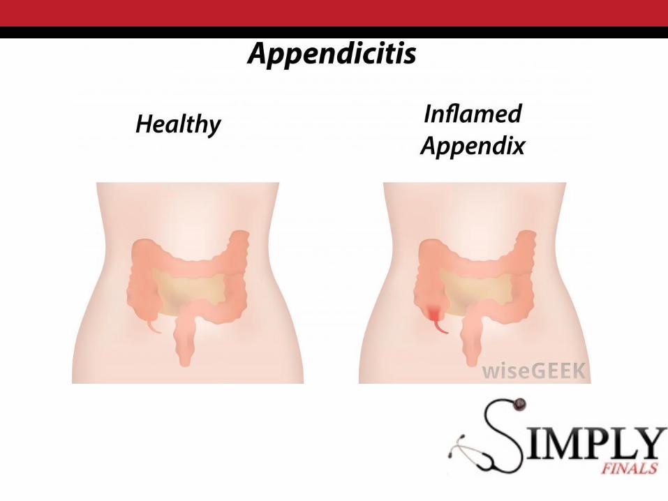
Acute Appendicitis
• Clinical diagnosis
• History– Migration of pain
– Duration of symptoms
– Anorexia is common
– Urinary symptoms
– Gynae symptoms
– LMP
Acute Appendicitis
• Clinical diagnosis
• History– Migration of pain
– Duration of symptom
– Anorexia is common
– Urinary symptoms
– Gynae symptoms
– LMP
Can affect any age
Perforation risk with age
Older patients – suspect
cancer
Acute Appendicitis
• Examination
– McBurney’s point
Acute Appendicitis
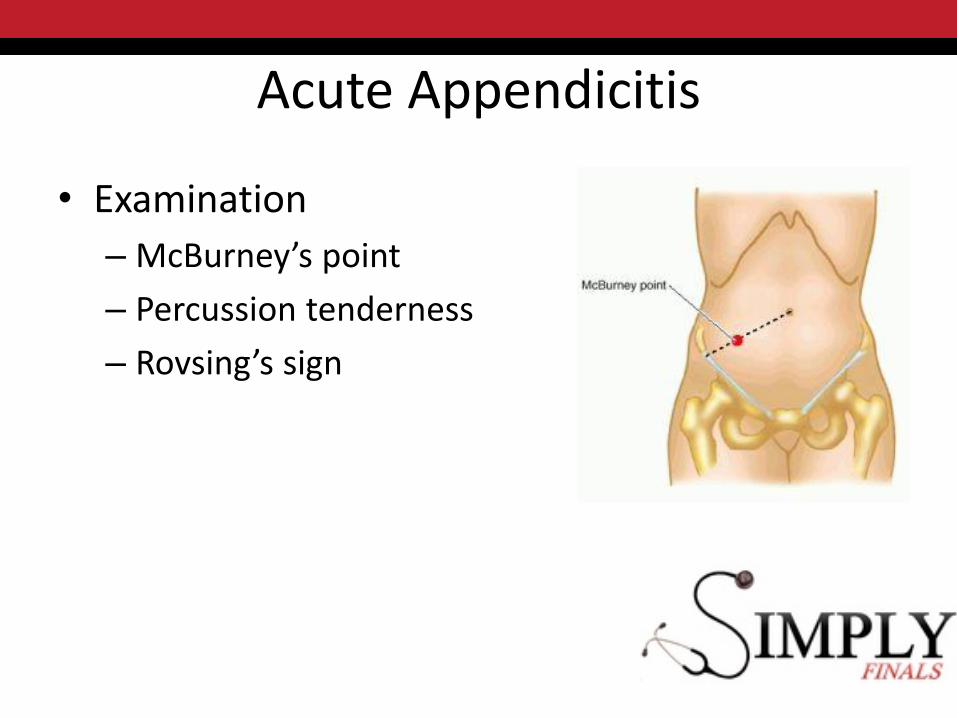
• Examination
– McBurney’s point
– Percussion tenderness
– Rovsing’s sign
Acute Appendicitis
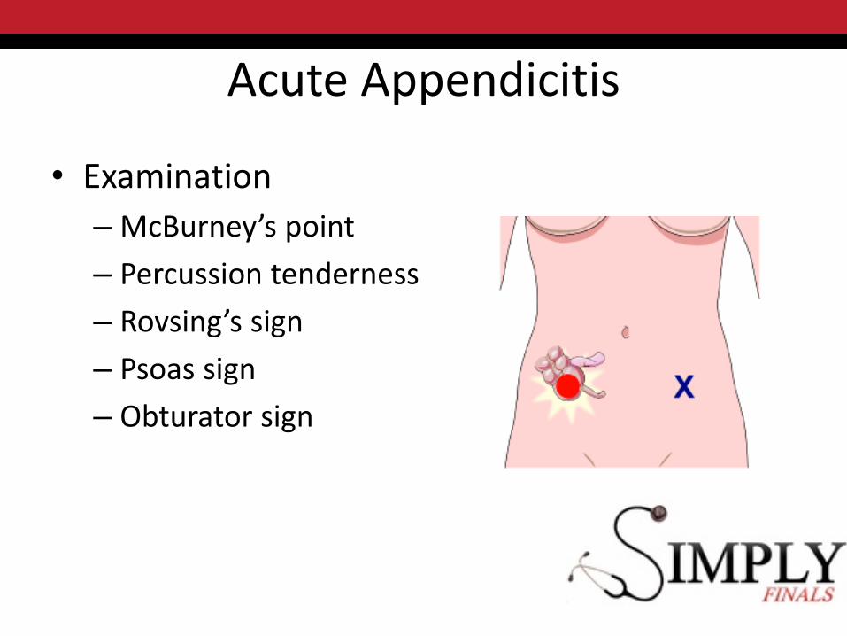
• Examination
– McBurney’s point
– Percussion tenderness
– Rovsing’s sign
– Psoas sign
– Obturator sign
Acute Appendicitis
• Examination
– Perforation
• Signs of sepsis
• Peritonism– Localised vs generalised
Acute Appendicitis - DDx
• Gynaecological– ECTOPIC PREGNANCY– Ovarian cyst accident– PID
• Genitourinary– TESTICULAR TORSION– Pyelonephritis– Ureteric colic
• Other– ‘Non specific abdominal pain’– Mesenteric adenitis– Meckel’s diverticulitis
Case 3 – What next?

Case 3 - Management
• IV fluids
• Symptomatic relief
– Opioids?
• Antibiotics?
• Operation?
Case 3 - Management
• Admit and observe if diagnosis in doubt
• Antibiotics if…
– Patient is septic
– You are booking for theatre
• Operation – when and how?
– Laparoscopic vs open
Case 4 – More abdominal pain
Case 4 – More abdominal pain
• 38F
• Severe central abdo pain for 1/7
• Constant
• Radiating to back
Examination
• A– SM
• B– RR 24/min– SaO2 93% (RA)– Chest clear
• C– Dehydrated– HR 120bpm– BP 110/50
• D– Alert
• E– T 37.6– Abdomen
• Sitting forward• Tender ++ periumbilical,
epigastrium & RUQ
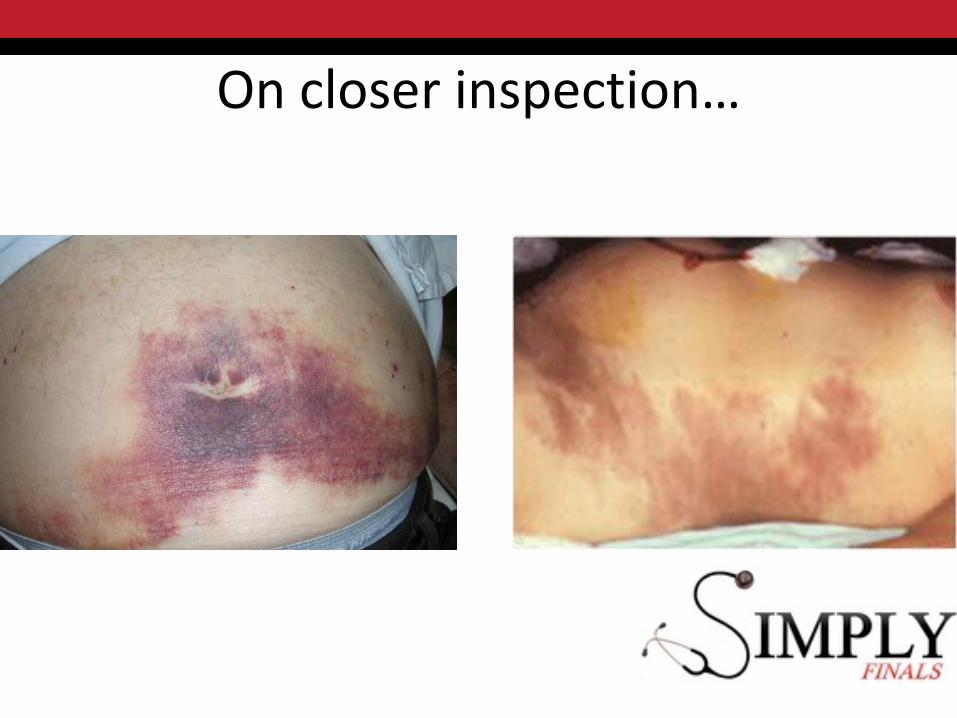
On closer inspection…
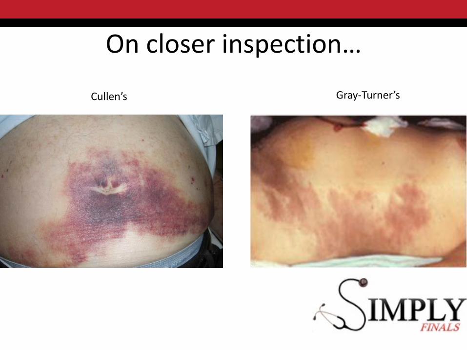
On closer inspection…
Cullen’s Gray-Turner’s

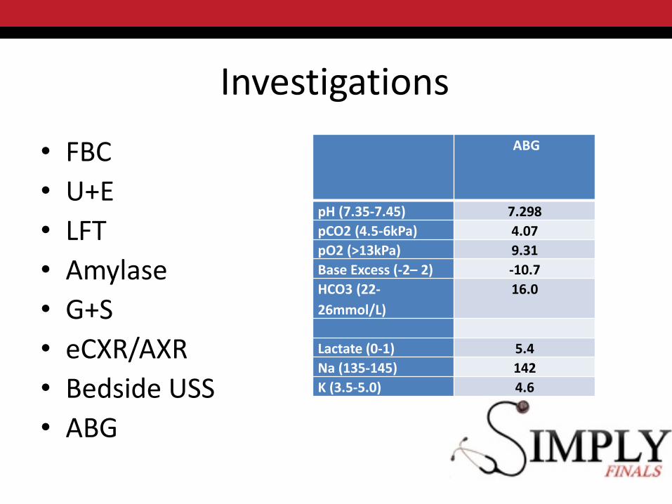
Investigations

Investigations
• FBC
• U+E
• LFT
• Amylase
• G+S
• eCXR/AXR
• Bedside USS
• ABG
Investigations
• FBC
• U+E
• LFT
• Amylase
• G+S
• eCXR/AXR
• Bedside USS
• ABG
ABG
pH (7.35-7.45) 7.298
pCO2 (4.5-6kPa) 4.07
pO2 (>13kPa) 9.31
Base Excess (-2– 2) -10.7
HCO3 (22-
26mmol/L)
16.0
Lactate (0-1) 5.4
Na (135-145) 142
K (3.5-5.0) 4.6
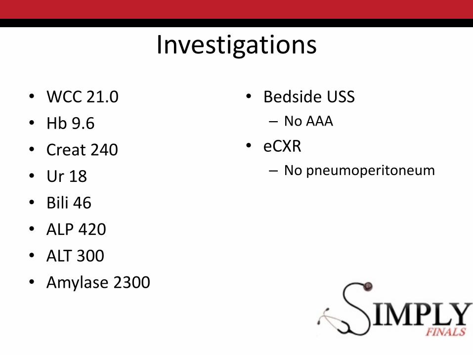
Investigations
• WCC 21.0
• Hb 9.6
• Creat 240
• Ur 18
• Bili 46
• ALP 420
• ALT 300
• Amylase 2300
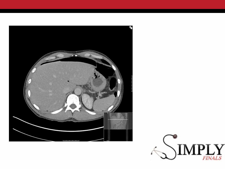
• Bedside USS
– No AAA
• eCXR
– No pneumoperitoneum
Investigations
• Imaging
– Rule out other causes
– USS Abdomen
• ?Gallstones
• ?intra/extrahepatic duct dilatation
– CT abdomen
• Usually not needed initially…
• …unless diagnosis still in doubt
• After 5-7 days if still unwell– To assess for complications
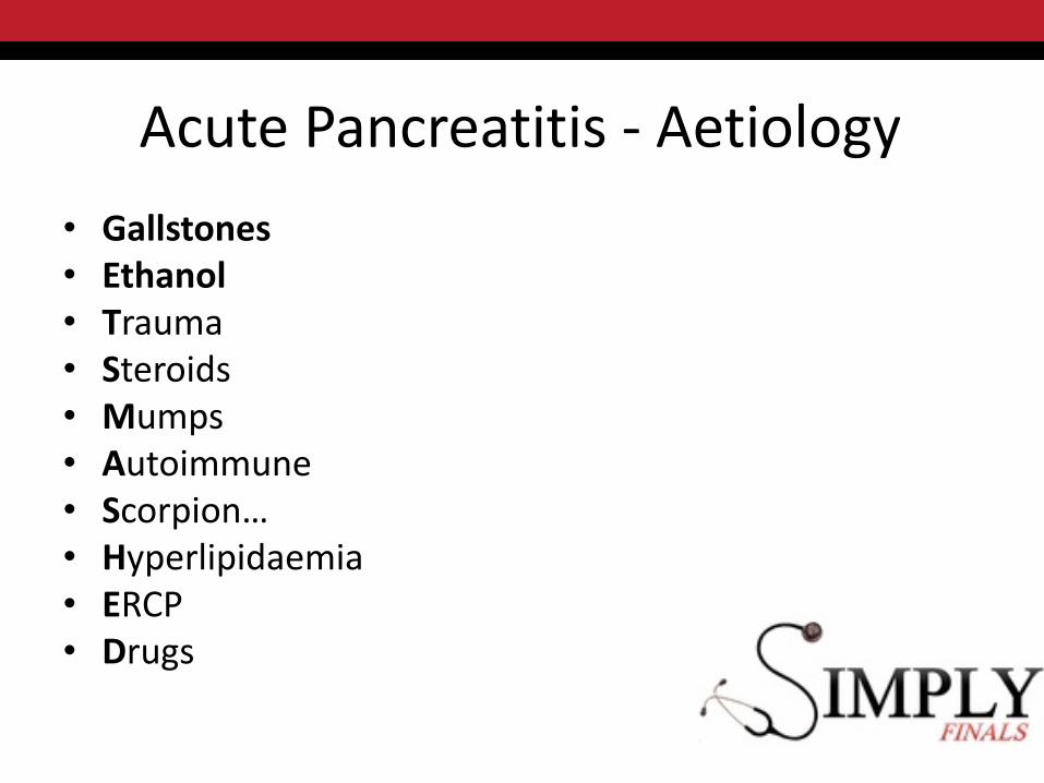
Acute Pancreatitis - Aetiology
• Gallstones• Ethanol• Trauma• Steroids• Mumps• Autoimmune• Scorpion…• Hyperlipidaemia• ERCP• Drugs
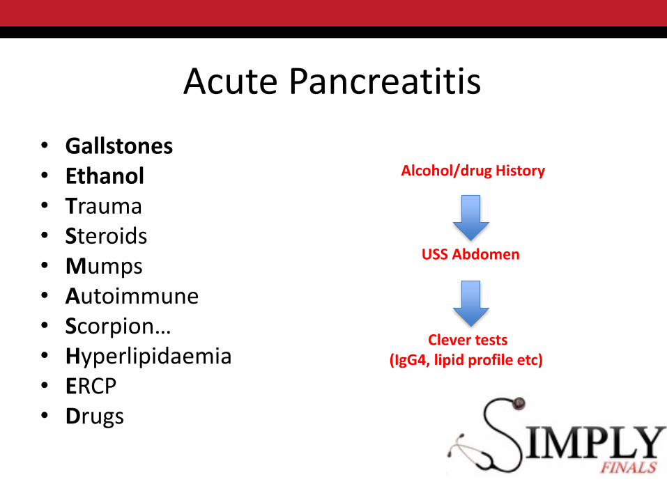
Acute Pancreatitis
• Gallstones• Ethanol• Trauma• Steroids• Mumps• Autoimmune• Scorpion…• Hyperlipidaemia• ERCP• Drugs
Alcohol/drug History
USS Abdomen
Clever tests(IgG4, lipid profile etc)
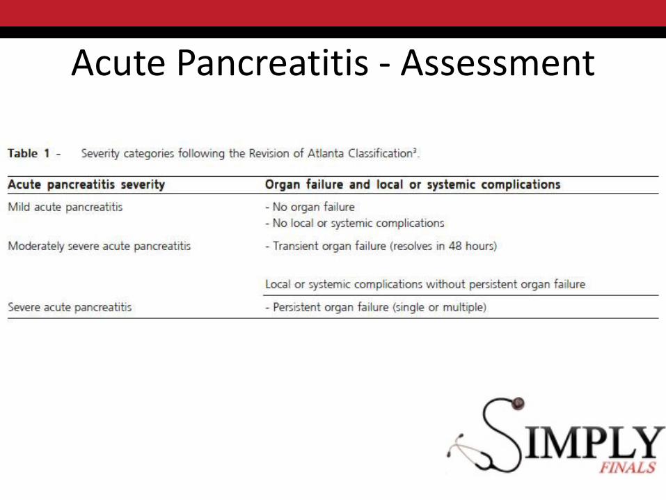
Acute Pancreatitis - Assessment
Acute Pancreatitis - Assessment
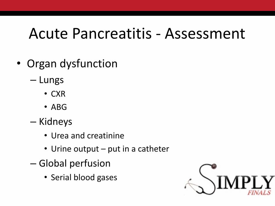
• Organ dysfunction
– Lungs
• CXR
• ABG
– Kidneys
• Urea and creatinine
• Urine output – put in a catheter
– Global perfusion
• Serial blood gases

Acute Pancreatitis - Assessment
• Glasgow score
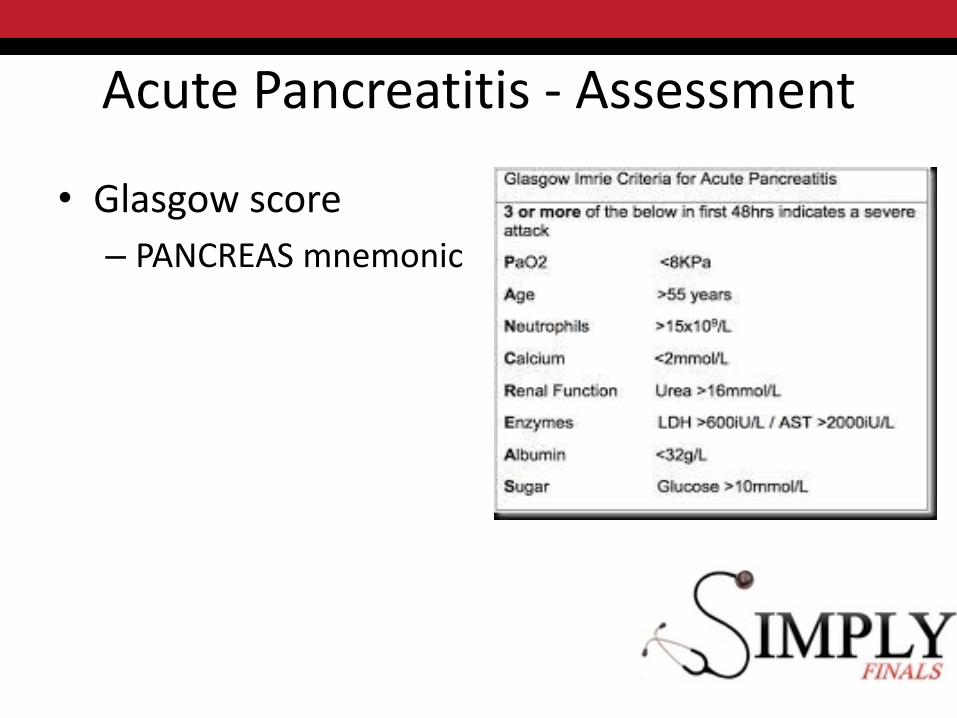
Acute Pancreatitis - Assessment
• Glasgow score
– PANCREAS mnemonic
Acute Pancreatitis - Assessment
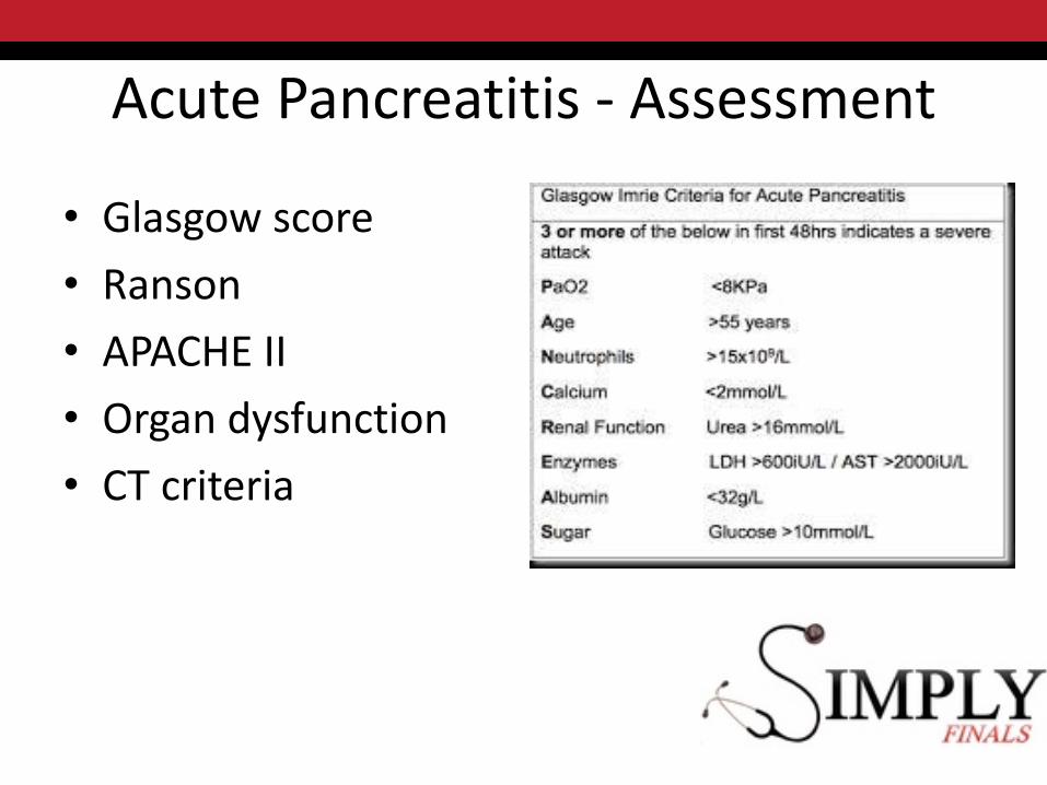
• Glasgow score
• Ranson
• APACHE II
• Organ dysfunction
• CT criteria
Acute Pancreatitis - Assessment
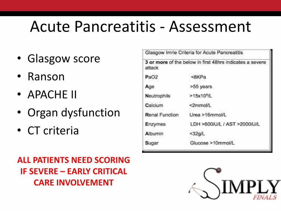
• Glasgow score
• Ranson
• APACHE II
• Organ dysfunction
• CT criteria
ALL PATIENTS NEED SCORINGIF SEVERE – EARLY CRITICAL
CARE INVOLVEMENT

Acute Pancreatitis – Immediate Management
• Big IV access
• Fluid, fluid, fluid
• Opiate analgesia
• Anti-emetics
• Urinary catheter and monitoring
• Antibiotics?
Acute Pancreatitis – Immediate Management
• Big IV access
• Fluid, fluid, fluid
• Opiate analgesia
• Anti-emetics
• Urinary catheter and monitoring
• Antibiotics?– No role in early management
– If established pancreatic necrosis:• Carbapenem
Acute Pancreatitis – Ongoing Management
• Organ support– Cardiovascular– Respiratory– Renal
• Nutrition– Early enteral nutrition improves outcome
• Treat cause…– ERCP– Steroids
• Surgery?
Acute Pancreatitis – Ongoing Management
• Organ support– Cardiovascular– Respiratory– Renal
• Nutrition– Early enteral nutrition improves outcome
• Treat cause…– ERCP– Steroids
• Surgery?– Necrosectomy– Cholecystectomy
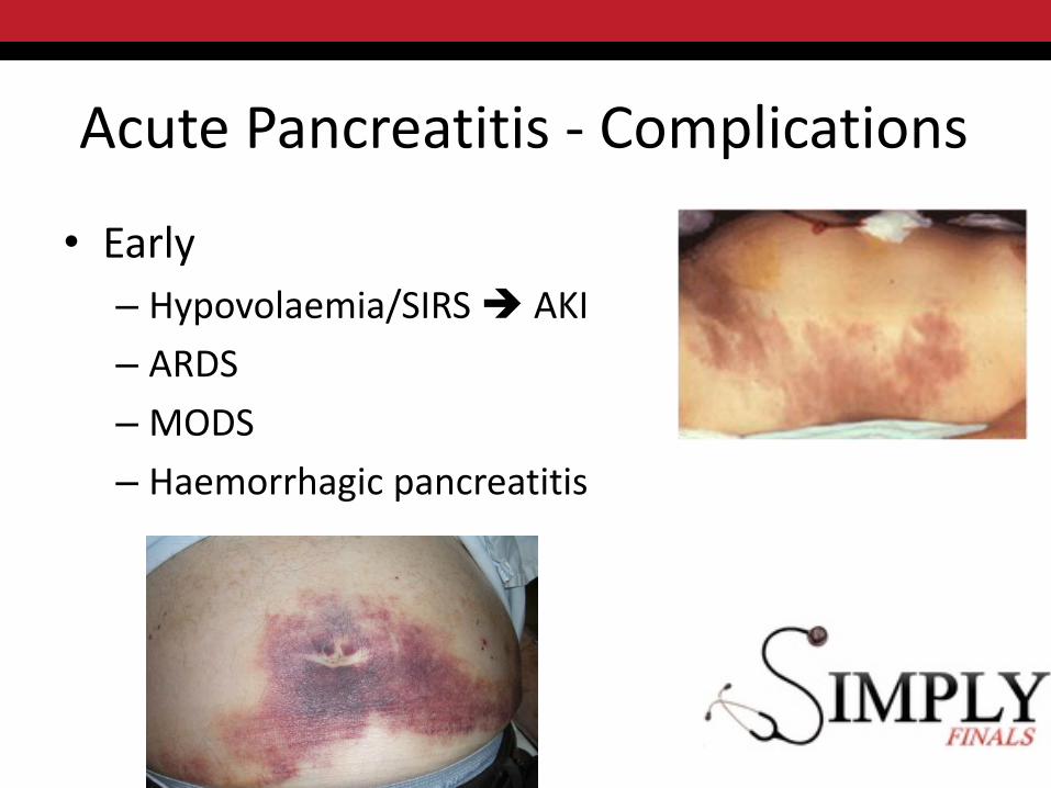
Acute Pancreatitis - Complications
• Early
– Hypovolaemia/SIRS AKI
– ARDS
– MODS
– Haemorrhagic pancreatitis
Acute Pancreatitis - Complications
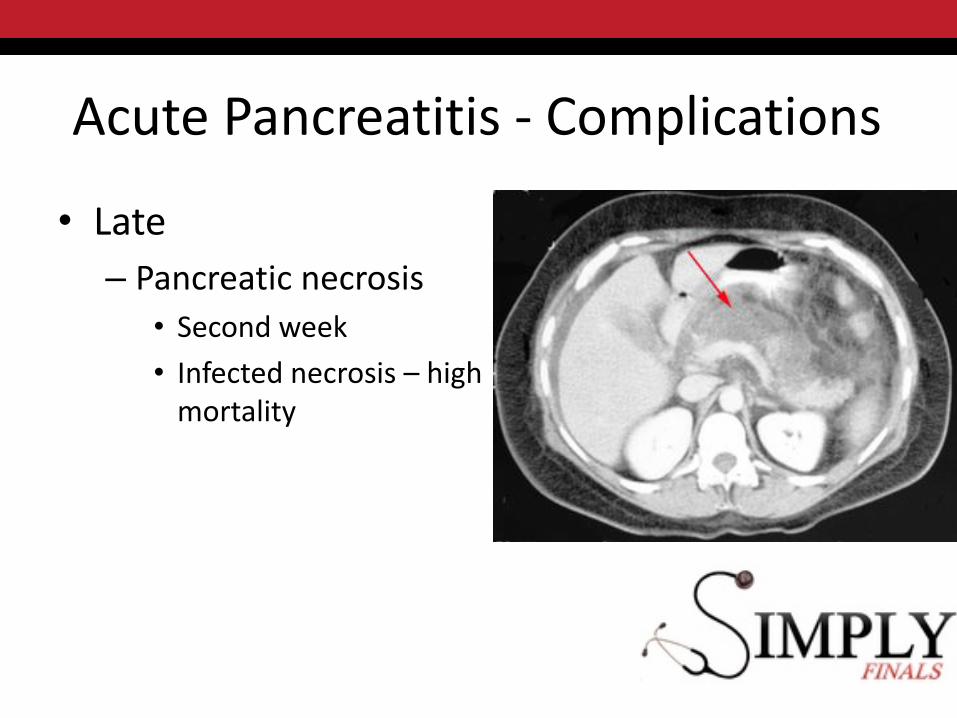
• Late
– Pancreatic necrosis
• Second week
• Infected necrosis – high mortality

Acute Pancreatitis - Complications
• Late
– Pancreatic necrosis
– Splenic vein thrombosis
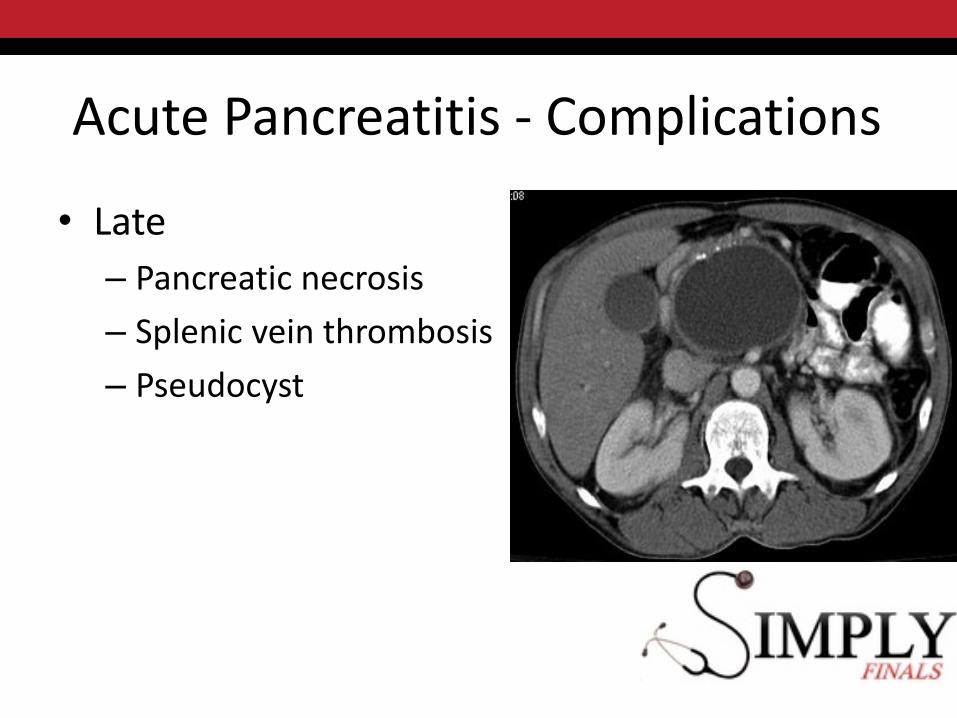
Acute Pancreatitis - Complications
• Late
– Pancreatic necrosis
– Splenic vein thrombosis
– Pseudocyst
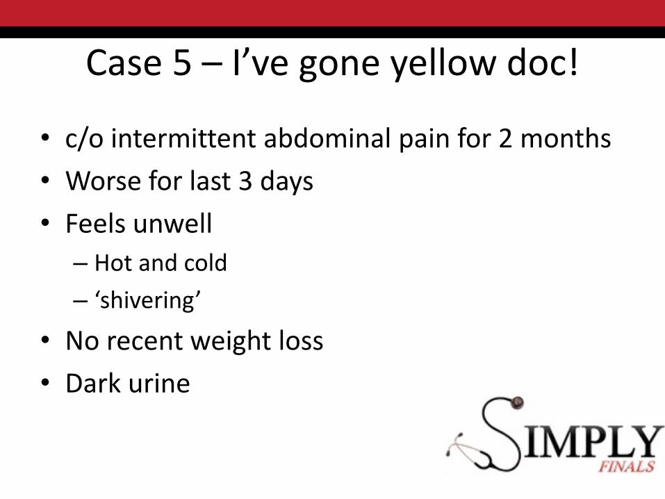
Case 5 – I’ve gone yellow doc!
Case 5 – I’ve gone yellow doc!
• 45M
• ‘big boned’
• Partner noticed yellowing of eyes
• c/o intermittent abdominal pain for 2 months
• Worse for last 3 days

Further Questions?
Further Questions?
• Symptoms of gallstones– Post-prandial RUQ pain
• Symptoms of obstructive jaundice– Dark urine– Pale stools– Pruritus
• Weight loss/anorexia– Malignancy
• Fever/rigors• Drug history• Alcohol
Case 5 – I’ve gone yellow doc!
• c/o intermittent abdominal pain for 2 months
• Worse for last 3 days
• Feels unwell
– Hot and cold
– ‘shivering’
• No recent weight loss
• Dark urine

Examination
• A– SM
• B– RR 18/min– SaO2 99% (RA)– Chest clear
• C– CRT 4 secs– HR 102bpm– BP 100/50
• D– Alert
• E– T 38.4– Jaundiced– Abdomen
• Tender RUQ• No mass• Murphy’s sign?
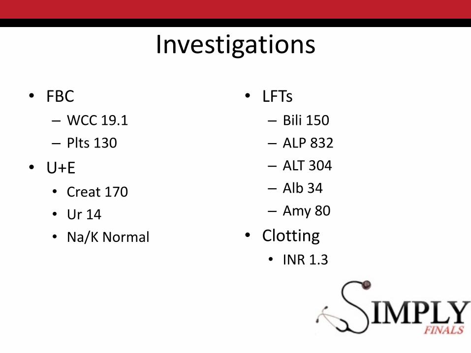
Investigations
• FBC
– WCC 19.1
– Plts 130
• U+E
• Creat 170
• Ur 14
• Na/K Normal
• LFTs
– Bili 150
– ALP 832
– ALT 304
– Alb 34
– Amy 80
• Clotting
• INR 1.3
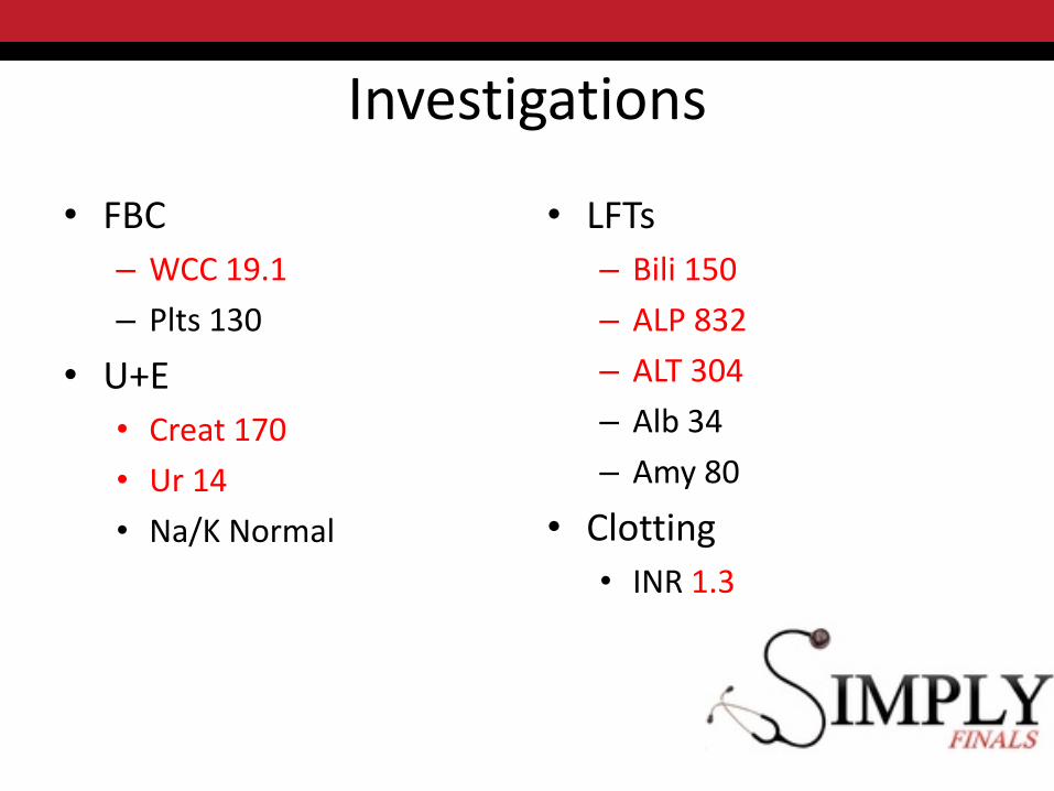
Investigations
• FBC
– WCC 19.1
– Plts 130
• U+E
• Creat 170
• Ur 14
• Na/K Normal
• LFTs
– Bili 150
– ALP 832
– ALT 304
– Alb 34
– Amy 80
• Clotting
• INR 1.3
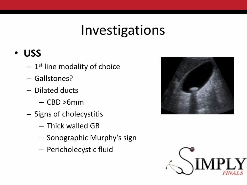
Investigations
• USS– 1st line modality of choice
– Gallstones?
– Dilated ducts
– CBD >6mm
– Signs of cholecystitis
– Thick walled GB
– Sonographic Murphy’s sign
– Pericholecystic fluid
Investigations• MRCP
– 2nd line– More sensitive for CBD stones– eg dilated ducts but cause unclear
• ERCP– Diagnostic +/- therapeutic– Decompression
• Stent/sphincterotomy
– Samples for cytology– Complications
• Pancreatitis• Perforation• Cholangitis
Jaundice
• Pre-hepatic– Haemolysis
– Drugs
– Gilbert’s
• Hepatic– Cirrhosis
– Hepatitis
– Drugs
– Budd-Chiari
• OBSTRUCTIVE– In the lumen
• Gallstones
– In the wall • Cholangiocarcinoma
• PSC
– External compression• Pancreatic Ca
• Lymph nodes
Gallstone Disease
• ‘Fat, Female, Fertile, Forty’
• Can affect any age
• Cholesterol (15%)
• Pigment (5%)
• Mixed (80%)
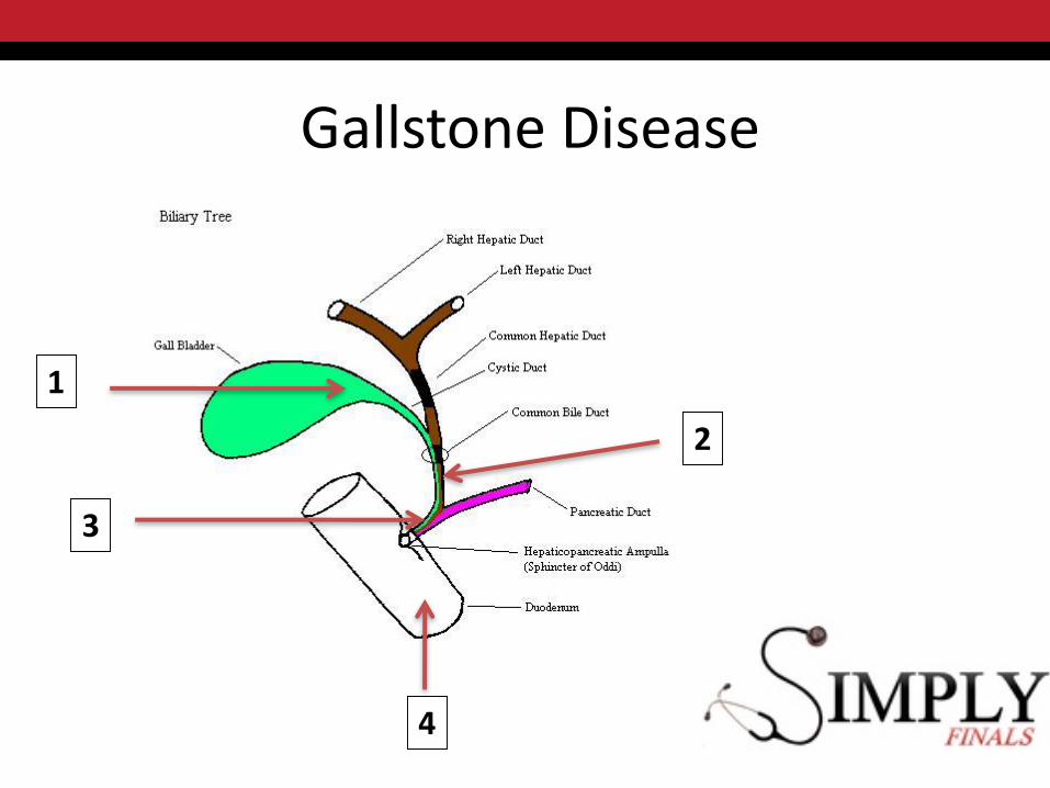
Gallstone Disease
1
2
3
4

Gallstone Disease
Acute cholecystitisBiliary colicEmpyema Obstructive jaundice
Ascending cholangitis
PancreatitisObstructive JaundiceAscending Cholangitis
Gallstone Ileus
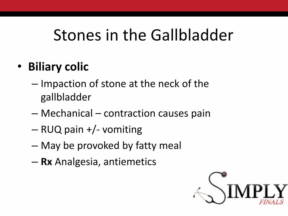
Stones in the Gallbladder
• Biliary colic
– Impaction of stone at the neck of the gallbladder
– Mechanical – contraction causes pain
– RUQ pain +/- vomiting
– May be provoked by fatty meal
– Rx Analgesia, antiemetics
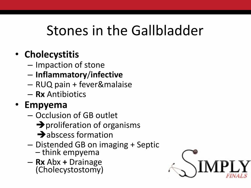
Stones in the Gallbladder
• Cholecystitis– Impaction of stone– Inflammatory/infective– RUQ pain + fever&malaise– Rx Antibiotics
• Empyema– Occlusion of GB outletproliferation of organismsabscess formation
– Distended GB on imaging + Septic – think empyema
– Rx Abx + Drainage (Cholecystostomy)
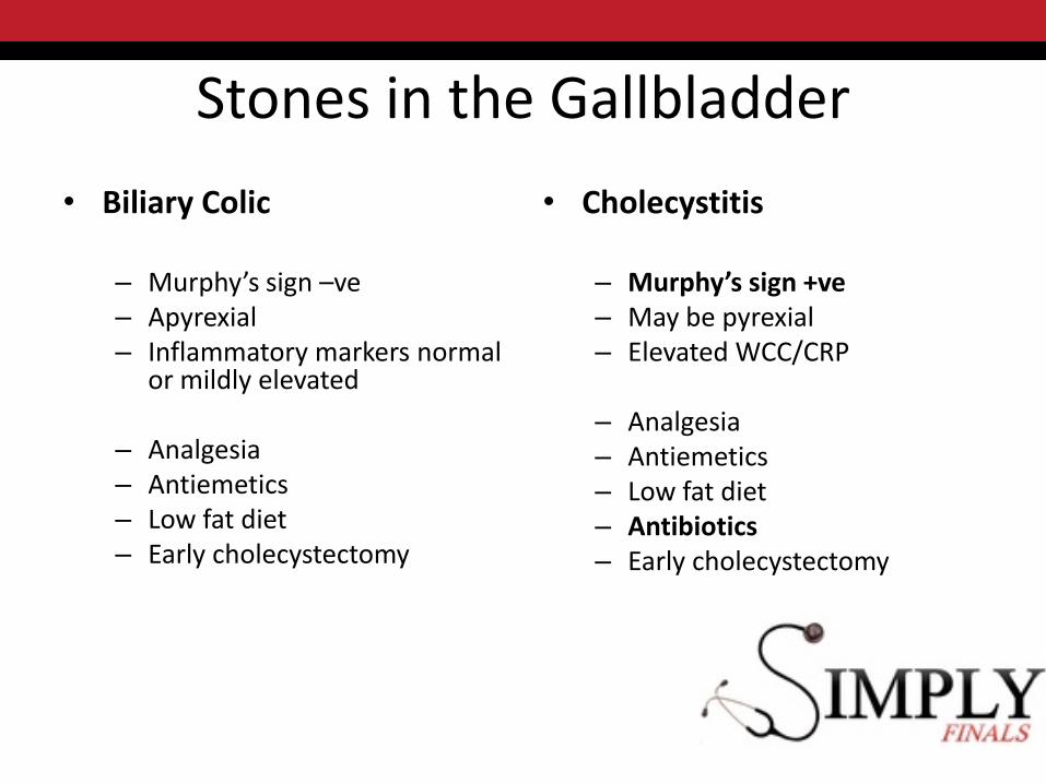
Stones in the Gallbladder
• Biliary Colic
– Murphy’s sign –ve– Apyrexial– Inflammatory markers normal
or mildly elevated
– Analgesia– Antiemetics– Low fat diet– Early cholecystectomy
• Cholecystitis
– Murphy’s sign +ve– May be pyrexial– Elevated WCC/CRP
– Analgesia– Antiemetics– Low fat diet– Antibiotics– Early cholecystectomy
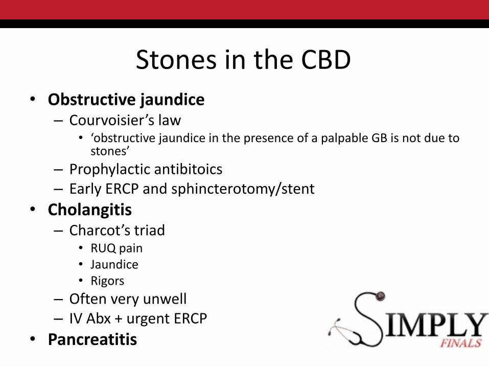
Stones in the CBD• Obstructive jaundice
– Courvoisier’s law• ‘obstructive jaundice in the presence of a palpable GB is not due to
stones’
– Prophylactic antibitoics– Early ERCP and sphincterotomy/stent
• Cholangitis– Charcot’s triad
• RUQ pain• Jaundice• Rigors
– Often very unwell– IV Abx + urgent ERCP
• Pancreatitis
Case Revisited• IV fluid• Antibiotics
– eg Tazocin 4.5g IV TDS• USS• ERCP
– Biliary trawl– Sphincterotomy
• Interval cholecystectomy
Quick Cases
• 64M
• Left loin pain for 8hrs
• Radiation to back
• Initially intermittent, now constant
• PMH– HTN, T2DM
Examination
• A– SM
• B– RR 28/min
– SaO2 96%
– Chest clear
• C– Cool peripheries
– CRT 4 secs
– HR 120bpm
– BP 100/75
• D– Alert but anxious
• E– T 36.9
– Abdomen• Mildly tender central/Lt
Loin

• Differential?
• What next?
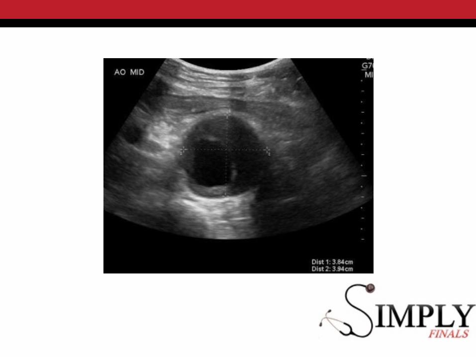

Abdominal Aortic Aneurysm
• Aneurysm = dilatation >150% of normal diameter– AAA = diameter >3cm
• True aneurysm = all three layers of vessel wall• False (pseudoaneurysm) = swelling contained by
adventitia• Majority Infra-renal• Aetiology:
– Atherosclerosis• CVS risk factors
– Connective tissue– Mycotic
Abdominal Aortic Aneurysm• Unruptured
– 3% >50years
– Often aysmptomatic
– Abdo/back pain
– Screening
– Indications for repair• Diameter >5.5cm in men, >5cm in women
• Rapid growth
• Symptomatic
BUT need to consider individual risk
Ruptured AAA
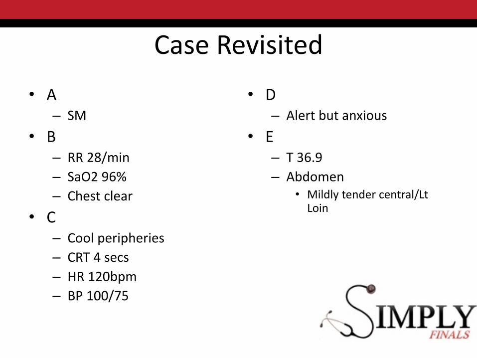
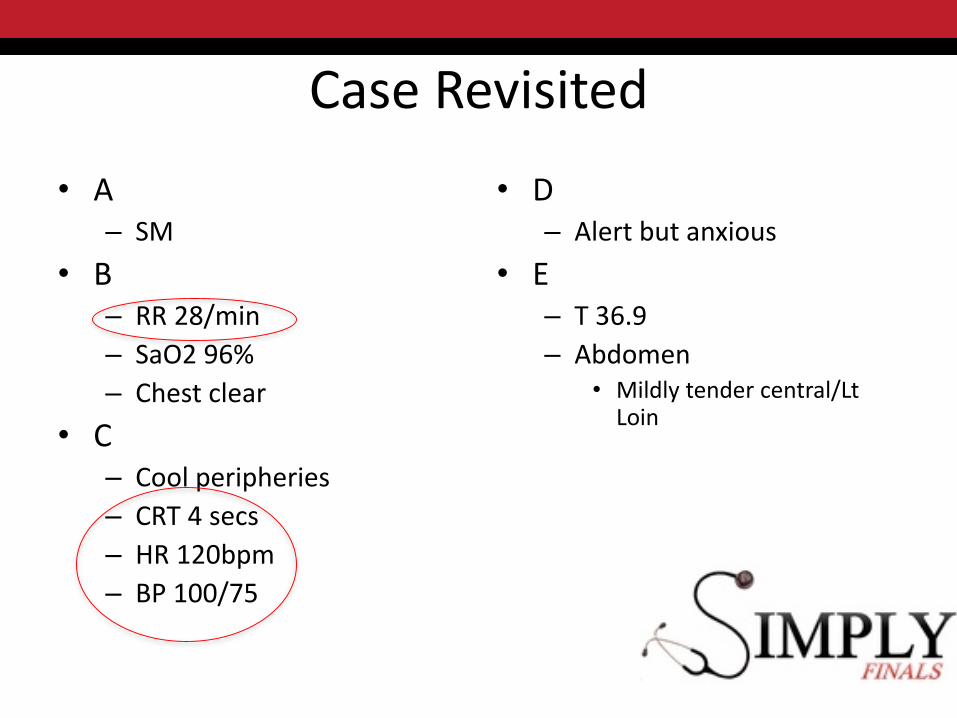
Case Revisited
• A– SM
• B– RR 28/min
– SaO2 96%
– Chest clear
• C– Cool peripheries
– CRT 4 secs
– HR 120bpm
– BP 100/75
• D– Alert but anxious
• E– T 36.9
– Abdomen• Mildly tender central/Lt
Loin
Case Revisited
• A– SM
• B– RR 28/min
– SaO2 96%
– Chest clear
• C– Cool peripheries
– CRT 4 secs
– HR 120bpm
– BP 100/75
• D– Alert but anxious
• E– T 36.9
– Abdomen• Mildly tender central/Lt
Loin
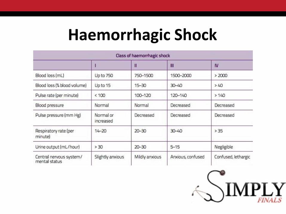
Haemorrhagic Shock
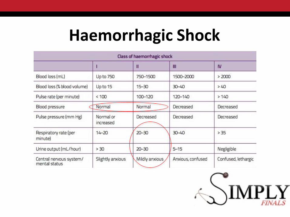
Haemorrhagic Shock
Management
• FBC/U+E/Clotting
• XM – blood and FFP
• Big IV access x2
• Urinary catheter
• Permissive hypotension
– Don’t ‘pop the clot’
– Keep sBP <100mmHg
Imaging?
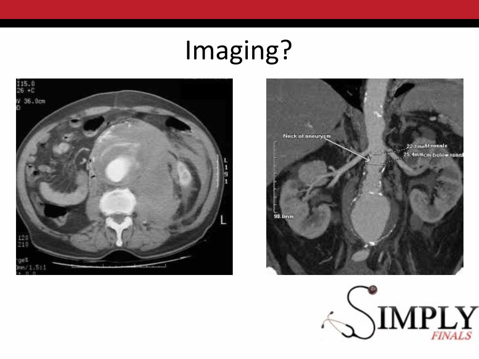
Imaging?
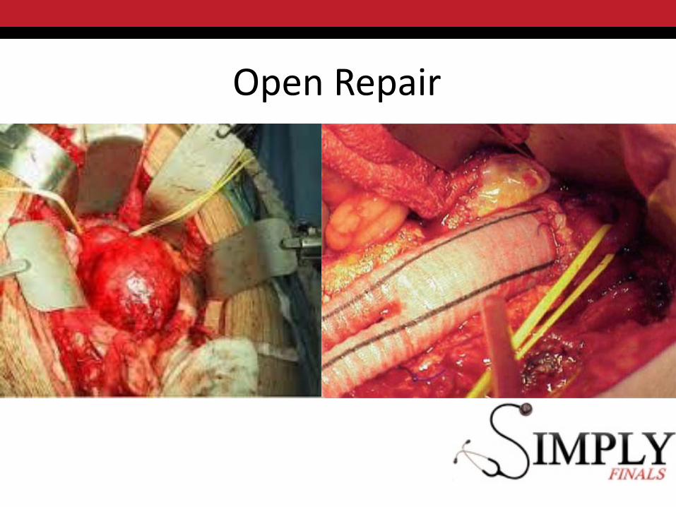
Open Repair
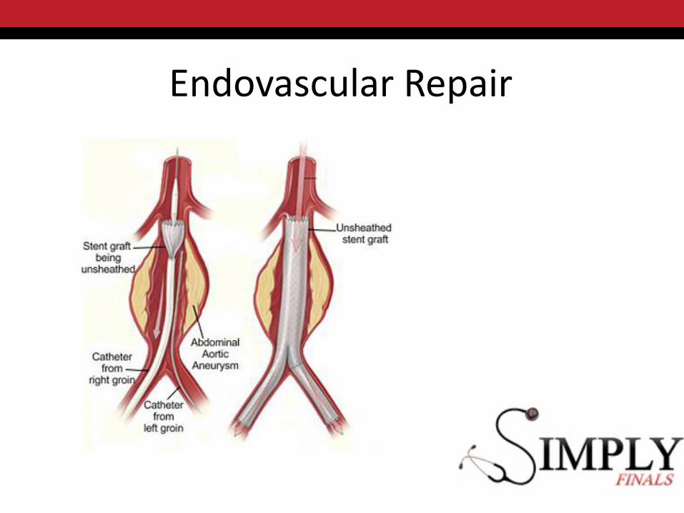
Endovascular Repair

80F - Resus
• Care home resident
• Central abdominal pain and vomiting
• O/E– HR 135bpm Irregular
– BP 95/60
– Abdomen• Generalised tenderness
• No peritonism
Mesenteric ischaemia
• AF + abdominal pain
= Mesenteric ischaemia until proven otherwise
• Pain out of proportion to clinical signs
• Elevated serum lactate
• CT mesenteric angiography is investigation of choice
– If stable…

Mesenteric ischaemia
• Management– Oxygen– IV Fluids– Antibiotics– Consider anticoagulation – IV heparin
• Embolus vs thrombus– Onset– Preceding symptoms
• Mesenteric angina
– Pattern of ischaemia
• Imaging findings– Non specific– Small bowel dilatation– Intramural gas
• Urgent surgery– Resection– (Revascularisation)
More cases
• 86F
• Painful right leg
• Onset of pain four hours ago
• PMH
– HTN, Previous MI, AF
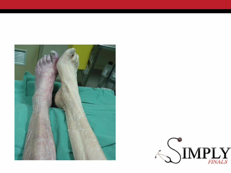
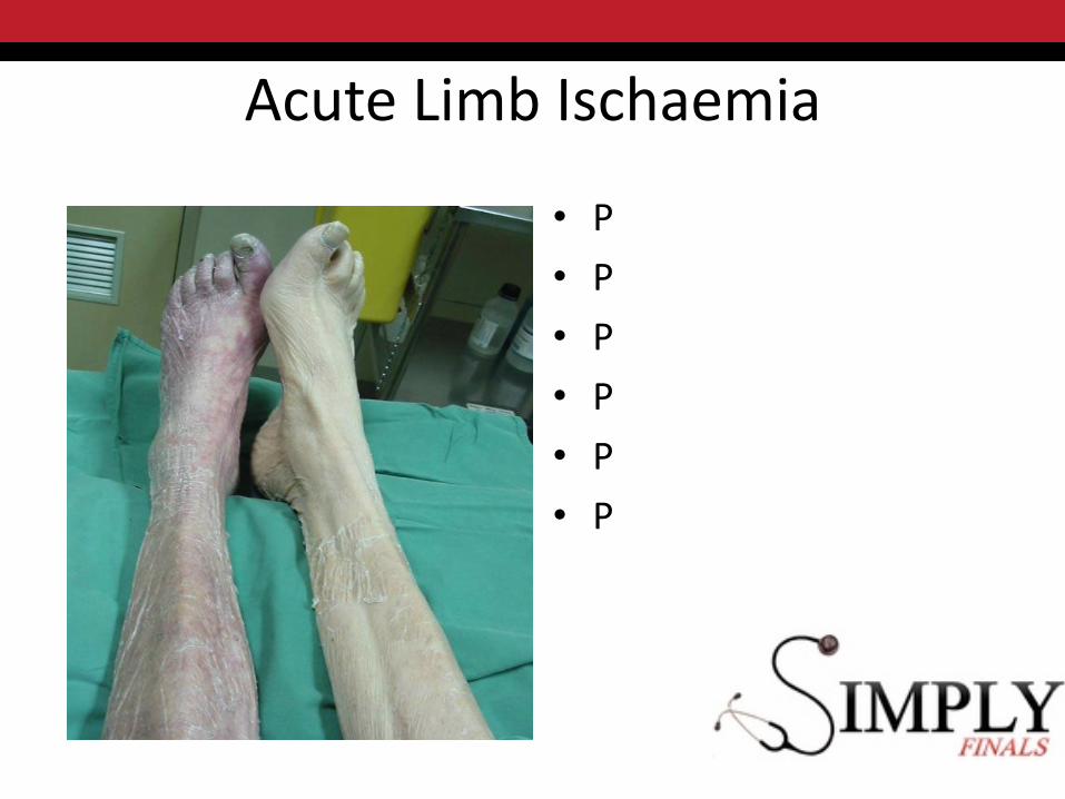
Acute Limb Ischaemia
• P
• P
• P
• P
• P
• P
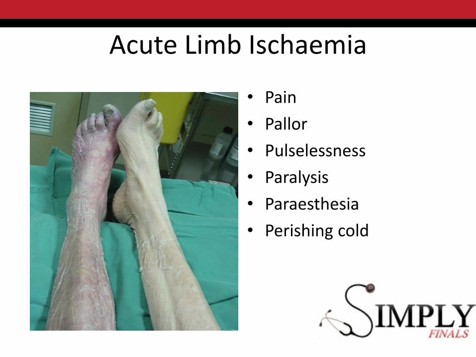
Acute Limb Ischaemia
• Pain
• Pallor
• Pulselessness
• Paralysis
• Paraesthesia
• Perishing cold
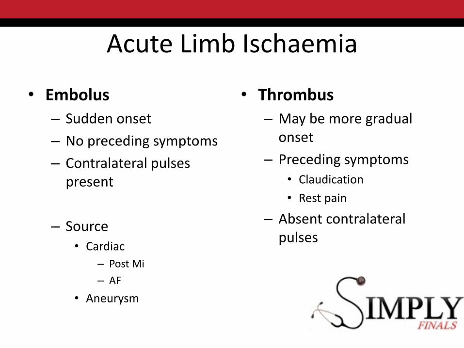
Acute Limb Ischaemia
• Embolus
– Sudden onset
– No preceding symptoms
– Contralateral pulses present
– Source• Cardiac
– Post Mi
– AF
• Aneurysm
• Thrombus
– May be more gradual onset
– Preceding symptoms• Claudication
• Rest pain
– Absent contralateral pulses
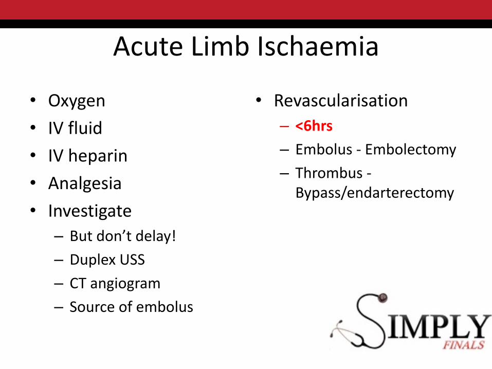
Acute Limb Ischaemia
• Oxygen
• IV fluid
• IV heparin
• Analgesia
• Investigate
– But don’t delay!
– Duplex USS
– CT angiogram
– Source of embolus
• Revascularisation
– <6hrs
– Embolus - Embolectomy
– Thrombus -Bypass/endarterectomy