Embed Size (px)
Citation preview

Surface chemical changes induced by mesophiles microbes on pyrite
S.M. La Vars1, J.S. Quinton1, S.L. Harmer1
1Flinders Centre for NanoScale Science and Technology, Flinders University School of Chemical andPhysical Sciences, South Australia
E-mail: [email protected]
Froth-flotation is a common process for separating valuable sulphide minerals from gangue usingchemical reagents to selectively alter the mineral surface hydrophobicity. Unfortunately, the xanthatecollectors and cyanide depressants used to bring about these changes in mineral hydrophobicity areoften environmentally hazardous. A potential solution to this problem is bioflotation, a new processthat uses microorganisms and their extracellular polymeric substances (EPS) to alter the surfacehydrophobicity. The mechanism by which this occurs varies with microbial species, pH and Eh.
This study has successfully identified several species that contribute to the hydrophobicity of pyriteusing Time of Flight Secondary Ion Mass Spectrometry. Pyrite, a gangue mineral, was exposed to twodifferent iron- and sulfur-oxidising microbes. The results indicate larger molecular weight fragments asa “fingerprint” region for proteins, polysaccharides and lipids.
Keywords: ToF-SIMS, pyrite, bioflotation, surface species
Spatially-resolved quantification of cataract-related metabolites in the aging humanlens
A.C. Grey1, N.J. Demarais2, B.J. West3, P.J. Donaldson1
1School of Medical Sciences, University of Auckland, New Zealand2Department of Physics, University of Auckland, New Zealand
3School of Biological Sciences, University of Auckland, New ZealandE-mail: [email protected]
To protect against oxidative-stress induced protein damage, the lens contains high levels ofantioxidants, such as glutathione (GSH) and ascorbic acid. The development of age-related cataractis thought to be related to changes in lens antioxidants and other metabolites in specific lens regions,and leads to the protein damage, insolubilisation and opacification that characterises lens cataract. Inthis study, lens metabolites have been mapped in the aging human lens to define lens metabolomechanges associated with normal lens aging, and a method to quantitatively map predominant lensantioxidants developed using GSH as a model.Cryosections from human lenses ranging in age from 29 to 82, and lens homogenates spiked withdifferent concentrations of stable isotopically-labelled GSH were coated with NEDC matrix. Thesamples were analysed by MALDI-FTICR mass spectrometry (Bruker Solarix-XR 7T) in negative ionmode with spatial resolution of 150mm. Data were imported into SCiLS Lab 2016b for analysis, whilestandard curves were plotted using Microsoft Excel.Signal for glutathione was detected in lenses of all ages, in addition to human lens-specific UV filters,which are known to cause pathological modifications to lens proteins with age. For example, 3-hydroxykynurenine glucoside (3-OHKG) was more abundant in the lens nucleus than the cortex, andsignal intensity decreased with lens age. In contrast, a signal for lens fluorophore 3-OHKG-GSHincreased in the aging lens nucleus. With age, glutathione signal declined, first in the nucleus, then inthe cortex. For glutathione quantitation, a standard curve using stable isotopically-labelled GSH wasused to quantitate GSH signal levels in the lens sections, which ranged from 5mmol/g in the cortex to0.7mmol/g in the nucleus. This study highlights the metabolic changes that take place in the aginghuman lens, providing a baseline measure for age-related changes to be compared with metabolicchanges associated with cataract formation.
Keywords: MALDI imaging, lens, metabolite, aging, MALDI FT-ICR
Ion mobility separation of N-Glycans directly from FFPE colon cancer tissue sectionin a MALDI imaging experiment.
S. Wilson4, E. Claude1, P. M. Angel2, R. R. Drake2, H. Olivos3, J Langridge1
1Waters Corporation, Wilmslow, United Kingdom2 Medical University of South Carolina, Charleston, SC, USA
3 Waters Corporation, Beverly, MA, USA4Waters Australia Pty Ltd, Sydney, Australia
E-mail: [email protected]
Colon cancer is currently the third most common cancer in both men and women. Research studieshave reported extensive alterations in protein glycosylation patterns in cancer tissues. However,during these studies, the tissues are homogenized and the spatial information showing thelocalization of the glycans is lost. Mass spectrometry imaging (MSI) is an established analytical toolfor biomolecular research which can accurately determine the spatial location of molecules in a tissuesection. Recently, methods have been developed to determine released N-Glycans directly fromtissues. A major challenge in the analysis of N-glycans is the large number of isobaric glycansresulting from their complex structures with branched chains and multiple additions residues. Here wereport the advantage of ion mobility separation to differentiate these glycans in a MALDI MSI workflowused in the analysis of human FFPE colon cancer tissue. 5µm FFPE tissue were deparaffinised inxylene, rehydrated in different composition of ethanol/water solutions, transferred in citraconicanhydride buffer for antigen retrieval and water washed. To release the glycans from their proteins, aPNGaseF solution was sprayed on the tissue. A solution of MALDI matrix was sprayed onto thetissue. Experiments were carried out on a SYNAPT G2-Si HDMS system(Waters) where the tri-waveseparated ions according to their ion mobility in the gas phase. The overall MALDI spectrum showsstrong signal for N-glycan molecules, demonstrating the effıcacy of the digestion step of themethodology. MALDI MSI could distinguish the tissue morphology and determine the tumor regionbased on specifıc ions. When the IMS was explored using the DriftScope software, it could clearly beobserved that two nested trendlines in m/z vs. DriftTime existed. The data will be displayed anddiscussed to derive knowledge of the species and distribution within the tissue.
Keywords: MALDI, Imaging, Ion Mobility, Tissue, Cancer
Mass Spectrometric Characterisation of a Synthetic REV-ERB Agonist (SR9009) andMetabolites for Human Sports Doping Control
N. Medojevic1, R.Chakrabarty1, V. Agon1, L. Brooker1, B. Chan2, S. Davies2, C. Goebel1
1Australian Sports Drug Testing Laboratory, National Measurement Institute, Australia2Certified Reference Materials, National Measurement Institute, Australia
E-mail: [email protected]
Mass spectrometric characterisation is vital for the identification of prohibited substances duringhuman sports doping control. SR9009 is a synthetic REV-ERB agonist that is not explicitly stated onthe World Anti-Doping Agency Prohibited List, but due to its nature would be considered banned viathe S0 “Non-Approved Substances” classification. SR9009 could potentially be classified as ametabolic modulator and is purported to have effect on the alteration of gene expression involved inlipid and glucose metabolism. The agonist, often referred to as “exercise in a pill”, is thought toprovide increased exercise capacity without any known detrimental side effects and is therefore ofgreat interest to athletes. Although SR9009 is not approved for therapeutic use, black market sourcesof the substance have now been identified. As the use of SR9009 as a doping agent is anticipated,anti-doping laboratories must rapidly develop fit-for-purpose testing methodologies. To support thisendeavour, the National Measurement Institute has synthesized two putative phase-I metabolites ofSR9009 (M2 and M6) and certified them for distribution as reference materials. Subsequently,comprehensive mass spectrometric characterisation of SR9009, M2 and M6 was undertaken todetermine fragmentation pathways and diagnostic ions to support development of drug testingmethods. LC-ESI-MS/MS analysis of SR9009, M2 and M6 verified their protonated molecular ions ([M+H]+ ) as m/z 438, 314 and 283 respectively. Product ion scans yielded the major ions m/z 268, 221and 142 for M2 and m/z 237, 125 and 112 for M6, which confirmed the proposed fragmentationpatterns published in the literature. The production and distribution of certified reference materials ofthe SR9009 metabolites M2 and M6 and their mass spectrometric characterisation provides the abilityto detect SR9009 use in routine anti-doping screening procedures world-wide which should helpdiscourage its use amongst athletes.
Keywords: SR9009, doping control, metabolites, mass spectrometry, LC-ESI-MS/MS
Importance of Traceability and Calibration using the Identical Treatment Principle forGC-C-IRMS in Doping Control
S.Salvacion1, L.Brooker1, C.Edey1 C.Goebel1
1Australian Sports Drug Testing Laboratory, National Measurement Institute, AustraliaE-mail: [email protected]
World Anti-Doping Agency (WADA) accredited laboratories utilise gas chromatography-combustion-isotope ratio mass spectrometry (GC-C-IRMS) to detect the administration of synthetic forms ofendogenous anabolic androgenic steroids (EAAS) via the measurement of the carbon isotope ratio(δ13C) of their metabolites present in urine samples. In order to obtain results that are comparablebetween other WADA laboratories, traceability to the international standard Vienna Pee DeeBelemnite (VPDB) must be demonstrated during calibration of the GC-C-IRMS system. The Identicaltreatment principle (IT principle) is acknowledged in the IRMS community as the most appropriateapproach to calibrate IRMS systems. However, there are no commercially available steroid metabolitecertified isotopic reference materials. In this poster, we will describe the process of the certification ofa suite of in-house steroid isotopic reference materials at the Australian Sports Drug TestingLaboratory (ASDTL) and demonstrate how their use ensures the δ13C values resulting from themeasurement of samples can be compared across all anti-doping laboratories world-wide.
Keywords: isotope ratio mass spectrometry, traceability, calibration, identical treatment principle,doping control

Chemical Profiling of Alcoholic Beverages in situ using Headspace GasChromatography Mass Spectrometry
V.J. Hedger1, G.S. Walker1
1 School of Chemical and Physical Sciences, Flinders University, Adelaide, AustraliaE-mail: [email protected]
Alcoholic beverages are commonly known to be consumed when people are in social environments orare experiencing personal issues. A variety of alcoholic beverages will be analysed to determinewhether or not they can be detected and distinguished from one another using Headspace GasChromatography Mass Spectrometry (HS-GC-MS) as well as a variety of other analytical techniques.The purpose of this research is to determine the type of alcoholic beverage, not just the amount ofethanol present as like the general alcohol detection methods commonly known (BAC). For example,the flavour and aroma content of oak-barrel whiskey will be very different from flavoured vodka or ajuniper based gin. Within this two-part project, HS-GC-MS is an analytical technique that will be usedto determine unique chemical compounds that are found in specific alcoholic beverages to achieve aunique chemical profile for each beverage. Secondly, the detection of the unique beverages added tosynthetic urine will be investigated to prepare for future research into the detection of alcoholicbeverages consumed and metabolised in the human body. This analysis may become a new crucialforensic tool in criminal justice. Initial results will be presented.
Keywords: Alcoholic Beverages, Headspace Gas Chromatography Mass Spectrometry, ChemicalProfile
Current FADS in Mass Spectrometry
R. Young1, B. Poad1, V. Narreddula1, R. Gupta1, M. Sadowski2, S. Blanksby1
1Central Analytical Research Facility, Queensland University of Technology, Australia.2Australian Prostate Cancer Research Centre – Queensland, Queensland University of Technology,Australia.E-mail: [email protected]
Within the field of lipidomics it is known that changes to lipid populations are indicative of cancer.Hence, detection of these changing patterns to lipid abundance via mass spectrometry is beingactively explored as a means to more fully define the composition of tumours and to aid medicaldiagnostics. While changes in the lipidome between healthy and diseased tissue can be profound,understanding the biochemical mechanisms behind these alterations requires a more detailedunderstanding.
Changes to the unsaturated lipid profile provide critical insight into the modulation of lipid uptake andbiosynthesis in cancer, but this is not easily visualised by conventional mass spectrometric means.Existing methods are adept at reporting the mass-to-charge ratio of ionized lipids that, whenmeasured with sufficiently high mass accuracy, can enable the assignment of lipid class, the numberor carbons in any acyl chains and the overall degree of unsaturation. Significantly, however, theposition of carbon-carbon double-bonds within the fatty-acyl chains cannot be derived fromconventional approaches, representing a crucial impediment to the elucidation of enzyme action.
To circumvent this, we have deployed a novel mass spectrometric technique known as ozone-induced dissociation (OzID) to investigate the site(s) of unsaturation in lipids present in a range ofprostate cancer cell lines. Preliminary results indicate that some metastatic cell lines reveal thepresence of positional isomeric lipids differing only in the site(s) of unsaturation. Significantly some ofthese unusual unsaturated lipids carry the monounsaturated 18-carbon fatty acyl chain – 18:1(n-10),which has never previously been reported in mammalian cell lines.
The presence of these lipids provides a clear biochemical marker of aberrant biosynthetic activity inthe metastatic cell lines, pointing to a change in substrate preference for the FADS2 desaturaseenzyme from palmitic acid (16:0) to stearic acid (18:0).
[291/300]
Keywords: lipidomics, fatty-acids, unsaturated, ozone, desaturase
Abstract Title
S.K. Duplock, K.M. Hemsley, M.F. Snel
Nutrition and Metabolism, South Australian Health & Medical Research Institute (SAHMRI), Australia
E-mail: [email protected]
Title: How specific is Filipin to cholesterol?
Filipin is a highly fluorescent histochemical stain that binds specifically to free cholesterol. This stain isused to diagnose the lysosomal storage disorder Niemann-Pick C and in a research setting to detectover-expression of cholesterol. But how specific is it? We provide evidence that Filipin stains tissuesthat are not enriched in cholesterol. Brain tissues from a mouse model of mucopolysaccharidosis IIIA(MPS IIIA) and normal mice were stained using Filipin. Cholesterol and cholesteryl esters werequantified using an LC-MS/MS assay. Free cholesterol was derivatised prior to analysis using acetylchloride to form the acetyl ester. Quantification was by comparison to deuterated cholesterol whichwas spiked into the sample prior to derivatisation. Cholesterol levels were confirmed with acolorimetric cholesterol quantification kit. Other lipids were also quantified using LC-MS/MS, includinggangliosides. All measurements were performed on an API 4000 QTrap coupled to a ProminenceHPLC system. Comparison of control and MPS IIIA mouse brains showed a positive result for Filipinin the MPS IIIA tissue but not in the control tissue. Interestingly, no increase in unesterified cholesterolwas found in either sample using mass spectrometry or the biometric assay for cholesterol. We foundthat levels of ganglioside species (GM1, GM2, GM3) were elevated in MPS IIIA mouse brains and wehypothesise that affinity of Filipin for these gangliosides may result in the observed fluorescence.Filipin staining is clearly is less specific for cholesterol than has previously been assumed. Furtherexperiments are planned to measure the spatial distribution of cholesterol and ganglioside in tissueusing a MALDI imaging approach. We also plan a series of simple in vitro tests to determine filipin’saffinity for different lipid species.
Keywords: Filipin, cholesterol, gangliosides, mass spectrometry, MALDI imaging
Analysis of plasma samples for identification of schizophrenia biomarkers
Simon Ovenden 1, Chad Bousman,2 Christos Pantelis,2 Ian Everall2
1Defence Science and Technology, 506 Lorimer Street Fishermans Bend, Victoria, 3207, Australia2Department of Psychiatry, University of Melbourne, 161 Barry Street, Carlton, Victoria, 3053,
AustraliaE-mail: [email protected]
Schizophrenia is a severe mental disorder that affects approximately 1% of the global population.[1-2]There are several common symptoms that characterise schizophrenia including delusions andhallucinations, dysfunction and altered cognitive behaviour. It is thought that the onset ofschizophrenia is caused by either or both of genetic makeup and environmental factors. Severalclasses of metabolites, including lipids and organic and amino acids, have been identified as potentialbiomarkers for schizophrenia.[1-4]
As part of an ongoing program looking at identifying biomarkers for mental illness, plasma from acohort of schizophrenia patients was analysed. Plasma samples were prepared for metabolomic andlipidomic sample analysis by both high resolution LC-MS and 1H NMR. Putative biomarkers wereidentified and confirmed through online database searches and additional chemical analysis. Thispresentation will discuss the current results of this ongoing research project, including samplepreparation and analysis, multivariate statistical analysis results and biomarker identification.
[1] Ying Qiao et al. Plasma metabolomics study of first-episode schizophrenia treated with olanzapinein female patients. Neuroscience Letters, 2016, 617, 270-276.[2] Y. He et al. Schizophrenia shows a unique metabolomics signature in plasma. TranslationalPsychiatry, 2012, 2, e149.[3] Matej Orešič et al. Metabolome in schizophrenia and other psychotic disorders: a generalpopulation-based study. Genome Medicine, 2011, 3:19.[4] Matej Orešič et al. Phospholipids and insulin resistance in psychosis: a lipidomics study of twinpairs discordant for schizophrenia. Genome Medicine, 2012, 4:1.
Keywords: Biomarkers, Schizophrenia, Lipidomics, Metabolomics
Palladium-mediated CO2 extrusion followed by insertion of carbodiimides as a routefor the synthesis of amidines: translating fundamental gas-phase studies to the
condensed phase.
Y. Yang,1 A. Noor,1 J. Li,1 A. J. Canty,2 P. S. Donnelly1, R. A. J. O’Hair 1
1School of Chemistry, University of Melbourne, Australia2School of Physical Sciences, University of Tasmania, Australia
E-mail: [email protected]
We recently described a new palladium mediated “one pot” synthesis of thioamides from aromaticcarboxylic acids (ArCO2H + RNCS→ ArC(S)NHR + CO2) that was discovered by gas-phaseexperiments and theoretical studies. Palladium-mediated decarboxylation of aromatic carboxylic acidsfollowed by addition of substituted isothiocyanates leads to the corresponding thioamide derivatives.Here we extend the concept of gas-phase directed reaction discovery to the synthesis of amidines, aclass of functional group found in natural products and used in organocatalysts, ionic liquids, and asligands for metal catalysts. A combination of multistage mass spectrometry (MSn), collision-induceddissociation (CID), ion-molecule reactions (IMR), and DFT calculations are used to examinedecarboxylation of palladium complexes of aromatic carboxylates to form aryl palladium complexesfollowed by insertion of carbodiimides (RNCNR) to form amidate complexes. We then used NMRexperiments to monitor the palladium mediated transformation of aromatic carboxylic acids toamidines. Finally, a one-pot synthetic method was developed that allowed the isolation and fullcharacterisation of the amidines.
Keywords: gas-phase, palladium complexes, collision-induced dissociation, ion-molecule reaction,DFT calculation
Mass spectrometry directed synthesis and gas-phase fragmentation reactions of themixed copper hydride dithioformate nanocluster [Cu3(μ3-H)(μ2-S2CH)LPh
3]BF4 (LPh =bis(diphenylphosphino)amine).
Howard Ma1, Jiaye Li1, Allan J. Canty2, Richard A. J. O’Hair1
1School of Chemistry, University of Melbourne, Australia2School of Physical Sciences, University of Tasmania, Australia
E-mail: [email protected]
Coinage metal nanoclusters have attracted a lot of attention due to their photophysical properties androles in catalysis. In particular, Stryker’s reagent, [HCu(PPh3)]6, is widely used in stereoselectivereductions. We have previously described the mass spectrometry directed synthesis of a range ofsilver and copper nanoclusters, including a ligated trinuclear copper hydride complex, [Cu3(μ3-H)(μ3-BH4)LPh
3]BF4. Here we describe the use of mass spectrometry experiments that facilitated thesynthesis of an unprecedented mixed copper hydride dithioformate nanocluster [Cu3(μ3-H)(μ2-S2CH)LPh
3]BF4 (LPh = bis(diphenylphosphino)amine), which was structurally characterised via anumber of techniques including X-ray crystallography, ESI-MS and NMR spectroscopy. The clustercation [Cu3(μ3-H)(μ2-S2CH)LPh
3]+ is observed upon electrospray ionisation mass spectrometry ofsolutions containing carbon disulfide and the mixed copper hydride borohydride nanocluster. By theaddition of excess CS2 to an acetonitrile solution of [Cu3(H)(BH4)LPh
3]BF4, cooled to 0 °C, we wereable to isolate the aforementioned dithioformate nanocluster for subsequent studies. Furthermore,combined gas-phase experiments (LPh) and DFT calculations (LMe) reveal how loss of a ligand fromthe cationic complex [Cu3(H)(S2CH)L3]+ provides a change in geometry that facilitates subsequentloss of CH2S to produce the sulfide cluster, [Cu3(S)L2]+. These results encouraged efforts to explorethe reactivity of these copper hydride nanoclusters with other heterocumulenes, which will bedescribed.
Keywords: gas-phase, copper hydride nanocluster, DFT, X-ray crystallography, electrosprayionization
Application of Metabolomics Tools to Understand Dynamics of Bitterness in ‘Kinnow’Mandarin and Possible Implications for Fresh and Juice Processing Quality
M. K. Saini1, 2, N. Caplash2, S.P. Singh1, 3
1National Agri–Food Biotechnology Institute (NABI), Mohali, Punjab, 160071 India2Department of Biotechnology, Panjab University, Chandigarh, 160014, India
3 New South Wales Department of Primary Industries, Central Coast Primary Industries Centre,Ourimbah, NSW 2258 Australia
E-mail: [email protected]
Bitterness is primarily caused by the limonoid compounds present in citrus fruits. The concentration oflimonoids and their chemical forms (aglycones or glycosides) play a critical role in determining theflavour of fresh fruits and processed products. During fruit maturation, the natural debittering processinvolves the conversion of bitter aglycones into tasteless glycones leaving small amount of aglyconesresponsible for bitterness. Limonoid aglycones above threshold levels (approx. 6ppm) negativelyinfluence the consumer acceptance of the products (such as juice). Several factors includingenvironment, rootstock, harvest maturity and postharvest storage can affect the level of limonoids injuice. However, the effects of such factors on limonoids in ‘Kinnow’ mandarin juice are currentlyunknown. Therefore, in the present study we used metabolic profiling employing liquidchromatography-mass spectrometry to identify and characterize the limonoids present in ‘Kinnow’ fruitjuice as influenced by preharvest (location, rootstock, maturity) and postharvest (low temperaturestorage) factors. In 'Kinnow' mandarin, limonin was found to be the major limonoid aglycone andlimonin glucoside, the major limonoid glycoside. Targeted metabolic profiling of limonoids suggestedthat the contents of limonoid aglycones (limonin, nomilin and obacunone) and limonoid glycosides(limonin glucoside) were significantly altered by factors such as location and rootstock. The data fromdifferent maturity stages revealed that juice from fruits harvested in early-February (maturity stage 6)showed lower aglycones and higher glycosides levels. Further, the low temperature storage alsoaffected the bitterness in juice by decreasing limonoid aglycones content. Thus, metabolomics incombination with liquid chromatography is a powerful technique for identification and characterizationof limonoids in citrus fruits providing insight into the factors responsible for bitterness in juice, makingit suitable for fresh and processing industry.
Keywords: Targeted, Metabolomics, Limonin, Bitterness, Kinnow
UV Photodissociation Action Spectroscopy of Protonated N-formylpyridines
B. McKinnon1, J. Bezzina1, A. J. Trevitt1
1School of Chemistry, University of Wollongong, NSW 2522, AustraliaE-mail: [email protected]
There is very little knowledge about the UV photostability and deactivation pathways ofprotonated substituted-pyridines, in particular oxygen-containing derivatives, despitecontaining the founding atoms of life: nitrogen, carbon, oxygen, and hydrogen. Thephotodissociation of protonated N-formylpyridines is investigated by performing UVphotodissociation action spectroscopy for the 2-, 3- and 4- isomers using a combination of iontrap mass spectrometry and tunable UV laser photodissociation spectroscopy. The detectedphotoproducts are m/z 78, 79 and 80 for all isomers corresponding to loss of 30, 29 and 28m/z, respectively. Each isomer exhibits a photofragmentation spectrum with some vibronicdetails, with spectra acquired over 285-225 nm (35000-44000 cm-1) using a =0.1nmresolution.
TD-DFT calculations guide the assignment of key spectral features of the 2- and 4-formylpyridineH+ isomers are due to excitation of a S2 S0 transition. Similarly, the 3-formylpyridineH+ is a S3 S0 excitation. In each case it is a * transition. Furthermore,quantum chemical calculations of the Franck-Condon factors for vibronic features allowassignment of some feature in the photodissociation spectrum.
Keywords: Formylpyridine, Action Spectroscopy
New Insights into Molecular Weight Growth Processes of Distonic PyridiniumRadicals with Propene and Acetylene.
C. Bright1, M. Prendergast1, P. Kelly1, A. Trevitt1.
1The University of Wollongong, New South Wales, AustraliaE-mail: [email protected]
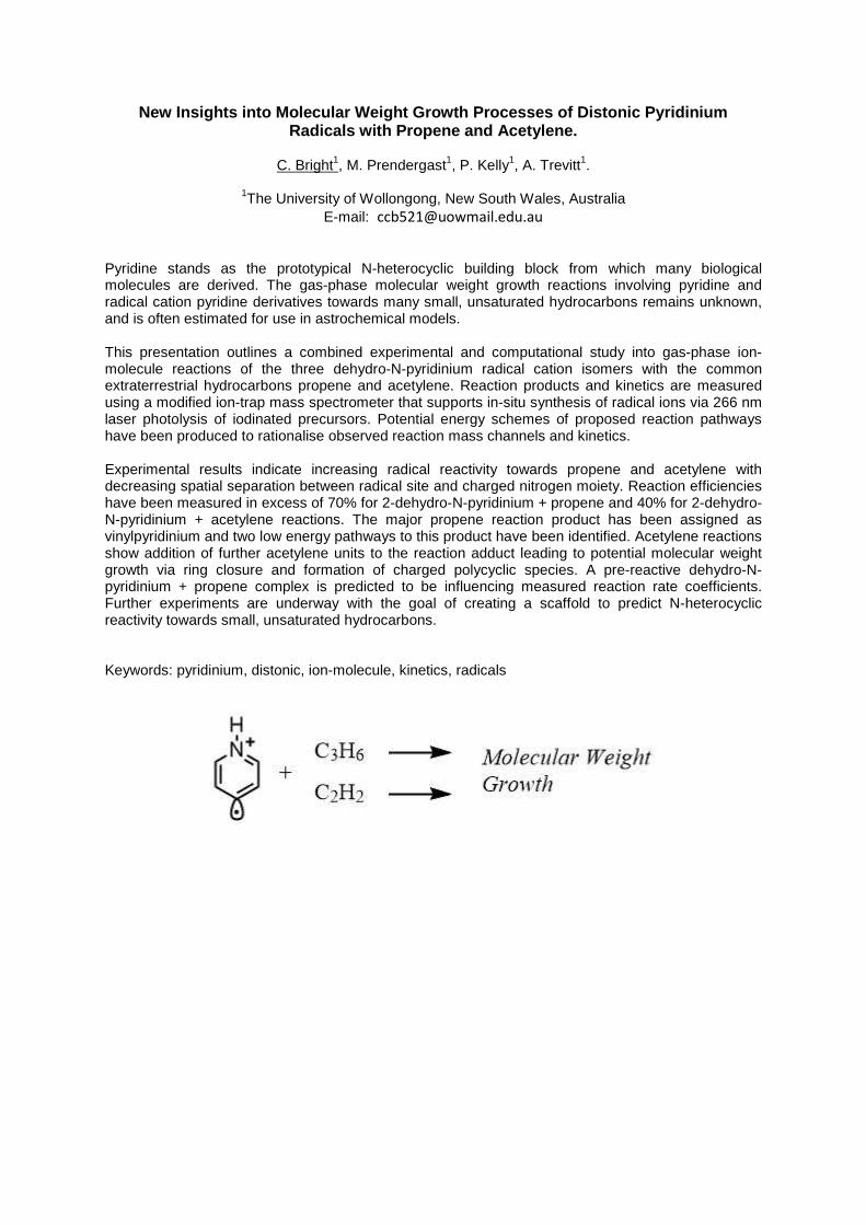
Pyridine stands as the prototypical N-heterocyclic building block from which many biologicalmolecules are derived. The gas-phase molecular weight growth reactions involving pyridine andradical cation pyridine derivatives towards many small, unsaturated hydrocarbons remains unknown,and is often estimated for use in astrochemical models.
This presentation outlines a combined experimental and computational study into gas-phase ion-molecule reactions of the three dehydro-N-pyridinium radical cation isomers with the commonextraterrestrial hydrocarbons propene and acetylene. Reaction products and kinetics are measuredusing a modified ion-trap mass spectrometer that supports in-situ synthesis of radical ions via 266 nmlaser photolysis of iodinated precursors. Potential energy schemes of proposed reaction pathwayshave been produced to rationalise observed reaction mass channels and kinetics.
Experimental results indicate increasing radical reactivity towards propene and acetylene withdecreasing spatial separation between radical site and charged nitrogen moiety. Reaction efficiencieshave been measured in excess of 70% for 2-dehydro-N-pyridinium + propene and 40% for 2-dehydro-N-pyridinium + acetylene reactions. The major propene reaction product has been assigned asvinylpyridinium and two low energy pathways to this product have been identified. Acetylene reactionsshow addition of further acetylene units to the reaction adduct leading to potential molecular weightgrowth via ring closure and formation of charged polycyclic species. A pre-reactive dehydro-N-pyridinium + propene complex is predicted to be influencing measured reaction rate coefficients.Further experiments are underway with the goal of creating a scaffold to predict N-heterocyclicreactivity towards small, unsaturated hydrocarbons.
Keywords: pyridinium, distonic, ion-molecule, kinetics, radicals
Electronic Spectra of Weakly Bound Diacetylene (HC4H+) Complexes in the Visibleand Ultraviolet Regions
G. Muller1, K. Catani2, E. Bieske1
1School of Chemistry, University of Melbourne, [email protected]
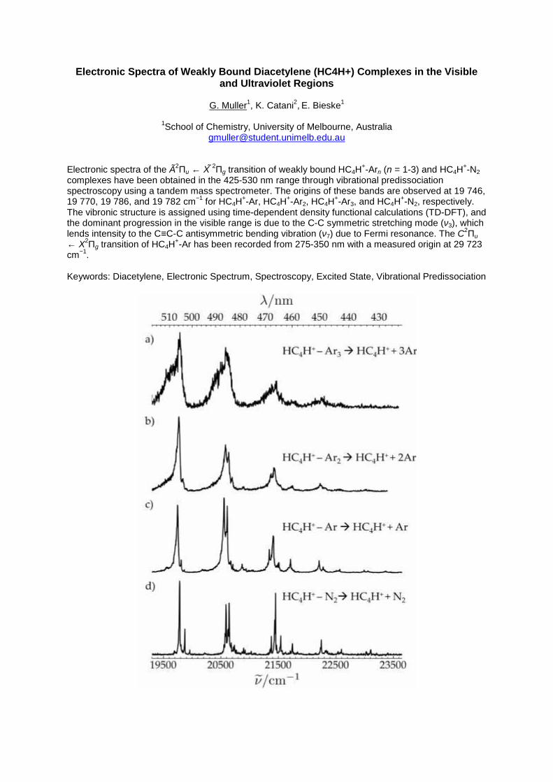
Electronic spectra of the Ã2Πu ← X ̃2Πg transition of weakly bound HC4H+-Arn (n = 1-3) and HC4H+-N2complexes have been obtained in the 425-530 nm range through vibrational predissociationspectroscopy using a tandem mass spectrometer. The origins of these bands are observed at 19 746,19 770, 19 786, and 19 782 cm−1 for HC4H+-Ar, HC4H+-Ar2, HC4H+-Ar3, and HC4H+-N2, respectively.The vibronic structure is assigned using time-dependent density functional calculations (TD-DFT), andthe dominant progression in the visible range is due to the C-C symmetric stretching mode (ν3), whichlends intensity to the C≡C-C antisymmetric bending vibration (ν7) due to Fermi resonance. The C2Πu← X2Πg transition of HC4H+-Ar has been recorded from 275-350 nm with a measured origin at 29 723cm−1.
Keywords: Diacetylene, Electronic Spectrum, Spectroscopy, Excited State, Vibrational Predissociation
Gas phase electronic spectroscopy of bare N3+ and the N3
+ - Ar and N5+ - Ar complexes
N. I. Bartlett1, K. J. Catani1, G. Muller,1 E.J. Bieske1
1School of Chemistry, The University of Melbourne, AustraliaE-mail: [email protected]
The electronic spectra of bare N3+ as well as the N3
+-Ar and N5+-Ar complexes were measured over
the 240-295 nm range using resonance enhanced photodissociation spectroscopy in a tandem massspectrometer. The spectra were measured by detecting N+ and N2
+ for the bare N3+ cation, whereas
spectra for the complexes were observed through detection of N3+ and N2
+- Ar for N3+-Ar and N3
+ andN+ for N5
+-Ar. The observed band system of the bare N3+ cation was previously seen using a similar
technique and was assigned to the A3u X3g- transition.1 This spectrum is reproduced in the
current study, in addition to the generation of other spectra from argon tagged clusters. The mainvibronic structure of the measured spectra are assigned based on the high resolution study of thesymmetric and antisymmetric N-N stretches (v1 and v3) with spacings of 1300 and 1700 cm-1.
The N3+ cation is known to exist in Earth’s atmosphere where it is postulated to form through the
ternary reaction of N+ and N2. Gas-phase spectroscopic characterisation of N3+ and its associated
clusters could offer insights into the composition and radiative processes in the terrestrial atmosphere.
Keywords: Ion spectroscopy, photodissociation
1. Friedmann, A.; Soliva, A.; Nizkorodov, S.; Bieske, E.; Maier, J., J. Phys. Chem. 1994, 98,8896
APCI-FTICR mass spectrometry reveals different reactivates of cyclohexyl-substitutedsilsesquinoxanes through isotopically labelled solvents
N. W. Proschogo1, A.J. Ward2, R. A. Lesic2, K. Fisher1, V. Fabos2, A. F. Masters2, T. Maschmeyer2,
1Mass Spectrometry Unit, School of Chemistry F11, The University of Sydney, Sydney 2006, Australia2Laboratory of Advanced Catalysis for Sustainability, School of Chemistry F11, The University of
Sydney, Sydney 2006, AustraliaE-mail: [email protected]
Amorphous silica plays an important role in heterogeneous catalysis as a support for metal catalystsand is frequently presumed to be “inert”. The structure of the supported catalyst is key tounderstanding the stability and reactivity of catalytic systems. To provide vital insights into the surfacereactivity of silica, polyhedral oligomeric silsesquioxanes (POSSs) can act as realistic homogeneousmolecular models for silica surfaces. It is difficult to study isolated solid phase reactions of surfaceswith the energetics of catalytic species. However, the reactivities of energetic model compounds canbe followed in the gas phase by mass spectroscopy. Here, we report novel reactivities associated withthe silica surface, derived from our insights obtained by means of such model systems with potentiallysignificant implications in catalysis when employing silica-supported catalysts. In this work, the gas-phase reactivities of two cyclohexylsubstituted POSSs, namely the completely condensed triganolprism [Si6cy6O9] (a6b0), and the incompletely-condensed partial cube [Si7cy7O9(OH)3] (a7b3), withcy=c-C6H11, were studied by using atmospheric pressure chemical ionisation (APCI) and collision-induced decomposition (CID) spectroscopies. Tentative structural assignment was achieved usingdeuterated and non-deuterated methanol and benzene solvents in combination with the highresolution and accurate mass Fourier transform ion cyclotron resonance (FTICR) mass spectrometryto determine reactivity with the solvent or the air using proton transfer and electron transfermechanisms. Silsesquioxane a6b0, containing three-membered rings, was found to be much morereactive, undergoing novel CH2-insertion on reaction with gas phase molecules—a reaction notobserved for a7b3, containing only four-membered rings. Both silsesquioxanes displayed the ability totrap ammonia formed in situ within the mass spectrometer from N2 in the instrument. This work alsodemonstrates the applicability of APCI and the role of CID in elucidating reactive POSS structures,highlighting novel gas-phase reactivities of POSS.
Keywords: Silsesquinoxanes, siloxane, atmospheric pressure chemical ionization, mass spectrometry,collision-induced dissociation
Gas-Phase Reactions of Distonic N-Substituted Aromatic Radical Cations with Low-Mass Unsaturated Hydrocarbons
P.D. Kelly1, M.B. Prendergast1, C.C. Bright1, S.J. Blanksby2, A.J. Trevitt1
1School of Chemistry, University of Wollongong, Australia2Queensland University of Technology, Brisbane, Queensland, Australia
E-mail: [email protected]
N-substituted aromatic radicals are commonly encountered in environments ranging from biologicalsystems to the atmosphere of Titan. In particular, aniline is a simple N-substituted aromatic molecule;thus, the reactivity of its radical derivatives forms an important case study in understanding thereactivity of N-substituted aryl radicals in general. It is notable that only a few of the reactions of theseradical derivatives of aniline have been investigated; specifically, significant research is yet to becompleted regarding distonic ammoniumylphenyl radical cations. In this study, the reactions of thesedistonic radical cations with short-chain unsaturated hydrocarbons such as propene, propyne, etheneand ethyne are investigated, with a particular focus on reaction kinetics and product branching ratios.
Experimentally, the target radicals are created in situ from UV laser photodissociation of thecorresponding iodoanilines in an ion-trap mass spectrometer modified to allow irradiation of trappedions with laser pulses. The hydrocarbon reactant (e.g. propene) is supplied to the mass spectrometeras a trace component of the He buffer gas. Mass spectra are obtained at varying storage times inorder to extract kinetic data. Concurrently, M06-2X/6-31G(2df,p) calculations are underway to assistin elucidating the reaction mechanism and rationalise the product branching fractions.
Calculations on the reaction of the different ammoniumylphenyl species with propene indicate thatcyclisation of the adduct (m/z 135) followed by hydrogen loss is a favourable process bothenergetically and entropically relative to other mechanisms of hydrogen loss; the presence of a m/z134 peak in the mass spectrum of this reaction shows that hydrogen loss is occurring to a non-negligible extent. Taken together, these results hint that such reactions may be important in formingnitrogenated polycyclic aromatic hydrocarbon (PANH) species thought to be responsible for the tholinhaze in Titan’s atmosphere.
Keywords: Distonic, aniline, hydrocarbons, kinetics, cyclisation
The Effect of Protonation on Photodissociation and Collision Induced DissociationProperties of Flavins and their Precursors.
S.J.P. Marlton1, A.J. Trevitt1
1School of Chemistry, University of Wollongong, 2533 NSW AustraliaE-mail: [email protected]
Flavins are blue light photoreceptors which contain an alloxazine group. Uracil, lumazine andlumichrome can be considered prototypes of biologically relevant flavins. Riboflavin (vitamin B2) is animportant biological molecule that decomposes under visible light (at wavelengths less than ~530nm).These molecules have several potentially viable protonation sites. The photostability of flavins andflavin precursors are not well understood, especially regarding the effect of protonation.
An LTQ XL ion trap mass spectrometer was used for collision induced dissociation (CID). Ultravioletlaser pulse redirected into the ion trap to initiate photo-dissociation (PD). A tuneable laser is used toscan wavelength dependence on PD in an LTQ Velos ion trap. Preliminary field asymmetric ion massspectrometry (FAIMS) experiments probe the presence of protonation isomers are also underway.
Preliminary results show differences in the CID and PD dissociation pathways of lumichrome andriboflavin. Because CID excites molecules gradually, the molecules can isomerise to less stableprotomers prior to dissociation. In contrast PD can access different pathways, perhaps with lessisomerisation. Photo-dissociation spectra of both riboflavin and lumichrome show unique dissociationpathways for distinct wavelength regions and this is currently being investigated. These spectracompared with excited state calculations (TD-DFT) aim to elucidate the presence of protomors. This iscompared to FAIMS experiments that indicate the presence of two protomers are present.
Gas-phase linkage photoisomerization in a ruthenium bis-sulfoxide photoswitch
M. S. Scholz1, J. N. Bull1, E. Carrascosa1, G. K. Kosgei2, J. J. Rack2, E. J. Bieske1
1School of Chemistry, University of Melbourne, Australia2Department of Chemistry and Chemical Biology, University of New Mexico, United States of America
E-mail: [email protected]
The ruthenium sulfoxide complexes [Ru(bpy)2L]2+ (bpy = bipyridine, L = bidentate bis-sulfoxide ligand)have three linkage isomers where each sulfoxide can be coordinated to the metal center through an Sor an O atom. These complexes have been proposed for use as molecular batteries, with solarenergy stored as a high-energy metastable O-bound state and released upon relaxation to the groundS-bound state. Previous time-resolved pump-probe spectroscopy measurements on these complexesin solution have shown sequential photoisomerization dynamics: starting from the S,S-bound isomer,the first photon produces the S,O-isomer on a ~150 ps timescale, and a second photon produces theO,O-isomer on a similar timescale. However, because photoisomerization occurs through a series ofcoupled metal-ligand charge transfer and metal-centered states of mixed multiplicity, it has beendifficult to compare solution-phase experimental results to gas phase ab initio calculations.
Using a custom-built tandem ion mobility-mass spectrometer coupled with laser spectroscopy, weprovide the first direct evidence for photoisomerization of a transition metal complex occuring in thegas phase. The S,S-bound isomer undergoes isomerization to both the S,O- and O,O-isomersfollowing absorption of a single UV photon, unlike in solution. The S,O-isomer predominantly gives theO,O-isomer upon irradiation with blue light. Minor reversion from the O,O-isomer to the S,O- and S,S-isomers is observed when irradiated in the green. Our experimental approach allows for study ofphotoactive transition metal complexes without the complication of solvation, which can dramaticallyalter their already-complex potential energy surfaces, and allows for straight-forward comparison tohigh-level theoretical calculations.
Keywords: photoisomerization, ion mobility, laser spectroscopy, ruthenium, linkage isomerism
A tale of two humps: using GCxGC-TOFMS to compare Caribbean oil seeps
A. G. Scarlett1, K. Grice1
1Western Australian Organic and Isotope Geochemistry Centre, Curtin University, Australia
E-mail: [email protected]
Oil oozing from the ground must have fascinated humankind since we first walked the Earth. Forthousands of years civilisations made use of oil seeps without understanding what they were. Ourunderstanding of the nature of oil has only taken shape in the last few decades, since massspectrometry has allowed us to interpret the chemical structures of thousands of compounds presentin oil. However, some oil seeps still remain a bit of a mystery.
The vast oil seep known as Pitch Lake in Trinidad has become a tourist destination and, like our earlyancestors, we come to gaze at oil emerging from the earth. Pitch Lake is the largest natural deposit ofasphalt in the world covering 40ha at La Brea in southwest Trinidad. At the opposite end of the chainof Caribbean islands lies Cuba. Here, oil seeps from mountains, sometimes emerging from rocks notnormally associated with oil. Due to the extent of biodegradation and the evaporative processes fromthe hot Caribbean sunshine on both these seeps, the oils are incredibly difficult to analyse usingtraditional GC-MS. The unresolved complex mixtures, or 'humps', of hydrocarbons,heterocyclic andpolar compounds make producing chromatographic peaks sufficiently resolved to obtain good massspectra challenging. Using two dimensional gas chromatography with time-of-flight massspectrometry (GCxGC-TOFMS) the resolution is dramatically improved allowing mass spectra to beobtained.
Biomarkers can be seen as molecular fossils providing us with a view of past climates andsedimentary conditions. By applying GCxGC-TOFMS following minimal sample preparation, we wereable to obtain good mass spectra for a range of biomarkers, including diamondoids, present in oilsfrom Cuba and Pitch Lake.
Keywords: oil, GCxGC-TOFMS, biomarkers, diamondoid, UCM
MALDI FT-ICR Imaging: Double the Performance or Doublethe Samples using Quadrupolar Detection
D.S. Cornett1, L.A. Woods2, C.J. Thompson1, M. Witt31Bruker Daltonics, Billerica, USA
2Bruker Pty Ltd, Preston, VIC, Australia3Bruker Daltonik, Bremen, Germany
E-mail: [email protected]
For FT-based MALDI imaging applications, reducing the acquisition time of each spectrum by a fewmilliseconds can shorten the total measurement time by hours. Quadrupolar detection (2ω) enhancesFTICR imaging studies by providing high MS performance with significantly reduced transient length.In this study, we evaluated the speed benefit of imaging with 2ω vs 1ω and compare MS performancefrom each.
Rat kidney and brain were sectioned at 12 μm thickness and coated with matrix using a pneumaticsprayer. 7T and 12T solarix systems were used. Each was fitted with a dynamically harmonized cellmodified with new electronics that improves the 2ω signal intensities and allows fast switchingbetween excitation and detection modes at the same electrodes. MALDI sensitivity was optimized form/z 200-900 and images were collected at different transient sizes and signal lengths.
A bottleneck for MALDI FT-ICR imaging is the low sample throughput, due to the compromise onemust make between acquisition speed and high MS performance. With a small electronic change to acommercial system, one can achieve the same mass resolving power in half the acquisition time, or2x the mass resolving power from the same acquisition time. For example, using a 7T system, a 2stransient of 1ω signal produces a resolution of 300k at m/z 800 while the 2ω signal producesresolution of 300k from a 1s transient. The significance being that each spectrum can be acquired inhalf the time without loss of mass accuracy or resolution. Alternatively, when comparing quadrupolardetection, 2w, with 1ω detection for the same 2s transient time, the 2ω detection yields 2x theresolving power, 600k at m/z 800. Although no speed benefit is realized in this mode of operationthere is benefit to resolving isotopic fine structure at higher m/z for added confidence in formulaidentification.
Keywords: FTMS, MALDI-imaging, isotopic fine structure, speed, ultra-high resolution
Optimization of SONAR DIA acquisition parameters for Omics experiments
S. Wilson1, J.P.C. Vissers2, C. Hughes2, K. Richardson, L. Gethings2 and Jim Langridge2
1Waters Corporation, Rydalmere, NSW, Australia2Waters MS Technologies, Wilmslow, UK
E-mail: [email protected]
Quadrupole time-of-flight (Q-TOF) MS is a well established tool for both discovery and quantitativeapplications. A new mode of DIA operation, SONAR, was recently introduced whereby a low-resolution quadrupole is scanned repetitively over alternating low (MS1) and elevated energy (MS2)scans, producing data of a similar format as Ion mobility enabled modes by utilising the same multi-dimensional acquisition system
Samples were analysed on a Xevo G2XS Q-TOF. The quadrupole mass filter of the MS wascharacterised to allow various transmission windows to be employed for different molecule classes;peptides, small molecules (metabolites and lipids). Initially, the full m/z range of the quadrupole wascontinuously and repetitively scanned and the MS2 CID energy applied in a linear fashion tocorrespond with the quadrupole mass position. Further optimisation of the collision energy can beachieved by segmenting the quadrupole m/z scale into multiple steps.
Experiments were carried out to determine optimum collision energy ramps using one quadrupolemass range; m/z 400 – 900 for analysis of complex peptide mixtures and m/z 500 – 1200 for smallmolecule applications with the starting and ending collision energy values altered in 5 V steps.Optimum ramps were initially determined as 14 to 40V for proteomic and 20 to 50 V (+ve) and 25 to55 V (-ve) for small molecules. The potential to further optimize collision energy ramps is apparent asthere are distinct retention time regions where SONAR experiments would potentially benefit fromusing multiple quadrupole m/z ranges and collision energies, i.e. retention order, dependent. Analysisof complex proteomics samples shows the multi-step method increases coverage and subsequentquantitation by > 20%. For small molecule lipidomics experiments the same method increased featureidentification rates significantly, improving the qualitative information by extraction of analyte classinformation based upon neutral loss or product ion extraction.
Keywords: Data independent acquisition, SONAR, TOF mass spectrometry
REAL-TIME AUTHENTICATION OF WHISKEYS USING DART-QDA ANALYSISHeather Patsiouras3Kari Organtini1, Steve Pringle2, Gareth Cleland1
1 Waters Corporation, Milford, United States of America2 Waters Corporation, Wilmslow, United Kingdom of Great Britain and Northern Ireland3 Waters Australia, Sydney, Australiae-mail:[email protected]
Whiskey labeling and branding is highly regulated to protect distillers and consumers. Developmentof analytical tools to quickly and easily authenticate whiskeys is therefore important to protectconsumers and distillers alike. Direct Analysis in Real Time (DART) is a highly useful ambientionization technique that benefits from the need for little to no sample preparation and nochromatography, allowing the user to directly analyze a sample within minutes. In this work, DARThas been coupled with simple mass detection (QDa) to provide a tool for rapid identification ofwhiskey samples. Twelve different brands of whiskey were analyzed, including samples of bourbons(Kentucky and Tennessee), Irish whiskey, blended Scotch, and single malt Scotch. A DART-QDamethod was established to provide the most unique mass spectra for each sample. The dataobtained for all twelve whiskeys analyzed were used to construct a PCA and LDA based statisticalmodel using a prototype model building software. The statistical model generated was used to makeidentifications of unknown whiskey samples as the mass spectra were being acquired or by a rawdata file provided. Using the model created, unknown samples of each of the whiskeys weresuccessfully identified in real time with greater than 97% confidence. Amongst all the samples, thefive bourbons analyzed were most similar when statistically modeled. Despite their similarity, theywere successfully identified by brand in the twelve whiskey model. The current study demonstratesthe utility of DART-QDa in the authentication of whiskeys. This technology could be used to rapidlyscreen bottles of whiskeys to determine the need for further analysis of suspect samples. The dataalso implies usefulness in applications in the distillery to monitor the quality of production andblending of whiskeys.
Keywords: DART, QDa, Authenticity, Whiskey
Capturing reactive high-valent iron(IV)-oxo intermediates of catalytic cycles usingtheta-capillary nanoelectrospray ionisation mass spectrometry
H. E. Lee and W. A. Donald
School of Chemistry, University of New South Wales, AustraliaE-mail: [email protected]
Efficient and selective functionalisation of C-H bonds is an important goal due to the extensive use oforganic molecules and polymers in many industries. The White-Chen (WC) catalyst is useful for itsability to selectively oxidise inert aliphatic hydrocarbons under mild conditions using hydrogenperoxide as a terminal oxidant. However, the active catalytic intermediate(s) of the WC catalyst,presumably a high valent Fe-oxo species, has not been directly detected by any experimental method(to our knowledge) because the intermediate(s) are highly reactive.
Here, we use theta nanoelectrospray ionisation (nESI) mass spectrometry, in which two solutions aremixed rapidly (sub-s) immediately prior to ion formation and detection; i.e., this is the fastest knownmixing system available that enables reaction products and intermediates to be detected. Theperformance of theta nESI for forming and detecting reactive intermediates of metal-mediatedcatalytic reactions was examined using a porphyrin-based iron-oxo catalyst. For theta nESI, the ionabundance of reactive intermediate species was over 15 times higher than that measured by use ofconventional nESI MS with a wide range of terminal oxidants.
Theta nESI MS was used to investigate the mechanism of WC catalysed C-H bond oxidation thatconverts methyl hexanoate to 5-oxo methyl hexanoate. By rapidly mixing an oxidant with the Fe-catalyst immediately prior to ESI, the accurate masses and isotope distributions of ions consistentwith the molecular formulae of reactive intermediates were detected by high-resolution massspectrometry. If an oxidant was not used, the reactive intermediates were not detected. By correlatingthe ion abundance of these ions to the concentrations of the substrate and oxidant prior to ESI, themechanism of the WC reaction can be inferred. Overall, these results are consistent with a high valentFe(IV)-oxo carboxylate as the active intermediate in the C-H bond oxidation reaction catalysed by theWC catalyst.
Keywords: theta nanoelectrospray ionisation, short lived intermediate, reactive intermediate, inorganiccoordination complexes, mass spectrometry
Rapid Screening of Non-Volatile Oak Wood Components of Oenological Importance
R.R. Farrell1, D. Stadler2, J. Krismer2, R. Zenobi2
1Department of Chemistry, University of Tasmania, Australia2Department of Chemistry and Applied Biosciences, ETH-Zurich, Switzerland
E-mail: [email protected]
We present a rapid and high-throughput method for screening important flavour compounds in oakwood used for maturing wine. Our approach utilizes a rapid extraction protocol (taking less than oneminute) followed by Matrix Assisted Laser Desorption Ionisation Mass Spectrometry (MALDI-MS).Triterpenoids responsible for sweet and bitter taste attributes and other important non-volatilecompounds from oak wood were successfully extracted and ionized by MALDI-MS. The elementalcomposition obtained from high-resolution MS and MSMS studies were used for compoundidentification. Internal standards were used for quantitative measurements. Fifteen different oakboards sourced from a cooperage in France were screened to validate the method and to elucidatedifferences in the non-volatile oak wood extractives. A high degree of variation in the non-volatilecomposition of the different oak boards was observed. The oak boards were categorized into differentflavour categories according to the levels of key flavour compounds. These compounds are of greatinterest due to their significant flavour impact on oak-aged wine and the high degree of naturalvariation in oak wood chemistry. This method provides a powerful approach for the rapid screening ofimportant non-volatile flavour components in oak wood and greatly improves sample throughput incomparison with current approaches based on liquid chromatography.
Oak wood, non-volatiles, rapid analysis, flavour control, mass spectrometry
EmporeTM assisted rapid fractionation improves protein identification in bovine milk
A. Thomas 1, S. Deb-Choudhury 1, A. Grosvenor 1, E. Lee 1, P. Middlewood 1, S. Clerens 1,3
1 Food and Bio-Based Products, AgResearch Limited, New Zealand3 Biomolecular Interaction Centre, University of Canterbury, Christchurch, New Zealand
E-mail: [email protected]
Heating processes such as pasteurisation and ultra-high temperature (UHT) treatment of milk improvefood safety and shelf life of liquid milk. However, these treatments can cause physical and chemicalmodifications to milk proteins thus introducing changes in the flavour and nutritional qualities of liquidmilk. Most studies involving bovine milk protein composition have focused on the most abundantproteins present because of the technical challenges associated with the characterisation of the lowabundant proteins. But it is important to maximise the identifications of low abundant proteins as theymay play significant roles in the protein modifications during processing treatments of bovine milk.
For this study, supermarket-bought UHT milk was prepared by acid precipitation into casein and wheyfractions. Protein extraction was performed by shaking the fractions overnight at 25°C in a buffercontaining 7 M urea, 2 M thiourea, 50 mM dithiothreitol and 50 mm Tris. The precipitated proteinswere subjected to standard reduction, alkylation and digestion methods.
EmporeTM assisted extraction is known to improve purification and concentration of analytes fromaqueous samples. In this study we explored a novel rapid fractionation approach involving C18EmporeTM. Tryptic peptides were first bound to EmporeTM at a high pH for 3 hours at roomtemperature with light vortexing. The bound peptides were then sequentially eluted using 10%followed by 50% acetonitrile in 10 mM ammonium formate solution, pH 10. The two fractions werethen individually dried down and analysed by LC-MSMS. This fractionation approach is rapid andsimple, compared to column-based pre-fractionation, with an added advantage of reduced sampleloss and enhanced protein identifications compared to the use of strong cation exchange (SCX)fractionation and clean up prior to LC-MSMS analysis.
Keywords: LC-MS/MS: Liquid chromatography-tandem mass spectrometry, UHT: Ultra-hightemperature processing, SCX: strong cation exchange, C18 EmporeTM.
Tape stripped stratum corneum samples prove to be suitable for comprehensiveproteomic investigation of actinic keratosis
Ali Azimi1, Marina Ali 1, Kim L. Kaufman2, 3, Graham Mann4, Pablo Fernandez-Penas1
1. Department of Dermatology, Westmead Hospital, The University of Sydney, WestmeadNSW 2145, Australia
2. School of Molecular Bioscience, Faculty of Science, The University of Sydney, DarlingtonNSW 2006, Australia
3. Brain and Mind Research Institute, The University of Sydney, Camperdown, NSW 20504. Westmead Institute for Medical Research, The University of Sydney, Westmead NSW 2145,
AustraliaE-mail: [email protected]
ABSTRACT
Actinic keratosis (AK) is a UV light–induced lesion of the skin that carries the risk of progressionto invasive squamous cell carcinoma. The only method to confirm a clinical diagnosis of AK isperforming a biopsy, which may lead to complications such as bleeding, wound infection, andscarring. Here we report an efficient and non-invasive method of Tape Stripping (TS) for thecollection and extraction of proteins from the stratum corneum and its use for subsequentdiagnostic biomarker analysis using proteomic techniques. Human stratum corneum proteinsfrom five patients with AK were obtained non-invasively by sequential application and removalof adhesive stripping tapes on the lesional skin surface and their matched normal skin.Following their extraction and proteolytic digestion, the peptide digests were analysed by label-free liquid chromatography tandem mass-spectrometry (LC-MS/MS). Of the total 613 uniqueproteins identified, 477 proteins (77.8%) were overlapping with those previously identified by ourproteomic analysis of formalin-fixed and paraffin-embedded (FFPE) AK samples. In addition,differential abundance analysis showed 32 proteins significantly increased and four proteinsdecreased in AK samples compared to the normal skin (p<0.05). Ingenuity Pathway Analysis(IPA) and Funrich bioinformatics analysis showed that differentially changed proteins in the TSsamples are involved in tumorigenic pathways including p38 signalling, PI3K/AKT signalling andmTOR signalling that were found to be comparably disrupted in the FFPE AK samples. Thispilot study is the first to demonstrate the application of a non-invasive method for sampling andextraction of proteins from AK lesions for subsequent proteomic investigations. Exploring thistechnique further will revolutionise early diagnosis and management of the skin tumours withoutthe need for traditional biopsy.
Keywords: tape stripping, proteomics, actinic keratosis,
NEW HETEROAROMATIC SCIFF BASE DERIVED CuII AND ZnII COMPLEXESAS POTENTIAL ANTICANCER AGENTS
AlAjmi, MF1, Afzal Hussain, A1, Ahmed, R2, Khan, A1, Alam, P1
1 Pharmacy, King Saud Univrsity, Riyadh, Saudi Arabia2 College of Science, King Saud University, Riyadh, Saudi Arabia
ABSTRACT:
Two new complexes of copper(II), [Complex 1], and zinc(II), [Complex 2], with atridentate –ONN– Schiff base ligand (L), a bioactive scaffold derived from 2-aminobenzimidazole and 1-aminoimizole-2-carboxyaldehyde, were synthesized andcharacterized using various spectroscopic techniques, viz, IR, 1H and 13C NMR,EPR, HRMS, and elemental analysis. Purity was confirmed by UPLC analysis. Bothcomplexes are nonelectrolytic by nature. The newly synthesized complexes werescreened for their anticancer activity in different cell lines i.e. HepG2 (Liver), SK-MEL-1 (Skin), HT018 (Colon), HeLa (Cervical) and MDA-MB 231(Breast). Interactionstudies of both the complexes with CT DNA were studied by using variousbiophysical techniques, which showed binding affinity of complexes with CT DNA. Invitro cytotoxicity of complexes was evaluated against different human cancer celllines of different histological origins by employing MTT Assay. The apoptosis andnecrosis were examined by the flow cytometry experiment. Complex 2 showedhighly potent and significant activity in SK-MEL-1 (Skin) (IC50 18 ± 2.3 µg/ml)whereas Complex 1 showed potent anticancer activity in MDA-MB 231(Breast) (IC50
3.5 ± 2.4 µg/ml). The mechanism of anticancer activity of these complexes werestudies.
Complexes 1 and 2 showed promising anticancer activities against SK-MEL-1 andMDA-MB respectively, compared to standard anticancer drug.
Synergistic Targeting of FLT3-activated Signalling Pathways in Acute Myeloid Leukaemia(AML): Pathways Identification by Phosphoproteomic Profiling
Heather C. Murray1, Juhura G. Al Mazi1, Richard G.S. Kahl1, Hayley M. Flanagan1, Charles E. deBock2, Nathan D. Smith3, Anoop K. Enjeti4, Martin R. Larsen5, Nicole M. Verrills1,*, Matthew D. Dun1,*
1 Cancer Research Program, Hunter Medical Research Institute, and School of Biomedical Sciencesand Pharmacy Faculty of Health and Medicine, University of Newcastle, Callaghan, NSW, 2308,
Australia.2 VIB Center for the Biology of Disease, Leuven 3000, Belgium, and KU Leuven Center for Human
Genetics, Belgium.3 Analytical and Biomolecular Research Facility Advanced Mass Spectrometry Unit, University of
Newcastle, Callaghan, NSW, 2308, Australia.4 Calvary Mater Hospital, Newcastle, 2298, NSW, Australia.
5 Department of Molecular Biology and Biochemistry, Protein Research Group, University of SouthernDenmark, Odense, 5230, Denmark.
*These authors contributed equally to this work.
Email: [email protected]
Acute Myeloid Leukaemia (AML) carries a 5-year survival rate of 24%. Current treatmentsinclude high dose chemotherapy and bone marrow transplantation; however development oftreatment resistance and relapse is common. Internal Tandem Duplication (ITD) mutation of thereceptor tyrosine kinase FLT3 is the most frequent driver mutation in AML (~25% of cases), and isassociated with poor prognosis. Therapeutic inhibition of FLT3 has however proven difficult toachieve, with FLT3 inhibitors displaying limited monotherapeutic success. Therefore, thecharacterisation of the oncogenic signalling pathways downstream of FLT3 mutation may identifyimproved therapeutic approaches in AML. To identify new drug targets the primary blast cells of 6AML patients (3 FLT3-ITD, 3 FLT3-wildtype) were isolated and tryptic peptides were labelled withisobaric tags prior to being subjected to a multistage process of phosphopeptide enrichment, offlineHPLC fractionation and high-resolution mass spectrometry. 4803 phosphosites on 4085 uniquephosphopeptides from 1905 proteins were identified across the 6 AML patient blast samples. Analysisof differentially expressed phosphoproteins in FLT3-ITD versus wildtype AML patients identifieddysregulation of DNA double strand break repair pathways with decreased phosphorylation ofHomologous Recombination Repair (HRR) proteins, including BRCA1 and increased phosphorylationof Non-Homologous End joining (NHEJ) proteins, especially DNA-PK, as well as increased ƴH2AX.Targeting DNA-PK induced selective toxicity in FLT3-ITD cell lines. DNA-PK inhibitors combined withFLT3 inhibitors including Sorafenib which enhanced this effect, leading to G1 phase accumulationfollowed by synergistic cell death in FLT3-ITD but not FLT3-wildtype AML cell lines. Similarly, DNA-PK inhibitors in combination with the DNA-damaging chemotherapy agent Cytarabine displayedsynergy in FLT3-ITD cells. Our phosphoproteomic data shows that FLT3-ITD AML is associated withactivation of the error-prone NHEJ pathway, which may contribute to genomic instability. TargetingDNA-PK in combination with standard therapeutic agents has the potential to improve outcomes forthis poor-prognosis AML subtype.
Keywords: AML, Phosphoproteomics, DNA repair
New Supercharging Additives for Improving Protein Sequencing by MassSpectrometry
E. D. B. Foley, W. A. Donald
School of Chemistry, University of New South Wales, AustraliaE-mail: [email protected]
Multiply charged protein ions can be formed by electrospray ionization (ESI), which enables accuratemeasurement of molecular weight and sequence information of proteins to be obtained by massspectrometry (MS) experiments. Protein charge states can be significantly increased by adding smallorganic molecules called superchargers (e.g., m-NBA and butylene carbonate) to ESI solutions.Higher protein charge states improve the signal-to-noise ratio in Fourier transform massspectrometers and increase the amount of non-specific fragmentation by electron capturedissociation, which significantly improves the amount of sequence information obtained.Superchargers can also increase the ion abundances of protein and peptide ions. It has been shownthat increasing the alkyl chain length of cyclic carbonates (e.g., from ethylene to butylene carbonate)increases the charge imparted on the protein.
Here, we synthesised a series of cyclic carbonates with longer alkyl chains than butylene carbonateand investigated the effects of these molecules on the charge states and sequence coverage valuesobtained for three proteins (ubiquitin, cytochrome c and carbonic anhydrase II). The charge states ofionised cytochrome c can be increased from butylene carbonate to 1,2-hexylene carbonate. Theseresults indicate that superchargers may function through their surfactant properties. The physicalproperties (e.g., dipole moment, surface tension, gas-phase basicity) were used to rationalise themechanism of how these superchargers improve the extent of protein charge. The new additives werealso tested on protein digests by LC-MS for their effect on detected ion abundances.
Keywords: Proteomics, electrospray ionization, supercharging top-down protein analysis

Comparative whole membrane proteomics analyses of marine cyanobacteria
Fallen Teoh1, Martin Ostrowski1, Ian T. Paulsen1
1Department of Chemistry and Biomolecular Sciences, Macquarie University, [email protected]
Abstract:Marine cyanobacteria Synechococcus are abundant, globally distributed and genetically diverse.Synechococcus strains have developed distinct strategies to cope with a wide range of marineenvironments, from turbid, estuarine waters to transparent oligotrophic waters and varioustemperatures across 120˚ of latitude. We analysed the membrane protein composition of strainsisolated from different regimes to gain an understanding of how these bacteria interact with theirenvironment. Whole membrane proteins of four Synechococcus strains namely CC9311(clade I),CC9605(clade II), WH8102(clade III) and CC9902(clade IV) have been analysed by a bottom-upproteomics approach using a Q-Exactive Orbitrap mass spectrometer. Synechococcus CC9311 andCC9902 represent temperate strains while CC9605 and WH8102 represent strains predominantlyfound in warm tropical waters. More than 200 proteins identified from each Synechococcus sp.contain transmembrane domain (TM). Interestingly, protein phosphatase 2C is one of the highestexpressed proteins in Synechococcus CC9311 and CC9902. This protein is suggested to play a rolein signal transduction but the exact role in marine cyanobacteria remains elusive. FutA (iron ABCtransporter substrate binding protein) unique to Synechococcus clade III has been identified as highlyexpressed in Synechococcus WH8102. In contrast, the iron Fe3+ ABC substrate binding proteinexpression level of sync_1545(CC9311), syncc9605_1578 (CC9605) and Syncc9902_2002 (CC9902)is relatively low. Overall, the high expression level of FutA suggested the high binding affinity of iron isimportant for iron scavenging in tropical oligotrophic waters. A large number of expressed membraneproteins have no known function, or only general predictions, which indicates that more work isrequired to understand the spectrum of chemical compounds that these cells are capable oftransporting. Among the top 50 expressed proteins, we also identified five proteins which are uniqueto Synechococcus CC9311 suggests there are specific adaptations in Synechococcus CC9311 forsurvival in coastal waters.
Keywords: membrane proteomics, cyanobacteria, Synechococcus, microbiology, transporter
Protein Correlation Profiling and the Dynamics of the Saccharomyces cerevisiaeIntracellular Methylation Network
Gene Hart-Smith1, Chi Nam Ignatius Pang1, Daniela-Lee Smith1, Joshua J. Hamey1, Marc R. Wilkins1
1 NSW Systems Biology Initiative, School of Biotechnology and Biomolecular Sciences, University ofNew South Wales, Sydney, New South Wales 2052, Australia
E-mail: [email protected]
Protein-protein interactions (PPIs) are fundamental to all biological processes. For this reason a keyaim of molecular systems biology is to better understand the dynamics of PPI networks. It is knownthat post-translational modifications (PTMs) can mediate the dynamics of PPIs, and our laboratoryhas devoted particular attention to the study of arginine and lysine methylation in this context. Tocreate a foundation for this work we have produced a detailed map of methyltransferase-substrateinteractions, embedded within a broader network of PPIs, in the model organism Saccharomycescerevisiae: the yeast intracellular methylation network.We are currently studying the dynamics of this network using a technique known as size exclusionchromatography-protein correlation profiling (SEC-PCP). This technique involves the fractionation ofnative protein complexes using SEC. Fractionation profiles for individual proteins are measured usingquantitative mass spectrometry techniques, allowing PPIs to be studied via the correlation of similarfractionation profiles under the assumption that proteins in a common complex co-fractionate.We have created a reference PCP dataset from 280 SEC fractions of wild-type yeast lysate (70fractions × 4 biological replicates), identifying >200 native core complexes, including complexes thatcontain known methyltransferase substrates. We are characterising the absolute stoichiometries ofindividual methylation sites in these complexes using a library of 34 internal standard heavy isotope-labelled synthetic peptides. This has, for example, identified that methyltransferase activity on EF1α –a protein observed in 4 distinct complexes – can change when EF1α is associated with different PPIs.We are also in the process of producing PCP data from methyltransferase knockout yeast strains.This has revealed that methylation on K316 of EF1α, which is catalysed by the methyltransferaseEFM4, influences the formation of the elongation factor 1 core complex.Together these studies are providing broad-scale insights into how methyltransferase-substrateinteractions can mediate the dynamics of PPI networks.
Keywords: protein-protein interactions, systems biology, protein-correlation profiling, post-translationalmethylation, quantitative mass spectrometry
Detecting crosslinks in milk using mass spectrometry
H. McKerchar1, 2, R.C.J. Dobson1, J.M. Dyer1, 2, J.A. Gerrard1,3, S. Clerens1,2
1Biomolecular Interaction Centre, University of Canterbury, New Zealand2Food & Bio-Based Products, AgResearch Lincoln Research Centre, New Zealand
3School of Biological Sciences and School of Chemical Sciences, University of Auckland, NewZealand
E-mail: [email protected]
During food processing and storage, crosslinks can form between proteins, which profoundlyinfluence the nutritional value and properties of food. One crosslink that can form with heating andbasic pH, two typical food processing procedures used in milk products, is lysinoalanine. Thiscrosslink is interesting because it has been shown to decrease essential amino acid availability(particularly lysine) and protein digestibility, protein quality and mineral bioavailability. Despite thepotential deleterious effects of lysinoalanine, its formation and biological fate is not well understood.Harnessing the highly accurate analysis of small quantities of analytes afforded by mass spectrometryprovides a means to detect and characterise lysinoalanine in milk proteins. The typically low relativeabundance of crosslinked peptides compared to linear peptides and the complex fragmentationpattern produced by crosslinks compared to linear peptides, together with several other factors,makes investigating crosslinked peptides in food is challenging. As a means of simplifying thecomplex fragmentation patterns and narrowing the potential number of analytes, we subjected twoshort synthetic peptides, each five amino acids long, to heat and alkaline treatment and identified aputative crosslink of lysinoalanine between the peptides. We are in the early stages of characterisingthe fragmentation pattern of this simple crosslinked unit. We anticipate that the characteristic patternof this simple model can be used as a diagnostic tool for identifying lysinoalanine crosslinks in largerprotein complexes and untangle the many unknowns of crosslinks food proteins.
Keywords: proteomics, crosslinking, food, lysinoalanine
Advanced Mass Spectrometry Methods for Unravelling the Mechanisms ofOrganophosphate Toxicity
H. X. Wang1, M. G. Leeming2, W.A. Donald1
1School of Chemistry, University of New South Wales, Australia2School of Chemistry, The University of Melbourne, Australia
E-mail: [email protected]
Organophosphates are widely used for insect pest control. Some of the most toxic organophosphatessynthesized by humankind have been used in warfare causing thousands of fatalities. Acute andchronic exposures to organophosphate insecticides have been documented to cause adverse healtheffects. However, the mechanism of the toxicity is still not clear because of the lack of an analyticalmethod for the global detection of proteins of that have been covalently modified byorganophosphates.
We developed and applied a novel mass spectrometry method to provide a solution to identify OP-modified adducted proteins in non-targeted fashion. We incubated human plasma with a mixture ofisotope labelled and unlabelled organophosphate pesticides, parathion and dichlorvos. All proteinsand any protein-adducts were digested followed by LC-MS analysis to locate “twin ion” peaks ofpeptides adducted by organophosphates. Tandem mass spectrometry was used to identify thepeptides and modification sites. The LC-MS/MS data was processed by the development of blendeddata analytics software called Xenophile, which is capable of identifying the amino acid residue of anyproteins that were modified by the labelled organophosphates. This approach has the advantage thatthere is no need for specific parameterisation of protein database search algorithms. Using thismethod, the number, location and combinations of organophosphate adductions to protein targets inhuman plasma was characterized. This information will be useful for unveiling the mechanism oforganophosphate toxicity and designing novel therapies for organophosphates exposure.
Keywords: Organophosphates, protein target, non-targeted identification, LC-MS/MS.
Does GelLC still have a place in proteomic analyses? A comparative study ofmammalian skin
Trevor S Loo1, Meekyung Ahn2, Gillian E Norris1
1Inst. Fundamental Sciences, Massey University, Palmerston North, New Zealand2Leather and Shoe Research Association, Palmerston North, New Zealand
E-mail: [email protected]
The skin is the largest organ of the mammalian body and serves as a first line of defence againstphysical trauma, microbial infection and UV-radiation. It consists of three distinct layers; theepidermis, dermis and the subcutaneous fat layer. The epidermis is the most superficial layer of theskin and functions as a barrier to the environment. The dermis is predominantly made up of collagensthat are responsible for the tensile and tear strength of skin and the subcutaneous fat layer is largelyfor insulation. These physical properties present challenges in extracting and solubilising proteins foranalysis by mass spectrometry.
Sheep, deer and cow skins are by-products from the NZ meat industry, which are subsequentlyprocessed to leather used for making garments, footwear and upholstery. Although there has been asubstantial body of research that has concentrated on the arrangement of collagen fibers/fibrils indifferent animal skins, the molecular factors responsible for these structural differences are less wellunderstood.
In this study, we describe sample preparation procedures to improve the solubilisation of sheepskinproteins and compare the merits of gel-free vs gel-based approaches using the number of totalprotein IDs obtained. In the gel-free method, proteins were sequentially extracted prior to the trypsindigestion. In the gel-based method, solubilised proteins were sequentially extracted before beingfractionated into broad molecular weight ranges using 1D-SDS-PAGE prior to trypsin digestion.Trypsin digested peptides were analysed using either a 60 or a 95 min gradient on a nano-LC-MS/MS(Q-Exactive Plus, ThermoFisher Scientific). Proteins were identified using Proteome Discoverer(v.2.2, ThermoFisher Scientific) searched against the ruminant NCBI database with a FDR of 1%. Theoptimised method was then used to analyse and compare extracted proteins from cow-, goat- anddeerskins.
Keywords: Animal Skin, Extraction, Gel Digestion, nanoLC-MS/MS
A validated high throughput mass spectrometry method for the analysis oferythrocyte fatty acids
Ayedh Alqarni1, 2, 3, Todd W. Mitchell1, 2 Barbara J. Meyer1’ 2
1Illawarra Health and Medical Research Institute, University of Wollongong, Australia2School of Medicine, University of Wollongong, Australia
3King Fahad Specialist Hospital, Dammam city, Saudi Arabia
E-mail: [email protected]
The omega-3 index is defined as the amount of EPA “eicosapentaenoic acid” plus DHA
“docosahexaenoic acid” in erythrocytes expressed as the percent of total erythrocytes fatty acids.
Several observational studies and randomised controlled trials indicate that a lower omega-3 index is
associated with an increased risk of coronary heart disease mortality. Large scale clinical studies
ideally should measure erythrocyte membrane fatty acids concentration in a large number of samples,
however the cost, time and sample volume are limiting factors. The aim of this study is to develop a
high throughput method that will reduce cost, time and sample volume. The current reference method
used for measuring erythrocyte membrane fatty acids concentration is a single-step extraction,
degradation and derivatisation method (Lepage & Roy, 1986) followed by gas chromatography flame
ionisation detector measurement. For the novel method erythrocyte lipids were extracted using an
automated Methyl tert-butyl ether method and measured by shotgun mass spectrometry. The
samples used for this study were synthetic PL mixtures and human erythrocytes in order to measure
fatty acids concentration and determine the omega-3 index. The range of recoveries over five
synthetic PL mixtures were 91.3–105.5% and the coefficient of variation ranged around 0.13-9.4%
(within-runs) and 1.2-13.5% (between-runs). Both methods showed very high correlation (R=0.997)
for the erythrocyte omega-3 index and this is further confirmed by good agreement by using the Bland
Altman plot with the majority of data points lying within the 95% confidence interval. The novel
automated method is more efficient than the reference method and offers considerable advantages in
terms of reduce cost, ease of preparation, speed of throughput and reduced use of solvents and
reagents.
Low level Estrogen Analysis by ESI negative Ion UHPLC Mass spectrometry with minimalsample preparation.
C.Bowen, N. Van
Shimadzu Scientific Australia, Bio 21 institute, Parkville, Victoria, 3052
10-16 South Street, Rydalmere, New South Wales 2116E-mail: [email protected]
.
Steroid Hormone analysis in biological matrices is well documented and discussed. The standardpanel of 17 compounds is becoming routine but the Estrogens still remain difficult due to thelack of ionisable functional groups and previous generation mass spectrometers performance innegative ion mode. The analysis of Estradiol and Esterol often involves time consumingsample prep including derivatisation and ( offline/online) solid phase extraction (SPE). Even withthese involved sample prep strategies low pMol levels are often difficult to quantify. Thismethodology shows UHPLC coupled to a modern LC QQQ for pMol concentrations of Estradiol &Estrogen in plasma with a simple protein precipitation and injection on a standard UHPLCLCMSMS system.
Conclusions
Low level detection of Estrogens is possible with minimal sample prep, direct injection UHPLCESI MSMS.
The Estrogen`s could be added to a larger panel of steroid analysis sharing a common extractionmethodology and pos/neg switching acquisition. Lower limits of detection could be achieved byadding a liquid/liquid extraction & concentration step if required.
Keywords
Clinical Mass spectrometry, Negative ion Mass spectrometry, Steroid hormone, Estradiol,Shimadzu.
Protein modifications in homogenised bovine milk
J. Gathercole1, A. Thomas, P. Harris1, M. Gomes Reis1, S. Clerens1, J. Dyer1, M. Weeks1 and B.Haigh1.
1Food and Bio-based products, AgResearch, New ZealandE-mail: [email protected]
Mass spectrometry along with protein libraries have provided us with tools to identify peptidesequences and protein modifications. These tools allow the evaluation of large numbers of proteinmodifications caused by food processing. It is hypothesised that protein modifications in food maychange the nutritional value of proteins and protein by-products. Identifying what changes occurduring different stages of food treatment will highlight processes that could be altered to improve thenutritional value of food.
Milk is a source of many nutrients but is generally treated in various ways prior to consumption, whichcan modify its nutritional components. Heat treatment (pasteurisation) of milk removes microbes andis often required by law prior to selling. Homogenisation is undertaken to eliminate the separation ofcream and milk and mix the cream into milk to produce a homogenous solution. Studies on heattreatment of milk have shown differences in protein modification due to the different heatingconditions but limited publications have discussed protein modification changes due tohomogenisation. Milk homogenisation involves using temperature and pressure to immerse the creaminto the milk. Temperature and pressure can both induce chemical changes, and changes in thedistribution of proteins are known to occur in homogenised milk. This led us to hypothesise thatprotein modifications would also occur in homogenised milk.
This study examines the effect of two different homogenisation methods on milk. Protein modificationsin the homogenised milk were compared to raw and pasteurised milk. Protein modifications wereidentified from LC-MS/MS (Amazon ion trap, Bruker Daltonics) and compared using an in-housedeveloped algorithm and scoring system. The results have determined modifications of interest thatchange due to homogenisation of milk. Understanding these molecular changes opens up new waysto optimise dairy processing steps and produce high quality products with maximum nutritional value.
Keywords: Protein modifications, homogenised milk, LC-MS/MS.
Painting a Picture of the N-Glycome
Matthew T. Briggs1,2,3, Yin Ying Ho1, Gurjeet Kaur4, Martin K. Oehler5,6, Arun V. Everest-Dass3,7,Nicolle H. Packer3,7, Peter Hoffmann1,2
1Adelaide Proteomics Centre, School of Biological Sciences, University of Adelaide, Adelaide,Australia, 50052Institute for Photonics and Advanced Sensing (IPAS), University of Adelaide, Adelaide, Australia50053ARC Centre for Nanoscale BioPhotonics (CNBP), University of Adelaide, Adelaide, Australia, 50054Institute for Research in Molecular Medicine (INFORMM), Universiti Sains Malaysia, Pulau Pinang,Malaysia5Department of Gynaecological Oncology, Royal Adelaide Hospital, Adelaide, South Australia, 5005,Australia6Robinson Institute, University of Adelaide, Adelaide, Australia, 50057Biomolecular Frontiers Research Centre, Macquarie University, Sydney, Australia, 2109
Email: [email protected]
Matrix-assisted laser desorption/ionization mass spectrometry imaging (MALDI-MSI) is a fine art. Arelatively recent advancement has been N-glycan MALDI-MSI, with only 3 groups worldwideorchestrating workflows of their own. In 2014, our group developed and published our very ownN-glycan MALDI-MSI workflow. Since then, we have applied our workflow to formalin-fixedparaffin-embedded (FFPE) tissues from ovarian cancer and osteoarthritis patients. However, furtherwork is required by our group to (i) improve and optimize this workflow, and (ii) verify whether MALDI-MSI can be used to discover potential early-stage diagnostic markers. This poster will provide readerswith (i) detailed instructions on how other groups can easily integrate our established workflow intotheir research laboratories, and (ii) specific examples of how this workflow can be used for clinical anddiagnostic applications in the future.
Keywords: N-glycan, MALDI, mass spectrometry imaging
Quantification of Enzalutamide in Cultured Prostate Tissue Using LC-MS/MS
Shadrack Mutuku1, 2, Bala Prabhala3, Margaret Centenera1, 2, Paul Trim4, Marten Snel4, Lisa Butler1, 2
1 Adelaide Medical School, University of Adelaide, Australia2 Prostate Cancer Research Group, South Australian Health and Medical Research Institute
(SAHMRI), Australia3 Department of Drug Design and Pharmacology, University of Copenhagen, Denmark
4 Lysosomal Diseases Research Unit, South Australian Health and Medical Research Institute(SAHMRI), Australia
E-mail: [email protected]
Prostate cancer is the most common cancer in Australian men and 16,665 new cases are estimatedto be diagnosed in 2017. Enzalutamide is used to treat patients with advanced prostate cancer byantagonising androgen binding to the androgen receptor. We are using an explant model of prostatecancer in which small pieces of biopsy tissue are cultured in the absence and presence ofenzalutamide to identify novel biomarkers. Here we describe the development of an LC-MS/MSassay for precise quantification of enzalutamide in the tissue and growth medium. Duplicate tissuepieces were cultured for 48 hours at 37°C on gelatin sponges pre-soaked in media containing 10 µMenzalutamide for the treated group and DMSO for the control group. Two extraction methods havebeen evaluated, protein precipitation and liquid/liquid extraction (Folch extraction). Drug quantificationwas by multiple reaction monitoring on an LC-MS/MS system (API 4000 QTrap coupled to an AcquityUPLC). Drug penetration into tissue was assessed using MALDI MS/MS imaging on a MALDI SynaptHDMS system. The pre-incubation signal intensity for both extraction methods was equivalent. Postincubation media measurements were lower, 83% (Folch extraction) and 62% (protein precipitation)of pre-incubation values. Signal intensity in tissue was 29% (Folch extraction) and 47% (proteinprecipitation) of pre-incubation media values. No quantifiable signal was detected in the blanksamples for all experiments. The imaging data showed that enzalutamide was distributed evenlythroughout the tissue after 24-48 hours. Both extraction methods performed similarly. The proteinprecipitation method was experimentally simpler and will be used as the standard method in futureexperiments. We aim to validate this method according to US Food and Drug AdministrationBioanalytical Method Validation guidelines.
Keywords: Enzalutamide, prostate tissue, tandem mass spectrometry
Word Count: 275
Direct Analysis of Methyl Nitrate using Selected Ion Flow Tube Mass Spectrometry(SIFT-MS)
W.-P. Hu1, Y. J. Mange2, M. J. McEwan,2,3
1Central Analytical Research Facility, Institute for Future Environments, Queensland University ofTechnology, Australia
2Syft Technologies Ltd, New Zealand3Department of Chemistry, University of Canterbury, New Zealand
E-mail: [email protected]
Selected Ion Flow Tube Mass Spectrometry (SIFT-MS) has proven to be an effective direct analysistechnique to monitor analytes at trace levels in air samples in real time. Analysis of methyl nitrateusing SIFT-MS has been investigated here using a certified standard of methyl nitrate in nitrogen withboth positive and negative reagent ions. The H3O+ ion exhibited collision rate proton transfer to formMeONO2H+; O2
+ reacts with methyl nitrate to form NO2+; O- and OH- produce NO2
- and O2- reacts to
form NO3-. Methyl nitrate has been reported as a non-invasive marker of hyperglycaemia in Type 1
diabetes. The combination of real-time monitoring of methyl nitrate in breath using the sensitivity ofSIFT-MS offers exciting possibilities for rapid diagnoses.
Keywords: Selected ion flow tube; VOCs; Methyl Nitrate, SIFT-MS
Investigation of Nucleic Acid Triplexes Implicated in Friedreich’s Ataxia ThroughNative IM-MS
A.J. Begbie, J. Li, D.M. Huang, T.L. Pukala
The neurodegenerative disease Friedreich’s Ataxia (FRDA) features the expansion of a trinucleotide repeat(GAA) in intron 1 of the frataxin gene to very large numbers (>500 repeats). This gives rise to triple strandednucleic acid structures which ultimately lead to transcriptional silencing of the frataxin protein, and in turn theassociated disease characteristics. Despite their known role in FRDA the triplex DNA structures are difficultto characterise and aspects of their molecular features and interactions are still poorly understood. Here wehave utilised native ion mobility-mass spectrometry (IM-MS) to gain insight into the formation of thesestructures. FRDA features the expansion of a trinucleotide repeat (GAA) in intron 1 of the FXN gene to verylarge numbers (>500 repeats), which presents itself as a triplex formation site, ultimately leading totranscriptional silencing. Model DNA triplexes based on the GAA repeat were prepared with 18, 27 and 36mer lengths, as well as an18mer triplex containing an RNA based triplex forming oligomer (TFO). All triplexes were formed using apyrimidine based TFO, without addition of metal cations to the buffer system, and were found to be favouredunder acidic conditions.
Triplex stability was found to increase with increased DNA strand length, however, it this was accompaniedby a substantial decrease in the amount of triplex formed. Triplexes formed with an RNA based TFOexhibited a substantial increase in stability, and were produced in a higher yield at neutral pH. These resultssuggest that the large scale triplex structures formed in FRDA are highly stable structures, though furtherstudy of triplexes on biologically relevant lengths is required to confirm their structure and composition.
Keywords: Triplex, IM-MS, DNA, Friedreich’s ataxia, FRDA
Network Map analysis using Native Mass Spectrometry as a novel approach toprogress TB fragment Hits
A. Di Capua, T.C. Mak, R.J. Quinn
1Griffith Institute of Drug Discovery, Griffith University, Brisbane, QueenslandE-mail: [email protected]
Tuberculosis (TB) is a lung infection and it is a re-emerging disease. Most of the common drugs usedfor the treatment of TB show a loss in activity against multidrug-resistant (MDR) and extensively drug-resistant (XDR) tuberculosis. There is an urgent need to develop new anti-TB drugs.
Fragment Based Drug Discovery (FBDD) is an approach that investigates small compound libraries’ability to bind with proteins. Native mass spectrometry (MS) is an approach used to study proteins inthe native folded state and useful to investigate non-covalent ligand protein complexes.
Merging these two approaches, we identified small molecules able to interact with a set of proteinsfrom Mycobacterium tuberculosis. We analysed our Natural Product (NP) fragment library (679compounds) with 20 TB proteins using Fourier Transform MS (FTMS). The result is a large amount ofdata difficult to analyse, as it is hard to have a whole picture by reading numbers from a spreadsheet.By converting list into an informatics network map, relationship between ligands and protein targetscan be easily sorted and understood. Some fragments interacted with one or more proteins,suggesting the presence of similar local binding sites.
The application of Native Mass Spectrometry is helping to identify new binding sites as well as identifynew targetable proteins. Fragment evolution and fragment linking approaches will be used to developnew candidate drugs based on natural fragments to treat TB.
Keywords: Tuberculosis, FBDD, FTMS, native mass spectrometry, natural products
Structural characterisation of non-covalent protein complexes using size exclusionchromatography- native MS
S. Wilson1, 2, J. Brown2 and Jon Williams2
1Waters Corporation, Rydalmere, NSW, Australia2Waters MS Technologies, Wilmslow, UK
E-mail: [email protected]
Insert In order for non-covalent protein complexes to be preserved and detected in the gas-phasethey need to be analysed at near physiological pH. The technique is known as "Native MassSpectrometry" and routinely used for the structural analysis of protein complexes, providinginformation that can complement traditional methods such as NMR, and X-ray crystallography. Herewe describe the use of native MS to detect and characterise large protein complexes using SizeExclusion Chromatography coupled with ion mobility and TOF MS. A number of critical parameterssuch as sample purity and instrumental pressure have been addressed which appears to make nativeMS analysis of non-covalent protein complexes robust and reproducible. The following proteins wereinvestigated: ADH (Sigma A3263), C-reactive protein and Streptavidin. Sprayed from an aqueoussolution containing ammonium acetate, ADH (~147 kDa), C-reactive protein (~115 kDa) andStreptavidin (~53 kDa) form non-covalent assemblies that are detected around m/z 5600, m/z 4800and m/z 3500 respectively. Size Exclusion Chromatography made use of a UPLC H-Class with anAcquity UPLC Protein BEH SEC Column (200Å, 1.7µM, 4.6mm x 150mm).
SEC-native MS allowed on-line sample clean up without manual de-salting procedures that can beboth time-consuming and error prone. Research software was used to provide optimum gas controlfor recording MS data of non-covalent protein complexes upon Synapt G2Si. The use of SECcombined with optimised instrumental gas flow produced a substantial reduction in the overall width ofthe non-covalent protein assemblies. For example, the peak width obtained using static needles orconventional standard ESI probe at flow rate (5uL/min) and manual de-salting procedures is typically9m/z (FWHM) for ADH. During this study the peak width of the tetramer ions was 1m/z (FWHM) -close to the theoretical isotopic peak width for the detected charge states of the native species.
Keywords: SEC, protein complexes, native mass spectrometry
Structural Investigation of Protein-DNA Interactions in Gene Switches Using IonMobility Mass Spectrometry
J. Li1, Truong, J1, Shearwin, K1, Pukala, T1
1 School of Physical Sciences, The University of Adelaide, Adelaide, Australia
E-mail: [email protected]
Lambda and 186 bacteriophages, known as “gene switches”, are both models for understanding generegulation as the lytic-lysogenic decision in bacteriophages depends on repressor proteins (such asthe CI protein) bind to a DNA operator site. Although numerous studies have investigated these geneswitches, some structural details of the protein-DNA interactions are still unclear. Here we developeda native mass spectrometry approach to probe these details.
Ion-mobility mass spectrometry (IM-MS) is a powerful and sensitive mass spectroscopic technique,which allows separation of ions based on both mass-to-charge ratio (m/z) and collision cross section(CCS), and is applicable to study of intact biomolecular assemblies in the native state. This researchaims to detect the details of structures for lambda CI protein and 186 CI protein binding to variousoperators, such as OL1, OL2 DNA, via IM-MS experiments.
Our data shows the oligomerisation states of both lambda CI protein and 186 CI protein. Furthermore,the lambda CI protein binds to the OL1 operator with pairwise cooperatively, such as dimer, tetramerand octamer stoichiometries, which is consistent with the reported results of repressor binding to longrange DNA loops.
Keywords: ion mobility mass spectrometry, protein-DNA interactions, lambda CI protein
Target validation using Native Mass spectrometry
Miaomiao Liu, Ronald J. Quinn
Griffith Institute for Drug Discovery, Griffith University, Brisbane, QLD 4111, [email protected]
The TB epidemic is larger than previously thought with an estimated 10.4 million new TB casesworldwide in 2015. There were an estimated 1.4 million TB deaths and an estimated 480,000 newcases of multi-drug resistant TB (MDR-TB) in 2015. New drugs are essential to contribute to the “EndTB strategy”.Modern drug discovery and development is heavily dependent on rapid and insightful analyticalmethods. FTMS is the highest performance MS method currently available. It provides ultra-high massresolution and mass accuracy, non-destructive detection, high sensitivity and multistage MSn, makingit an increasingly useful tool in drug discovery and development.Nature Bank is a unique storehouse of nature’s chemical diversity. It consists of over 63,000 biotasamples of plants, and marine invertebrates sourced from tropical and temperature Australia, Chinaand Papua New Guinea. A previous phenotypic HTS assay screening of the Nature Bank FractionLibrary (202,983 fractions) against M. tuberculosis H37Rv resulted in 752 single point actives, 452fractions showing a MIC following 11-point dose response analysis. With the guidance of NMRfingerprinting, we proceeded to direct isolate active compounds in the active fractions.Additional to the previous data, we have also been successful in a Target Identification strategy thatidentifies specific binding proteins using our native mass spectrometry platform. Rather thanhundreds of up or down regulated proteins from proteomics, we identified the specific protein targetusing the TB active fractions.
Keywords: FTMS, native mass spectrometry, target identification, natural products
Fragment-based Drug Discovery using Native Mass spectrometry
Mak, T. C. , Quinn, R. J.
Griffith Institute for Drug Discovery, Griffith University, Australia
E-mail: [email protected]
Fragment screening using Fourier Transform Mass Spectrometry (FTMS) is a label-free method thatpermits the observation of non-covalent protein-ligand complexes.
Malaria is a life-threatening disease. Parasites that are resistant to drugs have been reported whichmakes the development of new drugs against malaria critical.
FTMS can directly observe weak, non-covalent protein binding of low MW (fragment-sized)compounds. We report 95 low molecular weight natural products identified as binding partners of 31putative malarial targets, Twenty-nine of these fragments have direct growth inhibition activity againstPlasmodium falciparum validating 16 molecular targets for further drug discovery efforts.
Keywords: FTMS, fragment screening, Plasmodium falciparum
Fragment-linking Strategy Using Native Mass Spectrometry
Yang Yang, Ronald J. Quinn
Griffith Institute for Drug Discovery, Griffith University, Brisbane, QLD 4111, Australia
Fourier Transform mass spectrometry (FTMS) equipped with electrospray ionization was used for thescreening of low MW natural products and 26 fragments were detected to bind with 10 differenttuberculosis (TB) proteins. An interactive network map was developed, which showed fragments incommon with different proteins.
With the aid of molecular docking, binding site and binding mode of each fragment was analyzed in theprotein pockets. The analysis data showed examples of two fragments occupying a binding site close toeach other. The two fragments were screened together by FTMS to validate the docking results.
New molecules were designed in silico by linking pairs of fragments and docked into the protein pocket.The binding poses and important interactions with the residues in the binding site were analysed todetermine if the interactions were maintained in the new molecules. These molecules will besynthesized and tested by native mass spectrometry to determine the binding affinity. This study might
be able to discover new potential inhibitors for tuberculosis.