Embed Size (px)
Citation preview
Ciechańska D, Kazimierczak J, Wietecha J, Rom M. Surface Biomodification of Surgical Meshes Intended for Hernia Repair.FIBRES & TEXTILES in Eastern Europe 2012; 20, 6B (96): 107-114.
107
Surface Biomodification of Surgical Meshes Intended for Hernia Repair
Danuta Ciechańska, Janusz Kazimierczak,
Justyna Wietecha, *Monika Rom
Institute of Biopolymers and Chemical Fibres, ul. M. Skłodowskiej-Curie 19.27 Łódź, Poland
E-mail: [email protected]
*Institute of Textile Engineering and Polymer Materials,
University of Bielsko-Biala, ul. Willowa 2, 43-309 Bielsko-Biala, Poland
AbstractThe article presents a method of producing a composite surgical mesh. A synthetic, mac-roporous mesh with mechanical properties similar to that of the abdominal wall and with reduced surface density compared to conventional surgical meshes was made of polypro-pylene (PP) monofilament. The mesh was used as a substrate for a layer of modified bac-terial cellulose (MBC) produced by a microbial synthesis with the use of the Acetobacter xylinum strain. The PP mesh was coated with the MBC layer directly in the process of biosynthesis in liquid culture medium with the addition of chitosan modifier. During the resorption process, under the action of body enzymes, amino sugars are released from the MBC layer, which have the ability to stimulate tissue granulation and accelerate the wound healing process, while preventing formation of scars.
Key words: hernia, lightweight surgical mesh, semi-resorbable mesh, modified bacterial cellulose.
such implants also histiocytes may accu-mulate and contribute to the rejection of the implant as a foreign body [3].
Macroporous implants facilitate direct contact of macrophages with bacteria to prevent the build-up of bacteria in the pores as well as promote rapid processes of fibroplasia and angiogenesis, which significantly reduces the infiltration and growth of bacteria [4].
A new, third generation of medical de-vices used for hernia repair are partially resorbable meshes [5, 6]. Their use re-sults in a significant reduction in an intra-abdominal pressure and improves blood circulation in the tissues adjacent to the implant (mainly the peritoneum and the kidneys) [7, 8]. Non-resorbable meshes introduce a high risk of adhesions and fis-tulae when they are in direct contact with the abdominal organs (in 40-80% of cas-es), while in the case of partially or com-pletely absorbable meshes those compli-cations are rare, which is associated with a reduced risk of adhesion to the internal organs [2, 3]. The introduction of a com-pletely resorbable meshes, unfortunately, contributed to an increased risk of hernia recurrence, which makes it necessary to carry out subsequent operation, this time with the use of non-resorbable implant.
Bacterial cellulose is an interesting, mod-ern material widely applied in medicine. It is produced by bacteria, such as of the Acetobacter genus. It is secreted outside the cells in the form of fibrils, which are combined in ribbon-shaped nano-fibres forming a three-dimensional network. Bacterial cellulose in contrast to the plant cellulose is free from lignin, pectin and hemicellulose. An important characteris-
stitching them together to close the hernia gate. In this type of repair, stitches or sutures exert tension on tis-sue on each side of the hernia gate in order to keep it closed.
n Tension-free methods, which involve an implantation of a prosthesis, which is to complement the defect in the fas-cia and close the hernia gate without causing a tension in the surrounding tissue.
The use of plastic surgical meshes, most often made of polyester, polypropylene or expanded PTFE, enables a significant decrease in the recurrence rate, even be-low 10%, while the tension operations pose a risk of recurrence of up to 50% [1].
The disadvantage of tension-free meth-ods, especially for large abdominal her-nias, is the patient’s discomfort occurring a long time after implantation, which is associated with stiffening of the implant due to the ingrowth of a fibrous tissue. Another shortcoming is an introduction of a certain amount of alloplastic ma-terial into the patient’s body, which is necessary to obtain sufficient strength of the implant, but which also prolongs the healing process and may trigger a foreign body reaction.
Among the most serious complications resulting from the use of surgical mesh-es are infections. When the pores in the mesh are smaller than 10 microns, bacte-rial cells with an average size of 1 micron can freely enter and colonize the mesh. On the other hand, neutrophilic granulo-cytes, and macrophages, because of their large size, cannot penetrate the pores and destroy the bacteria. This may be a poten-tial source of secondary infection [2]. In
n Introduction A hernia is an abnormal protrusion of an organ or its part outside the body cavity which normally contains it. The open-ing through which organs bulge outside the body cavity is called the hernia gate. Most frequently, hernias occur in the groin or the abdomen, and statistically in four cases of inguinal hernia there is one case of abdominal hernia
The only effective treatment of a hernia is a surgery. Nowadays, the methods, by which hernia repairs are performed can be divided into two general categories:n Tension methods, which involve an
approximation of tissue edges and

FIBRES & TEXTILES in Eastern Europe 2012, Vol. 20, No. 6B (96)108
tic of the bacterial cellulose is its ability to be shaped in the place of its formation (in situ).
Bacterial cellulose may be used as a component of dietary foods [9], as mem-branes for electro-acoustic transducers [10], as a filtration material, extremely strong paper [11], or in the form of a suspension having coating, binding and thickening properties [12].
A number of applications of bacterial cel-lulose in medicine and veterinary medi-cine have also been reported [13 - 16]. Many advantages of bacterial cellulose have been observed, such as: fast pain re-lief, good adaptation to the injured place, less discomfort for patient after surgery, low risk of secondary infection, ease of observation of the healing progress due to its transparency, acceleration of the healing process, high ability to absorb exudates, easy dressing removal after re-generation of epithelium cells, reduction in treatment time and cost.
The results of previous work carried out at the Institute of Biopolymers and Chemical Fibres [17, 18] and literature data [19], indicate that the bacterial cellu-lose modified by chitosan or its oligom-ers, is a biocompatible biomaterial, which is not cytotoxic or genotoxic, causing no irritant and allergic reaction, exhibiting
n 96% ethanol (PPS Polmos Warszawa S.A., Poland) 20.0 cm3
Chitosan modifierThe culture medium was modified by 2 - 60 g per 1000 cm3 of Chito Oligo-100 chitosan oligomers (Amicogen, Korea) with the characteristic presented in Ta-ble 1. The chitosan modifier was selected on the basis of previous studies [15, 16].
Synthetic carrierPrototype surgical mesh Dallop M/MN produced by Tricomed S.A. Lodz, Po-land was used in the study as a carrier for a bacterial cellulose layer. The mesh was made of polypropylene (PP) monofila-ment using a knitting technique. It was characterized by a high macro-porosity and a low surface density. According to the declaration of the manufacturer the mesh meets the chemical purity require-ments for surgical meshes. Table 2 shows the physical-mechanical parameters of the mesh, as declared by the manufac-turer.
n MethodsMicrobial synthesis of MBCBiosynthesis of modified bacterial cel-lulose was carried out in sterile flat-bot-tomed glass vessels in a HS culture medi-um modified by Chito Oligo-100 chitosan oligomers, in amounts of 2 to 60 g per 1000 cm3. The culture was carried out for 2 to 7 days at 30 °C in an incubator. The obtained pellicles were washed with dis-tilled water until the complete removal of culture medium components (water con-ductivity after washing <20 µS). Next, the modified cellulose pellicles were soaked in 1% NaOH and autoclaved at 121 °C for 15 min. After that the pellicles were washed with distilled water until neutral pH and conductivity <20 µS.
Microbial synthesis of MBC on a synthetic carrier Biosynthesis of modified bacterial cel-lulose on DallopM/MN PP mesh was carried out in sterile flat-bottomed glass vessels in a HS culture medium modified by Chito Oligo-100 chitosan oligom-ers in the amount of 60 g per 1000 cm3. PP mesh was sterilized in 70% ethanol, placed at the bottom of the vessel and a liquid culture medium was added. The culture was carried out for 2 to 5 days at 30 °C in an incubator, either on one or on both sides of the mesh. The PP car-
no acute toxicity and integrates well with the living tissue. Based on studies [18], the ability has been shown to produce vascular prostheses of bacterial cellulose modified directly during its synthesis as well as to carry out the synthesis of cellulose on a pleated synthetic carrier or on a commercial unsealed vascular prosthesis produced by knitting. Control-ling the amount of glucosamine and N-acetylglucosamine in bacterial cellulose it is possible to prepare biomaterials with new properties. For example, a suitable amount of N-acetylglucosamine in the bacterial cellulose makes it susceptible to hydrolytic action of lysozyme in the body and results in a biodegradable ma-terial [20].
The aim of the study was to develop a new type of composite hernia mesh com-bining several advantages. In order to minimize the amount of alloplastic mate-rial introduced into the patient’s body and to reduce the risk of secondary infection, a non-resorbable, macroporous, light (weighing less than 80 g/m2) polypropyl-ene knitted mesh was applied as a base material. The use of a layer of modified bacterial cellulose (MBC) synthesized on a polypropylene mesh was assumed in order to reduce the risk of adhesion to the internal organs, and the formation of fistulas and scar tissue in the implant.
n Materials Bacterial strain In the study the Acetobacter xylinum strain (CCM 2360) obtained from the Czech Collection of Microorganisms was used. The strain was stored in a liq-uid nutrient medium containing 3% yeast extract, 0.3% calcium carbonate, 3% eth-anol.
Culture mediumIn the biosynthesis process of modified bacterial cellulose the Hestrin-Schramm (HS) culture medium [21] was used, which contained (in 1000 cm3):n glucose (Hasco-Lek S.A., Poland)
20.0 g,n yeast extract (Becton Dickinson,
USA) 5.0 gn Bacto™ Soytone (Becton Dickinson,
USA) 5.0 gn disodium phosphate, anhydrous
(POCh S.A., Poland) 2.7 gn citric acid 1-hydrate (POCh S.A.,
Poland) 1.2 g
Table 1. Characteristic of chitosan oligom-ers (manufacturer data).
Percentage of fractionmonomer 0dimer 2.40trimer 12.49tetramer 17.90pentamer 15.55hexamer 9.78heptamer 3.73octamer 1.54nanomer 1.43
Table 2. Physical-mechanical parameters of Dallop M/MN mesh.
Parameter Value
Surface density, g/m2 53.1 ± 1.4
Puncture resistance, N 1486 ± 77
Suture pull out strength, N 33.8 ± 7.8
Breaking force in the longitudinal direction, N 360 ± 25
Breaking force in the transverse direction, N 82 ± 5

109FIBRES & TEXTILES in Eastern Europe 2012, Vol. 20, No. 6B (96)
rier pre-treatment was carried out using three methods: UV-C irradiation by a wavelength of 366 nm during 6 hours be-fore biosynthesis, autoclaving at 121 °C for 15 minutes with direct contact with steam and low-temperature plasma treat-ment - one day before biosynthesis. Surface modification of the PP mesh by low-temperature plasma was carried out at the Department of Mechanical Engi-neering, Lodz University of Technology, under the following conditions: air flow of 5.7 cm3/min, pressure of 15 Pa, dis-charge power of 20 W and frequency of 14 MHz. After separation from the cul-ture medium the meshes covered in MBC were purified by the same method as used for the purification of MBC pellicles.
Assessment of MBC susceptibility to hydrolytic and enzymatic degradationSusceptibility of MBC to degradation was evaluated at in vitro conditions, using as a medium a phosphate buffer (pH 7.23) - in the case of hydrolytic deg-radation, and a solution of lysozyme in phosphate buffer - in the case of enzymat-ic degradation. Prior to degradation test the samples in phosphate buffer at a ratio of 0.1 g/100 mL were steam sterilized (121 °C, 15 min). A solution of lysozyme at a concentration of 4400 U/ml was add-ed under sterile conditions to the samples intended for enzymatic degradation. The degradation progress was assessed by the amount of amino sugars released after 14 days of incubation at 37 °C. Amino sugars content in the buffer solution and in the enzyme solution was determined by a colorimetric method with 3,5-dini-trosalicylic acid (DNS) using a calibra-tion curve prepared for amino glucose in the assay conditions [22].
n Analytical methodsEvaluation of structure and properties of MBCThe α-cellulose content in MBC samples was determined according to the Polish Standard PN-50099-62P.
GPC analysis was carried out according to the Turbak [23, 24] procedure using HP 1050 (Hewlett Packard, USA) appa-ratus equipped with a HP 1047A refrac-tometric detector and a set of columns with a rigid hydrophilic packing material based on a polymeric gel. Samples were prepared for the analysis according to the modified Ekmanis method [25].
The analysis of the crystal structure of MBC was performed by wide-angle X-ray scattering using URD-6 diffrac-tometer (Seifert) in a reflection mode. MBC pellicle was placed in a holder enabling the appropriate thickness of the sample. In the case of the composite MBC/PP surgical mesh only a cellulose layer was examined. WAXS measure-ments were made in the angular range of 4 - 60 degrees, in 0.10 degrees steps and counting time of 15 seconds. Diffrac-tion curves were separated into amor-phous and crystalline components. The degree of crystallinity was calculated using the modified Hindelech-Johnson method. Also the crystallite sizes were calculated.
Morphological structure analysis of MBC and composite MBC/PP surgical mesh was performed by scanning electron mi-croscopy (SEM) using a JSM-5500LV microscope (Jeol, Japan), equipped with a tungsten electron source. Test samples were coated with a layer of gold using a JFC 1200 sputter coater (Jeol, Japan). Sputter coating operation was carried out under a vacuum of 0.003 Pa. Mi-croscopic observations were carried out
with the accelerating voltage of 15 kV. Observations were made at various mag-nifications depending on the nature of the sample, from the magnification of 100×, in the case of a PP mesh to the magnifi-cation of 20 000× in the case of a well-defined MBC.
Physical and mechanical properties of composite surgical meshes were evalu-ated according to the relevant standards:n Width and length - PN EN 1773:2000n Surface density - PN EN 12127:2000n Puncture resistance - PN-EN ISO
12236:2007n Suture pull out strength - ISO
7198:1998 n Breaking force, elongation at max.
force in the transverse and longitudi-nal directions - PN-EN ISO 13934-1:2002
n Results and discussionStudies on the biosynthesis of MBC in a culture medium with chitosan oligomersIn the studies related to the biosynthe-sis of MBC, the Acetobacter CCM 2360 strain was used. In the first stage of re-search, the cultures were performed un-
Table 3. Biosynthesis yield of 7-day culture and selected properties of bacterial cellulose modified by chitosan oligomers with different concentrations.
Concentration of chitosan oligomers in the culture medium, %
Biosynthesis yield, g/dm3
Alpha-cellulose content, %
Surface density, g/m2
0.2 5.45 98.32 24.220.5 4.89 96.11 21.731.0 4.68 94.94 20.802.0 4.60 93.12 20.446.0 4.27 89.66 18.989.0 1.15 87.80 5.11
Figure 1. Influence of chitosan oligomers concentration in the culture medium and the time of culture on the yield of MBC biosynthesis.
FIBRES & TEXTILES in Eastern Europe 2012, Vol. 20, No. 6B (96)110
der stationary conditions for 7 days at 30 °C. Chito Oligo-100 chitosan oligom-ers were used as a modifier at concentra-tions of 0.2 to 9.0 wt%. The results are shown in Table 3.
In the next stage, the impact of chitosan oligomers concentration in the culture medium and the cultivation time on the yield of MBC biosynthesis process was studied.
The use of a biopolymer with the struc-ture required to maintain the quality parameters of partially resorbable mesh was assumed in the study on the compos-ite surgical mesh. the biopolymer layer should provide good adhesion to the syn-thetic carrier and maintain a low surface density that is characteristic for “light” meshes. Therefore, the cultivation time was reduced to two days (Figure 1). It was found that the amount of MBC ob-
tained in a 2-day culture is sufficient to uniformly cover the polypropylene mesh.
Samples of bacterial cellulose modified by chitosan oligomers at a concentration in the culture medium of 0.2 – 6 wt%, were subjected to hydrolytic and enzy-matic degradation in order to determine the amount of the released amino sugars (biologically active substances). MBC samples were subjected to 14-day tests of: hydrolytic degradation (in a phos-phate buffer) and an enzymatic degrada-tion (in a lysozyme solution of activity corresponding to 4400 U/cm3). The re-sults of degradation as measured by con-centration of released amino sugars are shown in Table 4.
Chitosan oligomers at a concentration of 6 wt% in the culture medium were cho-sen as a modifier in the further experi-ments. It was the highest concentration to achieve both the high yield of MBC biosynthesis and the high susceptibility to enzymatic degradation.
To evaluate the morphology and structure of MBC, gel permeation chromatography (GPC), Wide-Angle X-ray Scattering (WAXS) and scanning electron micros-copy (SEM) were used. This study was to compare the molecular structure of the MBC obtained with the use of chitosan oligomers in the culture medium:n at a concentration of 0.2 wt% - the
highest biosynthesis yield,n at a concentration of 6% wt% - the
highest concentration of the modi-fier in the culture medium, for which both the highest biosynthesis yield of 4.3 g/dm3 and a high content of chi-tosan modifier in cellulose was ob-tained.
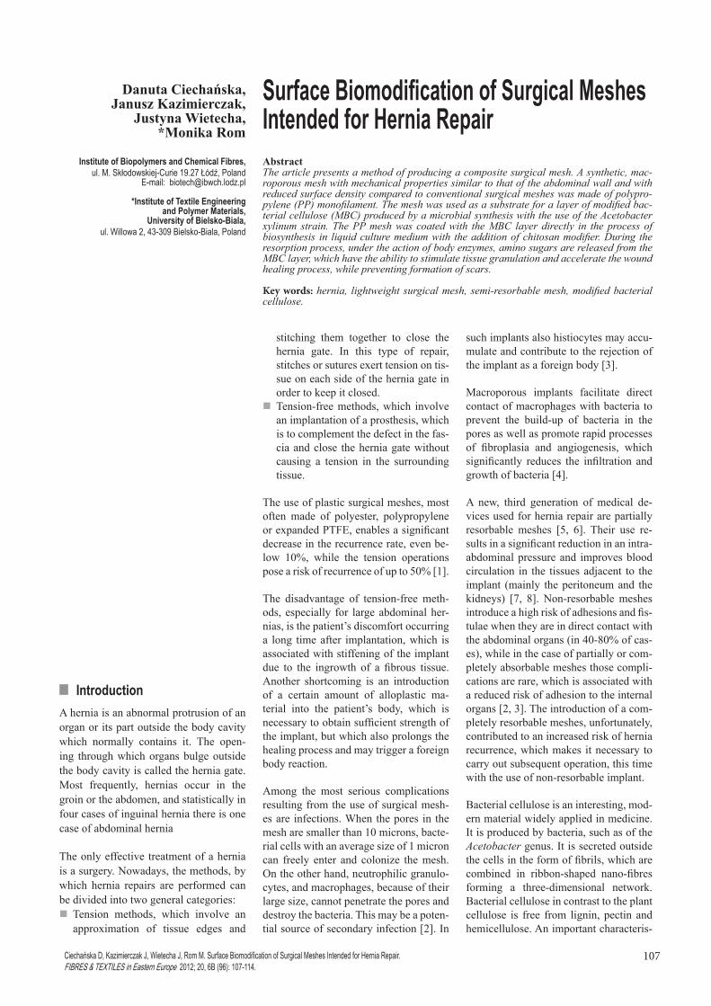
Figure 2 shows differential curves of the molecular weight distribution (MWD) for the following samples: unmodified bac-terial cellulose and MBC modified with chitosan oligomers at a concentration of 0.2 wt% and 6wt% in the culture medi-um. All the curves have similar shapes. A slight reduction in the average molecu-lar weight with the higher concentration of chitosan modifier was observed. The weight-average molecular weight Mw of unmodified bacterial cellulose was approximately 344000 g/mol, while of MBC modified with 6 wt%. oligomers in the culture medium was 305000 g/mol. Differential MWD curves of the MBC samples modified with 0.2 wt% and 6 wt% chitosan oligomers in the culture
Table 4. Influence of chitosan oligomers concentration in the culture medium on hydrolytic and enzymatic degradation of MBC.
Concentration of chitosan oligomers in the culture medium, %
Hydrolytic degradation Enzymatic degradationReleased amino sugars, mg/g MBC
0.2 0 7.70.5 0 9.01.0 0 10.63.0 0 12.36.0 0 13.99.0 0 15.2
Table 5. Molecular characteristic of unmodified bacterial cellulose and MBC modified with chitosan oligomers at a concentration of 0.2 wt% and 6 wt% in the culture medium.
Concentration of chitosan oligomers in the
culture medium, wt%Mn, g/
molMw, g/
molMw /Mn DPw
Percentage of DP fraction, %
DP<200 200<DP<550 DP>550
0 173000 344000 2.0 2.12 1 9 900.2 163000 321000 2.0 1.98 2 10 886.0 161000 305000 1.9 1.88 2 10 88
Figure 2. Molecular weight distribution curves of unmodified bacterial cellulose and MBC modified with chitosan oligomers at a concentration of 0.2 wt% and 6 wt% in the culture medium.
Table 6. Crystallinity degree and crystallite size, defined by WAXS, for unmodified bacterial cellulose and for MBC modified with chitosan oligomers at a concentration of 6wt% in the culture medium.
Sample Crystallinity degree, %
Crystallite size, nm D1 D2
Unmodified bacterial cellulose 74.6 6.15 6.12MBC modified with chitosan oligomers at a concentration of 6wt% in the culture medium 72.7 6.22 6.15
111FIBRES & TEXTILES in Eastern Europe 2012, Vol. 20, No. 6B (96)
medium are slightly shifted in the direc-tion of lower average molecular weights in relation to the unmodified bacterial cellulose. All differential MWD func-tions have a uniform molecular weight distribution, as evidenced by the polydis-persity ratio Mw/Mn of ≅ 2.0.
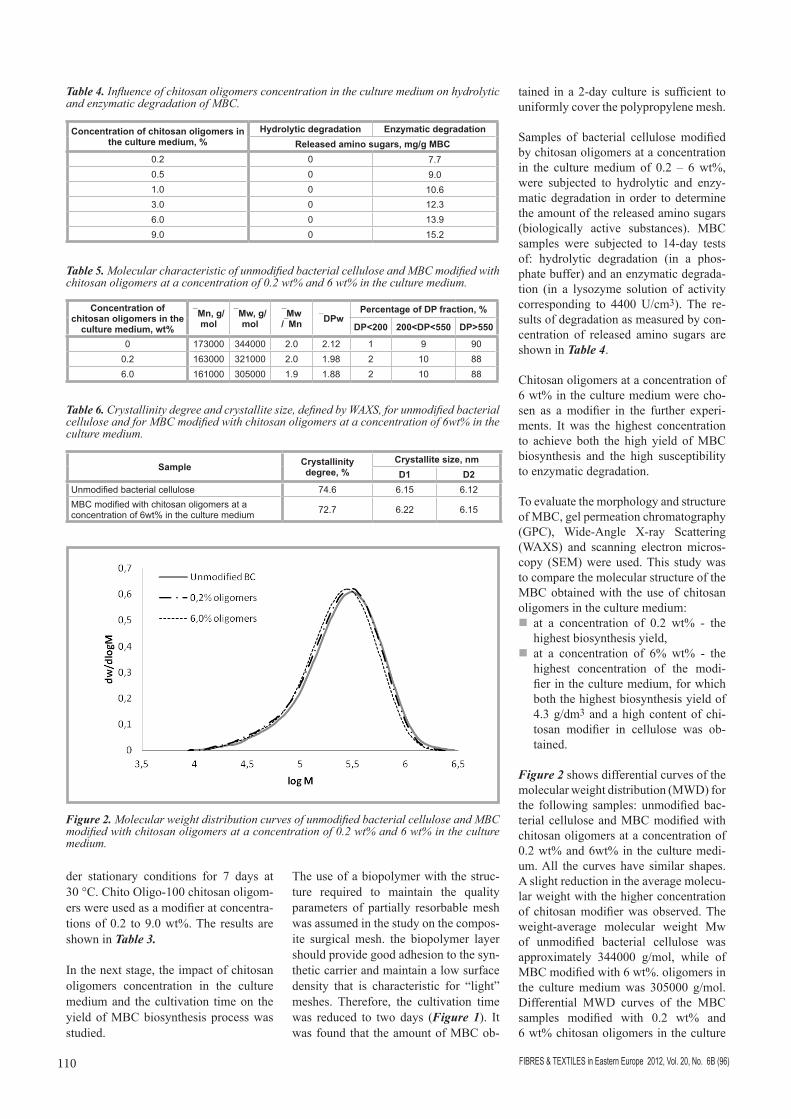
X-ray scattering curve recorded for sam-ples of MBC modified with chitosan oligomers at a concentration of 6 wt% in the culture medium is shown in Fig-ure 3. Calculated values of the degree of crystallinity and crystallite sizes are pre-sented in Table 6.
Diffractograms recorded for samples of unmodified bacterial cellulose and MBC modified with chitosan oligomers at a concentration of 6 wt% in the culture medium show patterns typical for cellu-lose I, with a crystallinity degree above 70%. Diffraction curves are dominated by two strong peaks, whose angle posi-tions are approximately at 15 and 23 de-grees (Figure 3). For all tested samples, these peaks have the greatest intensity of the peaks characteristic for cellulose I. Therefore, the crystallite sizes were de-termined from the analysis of both peaks. In Table 6, they are marked respectively as D1 and D2.
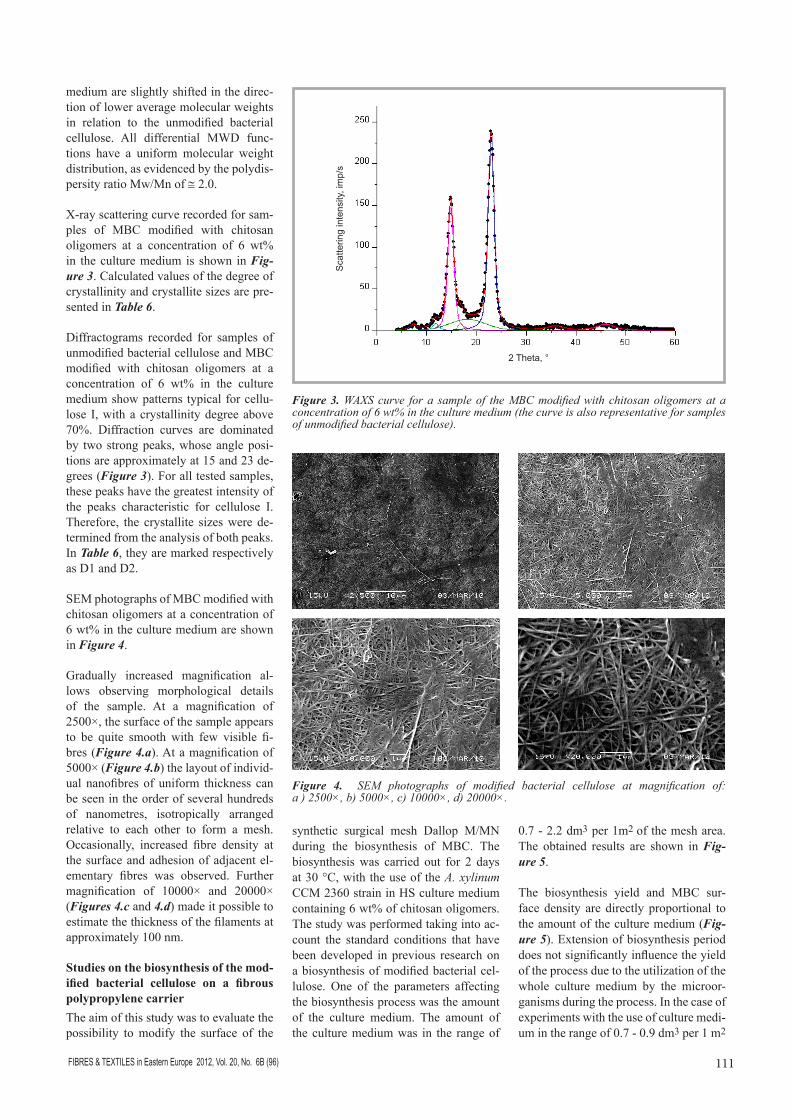
SEM photographs of MBC modified with chitosan oligomers at a concentration of 6 wt% in the culture medium are shown in Figure 4.
Gradually increased magnification al-lows observing morphological details of the sample. At a magnification of 2500×, the surface of the sample appears to be quite smooth with few visible fi-bres (Figure 4.a). At a magnification of 5000× (Figure 4.b) the layout of individ-ual nanofibres of uniform thickness can be seen in the order of several hundreds of nanometres, isotropically arranged relative to each other to form a mesh. Occasionally, increased fibre density at the surface and adhesion of adjacent el-ementary fibres was observed. Further magnification of 10000× and 20000× (Figures 4.c and 4.d) made it possible to estimate the thickness of the filaments at approximately 100 nm.
Studies on the biosynthesis of the mod-ified bacterial cellulose on a fibrous polypropylene carrierThe aim of this study was to evaluate the possibility to modify the surface of the
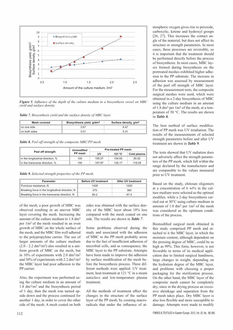
synthetic surgical mesh Dallop M/MN during the biosynthesis of MBC. The biosynthesis was carried out for 2 days at 30 °C, with the use of the A. xylinum CCM 2360 strain in HS culture medium containing 6 wt% of chitosan oligomers. The study was performed taking into ac-count the standard conditions that have been developed in previous research on a biosynthesis of modified bacterial cel-lulose. One of the parameters affecting the biosynthesis process was the amount of the culture medium. The amount of the culture medium was in the range of
0.7 - 2.2 dm3 per 1m2 of the mesh area. The obtained results are shown in Fig-ure 5.
The biosynthesis yield and MBC sur-face density are directly proportional to the amount of the culture medium (Fig-ure 5). Extension of biosynthesis period does not significantly influence the yield of the process due to the utilization of the whole culture medium by the microor-ganisms during the process. In the case of experiments with the use of culture medi-um in the range of 0.7 - 0.9 dm3 per 1 m2
Figure 3. WAXS curve for a sample of the MBC modified with chitosan oligomers at a concentration of 6 wt% in the culture medium (the curve is also representative for samples of unmodified bacterial cellulose).
Figure 4. SEM photographs of modified bacterial cellulose at magnification of: a ) 2500×, b) 5000×, c) 10000×, d) 20000×.
2 Theta, °
Sca
tterin
g in
tens
ity, i
mp/
s
FIBRES & TEXTILES in Eastern Europe 2012, Vol. 20, No. 6B (96)112
of the mesh, a poor growth of MBC was observed resulting in an uneven MBC layer covering the mesh. Increasing the amount of the culture medium to 1.8 dm3 per 1m2 of the mesh resulted in an even growth of MBC on the whole surface of the mesh, and the MBC film well adhered to the polypropylene carrier. The use of larger amounts of the culture medium (2.0 - 2.2 dm3/m2) also resulted in a uni-form growth of MBC on the mesh, but in 10% of experiments with 2.0 dm3/m2 and 30% of experiments with 2.2 dm3/m2 the MBC layer had poor adhesion to the PP carrier.
Also, the experiment was performed us-ing the culture medium in an amount of 1.8 dm3/m2 and the biosynthesis period of 1 day, then the mesh was turned up-side down and the process continued for another 1 day, in order to cover the other side of the mesh. A mesh coated on both
sides was obtained with the surface den-sity of the MBC layer about 18% less compared with the mesh coated on one side. The results are shown in Table 7.
Some problems observed during the study and associated with the adhesion of MBC to the PP mesh probably arose due to the fact of insufficient adhesion of microbial cells, and as consequence, the MBC layer to the PP substrate. Attempts have been made to improve the adhesion by surface modification of the mesh be-fore the biosynthesis process. Three dif-ferent methods were applied: UV treat-ment, heat treatment at 121 °C in a steam autoclave and low-temperature plasma treatment.
All the methods of treatment affect the changes in the structure of the surface layer of the PP mesh, by creating macro-radicals that under the influence of at-
mospheric oxygen gives rise to peroxide, carboxylic, ketone and hydroxyl groups [26, 27]. This increases the contact an-gle of the material, but does not affect its structure or strength parameters. In most cases, these processes are reversible, so it is important that the treatment should be performed directly before the process of biosynthesis. In most cases, MBC lay-ers formed during biosynthesis on the pretreated meshes exhibited higher adhe-sion to the PP substrate. The increase in adhesion was assessed by measurement of the peel off strength of MBC layer. For the measurement tests, the composite surgical meshes were used, which were obtained in a 2-day biosynthesis of MBC using the culture medium in an amount of 1.8 dm3 per 1m2 of the mesh, at a tem-perature of 30 °C. The results are shown in Table 8.
The best method of surface modifica-tion of PP mesh was UV irradiation. The results of the measurements of selected strength parameters before and after UV treatment are shown in Table 9.
The tests showed that UV radiation does not adversely affect the strength parame-ters of the PP mesh, which fall within the range declared by the manufacturer and are comparable to the values measured prior to UV treatment.
Based on the study, chitosan oligomers at a concentration of 6 wt% in the cul-ture medium were selected as the optimal modifier, while a 2-day biosynthesis car-ried out at 30°C using culture medium in amount of 1.8 dm3 per 1m2 of the mesh was considered as the optimum condi-tions of the process.
Biomodified surgical mesh obtained in this study comprised PP mesh and at-tached to it the MBC layer, in which the moisture content, although dependent on the pressing degree of MBC, could be as high as 99%. This form, however, is not favorable in terms of its medical appli-cation due to limited surgical handiness, large changes in weight, depending on the hydration degree of the MBC layer, and problems with choosing a proper packaging for the sterilization process. On the other hand, the MBC layer of the composite mesh cannot be completely dry, since in the drying process an exces-sive shrinkage and separation from the PP mesh takes place. Dry MBC layer is also less flexible and more susceptible to damage. Attempts were made to modify
Figure 5. Influence of the depth of the culture medium in a biosynthesis vessel on MBC yield and surface density.
Table 7. Biosynthesis yield and the surface density of MBC layer.
Mesh covered Biosynthesis yield, g/dm3 Surface density, g/m2
on one side 3.67 6.47on both sides 3.01 5.31
Table 8. Peel off strength of the composite MBC/PP mesh.
Peel off strength Unmodified PP mesh
Pre-treated PP meshUV 121 ºC Cold plasma
in the longitudinal direction, % 100 195.07 154.93 85.92in the transverse direction, % 100 197.87 136.17 118.09
Table 9. Selected strength properties of the PP mesh.
Parameter Before UV treatment After UV treatment Puncture resistance, N 1426 1420Breaking force in the longitudinal direction, N 370 360Breaking force in the transverse direction, N 80.2 87.4
Amount of the culture medium, l/m2
Bio
synt
hesi
s yi
eld,
g/d
m3
Sur
face
den
sity
, g/m
2
0.5 1.0 1.5 2.0 2.5

113FIBRES & TEXTILES in Eastern Europe 2012, Vol. 20, No. 6B (96)
the MBC layer by immersing the com-posite mesh for 24 hours in 10 or 15% glycerol solution and drying at 40 °C. Composite mesh immersed in a 10% glycerol solution remained flexible after drying and showed good surgical handi-ness. The sample immersed in 15% glyc-erol solution showed a much greater sur-face density and gave the feeling of too high a stickiness.
The surface density of composite meshes immersed in 10% glycerol was deter-mined. For the chosen biosynthesis vari-ant (using 1.8 dm3 per 1 m2 of the mesh), the final surface density of the composite mesh was about 170 g/m2. They were also the percentage of the individual components of the composite. The PP substrate accounted for 31.5 wt%, wa-ter for 10 wt% and glycerol for nearly 50 wt% of the total weight of the com-posite mesh.
Mechanical properties and performance of the selected composite surgical mesh were also assessed. On the basis of the re-sults obtained it was found that covering the PP mesh with an MBC layer does not significantly change the implant strength parameters. Despite the introduction of MBC material, elasticity of the compos-ite mesh did not decrease and allowed proper fit to hernia repair. Elasticity of the meshes was evaluated by measuring their flexural rigidity, which, in the lon-gitudinal direction was 0.052 mN/m for PP mesh, and 0.096 mN/m for composite mesh, while in the transverse direction it was identical for both types of meshes and amounted to 0.048 mN/m
Figures 6 - 9 show SEM photographs of the composite MBC/PP surgical mesh.
General view (Figure 6) shows a frag-ment of polypropylene knitted mesh coated with an MBC layer. In the places where stitches of PP monofilament inter-lace (Figure 7), a minor discontinuity of the MBC layer was observed. Apart from these points the MBC layer covers both the PP mesh surface and the spaces be-tween the PP filaments. In Figures 8 & 9 elementary nanofibres of modified bacte-rial cellulose can be seen.
n Conclusions1. A method was developed for the bio-
synthesis of modified bacterial cellu-lose susceptible to partial enzymatic degradation by lysozyme - an en-zyme found in body fluids. Based on the study, as a modifier with the best ability to degrade, chitosan oligom-ers were selected in a concentration of 60 g per 1 dm3 of HS culture medium.
2. An improved degradation of modi-fied bacterial cellulose obtained was possible by increasing the content of chitosan oligomers and due to a lower crystallinity and polymerization de-gree, compared to the unmodified bac-terial cellulose or MBC synthesized in a medium with a lower content of oli-gomers.
3. A method for bio-modification of PP surgical mesh with a layer of MBC di-rectly in the biosynthesis process was
developed. One- or two-side coated composite surgical meshes were ob-tained with different surface density depending on the amount of the cul-ture medium used.
4 In order to maintain sufficient elas-ticity of the MBC layer and func-tional properties of surgical meshes obtained, they were immersed in 10 wt% glycerol and dried at 40 °C for 4 hours. Surface density of the com-posite surgical mesh obtained in se-lected conditions was approximately 170 g/m2 (including MBC layer, wa-ter and glycerol). Studies on steriliza-tion methods, biocompatibility and biomedical research of developed
Figure 6. View of a stitch of the PP monofilament (100×) coated with MBC layer; A) - boundary area exposed by sample cutting, B) MBC layer covering the mesh.
A) B)
Figure 9. View of the ‘B’ area of MBC layer. a) magnification 5000×, b) magnification 10000×.
Figure 7. View of composite mesh (100×). Around interlacing PP monofilaments a discontinuity of the MBC layer is visible.
a) b)
Figure 8. View of the ‘A’ area of MBC layer; a) magnification 5000×, b) magnification 10000×.
a) b)
FIBRES & TEXTILES in Eastern Europe 2012, Vol. 20, No. 6B (96)114
composite hernia meshes shall be the subject of another paper.
AcknowledgmentThe study was carried out under the re-search and development project R 05 015 03 “Development of partially resorbable sur-gical meshes for hernia repair”.
References1. Trojanowski P, Witczak W, Najdecki M,
Stanowski E. Własne doświadczenia w leczeniu olbrzymich przepuklin poop-eracyjnych przy użyciu siatek dootrze-wnowych, Pol Merk Lek 2007; XXII, 131: 376-378 (in Polish).
2. Amid P.K. Clasification of biomateri-als and their related complications in abdominal wall hernia surgery. Hernia 1997; 1: 15-21.
3. Józefowicz M, Teresiński L, Biskup-ski A. Reakcja tkankowa na obecność niewchłanialnych siatek syntetycznych wszczepionych w ścianę brzucha i jej znaczenie w chirurgii przepuklin br-zusznych, www.hernia.pl (in Polish).
4. Cobb WS, Kercher KW, Heniford BT., The argument for lightweight polypropyl-ene mesh in hernia repair, Surg Innov. 2005 Mar; 12(1): 63-69.
5. Niekraszewicz A., Kucharska M., Wawro D., Struszczyk M., Kopias K., Rogacze-wska A., Development of a Manufactur-ing Method for Surgical Meshes Modified by Chitosan, Fibres and Textiles in East-ern Europe 2007; 15, 3(62): 105-108.
6. Niekraszewicz A, Kucharska M, Struszc-zyk MH, Rogaczewska A, Struszczyk K. Investigation into biological, composite surgical meshes, Fibres and Textiles in Eastern Europe 2008; 16, 6 (71): 117-121.
7. Saxe JM, Ledgerwood AM, Lucas CE. Management of the difficult abdominal closure. Surg. Clin. North Am., 1993; 73 (2): 243-251.
8. Gentile AT, Feliciano PD, Mullins RJ, Crass RA, Eidemiller LR, Sheppard BC. The utility of polyglycolic acid mesh for abdominal access in patients with ne-crotizing pancreatitis, J. Am. Coll. Surg., 1998; 196 (3): 313-318.
9. Jonas R, Farah LF. Production and ap-plication of microbial cellulose. Polym. Degrad. Stab., 1998; 59: 101-106.
10. Ciechańska D, Struszczyk H, Kazimierc-zak J, Guzińska K, Pawlak, Kozłowska E, Matusiak G, Dutkiewicz M. New Electro-Acoustic Transducers Based on Modified Bacterial Cellulose. Fibres & Textiles in Eastern Europe 2002; 10, 1 (36): 27-30.
11. Johnson DC, Winslow AR. Bacterial cel-lulose has potential application as new paper coating. Pulp. Pap. 1990; 64 (5): 105-107.
12. Kent RA, Stephens RS, Westland JA. Bacterial cellulose fiber provides an
alternative for thickening and coating. Food Technol 1991; 45: 108-111.
13. Salata LA, Craig GT, Brook IM. In-vivo evaluation of a new membrane (Gengi-flex) for guided bone regeneration. Di-visional abstracts: The British Society of Dental Research. J Dent Res 1995; 74(3): 825.
14. Rebello C, Almeida DAD. Lima EM Jr, Dornelas MDP. Biofill a new skin substi-tute, our experience. Rev Bras Cir 1987; 77(6).
15. Fontana JD, de Souza AM, Fontana CK, Torriani IL, Moreschi JC, Gallotti BJ. Acetobacter cellulose pellicle as a temporary skin substitute. Appl Biochem Biotechnol 1990; 24–25: 253–264.
16. Bielecki S, Kołodziejczyk M, Kowal-ska K, Krystynowicz A, Pankiewicz T. A method of production of a cartilage-like biomaterial designed for reconstruc-tive surgery, EP 2371401 A2.
17. Ciechańska D. Multifunctional Bacterial Cellulose/Chitosan Composite Materials for Medical Applications. Fibres & Tex-tiles in Eastern Europe 2004; 12, 4 (48): 69-72.
18. Ciechańska D, Wietecha J, Kaźmierczak D, Kazimierczak J. Biosynthesis of Mod-ified Bacterial Cellulose in a Tubular Form. Fibres & Textiles in Eastern Eu-rope 2010; 18, 5 (82): 98-104.
19. Helenius G, Bäckdahl H, Bodin A, Nann-mark U, Gatenholm P, Risberg B. In vivo biocompatibility of bacterial cellulose. J Biomed Mater Res 2006; 76A: 431–438.
20. Lee JW, Deng F, Yeomans WG, Al-len AL, Gross RA, Kaplan DL. Direct Incorporation of Glucosamine and N-Acetylglucosamine into Exopolymers by Gluconacetobacter xylinus (=Acetobac-ter xylinum) ATCC 10245: Production of Chitosan-Cellulose and Chitin-Cellulose Exopolymers. Applied and Environmen-tal Microbiology, Sept. 2001; 3970–3975.
21. Hestrin S, Schramm M. Synthesis of cel-lulose by Acetobacter xylinum. Biochem J 1954; 58: 345-352.
22. Miller GL, Analytical Chem 1959; 31: 426.
23. Turbak AF, El-Kafrawy A, Snyder FW, Auerbach AB. Solvent system for cellu-lose, Patent US 4,302,252, 1981.
24. Turbak AF. Newer Cellulose Solvent Systems. In: Wood and Agricultural Residues; Soltes E. J., Ed.; Academic Press: New York, 1983, pp. 87-99.
25. Ekmanis JL. Am Lab News, Jan/Feb 1987; pp. 10-11.
26. Bryjak M, Janecki T, Gancarz I, Smolińska K. Plazmowa modyfikacja membran polimerowych, Membrany. Teoria i Praktyka (in Polish) ed. R. Wódzki, Zeszyt III, Uniwersytet Mikołaja Kopernika, Toruń 2009.
27. Brzozowska-Stanuch A, Rabiej S, Sta-nuch W. Wpływ warunków przyspieszo-nego starzenia-promieniowania UV i temperatury na poliamidy i polipropylen, (in Polish) Mechanika. Czasopismo techniczne, 1-M/2009; 106, 3: Wyd. Po-litechniki Krakowskiej.
Received 04.05.2010 Reviewed 17.12.2012
U N I V E R S I T Y OF BIELSKO-BIAŁA
Faculty of Textile Engineering and Environmental Protection
The Faculty was founded in 1969 as
the Faculty of Textile Engineering of the
Technical University of Łódź, Branch in
Bielsko-Biała. It offers several courses
for a Bachelor of Science degree and a
Master of Science degree in the field of
Textile Engineering and Environmental
Engineering and Protection.
The Faculty considers modern trends in
science and technology as well as the
current needs of regional and national
industries. At present, the Faculty con-
sists of:
g The Institute of Textile Engineering and
Polymer Materials, divided into the following
Departments:
g Polymer Materials
g Physics and Structural Research
g Textile Engineering and Commodity
g Applied Informatics
g The Institute of Engineering and Environmental
Protection, divided into the following
Departments:
g Biology and Environmental Chemistry
g Hydrology and Water Engineering
g Ecology and Applied Microbiology
g Sustainable Development
g Processes and Environmental Technology
g Air Pollution Control
University of Bielsko-BiałaFaculty of Textile Engineering and Environmental Protection
ul. Willowa 2, 43-309 Bielsko-Białatel. +48 33 8279 114, fax. +48 33 8279 100
E-mail: [email protected]