Embed Size (px)
Citation preview
The Abdomen
Surface Anatomy, Vessels, Muscles, and Peritoneum

Surface Anatomy
• Anterior abdominal wall extends from costal margin to inferior boundaries: – Iliac crest – Anterior superior iliac spine – Inguinal ligament – Pubic crest
• Superior boundary – Diaphragm
• Central landmark – Umbilicus
• Linea alba (white line) – Tendinous line – Extends from xiphoid process
to pubic symphysis

Abdominal Quadrants
• 9 regions
• 4 quadrants
– Draw “line” through navel
– Right upper quadrant
– Left upper quadrant
– Left lower quadrant
– Right lower quadrant

Muscles • Function:
– Help contain abdominal organs – Move trunk – Forced breathing – Increase intraabdominal pressure
• Abdominal wall – Anterior (4)
• Innervated by intercostal nerves • Continuous with layers of intercostal muscles • Fibers of layers run in different directions for strength • Ends in aponeurosis which contains rectus abdominis muscle
– Posterior (3)


Anterior Abdominal Wall Muscles
Rectus Abdominis • Origin
– Pubic crest, symphysis
• Insertion
– Xiphoid process, costal cartilages of ribs 5-7
• Function
– Flex, rotate trunk, fix and depress ribs, stabilize pelvis, compress abdomen

Anterior Abdominal Wall
• External oblique (“hands-in-pocket”) – Origin
• Lower 8 ribs
– Insertion
• Aponeurosis to linea alba, pubic and iliac crest
– Function
• Flex trunk, compress abdominal wall (together), rotate trunk (separate sides)
• Internal oblique
– Origin
• Lumbar fascia, iliac crest, inguinal ligament
– Insertion
• Linea alba, pubic crest, last 3-4 ribs, costal margin
– Function
• Same for external obliques

• Transversus abdominis – Origin
• Inguinal ligament, lumbar fascia, cartilage of last 6 ribs, iliac crest
– Insertion • Linea alba, pubic crest
– Function • Compress abdominal contents
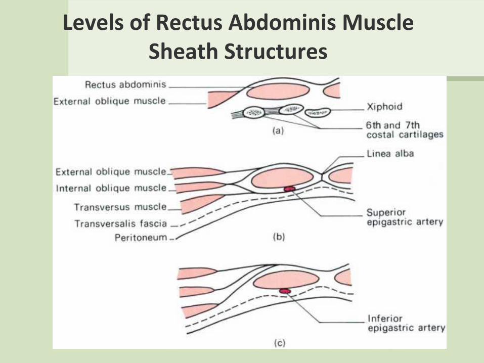
Levels of Rectus Abdominis Muscle Sheath Structures

ANTERIOR ABDOMINAL WALL LINEA ALBA
• Cord of connective tissue
• Extend – sternum (xyphoid process) –symphysis, pubic bones
• Aponeurotic parts of oblique muscles attache to the linea alba at the midline
• One of the surgical approaches to the peritoneal cavity (midline incision)

Weak Places of the Anterior Abdominal Wall

Layer Structure of the Anterior Abdominal Wall

Abdominal Incisions
• Must locate nearest to the organ.
• Must have sufficient length for surgeon activities.
• Must be atraumatic.

Skin Incisions of the Ventral Abdominal Wall

Male Inguinal Canal

Female Inguinal Canal

Superficial Inguinal Ring

Internal Inguinal Ring

Anterior Abdominal Wall (lower internal view)

Posterior Abdominal Wall Iliopsoas
• Psoas major – Origin
• Lumbar vertebrae, T12
– Insertion • Lesser trochanter of femur via iliopsoas tendon
– Function • Thigh flexion, trunk flexion, lateral flexion
– Innervation • Ventral rami L1-L3
• Iliacus – Origin
• Iliac fossa, ala of sacrum
– Insertion • Lesser trochanter of femur via iliopsoas tendon
– Function • Thigh flexion, trunk flexion
– Innervation • Femoral nerve (L2 and L3)
• Psoas minor – variable (40-60% do not have)

Posterior Abdominal Wall
• Quadratus lumborum – Origin
• Iliac crest and lumbar fascia
– Insertion • Transverse process of upper
lumbar vertebrae, lower margin of rib 12
– Function • Flex vertebral column,
maintains upright posture, assists in inspiration
– Innervation: • T12 and upper lumbar spinal
nerves (ventral rami)

Abdominopelvic Cavity
• Ventral body cavity – Thoracic
– Abdominopelvic
• Abdominopelvic – Abdominal
• Liver
• Stomach
• Kidneys
– Pelvic cavity
• Bladder
• Some reproductive organs
• Rectum

Abdominal cavity
The space bounded by:
• Anterolateral abdominal wall
• Posterior abdominal wall
• Diaphragm
• Pelvic walls and pelvic floor.
Subdivided into:
• True abdominal cavity (from
diaphragm to linea terminalis)
• Pelvic cavity (below linea terminalis).
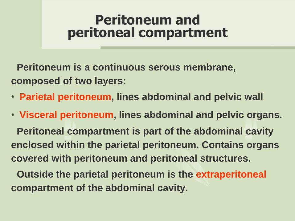
Peritoneum and peritoneal compartment
Peritoneum is a continuous serous membrane,
composed of two layers:
• Parietal peritoneum, lines abdominal and pelvic wall
• Visceral peritoneum, lines abdominal and pelvic organs.
Peritoneal compartment is part of the abdominal cavity
enclosed within the parietal peritoneum. Contains organs
covered with peritoneum and peritoneal structures.
Outside the parietal peritoneum is the extraperitoneal
compartment of the abdominal cavity.

Peritoneal cavity
Peritoneal cavity (PC) - the space between the two peritoneal layers, is a potential space, into which the organs are tightly packed against each other.
•PC contains thin layer of fluid, which lubricates the peritoneal surfaces and allows movement of the organs without friction.
•PC is closed in males, but communicates with the external environment in females through the uterine tubes, uterus and vagina.
•Peritoneum, peritoneal cavity and all the organs are situated in the abdominal cavity.
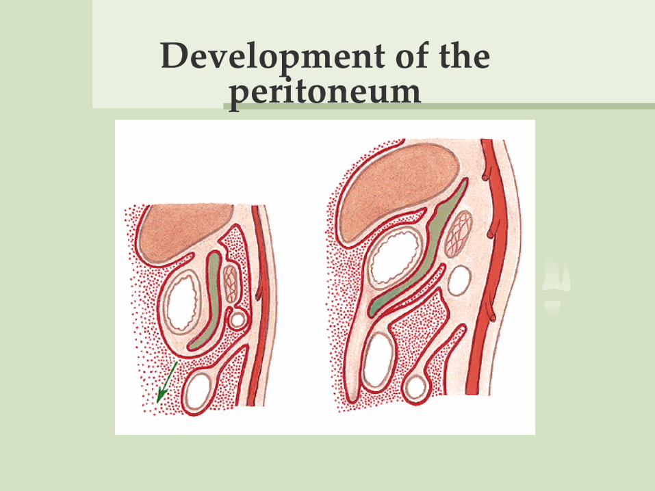
Development of the peritoneum

Relationship between the organs and peritoneum
Due to intraembryonal processes the organs have different
relationship with the peritoneum.
1. Intraperitoneal organs are entirely covered with peritoneum.
They are connected to the abdominal wall with ligaments or
meso, which ensures greater mobility.
2. Extraperitoneal organs are partially or entirely devoid of
peritoneum. They are slightly movable or immovable. According
to their position these are:
а) retroperitoneal – on the posterior abdominal wall
b) subperitoneal – in the lesser pelvis
c) preperitoneal – at the anterior abdominal wall.

Vertical layout of the peritoneum

Horizontal layout of the peritoneum

Passage of the parietal into visceral peritoneum

Peritoneal structures
1. Mesentery – double peritoneal layer, representing elongation
of the visceral peritoneum.
•М. connects the corresponding organ with the abdominal wall
(e.g., mesentery of the small intestine).
•М. contains connective tissue in which are embedded blood
vessels, nerves and lymph nodes.
•М. ensures mobility of the organs.

2. Omentum – double layered structure of
visceral peritoneal, extending from the
stomach to neighbouring organs.
• Lesser omentum (оmentum minus) connects
the lesser curvature of the stomach and intitial
portion of pars superior duodeni with liver.
• Greater omentum (оmentum majus)
descends from the greater curvature of the
stomach and intitial portion of pars superior
duodeni, covers the intestines, and then
ascends back to attache to the transverse colon.
Contains great amount of fat tissue.

3. Peritoneal ligaments – double layered
structures of visceral peritoneum, between
neighbouring organs or between organ and
abdominal wall (e.g., lig. falciforme, lig.
gastrophrenicum, lig. gastrolienale, lig.
gastrocolicum).
4. Peritoneal folds (plicae) formed over
underlying structures (e.g., plica iliocecalis
superior, plica umbilicalis mediana).
5. Peritoneal recessuses – spaces in the peritoneal
cavity заградени between peritoneal structures
and abdominal organs or abdominal wall (e.g.,
bursa omentalis, recessus subphrenicus, fossa
retrocecalis).

Divisions of the peritoneal cavity
By mesocolon transversum the peritoneal
compartment divites into:
1. Supracolic compartment – between diaphragm and
mesocolon transversum with its mesentery.
2. Infracolic compartment - between mesocolon
transversum and linea terminalis.
3. Pelvic compartment - below linea terminalis in the
pelvi cavity.

Supracolic compartment
Organs:
1. Esophagus, pars abdominalis - intraperitoneal
2. Stomach - intraperitoneal
3. Liver - intraperitoneal
4. Gall bladder - intraperitoneal
5. Spleen - intraperitoneal

Supracolic compartment. Projections of organs

Supracolic compartment
Peritoneal structures: 1. Lig. falciforme hepatis
– lig. teres hepatis
2. Lig. coronarium hepatis (dextum et sinistrum) – area nuda
3. Lig. triangulare (dextum et sinistrum)

Supracolic compartment
4. Omentum minus
– lig. hepatogastricum
– lig. hepatoduodenale
5. Omentum majus – lig. gastrocolicum
– lig. gastrolienale
– lig. gastrophrenicum
6. Lig. phrenicolienale

Supracolic compartment
Peritoneal spaces:
1. Recessus subphrenicus dexter - bursa hepatica
2. Recessus subphrenicus sinister - bursa pregastrica
3. Perilienal space
4. Recessus subhepaticus
а) anterior part
b) posterior part - recessus hepatorenalis
5. Bursa omentalis

Supracolic compartment
Bursa omentalis. Opened thru lig. gastrocolicum
Bursa omentalis. Opened thru lig. hepatogastricum
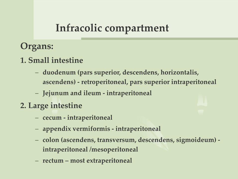
Infracolic compartment
Organs:
1. Small intestine
– duodenum (pars superior, descendens, horizontalis,
ascendens) - retroperitoneal, pars superior intraperitoneal
– Jejunum and ileum - intraperitoneal
2. Large intestine
– cecum - intraperitoneal
– appendix vermiformis - intraperitoneal
– colon (ascendens, transversum, descendens, sigmoideum) -
intraperitoneal /mesoperitoneal
– rectum – most extraperitoneal

Organs and projections

Peritoneal structures
1. Omentum majus - pars libera
2. Mesenterium
3. Mesocolon transversum
4. Mesocolon sigmoideum
5. Mesoappendix

Peritoneal structures
1. Plicae duodenalis
superior/inferior
- recessus duodenalis
superior/inferior
2. Plicae ileocecalis
superior/inferior
- recessus ileocecalis
superior/inferior

Peritoneal spaces
1. Canalis lateralis dexter
2. Sinus mesentericus dexter
3. Sinus mesentericus sinister
4. Canalis lateralis sinister
5. Recessus intersigmoideus
6. Recessus retrocecalis

Appendix vermiformis

Supracolic compartment. Blood
supply
Truncus celiacus
1. A. gastrica sisnistra - r. esophageus
2. A. hepatica communis - a. hepatica propria
- a. hepatica dextra/sinistra
- a. gastroduodenalis - a. gastroepiploica dextra
- aa. pancreaticoduodenales superiores (anterior/posterior)
- a. gastrica dextra
3. A. lienalis - aa. gastricae breves
- a. gastroepiploica sinistra

Supracolic compartment. Blood supply

Arteriogram of truncus celiacus

Infracolic compartment. Blood supply
A. mesenterica superior
1. A. pancreaticoduodenalis
inferior
2. Aa. intestinales (15-18)
3. A. iliocolica
4. A. colica dextra
5. A. colica media

Infracolic compartment. Blood supply
A. mesenterica inferior
1. A. colica sinistra
2. Aa. sigmoideae (3-4)
3. A. rectalis superior