Embed Size (px)
Citation preview
CASE REPORT
Supratentorial extraventricular anaplastic ependymomain an adult with repeated intratumoral hemorrhage
Naotaka Iwamoto • Yasuo Murai • Yoichiro Yamamoto •
Koji Adachi • Akira Teramoto
Received: 18 September 2012 / Accepted: 13 March 2013 / Published online: 2 April 2013
� The Author(s) 2013. This article is published with open access at Springerlink.com
Abstract We report the case of a 61-year-old man with
supratentorial extraventricular anaplastic ependymoma
who presented with repeated intratumoral hemorrhage. The
patient was admitted with headache. Computed tomogra-
phy and magnetic resonance imaging showed an enhancing
mass with intratumoral hemorrhage in the right temporal
lobe. Gross total resection was performed. The tumor was
well demarcated from the brain tissue, and showed no
continuity with the ventricular system. Histopathological
examination revealed the features of anaplastic ependy-
moma. Therefore, additional radiation therapy and adju-
vant chemotherapy were administered. Ten months later,
the tumor recurred with hemorrhage in the spinal canal.
This case showed rapid malignant progression and repeated
intratumoral hemorrhage within a short period of time, both
of which are characteristics of anaplastic ependymomas.
Close observation of the central nervous system and
adjuvant radiotherapy are mandatory, even if the ependy-
moma presents with repeated intratumoral hemorrhage.
Keywords Anaplastic ependymoma � Supratentorial
ependymoma � Hemorrhage
Introduction
Ependymomas are primary neoplasms of the central ner-
vous system (CNS) that account for about 3–5 % of all
adult intracranial gliomas [1, 2]. Ependymomas usually
arise from the cells lining the ventricular system and cen-
tral canal in the spinal cord [3–6]. In a minority of cases,
ependymomas arise from the supratentorial parenchyma
and show no continuity with the ventricular system. These
ependymoma variants are called ectopic, cortical, lobar, or
extraventricular ependymomas. Only a few such cases have
been reported in the literature [7–15]. In most of these
cases, the tumors were difficult to diagnose before surgery.
We present a patient with a supratentorial extraventricular
anaplastic ependymoma who presented with repeated
intratumoral hemorrhage in the brain and spine.
Case report
A 61-year-old man presented with severe headache on
November 30, 2008. A more detailed history revealed that
he had been suffering from severe headache of acute onset
from 3 days beforehand. Head computed tomography (CT)
demonstrated a high-density lesion in the right temporal
lobe (Fig. 1a) with a mean diameter of 40 mm. On the
magnetic resonance imaging (MRI) performed on
December 9, 2008, the lesion was visualized as mixed
intensity on T1- and T2-weighted images, and showed
strong enhancement following intravenous administration
of gadolinium diethylenetriaminepentaacetic acid (Fig. 1b–d).
The lesion was surrounded by perifocal cerebral edema.
Based on these findings, hemorrhage in the brain tumor was
suspected. Cerebral angiography showed that the tumor was
supplied by both the internal and external carotid arteries
N. Iwamoto (&) � Y. Murai � K. Adachi � A. Teramoto
Department of Neurosurgery, Nippon Medical School,
1-1-5 Bunkyo-ku Sendagi, Tokyo 113-8602, Japan
e-mail: [email protected]
Y. Yamamoto
Department of Diagnostic Pathology, Nippon Medical School,
1-1-5 Bunkyo-ku Sendagi, Tokyo 113-8602, Japan
123
Brain Tumor Pathol (2014) 31:138–143
DOI 10.1007/s10014-013-0146-0
(Fig. 2a, b). Neurological examination did not reveal any
neurological deficit during this admission. We planned the
operation for January 9, 2009, and the patient was discharged
from the hospital.
The patient presented to the hospital again with severe
headache on December 26, 2008. Repeat CT revealed
another high-density area within the tumor and more
extensive peripheral edema, the findings suggestive of
recurrence of the intratumoral hemorrhage (Fig. 3a). The
patient was treated conservatively until the operation.
During the preoperative period, the patient developed
consciousness disturbance. Follow-up CT scans obtained
after admission demonstrated another recurrence of the
intratumoral hemorrhage (Fig. 3b–d). On January 7, 2009,
the patient fell into a coma, and emergent right temporal
craniotomy was performed. Intraoperative findings con-
firmed that the tumor was attached to the dural membrane
of the middle fossa, showing no attachment to the
ventricular system. The tumor was clearly demarcated from
the surrounding brain tissue and gross total resection was
performed.
Histological examination revealed that the lesion was
very cellular and well vascularized. Many blood vessels,
hemorrhages, and vascular proliferation were seen, but
pseudopalisading necrosis was not seen in the specimen
(Fig. 4a, b). The nuclei were polymorphic; there were some
mitotic figures and numerous perivascular pseudo-rosette
formations (Fig. 4c). Immunohistochemical study revealed
positive staining of the tumor cells for glial fibrillary acidic
protein (GFAP) (Fig. 4d), epithelial membrane antigen
(EMA) (Fig. 4e), S-100 protein, and vimentin. However,
the tumor showed negative staining for CD34 and bcl2.
The MIB-1 labeling index was 10–30 %. The pathological
diagnosis was anaplastic ependymoma.
Postoperative MRI demonstrated gross total resection
(data not shown). After surgery, focal radiation therapy
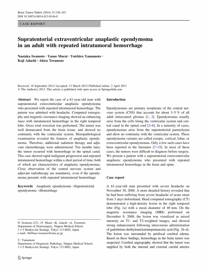
Fig. 1 Preoperative imaging
studies. a Axial-plane CT scan
showing a high-density lesion in
the right temporal lobe. b Axial
T1-weighted MR image
demonstrating the tumor with
heterogeneous intensity located
intraaxially and in the
extraventricular space. c Axial
contrast-enhanced T1-weighted
MR image demonstrating a
strongly enhancing mass.
d Coronal contrast-enhanced
T1-weighted MR image
demonstrating a strongly
enhancing mass. The tumor
occupies the right temporal lobe
Brain Tumor Pathol (2014) 31:138–143 139
123

Fig. 2 Lateral view of the right
internal carotid artery cerebral
angiogram (a) and lateral view
of the right external carotid
cerebral angiogram
(b) performed on December 9,
2008. Early venous filling and
tumor staining are observed
Fig. 3 Computed tomography
scans obtained on December 26,
2008 (a), December 31, 2008
(b), January 3, 2009 (c), and
January 7, 2009 (d). A high-
density area can be seen in the
tumor that gradually expands
(arrow head). This indicates
repeated intratumoral
hemorrhage. The perilesional
brain edema and displacement
of the midline structures
deteriorated
140 Brain Tumor Pathol (2014) 31:138–143
123
(60 Gy) and chemotherapy (temozolomide) were admin-
istered. The patient showed no neurological deficit after the
treatment, and was discharged.
The patient was admitted again with back pain and gait
disturbance on November 8, 2009. MRI of the thoracic
spine demonstrated a tumor with hematoma in the spinal
canal (Fig. 5). A second operation was performed, and
histopathological examination revealed recurrence and
dissemination of the anaplastic ependymoma.
Discussion
Ependymomas usually arise from the cells lining the ven-
tricular system and central canal of the spinal cord [3–6].
The clinical courses of patients with intracranial
ependymomas can be quite variable [16]. Supratentorial
ependymomas in adults are rare CNS tumors that continue
to generate considerable controversy with regard to their
clinical management [17]. Several negative prognostic
parameters have been identified, such as young age,
incomplete tumor resection, histological anaplasia, and
supratentorial localization [9, 18, 19]. To the best of our
knowledge, only 9 case reports of supratentorial extra-
ventricular anaplastic ependymoma, including our present
case, have been reported in the literature (Table 1). The
mean age of the 9 patients was 40 years, and the male-to-
female ratio was 5:4. The tumor was located in the frontal
lobe in 3 cases, the parietal lobe in 1 case, the temporal
lobe in 2 cases, the temporoparietal lobe in 2 cases, and the
parietooccipital lobe in 1 case. In 6 cases, the tumors were
contiguous with the brain surface as cortical ependymoma.
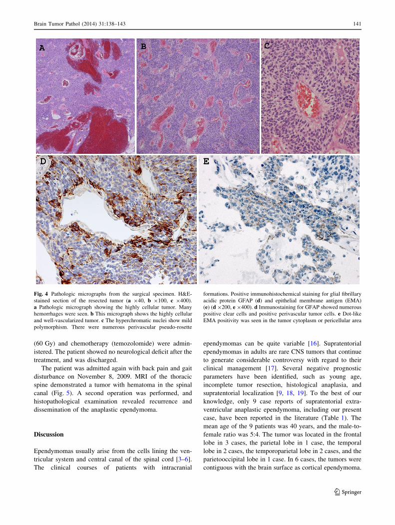
Fig. 4 Pathologic micrographs from the surgical specimen. H&E-
stained section of the resected tumor (a 940, b 9100, c 9400).
a Pathologic micrograph showing the highly cellular tumor. Many
hemorrhages were seen. b This micrograph shows the highly cellular
and well-vascularized tumor. c The hyperchromatic nuclei show mild
polymorphism. There were numerous perivascular pseudo-rosette
formations. Positive immunohistochemical staining for glial fibrillary
acidic protein GFAP (d) and epithelial membrane antigen (EMA)
(e) (d 9200, e 9400). d Immunostaining for GFAP showed numerous
positive clear cells and positive perivascular tumor cells. e Dot-like
EMA positivity was seen in the tumor cytoplasm or pericellular area
Brain Tumor Pathol (2014) 31:138–143 141
123

In 5 of these cases, intratumoral hemorrhage was observed.
Hemorrhage was observed in 4 cases of cortical
ependymoma.
The case we have reported here presented with at least 5
episodes of intratumoral hemorrhage over a period of
40 days. Only one previously reported case of supratento-
rial extraventricular ependymoma presented with repeated
intratumoral hemorrhage [20]. In that case, 3 episodes of
hemorrhage occurred over a period of 2 years. To the best
of our knowledge, none of the previously reported cases
had repeated intratumoral hemorrhage that occurred within
a period as short as that seen in our patient. Intratumoral
hemorrhage in supratentorial ependymomas is usually
considered a rare event [20, 21], although Romero et al.
[22] mentioned that intratumoral hemorrhage is not rare in
this tumor. The pathological finding in all of these cases
was anaplastic ependymoma. Hemorrhage caused by
intracranial neoplasm is usually associated with high-grade
malignancy and extensive, abnormal vascularization [23].
Kojima et al. [24] reported that the hemorrhage in the
tumor reflects the malignancy grade of the tumor. Ernestus
et al. [25] also mentioned that the factor that predisposes
the most for bleeding seems to be extensive and abnormal
vascularity, and endothelial proliferation or dilated thin-
walled vessels were common findings in ependymal tumors
with spontaneous hemorrhages. In our case, the histological
findings were compatible.
In our present case, the tumor recurred in the spine
between the lower thoracic and upper lumbar spinal cord,
showing both intratumoral and extratumoral hemorrhage.
Fig. 5 MRI of the thoracic spine. Sagittal T1-weighted MR image
(a), T2-weighted MR image (b), and T1-weighted MR image with
contrast enhancement (c). a Fluid–fluid level was seen in the dorsal
side of the spinal cord as a slightly high intensity signal (arrows).
b Fluid–fluid level was seen in the dorsal side of the spinal cord as a
low-intensity signal (arrows). c An intradural extramedullary enhanc-
ing mass was seen after intravenous administration of gadolinium
diethylenetriaminepentaacetic acid (arrow heads). This indicated
drop metastases (arrow heads) with hemorrhage (arrows)
Table 1 Summary of 9 cases of supratentorial extraventricular anaplastic ependymoma
Case no. Author (year) Age/sex Location of ependymoma Hemorrhage Staining on angiography Recurrence
1 Takeshima (2002) 70/F Frontal ? No study –
2 Kojima (2003) 56/F Temporoparietal ? No study Residual lesion
3 Moritani (2003) 50/F Temporal – Hypovascular Initial location
4 Miyazawa (2007) 32/M Parietal ? ? Initial location
5 Toba (2009) 36/F Frontal – No study –
6 Toba (2009) 18/M Temporoparietal ? No study Initial location, spine
7 Eika (2010) 15/M Parietooccipital – No study –
8 Flavio (2012) 23/M Frontal – No study –
9 Present case 61/M Temporal ? ? Spine
142 Brain Tumor Pathol (2014) 31:138–143
123

According to the previous literature, anaplastic ependy-
momas are characterized by a higher proliferative rate and
a greater tendency to disseminate into the cerebrospinal
fluid, causing drop metastases. Saito et al. [26] mentioned
that anaplastic ependymoma disseminated within the cen-
tral nervous system without local failure. Our case showed
a similar course. Cerebrospinal fluid dissemination of
anaplastic ependymoma has been reported to be one of the
factors that determine end-of-life prognosis [19, 26, 27].
In conclusion, meticulous MRI follow-up of the CNS is
mandatory in adult patients with intracranial anaplastic
ependymomas, even after gross total removal of the tumor.
Thus, our experience of this case indicates that supra-
tentorial extraventricular ependymoma with repeated in-
tratumoral hemorrhage should lead to a suspicion of an
anaplastic tumor histology. Neurosurgeons should not
hesitate to perform a radical initial surgery in such cases.
Even after gross total removal of the tumor, adjuvant
radiotherapy and close MRI follow-up of the central ner-
vous system are mandatory.
Acknowledgments We are grateful to Dr Shinichi Tsuchiya,
Division of Diagnostic Pathology, Nippon Medical School Hospital,
Tokyo, for providing several comments on the histological diagnosis
of this case.
Open Access This article is distributed under the terms of the
Creative Commons Attribution License which permits any use, dis-
tribution, and reproduction in any medium, provided the original
author(s) and the source are credited.
References
1. Metellus P, Barrie M, Figarella-Branger D et al (2007) Multi-
centric French study on adult intracranial ependymomas: prog-
nostic factors analysis and therapeutic considerations from a
cohort of 152 patients. Brain 130:1338–1349
2. Amirian ES, Armstrong TS, Gilbert MR et al (2012) Predictors of
survival among older adults with ependymoma. J Neurooncol
107:183–189
3. Barone BM, Elvidge AR (1970) Ependymomas. A clinical sur-
vey. J Neurosurg 33:428–438
4. Swartz JD, Zimmerman RA, Bilaniuk LT (1982) Computed
tomography of intracranial ependymomas. Radiology
143:97–101
5. Mork SJ, Loken AC (1977) Ependymoma: a follow-up study of
101 cases. Cancer 40:907–915
6. Coulon RA, Till K (1977) Intracranial ependymomas in children:
a review of 43 cases. Childs Brain 3:154–168
7. Hamano E, Tsutsumi S, Nonaka Y et al (2010) Huge supraten-
torial extraventricular anaplastic ependymoma presenting with
massive calcification—case report. Neurol Med Chir (Tokyo)
50:150–153
8. Shuangshoti S, Rushing EJ, Mena H et al (2005) Supratentorial
extraventricular ependymal neoplasms: a clinicopathologic study
of 32 patients. Cancer 103:2598–2605
9. Roncaroli F, Consales A, Fioravanti A et al (2005) Supratentorial
cortical ependymoma: report of three cases. Neurosurgery
57:E192 discussion E
10. Schwartz TH, Kim S, Glick RS et al (1999) Supratentorial
ependymomas in adult patients. Neurosurgery 44:721–731
11. Miyazawa T, Hirose T, Nakanishi K et al (2007) Supratentorial
ectopic cortical ependymoma occurring with intratumoral hem-
orrhage. Brain Tumor Pathol 24:35–40
12. Molina OM, Colina JL, Luzardo GD et al (1999) Extraventricular
cerebral anaplastic ependymomas. Surg Neurol 51:630–635
13. Niazi TN, Jensen EM, Jensen RL (2009) WHO Grade II and III
supratentorial hemispheric ependymomas in adults: case series
and review of treatment options. J Neurooncol 91:323–328
14. Ono S, Ichikawa T, Ono Y et al (2004) Large supratentorial
ectopic ependymoma with massive calcification and cyst forma-
tion—case report. Neurol Med Chir (Tokyo) 44:424–428
15. Satoshi N, Takahashi S, Eiji K et al (2012) Supratentorial pure
cortical ependymoma. J Clin Neurosci 19:1453–1455
16. Oya N, Shibamoto Y, Nagata Y et al (2002) Postoperative
radiotherapy for intracranial ependymoma: analysis of prognostic
factors and patterns of failure. J Neurooncol 56:87–94
17. Metellus P, Figarella-Branger D, Guyotat J et al (2008) Supra-
tentorial ependymomas: prognostic factors and outcome analysis
in a retrospective series of 46 adult patients. Cancer 113:175–185
18. Bostrom A, Bostrom J, Hartmann W et al (2011) Treatment
results in patients with intracranial ependymomas. Cen Eur
Neurosurg 72:127–132
19. Kawabata Y, Takahashi JA, Arakawa Y et al (2005) Long-term
outcome in patients harboring intracranial ependymoma. J Neu-
rosurg 103:31–37
20. Takeshima H, Kawahara T, Uchida H et al (2002) Brain surface
ependymoma with repeated episodes of intratumoral hemor-
rhage—case report. Neurol Med Chir (Tokyo) 42:166–169
21. Armington WG, Osborn AG, Cubberley DA et al (1985) Supra-
tentorial ependymoma: CT appearance. Radiology 157:367–372
22. Romero FR, Zanini MA, Ducati LG et al (2012) Purely cortical
anaplastic ependymoma. Case Rep Oncol Med 541431
23. Zulch KJ et al (1986) Neuropathology of intracranial hemor-
rhage. Prog Brain Res 30:151–165
24. Kojima A, Yamaguchi N, Okui S et al (2003) Parenchymal
anaplastic ependymoma with intratumoral hemorrhage: a case
report. Brain Tumor Pathol 20:85–88
25. Ernestus RI, Scnroder R, Klug N (1992) Spontaneous intracere-
bral hemorrhage from an unsuspected epenymoma in early
infancy. Child Nerv Syst 8:357–360
26. Saito R, Kumabe T, Kanamori M et al (2010) Dissemination
limits the survival of patients with anaplastic ependymoma after
extensive surgical resection, meticulous follow up, and intensive
treatment for recurrence. Neurosurg Rev 33:185–191, discussion
91-92
27. Schild SE, Nisi K, Scheithauer BW et al (1998) The results of
radiotherapy for ependymomas: the Mayo Clinic experience. Int J
Radiat Oncol Biol Phys 42:953–958
Brain Tumor Pathol (2014) 31:138–143 143
123