Embed Size (px)
Citation preview
* Author to whom correspondence should be addressed. (E-mail: [email protected] & [email protected])
CROATICA CHEMICA ACTA CCACAA, ISSN 0011-1643, e-ISSN 1334-417X
Croat. Chem. Acta 87 (2) (2014) 123–128. http://dx.doi.org/10.5562/cca2289
Original Scientific Article
Supramolecular Interactions Involved in the Solid State Structure of N,N'-[bis(pyridin-2-yl)formylidene]ethane-1,2-diamine
Arab K. El-Qisairi,a,* Hanan A. Qaseer,a Solhe F. Alshahateet,a M. K. Hasan Qaseer,b Mukarram H. Zaghal,c Wa'el Al-Btoush,a and Louise N. Dawed
aDepartment of Chemistry, Faculty of Science, Mu’tah University, Mu’tah 61710, Jordan bDepartment of Physics, Faculty of Science, Jordan University of Science and Technology, P.O.Box 3030, Irbid, Jordan
cDepartment of Chemistry, Faculty of Science, Yarmouk University, Irbid, Jordan dCentre for Chemical Analysis, Research and Training, Memorial University of Newfoundland St. John’s,
NL, A1B 3X7, Canada
RECEIVED MAY 3, 2013; REVISED FEBRUARY 5,2014; ACCEPTED FEBRUARY 25, 2014
Abstract. The structure of the symmetrical Schiff base, N,N'-[bis(pyridin-2-yl)formylidene]ethane-1,2-diamine (bpfd) has been characterized by single crystal X-ray diffraction. The non-covalent supramolecular chemistry involved in the crystal structure of this ligand has been carefully investigated. The structure adopted different motifs of nitrogen-hydrogen interactions that led to the formation of centrosymmetric dimers. In addition, edge-edge and face-face nitrogen-nitrogen interactions were ob-served and reported. The Schiff base (bpfd) ligand crystallizes in a monoclinic space group C12/c1 with a = 19.128(2) Å; b = 5.8776(6) Å; c = 13.1403(15) Å; α = 90o; β = 121.970o(4); γ = 90o and z = 4. This structure is an example of compounds with many symmetry-independent molecules in the asymmetric unit cell (Z > 2).
Keywords: centrosymmetric dimer interactions, unit cell packing arrangement, spectral properties, high Z-value structure
INTRODUCTION
Recently, there has been some interest in the preparation of various Schiff base ligands1 due to their preparative accessibilities, structural varieties and varied denticities. Metal-chelate Schiff base complexes have played an important role in developing stereochemical models in main group and transition metal coordination chemistry, mainly due to their stability, ease of preparation, and structural variability.2−6 Furthermore, they have been investigated for their ability to act as antibacterial agents,7−9 antifungal agents,10,11 antitumor drugs12−14 and catalysts.15−17 The N,N'-[bis(pyridin-2-yl)formylidene]e-thane-1,2-diamine (bpfd) ligand is among the ligands that have been studied and some of its complexes have been reported.1,18−23 As a part of our ongoing research into the structure and utility of Schiff base ligands, we report the crystal structure of N,N'-[bis(pyridin-2-yl)formylidene]ethane-1,2-diamine (bpfd) ligand, and study the supramolecular chemistry is based on non-covalent interactions involved in the crystal structure of (bpfd) ligand.
EXPERIMENTAL
General Experimental
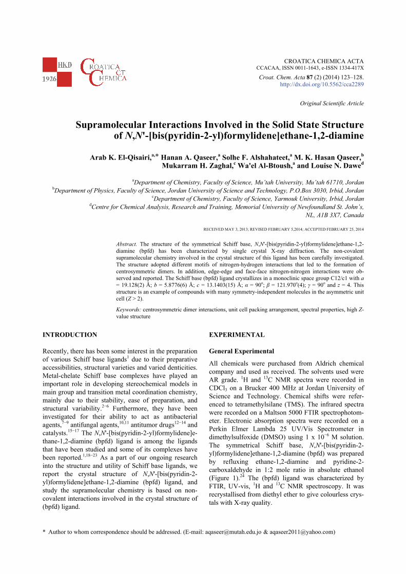
All chemicals were purchased from Aldrich chemical company and used as received. The solvents used were AR grade. 1H and 13C NMR spectra were recorded in CDCl3 on a Brucker 400 MHz at Jordan University of Science and Technology. Chemical shifts were refer-enced to tetramethylsilane (TMS). The infrared spectra were recorded on a Maltson 5000 FTIR spectrophotom-eter. Electronic absorption spectra were recorded on a Perkin Elmer Lambda 25 UV/Vis Spectrometer in dimethylsulfoxide (DMSO) using 1 x 10⁻⁶ M solution. The symmetrical Schiff base, N,N'-[bis(pyridin-2-yl)formylidene]ethane-1,2-diamine (bpfd) was prepared by refluxing ethane-1,2-diamine and pyridine-2-carboxaldehyde in 1:2 mole ratio in absolute ethanol (Figure 1).24 The (bpfd) ligand was characterized by FTIR, UV-vis, 1H and 13C NMR spectroscopy. It was recrystallised from diethyl ether to give colourless crys-tals with X-ray quality.
124 A. K. El-Qisairi et al., Supramolecular Interactions Involved in the Solid State Structure
Croat. Chem. Acta 87 (2014) 123.
Preparation of C14H14N4 (bpfd)
To a stirred solution of ethylenediamine (0.157 g; 2.61 mmol) in 20 ml ethanol, a solution of 2-pyridinecarboxaldehyde (0.560 g; 5.23 mmol) in ethanol (5 ml) was added dropwise over a period of 1−2 hours. The reaction mixture was then stirred and refluxed for 24 hours. The mixture was evaporated and recrystallized from diethyl ether. The product was filtered and dried in oven at 40 oC. The yield was 0.30 g (48 %). The com-pound melted at 57−59 oC. FTIR (neat, cm−1): 2920 v(C−H, aliphatic), 1647 v(C=N, imine), 1586, and 1427 v(C=N, and C=C pyridine). UV-vis; max(): 272 nm (22900 M−1 cm−1), 282 (15500 M−1 cm−1). 1H NMR (400 MHz, CDCl3): = 8.59 (d, 2H, J = 6.1), 8.40 (s, 2H), 7.95 (d, 2H, J = 6.1), 7.69 (t, 2H, J = 7.6), 7.25 (dt, 2H, J = 1.6, 6.1), 4.03 (s, 4H) ppm. 13C NMR (100 MHz, CDCl3): = 61.3, 121.3, 124.4, 136.5, 149.3, 154.3, 163.2 ppm. The solid state structure of the (bpfd) ligand has been determined by single-crystal X-ray diffraction.
Crystal Data
Empirical Formula C14H14N4 Formula Weight 238.29 g/mol Crystal Color, Habit chunk, colorless Crystal Dimensions 0.80 X 0.80 X 0.78 mm Crystal System monoclinic Lattice Type C-centered Detector Position 40.03 mm Pixel Size 0.137 mm Lattice Parameters a = 19.128(2) Å b = 5.8776(6) Å c = 13.1403(15) Å = 121.970(4)° V = 1253.3(2) Å3
Space Group C12/c1 Z value 4
Dcalc 1.263 g/cm3 F000 504 (MoK) 0.79 cm−1 Intensity Measurements
Detector Rigaku Saturn Goniometer Rigaku AFC8 Radiation MoK ( = 0.71070 Å) graphite monochromated Detector Aperture 70 mm × 70 mm Data Images 864 exposures oscillation Range (= 0.0, = 0.0) −75.0–105.0° Exposure Rate 22.0 sec./° Detector Swing Angle 15.08° oscillation Range ( = 0.0, = 180.0) −45.0–75.0° Exposure Rate 22.0 sec./° Detector Swing Angle 15.08° oscillation Range (= 45.0, = 90.0) −42.0–90.0° Exposure Rate 22.0 sec./° Detector Swing Angle 15.08° Detector Position 40.00 mm Pixel Size 0.068 mm 2max 80.6° No. of Reflections Measured Total: 2592 Unique: 1420 (Rint = 0.0214) Corrections Lorentz-polarization Secondary Extinction (coefficient: 0.0066(16)) Absorption (trans. factors:0.9394–0.9408)
Figure 1. Preparation of bpfd ligand
A. K. El-Qisairi et al., Supramolecular Interactions Involved in the Solid State Structure 125
Croat. Chem. Acta 87 (2014) 123.
Structure Solution and Refinement
Structure Solution Direct Methods (SHELX97) Refinement Full-matrix least-squares on F2 Function Minimized w (Fo2 − Fc2)2 Least Squares Weights w = 1/ [ (Fo2) + (0.0202 . P)2 + 1.2836 . P] where P = (Max(Fo2,0) + 2Fc2)/3 2max cut off 55.0° Anomalous Dispersion All non-hydrogen atoms No. Observations (All reflections) 1420 No. Variables 84 Reflection/Parameter Ratio 16.90 Residuals: R1 (I > 2.00 (I)) 0.0458 Residuals: R (All reflections) 0.0482 Residuals: wR2 (All reflections) 0.0944 Goodness of Fit Indicator 1.223 Max Shift/Error in Final Cycle 0.000 Maximum peak in Final Diff. Map 0.29 e−/Å3 Minimum peak in Final Diff. Map −0.17 e
−/Å3 Solution and Refinement of the Crystal Structure
Single-crystal X-ray diffraction experiment was carried out on a Rigaku Saturn CCD area detector with graphite monochromate Mo-Kα radiation. The data were collected at a temperature of −135 ± 1 °C to a maximum 2θ value of 80.6°. Data were collected and processed using Crystal Clear.25 The linear absorption coefficient, μ, for Mo-Kα radiation is 0.79 cm−1. A numerical absorption correction was applied which resulted in transmission factors rang-ing from 0.9394 to 0.9408. The data were corrected for Lorentz and polarization effects.26 The structure was solved by direct methods27 and expanded using Fourier techniques.28 The non-hydrogen atoms were refined anisotropically. Hydrogen atoms were refined using the riding model. The final cycle of full-matrix least-squares refinement29 on F2 was based on 1420 observed reflec-tions and 84 variable parameters and converged. Crys-tallographic data for the structural analysis reported in this paper have been deposited with the Cambridge Crystallographic Data Centre, CCDC, Number (721559). This data can be obtained free of charge at http://www.ccdc.cam.ac.uk/conts/retrieving.html [or from the Cambridge Crystallographic Data Centre
(CCDC), 12 Union Road, Cambridge CB2 1EZ, UK; fax: +44-1223-336033; email: [email protected]].
Neutral atom scattering factors were taken from Cromer and Waber.30 Anomalous dispersion effects were included in Fcalc;
31 the values for Δf′ and Δf″ were those of Creagh and McAuley.32 The values for the mass attenuation coefficients are those of Creagh and Hubbell.33 All calculations were performed using the CrystalStructure25,34 crystallographic software package except for refinement, which was performed using SHELXL-97.29
RESULTS AND DISCUSSION
IR and UV/vis Spectroscopy
The Schiff base (bpfd) ligand has a strong absorbance at 1647 cm−1 in the infrared region, confirming the pres-ence of the imine moiety. The stretching vibration of C=N and C=C of the pyridine ring are observed at 1586 and 1427 cm−1, respectively. Additionally, the UV/vis spectrum has strong absorptions near 272 and 282 nm. These bands are assigned to the ligand centered (LC) or -* transitions. The IR data are consistent with the reported value.35
NMR Spectroscopy
The 1H NMR spectrum for the (bpfd) ligand give signals corresponding to the aromatic protons in the range 7.25–8.59 ppm. The 1H NMR signals arising from ethylene protons appear as singlet at 4.02 ppm. 13C NMR spectrum supports the formation of the Schiff base (bpfd) ligand. The 13C NMR signals corre-sponding to the aromatic carbons are visible in the region 117–166 ppm. Signal corresponding to the aliphatic and vinylic carbons are found at 61 and 154 ppm, respectively. Supramolecular Interactions Involved in the Solid State Structure of (bpfd) Ligand
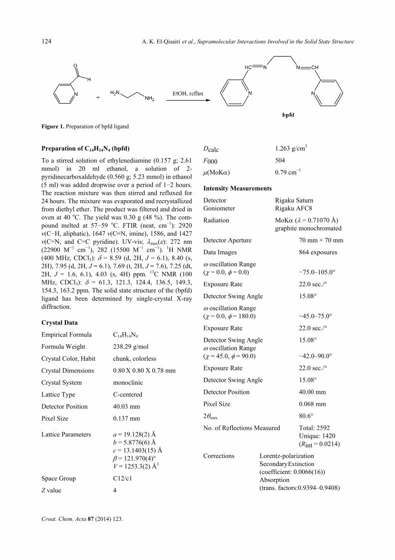
A solvent free sample of the N,N'-[bis(pyridine-2-yl)-formylidene] ethane-1,2-diamine ligand (bpfd) (Figure 1) was obtained after crystallization from diethyl ether to give material that was good enough to be examined by the single crystal X-ray technique. The (bpfd) ligand crystallizes in a monoclinic space group C12/c1 with a = 19.128(2) Å; b = 5.8776(6) Å; c = 13.1403(15) Å; α = 90°; β = 121.970°(4); γ = 90° and z = 4. Numerical details of the crystal structure are presented in experi-mental section. ORTEP drawing of (bpfd) molecular structure is shown in Figure 2. The bond distances and angles for (bpfd) ligand (Tables 1, 2) are as ex-pected.36,37 The crystal structure of (bpfd) is an example of compounds with many symmetry-independent mole-
126 A. K. El-Qisairi et al., Supramolecular Interactions Involved in the Solid State Structure
Croat. Chem. Acta 87 (2014) 123.
cules in the asymmetric unit cell (Z > 2). This phenom-enon was extensively analyzed by Bernstein et al.38 and accordingly reported that high-Z structures have been described as ‘‘snapshot pictures’’ and also as ‘‘fossil relics’’ of early stages in crystallization. Such descrip-tion has a certain charm. However, as we are almost totally ignorant of the nucleation process, the early stages in crystallization, these picturesque descriptions have the disadvantage that they are almost impossible to confirm or to refute.
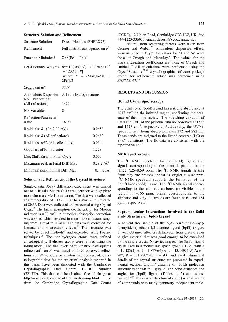
Detailed analysis of (bpfd) crystal structure showed the presence of edge-edge C6−H5…N1 centro-symmetric dimer interactions. This centrosymme-tric C−H…N dimer is composed of C−H…N and C−H…N interactions with distances of 2.7 and 3.55 Å, respec-
Figure 2. ORTEP drawing of (bpfd). Thermal ellipses areshown in 50 % probability.
Table 1. Summary of bond distances (Å) for (bpfd) ligand
atom atom distance atom atom distance
N(1) C(1) 1.3459(18) N(1) C(5) 1.3547(18)
N(2) C(6) 1.2715(16) N(2) C(7) 1.4666(18)
C(1) C(2) 1.396(2) C(2) C(3) 1.391(2)
C(3) C(4) 1.394(2) C(4) C(5) 1.400(2)
C(5) C(6) 1.4870(18) C(7) C(7) 1.5272(14)
Table 2. Summary of bond angles (°) for (bpfd) ligand
atom atom atom angle
C(1) N(1) C(5) 117.22(14)
N(1) C(1) C(2) 123.44(13)
C(2) C(3) C(4) 119.27(17)
N(1) C(5) C(4) 123.33(12)
C(4) C(5) C(6) 121.60(12)
N(2) C(7) C(7) 111.46(11)
C(6) N(2) C(7) 116.99(13)
C(1) C(2) C(3) 118.56(13)
C(3) C(4) C(5) 118.16(14)
N(1) C(5) C(6) 115.07(13)
N(2) C(6) C(5) 121.82(14)
Figure 3. Centrosymmetric dimer composed of two molecules of (bpfd) through C6−H5…N1 interactions with a bond dis-tance of 2.7 Å and bond angle of 145.68°.
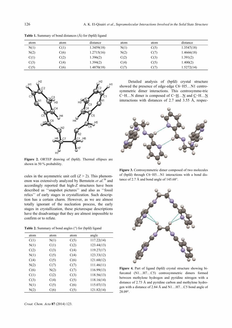
Figure 4. Part of ligand (bpfd) crystal structure showing bi-furcated (N1…H7…C5) centrosymmetric dimers formed between methylene hydrogen and pyridine nitrogen with a distance of 2.73 Å and pyridine carbon and methylene hydro-gen with a distance of 2.84 Å and N1…H7…C5 bond angle of 28.09°.
A. K. El-Qisairi et al., Supramolecular Interactions Involved in the Solid State Structure 127
Croat. Chem. Acta 87 (2014) 123.
tively and C6−H5…N1 bond angle of 145.68° as shown in Figure 3.
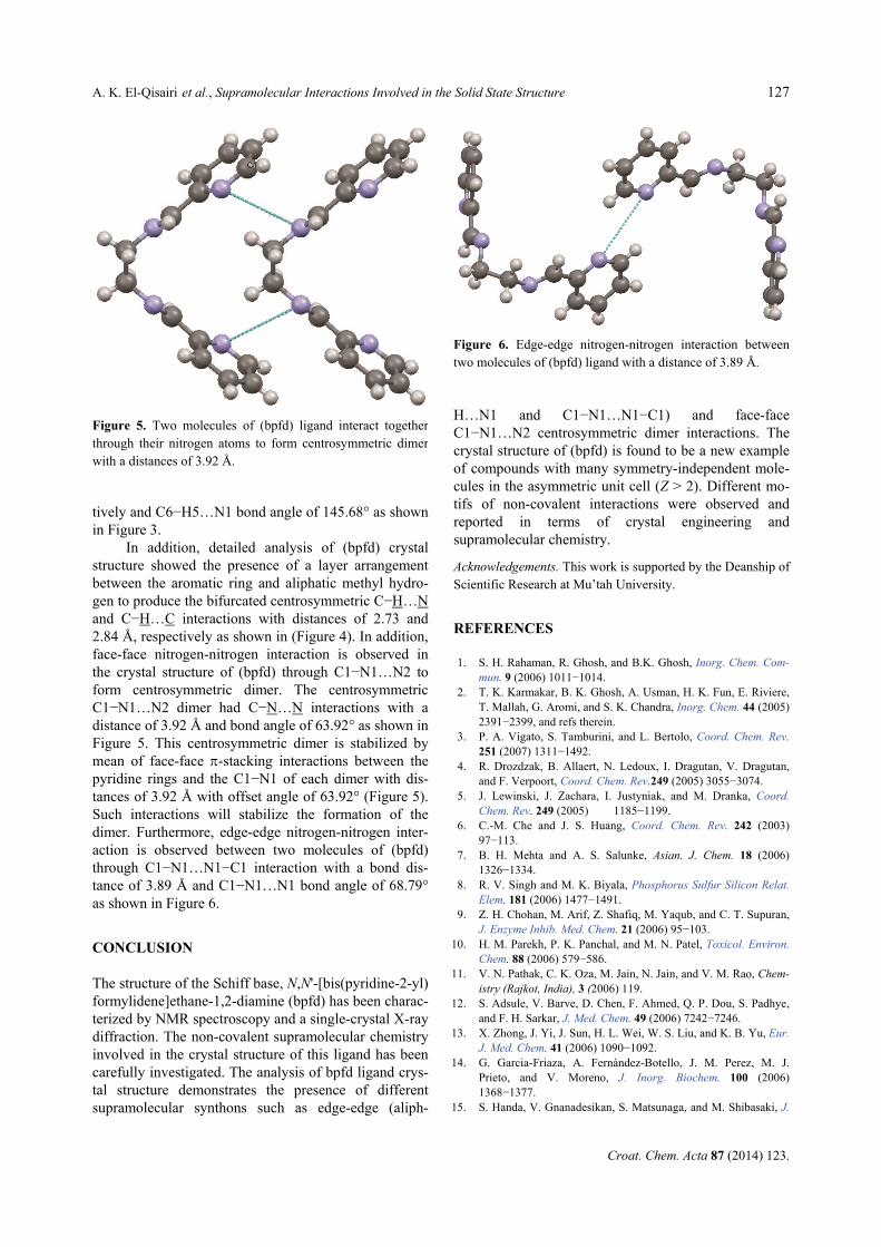
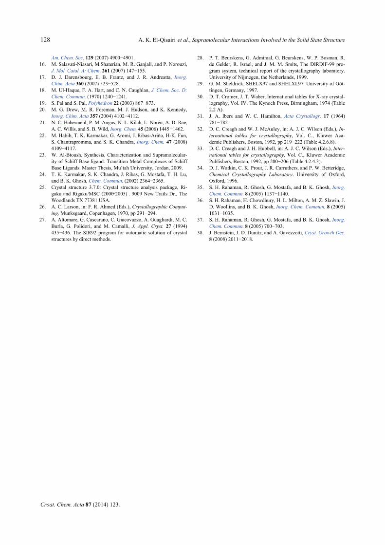
In addition, detailed analysis of (bpfd) crystal structure showed the presence of a layer arrangement between the aromatic ring and aliphatic methyl hydro-gen to produce the bifurcated centrosymmetric C−H…N and C−H…C interactions with distances of 2.73 and 2.84 Å, respectively as shown in (Figure 4). In addition, face-face nitrogen-nitrogen interaction is observed in the crystal structure of (bpfd) through C1−N1…N2 to form centrosymmetric dimer. The centrosymmetric C1−N1…N2 dimer had C−N…N interactions with a distance of 3.92 Å and bond angle of 63.92° as shown in Figure 5. This centrosymmetric dimer is stabilized by mean of face-face -stacking interactions between the pyridine rings and the C1−N1 of each dimer with dis-tances of 3.92 Å with offset angle of 63.92° (Figure 5). Such interactions will stabilize the formation of the dimer. Furthermore, edge-edge nitrogen-nitrogen inter-action is observed between two molecules of (bpfd) through C1−N1…N1−C1 interaction with a bond dis-tance of 3.89 Å and C1−N1…N1 bond angle of 68.79° as shown in Figure 6.
CONCLUSION
The structure of the Schiff base, N,N'-[bis(pyridine-2-yl) formylidene]ethane-1,2-diamine (bpfd) has been charac-terized by NMR spectroscopy and a single-crystal X-ray diffraction. The non-covalent supramolecular chemistry involved in the crystal structure of this ligand has been carefully investigated. The analysis of bpfd ligand crys-tal structure demonstrates the presence of different supramolecular synthons such as edge-edge (aliph-
H…N1 and C1−N1…N1−C1) and face-face C1−N1…N2 centrosymmetric dimer interactions. The crystal structure of (bpfd) is found to be a new example of compounds with many symmetry-independent mole-cules in the asymmetric unit cell (Z > 2). Different mo-tifs of non-covalent interactions were observed and reported in terms of crystal engineering and supramolecular chemistry.
Acknowledgements. This work is supported by the Deanship of Scientific Research at Mu’tah University.
REFERENCES
1. S. H. Rahaman, R. Ghosh, and B.K. Ghosh, Inorg. Chem. Com-mun. 9 (2006) 1011−1014.
2. T. K. Karmakar, B. K. Ghosh, A. Usman, H. K. Fun, E. Riviere, T. Mallah, G. Aromi, and S. K. Chandra, Inorg. Chem. 44 (2005) 2391−2399, and refs therein.
3. P. A. Vigato, S. Tamburini, and L. Bertolo, Coord. Chem. Rev. 251 (2007) 1311−1492.
4. R. Drozdzak, B. Allaert, N. Ledoux, I. Dragutan, V. Dragutan, and F. Verpoort, Coord. Chem. Rev.249 (2005) 3055−3074.
5. J. Lewinski, J. Zachara, I. Justyniak, and M. Dranka, Coord. Chem. Rev. 249 (2005) 1185−1199.
6. C.-M. Che and J. S. Huang, Coord. Chem. Rev. 242 (2003) 97−113.
7. B. H. Mehta and A. S. Salunke, Asian. J. Chem. 18 (2006) 1326−1334.
8. R. V. Singh and M. K. Biyala, Phosphorus Sulfur Silicon Relat. Elem. 181 (2006) 1477−1491.
9. Z. H. Chohan, M. Arif, Z. Shafiq, M. Yaqub, and C. T. Supuran, J. Enzyme Inhib. Med. Chem. 21 (2006) 95−103.
10. H. M. Parekh, P. K. Panchal, and M. N. Patel, Toxicol. Environ. Chem. 88 (2006) 579−586.
11. V. N. Pathak, C. K. Oza, M. Jain, N. Jain, and V. M. Rao, Chem-istry (Rajkot, India), 3 (2006) 119.
12. S. Adsule, V. Barve, D. Chen, F. Ahmed, Q. P. Dou, S. Padhye, and F. H. Sarkar, J. Med. Chem. 49 (2006) 7242−7246.
13. X. Zhong, J. Yi, J. Sun, H. L. Wei, W. S. Liu, and K. B. Yu, Eur. J. Med. Chem. 41 (2006) 1090−1092.
14. G. Garcia-Friaza, A. Fernàndez-Botello, J. M. Perez, M. J. Prieto, and V. Moreno, J. Inorg. Biochem. 100 (2006) 1368−1377.
15. S. Handa, V. Gnanadesikan, S. Matsunaga, and M. Shibasaki, J.
Figure 5. Two molecules of (bpfd) ligand interact togetherthrough their nitrogen atoms to form centrosymmetric dimerwith a distances of 3.92 Å.
Figure 6. Edge-edge nitrogen-nitrogen interaction between two molecules of (bpfd) ligand with a distance of 3.89 Å.
128 A. K. El-Qisairi et al., Supramolecular Interactions Involved in the Solid State Structure
Croat. Chem. Acta 87 (2014) 123.
Am. Chem. Soc. 129 (2007) 4900−4901. 16. M. Salavati-Niasari, M.Shaterian, M. R. Ganjali, and P. Norouzi,
J. Mol. Catal. A: Chem. 261 (2007) 147−155. 17. D. J. Darensbourg, E. B. Frantz, and J. R. Andreatta, Inorg.
Chim. Acta 360 (2007) 523−528. 18. M. Ul-Haque, F. A. Hart, and C. N. Caughlan, J. Chem. Soc. D:
Chem. Commun. (1970) 1240−1241. 19. S. Pal and S. Pal, Polyhedron 22 (2003) 867−873. 20. M. G. Drew, M. R. Foreman, M. J. Hudson, and K. Kennedy,
Inorg. Chim. Acta 357 (2004) 4102−4112. 21. N. C. Habermehl, P. M. Angus, N. L. Kilah, L. Norén, A. D. Rae,
A. C. Willis, and S. B. Wild, Inorg. Chem. 45 (2006) 1445 −1462. 22. M. Habib, T. K. Karmakar, G. Aromí, J. Ribas-Ariňo, H-K. Fun,
S. Chantrapromma, and S. K. Chandra, Inorg. Chem. 47 (2008) 4109−4117.
23. W. Al-Btoush, Synthesis, Characterization and Supramolecular-ity of Schiff Base ligand. Transition Metal Complexes of Schiff Base Ligands. Master Thesis, Mu’tah University, Jordan, 2009.
24. T. K. Karmakar, S. K. Chandra, J. Ribas, G. Mostafa, T. H. Lu, and B. K. Ghosh, Chem. Commun. (2002) 2364−2365.
25. Crystal structure 3.7.0: Crystal structure analysis package, Ri-gaku and Rigaku/MSC (2000−2005) . 9009 New Trails Dr., The Woodlands TX 77381 USA.
26. A. C. Larson, in: F. R. Ahmed (Eds.), Crystallographic Comput-ing, Munksgaard, Copenhagen, 1970, pp 291−294.
27. A. Altomare, G. Cascarano, C. Giacovazzo, A. Guagliardi, M. C. Burla, G. Polidori, and M. Camalli, J. Appl. Cryst. 27 (1994) 435−436. The SIR92 program for automatic solution of crystal structures by direct methods.
28. P. T. Beurskens, G. Admiraal, G. Beurskens, W. P. Bosman, R. de Gelder, R. Israel, and J. M. M. Smits, The DIRDIF-99 pro-gram system, technical report of the crystallography laboratory. University of Nijmegen, the Netherlands, 1999.
29. G. M. Sheldrick, SHELX97 and SHELXL97. University of Göt-tingen, Germany, 1997.
30. D. T. Cromer, J. T. Waber, International tables for X-ray crystal-lography, Vol. IV. The Kynoch Press, Birmingham, 1974 (Table 2.2 A).
31. J. A. Ibers and W. C. Hamilton, Acta Crystallogr. 17 (1964) 781−782.
32. D. C. Creagh and W. J. McAuley, in: A. J. C. Wilson (Eds.), In-ternational tables for crystallography, Vol. C., Kluwer Aca-demic Publishers, Boston, 1992, pp 219−222 (Table 4.2.6.8).
33. D. C. Creagh and J. H. Hubbell, in: A. J. C. Wilson (Eds.), Inter-national tables for crystallography, Vol. C., Kluwer Academic Publishers, Boston, 1992, pp 200−206 (Table 4.2.4.3).
34. D. J. Watkin, C. K. Prout, J. R. Carruthers, and P. W. Betteridge, Chemical Crystallography Laboratory. University of Oxford, Oxford, 1996.
35. S. H. Rahaman, R. Ghosh, G. Mostafa, and B. K. Ghosh, Inorg. Chem. Commun. 8 (2005) 1137−1140.
36. S. H. Rahaman, H. Chowdhury, H. L. Milton, A. M. Z. Slawin, J. D. Woollins, and B. K. Ghosh, Inorg. Chem. Commun. 8 (2005) 1031−1035.
37. S. H. Rahaman, R. Ghosh, G. Mostafa, and B. K. Ghosh, Inorg. Chem. Commun. 8 (2005) 700−703.
38. J. Bernstein, J. D. Dunitz, and A. Gavezzotti, Cryst. Growth Des. 8 (2008) 2011−2018.


![Advantageous Supramolecular System Through Self ... polymer, xanthan gum self-associates through electrostatic interactions with functional materials [6]. Xanthan gum shows re - markable](https://img.pdfslide.us/doc/110x75/5eccdc8cc03a16731d06ddd9/advantageous-supramolecular-system-through-self-polymer-xanthan-gum-self-associates.jpg)


![7. Supramolecular structures - Acclab h55.it.helsinki.fiknordlun/nanotiede/nanosc7nc.pdf · 7. Supramolecular structures [Poole-Owens 11.5] Supramolecular structures are large molecules](https://img.pdfslide.us/doc/110x75/5f071ded7e708231d41b63bf/7-supramolecular-structures-acclab-h55it-knordlunnanotiedenanosc7ncpdf.jpg)


![Index [] · Supramolecular chemistry is intrinsically a dynamic chemistryin view of the lability of the interactions ... Supramolecular Chemistry: Concepts and Perspectives,VCH Weinheim,](https://img.pdfslide.us/doc/110x75/5f2285620fd639682f41fe10/index-supramolecular-chemistry-is-intrinsically-a-dynamic-chemistryin-view-of.jpg)