Embed Size (px)
Citation preview

Supracondylar Humerus Fractures.-Urgency and Open Reduction
5th Annual SLAOTI meetingSao Paolo, BrazilOctober 12-14, 2017
Richard M Schwend MDProfessor Orthopaedics and PediatricsDirector of ResearchChildren’s Mercy HospitalKansas City MO, [email protected] © The Children's Mercy Hospital 2015
1

Disclosures
• K2M Consultant
• Medtronic Consultant
• POSNA President and BOD member
• AAP Immediate Past Chair and Section on Orthopaedics Executive Committee
• Project Perfect World Board of Directors.
• Miracle Feet Medical Advisory Board.

Learning Objectives
•When is time essential?
•When do you need to do open reduction?

Extension Type Supracondylar Humerus Fx
Type I Type II Type III

RIGHT ARM
Posterior lateral displacement

RIGHT ARM
Posterior medial displacement
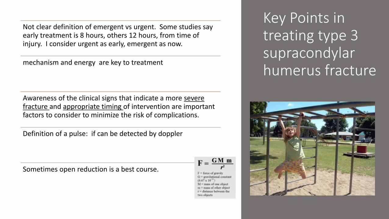
Key Points in treating type 3 supracondylar humerus fracture
Not clear definition of emergent vs urgent. Some studies say early treatment is 8 hours, others 12 hours, from time of injury. I consider urgent as early, emergent as now.
mechanism and energy are key to treatment
Awareness of the clinical signs that indicate a more severe fracture and appropriate timing of intervention are important factors to consider to minimize the risk of complications.
Definition of a pulse: if can be detected by doppler
Sometimes open reduction is a best course.


Importance of Perfusion Examination
The perfusion status of the extremity should be noted. Important findings include warmth, capillary refill, and the presence or absence of a radial pulse by palpation and/or Doppler
ultrasound.
POSNA study guide

General Principles type 3 fractures
These are soft tissue injuries that happen to have a fracture
Besides the fracture type, evaluate:
• Vascular status- <3 sec cap refill, warm, pink, palpable pulse (dopplerable)
• Associated nerves injuries
• Condition of soft tissue- open? Dirty?
• Ipsilateral radius or ulnar fracture?
• Degree of swelling- typical or severe

General Principles type 3 fractures
These are soft tissue injuries that happen to have a fracture
Besides the fracture type, evaluate:
• Vascular status- <3 sec cap refill,warm, pink, palpable pulse (dopplerable)
• Associated nerves injuries
• Condition of soft tissue- open? Dirty?
• Ipsilateral radius or ulnar fracture?
• Degree of swelling- typical or severe

Complications to Avoid
• Compartment syndrome • Minimize risk with immediate treatment of vascular injuries• Significant swelling, wide displacement, antecubital ecchymosis,
ipsilateral fractures, and neurologic deficit are relative indications for early intervention
• 3 A’s in pediatric patients (anxiety, agitation, analgesic requirement) as potential signs of impending compartment syndrome
• Immobilize in relative extension (≤ 70 degrees for displaced fractures) to reduce compartment pressures.
• Vascular injury- and resulting Volkmann
• Nerve injury- most are resolving neuropraxias
• Infection- urgent and appropriate treatment of open fractures
• Loss of reduction or mal reduction
• StiffnessPOSNA Study Guide

Treatment Case Example
• 7 year old boy with type 3 supracondylar humerus fracture
• Perfused hand (>3 sec cap refill) but no distal palpable or dopplerable pulse
• Anterior interosseous nerve injury
• Skin is intact (closed soft tissue envelope)
• No ipsilateral radius or ulna fracture
• Severe swelling and puckering of the skin.
• What to do? Emergent, urgent or not?

Go to AAOS AUCAppropriate Use CriteriaPlug in the dataYou get an answer!

2009 -2016 study at Children’s Mercy HospitalPercentage that deviated from AUC
Out of 585 patients:• 560 (95.7%) “appropriate”
• 25 (4.3%) “maybe appropriate”
• 0 “rarely appropriate”
Significant decrease in the proportion
that deviated from the guidelines
(p = 0.0076)
CPG 2011

Treatment for type 3
• Type 3 – Treatment consists of closed reduction and percutaneous pinning. Timingof intervention is a key point. In the presence of vascular compromise or compartment syndrome, emergentintervention is essential
• POSNA study guide

So when do you?
• When to sit back and coast?
• Have to emergently treat a fracture?
• Open a fracture and explore

What does the evidence say to do? 14 recommendations- 4 related to timing and opening

If fracture does not reduce?
You may choose to open the fracture to get better reduction

If Presents with Poor Perfusion?
Then do emergent closed reduction

Pale, Pulseless Hand
• Emergent operative reduction and pinning of the fracture
• If no restoration of perfusion, immediate brachial artery exploration
• If trapped in the fracture then release the pins and repair it.
• If the artery is injured or torn then repair it.

Pale, Pulseless Hand
• Emergent operative reduction and pinning of the fracture
• If no restoration of perfusion, immediate brachial artery exploration
• If trapped in the fracture then release the pins and remove it.
• If the artery is injured or torn then repair it.

Pale, Pulseless Hand
• Emergent operative reduction and pinning of the fracture
• If no restoration of perfusion, immediate brachial artery exploration
• If trapped in the fracture then release the pins and remove it.
• If the artery is injured or torn then repair it.

Pale, Pulseless Hand
• Emergent operative reduction and pinning of the fracture
• If no restoration of perfusion, immediate brachial artery exploration
• If trapped in the fracture then release the pins and repair it.
• If the artery is injured or torn then repair it.
But don’t do this!

If absent pulses and poor perfusion post reduction pinning?
Explore the site

If absent pulses and poor perfusion post reduction pinning?
Explore the site


Lots of studies but all low power

Pink Pulseless Hand
• Pink, warm hand with capillary refill symmetric to the contralateral side
• Radial pulse NOT palpable
• AND Normal radial pulse (triphasic) NOT audible with Doppler ultrasound

Pulse absent/hand perfused after
reduction?
Use your judgement to open
or not.

Problems with evidence based recommendations
Early, emergent and urgent time periods are not defined.
Some scenarios with AUC don’t make sense. ok to not wash out contaminated open fx, really?
Needs real life data to determine if recommendations are really working properly.
Legal concerns if you stray too far.

Summary-From AUC
Try using the AAOS AUC but understand it is not perfect!
AUC does not define what is early treatment, urgent or emergent
Emergent
•No perfusion- open treatment if cannot restore perfusion with closed.
•No pulse, perfused, nerve out- use your judgement to open
•Contaminated open fracture- open treatment
Urgent
•No pulse, but perfused- use your judgment to open
•Open fracture, not contaminated- open treatment
You may want to open the fracture if cant get adequate reduction

References
• Abzug, Joshua M.; Herman, Martin J. Management of Supracondylar Humerus Fractures in Children: Current Concepts. Journal of the American Academy of Orthopaedic Surgeons. 20(2):69-77, February 2012
• Babal JC, Mehlmann CT, Klein G. Nerve injuries associated with pediatric supracondylar humeral fractures: a meta-analysis. J Pediatr Orthop. 2010; 30 (3): 253-263.
• Bae DS, Kadiyala RK, Waters PM. Acute compartment syndrome in children. J Pediatr Orthop. 2001; 21 (5): 680-688.
• Barton KL, Kaminsky CK, Green DW, Shean CJ, Kautz SM, Skaggs DL. Reliability of a modified Gartland classification of supracondylar humerus fractures. J Pediatr Orthop. 2001; 21 (1): 27-30.
• Keppler P, Salem K, Schwarting B, Kinzl L. The effectiveness of physiotherapy after operative treatment of supracondylar humeral fractures in children. J Pediatr Orthop2005;25(3):314-316
• Kocher MS, Kasser JR, Waters PM et al. Lateral entry compared with medial and lateral entry pin fixation for completely displaced supracondylar humeral fractures in children. A randomized clinical trial. J Bone Joint Surg Am 2007;89(4):706-712
• Moraleda L, Valencia M, Barco R, Gonzalez-Moran G. Natural history of unreduced Gartland type-II supracondylar fractures of the humerus in children: a two to thirteen-year follow-up study. J Bone Joint Surg Am. 2013; 95 (1): 28-34.
• Omid R, Choi PD, Skaggs DL. Supracondylar humeral fractures in children. J Bone Joint Surg Am. 2008; 90 (5): 1121-1132.
• Scannell BP, Jackson B, Bray C, Roush TS, Brighton BK, Frick SL. The perfused, pulseless supracondylar humeral fracture: intermediate-term follow-up of vascular status and function. J Bone Joint Surg Am. 2013; 95: 1913-9.
• Skaggs DL, Sankar WN, Albrektson J, Vaishnav S, Choi PD, Kay RM. How safe is the operative treatment of Gartland type 2 supracondylar humerus fractures in children? J PediatrOrthop. 2008; 28 (2):139-141.
• Tripuraneni KR, Bosch PP, Schwend RM, Yaste JJ. Prospective, surgeon randomized evaluation of crossed pins versus lateral pins for unstable supracondylar humerus fractures in children. J Pediatr Orthop B 2009;18(2):93-98
• White L, Mehlman CT, Crawford AH. Perfused, pulseless, and puzzling: a systematic review of vascular injuries in pediatric supracondylar humerus fractures and results of a POSNA questionnaire. J Pediatr Orthop. 2010; 30 (4): 328-335


![Pediatric Supracondylar Fractures: Are Medial Pins Indicated?are the supracondylar fractures of the humerus that can be managed by both operative and non-operative modalities [1]](https://img.pdfslide.us/doc/110x75/6087220d2ec1ae7c713805b2/pediatric-supracondylar-fractures-are-medial-pins-indicated-are-the-supracondylar.jpg)