Embed Size (px)
Citation preview

Supporting Information
for
Gallium and functionalised-porphyrins combine to form potential
lysosome-specific multi-modal bio-probes
Jie Pan,a# Bethany I. Harriss,b# Chi-Fai Chan,a Lijun Jiang,a Tik-Hung Tsoi,c Nicholas
J. Long, b,* Wing-Tak Wong,c Wai-Kwok Wong,a* and Ka-Leung Wonga*
a. Department of Chemistry, Hong Kong Baptist University, Kowloon Tong,
Kowloon, Hong Kong SAR. E-mail: [email protected], [email protected]
b. Department of Chemistry, Imperial College London, South Kensington Campus,
London SW7 2AZ, UK. E-mail: [email protected]; Fax: +44(0)20 7594 5804;
Tel: +44 (0)20 7594 5781
c. Department of applied biology and chemical technology, Hong Kong Polytechnic
University, Hung Hom, Hong Kong SAR.
#These authors contributed equally to this work and should be considered co-first
authors.

Experimental
General synthesis for the PorRu-1
All the reagents were purchased commercially and used without further purification.
All reactions were performed under a nitrogen atmosphere, unless otherwise noted.
The anhydrous solvents were dried according to standard protocols in literature. The
synthetic route for the preparation of PorRu-1, ZnporRu-1 and GaporRu-1 is shown
in Scheme S1. The intermediates and products were characterized by 1H NMR, 13C
NMR, MALDI-TOF MS and UV-Vis spectroscopy. NMR spectra were recorded on
either a Brüker Advance 400 (1H: 400 MHz, 13C: 100 MHz) spectrometer. The
following abbreviations were used to depict the multiplicities: s = singlet, d = doublet,
t = triplet, q = quartet, dd = doublet of doublets, m = multiplet, br = broad. PorRu-1
was synthesized by using our developed method in the literature.1
CHO
TMS
N
NH N
HN
OMeOMe
MeO
MeO
MeO
MeO
MeOOMe
OMe
TMS
OMeOMeMeO
CHO
HN Propionic acid
140oC+ + TBAF,THF
N
NH N
HN
OMeOMe
MeO
MeO
MeO
MeO
MeOOMe
OMe
HPd(PPh3)4,CuI,DMF
40-50oC,8hK2CO3
p-Acetylene Por
Por-triple
N
NH N
HN
OMeOMe
MeO
MeO
MeO
MeO
MeOOMe
OMe
N
N
N
NH N
HN
OMeOMeMeO
OMeOMe
MeO
MeO
MeO
MeOTHF,Ethanol,cis-Ru(bpy)2Cl2
N
NN
N
NN
Ru
2+
L1
PorRu-1
5-bromo-1,10-phenanthroline
PorRu-‐1 GaporRu-‐1
Scheme S1. Synthetic routes of GaporRu-‐1 and ZnporRu-‐1.
Preparation of 5,10,15-Tris (3’,4’,5’-trimethoxylphenyl)-20-[4’-(2i”-
trimethylsilylethyl)phenyl]-21H,23H-porphyrin (p-Acetylene Por). 3,4,5-Trimethoxylbenzaldehyde (8.00 g, 0.041 mol) and 4-[2-
(trimethylsilyl)ethynyl]benzaldehyde (2.75 g, 0.014 mol) were added in 300 mL
propionic acid. The reaction temperature was 120 °C and a solution of pyrrole (3.65 g,
0.054 mol) in 50 mL propionic acid was added dropwise. After all the pyrrole was
added to the reaction solution, the reaction temperature was increased to 140 °C and
refluxed for 3 h. Once the reaction was complete, the propionic acid was distilled off.
The residue was purified on silica gel for column chromatography by using DCM as
eluent. A purple product was isolated. Yield 2.00 g (15%). 1H NMR (CDCl3) δ -2.68
(s, 2H), 0.45 (s, 9H), 4.05 (d, 18H, J = 3.6 Hz), 4.25 (s, 9H), 7.57(d, 6H, J = 2.0 Hz),
7.96 (d, 2H, J = 8.4 Hz), 8.25 (d, 2H, J = 8.0 Hz), 8.92 (d, 2H, J = 4.8 Hz), 9.07 (s,
6H). HRMS (MALDI-TOF) ([M]+, m/z): Calcd. for C58H56N4O9Si, 981.2; Found for
[M]+, 981.2.
Preparation of Por-triple
p-Acetylene Por (200 mg, 0.192 mmol) was dissolved in 30 mL DCM and the
protecting group (TMS) in the compound was deprotected by 210 μL TBAF (1 M
TBAF in THF). The solution mixture was stirred at room temperature for 30 min. The
solvent was removed and the residue was purified on silica gel for column
N
NH N
HN
OMeOMeMeO
OMeOMe
MeO
MeOMeO
MeO
N
N NN
NN
Ru
Zn(OAc)2.2H2O
Methanol,CHCl3reflux 3h
N
N N
N
OMeOMeMeO
OMeOMe
MeO
MeOMeO
MeO
N
N NN
NN
RuZn
N
N N
N
OMeOMeMeO
OMeOMe
MeO
MeO
MeO
MeO
N
NN
N
NN
RuGaGaCl3, AcONa
Acetic acidMicrowave, 110oc 15min
N
NH N
HN
OMeOMeMeO
OMeOMe
MeO
MeO
MeO
MeO
N
NN
N
NN
Ru
3+2+
2+ 2+

chromatography by using DCM as eluent. Yield 180 mg (98%). 1H NMR (CDCl3) δ
-2.8 (s, 2H), 3.06 (s, 18H), 4.15 (s, 9H), 7.46 (s, 6H), 7.89 (d, 2H, J = 8.0 Hz), 8.17 (d,
2H, J = 7.8Hz), 8.82 (d, 2H, J = 4.4 Hz), 8.96 (s, 6H).
Preparation of Phenylacetylene-Linked Porphyrin-Phen . L1
Pd(PPh)4 (20 mg), CuI (20 mg), K2CO3, Por-triple (100 mg, 0.11 mmol) and
5-bromo-1,10-phenanthroline (80 mg, 0.313 mmol) were dissolved in 50 mL
anhydrous DMF. The reaction temperature was 50 °C and stirred for 12 hours. The
organic phase of the reaction mixture was washed by chloroform and water three
times. The residue was purified on silica gel for column chromatography by using
CH3Cl/MeOH (v/v = 30:1) as eluent. Yield 64.5 mg, 51%. 1H NMR (CDCl3) δ -2.75(s,
2H), 3.97 (s, 18H), 4.18 (s, 9H), 7.48 (s, 6H), 7.70−7.74 (dd, 1H, J = 4.4 Hz),
7.83-7.87 (m, 1H), 8.09 (d, 2H, J = 8.0 Hz) 8.27-8.34 (m, 4H), 8.89 (d, 2H, J = 4.8
Hz), 8.97-9.00 (m, 6H), 9.01−9.04 (m, 1H), 9.25(d, 1H, J = 1.6 Hz), 9.28(d, 1H, J =
1.6Hz). 13C NMR (DMSO-d6), δ 137.42, 136.72, 134.57, 133.23, 132.21, 132.06, 132.
03, 131.53, 131.44, 128.83, 128.71, 60.42, 56.20.
Preparation of Phenylacetylene-Linked [(Porphyrin-Phen)Ru(bpy)2]-[Cl]2
(PorRu-1)
L1 (20 mg, 0.018 mmol) and cis-Ru(bpy)2Cl2 (34 mg, 0.070 mmol) were dissolved in
a mixture of THF (15 mL) and ethanol (15 mL) solution. After refluxing for 12 h, the
solvent was removed under reduced pressure. The crude solid was purified on neutral
Al2O3 for column chromatography with DCM/MeOH (v/v = 20:1) as eluent. Yield 20
mg, 74%. 1H NMR (DMSO-d6) δ −2.90 (s, 2H), 3.90 (s, 18H), 4.00 (s, 9H), 7.40-7.56
(m, 6H), 7.68−7.96 (m, 12H), 8.13-8.27 (m, 6H), 8.40 (s, 2H), 8.85−9.00 (m, 14H). 13C NMR (DMSO-d6) δ 156.87, 156.80, 156.57, 156.48, 153.16, 152.95, 151.96,
151.72, 151.51, 151.19, 151.42, 151.19, 147.61, 147.20, 146.93, 142.97, 138.08,
137.96, 137.43, 136.79, 136.70, 131.15, 130.62, 130.15, 129.74, 127.91, 127.86,
127.27, 126.99, 124.58, 124.47, 120.14, 118.64, 112.78, 60.43, 56.21. HRMS
(MALDI-TOF, positive mode) ([M]+, m/z): Calcd. for C87H70N10O9Ru, 1500.6; Found
for [M]+, 1500.43.

Preparation of Phenylacetylene-Linked [(Zinc-Porphyrin-Phen)Ru(bpy)2]-[Cl]2
(ZnporRu-1)
PorRu-1 (20 mg, 0.013 mmol) and excess amount of Zn(OAc)2.2H2O was dissolved
in methanol (15 mL) and chloroform (15 mL). After refluxing for 3h, the solvent was
removed under reduced pressure. The crude solid was purified on neutral Al2O3 for
column chromatography with DCM/MeOH (v/v=20:1) as eluent. Yield 17 mg, 83%. 1H NMR (DMSO-d6) δ 3.91 (s, 18H), 4.00 (s, 9H), 7.45 (d, 2H, J = 5.8Hz), 7.53-7.57
(m, 6H), 7.70 (d, 2H, J = 5.4Hz), 7.74 (d, 4H, J = 4.9Hz), 7.88 (m, 2H, J = 4.2Hz),
7.95−7.99 (m, 1H), 8.15 (s, 1H), 8.16 (d, 2H, J = 1.3Hz), 8.22−8.28 (m, 3H),
8.32-8.35 (m, 1H), 8.82 (d, 2H, J = 4.6Hz), 8.86−8.90 (m, 8H), 8.93−8.96 (m, 4H),
9.28 (d, 1H, J = 9.3Hz). HRMS (MALDI-TOF) ([M−2Cl]+, m/z): Calcd. for
C87H68N10O9RuZn, 1562.9; Found for [M+H]+, 1564.35; [M-bpy]+, 1408.31.
Preparation of GaporRu-1
PorRu-1 (10 mg, 0.0093 mmol) and GaCl3 (4.8 mg, 0.028 mmol) were dissolved in
acetic acid (10 mL) and excess amount of NaOAc, heated by microwave at 110 oC for
10 minutes. The crude product was purified by column chromatography on neutral
Al2O3 by using CHCl3 as eluent (product yield of GaporRu-1 ~ 96%).
GaporRu-1 1H NMR (DMSO-d6) δ 3.89 (s, 18H), 4.0 (s, 9H), 7.34-7.58 (m, 7H),
7.67-7.80 (m, 4H), 8.10-8.21 (m, 16H), 8.73-8.89 (m, 10H), 9.04-9.14 (m, 4H). 13C
NMR (DMSO-d6) δ 156.88, 156.81, 156.56, 156.50, 153.10, 152.98, 152.72, 151.77,
151.68, 151.50, 151.43, 151.19, 147.62, 147.20, 146.65, 138.09, 137.96, 136.15,
136.07, 131.18, 130.57, 130.16, 129.74, 127.92, 127.80, 127.41, 127.01, 124.59,
124.49, 121.33, 112.62, 60.47, 56.23. HRMS (MALDI-TOF) ([M]+): Calcd for
C87H68ClGaN10O9Ru, 1603.78; Found for [M+H]+, 1604.26.
Radiochemistry
Labelling with 68Ga 68GaCl3 was produced by using an Eckert & Ziegler IGG100 68Ge/68Ga-Generator in a
fully-automated Modular-Lab system. 68GaCl3 was eluted in 10 mL of a 0.1 M HCl
solution onto a cation exchange cartridge (Strata™-XC, 33 μm, Strong Cation, 30
mg/mL, Phenomenex) and was removed using 0.8 mL of 98 % acetone and 2% 0.1 M
HCl solution. This was dried at 150 oC for 20 min before the reaction was performed.

All microwave radiolabelling was performed using a Biotage Initiator + Microwave
Synthesizer in high precision glass 0.2 - 0.5 mL reaction vials.
HPLC was performed on an Agilent 1100 series HPLC system (Agilent Technologies,
Stockport, UK) equipped with a UV (254 nm) detector and a LabLogic Flow-Count
radio-detector. A flow rate of 1 mL/ min was used with a Phenomenex Gemini C18
column (150 mm x 4.6 mm) at the following gradient: 95-5 % A over 15 min, 5 % A
for 1 min, 5-95 % A over 1 min and 95 % A for 1 min. A = H2O (0.1% TFA) B =
MeCN (0.1% TFA). Laura 3 software (LabLogic, Sheffield, UK) was used to analyze
HPLC chromatograms.
Cartridge purification was performed using a SepPac tC18 light cartridge. This was
preconditioned using EtOH (5 mL) and NaOAc buffer (5 mL 0.2 M pH 4). The
sample was then loaded onto the cartridge, washed with NaOAc buffer (5 mL) and
eluted in EtOH (0.5 mL).
Preparation of 68GaporRu-1
PorRu-1 (200 μL 2 mg / mL in acetic acid) and NaOAc (1.2 mg) were added to 445
μCi 68GaCl3. This solution was microwaved at 150 oC for 15 min, with 85 % of the
activity incorporated into the product (Figure S3). This solution was diluted with
NaOAc buffer (1 mL 0.2 M pH 4) and purified using a tC18 light cartridge.
The radiolabelling of 68Ga with PorRu-1 can also be performed by conventional
heating method. PorRu-1 (200 μL 1 mg/ mL in 0.2 M pH 4 NaOAc buffer) and
NaOAc buffer (1 mL, 0.2 M, pH 4) were added to 310 μCi 68GaCl3. This solution was
heated at 100 oC for 1 h, with 4 % of the activity incorporated into the product (Figure
S4).
Photophysical Measurements. UV-visible absorption spectra (200 - 1100 nm) were
recorded by an HP Agilent UV-8453 spectrophotometer. Single-photon luminescence
spectra were recorded using an Edinburgh Instrument FLS920 combined steady state
spectrophotometer that was equipped with a visible to near-infrared-sensitive
photomultiplier in nitrogen-cooled housing. The spectra were corrected for detector

response and stray background light phosphorescence. The quantum yields of the
compounds were measured by the comparative method and integrated sphere. Singlet
oxygen was detected directly by its phosphorescence emission at 1270 nm using an
InGaAs detector on a PTI QM4 luminescence spectrometer. The emission quantum
yields (Фem) of the test compounds were measured by comparative method and
integrated sphere (compared with 5,10,15,20-tetraphenyl-zinc-porphyrin, Φem = 0.033
in toluene, Abs = 0.05). Singlet oxygen quantum yields (ΦΔ) were determined in
chloroform by comparing their singlet oxygen phosphorescence emission signals to
that of a reference compound (H2TPP, ΦΔ = 0.55 in chloroform).2,3
Cell Culture. Human cervical carcinoma HeLa cells were cultured in Dulbecco’s
Modified Eagle Medium (DMEM) supplemented with 10% fetal bovine serum (FBS)
and 1% penicillin/streptomycin at 37 ˚C and 5% CO2.
Breast cancer MCF-7 cells were cultured in Eagle’s Minimum Essential Medium
(EMEM) supplemented with 10% fetal bovine serum (FBS) and 1%
penicillin/streptomycin at 37 ˚C and 5% CO2.
In vitro fluorescence imaging. HeLa cells were seeded onto a coverslip in 35-mm
culture dishes overnight. The cells were then incubated with 10 μM complexes
GaporRu-1 for 20 h at 37 ˚C and 5% CO2. For colocalization experiments, the cells
were then washed with 1X PBS and stained with 50 nM Lyso Tracker Green for 15
min. The emitted fluorescent signals of the complexes and the tracker were examined
with an inverted fluorescence microscope (Zeiss Axio Observer Z1, Zeiss, Germany)
equipped with a UV lamp, mercury bulbs and a customized fluorescence filter
(excitation wavelength = 365 nm, emission wavelength = 610 nm). A 63X oil
immersion objective was used for imaging. The same procedure was applied on
Mitochondria Tracker for imaging.
Flow Cytometric Cellular Uptake. HeLa cells were seeded onto wells of a 6-well
plate overnight. The cells were then incubated with 10 μM complexes GaporRu-1

and ZnporRu-1 for 6 h at 37 ˚C and 5 % CO2. Cells were then trypsinized and
washed with 1X PBS for twice. The uptakes were then monitored with flow cytometer
(BD FACSaria Cell Sorting System, BD Biosciences, China). The cells were excited
by a 532 nm laser, and the emissions were recorded using a suitable optical filter
(APC-Alexa Fluor ® 660nm). At least 10000 events were analyzed for each
measurement.
Dark-cytotoxicity assay: The MTT viability assay was performed according to
standard methods. In brief, HeLa cells (3×10³ / well) were seeded in 96 - well plates
24 hours prior to exposure to drugs. The cells were treated with samples GaporRu-1
and ZnporRu-1 overnight in the dark. The cytotoxicity was determined by the MTT
reduction assay. The cell monolayers were rinsed twice with phosphate-buffered
saline (PBS) and then incubated with 50 μL MTT
[3-(4,5-dimethylthiazol-2-yl)-2,5-diphenyltetrazolium bromide] solution (0.5 mg / mL)
at 37 °C for 3 hours. Then the media were removed, and 100 μL of DMSO
solubilizing reagent was added and shaken for 30 minutes to dissolve the formed
formazan crystals in living cells. The absorbance was measured at dual wavelength,
540 nm and 690 nm, on a Labsystem Multiskan microplate reader (Merck Eurolab,
Switzerland). Each dosed concentration was performed in triplicate wells, and
repeated twice for the MTT assay.4
Photo-cytotoxicity assay: The PDT cytotoxicity assay was performed according to
standard methods. In general, HeLa cells (3×10³ / well) were incubated in 96-well
plates 24 hours prior to exposure to drugs. The cells were treated with samples
GaporRu-1 and ZnporRu-1 in the dark overnight. Afterwards, the cells were
exposed to yellow light (1-4 J/ cm2) produced from a 400 W tungsten lamp fitted with
a heat-isolation filter and a 550 nm long-pass filter. The fluence rate was 6 mW/ cm2.
Cell viability was determined by the MTT reduction assay at 24 h post-PDT.5 The cell
monolayers were rinsed twice with PBS and then incubated with 50 μL MTT solution
(0.5 mg / mL) at 37 °C for 3 h. Then the media were removed, and 100 μL of DMSO

solubilizing reagent was added and shaken for 30 minutes to dissolve the formed
formazan crystals in living cells. The absorbance was measured at dual wavelength,
540 nm and 690 nm, on a Labsystem Multiskan microplate reader (Merck Eurolab,
Switzerland). Each dosed concentration at individual light exposure was performed in
quadruplicate wells for the PDT assay.
Figure S1. 400 MHz-1H-NMR (DMSO-d6) spectrum of GaporRu-1
−112 11 10 9 8 7 6 5 4 3 2 1 0 ppm
3.899
4.003
7.346
7.425
7.511
7.531
7.547
7.586
7.672
7.725
7.735
7.807
7.875
7.976
8.106
8.165
8.183
8.204
8.738
8.758
8.841
8.861
8.892
9.047
9.144
17.2
29.
00
7.40
4.23
15.9
4
10.6
64.
21
PC 1.00GB 0LB 0.30 HzSSB 0WDW EMSF 400.2300092 MHzSI 65536F2 − Processing parameters
SFO1 400.2324716 MHzPLW1 20.00000000 WP1 11.80 usecNUC1 1H======== CHANNEL f1 ========
TD0 1D1 1.00000000 secTE 297.6 KDE 6.50 usecDW 60.800 usecRG 90.5AQ 3.9845889 secFIDRES 0.125483 HzSWH 8223.685 HzDS 2NS 16SOLVENT DMSOTD 65536PULPROG zg30PROBHD 5 mm PABBO BB−INSTRUM spectTime 15.21Date_ 20150611F2 − Acquisition Parameters
PROCNO 1EXPNO 10NAME 06112015Current Data Parameters
HKBU_PROTON�
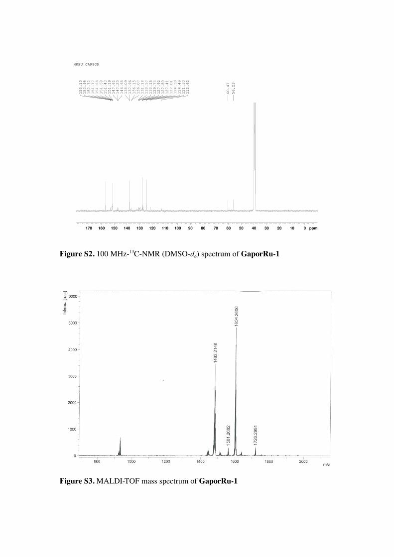
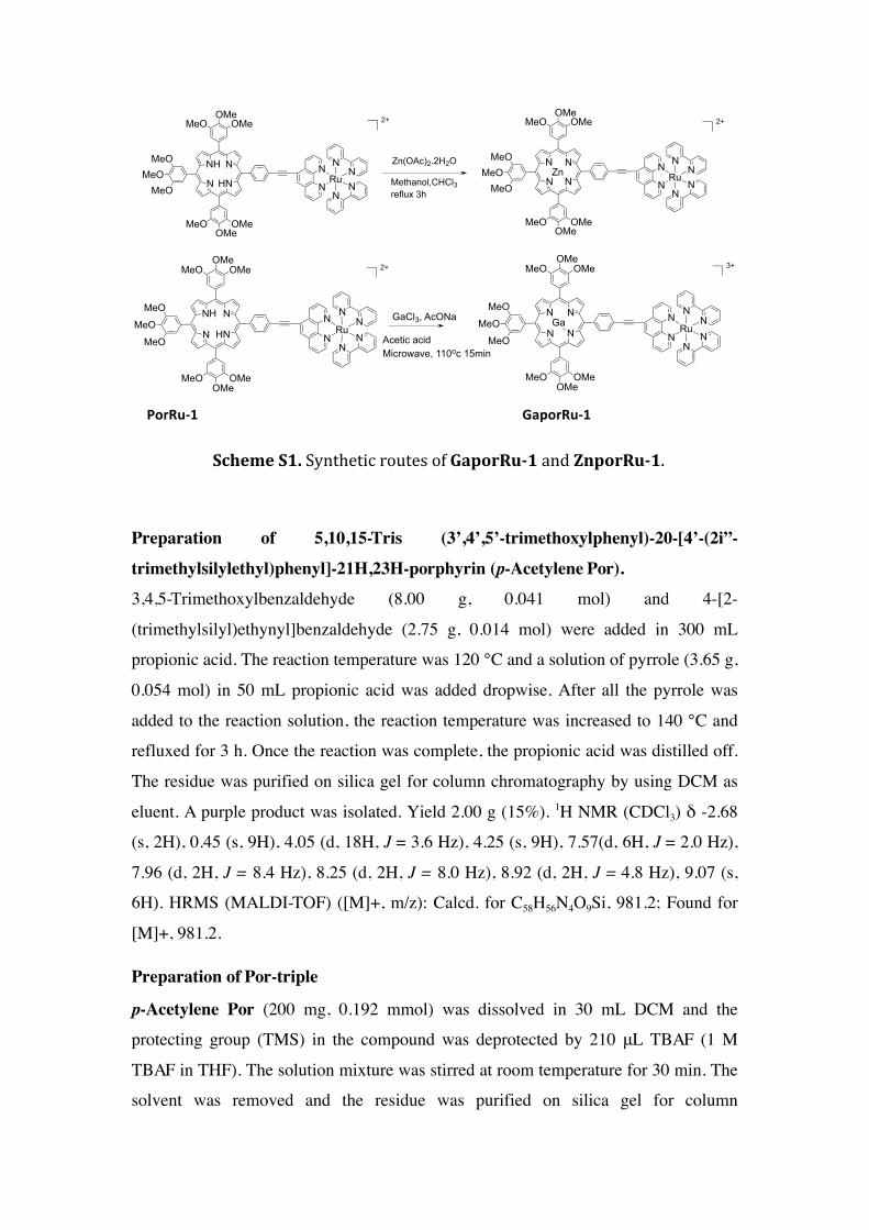
Figure S2. 100 MHz-13C-NMR (DMSO-d6) spectrum of GaporRu-1
Figure S3. MALDI-TOF mass spectrum of GaporRu-1
170 160 150 140 130 120 110 100 90 80 70 60 50 40 30 20 10 0 ppm56.23
60.47
112.62
121.33
124.49
124.59
127.01
127.41
127.80
127.92
129.74
130.16
130.57
131.18
136.07
136.15
137.96
138.09
146.65
147.20
147.62
151.19
151.43
151.50
151.68
151.77
152.72
152.98
153.10
HKBU_CARBON

Figure S4. The HPLC spectra of crude 68GaporRu-1 after 15 minutes microwave
heating at 150 oC (85 % incorporation of radiolabel into the desired product).
Figure S5. The HPLC spectra of crude 68Ga radiolabelling for 68GaporRu-1 without
microwave heating (with only conventional heating up at 100 oC, 1 h, yield 4 %).
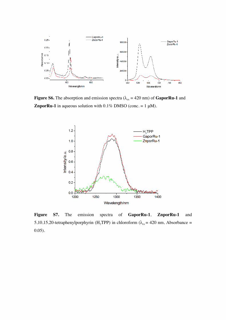
Figure S6. The absorption and emission spectra (λex = 420 nm) of GaporRu-1 and
ZnporRu-1 in aqueous solution with 0.1% DMSO (conc. = 1 μM).
Figure S7. The emission spectra of GaporRu-1, ZnporRu-1 and
5,10,15,20-tetraphenylporphyrin (H2TPP) in chloroform (λex = 420 nm, Absorbance =
0.05).

GaporRu-1 ZnporRu-1
Figure S8. The plot of the emission intensity (λex = 430 nm) of GaporRu-1 (λem =
607 nm) and ZnporRu-1 (λem = 613 nm) in different pH in aqueous solution with 5%
DMSO (pKa values of GaporRu-1 = 3.45 ; ZnporRu-1 = 4.09).
Figure S9. The MTT assay of GaporRu-1 and ZnporRu-1 in MCF-7 cells (24 hours
incubation, conc. 1-50 μM).

Figure S10. The MTT assay of GaporRu-1 and ZnporRu-1 in HeLa cells. (24 hours
incubation, conc. 1-50 μM)
Reference
[1] J. X. Zhang, J. W. Zhou, C. F. Chan, T. C. Lau, D. W. Kwong, H. L. Tam, N. K.
Mak, K. L. Wong and W. K. Wong, Bioconjugate Chem., 2012, 23, 1623-1638.
[2] I. Gupta and M. Ravikanth, J. Chem. Sci., 2005, 117, 161-166.
[3] J. Zhang, K. L. Wong, W. K. Wong, N. K. Mak, W. J. D. Kwong and H. L. Tam,
Org. Biomol. Chem., 2011, 50, 5517-5525.
[4] Y. Li, T. M. Pritchett, J. Huang, M. Ke, P. Shao, and W. Sun, J. Phys. Chem. A
2008, 112, 7200-7207.
[5] P. A. Wilson, Oxford University Press, eds Masters JRW., 2000.
[6] N. K. Mak, K. M. Li, W. N. Leung, N. S. Wong, D. P Huang, M. L. Lung, Y. K.
Lau, C. K. Chang, Biochem Pharmacol., 2004, 68, 2387-2396.