Embed Size (px)
Citation preview
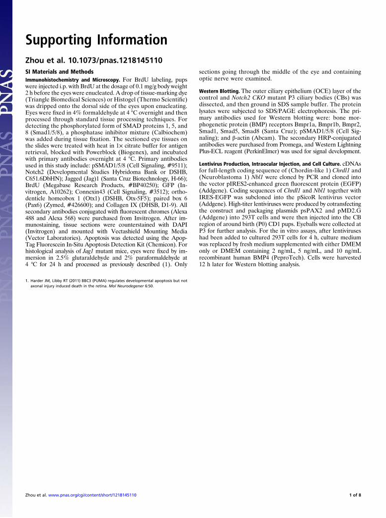
Supporting InformationZhou et al. 10.1073/pnas.1218145110SI Materials and MethodsImmunohistochemistry and Microscopy. For BrdU labeling, pupswere injected i.p. with BrdU at the dosage of 0.1 mg/g body weight2 h before the eyes were enucleated. A drop of tissue-marking dye(Triangle Biomedical Sciences) or Histogel (Thermo Scientific)was dripped onto the dorsal side of the eyes upon enucleating.Eyes were fixed in 4% formaldehyde at 4 °C overnight and thenprocessed through standard tissue processing techniques. Fordetecting the phosphorylated form of SMAD proteins 1, 5, and8 (Smad1/5/8), a phosphatase inhibitor mixture (Calbiochem)was added during tissue fixation. The sectioned eye tissues onthe slides were treated with heat in 1× citrate buffer for antigenretrieval, blocked with Powerblock (Biogenex), and incubatedwith primary antibodies overnight at 4 °C. Primary antibodiesused in this study include: pSMAD1/5/8 (Cell Signaling, #9511);Notch2 (Developmental Studies Hybridoma Bank or DSHB,C651.6DbHN); Jagged (Jag)1 (Santa Cruz Biotechnology, H-66);BrdU (Megabase Research Products, #BP40250); GFP (In-vitrogen, A10262); Connexin43 (Cell Signaling, #3512); ortho-denticle homeobox 1 (Otx1) (DSHB, Otx-5F5); paired box 6(Pax6) (Zymed, #426600); and Collagen IX (DHSB, D1-9). Allsecondary antibodies conjugated with fluorescent chromes (Alexa488 and Alexa 568) were purchased from Invitrogen. After im-munostaining, tissue sections were counterstained with DAPI(Invitrogen) and mounted with Vectashield Mounting Media(Vector Laboratories). Apoptosis was detected using the Apop-Tag Fluorescein In-Situ Apoptosis Detection Kit (Chemicon). Forhistological analysis of Jag1 mutant mice, eyes were fixed by im-mersion in 2.5% glutaraldehyde and 2% paraformaldehyde at4 °C for 24 h and processed as previously described (1). Only
sections going through the middle of the eye and containingoptic nerve were examined.
Western Blotting. The outer ciliary epithelium (OCE) layer of thecontrol and Notch2 CKO mutant P3 ciliary bodies (CBs) wasdissected, and then ground in SDS sample buffer. The proteinlysates were subjected to SDS/PAGE electrophoresis. The pri-mary antibodies used for Western blotting were: bone mor-phogenetic protein (BMP) receptors Bmpr1a, Bmpr1b, Bmpr2,Smad1, Smad5, Smad8 (Santa Cruz); pSMAD1/5/8 (Cell Sig-naling); and β-actin (Abcam). The secondary HRP-conjugatedantibodies were purchased from Promega, and Western LightningPlus-ECL reagent (PerkinElmer) was used for signal development.
Lentivirus Production, Intraocular Injection, and Cell Culture. cDNAsfor full-length coding sequence of (Chordin-like 1) Chrdl1 and(Neuroblastoma 1) Nbl1 were cloned by PCR and cloned intothe vector pIRES2-enhanced green fluorescent protein (EGFP)(Addgene). Coding sequences of Chrdl1 and Nbl1 together withIRES-EGFP was subcloned into the pSicoR lentivirus vector(Addgene). High-titer lentiviruses were produced by cotransfectingthe construct and packaging plasmids psPAX2 and pMD2.G(Addgene) into 293T cells and were then injected into the CBregion of around birth (P0) CD1 pups. Eyeballs were collected atP3 for further analysis. For the in vitro assays, after lentiviruseshad been added to cultured 293T cells for 4 h, culture mediumwas replaced by fresh medium supplemented with either DMEMonly or DMEM containing 2 ng/mL, 5 ng/mL, and 10 ng/mLrecombinant human BMP4 (PeproTech). Cells were harvested12 h later for Western blotting analysis.
1. Harder JM, Libby RT (2011) BBC3 (PUMA) regulates developmental apoptosis but notaxonal injury induced death in the retina. Mol Neurodegener 6:50.
Zhou et al. www.pnas.org/cgi/content/short/1218145110 1 of 8
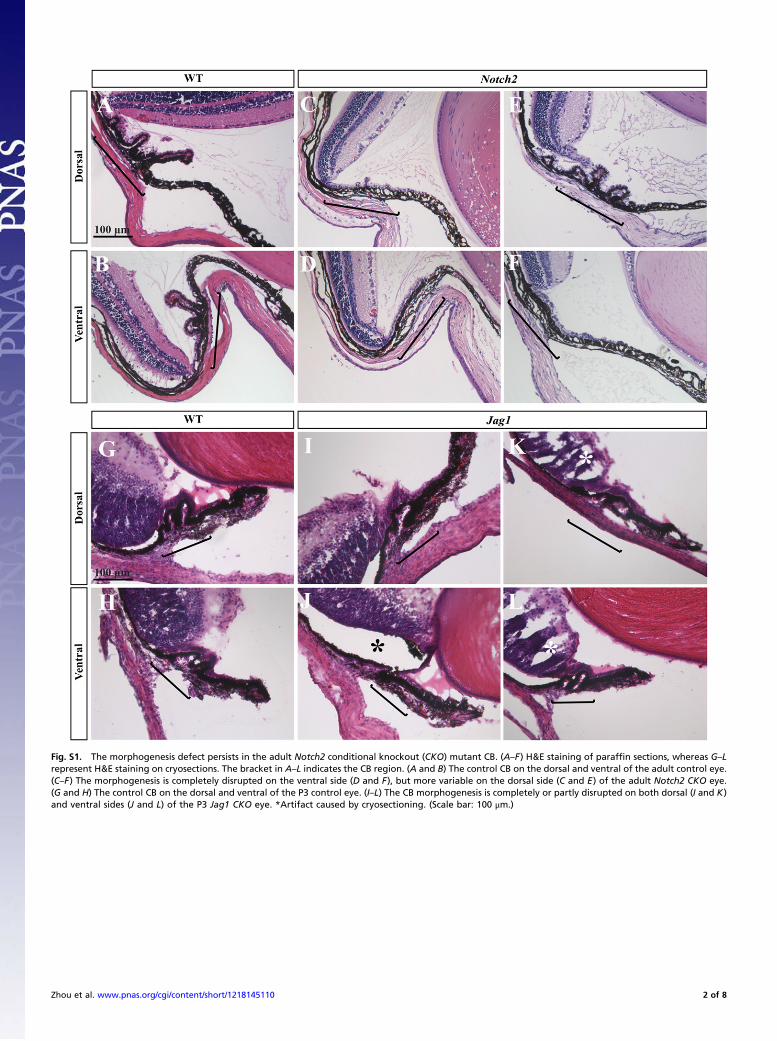
Fig. S1. The morphogenesis defect persists in the adult Notch2 conditional knockout (CKO) mutant CB. (A–F) H&E staining of paraffin sections, whereas G–Lrepresent H&E staining on cryosections. The bracket in A–L indicates the CB region. (A and B) The control CB on the dorsal and ventral of the adult control eye.(C–F) The morphogenesis is completely disrupted on the ventral side (D and F), but more variable on the dorsal side (C and E) of the adult Notch2 CKO eye.(G and H) The control CB on the dorsal and ventral of the P3 control eye. (I–L) The CB morphogenesis is completely or partly disrupted on both dorsal (I and K)and ventral sides (J and L) of the P3 Jag1 CKO eye. *Artifact caused by cryosectioning. (Scale bar: 100 μm.)
Zhou et al. www.pnas.org/cgi/content/short/1218145110 2 of 8
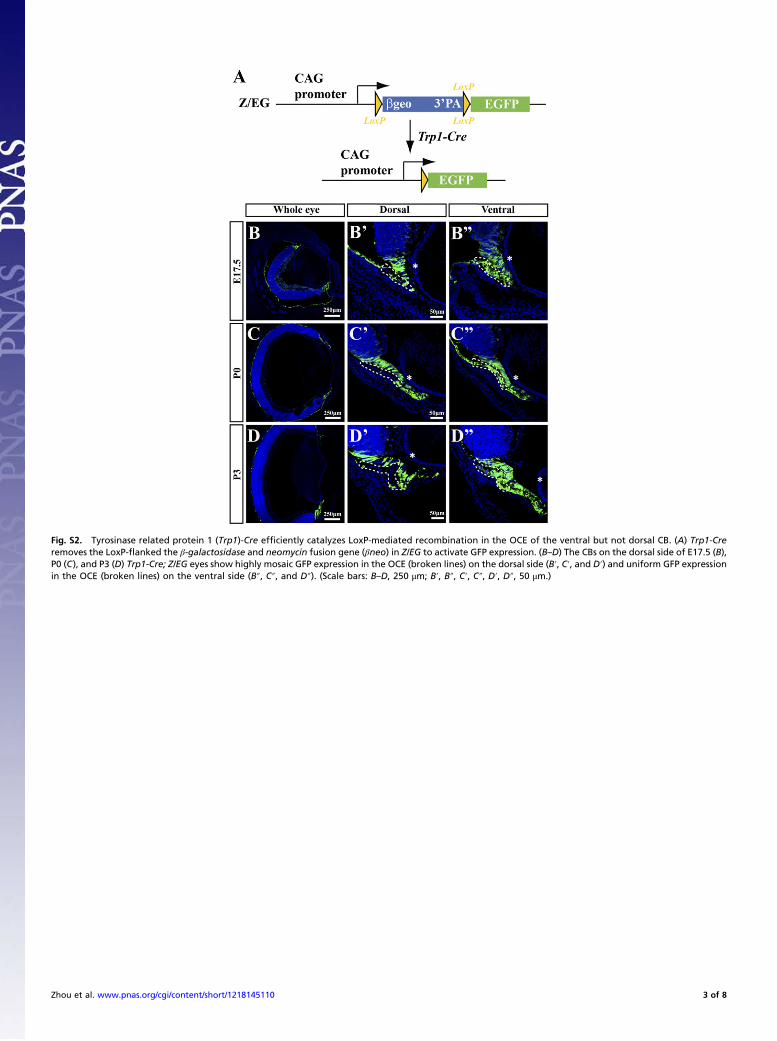
Fig. S2. Tyrosinase related protein 1 (Trp1)-Cre efficiently catalyzes LoxP-mediated recombination in the OCE of the ventral but not dorsal CB. (A) Trp1-Creremoves the LoxP-flanked the β-galactosidase and neomycin fusion gene (βneo) in Z/EG to activate GFP expression. (B–D) The CBs on the dorsal side of E17.5 (B),P0 (C), and P3 (D) Trp1-Cre; Z/EG eyes show highly mosaic GFP expression in the OCE (broken lines) on the dorsal side (B′, C′, and D′) and uniform GFP expressionin the OCE (broken lines) on the ventral side (B″, C″, and D″). (Scale bars: B–D, 250 μm; B′, B″, C′, C″, D′, D″, 50 μm.)
Zhou et al. www.pnas.org/cgi/content/short/1218145110 3 of 8
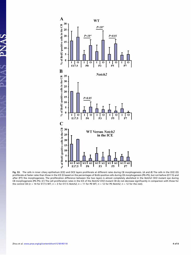
Fig. S3. The cells in inner ciliary epithelium (ICE) and OCE layers proliferate at different rates during CB morphogenesis. (A and B) The cells in the OCE (O)proliferate at faster rates than those in the ICE (I) based on the percentages of BrdU-positive cells during CB morphogenesis (P0–P5), but not before (E17.5) andafter (P7) the morphogenesis. The proliferation difference between the two layers is almost completely abolished in the Notch2 CKO mutant eye duringCB morphogenesis (P0–P5). (C) The cell proliferation rates in the ICE of the Notch2 CKO mutant CB do not decrease significantly in comparison with those forthe control CB (n = 14 for E17.5 WT; n = 3 for E17.5 Notch2; n = 11 for P0 WT; n = 12 for P0 Notch2; n = 12 for the rest).
Zhou et al. www.pnas.org/cgi/content/short/1218145110 4 of 8
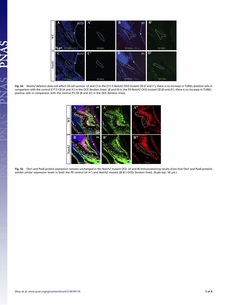
Fig. S4. Notch2 deletion does not affect CB cell survival. (A and C) In the E17.5 Notch2 CKO mutant CB (C and C′), there is no increase in TUNEL-positive cells incomparison with the control E17.5 CB (A and A′) in the OCE (broken lines). (B and D) In the P3 Notch2 CKO mutant CB (D and D′), there is no increase in TUNEL-positive cells in comparison with the control P3 CB (B and B′) in the OCE (broken lines).
Fig. S5. Otx1 and Pax6 protein expression remains unchanged in the Notch2 mutant OCE. (A and B) Immunostaining results show that Otx1 and Pax6 proteinsexhibit similar expression levels in both the P0 control (A–A″) and Notch2 mutant (B–B″) OCEs (broken lines). (Scale bar: 50 μm.)
Zhou et al. www.pnas.org/cgi/content/short/1218145110 5 of 8
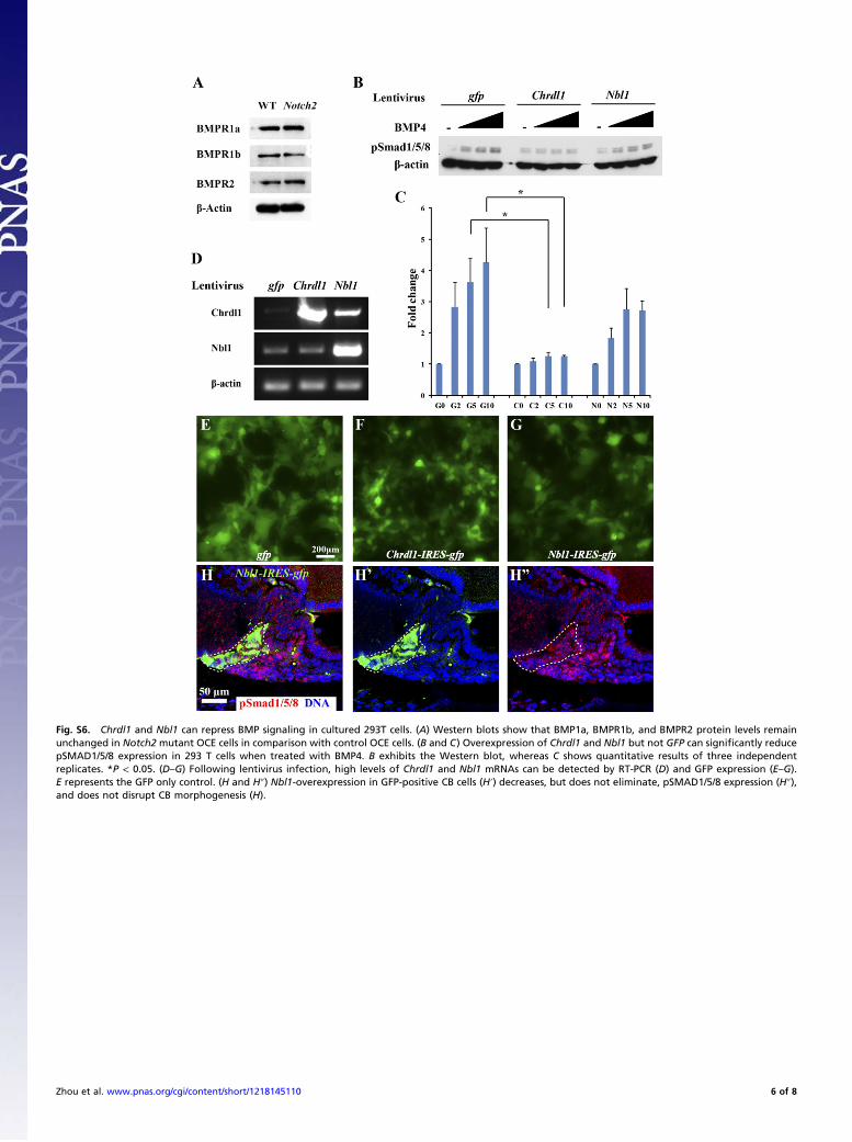
Fig. S6. Chrdl1 and Nbl1 can repress BMP signaling in cultured 293T cells. (A) Western blots show that BMP1a, BMPR1b, and BMPR2 protein levels remainunchanged in Notch2 mutant OCE cells in comparison with control OCE cells. (B and C) Overexpression of Chrdl1 and Nbl1 but not GFP can significantly reducepSMAD1/5/8 expression in 293 T cells when treated with BMP4. B exhibits the Western blot, whereas C shows quantitative results of three independentreplicates. *P < 0.05. (D–G) Following lentivirus infection, high levels of Chrdl1 and Nbl1 mRNAs can be detected by RT-PCR (D) and GFP expression (E–G).E represents the GFP only control. (H and H″) Nbl1-overexpression in GFP-positive CB cells (H′) decreases, but does not eliminate, pSMAD1/5/8 expression (H″),and does not disrupt CB morphogenesis (H).
Zhou et al. www.pnas.org/cgi/content/short/1218145110 6 of 8
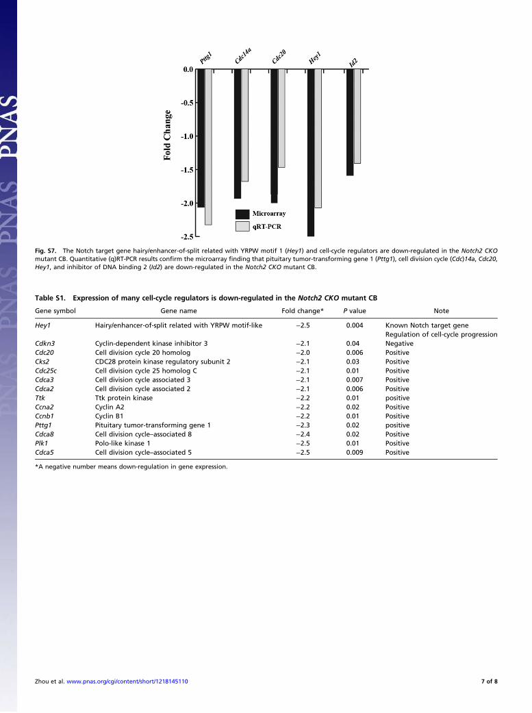
Fig. S7. The Notch target gene hairy/enhancer-of-split related with YRPW motif 1 (Hey1) and cell-cycle regulators are down-regulated in the Notch2 CKOmutant CB. Quantitative (q)RT-PCR results confirm the microarray finding that pituitary tumor-transforming gene 1 (Pttg1), cell division cycle (Cdc)14a, Cdc20,Hey1, and inhibitor of DNA binding 2 (Id2) are down-regulated in the Notch2 CKO mutant CB.
Table S1. Expression of many cell-cycle regulators is down-regulated in the Notch2 CKO mutant CB
Gene symbol Gene name Fold change* P value Note
Hey1 Hairy/enhancer-of-split related with YRPW motif-like −2.5 0.004 Known Notch target geneRegulation of cell-cycle progression
Cdkn3 Cyclin-dependent kinase inhibitor 3 −2.1 0.04 NegativeCdc20 Cell division cycle 20 homolog −2.0 0.006 PositiveCks2 CDC28 protein kinase regulatory subunit 2 −2.1 0.03 PositiveCdc25c Cell division cycle 25 homolog C −2.1 0.01 PositiveCdca3 Cell division cycle associated 3 −2.1 0.007 PositiveCdca2 Cell division cycle associated 2 −2.1 0.006 PositiveTtk Ttk protein kinase −2.2 0.01 positiveCcna2 Cyclin A2 −2.2 0.02 PositiveCcnb1 Cyclin B1 −2.2 0.01 PositivePttg1 Pituitary tumor-transforming gene 1 −2.3 0.02 positiveCdca8 Cell division cycle–associated 8 −2.4 0.02 PositivePlk1 Polo-like kinase 1 −2.5 0.01 PositiveCdca5 Cell division cycle–associated 5 −2.5 0.009 Positive
*A negative number means down-regulation in gene expression.
Zhou et al. www.pnas.org/cgi/content/short/1218145110 7 of 8
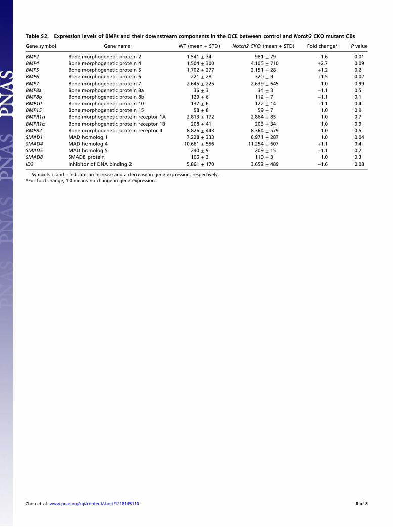
Table S2. Expression levels of BMPs and their downstream components in the OCE between control and Notch2 CKO mutant CBs
Gene symbol Gene name WT (mean ± STD) Notch2 CKO (mean ± STD) Fold change* P value
BMP2 Bone morphogenetic protein 2 1,541 ± 74 981 ± 79 −1.6 0.01BMP4 Bone morphogenetic protein 4 1,504 ± 300 4,105 ± 710 +2.7 0.09BMP5 Bone morphogenetic protein 5 1,702 ± 277 2,151 ± 28 +1.2 0.2BMP6 Bone morphogenetic protein 6 221 ± 28 320 ± 9 +1.5 0.02BMP7 Bone morphogenetic protein 7 2,645 ± 225 2,639 ± 645 1.0 0.99BMP8a Bone morphogenetic protein 8a 36 ± 3 34 ± 3 −1.1 0.5BMP8b Bone morphogenetic protein 8b 129 ± 6 112 ± 7 −1.1 0.1BMP10 Bone morphogenetic protein 10 137 ± 6 122 ± 14 −1.1 0.4BMP15 Bone morphogenetic protein 15 58 ± 8 59 ± 7 1.0 0.9BMPR1a Bone morphogenetic protein receptor 1A 2,813 ± 172 2,864 ± 85 1.0 0.7BMPR1b Bone morphogenetic protein receptor 1B 208 ± 41 203 ± 34 1.0 0.9BMPR2 Bone morphogenetic protein receptor II 8,826 ± 443 8,364 ± 579 1.0 0.5SMAD1 MAD homolog 1 7,228 ± 333 6,971 ± 287 1.0 0.04SMAD4 MAD homolog 4 10,661 ± 556 11,254 ± 607 +1.1 0.4SMAD5 MAD homolog 5 240 ± 9 209 ± 15 −1.1 0.2SMAD8 SMAD8 protein 106 ± 3 110 ± 3 1.0 0.3ID2 Inhibitor of DNA binding 2 5,861 ± 170 3,652 ± 489 −1.6 0.08
Symbols + and – indicate an increase and a decrease in gene expression, respectively.*For fold change, 1.0 means no change in gene expression.
Zhou et al. www.pnas.org/cgi/content/short/1218145110 8 of 8