Embed Size (px)
Citation preview
Kang et al.
Supplementary Information
Title
Degeneration and impaired regeneration of gray matter oligodendrocytes in
amyotrophic lateral sclerosis
Authors
Shin H. Kang, Ying Li, Masahiro Fukaya, Ileana Lorenzini, Don W. Cleveland, Lyle W.
Ostrow, Jeffrey D. Rothstein, and Dwight E. Bergles
Nature Neuroscience: doi:10.1038/nn.3357

Kang et al.
Supplementary Fig. 1 Analysis of NG2+ cell proliferation and differentiation in the spinal cord.
(a) Schematic showing the three spinal cord regions that were examined for cell proliferation and fate
analyses of NG2+ cells and oligodendrocytes. (b) Confocal images of spinal cord sections from
Pdgfra-creER;Z/EG mice 4 days after 4HT administration at P30 (P30+4) showing that EGFP
expression was restricted to NG2+ cells (PDGFR+Olig2+) (c) Confocal images of spinal cord sections
from Pdgfra-creER;Z/EG mice two months after administration of 4HT at P30 (P30+60), showing that
many EGFP+ cells were now CC1+ and PDGFR– (white arrowheads), indicating that they had
differentiated into oligodendrocytes; other cells remained in the progenitor state (PDGFR+) (yellow
arrows). Scale bars: 20 m. (d - f) Graphs showing the density of EGFP+ cells in the ventral gray
matter (d), ventral white matter (e), and dorsal gray matter (f) of the spinal cord from Pdgfra-
creER;Z/EG ± SOD1 (G93A) mice. 4HT was administered either at P30 or P60, and mice were
examined at P90 or P120. Data are presented as mean + s.e.m. (n = 9 sections from 3 mice per
group). * P < 0.05, ** P < 0.0005, *** P < 0.0001, unpaired Student’s t test.
Nature Neuroscience: doi:10.1038/nn.3357
Kang et al.
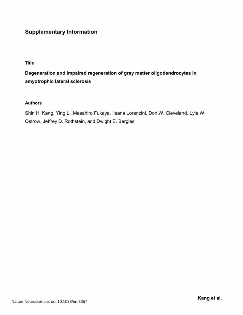
Supplementary Fig. 2 Genetic labeling of spinal cord oligodendrocytes. (a) Fluorescence image
of the ventral horn of the spinal cord from a Plp1-creER;ROSA26-EYFP mouse ten days after
administration of 4HT at P30 (P30+10). (b) High magnification confocal image of the region
highlighted by the yellow box in (a) showing EYFP+ cells that were PDGFR– and NG2– (white
arrowheads). (c) Confocal images of spinal cord showing that EYFP+ cells were CC1+ and Olig2+
(yellow arrows). (d) Confocal images showing the presence of EYFP+ astrocytes that were GFAP+
(asterisk). Astrocytes could be clearly distinguished from oligodendrocytes by their morphology and
lack of CC1 immunoreactivity (yellow arrows). Scale bars: 20 m (b - d).
Nature Neuroscience: doi:10.1038/nn.3357

Kang et al.
Supplementary Fig. 3 Oligodendrocyte-specific expression of EGFP in Mobp-EGFP mice.
(a,b) Fluorescence images of spinal cord from an adult Mobp-EGFP mouse (P70) immunostained for
EGFP (a) and NG2 (b). (c) High magnification confocal images of spinal cord from a Mobp-EGFP
mouse showing co-localization between EGFP, CC1, and Olig2 (yellow arrows), indicating that EGFP
is expressed by oligodendrocytes. (d) Confocal images of spinal cord sections immunostained for
EGFP, NG2 and PDGFR, indicating that EGFP is not expressed by NG2+ cells in these mice. Scale
bars: 20 m.
Nature Neuroscience: doi:10.1038/nn.3357

Kang et al.
Supplementary Fig. 4 Ablation of spinal motor neurons does not induce reactive changes in
NG2+ cells. (a) Transverse section of lumbar spinal cord from a mouse injected with ricin showing
NG2 immunoreactivity (brown) and cresyl violet (purple). Scale bar: 200 μm. (b) Magnified views of
boxed areas in (a). Boxed areas in upper panel are magnified in lower panels. Note that most motor
neurons (arrowheads, blue cell bodies) were lost from the ipsilateral side, but preserved on the
contralateral side. NG2 immunoreactivity was similar between the contralateral and ipsilateral sides
(black arrows). Red arrows highlight thin NG2+ cell processes. Scale bar: 50 μm. (c) Quantification of
motor neurons in the lumbar spinal cord. Data are presented as mean + s.e.m. (n = 14 sections from
3 mice per group) P = 0.0001, paired Student’s t test. (d) Quantification of Ki67+ NG2+ cells. Data are
presented as mean + s.e.m. (n = 4 sections from 3 mice per group) P > 0.05, paired Student’s t test.
Nature Neuroscience: doi:10.1038/nn.3357

Kang et al.
Supplementary Fig. 5 Progressive alterations in the morphology of oligodendrocytes in the
spinal cord of ALS mice. (a) Fluorescence images from Mobp-EGFP (top panels) and Mobp-
EGFP;SOD1 (G93A) mice (bottom panels). In P120 control mice, EGFP was primarily restricted to
the somata of oligodendrocytes, whereas in end stage SOD1 (G93A) mice, numerous large EGFP+
structures were visible that lacked DAPI+ nuclei and were Olig2– (yellow arrows). (b) Images of
EGFP+ structures from these mice at P90 and end stage SOD1 (G93A) mice. (c) Selected images of
EGFP+ structures from cells highlighted by yellow squares in (b), showing that EGFP+ Olig2–
structures (oligodendrocyte somata and putative apoptotic bodies) are present by P90 in SOD1
(G93A) mice (middle panels). Scale bars: 20 m (a) and 5 m (c). (d) Quantification of
oligodendrocyte-derived apoptotic bodies. Schematic at right shows how images were processed and
left panels show representative images from the ventral gray matter of Mobp-EGFP (P120, control)
and end stage Mobp-EGFP;SOD1 (G93A) mice. Sections of spinal cord were immunostained for
EGFP, Olig2 and CC1 and imaged using a confocal fluorescence microscope. Surface rendering of
EGFP+ structures was performed using Imaris, and Olig2+EGFP+ structures (oligodendrocyte somata)
were digitally subtracted. The volume of the remaining EGFP+Olig2– structures was determined. In
this example, the volume of these putative apoptotic bodies was 89 m3 in control mice and 1705 m3
in SOD1 (G93A) mice. Scale bars: 20 m.
Nature Neuroscience: doi:10.1038/nn.3357
Kang et al.
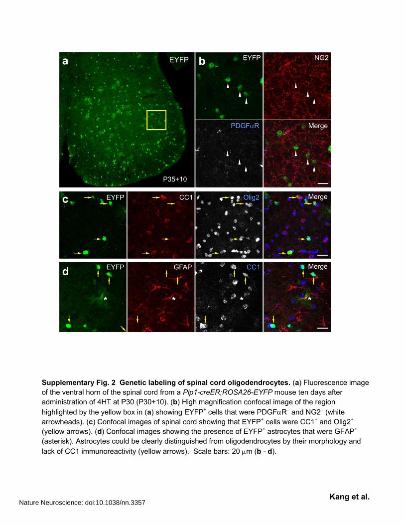
Supplementary Fig. 6 Degradation of myelinated axons and altered myelin thickness in the
spinal cord gray matter of end stage ALS mice. (a) Electron micrograph showing degenerating
axons with swollen myelin (white arrowheads) in the spinal cord ventral gray matter of end stage
SOD1 (G93A) mice. Scale bar: 1 m. (b) Cumulative plot showing the distribution of g ratios for
compact myelin surrounding non-degenerating axons in control mice at P120 and SOD1 (G93A) mice
at end stage. (Control, n = 119 axons from 3 mice; SOD1 (G93A), n = 103 axons from 3 mice) (c,d)
Scatter plot of g-ratio vs. axon diameter. The g ratio of end stage SOD1 (G93A) mice was significantly
smaller than that of P120 control mice. P = 0.0001, Kolmogorov-Smirnov test.
Nature Neuroscience: doi:10.1038/nn.3357

Kang et al.
Supplementary Fig. 7 Decrease in myelin protein abundance in the spinal cord of ALS mice.
(a) Western blotting of myelin proteins (MBP, MOG, and CNPase) and PDGFR in wild type and
SOD1 (G93A) mice and different stages of disease. (b) Graphs showing the protein expression levels
in ALS mice relative to control, after normalization to GAPDH. Data are presented as mean ± s.e.m.
(n = 3) * P < 0.05, ** P < 0.01, *** P < 0.005. Student’s t test. (c) Luxol fast blue staining of lumbar
spinal cord from control (P120) and end stage SOD1 (G93A) mice. Lower panels are magnified
images of boxed areas in upper panels. GM, gray matter, WM, white matter.
Nature Neuroscience: doi:10.1038/nn.3357
Kang et al.
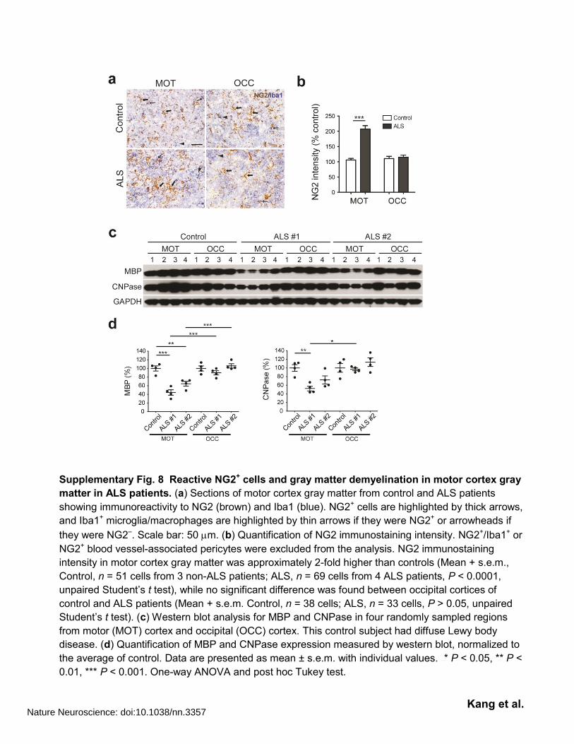
Supplementary Fig. 8 Reactive NG2+ cells and gray matter demyelination in motor cortex gray
matter in ALS patients. (a) Sections of motor cortex gray matter from control and ALS patients
showing immunoreactivity to NG2 (brown) and Iba1 (blue). NG2+ cells are highlighted by thick arrows,
and Iba1+ microglia/macrophages are highlighted by thin arrows if they were NG2+ or arrowheads if
they were NG2–. Scale bar: 50 m. (b) Quantification of NG2 immunostaining intensity. NG2+/Iba1+ or
NG2+ blood vessel-associated pericytes were excluded from the analysis. NG2 immunostaining
intensity in motor cortex gray matter was approximately 2-fold higher than controls (Mean + s.e.m.,
Control, n = 51 cells from 3 non-ALS patients; ALS, n = 69 cells from 4 ALS patients, P < 0.0001,
unpaired Student’s t test), while no significant difference was found between occipital cortices of
control and ALS patients (Mean + s.e.m. Control, n = 38 cells; ALS, n = 33 cells, P > 0.05, unpaired
Student’s t test). (c) Western blot analysis for MBP and CNPase in four randomly sampled regions
from motor (MOT) cortex and occipital (OCC) cortex. This control subject had diffuse Lewy body
disease. (d) Quantification of MBP and CNPase expression measured by western blot, normalized to
the average of control. Data are presented as mean ± s.e.m. with individual values. * P < 0.05, ** P <
0.01, *** P < 0.001. One-way ANOVA and post hoc Tukey test.
Nature Neuroscience: doi:10.1038/nn.3357

Kang et al.
Supplementary Fig. 9 Excision of SOD1 (G37R) from NG2+ cells reduced gliosis. (a) Delayed
gliosis at normal age of disease onset after SOD1 (G37R) deletion in oligodendroglia. Images show
sections of lumbar spinal cord ventral horn from Pdgfra-creER;loxSOD1 (G37R) mice ± 4HT that
were immunostained for GFAP and Iba1. White arrowheads in upper left panel highlight region of
increased GFAP immunoreactivity in the ventral horn gray matter. Note the reduced gliosis in
sections from 4HT treated mice, as indicated by the lower GFAP and Iba1immunoreactivity. Dashed
lines indicate the border between gray and white matter. Scale bar: 100 μm. (b) Immunofluorescence
staining showing preservation of mutant SOD1 expression (green) in lumbar spinal cord neurons
(SMI32+, red) in Pdgfra-creER;loxSOD1 (G37R) mice with and without 4HT treatment. Scale bar: 50
μm. (c, d) Western blot showing that SOD1 expression was slightly lower (P = 0.0512) in the lumbar
spinal cord in 4HT treated mice. Data are presented as mean + s.e.m. (–4HT, n = 3; +4HT, n = 4),
unpaired Student’s t test.
Nature Neuroscience: doi:10.1038/nn.3357

Kang et al.
Supplementary Fig. 10 Abnormal dynamics of gray matter oligodendrocytes and their progenitors in ALS. (a) Schematic representation of the change in oligodendrocyte number in the normal and ALS spinal cord. Black line represents the total number of oligodendrocytes, the red line shows the fate of oligodendrocytes born early in postnatal life, and the blue line show the generation of new oligodendrocytes in adult life (note this shows only net accumulation). The progressive degeneration of early-born oligodendrocytes in ALS enhances oligodendrogenesis to maintain their density. (b) Model derived from genetic fate tracing of both NG2+ cells (green, middle) and oligodendrocytes showing the relationship between NG2+ cell proliferation and oligodendrocyte generation in the normal spinal cord, and oligodendrocyte degeneration and enhanced proliferation and differentiation of NG2+ cells in the ALS spinal cord. The death of early-born oligodendrocytes (red) triggers an increase in differentiation of NG2+ cells to maintain the oligodendrocyte population. NG2+ cell proliferation is enhanced to replace cells that have differentiated (blue), thereby maintaining their density.
Nature Neuroscience: doi:10.1038/nn.3357

Kang et al.
Supplementary Fig. 11 Full-length pictures of the blots presented in the main figures.
Nature Neuroscience: doi:10.1038/nn.3357
Kang et al.
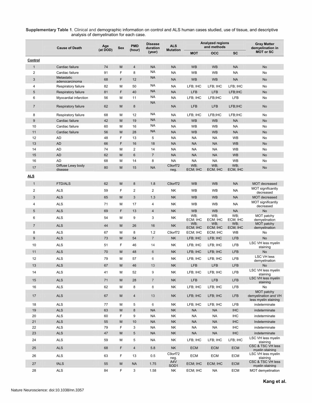
Supplementary Table 1. Clinical and demographic information on control and ALS human cases studied, use of tissue, and descriptive analysis of demyelination for each case.
Cause of Death
Age (at DOD)
Sex PMD
(hour)
Disease duration
(year)
ALS Mutation
Analyzed regions and methods
Gray Matter demyelination in
MOT or SC MOT OCC SC
Control
1 Cardiac failure 74 M 4 NA NA WB WB NA No
2 Cardiac failure 91 F 8 NA NA WB WB NA No
3 Metastatic adenocarcinoma
68 F 12 NA
NA WB WB NA No
4 Respiratory failure 82 M 50 NA NA LFB; IHC LFB; IHC LFB; IHC No
5 Respiratory failure 81 F 40 NA NA LFB LFB LFB;IHC No
6 Myocardial infarction 56 M 11 NA NA LFB; IHC LFB;IHC LFB No
7 Respiratory failure 62 M 8 NA
NA LFB LFB LFB;IHC No
8 Respiratory failure 68 M 12 NA NA LFB; IHC LFB;IHC LFB;IHC No
9 Cardiac failure 42 M 19 NA NA WB WB NA No
10 Cardiac failure 60 M 16 NA NA WB WB NA No
11 Cardiac failure 56 M 28 NA NA WB WB NA No
12 AD 48 F 13 5 NA NA NA WB No
13 AD 66 F 16 18 NA NA NA WB No
14 AD 74 M 2 14 NA NA NA WB No
15 AD 62 M 6 7 NA NA NA WB No
16 AD 68 M 14 8 NA NA NA WB No
17 Diffuse Lewy body disease
80 M 15 NA C9orf72
neg. WB;
ECM; IHC WB;
ECM, IHC WB;
ECM, IHC No
ALS
1 FTD/ALS 62 M 8 1.8 C9orf72 WB WB NA MOT decreased
2 ALS 59 F 2 2 NK WB WB NA MOT significantly
decreased
3 ALS 65 M 3 1.3 NK WB WB NA MOT decreased
4 ALS 71 M 17 4 NK WB WB NA MOT significantly
decreased
5 ALS 69 F 13 4 NK WB WB NA No
6 ALS 54 M 9 3 NK WB;
ECM; IHC WB;
ECM; IHC WB;
ECM; IHC MOT patchy
demyelination
7 ALS 44 M 26 16 NK WB;
ECM; IHC WB;
ECM; IHC WB;
ECM; IHC MOT patchy
demyelination
8 ALS 67 M 8 1.2 C9orf72 ECM; IHC ECM; IHC WB No
9 ALS 73 M 54 7 NK LFB; IHC LFB; IHC LFB No
10 ALS 51 F 46 14 NK LFB; IHC LFB; IHC LFB LSC VH less myelin
staining
11 ALS 70 M 48 6 NK LFB; IHC LFB; IHC LFB No
12 ALS 79 M 57 6 NK LFB; IHC LFB; IHC LFB LSC VH less demyelination
13 ALS 67 M 46 13 NK LFB LFB LFB No
14 ALS 41 M 52 9 NK LFB; IHC LFB; IHC LFB LSC VH less myelin
staining
15 ALS 71 M 28 7 NK LFB LFB LFB LSC VH less myelin
staining
16 ALS 62 M 8 8 NK LFB; IHC LFB; IHC LFB No
17 ALS 67 M 4 13 NK LFB; IHC LFB; IHC LFB MOT patchy
demyelination and VH less myelin staining
18 ALS 77 M 5 6 NK LFB; IHC LFB; IHC LFB indeterminate
19 ALS 63 M 8 NA NK NA NA IHC indeterminate
20 ALS 60 F 9 NA NK NA NA IHC indeterminate
21 ALS 55 M 10 NA NK NA NA IHC Indeterminate
22 ALS 79 F 3 NA NK NA NA IHC indeterminate
23 ALS 47 M 5 NA NK NA NA IHC indeterminate
24 ALS 59 M 5 NA NK LFB; IHC LFB; IHC LFB; IHC LSC VH less myelin
staining
25 ALS 68 F 4 5.8 NK ECM ECM ECM CSC & TSC VH less
myelin staining
26 ALS 63 F 13 0.5 C9orf72
neg. ECM ECM ECM
LSC VH less myelin staining
27 fALS 55 M NA 1.75 A4V
SOD1 ECM; IHC ECM; IHC ECM
CSC & TSC VH less myelin staining
28 ALS 84 F 3 1.58 NK ECM; IHC NA ECM MOT demyelination
Nature Neuroscience: doi:10.1038/nn.3357

Kang et al.
Notes: AD: Alzheimer’s disease; fALS: familial ALS; C9orf72 neg: C9orf72 repeats negative; CSC: cervical spinal cord; DOD: Date of death; ECM: Erichrome Cyanine R myelin staining; F: female; FTD: frontal-temporal dementia; GM: gray matter; IHC: immunohistochemistry; LFB: Luxol fast blue myelin staining; LSC: Lumbar spinal cord; M: male; MOT: motor cortex; NA: not applicable/available; NK: not known; PMD: postmortem duration; SOD1: superoxide dismutase 1; TSC: thoracic spinal cord; VH: ventral horn; WB: Western blot analysis. c9orf72: c9orf72 repeat expansion and c9orf72 neg: c9orf72 repeat expansion negative. Post mortem delay average (± s.e.m.): 16.2 ± 3 hrs Control (range: 2-50), 15.5 ± 3 hrs ALS (range: 2-57) Median: Control 13 hrs; ALS, 8 hrs (No significant differences between post mortem delay) Mean age (± s.e.m.): Control: 66 ± 3 yrs ALS: 62 ± 2 yrs (No significant difference in ages)
29 ALS 65 F NA NA NK ECM; IHC NA NA MOT demyelination
30 ALS 56 F 4 3 C9orf72
repeats + ECM ECM ECM; WB indeterminate
31 ALS 64 F 13 1 NK NA NA ECM indeterminate
32 fALS 66 M 6 1.8 C9orf72 ECM; IHC ECM; IHC ECM; IHC indeterminate
33 ALS 49 F 6 2.5 NK NA NA WB; IHC LSC VH
demyelination
34 ALS 71 M 13 0.5 NK NA NA WB; IHC No
35 ALS 62 F 5 3 NK NA NA WB; IHC LSC VH
demyelination
36 ALS 68 M 11 2 NK NA NA WB; IHC No
37 ALS 61 M 10 10 NK NA NA WB; IHC No
38 ALS 34 F 5 1 C9orf72 NA NA ECM LSC VH
demyelination
39 fALS 47 M 5 3 C9orf72 NA NA ECM indeterminate
Nature Neuroscience: doi:10.1038/nn.3357