Embed Size (px)
Citation preview
1
Supplementary Information for ‘Ultrasound and pH Dually Responsive Polymer Vesicles
for Anticancer Drug Delivery’
Wenqin Chen and Jianzhong Du*
School of Materials Science and Engineering, 4800 Caoan Road, Shanghai, 201804, China. E-mail:
Supplementary Figures
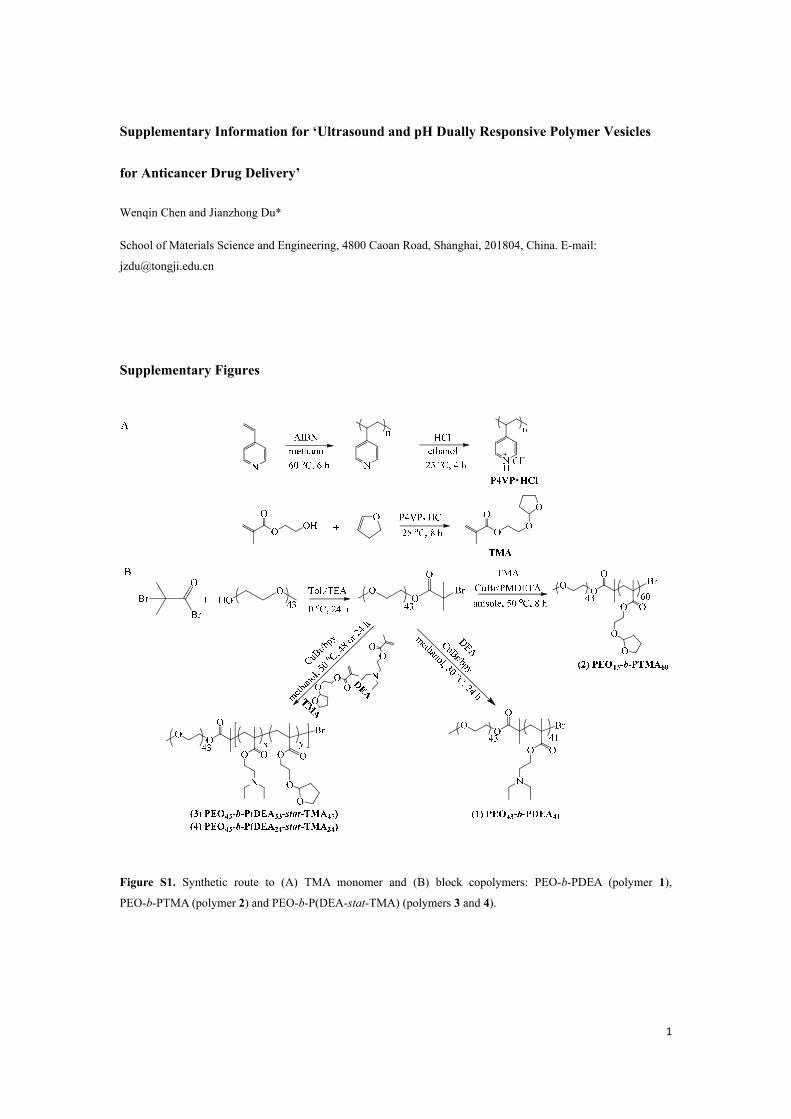
Figure S1. Synthetic route to (A) TMA monomer and (B) block copolymers: PEO-b-PDEA (polymer 1),
PEO-b-PTMA (polymer 2) and PEO-b-P(DEA-stat-TMA) (polymers 3 and 4).
2
6.5 6.0 5.5 5.0 4.5 4.0 3.5 3.0 2.5 2.0 1.5
δ (ppm)
h i fe
da
b+c +g
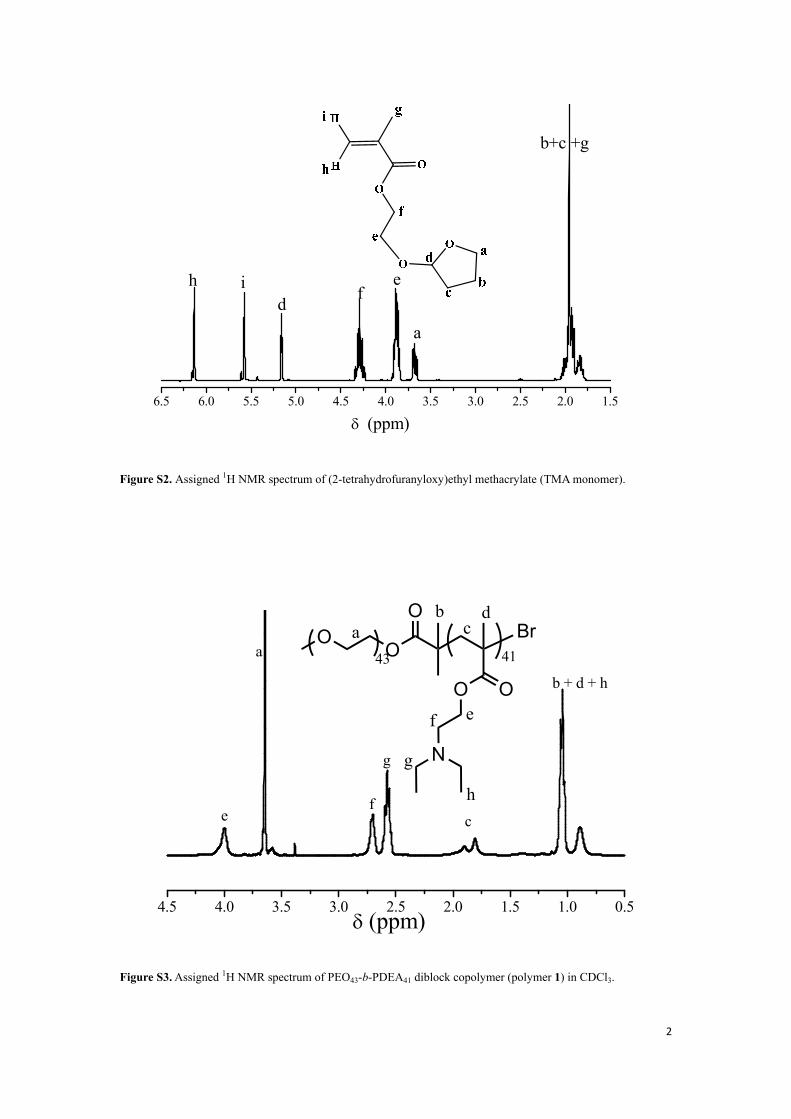
Figure S2. Assigned 1H NMR spectrum of (2-tetrahydrofuranyloxy)ethyl methacrylate (TMA monomer).
4.5 4.0 3.5 3.0 2.5 2.0 1.5 1.0 0.5
g
ef
a
c
b + d + h
OO43
O
OO
N
Br41
adb
c
ef
g
h
δ (ppm)
Figure S3. Assigned 1H NMR spectrum of PEO43-b-PDEA41 diblock copolymer (polymer 1) in CDCl3.
3
5 4 3 2 1
D
C
B
s'+t'
p'+w'+a'+m'h'+i'+n'+q'
h+i+n+q p+w+a+ms+t
g'c'r'+k'+j'+l'
r+k
j+la+m
a
h+i+n
d
k
c
g
δ (ppm)
OO43
O
O O
N
33
p'q'
r' s'
t'w'
Br
OO
O
O
47
k'
g'l'
i'h'
n'
j'
d'
m'
c'
a'
OO43
O
O O
N
33
pq
r s
tw
Br
OO
O
O
47
43
g hi
j
l
mnO
OBr
O
OO
60
O
O
k
b A
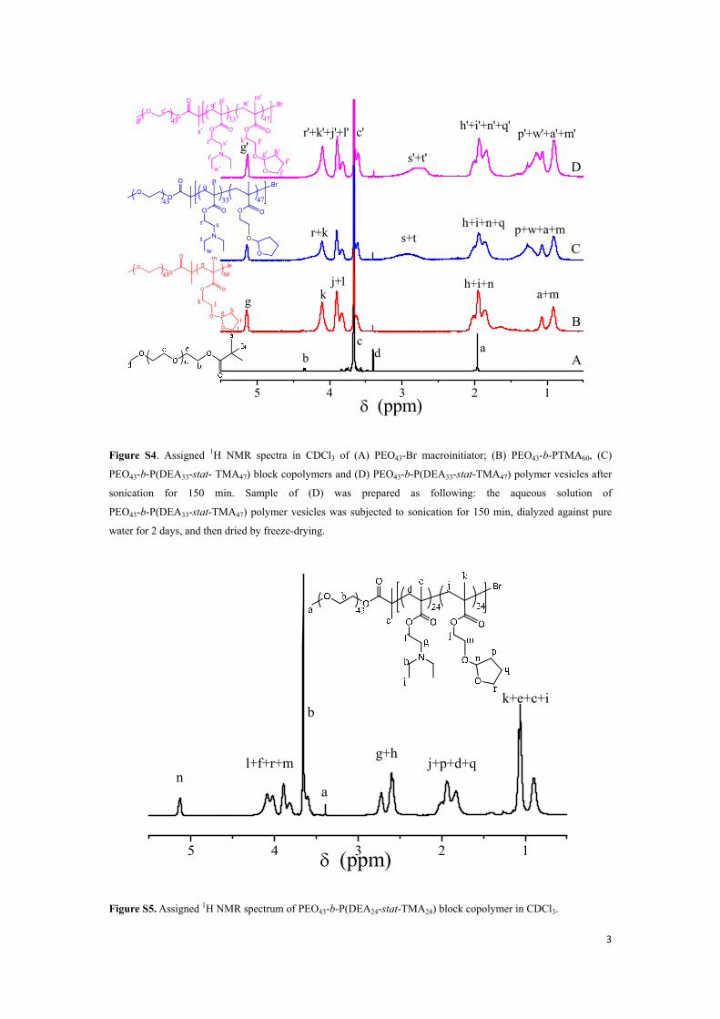
Figure S4. Assigned 1H NMR spectra in CDCl3 of (A) PEO43-Br macroinitiator; (B) PEO43-b-PTMA60, (C)
PEO43-b-P(DEA33-stat- TMA47) block copolymers and (D) PEO43-b-P(DEA33-stat-TMA47) polymer vesicles after
sonication for 150 min. Sample of (D) was prepared as following: the aqueous solution of
PEO43-b-P(DEA33-stat-TMA47) polymer vesicles was subjected to sonication for 150 min, dialyzed against pure
water for 2 days, and then dried by freeze-drying.
5 4 3 2 1
k+e+c+i
j+p+d+qn
g+hl+f+r+m
a
δ (ppm)
b
Figure S5. Assigned 1H NMR spectrum of PEO43-b-P(DEA24-stat-TMA24) block copolymer in CDCl3.
4
12 15 18
(a)(b)(c)(d)
Elution Time (min)
Mn Mw/Mn
(a) 7300 1.16(b) 7700 1.18(c) 11000 1.26(d) 19000 1.25
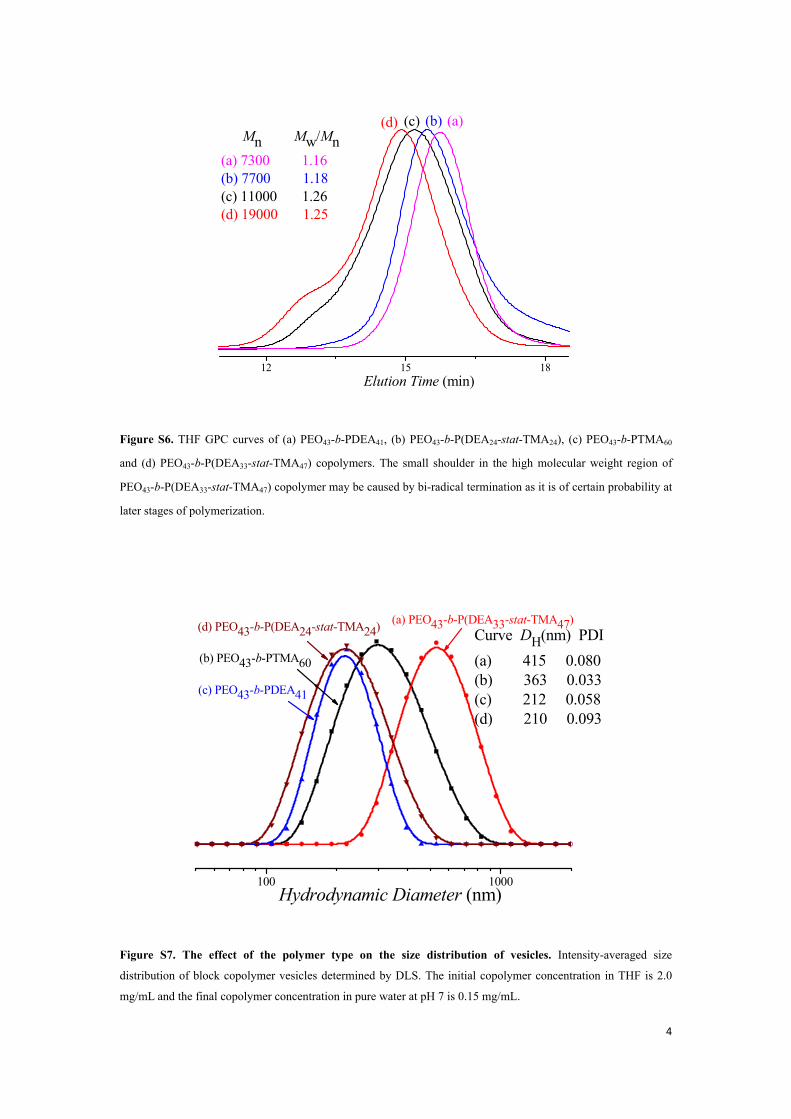
Figure S6. THF GPC curves of (a) PEO43-b-PDEA41, (b) PEO43-b-P(DEA24-stat-TMA24), (c) PEO43-b-PTMA60
and (d) PEO43-b-P(DEA33-stat-TMA47) copolymers. The small shoulder in the high molecular weight region of
PEO43-b-P(DEA33-stat-TMA47) copolymer may be caused by bi-radical termination as it is of certain probability at
later stages of polymerization.
100 1000
Curve DH(nm) PDI(a) 415 0.080(b) 363 0.033(c) 212 0.058(d) 210 0.093
(d) PEO43-b-P(DEA24-stat-TMA24)
(c) PEO43-b-PDEA41
(b) PEO43-b-PTMA60
(a) PEO43-b-P(DEA33-stat-TMA47)
Hydrodynamic Diameter (nm)
Figure S7. The effect of the polymer type on the size distribution of vesicles. Intensity-averaged size
distribution of block copolymer vesicles determined by DLS. The initial copolymer concentration in THF is 2.0
mg/mL and the final copolymer concentration in pure water at pH 7 is 0.15 mg/mL.
5
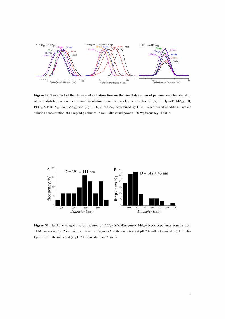
Figure S8. The effect of the ultrasound radiation time on the size distribution of polymer vesicles. Variation
of size distribution over ultrasound irradiation time for copolymer vesicles of (A) PEO43-b-PTMA60, (B)
PEO43-b-P(DEA33-stat-TMA47) and (C) PEO43-b-PDEA41 determined by DLS. Experimental conditions: vesicle
solution concentration: 0.15 mg/mL; volume: 15 mL. Ultrasound power: 180 W; frequency: 40 kHz.
Figure S9. Number-averaged size distribution of PEO43-b-P(DEA33-stat-TMA47) block copolymer vesicles from
TEM images in Fig. 2 in main text: A in this figure→A in the main text (at pH 7.4 without sonication); B in this
figure→C in the main text (at pH 7.4, sonication for 90 min).
100 150 200 250 300 350 4000
5
10
15
20
25
30
D = 148 ± 43 nm
freq
uenc
y(%
)
Diameter (nm)200 300 400 500
0
6
12
18
24
D = 391 ± 111 nm
freq
uenc
y(%
)
Diameter (nm)
100 1000 Hydrodynamic Diameter (nm)
150 min120 min
90 min60 min 30 min
10 min0 min
A: PEO43-b-PTMA60
100 1000
C: PEO43-b-PDEA41
150 min120 min 60 min
30 min
90 min
10 min
Hydrodynamic Diameter (nm)
0 min
100 1000
B: PEO43-b-P(DEA33-stat-TMA47)
Hydrodynamic Diameter (nm)
90 min150 min
60 min 30 min 10 min 0 min
A B
6
-20 0 20 40 60-2.5
-2.0
-1.5
-1.0
-0.5
Tg,4= 42.2 oC
Tg,3= 7.7 oC
Tg,2= 39.0 oC(b) PEO43-b-PTMA60
(a) PEO43-b-P(DEA33-stat-TMA47)
Tg,1= -6.6 oC
Hea
t Flo
w (W
/g)
Temperature (oC)
Tc = 20-30 oC
�
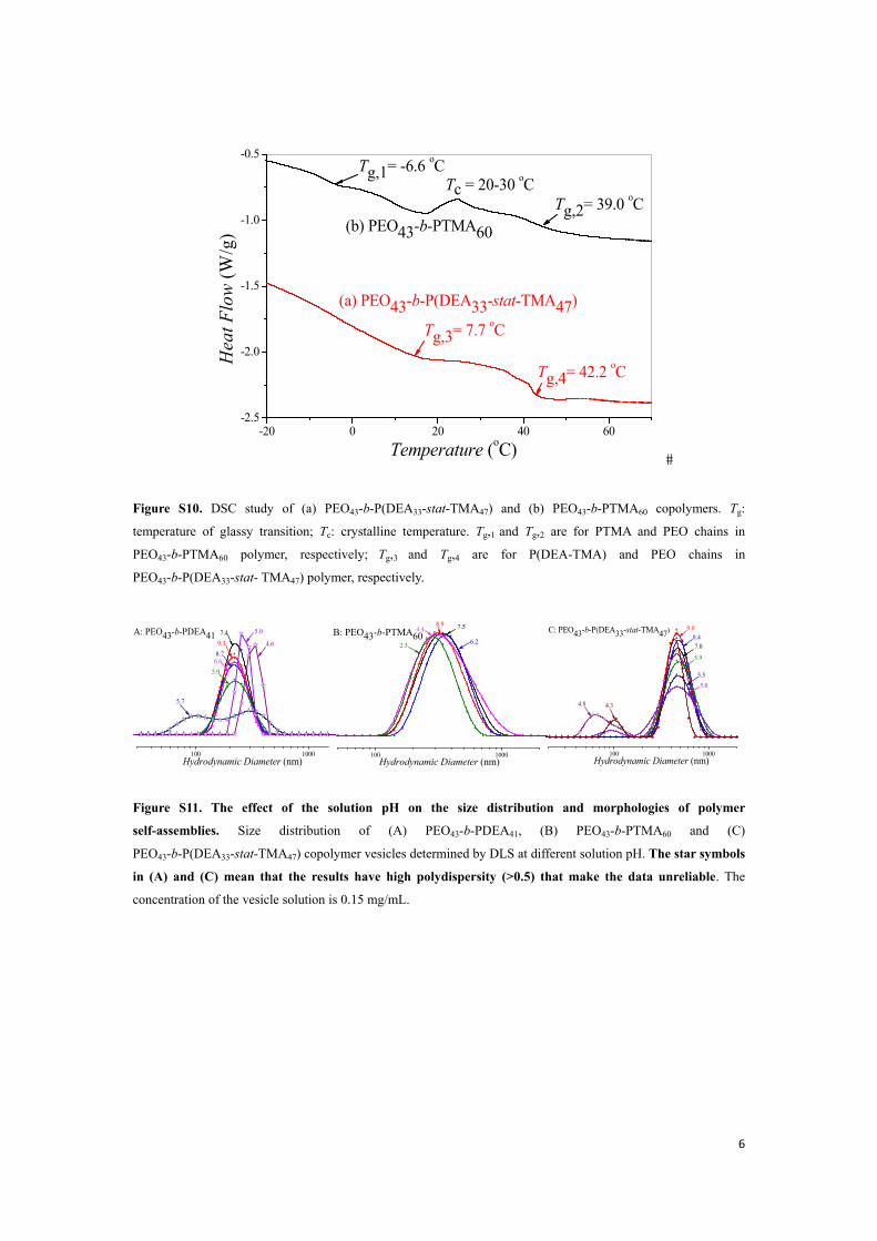
Figure S10. DSC study of (a) PEO43-b-P(DEA33-stat-TMA47) and (b) PEO43-b-PTMA60 copolymers. Tg:
temperature of glassy transition; Tc: crystalline temperature. Tg,1 and Tg,2 are for PTMA and PEO chains in
PEO43-b-PTMA60 polymer, respectively; Tg,3 and Tg,4 are for P(DEA-TMA) and PEO chains in
PEO43-b-P(DEA33-stat- TMA47) polymer, respectively.
Figure S11. The effect of the solution pH on the size distribution and morphologies of polymer
self-assemblies. Size distribution of (A) PEO43-b-PDEA41, (B) PEO43-b-PTMA60 and (C)
PEO43-b-P(DEA33-stat-TMA47) copolymer vesicles determined by DLS at different solution pH. The star symbols
in (A) and (C) mean that the results have high polydispersity (>0.5) that make the data unreliable. The
concentration of the vesicle solution is 0.15 mg/mL.
100 1000
4.6
A: PEO43-b-PDEA41
5.96.6
7.49.3
Hydrodynamic Diameter (nm)
8.7
5.7
5.0
100 1000Hydrodynamic Diameter (nm)
2.5 6.2
8.9 7.54.4B: PEO43-b-PTMA60
100 1000
5.0 5.5
7.0 8.4
C: PEO43-b-P(DEA33-stat-TMA47)
Hydrodynamic Diameter (nm)
4.8 4.3
9.0
5.9
7
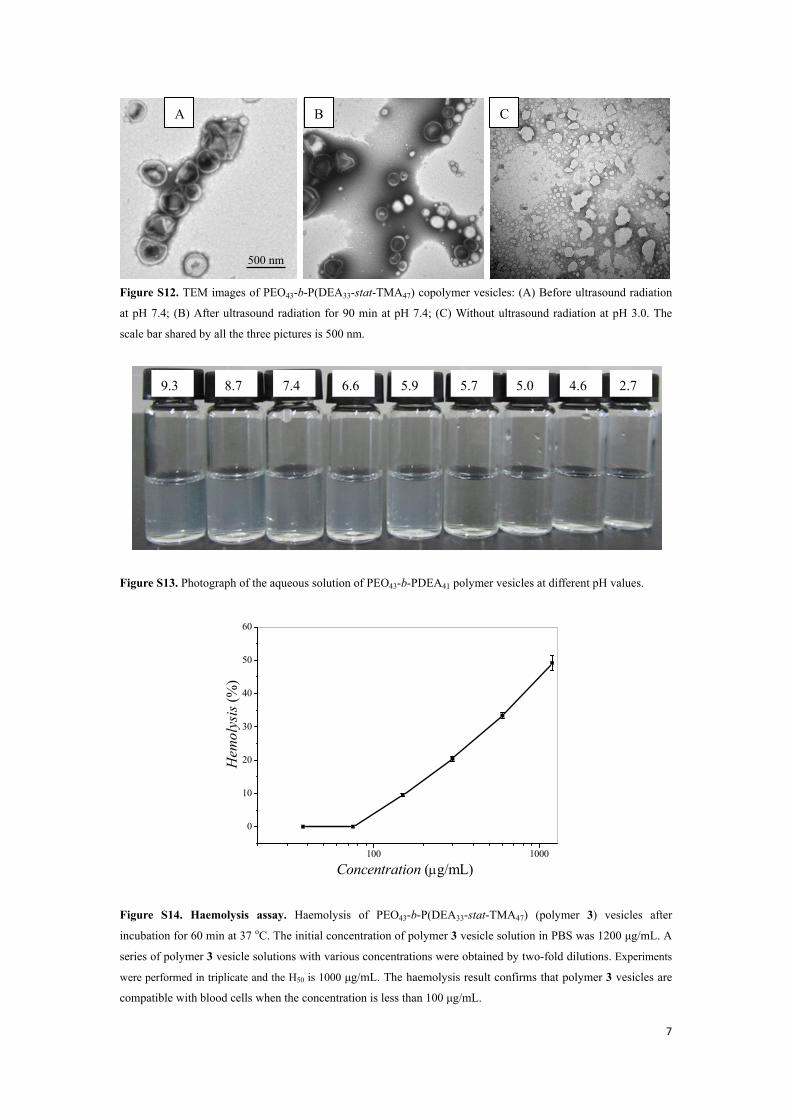
Figure S12. TEM images of PEO43-b-P(DEA33-stat-TMA47) copolymer vesicles: (A) Before ultrasound radiation
at pH 7.4; (B) After ultrasound radiation for 90 min at pH 7.4; (C) Without ultrasound radiation at pH 3.0. The
scale bar shared by all the three pictures is 500 nm.
Figure S13. Photograph of the aqueous solution of PEO43-b-PDEA41 polymer vesicles at different pH values.
100 1000
0
10
20
30
40
50
60
Concentration (μg/mL)
Hem
olys
is (%
)
Figure S14. Haemolysis assay. Haemolysis of PEO43-b-P(DEA33-stat-TMA47) (polymer 3) vesicles after
incubation for 60 min at 37 oC. The initial concentration of polymer 3 vesicle solution in PBS was 1200 μg/mL. A
series of polymer 3 vesicle solutions with various concentrations were obtained by two-fold dilutions. Experiments
were performed in triplicate and the H50 is 1000 μg/mL. The haemolysis result confirms that polymer 3 vesicles are
compatible with blood cells when the concentration is less than 100 μg/mL.
500 nm
A C B
2.7 4.6 5.05.75.96.67.4 8.79.3
8
-0.5 0.0 0.5 1.0 1.5 2.0 2.5 3.0
0
5000
10000
15000
20000
25000
Log C (μg/mL)
Inte
nsity
(a.
u.)
CVC=13.5 μg/mL
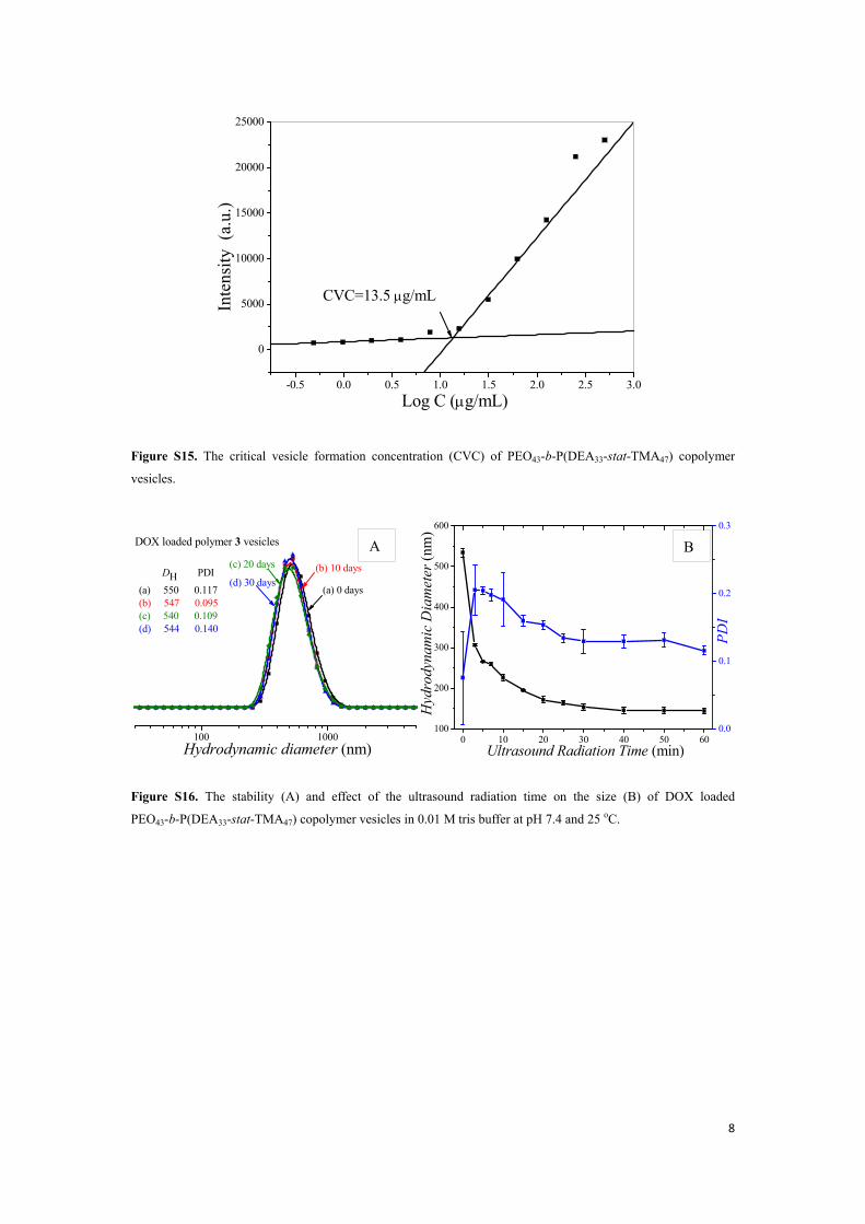
Figure S15. The critical vesicle formation concentration (CVC) of PEO43-b-P(DEA33-stat-TMA47) copolymer
vesicles.
100 1000
DH PDI (a) 550 0.117 (b) 547 0.095(c) 540 0.109(d) 544 0.140
(a) 0 days(d) 30 days
(c) 20 days
Hydrodynamic diameter (nm)
DOX loaded polymer 3 vesicles
(b) 10 days
0 10 20 30 40 50 60100
200
300
400
500
600
Hyd
rody
nam
ic D
iam
eter
(nm
)
Ultrasound Radiation Time (min)0.0
0.1
0.2
0.3
PD
I
Figure S16. The stability (A) and effect of the ultrasound radiation time on the size (B) of DOX loaded
PEO43-b-P(DEA33-stat-TMA47) copolymer vesicles in 0.01 M tris buffer at pH 7.4 and 25 oC.
A B
9
0 100 200 300 400 5000
2000
4000
6000
8000
10000
12000
14000
16000
18000
Inte
nsity
(a.u
.)
Concentration (0.01 μg/mL)
y = 35.1035x + 11.7306 R2 = 0.99931
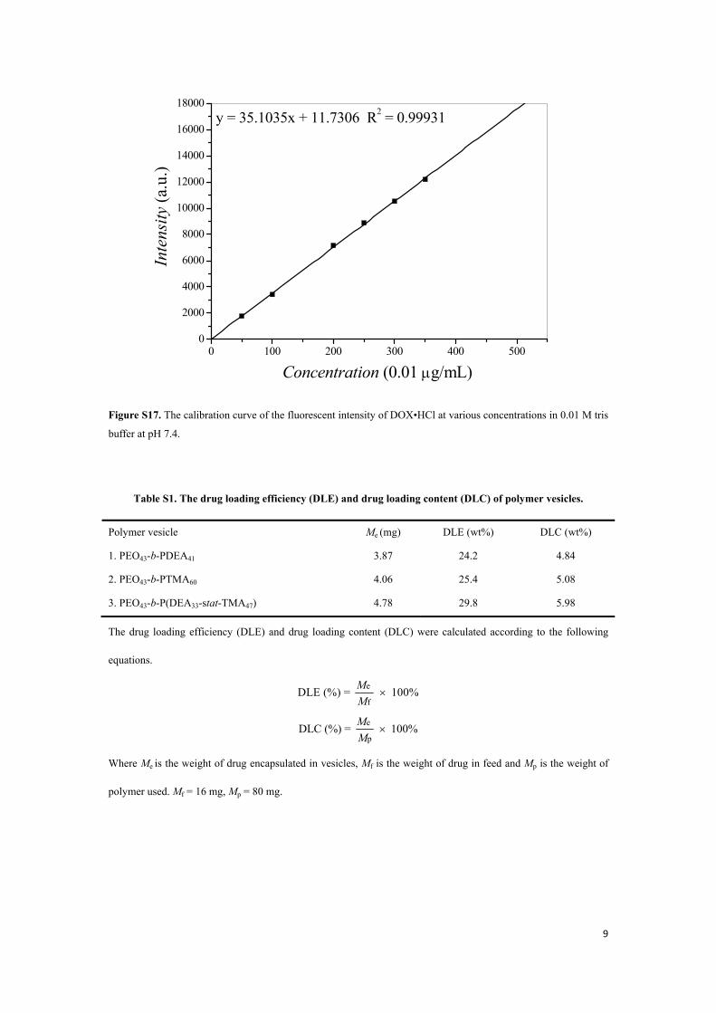
Figure S17. The calibration curve of the fluorescent intensity of DOX•HCl at various concentrations in 0.01 M tris
buffer at pH 7.4.
Table S1. The drug loading efficiency (DLE) and drug loading content (DLC) of polymer vesicles.
Polymer vesicle Me (mg) DLE (wt%) DLC (wt%)
1. PEO43-b-PDEA41 3.87 24.2 4.84
2. PEO43-b-PTMA60 4.06 25.4 5.08
3. PEO43-b-P(DEA33-stat-TMA47) 4.78 29.8 5.98
The drug loading efficiency (DLE) and drug loading content (DLC) were calculated according to the following
equations.
e
fDLE (%) = 100%M
M×
e
pDLC (%) = 100%M
M×
Where Me is the weight of drug encapsulated in vesicles, Mf is the weight of drug in feed and Mp is the weight of
polymer used. Mf = 16 mg, Mp = 80 mg.

10
Supplementary Methods
Materials
2-Bromoisobutyryl bromide, copper(I) bromide (Cu(I)Br, 99.99%), 2,3-dihydrofuran, 2-hydroxyethyl methacrylate
(HEMA), 4-vinylpyridine (4VP), azobisisobutyronitrile (AIBN), 2,2'-bipyridine (bpy),
N,N,N',N",N"-pentamethyldiethylenetri-amine (PMDETA, 98%) and triethylamine were purchased from Aladdin
Chemistry, Co. and used as received. Polyethylene glycol monomethylether (MeO-PEO-OH; Mn = 1900) was
purchased from Alfa Aesar and dried azeotropically by using anhydrous toluene to remove traces of water before
use. 2-(Diethylamino)ethyl methacrylate (DEA) was purchased from Aladdin Chemistry, Co. and was passed
through a basic Al2O3 column before use. Anisole, methanol, dichloromethane (DCM), tetrahydrofuran (THF) and
dialysis tubing with molecular weight cutoff from 8000 to 14000 were purchased from Sinopharm Chemical
Reagent Co., Ltd (SCRC, Shanghai, China).
General Experimental
Proton nuclear magnetic resonance (1H NMR) spectra were recorded using a Bruker AV 400 MHz spectrometer at
ambient temperature using CDCl3 as solvent. Chemical shifts are in ppm with respect to TMS (tetramethylsilane)
using the manufacture indirect referencing method. All chemical shifts are quoted on the scale in ppm using
residual solvent as the internal standard (1H NMR: δ = 7.26 for CDCl3). Coupling constants (J) are reported in Hz
with the following splitting abbreviations: s = singlet, d = doublet, t = triplet and m = multiplet.
Gel permeation chromatography (GPC) analysis were carried out with a Waters Breeze 1525 GPC analysis system
with two PL mix-D columns, using THF as the eluent at a flow rate of 1.0 mL/min at 35 oC. The copolymers were
dissolved in THF and filtered prior to analysis.
Dynamic light scattering (DLS) measurements were carried out with Zetasizer Nano series instrument (Malvern
Instruments ZS 90) equipped with a multipurpose autotitrator (MPT-2). DLS studies of aqueous polymer vesicles
were carried out at 25 oC and a fixed scattering angle of 90o. Each reported measurement was conducted three
runs.
Transmission electron microscopy (TEM) images were obtained using a H7560 (Hitachi Limited Corporation)
electron microscope operating at an acceleration voltage of 80 kV. To prepare TEM samples, a drop of aqueous
11
vesicle solution (0.4 mg/mL) was placed on a copper grid coated with thin carbon film. The pH of phosphotungstic
acid (PTA, 1.0 wt%) was adjusted by NaOH aqueous solution to ca. 7.4 and then used as the stain. The solution
droplet was dried by evaporation under ambient conditions overnight.
Differential scanning calorimetry (DSC) data were recorded by DSC Q100 (TA Instruments). In the dynamic DSC
measurements, freshly extruded samples were kept for 3 min at –80 oC and heated at the rate of 10 oC/min to 80
oC.
Determination of critical vesicle formation concentration of PEO43-b-P(DEA33-stat-TMA47) diblock copolymer An initial solution of pyrene was made by dissolving pyrene (3.0 mg, 15 μmol) in acetone (25 mL) to form a 6 ×
10-5 M solution. The pyrene solution (10 μL) was dropped into 11 centrifuge tubes. The acetone was evaporated
overnight in a vacuum oven. The PEO43-b-P(DEA33-stat-TMA47) polymer vesicle stock solution was serially
diluted with deionized water starting with a concentration of 0.5 mg/mL down to 4.9 × 10⁻⁴ mg/mL by
half-and-half dilution. Each polymer solution (4.0 mL) was transferred to a centrifuge tube containing pyrene and
stirred overnight.1 Fluorescence determinations were made by exciting samples at 334 nm, using a 5 nm slit width
for excitation and a 5 nm slit width for emission. Emission wavelengths were scanned from 350 to 500 nm. The
intensities of the I1 (372.1 nm) vibronic bands were evaluated for each sample. The intensity values were plotted
against the log of the concentration of each polymer vesicle sample. The critical vesicle formation concentration
(CVC) was taken as the intersection of two regression lines calculated from the linear portions of the graphs.
Synthesis of poly(4-vinylpyridine) (P4VP)
Distilled 4-vinylpyridine (2.00 g, 19.0 mmol) and recrystallized AIBN (0.0300 g, 0.200 mmol) were dissolved in
ethanol, and then the solution was heated at 60 oC reacting for 24 h under argon atmosphere while stirring. The
obtained viscous solution was poured into deionized water to obtain poly(4-vinylpyridine) (P4VP). The polymer
was purified by reprecipitation with water from ethanol solution to remove unreacted 4VP monomers. At last it
was dried in a vacuum oven to constant weight at 40 oC.
Preparation of poly(4-vinylpyridine) hydrochloride (P4VP•HCl)
P4VP (0.62 g) was dissolved in ethanol, and then hydrochloric acid (1.0 M, 2.5 mL) was added into the solution
while stirring at room temperature. The reaction was carried out for 4.0 h when a fit amount of white precipitates
appeared. A white powder product (0.56 g) was obtained after vacuum filtration and vacuum dry for 24 h.
Synthesis of (2-tetrahydrofuranyloxy)ethyl methacrylate (TMA)
12
TMA was synthesized by the addition reaction between 2-hydroxyethyl methacrylate and 2,3-dihydrofuran in
methanol with P4VP•HCl as out-phase catalyst. A literature method2 was modified as following. 2-Hydroxyethyl
methacrylate (7.27 g, 54.7 mmol), 2,3-dihydrofuran (5.91 g, 83.5 mmol), and P4VP•HCl (0.260 g, 1.70 mmol)
were charged in a round-bottom flask. The solution was heated to 45 oC and stirred overnight. Afterward, the
mixture was filtered to remove P4VP•HCl. The excess of 2,3-dihydrofuran was removed by evaporation under
reduced pressure. Finally, the solution was passed through a basic Al2O3 column to give a transparent liquid.
1H NMR (400 MHz, CDCl3): δ 6.13 (s, J = 4.1 Hz, 1H, CH2CC), 5.58 (s, J = 4.7 Hz, 1H, CH2CC), 5.16 (t, J = 4.5
Hz, 1H, OCHO), 4.29 (m, J = 3.2 Hz, 2H, CH2CH2OCH), 3.89 (m, J = 5.4 Hz, 2H, CH2OCH), 3.68 (m, J = 3.6 Hz,
2H, CH2CH2CH2CH), 1.96-1.83 (m, 7H, CH3C, CH2CH2CH). 1H NMR spectrum of TMA is shown in Fig. S2.
Synthesis of PEO43-Br macroinitiator
PEO43-Br macroinitiator was prepared by the reaction of MeO-PEO43-OH with 2-bromoisobutyryl bromide in the
presence of triethylamine on the basis of a previously reported method.3 MeO-PEO43-OH (10.0 g, 5.30 mmol) was
dissolved in toluene (250 mL) to remove traces of water by azeotropic distillation at 135 oC. Triethylamine (2.0
mL) and 2-bromoisobutyryl bromide (1.90 mL, 15.0 mmol) dissolved in anhydrous toluene (20 mL) were
sequentially added to the flask at a rate of approximately 5 s per drop. After 36 h reaction, the precipitated
byproducts were removed by filtration. The organic solution was then washed with pure water, 1.0 M HCl (50 mL)
and 1.0 M NaOH (50 mL) aqueous solutions and dried over anhydrous MgSO4. The crude product was
precipitated in 400 mL of diethyl ether twice and dried in vacuum. Yield: 80%.
1H NMR (400 MHz, CDCl3): δ 4.35 (t, J = 1.5 Hz, 2H, CH2OCO), 3.66 (broad, 170H, OCH2CH2O), 3.40 (s, J =
1.1 Hz, 3H, CH3O), 1.96 (s, 6H, (CH3)2C). 1H NMR spectrum is shown in Fig. S4 A.
Supplementary References
1. Greene, A. C., Zhu, J., Pochan, D. J., Jia, X. & Kiick, K. L. Poly(acrylic acid-b-styrene) Amphiphilic Multiblock Copolymers as Building Blocks for the Assembly of Discrete Nanoparticles. Macromolecules 44, 1942-1951, (2011).
2. Xuan, J. A., Pelletier, M., Xia, H. S. & Zhao, Y. Ultrasound-Induced Disruption of Amphiphilic Block Copolymer Micelles. Macromol. Chem. Phys. 212, 498-506, (2011).
3. Lu, H. et al. Preparation of water-dispersible silver-decorated polymer vesicles and micelles with excellent antibacterial efficacy. Polym. Chem. 3, 2217-2227, (2012).