Embed Size (px)
Citation preview
1
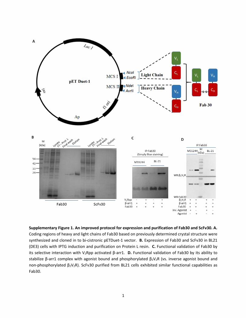
Supplementary Figure 1. An improved protocol for expression and purification of Fab30 and ScFv30. A.
Coding regions of heavy and light chains of Fab30 based on previously determined crystal structure were
synthesized and cloned in to bi-cistronic pETDuet-1 vector. B. Expression of Fab30 and ScFv30 in BL21
(DE3) cells with IPTG induction and purification on Protein L resin. C. Functional validation of Fab30 by
its selective interaction with V2Rpp activated β-arr1. D. Functional validation of Fab30 by its ability to
stabilize β-arr1 complex with agonist bound and phosphorylated β2V2R (vs. inverse agonist bound and
non-phosphorylated β2V2R). ScFv30 purified from BL21 cells exhibited similar functional capabilities as
Fab30.
2
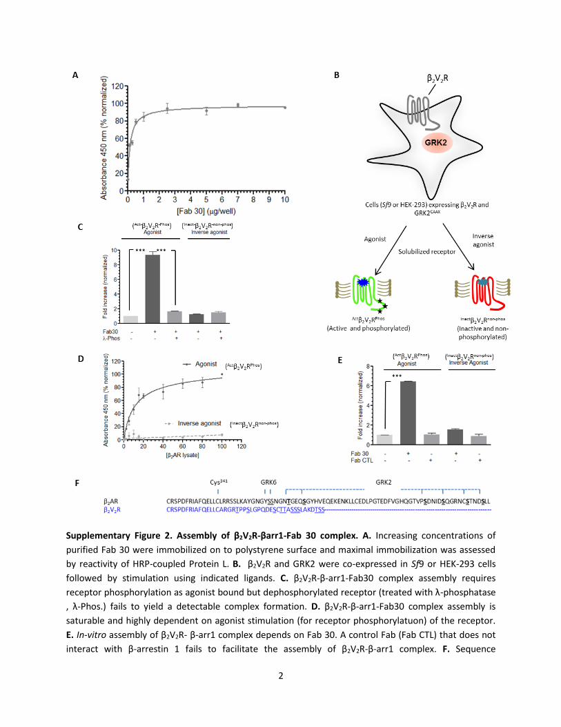
Supplementary Figure 2. Assembly of β2V2R-βarr1-Fab 30 complex. A. Increasing concentrations of
purified Fab 30 were immobilized on to polystyrene surface and maximal immobilization was assessed
by reactivity of HRP-coupled Protein L. B. β2V2R and GRK2 were co-expressed in Sf9 or HEK-293 cells
followed by stimulation using indicated ligands. C. β2V2R-β-arr1-Fab30 complex assembly requires
receptor phosphorylation as agonist bound but dephosphorylated receptor (treated with λ-phosphatase
, λ-Phos.) fails to yield a detectable complex formation. D. β2V2R-β-arr1-Fab30 complex assembly is
saturable and highly dependent on agonist stimulation (for receptor phosphorylatuon) of the receptor.
E. In-vitro assembly of β2V2R- β-arr1 complex depends on Fab 30. A control Fab (Fab CTL) that does not
interact with β-arrestin 1 fails to facilitate the assembly of β2V2R-β-arr1 complex. F. Sequence

3
comparison of the carboxyl-terminus of β2AR and β2V2R (blue). Potential phosphorylation sites in β2V2R
are underlined and known phosphorylation sites in β2AR are in bold and underlined. Data in panels C
and E represent mean±SEM of three independent experiments analyzed by ONE-WAY ANOVA with
Bonferroni post-test (***p<0.001).
4
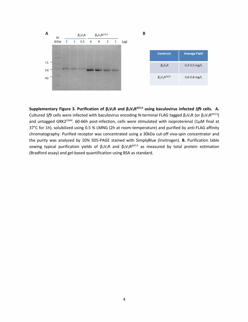
Supplementary Figure 3. Purification of β2V2R and β2V2RΔICL3 using baculovirus infected Sf9 cells. A.
Cultured Sf9 cells were infected with baculovirus encoding N-terminal FLAG tagged β2V2R (or β2V2RΔICL3)
and untagged GRK2CAAX. 60-66h post-infection, cells were stimulated with isoproterenol (1μM final at
37°C for 1h), solubilized using 0.5 % LMNG (2h at room-temperature) and purified by anti-FLAG affinity
chromatography. Purified receptor was concentrated using a 30kDa cut-off viva-spin concentrator and
the purity was analyzed by 10% SDS-PAGE stained with SimplyBlue (Invitrogen). B. Purification table
sowing typical purification yields of β2V2R and β2V2RΔICL3 as measured by total protein estimation
(Bradford assay) and gel-based quantification using BSA as standard.
5
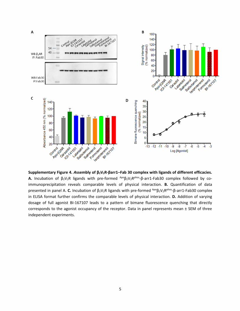
Supplementary Figure 4. Assembly of β2V2R-βarr1–Fab 30 complex with ligands of different efficacies.
A. Incubation of β2V2R ligands with pre-formed Apoβ2V2Rphos-β-arr1-Fab30 complex followed by co-
immunoprecipitation reveals comparable levels of physical interaction. B. Quantification of data
presented in panel A. C. Incubation of β2V2R ligands with pre-formed Apoβ2V2Rphos-β-arr1-Fab30 complex
in ELISA format further confirms the comparable levels of physical interaction. D. Addition of varying
dosage of full agonist BI-167107 leads to a pattern of bimane fluorescence quenching that directly
corresponds to the agonist occupancy of the receptor. Data in panel represents mean ± SEM of three
independent experiments.
6
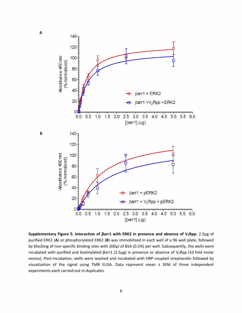
Supplementary Figure 5. Interaction of βarr1 with ERK2 in presence and absence of V2Rpp. 2.5μg of
purified ERK2 (A) or phosphorylated ERK2 (B) was immobilized in each well of a 96 well plate, followed
by blocking of non-specific binding sites with 200μl of BSA (0.5%) per well. Subsequently, the wells were
incubated with purified and biotinylated βarr1 (2.5μg) in presence or absence of V2Rpp (10 fold molar
excess). Post-incubation, wells were washed and incubated with HRP-coupled streptavidin followed by
visualization of the signal using TMB ELISA. Data represent mean ± SEM of three independent
experiments each carried out in duplicates.
7
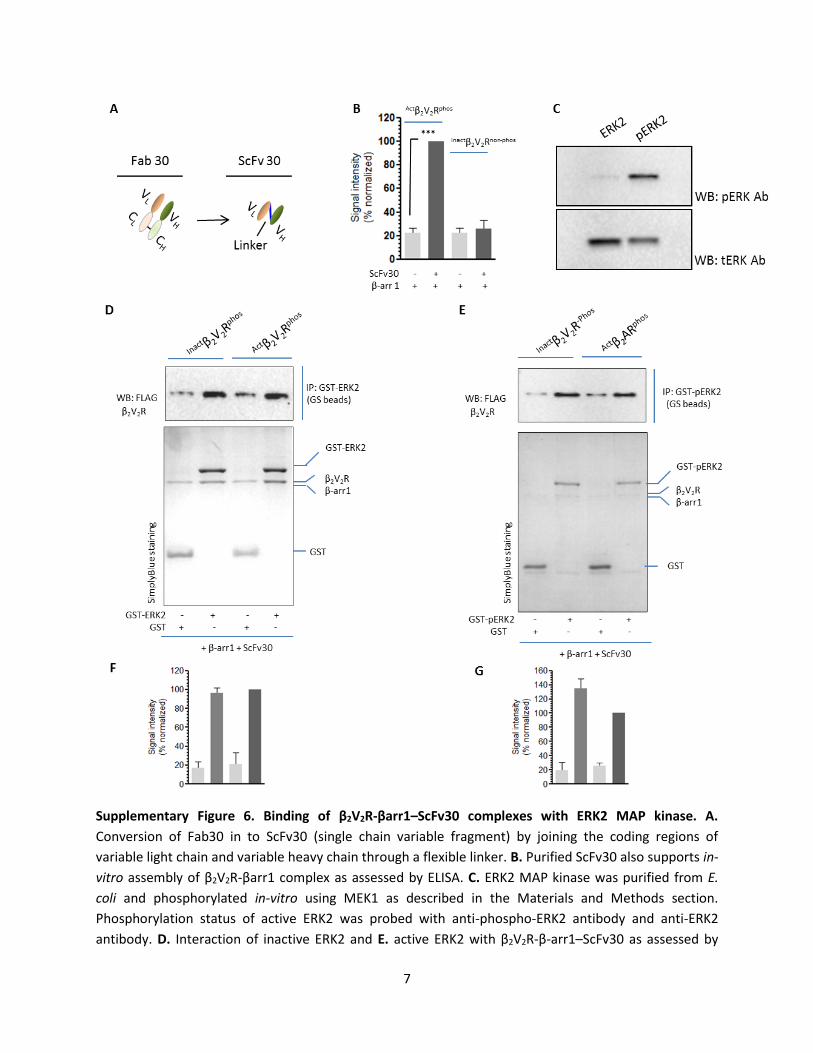
Supplementary Figure 6. Binding of β2V2R-βarr1–ScFv30 complexes with ERK2 MAP kinase. A.
Conversion of Fab30 in to ScFv30 (single chain variable fragment) by joining the coding regions of
variable light chain and variable heavy chain through a flexible linker. B. Purified ScFv30 also supports in-
vitro assembly of β2V2R-βarr1 complex as assessed by ELISA. C. ERK2 MAP kinase was purified from E.
coli and phosphorylated in-vitro using MEK1 as described in the Materials and Methods section.
Phosphorylation status of active ERK2 was probed with anti-phospho-ERK2 antibody and anti-ERK2
antibody. D. Interaction of inactive ERK2 and E. active ERK2 with β2V2R-β-arr1–ScFv30 as assessed by

8
coimmunoprecipitation assay (representative image from three independent experiments).
Quantification of complex interaction with F. ERK2 and G. pERK2. Data in panel B represent mean±SEM
of three independent experiments (ONE-WAY ANOVA with Bonferroni post-test; ***p<0.001).
9
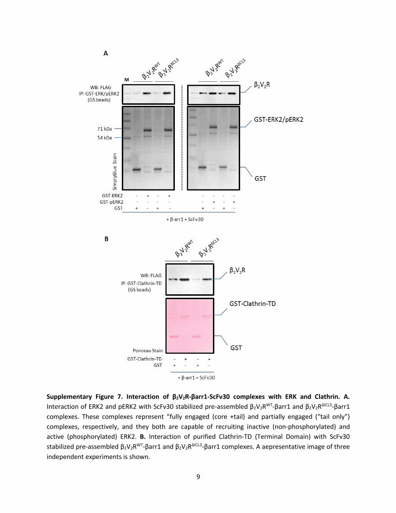
Supplementary Figure 7. Interaction of β2V2R-βarr1-ScFv30 complexes with ERK and Clathrin. A.
Interaction of ERK2 and pERK2 with ScFv30 stabilized pre-assembled β2V2RWT-βarr1 and β2V2RΔICL3-βarr1
complexes. These complexes represent “fully engaged (core +tail) and partially engaged (“tail only”)
complexes, respectively, and they both are capable of recruiting inactive (non-phosphorylated) and
active (phosphorylated) ERK2. B. Interaction of purified Clathrin-TD (Terminal Domain) with ScFv30
stabilized pre-assembled β2V2RWT-βarr1 and β2V2RΔICL3-βarr1 complexes. A aepresentative image of three
independent experiments is shown.
10
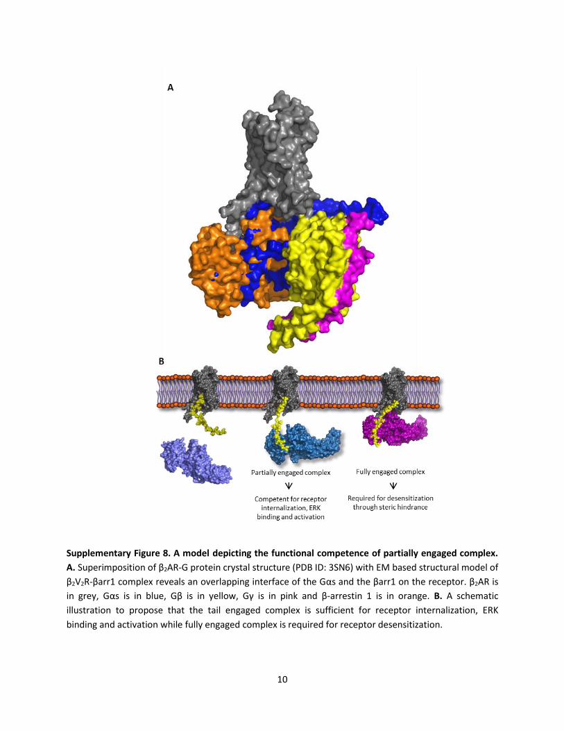
Supplementary Figure 8. A model depicting the functional competence of partially engaged complex.
A. Superimposition of β2AR-G protein crystal structure (PDB ID: 3SN6) with EM based structural model of
β2V2R-βarr1 complex reveals an overlapping interface of the Gαs and the βarr1 on the receptor. β2AR is
in grey, Gαs is in blue, Gβ is in yellow, Gγ is in pink and β-arrestin 1 is in orange. B. A schematic
illustration to propose that the tail engaged complex is sufficient for receptor internalization, ERK
binding and activation while fully engaged complex is required for receptor desensitization.
11
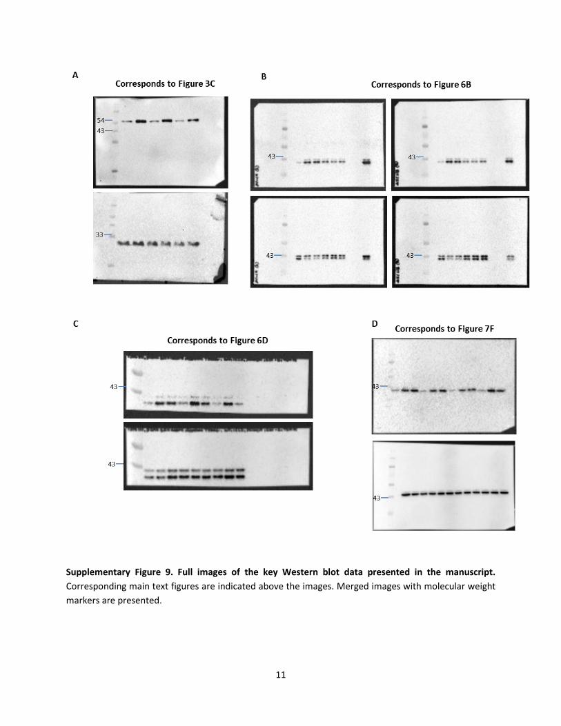
Supplementary Figure 9. Full images of the key Western blot data presented in the manuscript.
Corresponding main text figures are indicated above the images. Merged images with molecular weight
markers are presented.


![An improved energy aware distributed unequal clustering protocol … · 2017-01-17 · pan of an EADUC protocol used in continuous monitoring applications[38].TheEADUCemploysnon-uniformclusteringal-](https://img.pdfslide.us/doc/110x75/5e7033baeef98f26256f97bb/an-improved-energy-aware-distributed-unequal-clustering-protocol-2017-01-17-pan.jpg)
![The Type-B Cytokinin Response Regulator ARR1 Inhibits ... · The Type-B Cytokinin Response Regulator ARR1 Inhibits Shoot Regeneration in an ARR12-Dependent Manner in Arabidopsis[OPEN]](https://img.pdfslide.us/doc/110x75/5fb392200a417e793863db73/the-type-b-cytokinin-response-regulator-arr1-inhibits-the-type-b-cytokinin-response.jpg)
![Pointers and Memory Management · The memory location arr1[k]can be expressed in the form arr+k, i.e. the following is always true: arr1 +k≡&arr1[k] When one of the operands of](https://img.pdfslide.us/doc/110x75/5f4d32668c0e6e47d977d13f/pointers-and-memory-management-the-memory-location-arr1kcan-be-expressed-in-the.jpg)