Embed Size (px)
Citation preview
Practical Radiation Oncology (2011)
SUPPLEMENTAL MATERIAL
Quality and Safety Considerations in Stereotactic Radiosurgery and Stereotactic Body Radiation TherapyTimothy D. Solberg, Ph.D.1, James M. Balter, Ph.D.2, Stanley H. Benedict, Ph.D.3, Benedick A. Fraass, Ph.D.2, Brian Kavanagh, M.D.4, Curtis Miyamoto, M.D.5, Todd Pawlicki, Ph.D.6, Louis Potters, M.D.7, Yoshiya Yamada, M.D.8
1Department of Radiation Oncology, University of Texas Southwestern Medical Center, Dallas, Texas; 2Department of Radiation Oncology, University of Michigan Health System, Ann Arbor, Michigan; 3Department of Radiation Oncology, University of Virginia Health System, Charlottesville, Virginia; 4Department of Radiation Oncology, University of Colorado, Denver, Aurora, Colorado; 5Department of Radiation Oncology, Temple University, Philadelphia, Pennsylvania; 6Department of Radiation Oncology, University of California, San Diego, California; 7Department of Radiation Medicine, Long Island Jewish Medical Center, New Hyde Park, New York; 8Department of Radiation Oncology, Memorial Sloan Kettering Cancer Center, New York, New York
Reprint requests to: Timothy D. Solberg, Ph.D.
This document was prepared by the SBRT experts invited by the Multidisciplinary Quality Assurance Subcommittee of the Clinical Affairs and Quality Committee of the American Society for Radiation Oncology (ASTRO) as a part of ASTRO’s Target Safely Campaign.
The SBRT white paper was reviewed by 8 experts from the field of SBRT. All the comments were reviewed and discussed by the entire task group and appropriate revisions were incorporated in the paper with task group consensus.
We received approximately 22 comments from physicians, physicists, therapists, and representatives from radiation therapy manufacturers. Additionally, we received general and specific comments from the Associa-tion of Physicists in Medicine (AAPM), the American Association of Neurological Surgeons (AANS), and the Medical Imaging and Technology Alliance (MITA).
ASTRO white papers present scientific, health, and safety information and may to some extent reflect scien-tific or medical opinion. They are made available to ASTRO members and to the public for educational and informational purposes only. Any commercial use of any content in this white paper without the prior written consent of ASTRO is strictly prohibited.
Adherence to this white paper will not ensure successful treatment in every situation. Furthermore, this white paper should not be deemed inclusive of all proper methods of care or exclusive of other methods of care reasonably directed to obtaining the same results. The ultimate judgment regarding the propriety of any specif-ic therapy must be made by the physician and the patient in light of all circumstances presented by the individ-ual patient. ASTRO assumes no liability for the information, conclusions, and findings contained in its white papers. In addition, this white paper cannot be assumed to apply to the use of these interventions performed in the context of clinical trials, given that clinical studies are designed to evaluate or validate innovative approaches in a disease for which improved staging and treatment are needed or are being explored.
This white paper was prepared on the basis of information available at the time the Task Group was conduct-ing its research and discussions on this topic. There may be new developments that are not reflected in this white paper and that may, over time, be a basis for ASTRO to consider revisiting and updating the white paper.
2 TD Solberg et al Safety Considerations for SRS and SBRT 2Practical Radiation Oncology: August 2011 Safety Considerations for SRS and SBRT 3
Conflict of Interest Notification:
Before initiation of this white paper all members of the White Paper Task Group were required to complete disclosure statements. These statements are maintained at ASTRO Headquarters in Fairfax, VA and pertinent disclosures are published with the report. The ASTRO COI Disclosure Statement seeks to provide a broad disclosure of outside inter-ests. Where a potential conflict is detected, remedial measures to address any potential conflict are taken and will be noted in the disclosure statement. Dr. Timothy Solberg has a consulting service, Global Radiosurgery Services, LLC, that has provided services to BrainLab AG and to individual healthcare institutions. He also has research funded by grants to the University of Texas from Varian and Elekta. Dr. Benedick Fraass is a mem-ber of the Patient Safety Council for Varian. He receives no compensation or reimburse-ment for this work. Dr. James Balter is a consultant for Calypso Medical Technologies. These disclosures were reviewed according to ASTRO policies and determined to not present a conflict with respect to these Task Group members’ work on this White Paper. Safety Considerations for SRS and SBRT
1. Introduction1.1 Scope of this Document on Patient Safety for SRS and SBRT1.2 Nomenclature1.3 Safety Concerns2. Elements of Successful SRS / SBRT Quality Assurance2.1 Establishing Program Goals2.2 Technology Requirements2.3 Personnel Requirements3. SRS / SBRT Systems Acceptance and Commissioning4. SRS / SBRT Quality Assurance4.1 General QA Concepts4.2 Equipment QA4.3 Patient / Process QA5. Processes for Ongoing Quality Improvement6. Documentation7. Other Recommendations8. Summary
Appendix 1. Recommendations to Guard Against Catastrophic Failures in SRS and SBRTAppendix 2. Example Checklists for SRS and SBRTAppendix 3. Example Simulation and SBRT Process Documents
Note: Example checklists are intended to serve as a template, and should not be adopted in whole or in part. They are institution and technology specific and are meant solely for illustration.
Safety Considerations for SRS and SBRT 2 Safety Considerations for SRS and SBRT 3Practical Radiation Oncology: August 2011
Introduction
1.1. Scope of this Document on Patient Safety for SRS and SBRT This report on Stereotactic Radiosurgery (SRS) and Stereotactic Body Radiation Therapy (SBRT) is part of a series of white papers addressing patient safety commis-sioned by the American Society for Radiation Oncology (ASTRO) under the umbrella of the ASTRO/American Association of Physicists in Medicine (AAPM)/Ameri-can College of Radiology (ACR) Safety Task Force. This document was approved by the ASTRO Board of Directors April 11, 2011. It has been endorsed by the American Association of Physicists in Medicine, the American Society of Radiologic Technologists, and the American Association of Medical Dosimetrists. It has been reviewed and accepted by the American College of Radiology’s Commission on Radiation Oncology. In addition to many academic papers, profession-al organizations in North America have previously published several “guidance” reports on various aspects of SRS/SBRT. Notably, these include: • Current radiosurgery practice: results of an ASTRO
survey. Task Force on Stereotactic Radiosurgery, American Society for Therapeutic Radiology and Oncology, published in 1994.
• Stereotactic radiosurgery: report of AAPM Task Group 54, published in 1995.
• American Society for Therapeutic Radiology and Oncology (ASTRO) and American College of Radiology (ACR) Practice guideline for the performance of stereotactic radiosurgery, published in 2006.
• Quality Control Standards: Stereotactic Radiosur-gery/Radiotherapy. Standards for Quality Control at Canadian Radiation Treatment Centres, Cana-dian Association of Provincial Cancer Agencies (CAPCA), published in 2006.
• Stereotactic body radiation therapy: the report of AAPM Task Group 101, published in 2010.
• American Society for Therapeutic Radiology and Oncology (ASTRO) and American College of Radiology (ACR) Practice Guideline for the Performance of Stereotactic Body Radiation Therapy, published in 2010.
In addition, there are several recent international efforts specifically addressing safety in radiotherapy: Radiotherapy Risk Profile Technical Manual, published in 2006 by the World Health Organization (WHO).• Towards Safer Radiotherapy, published in 2008
by the Royal College of Radiologists, Society and College of Radiographers, Institute of Physics and Engineering in Medicine, National Patient Safety Agency, and the British Institute of Radiology.
• HTA Initiative #22: A Reference Guide for Learning from Incidents in Radiation Treatment,
published in 2006 by the Alberta Heritage Founda-tion for Medical Research.
• Preventing Accidental Exposures from New External Beam Radiation Therapy Technologies, published in 2010 by the International Commission on Radiological Protection.
It is important to understand that the SRS/SBRT QA measures described and recommended in this document are just one component of a broader process of ongoing quality assurance for the entire scope of practice within a radiation oncology department that includes period-ic review of errors, incidents, and near misses for the purpose of developing or refining standard operating procedures that minimize the risk of such events. Similarly, detailed equipment specifications and tolerances have been described in a number of docu-ments, and while some of these aspects may be reiter-ated and/or emphasized in this paper, it is not intended to be comprehensive in this regard. Rather, this report builds on these and other documents, broadly addressing SRS/SBRT delivery with a primary focus on program-matic elements and human processes that can identify and correct potential sources of error, particularly those which can result in catastrophic consequences. One can make a distinction between quality improvement efforts and safety improvement efforts, but for this document, they are considered the same.
1.2. Nomenclature
The adjective “stereotactic” describes a procedure during which a target lesion is localized relative to a known three dimensional reference system. Stereotactic body radiation therapy (SBRT) and Stereotactic Radio-surgery (SRS) are specialized forms of cancer treatment whereby high doses of radiation are delivered in large fraction sizes over a short course of treatment, gener-ally limited to 5 or fewer fractions. SRS is defined as treatment delivery to the brain or spine, while SBRT is defined as treatment delivery elsewhere within the body (which can include the spine); for completeness, fractionated stereotactic treatment for brain neoplasms is historically referred to as either stereotactic radio-therapy (SRT) or fractionated stereotactic radiotherapy (FSRT). These definitions SRS, SRT and FSRT are used to differentiate neurosurgical cases, which typically include participation of neurosurgical colleagues, from treatment of other sites where dose to nervous system structures is not a limiting normal tissue constraint. In contrast, the clinical activities of the radiation oncolo-gist encompass the full range of disease sites, from brain to spine to body sites, with prescriptions from one to five fractions. The quality assurance and safety issues are similar for SRS and SBRT, and the acronyms are used somewhat interchangeably, though differences are highlighted where specific emphasis is required.
4 TD Solberg et al Safety Considerations for SRS and SBRT 4Practical Radiation Oncology: August 2011 Safety Considerations for SRS and SBRT 5
From the earliest days of radiosurgery, the use of a stereotactic head frame has been a prerequisite for accurate cranial localization. The head frame both defines the stereotactic space and provides the means for posi-tioning and immobilizing the patient. While frameless techniques which incorporate image guidance can now provide equivalent accuracy for cranial applications, the use of a head frame remains widely used. In contrast, while SBRT localization may be assisted through the use of “body frames,” final SBRT localization must be performed using a sophisticated form of 3-dimensional image guidance, tightly integrated with the delivery system, to confirm proper patient positioning and tumor localization within the reference space. To mini-mize intra-treatment tumor displacement associated with breathing or other motion, some method to address intra-fraction target movement is often required. This can take the form of passive motion management, such as the design of patient/respiration-specific target margins from 4D computed tomography (4DCT), or active motion management such as abdominal compres-sion and beam gating/tracking. Management of respira-tory motion is covered in detail in the report of AAPM Task Group 76(1). SBRT may be delivered in 1 to 5 frac-tions, and each and every fraction requires an identical degree of precision, target localization accuracy, and quality of image guidance. SRS has been used for decades in the treatment of brain metastases and a variety of functional disor-ders, and its efficacy and toxicity profile has been well described as an efficient and effective means of achiev-ing a high rate of local control and, in some settings, improved survival (2). Acute side effects, including headache, pin-site infection, and short-term exacerba-tion of neurologic symptoms are relatively minor and readily managed. Late side effects, including radiation necrosis, brain edema, and exacerbation of preexist-ing, or development of new neurologic deficits occur in less than 5% of patients(3). Five year local control rates following SRS or fractionated stereotactic radiation therapy (FSRT) for acoustic neuromas exceed 95% (4). Current doses of 13 Gy (single fraction) or ~50 Gy (in 1.8 Gy fractions) yield excellent rates of hearing preservation as well as very low rates of facial and trigeminal neuropathies(4). Similarly, excellent rates of local control can be expected following either SRS or FSRT treatment of meningiomas, though the grade and location of these tumors plays a significant factor in both tumor control and potential complications (5-7). SBRT is a much more recent modality, with unique technological and clinical considerations. Neverthe-less, initial clinical results from prospective single institution, and more recently, multi-institutional clini-cal trials of SBRT have documented similar high rates of tumor control coupled with a low incidence of serious toxicity despite the high dose fractions of radiation
being delivered. The efficacy of SBRT is established for a variety of clinical indications as primary treatment for selected early stage cancers or as treatment for discrete tumors in patients with oligometastatic disease, selected benign neoplasms in or near the central nervous system, or recurrent cancer in previously irradiated regions. The utility of SBRT is perhaps best exemplified in the case of inoperable early stage lung cancer (8), where the three year primary tumor control rate of 98% is roughly twice what would be expected from conventional RT given over a six to seven week period. To date reports of prospective clinical trials of SBRT have typically documented similar high rates of tumor control coupled with a low incidence of serious toxicity despite the high dose fractions of radiation given to tumors (9-19). This favorable therapeutic ratio is achieved because SBRT couples a high degree of anatomic targeting accuracy and reproducibility with very high doses of precisely delivered radiation, thereby maximizing the cell-killing effect on the target(s) while minimizing radiation-relat-ed injury in adjacent normal tissues.
1.3. Safety Concerns
Given that very high dose fractions of radiation are delivered, the margin of error for SRS and SBRT is significantly smaller than that of conventional radio-therapy and therefore requires special attention and diligence. A small error in target localization for any one fraction risks undertreatment of portions of the tumor by 20% or more. Inadvertent overdosage of adjacent normal tissues in even a single fraction could escalate the risk of serious injury to a much greater degree than an equivalent treatment error in a course of radiother-apy where a substantially lower dose per fraction is used (15, 20-24). Low output factors associated with small fields necessitate delivery of a high number of monitor units, further increasing the associated risk. Many in the community are aware of recent events, publicized in the media, in which serious errors have occurred. These include: a calibration error on a radiosurgery linac that affected 77 patients in Florida in 2004-2005; similar errors in measurement of output factors affecting 145 patients in Toulouse, France between April 2006 and April 2007 (25-27), and 152 patients in Springfield, Missouri between late 2004 and late 2009; an error in a cranial localization acces-sory that affected seven centers in France, Spain and the U.S.; and errors in failure to properly set backup jaws for treatments using small circular collimators affecting a single AVM patient at an institution in France (26), and three patients (at least one of whom received radiosurgery for trigeminal neuralgia) at an institution in Evanston, Illinois (28). While no side effects related to the Florida calibra-tion error have been reported, that is not the case with
Safety Considerations for SRS and SBRT 4 Safety Considerations for SRS and SBRT 5Practical Radiation Oncology: August 2011
Elements of Successful SRS / SBRT Quality Assurance
2.1. Establishing Program Goals It is important to emphasize that SRS and SBRT are not one treatment technique or modality. The implementation and accompanying requirements for immobilization, simulation, treatment planning, delivery and quality assurance can vary significantly with disease site. Clinical and technical proficiency for one site (e.g., spine) does not always translate to proficiency in another site (e.g., lung). This complex nature of the stereotactic treatment process, and the consequences of errors when delivering high dose fractions of radia-tion, mandates a systematic and prospective approach to each disease site. In 2008, a consortium of British orga-nizations published a document entitled Towards Safer Radiotherapy (32). Many of the overall recommenda-tions are appropriate for SRS and SBRT programmatic development, including: a multidisciplinary working environment with a culture that fosters clear commu-nication and guards against inappropriate interruptions; careful planning and thorough risk assessment when introducing new techniques and technologies; a review of staffing levels and skills, with specific training in each new treatment technique or process prior to clinical use. Training on specific technologies, often provided by the equipment vendor(s) is an essential training element. Vendor training by itself, however, does not provide the comprehensive instruction needed to competently perform SRS or SBRT. AAPM Task Group 101 has called for a thorough feasibility analysis of existing resources to achieve the clinical and technical goals of any proposed SBRT program (33). Treatment of various disease sites should be considered within the context of nationally accepted clinical standards. It is strongly recommended that each department collect a library of published studies that document patient selection criteria and treatment planning and delivery parameters that are relevant to the population of patients to be treated with SBRT. Individual disease sites require unique and specialized technical elements and processes. Based on program goals and patient selection criteria, it is likely that treatment guidelines and procedures will be site-specific. Prior to initiating an SRS or SBRT program, this report strongly recom-mends that plans for patient selection and treatment guidelines be developed and clearly documented within each institution.
several of the other events. Gourmelon et al reported at 31% 12 month actuarial rate of trigeminal neuropa-thy in 32 acoustic neuroma patients overdosed in the Toulouse accident (27). In contrast, despite a mean over-dose of 61.2%, no treatment-related morbidity was observed in the 33 patients treated for brain metastases (25). In the case of the French patient treated with the incor-rect backup collimator setting, a subsequent dosimetric evaluation indicated that a large portion of normal brain received doses in excess of those intended for the AVM. The patient developed an oeso-tracheal fistula requiring surgery, experienced a hemorrhage and subsequently died (26). One of the three Evanston patients is described as being in a vegetative state (28). Radiosurgery errors are not limited to any particu-lar technology. As an example, challenges in accurate measurement of output factors, such as those encoun-tered on linacs in Toulouse, France and Springfield, Missouri, have also been encountered on gamma devices. In 1998, the output factor for a 4 mm gamma collimator was corrected by approximately 10%, from 0.80 to 0.87, by the manufacturer (29-30). A review of the Nuclear Regulatory Commission (NRC) Radiation Event Report Notification database yielded 13 gamma-based radiosurgery-related events from 2005 to present, 12 of which resulted in a deviation from the original prescription. Seven of the events involved the treat-ment of the wrong location, while three events involved delivery of an incorrect dose. Wrong-site errors continue to plague all medical disciplines, and are not unique to radiotherapy (31). While patient outcome is not described on the NRC site, several of the events listed, including treatment of the wrong location with single fraction doses as high as 90 Gy, would likely be accompanied by significant morbidity. The accidents described can largely be attributed to human error, mirroring the radiotherapy experience throughout the United Kingdom, in which only 2 out of 181 incidents reported since 2000 were determined to be non-related to human error (32). However, other factors also contributed. These include limits in equip-ment safety design and the inadequacy of systems and procedures to ensure that the stereotactic treatment was robust to the sources of error that eventually contributed to failure. Clearly then, improvement in human knowl-edge, training standards, and implementation of robust quality assurance processes is needed to minimize these errors, which in the case of SRS and SBRT, can have catastrophic consequences. A set of recommendations designed to guard against catastrophic failure in SRS and SBRT is provided in Appendix 1.
6 TD Solberg et al Safety Considerations for SRS and SBRT 6Practical Radiation Oncology: August 2011 Safety Considerations for SRS and SBRT 7
2.2. Personnel Requirements
SRS and SBRT require the coordinated efforts of a team of properly trained individuals who assume essential roles during the patient evaluation and treat-ment process (33-36). In addition to clinic nurses and other staff who provide general support for all patients receiving RT, for SRS / SBRT the essential personnel include the following individuals who have the indi-cated credentials and responsibilities:
Radiation Oncologist
1. The radiation oncologist participating in an SRS or SBRT program must have completed an Accred-itation Council for Graduate Medical Education (ACGME) approved residency program or an American Osteopathic Association (AOA) approved residency program in radiation on-cology, and must have obtained certification in Radiation Oncology or Therapeutic Radiology by the American Board of Radiology (ABR) or an equivalent national or international board. If the radiation oncologist’s formal training did not include SRS/ SBRT, then specific training in SRS/SBRT, including a minimum of 5 CME credit hours direct observation of treatment of at least 3 different patients, should be obtained prior to performing any SRS or SBRT procedures.
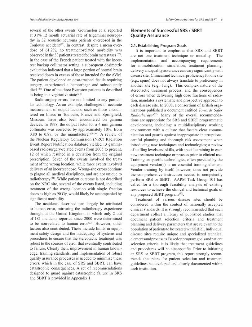
Table 1. Essential planning aspects for developing a new SBRT program and/or considering new disease sites.
Recommendation Duration or Frequency ReferenceEstablish clinical program goals, specify disease sites, identify program specialists, develop guidelines for treatment, follow-up and assessment.
Initially 33-34, 36
Identify required resources: expertise, personnel, technology, time. Initially, and for each new technology and/or disease site 32-33
Perform technology assessment commensurate with clinical goals, identify equipment and processes for simulation, immobilization, image guidance, management of organ motion, treatment delivery.
Initially, and for each new technology and/or disease site 32-33
Perform assessment of staffing levels, develop processes for initial and ongoing training of all program staff. Initially, and for each new technology and/or disease site 32-35
Develop and use checklists for all aspects of SRS/SBRT processes. Initially, and for each new technology and/or disease site 34-36
Provide documentation for a culture and environment fostering clear and open communication. Ongoing 32
Develop quality assurance processes that encompass all clinical and technical SBRT program aspects, clearly following available guidance, with regard to procedures and tolerances.
Initially, and for each new technology and/or disease site 32-36, 43
Conduct clinical SBRT patient conferences for pre-treatment planning and post-treatment review. Ongoing
Develop processes for documentation and reporting, peer review, regular review of processes and procedures, updating clinical guidelines and recommendations, ongoing needs assessment, and continuous quality improvement.
Ongoing 32-35
2. The responsibilities of the radiation oncologist include management of the overall disease- specific treatment regimen. The radiation oncologist will prescribe and supervise the means of patient positioning and immobilization, devices or tech-niques to manage any motion-related concerns, and simulation and planning image acquisition in the treatment position. The Radiation Oncologist must be provide direct supervision at the time of simu-lation, be present for critical decision making, and approve of the immobilization and imaging prior to completion of the simulation session.
3. The radiation oncologist is responsible for defin-ing the target volumes, verifying image fusion and defining and approving the contours of all the critical normal structures (e.g., brachial plexus, trachea, spinal cord, etc.). The radiation oncolo-gist works closely with the medical physicist and dosimetrist to design a treatment plan that provides proper dose to the tumor while respecting normal tissue dose constraints.
4. On the day(s) of patient treatment, the radiation oncologist must be present at the start of the treat-ment fraction (prior to irradiation) to verify the integrity of the patient setup at the treatment machine, patient repositioning using image guidance, and directly manage any clinical issues and/or treatment related toxicities. Thereafter, the Radiation Oncologist must be present for critical decision making and otherwise immediately available.
Safety Considerations for SRS and SBRT 6 Safety Considerations for SRS and SBRT 7Practical Radiation Oncology: August 2011
Medical Physicist
1. The medical physicist participating in an SRS or SBRT program must be certified in Therapeutic Radiological Physics or Radiological Physics by the American Board of Radiology (ABR), the American Board of Medical Physics (ABMP), or an equivalent international organization. The medical physicist must meet the American College of Radiology (ACR) Practice Guideline for Con-tinuing Medical Education (CME). If the medical physicist’s formal training did not include SRS / SBRT, then specific training in SRS / SBRT, includ-ing at least 5 CME credits and direct observation of at least 3 patient treatments, should be obtained prior to performing any SRS or SBRT procedures.
2. The medical physicist is responsible for the tech-nical aspects of an SBRT program, which includes simulation, planning and treatment, and verification of output calibration.
3. The medical physicist is responsible for initial commissioning and acceptance testing of all plan-ning, imaging, localization and immobilization, and delivery equipment, as well as periodic QA assess-ment to ensure proper performance of the treatment system(s) used.
4. On the day(s) of patient treatment, the medical physicist verifies the integrity of the patient setup at the treatment machine, verifies patient reposi-tioning using image guidance, and is responsible for performing and/or supervising that the treat-ment plan meets or exceeds the radiation oncolo-gist’s prescription and is able to be delivered with a minimal chance for errors and with the highest quality. The medical physicist must be present for the entirety of each treatment.
Medical Dosimetrist:
1. Works with radiation oncologist and physicist in devising a treatment plan per the physician-defined clinical goals. This may include assistance with positioning and immobilization, segmentation, beam placement, and margin recommendation, and plan review to assure that the goals of the treatment directive are met.
2. Enters the approved plan information into the patient’s chart and/or treatment management system.
3. Assists therapists with understanding the IMRT plan, and with treatment delivery as needed.
Neurosurgeon
1. The neurosurgeon participating in an SRS program must have completed an Accreditation Council for Graduate Medical Education (ACGME) approved residency program in neurosurgery, and must have obtained certification or be board-eligible in Neurosurgery by the American Board of Neuro-logical Surgery (ABNS) or an equivalent national or international board. If the neurosurgeon’s formal training did not include SRS, then specific training in SRS should be obtained prior to performing any SRS procedures.
2. The neurosurgeon generally participates in SRS cases of the brain or spine and works with the radia-tion oncologist in target definition and in assessing normal tissue structures and vital neurologic path-ways close to the planned target.
3. The Neurosurgeon will review target and nor-mal tissue contours and assess, with the radiation oncologist, the dose distribution.
4. In cases where head frames are required, the neuro-surgeon will manage the care of placing and remov-ing the head frame.
Radiation Therapist
1. A radiation therapist must fulfill any applicable state licensing or registration requirements and must have American Registry of Radiologic Technolo-gists (ARRT) certification in radiation therapy. If the radiation therapist’s formal training did not include SRS/SBRT, then specific initial and periodic training in SRS/SBRT should be obtained prior to performing any SRS or SBRT procedures.
2. The responsibilities of the radiation therapist include preparing the treatment room for the SRS or SBRT procedure, performing patient positioning/immobi-lization and assisting the treatment team answering any questions about the patient’s setup, and operat-ing the treatment unit after the radiation oncologist and medical physicist have approved the clinical and technical aspects for beam delivery.
Other Specialists
1. Other physicians may participate in the care of the patient undergoing SRS or SBRT by offering assistance derived from their own subspecialty training and expertise in the evaluation and treat-ment of the type of patient receiving SRS or SBRT. Typically, their participation could involve assistance in interpreting key imaging studies that facilitate the precise contouring of targets and delineation of normal tissue interfaces in order to aid the radiation oncologist and medical physicist in the planning process.
8 TD Solberg et al Safety Considerations for SRS and SBRT 8Practical Radiation Oncology: August 2011 Safety Considerations for SRS and SBRT 9
Administration
Due to the technical nature of SRS and SBRT, depart-ment administrators are responsible for providing full support to programs in:1. Ensuring adequate resources for personnel, equip-
ment, and time for commissioning.2. Supporting time required for development of
standard operating procedures and for ongoing documentation.
3. Supporting training and continuing education for all personnel.
4. Ensuring all program personnel have the ability to halt any procedures that are deemed unsafe.
5. Encouraging open communication among all team members, without fear of reprisal.
SRS and SBRT require a high-precision of treat-ment delivery, use a wide range of technologies with-in and across institutions, and require a large resource commitment involved in patient care, quality assurance, and documentation. The personnel resources required for proper operation of an SBRT program would therefore be expected to be significantly larger than for a tradition-al radiation therapy program. AAPM Task Group 101 (33) and the AAPM-sponsored ABT surveys (37) provide some guidance on the additional physics personnel levels required for best-practice SRS and SBRT programs. Similar references should be developed to guide person-nel decisions on the radiation oncologist, dosimetrist, and radiation therapist roles for SBRT. Nagata et al published the results of a recent survey of 53 institu-tions performing SBRT in Japan (38). While practice patterns in Japan may differ from those in the United States, the document is nonetheless instructive for assessing resources needed to initiate and maintain a clinical SBRT
program. Adequate levels of specialty staff is closely related to a reduction in medical errors (36, 39). This re-port strongly recommends that institutions hire addi-tional personnel to support SRS and/or SBRT programs. This report strongly recommends that the physician and physicist directing the initiation of the SRS and/or SBRT program consult with administration regarding the extent of additional resources needed to ensure safe-ty. Institutions planning to begin an SRS or SBRT program must ensure they have adequately planned for the staff required to carry out all necessary tasks without undue pressure.
2.3. Technology Requirements
SRS and SBRT require the use of technology at a standard above that routinely considered necessary for conformal radiotherapy and initial image guided radio-therapy applications. The extreme demands imposed by the ablative paradigm of dose delivery amplify concerns over the volume of tissue irradiated to high doses as well as doses in serial organs and regions near the skin that may otherwise be ignored. To achieve these demands, small margins around the clinical target volume are necessary to such an extent that conventional radio-graphic localization based on bony anatomy is gener-ally insufficient. A comprehensive image guidance and motion management strategy needs to be applied and maintained with sufficient technology and procedures to ensure safe and effective positioning for treatment. Furthermore, the dose distributions considered accept-able for SRS and SBRT require using large numbers of non-opposing beams often inclusive of multiple non- axial approaches. Dose needs to be calculated accu-rately through complex heterogeneities and represented over the entire irradiated volume. Isocenter placement
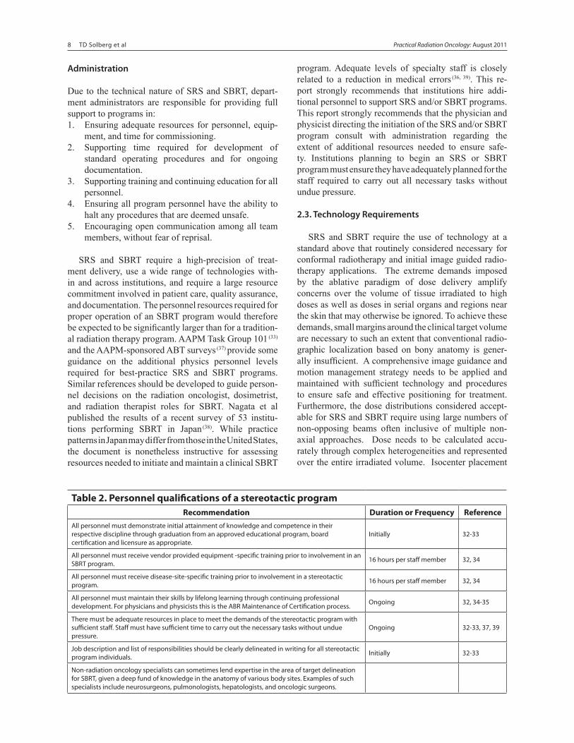
Table 2. Personnel qualifications of a stereotactic program Recommendation Duration or Frequency Reference
All personnel must demonstrate initial attainment of knowledge and competence in their respective discipline through graduation from an approved educational program, board certification and licensure as appropriate.
Initially 32-33
All personnel must receive vendor provided equipment -specific training prior to involvement in an SBRT program. 16 hours per staff member 32, 34
All personnel must receive disease-site-specific training prior to involvement in a stereotactic program. 16 hours per staff member 32, 34
All personnel must maintain their skills by lifelong learning through continuing professional development. For physicians and physicists this is the ABR Maintenance of Certification process. Ongoing 32, 34-35
There must be adequate resources in place to meet the demands of the stereotactic program with sufficient staff. Staff must have sufficient time to carry out the necessary tasks without undue pressure.
Ongoing 32-33, 37, 39
Job description and list of responsibilities should be clearly delineated in writing for all stereotactic program individuals. Initially 32-33
Non-radiation oncology specialists can sometimes lend expertise in the area of target delineation for SBRT, given a deep fund of knowledge in the anatomy of various body sites. Examples of such specialists include neurosurgeons, pulmonologists, hepatologists, and oncologic surgeons.
Safety Considerations for SRS and SBRT 8 Safety Considerations for SRS and SBRT 9Practical Radiation Oncology: August 2011
may be non-traditional due to needs of clearance for beam angles and imaging. Common technological and procedural requirements can be described by the SRS and SBRT processes.
Simulation SRS/SBRT begin to deviate from conventional treat-ments at simulation. Immobilization, both physical as well as physiological, need to be devised as necessary. Images used for simulation and planning may require motion estimation (e.g. 4DCT), inclusion of soft tissue (MRI), or metabolic (PET) imaging. Paraspinal SBRT may require enhanced visualization of the spinal cord (e.g. through MR or CT myelography). Typical immobilization equipment for SBRT includes custom formed devices that cover a large extent of the patient above and below the tumor (e.g., evacu-ated bean bags). The use of other technologies, such as surface imaging techniques, implanted radiographic markers and electromagnetic transponders may play a role in specific disease sites. For each of these devices and indications for use, the operational team (RTT, MD, physicist) should establish procedures for assessing the residual positioning uncertainty that is possible when combining these immobilization means with specific image guidance strategies. 4D CT or comparable imaging that is inclusive of the full range of motion of the target should be available for encompassing movement estimates into target volume construction. If gating, breath hold, or abdominal compression are to be used for treatment, then sufficient means must be available at simulation to image the patient appropriately for planning as well as to prepare for use, which includes an estimation of possible residual movement with any breathing management technique. Imaging must be performed over a sufficient-ly large volume to encompass the passage of non-axial beams through the patient.
Planning The treatment planning environment must be capable of supporting both multimodality as well as multidimensional input data for SRS and SBRT plan-ning. Specifically, MRI, PET, and multiple CT scans (e.g. non-contrast and contrast, 4D) must be able to be combined to facilitate target and normal tissue defini-tion, establishment of a patient data set for use in image guidance, and generation of the appropriate density grid for dose calculation. The planning system must be able to support dose calculation algorithms that represent dose deposition in the face of heterogeneities with sufficient accuracy. Commercial planning systems using pencil beam algorithms generally do not meet this require-
ment. Demonstration of calculation accuracy during the commissioning process, e.g. via an independent dosimetric check of a planned and irradiated phantom containing heterogeneities by an entity such as the Radiological Physics Center (RPC), is strongly recom-mended prior to initiating an SBRT program. The dosimetric goal of stereotactic techniques, namely, confining the high dose region to the volume of interest while effectively minimizing peripheral dose, is optimally accomplished through the use of many non-overlapping beams which converge on the target. RTOG has described a compactness constraint, which consists of a volume encompassing the PTV + 2 cm (40,41). Meeting such a constraint generally requires a signifi-cantly larger number of beams (on the order of 10-12 beams) than typically used in conventional radiotherapy (4-6 beams). SBRT treatment delivered using a small number of beams has been associated with significant morbidity (42). The addition of non-coplanar beams can substantially improve plan quality, in terms of dose compactness and OAR avoidance, though attention to potential gantry/couch/patient collisions is important when doing so.
Localization SBRT requires image-guided localization. Ideally, this guidance should involve tumor-based positioning at the start of each treatment fraction. In the absence of direct tumor localization, reliable soft tissue surrogates, e.g., implanted fiducial markers, may be necessary as a means of estimating position. Conventional radiographic localization based on bony anatomy is generally insuffi-cient to meet the precision demands of stereotactic treat-ments for soft tissue targets. Appropriate equipment for localization (e.g. cone beam CT or other 3D image-based method) must be used and maintained with sufficient quality assurance procedures to ensure the usefulness (image quality) and accuracy of positioning. In addi-tion to end-to-end tests at commissioning of any new image guidance technology and procedure, daily (or more frequent if needed) validation of the image-to-accelera-tor geometric relationship must be implemented. In addition to pre-treatment positioning, the manage-ment of intra-fraction patient body movement as well as physiological motions such as breathing must be accounted for. Some examples of such technologies include in-room surface monitoring systems, fluoro-scopic observation, external gating systems, and exter-nal interventional mechanisms such as abdominal com-pression and active breathing control systems. Sufficient technology and procedures need to be in place, with suf-ficient quality assurance in support of their role for intra-fraction monitoring, position correction, and/or gating.
10 TD Solberg et al Safety Considerations for SRS and SBRT 10Practical Radiation Oncology: August 2011 Safety Considerations for SRS and SBRT 11
3. SRS / SBRT Systems Acceptance and Commissioning
Acceptance testing and commissioning are essen-tial technical components of an SRS/SBRT program that must be performed and documented completely and thoroughly prior to clinical application. Accep-tance testing is performed in cooperation with an equip-ment vendor to ensure that the equipment is operating within stated specifications and in compliance with regulatory requirements. As SRS/SBRT requires a high level of precision in target and dose localization, it is necessary for vendors to demonstrate that capabili-ties are commensurate with the requirements of SBRT. Specific SRS/SBRT equipment requirements are pro-vided in the report of AAPM Task Group 142, with clear specifications and tolerances as well as requirements for daily, monthly and annual quality assurance tests (43). Further, acceptance testing must be performed in a manner that assesses both the individual and integrated components that comprise the SRS/SBRT process. Inte-grated, end-to-end testing is clearly emphasized in several guidance documents (33, 34, 43). For example, immobiliza-tion, image guidance and management of organ motion are all essential elements of SBRT. It is important to demonstrate that components operate properly within an integrated process. Commissioning is a more extensive process in which detailed measurements are performed to characterize every aspect of the operation of the equip-ment for its eventual clinical use. A common example of a commissioning task is the measurement of radiation data for subsequent use in dose calculation and treatment planning. Again, a critical aspect of commissioning is to verify the proper integration and operation of the vari-ous pieces of equipment that make up the combined SRS or SBRT system. This would include equipment and processes for CT simulation, treatment planning, treat-ment management systems (electronic radiotherapy record, including record-and-verify), image guidance and localization, and treatment delivery. Electronic treatment management systems in particular are an integral part of the radiotherapy process. Errors in configuring the treatment management system can be propagated through every treatment and patient. A com-plete commissioning process, therefore, must include thorough tests of the treatment management system. Generally the commissioning task begins with the measurement of the radiation characteristics of a machine. Beam data acquisition is a common task performed routinely by medical physicists; the process has been described in detail in the report of AAPM Task Group 106 (44). Acquisition of beam data for SRS and SBRT can be particularly challenging, however, due to the small size of the fields employed. There are several efforts aimed at improving accuracy and reducing
errors in small field dosimetry, and addressing calibration issues in treatment modalities that cannot establish conventional reference conditions (45). These include a recent publication by the Institute of Physics and En-gineering in Medicine (IPEM) (46) and ongoing effort of AAPM Task Group 155. Small field measurements require appropriately small detectors; TG 101 recommends the use of a dosimeter with an active area of 1 mm2 or less(33, 35). Centering of the dosimeter in the beam is also challeng-ing, and improper alignment of the beam and detector can introduce significant uncertainties. Further, small photon beams exhibit a loss of lateral electronic equilib-rium on the central axis, producing output factors that falloff rapidly for fields below 10 mm in diameter (47-48). Due to the challenges associated with beam data acqui-sition, and the profound clinical consequences of wrong data that are now well known in the recent media, this report strongly recommends that steps be taken to inde-pendently assess small field measurements. This could include comparison against published data, compari-son against un-published data from similar treatment units, or by verifying the data through a completely independent set of measurements. Similarly, independent verification of the absolute calibration, utilizing a service such as that provided by the Radiologic Physics Center (RPC), is essential. Following beam data acquisition, the treatment plan-ning system must be fully commissioned to ensure accu-rate calculation of dose and monitor units. This involves a systematic comparison of calculation and measure-ment, ranging from simple configurations, such as a single beam, to sophisticated arrangements of beams encompassing any and all situations encountered in clin-ical practice. Non-equilibrium effects are exacerbated at higher energies, and in the presence of low density tissue heterogeneities (49-53). It is for these reasons that the RTOG excluded the use of energies above 10 MV, and field sizes smaller than 3.5 cm, in the initial lung SBRT trials (40-41). Deficiencies with some dose algo-rithms in accounting for non-equilibrium effects also led the RTOG to prohibit treatment planning using hetero-geneity-corrected pencil beam algorithms; monitor unit calculations were required to be performed assum-ing only water density within the patient. The use of pencil-beam algorithms in lung SBRT applications where a target is surrounded by low-density tissue is also specifically disallowed in both AAPM Task Group reports 85 and 101 (33, 54). In subsequent lung SBRT protocols, RTOG has mandated the application of heterogeneity corrections with sophisticated dose algorithms, including superposition/convolution or Monte Carlo (55-56). In contrast, the use of a PB algorithm is appropriate for cranial disease sites. In any event, com-missioning must assess capabilities of the dose algorithm by incorporating appropriate, site-specific phantoms.
Safety Considerations for SRS and SBRT 10 Safety Considerations for SRS and SBRT 11Practical Radiation Oncology: August 2011
Other aspects of commissioning and quality assurance of treatment planning systems can be found in the report of AAPM Task Group 53 (57). While the use of body frames has been described for localization purposes, these devices by themselves are inadequate for ensuring targeting accuracy at the level required for SBRT. Image guidance, utilizing volumet-ric techniques such as cone beam CT, or multiple 2D projections, is a prerequisite for SBRT localization (33,34). As such, thorough commissioning and systematic assessment of the random and systematic imaging er-rors are essential. It is important to evaluate end-to-end localization capabilities (simulate-plan-localize-treat), as well as individual imaging components, such that the information obtained by the imaging system prop-erly directs the selected beams to the position within the patient determined by the treatment planning process. Guidance for commissioning and quality assurance of image guidance systems is described at length in AAPM reports 101 and 104 (33, 58). SRS and SBRT require precise delineation of patient anatomy, targets for planning, and clear visualiza-tion for localization during treatment delivery. It is also during the simulation process that immobilization devices are constructed. As such, acceptance testing, com-missioning, and quality assurance of CT simulators and other imaging modalities takes on added significance. Commissioning and quality assurance of the simulation process is described in length in the report of AAPM Task Group 66 (59). Management of respiratory motion is a critical aspect of SBRT planning and delivery of moving tumors. Some mechanism must be provided to minimize or
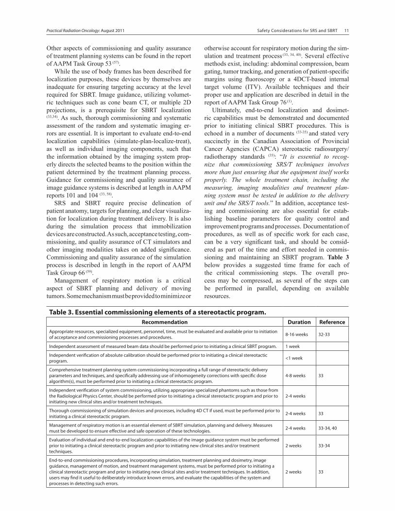
otherwise account for respiratory motion during the sim-ulation and treatment process (33, 34, 40). Several effective methods exist, including: abdominal compression, beam gating, tumor tracking, and generation of patient-specific margins using fluoroscopy or a 4DCT-based internal target volume (ITV). Available techniques and their proper use and application are described in detail in the report of AAPM Task Group 76 (1). Ultimately, end-to-end localization and dosimet-ric capabilities must be demonstrated and documented prior to initiating clinical SBRT procedures. This is echoed in a number of documents (33-35) and stated very succinctly in the Canadian Association of Provincial Cancer Agencies (CAPCA) stereotactic radiosurgery/ radiotherapy standards (35): “It is essential to recog-nize that commissioning SRS/T techniques involves more than just ensuring that the equipment itself works properly. The whole treatment chain, including the measuring, imaging modalities and treatment plan-ning system must be tested in addition to the delivery unit and the SRS/T tools.” In addition, acceptance test-ing and commissioning are also essential for estab-lishing baseline parameters for quality control and improvement programs and processes. Documentation of procedures, as well as of specific work for each case, can be a very significant task, and should be consid-ered as part of the time and effort needed in commis-sioning and maintaining an SBRT program. Table 3 below provides a suggested time frame for each of the critical commissioning steps. The overall pro-cess may be compressed, as several of the steps can be performed in parallel, depending on available resources.
Table 3. Essential commissioning elements of a stereotactic program. Recommendation Duration Reference
Appropriate resources, specialized equipment, personnel, time, must be evaluated and available prior to initiation of acceptance and commissioning processes and procedures. 8-16 weeks 32-33
Independent assessment of measured beam data should be performed prior to initiating a clinical SBRT program. 1 week
Independent verification of absolute calibration should be performed prior to initiating a clinical stereotactic program. <1 week
Comprehensive treatment planning system commissioning incorporating a full range of stereotactic delivery parameters and techniques, and specifically addressing use of inhomogeneity corrections with specific dose algorithm(s), must be performed prior to initiating a clinical stereotactic program.
4-8 weeks 33
Independent verification of system commissioning, utilizing appropriate specialized phantoms such as those from the Radiological Physics Center, should be performed prior to initiating a clinical stereotactic program and prior to initiating new clinical sites and/or treatment techniques.
2-4 weeks
Thorough commissioning of simulation devices and processes, including 4D CT if used, must be performed prior to initiating a clinical stereotactic program. 2-4 weeks 33
Management of respiratory motion is an essential element of SBRT simulation, planning and delivery. Measures must be developed to ensure effective and safe operation of these technologies. 2-4 weeks 33-34, 40
Evaluation of individual and end-to-end localization capabilities of the image guidance system must be performed prior to initiating a clinical stereotactic program and prior to initiating new clinical sites and/or treatment techniques.
2 weeks 33-34
End-to-end commissioning procedures, incorporating simulation, treatment planning and dosimetry, image guidance, management of motion, and treatment management systems, must be performed prior to initiating a clinical stereotactic program and prior to initiating new clinical sites and/or treatment techniques. In addition, users may find it useful to deliberately introduce known errors, and evaluate the capabilities of the system and processes in detecting such errors.
2 weeks 33
12 TD Solberg et al Safety Considerations for SRS and SBRT 12Practical Radiation Oncology: August 2011 Safety Considerations for SRS and SBRT 13
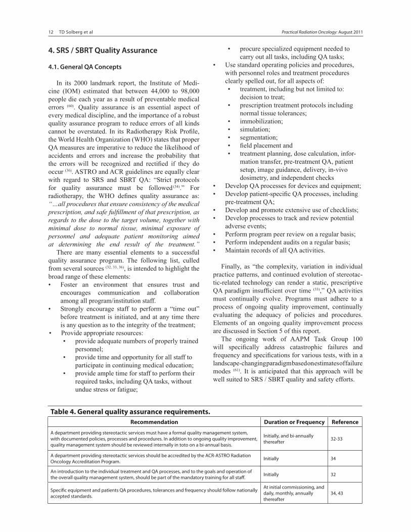
Table 4. General quality assurance requirements. Recommendation Duration or Frequency Reference
A department providing stereotactic services must have a formal quality management system, with documented policies, processes and procedures. In addition to ongoing quality improvement, quality management system should be reviewed internally in toto on a bi-annual basis.
Initially, and bi-annually thereafter 32-33
A department providing stereotactic services should be accredited by the ACR-ASTRO Radiation Oncology Accreditation Program. Initially 34
An introduction to the individual treatment and QA processes, and to the goals and operation of the overall quality management system, should be part of the mandatory training for all staff. Initially 32
Specific equipment and patients QA procedures, tolerances and frequency should follow nationally accepted standards.
At initial commissioning, and daily, monthly, annually thereafter
34, 43
• procure specialized equipment needed to carry out all tasks, including QA tasks;
• Use standard operating policies and procedures, with personnel roles and treatment procedures clearly spelled out, for all aspects of:• treatment, including but not limited to:
decision to treat;• prescription treatment protocols including
normal tissue tolerances;• immobilization; • simulation;• segmentation; • field placement and • treatment planning, dose calculation, infor-
mation transfer, pre-treatment QA, patient setup, image guidance, delivery, in-vivo dosimetry, and independent checks
• Develop QA processes for devices and equipment;• Develop patient-specific QA processes, including
pre-treatment QA;• Develop and promote extensive use of checklists;• Develop processes to track and review potential
adverse events;• Perform program peer review on a regular basis; • Perform independent audits on a regular basis; • Maintain records of all QA activities.
Finally, as “the complexity, variation in individual practice patterns, and continued evolution of stereotac-tic-related technology can render a static, prescriptive QA paradigm insufficient over time (33),” QA activities must continually evolve. Programs must adhere to a process of ongoing quality improvement, continually evaluating the adequacy of policies and procedures. Elements of an ongoing quality improvement process are discussed in Section 5 of this report. The ongoing work of AAPM Task Group 100 will specifically address catastrophic failures and frequency and specifications for various tests, with in a landscape-changing paradigm based on estimates of failure modes (61). It is anticipated that this approach will be well suited to SRS / SBRT quality and safety efforts.
4. SRS / SBRT Quality Assurance
4.1. General QA Concepts
In its 2000 landmark report, the Institute of Medi-cine (IOM) estimated that between 44,000 to 98,000 people die each year as a result of preventable medical errors (60). Quality assurance is an essential aspect of every medical discipline, and the importance of a robust quality assurance program to reduce errors of all kinds cannot be overstated. In its Radiotherapy Risk Profile, the World Health Organization (WHO) states that proper QA measures are imperative to reduce the likelihood of accidents and errors and increase the probability that the errors will be recognized and rectified if they do occur (36). ASTRO and ACR guidelines are equally clear with regard to SRS and SBRT QA: “Strict protocols for quality assurance must be followed (34).” For radiotherapy, the WHO defines quality assurance as: “…all procedures that ensure consistency of the medical prescription, and safe fulfillment of that prescription, as regards to the dose to the target volume, together with minimal dose to normal tissue, minimal exposure of personnel and adequate patient monitoring aimed at determining the end result of the treatment.” There are many essential elements to a successful quality assurance program. The following list, culled from several sources (32, 33, 36), is intended to highlight the broad range of these elements: • Foster an environment that ensures trust and
encourages communication and collaboration among all program/institution staff.
• Strongly encourage staff to perform a “time out” before treatment is initiated, and at any time there is any question as to the integrity of the treatment;
• Provide appropriate resources:• provide adequate numbers of properly trained
personnel;• provide time and opportunity for all staff to
participate in continuing medical education;• provide ample time for staff to perform their
required tasks, including QA tasks, without undue stress or fatigue;
Safety Considerations for SRS and SBRT 12 Safety Considerations for SRS and SBRT 13Practical Radiation Oncology: August 2011
4.2. Equipment QA Specific quality assurance processes and procedures will necessarily cover a broad range of stereotactic program elements, but generally can be grouped in two broad categories: equipment-related and patient-related. As with other delivery modalities, it is recommended for stereotactic programs to create daily, monthly and annual equipment quality assurance procedures. Daily QA activities should be designed to veri-fy the basic functionality and safe operation of the delivery and imaging equipment, especially the integri-ty of individual delivery and imaging devices, localiza-tion capabilities, and verification of the coincidence of imaging and therapeutic radiation isocenters of the treat-ment unit. Monthly QA procedures should be designed to detect trends in performance away from baseline and are focused on the imaging and delivery devices most likely to affect patient treatment. Annual QA procedures should be a thorough test of all aspects of the individ-ual and integrated stereotactic system, including imag-ing, treatment planning, localization, R/V, and delivery devices and processes. These QA procedures should be designed to detect any deviation from the baseline performance of the system determined at commissioning. AAPM Task Group
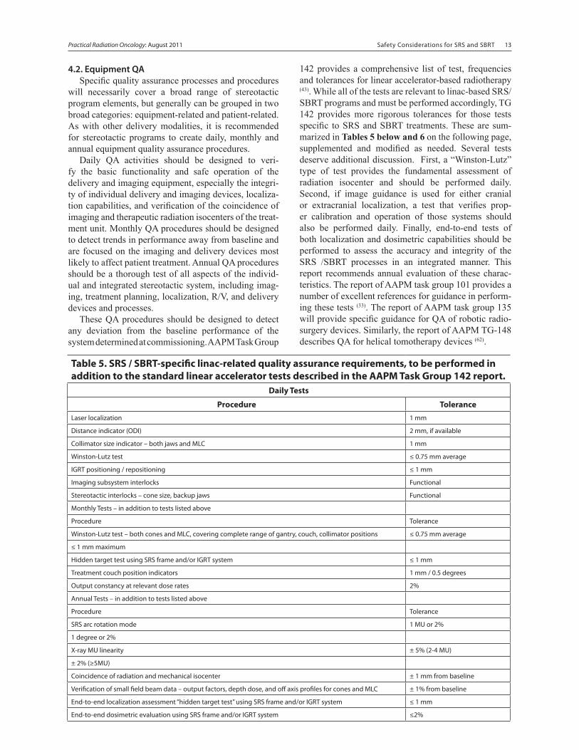
142 provides a comprehensive list of test, frequencies and tolerances for linear accelerator-based radiotherapy (43). While all of the tests are relevant to linac-based SRS/SBRT programs and must be performed accordingly, TG 142 provides more rigorous tolerances for those tests specific to SRS and SBRT treatments. These are sum-marized in Tables 5 below and 6 on the following page, supplemented and modified as needed. Several tests deserve additional discussion. First, a “Winston-Lutz” type of test provides the fundamental assessment of radiation isocenter and should be performed daily. Second, if image guidance is used for either cranial or extracranial localization, a test that verifies prop-er calibration and operation of those systems should also be performed daily. Finally, end-to-end tests of both localization and dosimetric capabilities should be performed to assess the accuracy and integrity of the SRS /SBRT processes in an integrated manner. This report recommends annual evaluation of these charac-teristics. The report of AAPM task group 101 provides a number of excellent references for guidance in perform-ing these tests (33). The report of AAPM task group 135 will provide specific guidance for QA of robotic radio-surgery devices. Similarly, the report of AAPM TG-148 describes QA for helical tomotherapy devices (62).
Table 5. SRS / SBRT-specific linac-related quality assurance requirements, to be performed in addition to the standard linear accelerator tests described in the AAPM Task Group 142 report.
Daily Tests
Procedure ToleranceLaser localization 1 mm
Distance indicator (ODI) 2 mm, if available
Collimator size indicator – both jaws and MLC 1 mm
Winston-Lutz test ≤ 0.75 mm average
IGRT positioning / repositioning ≤ 1 mm
Imaging subsystem interlocks Functional
Stereotactic interlocks – cone size, backup jaws Functional
Monthly Tests – in addition to tests listed above
Procedure Tolerance
Winston-Lutz test – both cones and MLC, covering complete range of gantry, couch, collimator positions ≤ 0.75 mm average
≤ 1 mm maximum
Hidden target test using SRS frame and/or IGRT system ≤ 1 mm
Treatment couch position indicators 1 mm / 0.5 degrees
Output constancy at relevant dose rates 2%
Annual Tests – in addition to tests listed above
Procedure Tolerance
SRS arc rotation mode 1 MU or 2%
1 degree or 2%
X-ray MU linearity ± 5% (2-4 MU)
± 2% (≥5MU)
Coincidence of radiation and mechanical isocenter ± 1 mm from baseline
Verification of small field beam data – output factors, depth dose, and off axis profiles for cones and MLC ± 1% from baseline
End-to-end localization assessment “hidden target test” using SRS frame and/or IGRT system ≤ 1 mm
End-to-end dosimetric evaluation using SRS frame and/or IGRT system ≤2%
14 TD Solberg et al Safety Considerations for SRS and SBRT 14Practical Radiation Oncology: August 2011 Safety Considerations for SRS and SBRT 15
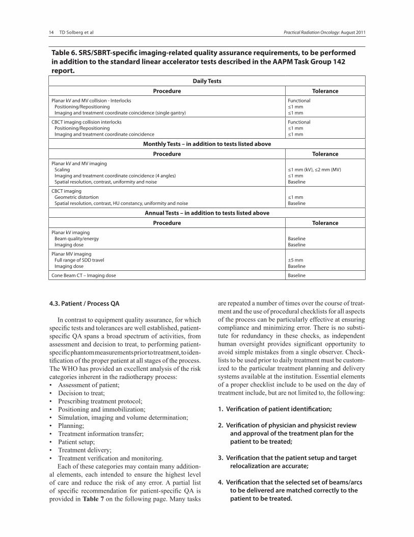
Table 6. SRS/SBRT-specific imaging-related quality assurance requirements, to be performed in addition to the standard linear accelerator tests described in the AAPM Task Group 142 report.
Daily Tests
Procedure TolerancePlanar kV and MV collision - Interlocks
Positioning/RepositioningImaging and treatment coordinate coincidence (single gantry)
Functional≤1 mm≤1 mm
CBCT imaging collision interlocksPositioning/RepositioningImaging and treatment coordinate coincidence
Functional≤1 mm≤1 mm
Monthly Tests – in addition to tests listed above
Procedure TolerancePlanar kV and MV imaging
ScalingImaging and treatment coordinate coincidence (4 angles)Spatial resolution, contrast, uniformity and noise
≤1 mm (kV), ≤2 mm (MV)≤1 mmBaseline
CBCT imagingGeometric distortionSpatial resolution, contrast, HU constancy, uniformity and noise
≤1 mmBaseline
Annual Tests – in addition to tests listed above
Procedure TolerancePlanar kV imaging
Beam quality/energyImaging dose
BaselineBaseline
Planar MV imagingFull range of SDD travelImaging dose
±5 mmBaseline
Cone Beam CT – Imaging dose Baseline
4.3. Patient / Process QA
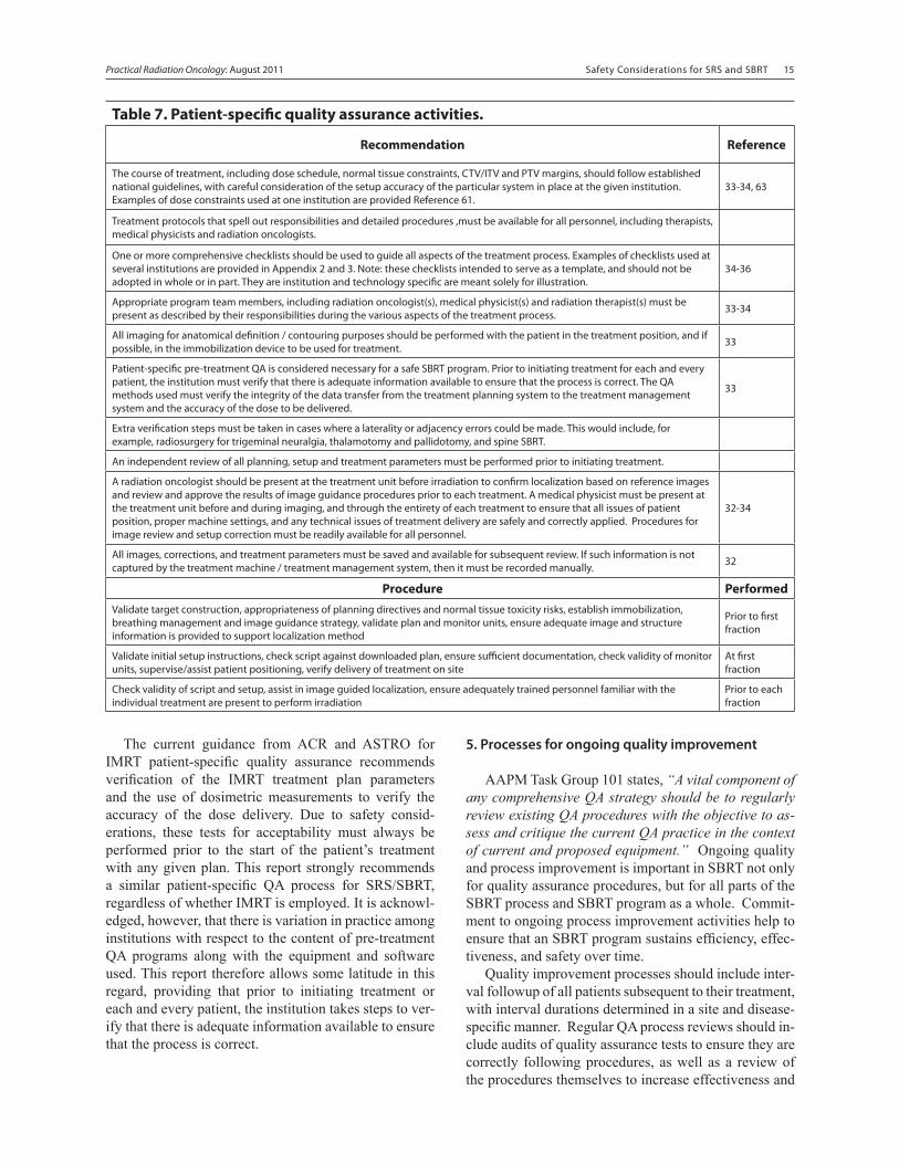
In contrast to equipment quality assurance, for which specific tests and tolerances are well established, patient-specific QA spans a broad spectrum of activities, from assessment and decision to treat, to performing patient-specific phantom measurements prior to treatment, to iden-tification of the proper patient at all stages of the process. The WHO has provided an excellent analysis of the risk categories inherent in the radiotherapy process:• Assessment of patient;• Decision to treat;• Prescribing treatment protocol;• Positioning and immobilization;• Simulation, imaging and volume determination;• Planning;• Treatment information transfer;• Patient setup;• Treatment delivery;• Treatment verification and monitoring. Each of these categories may contain many addition-al elements, each intended to ensure the highest level of care and reduce the risk of any error. A partial list of specific recommendation for patient-specific QA is provided in Table 7 on the following page. Many tasks
are repeated a number of times over the course of treat-ment and the use of procedural checklists for all aspects of the process can be particularly effective at ensuring compliance and minimizing error. There is no substi-tute for redundancy in these checks, as independent human oversight provides significant opportunity to avoid simple mistakes from a single observer. Check-lists to be used prior to daily treatment must be custom-ized to the particular treatment planning and delivery systems available at the institution. Essential elements of a proper checklist include to be used on the day of treatment include, but are not limited to, the following:
1. Verification of patient identification;
2. Verification of physician and physicist review and approval of the treatment plan for the patient to be treated;
3. Verification that the patient setup and target relocalization are accurate;
4. Verification that the selected set of beams/arcs to be delivered are matched correctly to the patient to be treated.
Safety Considerations for SRS and SBRT 14 Safety Considerations for SRS and SBRT 15Practical Radiation Oncology: August 2011
Table 7. Patient-specific quality assurance activities.
Recommendation Reference
The course of treatment, including dose schedule, normal tissue constraints, CTV/ITV and PTV margins, should follow established national guidelines, with careful consideration of the setup accuracy of the particular system in place at the given institution. Examples of dose constraints used at one institution are provided Reference 61.
33-34, 63
Treatment protocols that spell out responsibilities and detailed procedures ,must be available for all personnel, including therapists, medical physicists and radiation oncologists.
One or more comprehensive checklists should be used to guide all aspects of the treatment process. Examples of checklists used at several institutions are provided in Appendix 2 and 3. Note: these checklists intended to serve as a template, and should not be adopted in whole or in part. They are institution and technology specific are meant solely for illustration.
34-36
Appropriate program team members, including radiation oncologist(s), medical physicist(s) and radiation therapist(s) must be present as described by their responsibilities during the various aspects of the treatment process. 33-34
All imaging for anatomical definition / contouring purposes should be performed with the patient in the treatment position, and if possible, in the immobilization device to be used for treatment. 33
Patient-specific pre-treatment QA is considered necessary for a safe SBRT program. Prior to initiating treatment for each and every patient, the institution must verify that there is adequate information available to ensure that the process is correct. The QA methods used must verify the integrity of the data transfer from the treatment planning system to the treatment management system and the accuracy of the dose to be delivered.
33
Extra verification steps must be taken in cases where a laterality or adjacency errors could be made. This would include, for example, radiosurgery for trigeminal neuralgia, thalamotomy and pallidotomy, and spine SBRT.
An independent review of all planning, setup and treatment parameters must be performed prior to initiating treatment.
A radiation oncologist should be present at the treatment unit before irradiation to confirm localization based on reference images and review and approve the results of image guidance procedures prior to each treatment. A medical physicist must be present at the treatment unit before and during imaging, and through the entirety of each treatment to ensure that all issues of patient position, proper machine settings, and any technical issues of treatment delivery are safely and correctly applied. Procedures for image review and setup correction must be readily available for all personnel.
32-34
All images, corrections, and treatment parameters must be saved and available for subsequent review. If such information is not captured by the treatment machine / treatment management system, then it must be recorded manually. 32
Procedure PerformedValidate target construction, appropriateness of planning directives and normal tissue toxicity risks, establish immobilization, breathing management and image guidance strategy, validate plan and monitor units, ensure adequate image and structure information is provided to support localization method
Prior to first fraction
Validate initial setup instructions, check script against downloaded plan, ensure sufficient documentation, check validity of monitor units, supervise/assist patient positioning, verify delivery of treatment on site
At first fraction
Check validity of script and setup, assist in image guided localization, ensure adequately trained personnel familiar with the individual treatment are present to perform irradiation
Prior to each fraction
5. Processes for ongoing quality improvement
AAPM Task Group 101 states, “A vital component of any comprehensive QA strategy should be to regularly review existing QA procedures with the objective to as-sess and critique the current QA practice in the context of current and proposed equipment.” Ongoing quality and process improvement is important in SBRT not only for quality assurance procedures, but for all parts of the SBRT process and SBRT program as a whole. Commit-ment to ongoing process improvement activities help to ensure that an SBRT program sustains efficiency, effec-tiveness, and safety over time. Quality improvement processes should include inter-val followup of all patients subsequent to their treatment, with interval durations determined in a site and disease-specific manner. Regular QA process reviews should in-clude audits of quality assurance tests to ensure they are correctly following procedures, as well as a review of the procedures themselves to increase effectiveness and
The current guidance from ACR and ASTRO for IMRT patient-specific quality assurance recommends verification of the IMRT treatment plan parameters and the use of dosimetric measurements to verify the accuracy of the dose delivery. Due to safety consid-erations, these tests for acceptability must always be performed prior to the start of the patient’s treatment with any given plan. This report strongly recommends a similar patient-specific QA process for SRS/SBRT, regardless of whether IMRT is employed. It is acknowl-edged, however, that there is variation in practice among institutions with respect to the content of pre-treatment QA programs along with the equipment and software used. This report therefore allows some latitude in this regard, providing that prior to initiating treatment or each and every patient, the institution takes steps to ver-ify that there is adequate information available to ensure that the process is correct.
16 TD Solberg et al Safety Considerations for SRS and SBRT 16Practical Radiation Oncology: August 2011 Safety Considerations for SRS and SBRT 17
efficiency. External audits of stereotactic programs are strongly recommended. Offline monitoring and analysis of uncertainties and trends can help to detect system-atic and emergent problems in equipment and treatment procedures. A commitment to formal feedback to vendors helps to tie institutional quality improvement processes into the vendor’s own quality processes and guides future product development. In addition, institutions providing SBRT services are encouraged to investigate formalized tools for process improvement such as process mapping, process control and fault-tree analysis. These tools can help take guess-work out of processes and can help analyze risk and mitigation strategies on a quantitative basis. (64)
Proper ongoing quality improvement should at a minimum include interval follow-up of all patients subsequent to their treatment, offline monitoring and analysis of uncertainties and trends, periodic reviews, staff evaluations, and formal feedback to vendors.
6. Documentation
Proper documentation of all aspects of an SBRT program is essential to the program’s success and is critical to any ongoing practice quality improvement (PQI) program. Documentation must occur at all levels of the program, including personnel, equipment commis-sioning and QA, patient and treatment-specific records, and offline analysis and monitoring of uncertainties and trends. Documentation of personnel credentials, ongoing operational and safety training, time spent on any given task, and lifelong continuing education is important for ensuring the quality of the treatment team. Proper docu-mentation makes it possible to remind team members if they are overdue on any required training or continu-ing education. It also allows the team to track resource allocations and detect a need for additional staff in any given area. Documentation of equipment commissioning and quality assurance processes and test results help ensure tests are performed in a repeatable, systematic way. They allow the team to detect emerging problems in the system that can then be remedied before they become severe. Documentation of service requests and resolu-tions help the team estimate reliability, budget repair costs, and detect systematic equipment deficiencies that need to be addressed. Offline monitoring of uncertain-ties and trends can help the team refine procedures and equipment usage patterns. Patient-specific documentation should be in accordance with good medical practice as appropri-ate for the stage and site of disease treated. It should include clinical histories and treatment rationale, as well as treatment plans, setup notes, ongoing treat-ment records, patient-specific quality assurance checks,
treatment modifications, etc. Proper documentation of patient follow-up examinations allows retrospec-tive analysis for trends in treatment efficacy. AAPM Task Group 101 includes recommendations for specific data to document for SBRT treatments (33).
7. Other Recommendations
While this report deals primarily with institutions and professional staff, there are many stakeholders in the QA process, with common goals and shared respon-sibilities. In this regard, improvement of patient safety would be facilitated by collaborative efforts between the manufacturers and the users. It is hoped that there will be increased discussions and interaction between manufacturers and users in designing safer systems, in developing QA methods and training programs, and in promoting patient safety for SRS and SBRT. There are many areas for collaborative efforts between equipment vendors and end users to enhance the patient safety aspects of SRS and SBRT systems. For example, there must be dialogue and communication between equipment manufacturers and end-users on the approaches, system design, QA methodology, and clinical implementation of SRS and SBRT. Vendors must understand the needs and requirements of the clinicians, medical physicists and radiation therapists relative to the systems and processes for SRS and SBRT. With such understand-ing they must exert all the necessary efforts to incor-porate features and safeguards to assure efficacious and safe operation of their products. By the same token, the end-users need to work with the manufacturers in developing commissioning, safety and quality assur-ance tools, programs and procedures for the SRS and SBRT systems. There are many steps equipment vendors can take to improve the safety of their systems. Adequate training of all the SRS/SBRT team members, in their respective areas of responsibilities, is of paramount importance. Vendors must provide additional opportunities for specialized training, emphasizing implementation, clinical and quality assurance in addition to technical aspects, and the home institution must make available resources and time for such training. It is not adequate to train users on the basic aspects of system operation if the systems are sold and used to specialized purpos-es such as SRS and SBRT. Vendors must do more to emphasize all QA aspects, not only equipment QA, but process QA. SRS / SBRT systems consist of multiple components, and vendors must ensure and demonstrate full mechanical, electronic and information connectiv-ity of these components. In situations where compo-nents or subsystems come from more than one manu-facturer, it is the responsibilities of the manufacturers to collaboratively demonstrate compatibility of the vari-ous subsystems, and their safe operation when used in
Safety Considerations for SRS and SBRT 16 Safety Considerations for SRS and SBRT 17Practical Radiation Oncology: August 2011
combination. The users must assure that such demon-stration are documented and are satisfactory in terms of safe SRS and SBRT implementation. Departments must remember that the final responsibility of safe operation of complex treatment technology and procedures lies with the department, and that vendor documentation is only one part of the safety process. Adequately trained staff, with sufficient time, resources and support, are critical to implementation of stereotactic procedures with modern technology. Finally, while a turn-key approach to the use of complex clinical systems is appealing in terms of pro-cedural simplicity, inadequate understanding of the internal workings of such complex systems by the end-users is of concern. Rather, vendors should take an “all-inclusive” approach of safe equipment design, under-standing the need for QA equipment and procedures, and emphasizing commissioning, safety and quality assurance requirements and procedures. Professional organizations must do more to fa-cilitate proper training in specialized procedures such as SRS and SBRT, and to ensure that only qualified practitioners are involved in such procedures. Specialized accreditation programs may be an effective mechanism to realize this. The American College of Radiology presently accredits several imaging specialties, including CT, MRI mammography and stereotactic breast biopsy. Extend-ing accreditation to SRS and SBRT would be a strong step in emphasizing and recognizing quality practices. The current ACR- ASTRO Radiation Oncology Accred-itation Program of radiation oncology practices should no longer be voluntary. There are many steps that government agen-cies can take to enhance safety within the profession. There are numerous inconsistencies in regulation and radiation event reporting between state and federal agencies, and with regard to radioactive versus x-ray sources. The findings of an earlier investigation on regulatory reform in radiation medicine pointed to the need for improved databases on the actual incidence of adverse events and severe misadministrations (65). Such a system is mandated by law in the United Kingdom. Centralized registries for event-reporting, ensuring appropriate transparency regarding event details, are an effective mechanism for all stakeholders to learn from mistakes. Several voluntary efforts currently exist, nota-bly the Radiation Oncology Safety Information System (ROSIS) (66) and the system implemented at Washington University by Sasa Mutic and colleagues (67).
8. Summary
In summary, SRS and SBRT require a team-based approach, staffed by qualified radiation oncologists, medical physicists, and radiation therapists. Other spe-cialists, including disease-site-specific physicians, may also participate as needed. Treatment of SRS / SBRT patients should adhere to established national guidelines. Appropriately trained radiation oncologist(s), medical physicist(s) and radiation therapist(s) should be present at specified components of, if not for the entire duration of, each SBRT treatment. SRS and SBRT require significant resources in personnel, specialized technology, and implementa-tion time. A thorough feasibility analysis of resources required to achieve the clinical and technical goals must be performed and discussed with all personnel, includ-ing medical center administration. Because various dis-ease sites may have different clinical and/or technical requirements, feasibility and planning discussions are needed prior to undertaking new disease sites. Program personnel must be certified in their particu-lar specialty by a national certifying board, and licensed and credentialed as appropriate. Program personnel should maintain their certification and keep licenses and credentials current. Professional organizations are encouraged to develop specialized SRS / SBRT accredi-tation programs, similar to ACR- ASTRO Radiation Oncology Accreditation Program for specialty imaging programs. Specific training in clinical and technical aspects of SRS and SBRT will become increasingly important in the future. SRS and SBRT training should become a required part of radiation oncology residency training and of (CAMPEP-accredited) clinical medical phys-ics training. Current practitioners are strongly encour-aged to participate in ASTRO and/or AAPM-sponsored SRS and SBRT CME prior to treating patients. This should include general SRS / SBRT training, as well as specific training in each disease site in which a stereotactic approach is used. Proctoring is an essential component of SRS and SBRT training. Program personnel must participate in continuing medical education specific to SRS and SBRT. Acceptance and commissioning protocols and tests must be developed to explore in detail every aspect of the system with the goal of ensuring safe and effective operation. A comprehensive quality assurance program, encompassing all clinical, technical and patient-specific treatment aspects, must be developed to ensure SRS and SBRT are performed in a safe and effective manner.

18 TD Solberg et al Safety Considerations for SRS and SBRT 18Practical Radiation Oncology: August 2011 Safety Considerations for SRS and SBRT 19
References
1. Keall PJ, Mageras GS, Balter JM, et al. The management of respiratory motion in radiation oncology report of AAPM Task Group 76. Med Phys 2006;33:3874-3900.
2. Andrews DW, Scott CB, Sperduto PW, et al. Whole brain radia-tion therapy with or without stereotactic radiosurgery boost for patients with one to three brain metastases: phase III results of the RTOG 9508 randomised trial. Lancet. 2004;363:1665-1672.
3. Suh J. Stereotactic radiosurgery for the management of brain metastases. NEJM. 2010;362:1119-1127.
4. Combs SE, Welzel T, Schulz-Ertner D, et al. Differences in clinical results after linac-based single-dose radiosurgery versus fractionated stereotactic radiotherapy for patients with vestibular schwannomas. Int J Radiat Oncol Biol Phys. 2010;76:193-200.
5. Selch MT, Ahn E, Laskari A, et al. Stereotactic radiotherapy for
treatment of cavernous sinus meningiomas. Int J Radiat Oncol Biol Phys. 2004;59:101-111.
6. Spiegelmann R, Cohen ZR, Nissim O, et al. Cavernous sinus meningiomas: a large LINAC radiosurgery series. J Neuroon-col. 2010;98:195-202.
7. Sheehan JP, Williams B, Yen CP. Stereotactic radiosurgery for WHO grade I meningiomas. J Neurooncol. 2010;99:407-416.
8. Timmerman R, Paulus R, Galvin J, et al. Stereotactic body radiation therapy for inoperable early stage lung cancer. JAMA. 2010;303:1070-6.
9. Heron DE, Ferris RL, Karamouzis M, et al. Stereotactic body radiotherapy for recurrent squamous cell carcinoma of the head and neck: results of a phase I dose-escalation trial. Int J Radiat Oncol Biol Phys. 2009;75(5):1493-500.
10. Baumann P, Nyman J, Hoyer M, et al. Outcome in a prospec-tive phase II trial of medically inoperable stage I non-small-cell lung cancer patients treated with stereotactic body radiotherapy. J Clin Oncol. 2009;27:3290-6.
11. Rusthoven KE, Kavanagh BD, Cardenes H, et al. Multi-institu-tional phase I/II trial of stereotactic body radiation therapy for liver metastases. J Clin Oncol. 2009;27:1572-8.
12. Rusthoven KE, Kavanagh BD, Burri SH, et al. Multi-institu-tional phase I/II trial of stereotactic body radiation therapy for lung metastases. J Clin Oncol. 2009;27:1579-84.
13. Lee MT, Kim JJ, Dinniwell R, et al. Phase I study of individual-ized stereotactic body radiotherapy of liver metastases. J Clin Oncol. 2009;27:1585-91.
14. Fakiris AJ, McGarry RC, Yiannoutsos CT, et al. Stereotac-tic body radiation therapy for early-stage non-small-cell lung carcinoma: four-year results of a prospective phase II study. Int J Radiat Oncol Biol Phys. 2009;75:677-82.
15. Baumann P, Nyman J, Hoyer M, et al. Stereotactic body radio-therapy for medically inoperable patients with stage I non-small cell lung cancer - a first report of toxicity related to COPD/CVD in a non-randomized prospective phase II study. Radiother Oncol. 2008;88:359-67.
16. King CR, Brooks JD, Gill H, et al. Stereotactic body radio-therapy for localized prostate cancer: interim results of a prospective phase II clinical trial. Int J Radiat Oncol Biol Phys. 2009;73:1043-8.
17. Tse RV, Hawkins M, Lockwood G, et al. Phase I study of in-dividualized stereotactic body radiotherapy for hepatocellular carcinoma and intrahepatic cholangiocarcinoma. J Clin Oncol. 2008;26:657-64.
18. Chang EL, Shiu AS, Mendel E, et al. Phase I/II study of stereo-tactic body radiotherapy for spinal metastasis and its pattern of failure. J Neurosurg Spine. 2007;7:151-60.
19. Milano MT, Katz AW, Muhs AG, et al. A prospective pilot study of curative-intent stereotactic body radiation therapy in patients with 5 or fewer oligometastatic lesions. Cancer. 2008;112:650-8.
20. Blomgren H, Lax I, Näslund I, Svanström R. Stereotactic high dose fraction radiation therapy of extracranial tumors using an accelerator. Clinical experience of the first thirty-one patients. Acta Oncol. 1995;34:861-70.
21. Sawrie SM, Fiveash JB, Caudell JJ. Stereotactic body radiation
therapy for liver metastases and primary hepatocellular carci-noma: normal tissue tolerances and toxicity. Cancer Control. 2010;17:111-9.
22. Hoppe BS, Laser B, Kowalski AV, et al. Acute skin toxicity following stereotactic body radiation therapy for stage I non-small-cell lung cancer: who’s at risk? Int J Radiat Oncol Biol Phys. 2008;72:1283-6.
23. Bradley J. Radiographic response and clinical toxicity follow-ing SBRT for stage I lung cancer. J Thorac Oncol. 2007;2(7 Suppl 3):S118-24.
24. Timmerman R, McGarry R, Yiannoutsos C, et al. Excessive toxicity when treating central tumors in a phase II study of stereotactic body radiation therapy for medically inoperable early-stage lung cancer. J Clin Oncol. 2006;24:4833-9.
25. Borius PY, Debono B, Latorzeff I, et al. Dosimetric stereotac-tic radiosurgical accident: Study of 33 patients treated for brain metastases. Neurochirurgie. 2010;56:368-373.
26. Derreumaux S, Etard C, HuetI C, et al. Lessons from recent accidents in radiation therapy in France. Rad Prot Dosim. 2010;131:130-135.
27. Gourmelon P, Bey E, De Revel T, et al. The French radiation accident experience: emerging concepts in radiation burn and ARS therapies and in brain radiopathology. Radioprotection. 2008;43:23-26.

Safety Considerations for SRS and SBRT 18 Safety Considerations for SRS and SBRT 19Practical Radiation Oncology: August 2011
28. Bogdanich W, Rebelo K. (2010). The Radiation Boom—A Pinpoint Beam Strays Invisibly, Harming Instead of Healing. The New York Times. New York: A1.
29. Elekta Bulletin 98-02-17, Leksell Gamma Knife, ‘‘New 4-mm Helmet Output Factor,’’ Feb. 17, 1998.
30. Gibbons JP, Mihailidis D, Worthington C, et al. Technical note: The effect of the 4-mm-collimator output factor on gamma knife dose distributions. J Appl Clin Med Phys 2003;4:386-389.
31. Stahel PF, Sabel AL, Victoroff MS, et al. Wrong-site and wrong-patient procedures in the universal protocol era. Arch Surg. 2010;145:978-984.
32. The Royal College of Radiologists, Society and College of Radiographers, Institute of Physics and Engineering in Medi-cine, National Patient Safety Agency, British Institute of Radiology. Towards Safer Radiotherapy. London: The Royal College of Radiologists, 2008.
33. Benedict SH, Yenice KM, Followill D, et al. Stereotactic Body Radiation Therapy: The Report of AAPM Task Group 101. Med Phys 2010; 37(8): 4078-4101.
34. Potters L, Kavanagh B, Galvin JM et al. American Society for Therapeutic Radiology and Oncology (ASTRO) and American College of Radiology (ACR) Practice Guideline for the Perfor-mance of Stereotactic Body Radiation Therapy. Int J Radiat Oncol Biol Phys 2010;76:326-332.
35. CAPCA Quality Control Standards: Stereotactic Radiosur-gery/Radiotherapy. Standards for Quality Control at Canadian Radiation Treatment Centres, Canadian Association of Provin-cial Cancer Agencies (CAPCA), 2006.
36. World Health Organization (WHO). Radiotherapy Risk Profile Technical Manual, 2006.
37. ABT Associates I 2008 The ABT Study of Medical Physicist Work Values for Radiation Oncology Physics Services, Round III. American Association of Physicists in Medicine (AAPM). Available at http://www.aapm.org/pubs/reports/ABTIIIReport.pdf.
38. Nagata Y, Hiraoka M, Mizowaki T, et al. Survey of stereotactic body radiation therapy in Japan by the Japan 3-D Conformal External Beam Radiotherapy Group. Int J Radiat Oncol Biol Phys 2009;75:343-7.
39. Lawrence G, Bennett D, Branson A, et al. The Impact of chang-ing technology and working practices on errors at the NCCT. 1998–2006. Clinical Oncology (Royal College of Radiologists). 2007;19:S37.
40. Radiation Therapy Oncology Group. RTOG 0236: A Phase II Trial of Stereotactic Body Radiation Therapy (SBRT) in the Treatment of Patients with Medically Inoperable Stage I/II Non-Small Cell Lung Cancer. 2005. Available at:
http://www.rtog.org/ClinicalTrials/ProtocolTable/StudyDetails.aspx?action=openFile&FileID=4612.
41. Radiation Therapy Oncology Group. RTOG 0618: A Phase II Trial of Stereotactic Body Radiation Therapy (SBRT) in the Treatment of Patients with Operable Stage I/II Non-Small Cell Lung Cancer. 2007. Available at:
http://www.rtog.org/ClinicalTrials/ProtocolTable/StudyDetails.aspx?action=openFile&FileID=4650
42. Hoppe BS, Laser B, Kowalski AV, et al. Acute skin toxicity following stereotactic body radiation therapy for stage I non-small-cell lung cancer: Who’s at risk? Int J Radiat Oncol Biol Phys 2008;72:1283-1286.
43. Klein EE, Hanley J, Bayouth J, et al. Task Group 142 report: Quality assurance of medical accelerators. Med Phys 2009;36:4197-4212.
44. Das IJ, Cheng C-W, Watts RJ, et al. Accelerator beam data com-missioning equipment and procedures: Report of the TG-106 of the Therapy Physics Committee of the AAPM. Med Phys 2010; 35:4186-4215.
45. Alfonso A, Andreo P, Capote R, et al. A new formalism for ref-erence dosimetry of small and nonstandard fields. Med Phys. 2008;35:5179-5186.
46. Report 103: Small Field MV Photon Dosimetry. Aspradakis MA, Ed. Institute of Physics and Engineering in Medicine Pub-lishing, Bristol, United Kingdom, 2010.
47. Bjarngard BE, Tsai JS, Rice RK. Doses on the central axes of narrow 6-MV x-ray beams. Med Phys 1990;17:794-799.
48. Ding GX, Duggan DM, Coffey CW. Commissioning stereotac-tic radiosurgery beams using both experimental and theoretical methods. Phys Med Biol 2006;51:2549–2566.
49. Epp ER, Boyer AL, Doppke KP. Underdosing of lesions result-ing from lack of electronic equilibrium in upper respiratory air cavities irradiated by 10MV x-ray beams. Int J Radiat Oncol Biol Phys 1977;2:613-619.
50. Solberg TD, Holly FE, DeSalles AAF, Smathers JB. Implica-tions of Tissue Heterogeneity for Radiosurgery in Head and Neck Tumors. Int J Radiat Oncol Biol Phys 1995;32: 235-239.
51. Rustgi AK, Samuels A, Rustgi SN. Influence of air inhomoge-neities in radiosurgical beams. Med Dosim 1997;22:95-100.
52. Cheung JY, Yu KN, Ho RT, et al. Monte Carlo calculated output factors of a Leksell Gamma Knife unit. Phys Med Biol 1999;44:N247-249.
53. Martens C, Reynaert N, De Wagter C, et al. Underdosage of the upper-airway mucosa for small fields as used in intensity-mod-ulated radiation therapy: a comparison between radiochromic film measurements, Monte Carlo simulations, and collapsed cone convolution calculations. Med Phys 2002;29:1528-1535.
54. Papanikolaou N, Battista J, Boyer A, et al. AAPM Report No. 85: Tissue Inhomogeneity Corrections for Megavoltage Photon Beams. Report of Task Group No. 65 of the Radiation Therapy Committee of the American Association of Physicists in Medicine, 2004.

20 TD Solberg et al Safety Considerations for SRS and SBRT 20Practical Radiation Oncology: August 2011 Safety Considerations for SRS and SBRT 21
55. Radiation Therapy Oncology Group. RTOG 0813: Seamless Phase I/II Study of Stereotactic Lung Radiotherapy (SBRT) for Early Stage, Centrally Located, Non-Small Cell Lung Cancer (NSCLC) in Medically Inoperable Patients. 2009.
56. Radiation Therapy Oncology Group. RTOG 0915: A Randomized Phase II Study Comparing Two Stereotactic Body Radiation Therapy (SBRT) Schedules for Medically Inoper-able Patients with Stage I Peripheral Non-Small Cell Lung Cancer. 2009.
57. Fraass B, Doppke K, Hunt M, et al. American Association of Physicists in Medicine Radiation Therapy Committee Task Group 53: Quality assurance for clinical radiotherapy treatment planning. Med Phys 1998;25:1773-1829.
58. Yin F-F, Wong J, Balter J, et al. AAPM Report No. 104: The Role of In-Room kV X-Ray Imaging for Patient Setup and Tar-get Localization. Report of Task Group No. 104 of the Therapy Imaging Committee of the American Association of Physicists in Medicine, 2009.
59. Mutic S, Palta JR, Butket EK, et al. Quality assurance for com-puted-tomography simulators and the computed-tomography simulation process: Report of the AAPM Radiation Therapy Committee Task Group No. 66. Med Phys 2003;30:2762-2792.
60. To Err Is Human: Building a Safer Health System. Kohn LT, Corrigan JM, and Donaldson MS, Eds; The National Academies Press, Washington, DC, 2000.
61. Huq MS, Fraass BA, Dunscombe PB, et al. A method for evalu-ating quality assurance needs in radiation therapy. Int J Rad Onc Biol Phys. 2008;71(Suppl):S170-S173.
62. Langen KM, Papanikolaou N, Balog J, et al. QA for helical tomotherapy: Report of the AAPM Task Group 148. Med Phys, 2010;37:4817-4853.
63. Timmerman RD. An overview of hypofractionation and intro-duction to this issue of seminars in radiation oncology. Semin Radiat Oncol 2008;18:215-222.
64. Rath F. Tools for developing a quality management program: proactive tools (process mapping, value stream mapping, fault tree analysis, and failure mode and effects analysis) Int J Radiat Oncol Biol Phys 2008;71:S187-90.
65. Radiation in Medicine: A Need for Regulatory Reform. Gott-fried KD and Penn G, Eds; The National Academies Press, Washington, DC, 1996.
66. Cunningham J, Coffey M, Knöös T, Holmberg O. Radiation Oncology Safety Information System (ROSIS)--profiles of par-ticipants and the first 1074 incident reports. Radiother Oncol 2010;97:601-607.
67. Mutic S, Brame RS, Oddiraju S, et al. Event (error and near-miss) reporting and learning system for process improvement in radiation oncology. Med Phys 2010;37:5027-5036.
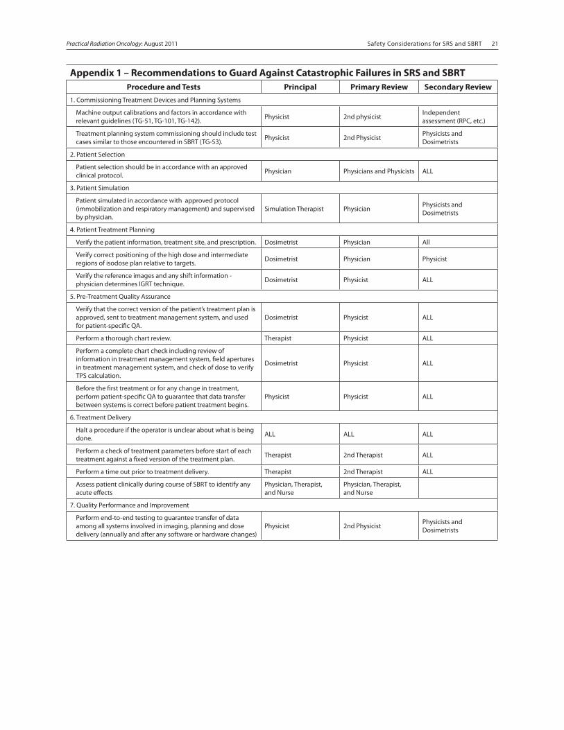
Safety Considerations for SRS and SBRT 20 Safety Considerations for SRS and SBRT 21Practical Radiation Oncology: August 2011
Appendix 1 – Recommendations to Guard Against Catastrophic Failures in SRS and SBRTProcedure and Tests Principal Primary Review Secondary Review
1. Commissioning Treatment Devices and Planning Systems
Machine output calibrations and factors in accordance with relevant guidelines (TG-51, TG-101, TG-142). Physicist 2nd physicist Independent
assessment (RPC, etc.)
Treatment planning system commissioning should include test cases similar to those encountered in SBRT (TG-53). Physicist 2nd Physicist Physicists and
Dosimetrists
2. Patient Selection
Patient selection should be in accordance with an approved clinical protocol. Physician Physicians and Physicists ALL
3. Patient Simulation
Patient simulated in accordance with approved protocol (immobilization and respiratory management) and supervised by physician.
Simulation Therapist Physician Physicists and Dosimetrists
4. Patient Treatment Planning
Verify the patient information, treatment site, and prescription. Dosimetrist Physician All
Verify correct positioning of the high dose and intermediate regions of isodose plan relative to targets. Dosimetrist Physician Physicist
Verify the reference images and any shift information - physician determines IGRT technique. Dosimetrist Physicist ALL
5. Pre-Treatment Quality Assurance
Verify that the correct version of the patient’s treatment plan is approved, sent to treatment management system, and used for patient-specific QA.
Dosimetrist Physicist ALL
Perform a thorough chart review. Therapist Physicist ALL
Perform a complete chart check including review of information in treatment management system, field apertures in treatment management system, and check of dose to verify TPS calculation.
Dosimetrist Physicist ALL
Before the first treatment or for any change in treatment, perform patient-specific QA to guarantee that data transfer between systems is correct before patient treatment begins.
Physicist Physicist ALL
6. Treatment Delivery
Halt a procedure if the operator is unclear about what is being done. ALL ALL ALL
Perform a check of treatment parameters before start of each treatment against a fixed version of the treatment plan. Therapist 2nd Therapist ALL
Perform a time out prior to treatment delivery. Therapist 2nd Therapist ALL
Assess patient clinically during course of SBRT to identify any acute effects
Physician, Therapist, and Nurse
Physician, Therapist, and Nurse
7. Quality Performance and Improvement
Perform end-to-end testing to guarantee transfer of data among all systems involved in imaging, planning and dose delivery (annually and after any software or hardware changes)
Physicist 2nd Physicist Physicists and Dosimetrists
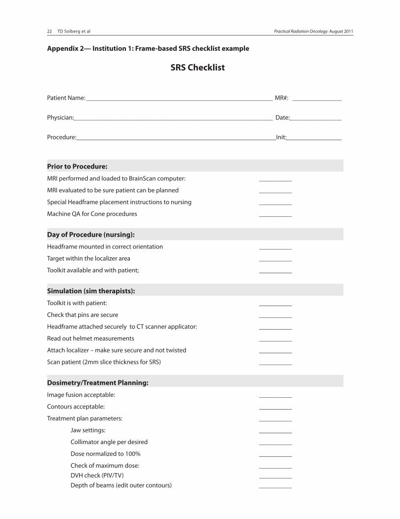
22 TD Solberg et al Safety Considerations for SRS and SBRT 22Practical Radiation Oncology: August 2011 Safety Considerations for SRS and SBRT 23
Appendix 2— Institution 1: Frame-based SRS checklist example
SRS Checklist
Patient Name: _________________________________________________________ MR#: _______________
Physician: _____________________________________________________________ Date: ________________
Procedure: _____________________________________________________________Init: _________________
Prior to Procedure:
MRI performed and loaded to BrainScan computer: __________
MRI evaluated to be sure patient can be planned __________
Special Headframe placement instructions to nursing __________
Machine QA for Cone procedures __________
Day of Procedure (nursing):
Headframe mounted in correct orientation __________
Target within the localizer area __________
Toolkit available and with patient; __________
Simulation (sim therapists):
Toolkit is with patient: __________
Check that pins are secure __________
Headframe attached securely to CT scanner applicator: __________
Read out helmet measurements __________
Attach localizer – make sure secure and not twisted __________
Scan patient (2mm slice thickness for SRS) __________
Dosimetry/Treatment Planning:
Image fusion acceptable: __________
Contours acceptable: __________
Treatment plan parameters: __________
Jaw settings: __________
Collimator angle per desired __________
Dose normalized to 100% __________
Check of maximum dose: __________ DVH check (PIV/TV) __________ Depth of beams (edit outer contours) __________
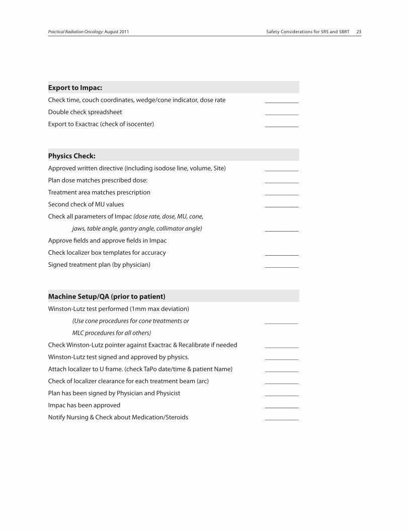
Safety Considerations for SRS and SBRT 22 Safety Considerations for SRS and SBRT 23Practical Radiation Oncology: August 2011
Export to Impac:
Check time, couch coordinates, wedge/cone indicator, dose rate __________
Double check spreadsheet __________
Export to Exactrac (check of isocenter) __________
Physics Check:
Approved written directive (including isodose line, volume, Site) __________
Plan dose matches prescribed dose: __________
Treatment area matches prescription __________
Second check of MU values __________
Check all parameters of Impac (dose rate, dose, MU, cone,
jaws, table angle, gantry angle, collimator angle) __________
Approve fields and approve fields in Impac
Check localizer box templates for accuracy __________
Signed treatment plan (by physician) __________
Machine Setup/QA (prior to patient)
Winston-Lutz test performed (1mm max deviation)
(Use cone procedures for cone treatments or __________
MLC procedures for all others)
Check Winston-Lutz pointer against Exactrac & Recalibrate if needed __________
Winston-Lutz test signed and approved by physics. __________
Attach localizer to U frame. (check TaPo date/time & patient Name) __________
Check of localizer clearance for each treatment beam (arc) __________
Plan has been signed by Physician and Physicist __________
Impac has been approved __________
Notify Nursing & Check about Medication/Steroids __________
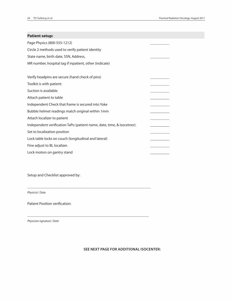
24 TD Solberg et al Safety Considerations for SRS and SBRT 24Practical Radiation Oncology: August 2011 Safety Considerations for SRS and SBRT 25
Patient setup:
Page Physics (800-555-1212) __________
Circle 2 methods used to verify patient identity
State name, birth date, SSN, Address, __________
MR number, hospital tag if inpatient, other (indicate)
Verify headpins are secure (hand check of pins) __________
Toolkit is with patient: __________
Suction is available. __________
Attach patient to table __________
Independent Check that frame is secured into Yoke __________
Bubble helmet readings match original within 1mm __________
Attach localizer to patient __________
Independent verification TaPo (patient name, date, time, & Isocetner): __________
Set to localization position __________
Lock table locks on couch (longitudinal and lateral) __________
Fine adjust to BL localizer. __________
Lock motors on gantry stand __________
Setup and Checklist approved by:
_________________________________________________________________Physicist / Date
Patient Position verification: ________________________________________________________________Physician signature / Date
SEE NEXT PAGE FOR ADDITIONAL ISOCENTER:
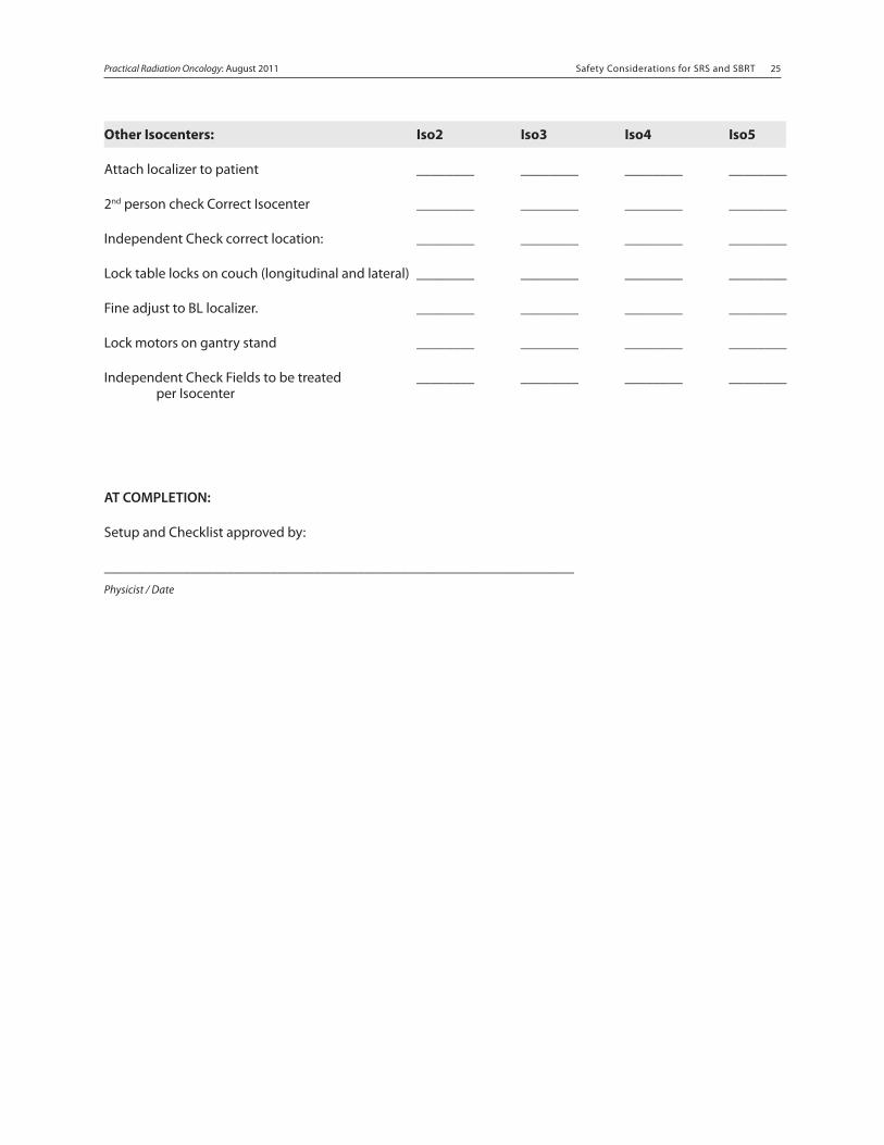
Safety Considerations for SRS and SBRT 24 Safety Considerations for SRS and SBRT 25Practical Radiation Oncology: August 2011
Other Isocenters: Iso2 Iso3 Iso4 Iso5
Attach localizer to patient ________ ________ ________ ________
2nd person check Correct Isocenter ________ ________ ________ ________
Independent Check correct location: ________ ________ ________ ________
Lock table locks on couch (longitudinal and lateral) ________ ________ ________ ________
Fine adjust to BL localizer. ________ ________ ________ ________
Lock motors on gantry stand ________ ________ ________ ________
Independent Check Fields to be treated ________ ________ ________ ________ per Isocenter
AT COMPLETION:
Setup and Checklist approved by:
_________________________________________________________________Physicist / Date
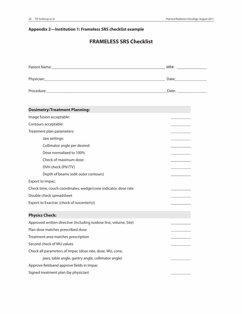
26 TD Solberg et al Safety Considerations for SRS and SBRT 26Practical Radiation Oncology: August 2011 Safety Considerations for SRS and SBRT 27
Appendix 2—Institution 1: Frameless SRS checklist example
FRAMELESS SRS Checklist
Patient Name: _________________________________________________________ MR#: _______________
Physician: _____________________________________________________________ Date: ________________
Procedure: _____________________________________________________________Date: _______________
Dosimetry/Treatment Planning:
Image fusion acceptable: __________
Contours acceptable: __________
Treatment plan parameters: __________
Jaw settings: __________
Collimator angle per desired __________
Dose normalized to 100% __________
Check of maximum dose: __________
DVH check (PIV/TV) __________
Depth of beams (edit outer contours) __________
Export to Impac:
Check time, couch coordinates, wedge/cone indicator, dose rate __________
Double check spreadsheet __________
Export to Exactrac (check of isocenter(s)) __________
Physics Check:
Approved written directive (including isodose line, volume, Site) __________
Plan dose matches prescribed dose: __________
Treatment area matches prescription __________
Second check of MU values __________
Check all parameters of Impac (dose rate, dose, MU, cone,
jaws, table angle, gantry angle, collimator angle) __________
Approve fieldsand approve fields in Impac
Signed treatment plan (by physician) __________
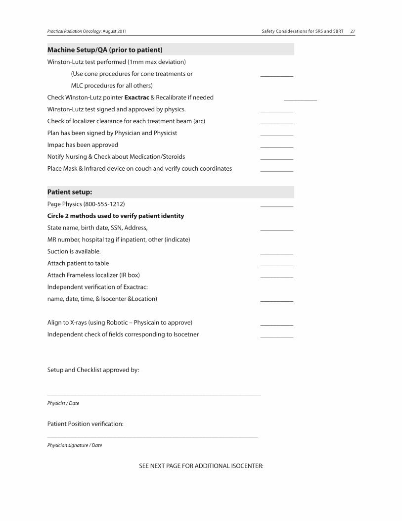
Safety Considerations for SRS and SBRT 26 Safety Considerations for SRS and SBRT 27Practical Radiation Oncology: August 2011
Machine Setup/QA (prior to patient)
Winston-Lutz test performed (1mm max deviation)
(Use cone procedures for cone treatments or __________
MLC procedures for all others)
Check Winston-Lutz pointer Exactrac & Recalibrate if needed __________
Winston-Lutz test signed and approved by physics. __________
Check of localizer clearance for each treatment beam (arc) __________
Plan has been signed by Physician and Physicist __________
Impac has been approved __________
Notify Nursing & Check about Medication/Steroids __________
Place Mask & Infrared device on couch and verify couch coordinates __________
Patient setup:
Page Physics (800-555-1212) __________
Circle 2 methods used to verify patient identity
State name, birth date, SSN, Address, __________
MR number, hospital tag if inpatient, other (indicate)
Suction is available. __________
Attach patient to table __________
Attach Frameless localizer (IR box) __________
Independent verification of Exactrac:
name, date, time, & Isocenter &Location) __________
Align to X-rays (using Robotic – Physicain to approve) __________
Independent check of fields corresponding to Isocetner __________
Setup and Checklist approved by:
_________________________________________________________________
Physicist / Date
Patient Position verification:
________________________________________________________________
Physician signature / Date
SEE NEXT PAGE FOR ADDITIONAL ISOCENTER:
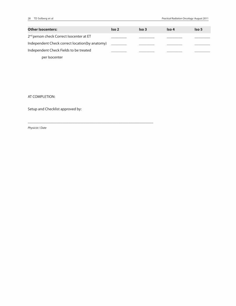
28 TD Solberg et al Safety Considerations for SRS and SBRT 28Practical Radiation Oncology: August 2011 Safety Considerations for SRS and SBRT 29
Other Isocenters: Iso 2 Iso 3 Iso 4 Iso 5
2nd person check Correct Isocenter at ET ________ ________ ________ ________
Independent Check correct location(by anatomy) ________ ________ ________ ________
Independent Check Fields to be treated ________ ________ ________ ________
per Isocenter
AT COMPLETION:
Setup and Checklist approved by:
_________________________________________________________________
Physicist / Date
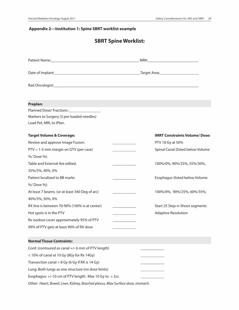
Safety Considerations for SRS and SBRT 28 Safety Considerations for SRS and SBRT 29Practical Radiation Oncology: August 2011
Appendix 2—Institution 1: Spine SBRT worklist example
SBRT Spine Worklist:
Patient Name:_____________________________________________ MR#:__________________________
Date of Implant:____________________________________________Target Area:____________________
Rad Oncologist:__________________________________________________________________________
Preplan:
Planned Dose/ Fractions :________________
Markers to Surgery (3 pre-loaded needles)
Load Pet, MRI, to iPlan.
Target Volume & Coverage: IMRT Constraints Volume/ Dose:
Review and approve Image Fusion: ____________ PTV 18 Gy at 50%
PTV = 1-5 mm margin on GTV (per case) ____________ Spinal Canal (listed below Volume
%/ Dose %):
Table and External Are edited. ____________ 100%/0%, 90%/25%, 55%/30%,
35%/5%, 40%, 0%
Patient localized to BB marks ____________ Esophagus (listed below Volume
%/ Dose %):
At least 7 beams. (or at least 340 Deg of arc) ____________ 100%/0%, 90%/25%, 60%/35%,
40%/5%, 50%, 0%
RX line is between 70-90% (100% is at center) ____________ Start 25 Step-n-Shoot segments
Hot spots is in the PTV ____________ Adaptive Resolution
Rx isodose cover approximately 95% of PTV ____________
99% of PTV gets at least 90% of RX dose ____________
Normal Tissue Contraints:
Cord: (contoured as canal +/- 6 mm of PTV length) ____________
< 10% of canal at 10 Gy (8Gy for Rx 14Gy) ____________
Transection canal < 8 Gy (6 Gy if RX is 14 Gy) ____________
Lung: Both lungs as one structure (no dose limits) ____________
Esophagus: +/-10 cm of PTV length: Max 10 Gy to > 2cc ____________
Other: Heart, Bowel, Liver, Kidney, Brachial plexus, Max Surface dose, stomach.
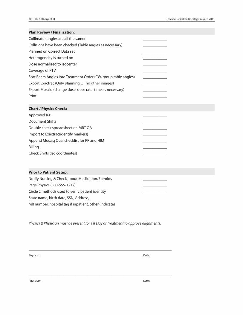
30 TD Solberg et al Safety Considerations for SRS and SBRT 30Practical Radiation Oncology: August 2011 Safety Considerations for SRS and SBRT 31
Plan Review / Finalization:
Collimator angles are all the same: ____________
Collisions have been checked (Table angles as necessary) ____________
Planned on Correct Data set ____________
Heterogeneity is turned on ____________
Dose normalized to isocenter ____________
Coverage of PTV. ____________
Sort Beam Angles into Treatment Order (CW, group table angles) ____________
Export Exactrac (Only planning CT no other images) ____________
Export Mosaiq (change dose, dose rate, time as necessary) ____________
Print ____________
Chart / Physics Check:
Approved RX: ____________
Document Shifts ____________
Double check spreadsheet or IMRT QA ____________
Import to Exactrac(identify markers) ____________
Append Mosaiq Qual checklist for PR and HIM ____________
Billing ____________
Check Shifts (Iso coordinates) ____________
Prior to Patient Setup:
Notify Nursing & Check about Medication/Steroids ____________
Page Physics (800-555-1212) ____________
Circle 2 methods used to verify patient identity ____________
State name, birth date, SSN, Address,
MR number, hospital tag if inpatient, other (indicate)
Physics & Physician must be present for 1st Day of Treatment to approve alignments.
________________________________________________________________________
Physicist: Date:
________________________________________________________________________
Physician: Date:
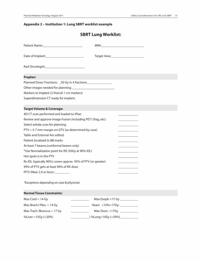
Safety Considerations for SRS and SBRT 30 Safety Considerations for SRS and SBRT 31Practical Radiation Oncology: August 2011
Appendix 2 – Institution 1: Lung SBRT worklist example
SBRT Lung Worklist:
Patient Name:________________________ MR#:__________________________
Date of Implant:_______________________ Target Area:____________________
Rad Oncologist:________________________
Preplan:
Planned Dose/ Fractions: _50 Gy in 4 fractions_______________
Other images needed for planning:__________________________
Markers to Implant (2 Visicoil 1 cm markers)
Superdimension CT ready for implant.
Target Volume & Coverage:
4D CT scan performed and loaded to iPlan ____________
Review and approve Image Fusion (including PET/ Diag, etc): ____________
Select exhale scan for planning ____________
PTV = 3-7 mm margin on GTV (as determined by case) ____________
Table and External Are edited. ____________
Patient localized to BB marks ____________
At least 7 beams.(conformal beams only) ____________
*Use Normalization point for RX (50Gy at 90% IDL) ____________
Hot spots is in the PTV ____________
Rx IDL (typically 90%) covers approx 95% of PTV (or greater) ____________
99% of PTV gets at least 90% of RX dose ____________
PITV (Near 2.0 or less)=_________ ____________
*Exceptions depending on case & physician
Normal Tissue Constraints:
Max Cord < 14 Gy ___________ Max Esoph.<17 Gy ___________
Max Brach.l Plex. < 14 Gy ___________ Heart <10%>17Gy: ___________
Max Trach /Broncus < 17 Gy ___________ Max Stom. <17Gy ___________
%Liver >15Gy (<20%) ___________\ %Lung>10Gy (<30%)___________
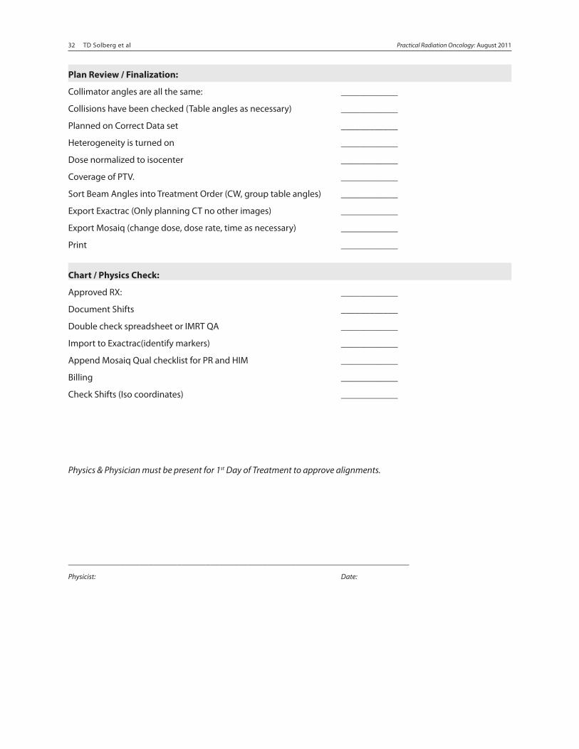
32 TD Solberg et al Safety Considerations for SRS and SBRT 32Practical Radiation Oncology: August 2011 Safety Considerations for SRS and SBRT 33
Plan Review / Finalization:
Collimator angles are all the same: ____________
Collisions have been checked (Table angles as necessary) ____________
Planned on Correct Data set ____________
Heterogeneity is turned on ____________
Dose normalized to isocenter ____________
Coverage of PTV. ____________
Sort Beam Angles into Treatment Order (CW, group table angles) ____________
Export Exactrac (Only planning CT no other images) ____________
Export Mosaiq (change dose, dose rate, time as necessary) ____________
Print ____________
Chart / Physics Check:
Approved RX: ____________
Document Shifts ____________
Double check spreadsheet or IMRT QA ____________
Import to Exactrac(identify markers) ____________
Append Mosaiq Qual checklist for PR and HIM ____________
Billing ____________
Check Shifts (Iso coordinates) ____________
Physics & Physician must be present for 1st Day of Treatment to approve alignments.
________________________________________________________________________
Physicist: Date:
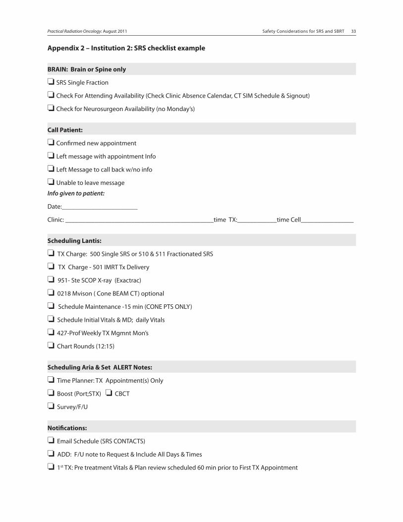
Safety Considerations for SRS and SBRT 32 Safety Considerations for SRS and SBRT 33Practical Radiation Oncology: August 2011
Appendix 2 – Institution 2: SRS checklist example
BRAIN: Brain or Spine only
❏ SRS Single Fraction
❏ Check For Attending Availability (Check Clinic Absence Calendar, CT SIM Schedule & Signout)
❏ Check for Neurosurgeon Availability (no Monday’s)
Call Patient:
❏ Confirmed new appointment
❏ Left message with appointment Info
❏ Left Message to call back w/no info
❏ Unable to leave message
Info given to patient:
Date:_______________________
Clinic: _____________________________________________time TX:____________time Cell________________
Scheduling Lantis:
❏ TX Charge: 500 Single SRS or 510 & 511 Fractionated SRS
❏ TX Charge - 501 IMRT Tx Delivery
❏ 951- Ste SCOP X-ray (Exactrac)
❏ 0218 Mvison ( Cone BEAM CT) optional
❏ Schedule Maintenance -15 min (CONE PTS ONLY)
❏ Schedule Initial Vitals & MD; daily Vitals
❏ 427-Prof Weekly TX Mgmnt Mon’s
❏ Chart Rounds (12:15)
Scheduling Aria & Set ALERT Notes:
❏ Time Planner: TX Appointment(s) Only
❏ Boost (Port;STX) ❏ CBCT
❏ Survey/F/U
Notifications:
❏ Email Schedule (SRS CONTACTS)
❏ ADD: F/U note to Request & Include All Days & Times
❏ 1st TX: Pre treatment Vitals & Plan review scheduled 60 min prior to First TX Appointment
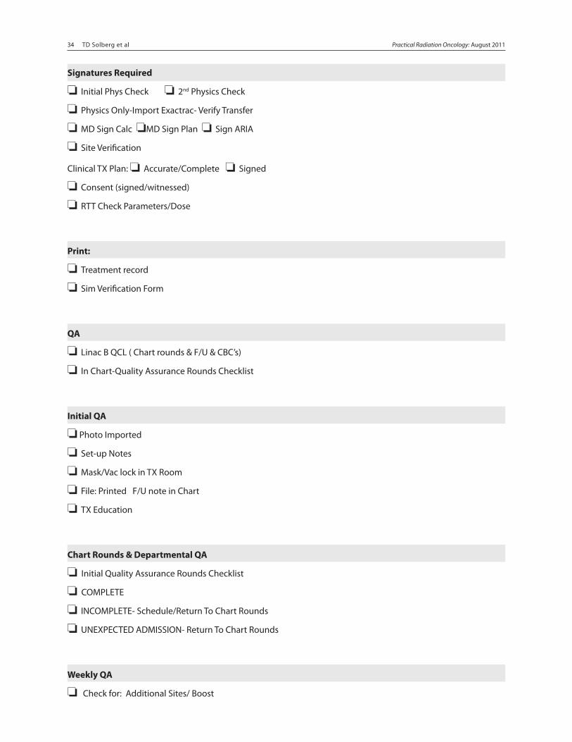
34 TD Solberg et al Safety Considerations for SRS and SBRT 34Practical Radiation Oncology: August 2011 Safety Considerations for SRS and SBRT 35
Signatures Required
❏ Initial Phys Check ❏ 2nd Physics Check
❏ Physics Only-Import Exactrac- Verify Transfer
❏ MD Sign Calc ❏MD Sign Plan ❏ Sign ARIA
❏ Site Verification
Clinical TX Plan: ❏ Accurate/Complete ❏ Signed
❏ Consent (signed/witnessed)
❏ RTT Check Parameters/Dose
Print:
❏ Treatment record
❏ Sim Verification Form
QA
❏ Linac B QCL ( Chart rounds & F/U & CBC’s)
❏ In Chart-Quality Assurance Rounds Checklist
Initial QA
❏ Photo Imported
❏ Set-up Notes
❏ Mask/Vac lock in TX Room
❏ File: Printed F/U note in Chart
❏ TX Education
Chart Rounds & Departmental QA
❏ Initial Quality Assurance Rounds Checklist
❏ COMPLETE
❏ INCOMPLETE- Schedule/Return To Chart Rounds
❏ UNEXPECTED ADMISSION- Return To Chart Rounds
Weekly QA
❏ Check for: Additional Sites/ Boost
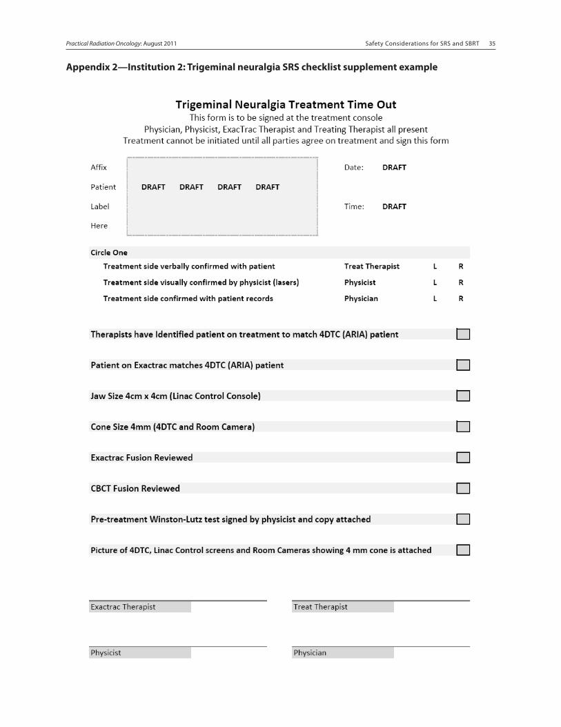
Safety Considerations for SRS and SBRT 34 Safety Considerations for SRS and SBRT 35Practical Radiation Oncology: August 2011
Appendix 2—Institution 2: Trigeminal neuralgia SRS checklist supplement example
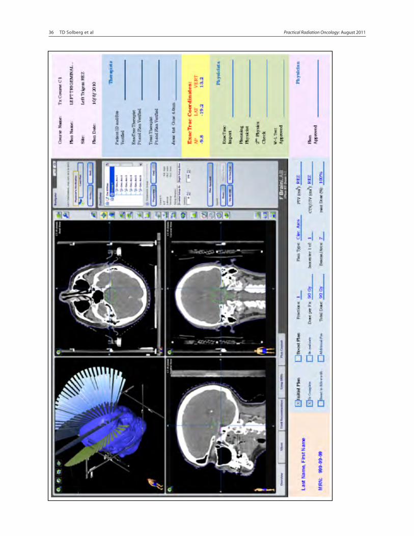
36 TD Solberg et al Safety Considerations for SRS and SBRT 36Practical Radiation Oncology: August 2011 Safety Considerations for SRS and SBRT 37
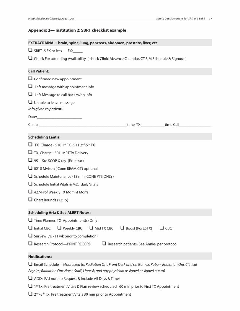
Safety Considerations for SRS and SBRT 36 Safety Considerations for SRS and SBRT 37Practical Radiation Oncology: August 2011
Appendix 2— Institution 2: SBRT checklist example
EXTRACRAINAL: brain, spine, lung, pancreas, abdomen, prostate, liver, etc
❏ SBRT 5 FX or less FX:_____
❏ Check For attending Availability ( check Clinic Absence Calendar, CT SIM Schedule & Signout )
Call Patient:
❏ Confirmed new appointment
❏ Left message with appointment Info
❏ Left Message to call back w/no info
❏ Unable to leave message
Info given to patient:
Date:_______________________
Clinic: _____________________________________________time TX:____________time Cell________________
Scheduling Lantis:
❏ TX Charge - 510 1st FX ; 511 2nd-5th FX
❏ TX Charge - 501 IMRT Tx Delivery
❏ 951- Ste SCOP X-ray (Exactrac)
❏ 0218 Mvison ( Cone BEAM CT) optional
❏ Schedule Maintenance -15 min (CONE PTS ONLY)
❏ Schedule Initial Vitals & MD; daily Vitals
❏ 427-Prof Weekly TX Mgmnt Mon’s
❏ Chart Rounds (12:15)
Scheduling Aria & Set ALERT Notes:
❏ Time Planner: TX Appointment(s) Only
❏ Initial CBC ❏ Weekly CBC ❏ Mid TX CBC ❏ Boost (Port;STX) ❏ CBCT
❏ Survey/F/U - (1 wk prior to completion)
❏ Research Protocol—PRINT RECORD ❏ Research patients- See Annie- per protocol
Notifications:
❏ Email Schedule—(Addressed to: Radiation Onc Front Desk and cc: Gomez, Ruben; Radiation Onc Clinical
Physics; Radiation Onc Nurse Staff; Linac B; and any physician assigned or signed out to)
❏ ADD: F/U note to Request & Include All Days & Times
❏ 1st TX: Pre treatment Vitals & Plan review scheduled 60 min prior to First TX Appointment
❏ 2nd–5th TX: Pre treatment Vitals 30 min prior to Appointment
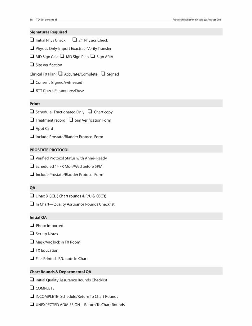
38 TD Solberg et al Safety Considerations for SRS and SBRT 38Practical Radiation Oncology: August 2011 Safety Considerations for SRS and SBRT 39
Signatures Required
❏ Initial Phys Check ❏ 2nd Physics Check
❏ Physics Only-Import Exactrac- Verify Transfer
❏ MD Sign Calc ❏ MD Sign Plan ❏ Sign ARIA
❏ Site Verification
Clinical TX Plan: ❏ Accurate/Complete ❏ Signed
❏ Consent (signed/witnessed)
❏ RTT Check Parameters/Dose
Print:
❏ Schedule- Fractionated Only ❏ Chart copy
❏ Treatment record ❏ Sim Verification Form
❏ Appt Card
❏ Include Prostate/Bladder Protocol Form
PROSTATE PROTOCOL
❏ Verified Protocol Status with Anne- Ready
❏ Scheduled 1st FX Mon/Wed before 5PM
❏ Include Prostate/Bladder Protocol Form
QA
❏ Linac B QCL ( Chart rounds & F/U & CBC’s)
❏ In Chart—Quality Assurance Rounds Checklist
Initial QA
❏ Photo Imported
❏ Set-up Notes
❏ Mask/Vac lock in TX Room
❏ TX Education
❏ File: Printed F/U note in Chart
Chart Rounds & Departmental QA
❏ Initial Quality Assurance Rounds Checklist
❏ COMPLETE
❏ INCOMPLETE- Schedule/Return To Chart Rounds
❏ UNEXPECTED ADMISSION—Return To Chart Rounds
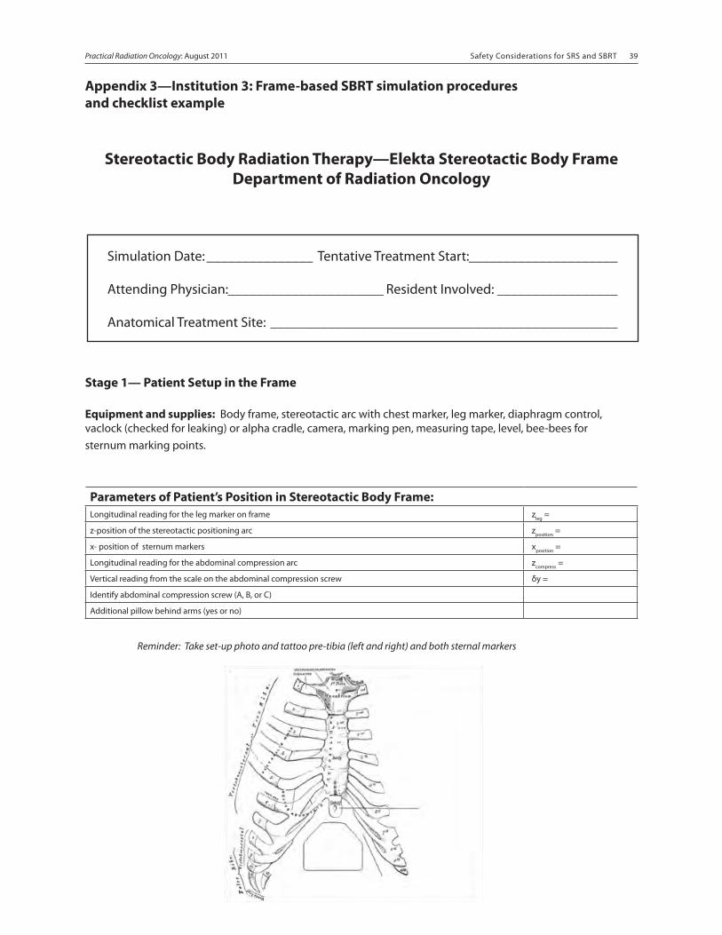
Safety Considerations for SRS and SBRT 38 Safety Considerations for SRS and SBRT 39Practical Radiation Oncology: August 2011
Appendix 3—Institution 3: Frame-based SBRT simulation procedures and checklist example
Stereotactic Body Radiation Therapy—Elekta Stereotactic Body FrameDepartment of Radiation Oncology
Stage 1— Patient Setup in the Frame
Equipment and supplies: Body frame, stereotactic arc with chest marker, leg marker, diaphragm control, vaclock (checked for leaking) or alpha cradle, camera, marking pen, measuring tape, level, bee-bees for sternum marking points.
Parameters of Patient’s Position in Stereotactic Body Frame:Longitudinal reading for the leg marker on frame zleg =
z-position of the stereotactic positioning arc zposition =
x- position of sternum markers xposition =
Longitudinal reading for the abdominal compression arc zcompress =
Vertical reading from the scale on the abdominal compression screw δy =
Identify abdominal compression screw (A, B, or C)
Additional pillow behind arms (yes or no)
Reminder: Take set-up photo and tattoo pre-tibia (left and right) and both sternal markers
Simulation Date: _______________ Tentative Treatment Start: _____________________
Attending Physician: ______________________ Resident Involved: _________________
Anatomical Treatment Site: _________________________________________________
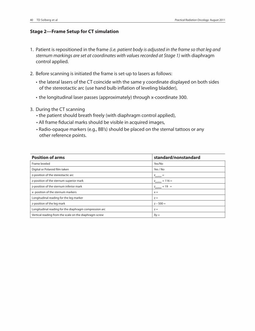
40 TD Solberg et al Safety Considerations for SRS and SBRT 40Practical Radiation Oncology: August 2011 Safety Considerations for SRS and SBRT 41
Stage 2—Frame Setup for CT simulation
1. Patient is repositioned in the frame (i.e. patient body is adjusted in the frame so that leg and sternum markings are set at coordinates with values recorded at Stage 1) with diaphragm control applied.
2. Before scanning is initiated the frame is set-up to lasers as follows:
• the lateral lasers of the CT coincide with the same y coordinate displayed on both sides of the stereotactic arc (use hand bulb inflation of leveling bladder),
• the longitudinal laser passes (approximately) through x-coordinate 300.
3. During the CT scanning • the patient should breath freely (with diaphragm control applied), • All frame fiducial marks should be visible in acquired images, • Radio-opaque markers (e.g., BB’s) should be placed on the sternal tattoos or any other reference points.
Position of arms standard/nonstandardFrame leveled Yes/No
Digital or Polaroid film taken Yes / No
z-position of the stereotactic arc zposition =
z-position of the sternum superior mark zposition + 116 =
z-position of the sternum inferior mark zposition + 19 =
x- position of the sternum markers x =
Longitudinal reading for the leg marker z =
z-position of the leg mark z – 500 =
Longitudinal reading for the diaphragm compression arc z =
Vertical reading from the scale on the diaphragm screw δy =
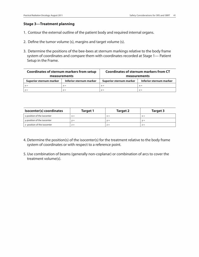
Safety Considerations for SRS and SBRT 40 Safety Considerations for SRS and SBRT 41Practical Radiation Oncology: August 2011
Stage 3—Treatment planning
1. Contour the external outline of the patient body and required internal organs.
2. Define the tumor volume (s), margins and target volume (s).
3. Determine the positions of the bee-bees at sternum markings relative to the body frame system of coordinates and compare them with coordinates recorded at Stage 1— Patient Setup in the Frame.
4. Determine the position(s) of the isocenter(s) for the treatment relative to the body frame system of coordinates or with respect to a reference point.
5. Use combination of beams (generally non-coplanar) or combination of arcs to cover the treatment volume(s).
Coordinates of sternum markers from setup measurements
Coordinates of sternum markers from CT measurements
Superior sternum marker Inferior sternum marker Superior sternum marker Inferior sternum markerx = x = x = x =
z = z = z = z =
Isocenter(s) coordinates Target 1 Target 2 Target 3x-position of the isocenter x = x = x =
y-position of the isocenter y = y = y =
z- position of the isocenter z = z = z =
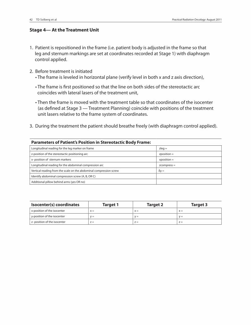
42 TD Solberg et al Safety Considerations for SRS and SBRT 42Practical Radiation Oncology: August 2011 Safety Considerations for SRS and SBRT 43
Stage 4— At the Treatment Unit
1. Patient is repositioned in the frame (i.e. patient body is adjusted in the frame so that leg and sternum markings are set at coordinates recorded at Stage 1) with diaphragm control applied.
2. Before treatment is initiated • The frame is leveled in horizontal plane (verify level in both x and z axis direction),
• The frame is first positioned so that the line on both sides of the stereotactic arc coincides with lateral lasers of the treatment unit,
• Then the frame is moved with the treatment table so that coordinates of the isocenter (as defined at Stage 3 — Treatment Planning) coincide with positions of the treatment unit lasers relative to the frame system of coordinates.
3. During the treatment the patient should breathe freely (with diaphragm control applied).
Parameters of Patient’s Position in Stereotactic Body Frame:Longitudinal reading for the leg marker on frame zleg =
z-position of the stereotactic positioning arc zposition =
x- position of sternum markers xposition =
Longitudinal reading for the abdominal compression arc zcompress =
Vertical reading from the scale on the abdominal compression screw δy =
Identify abdominal compression screw (A, B, OR C)
Additional pillow behind arms (yes OR no)
Isocenter(s) coordinates Target 1 Target 2 Target 3x-position of the isocenter x = x = x =
y-position of the isocenter y = y = y =
z- position of the isocenter z = z = z =
Safety Considerations for SRS and SBRT 42 Safety Considerations for SRS and SBRT 43Practical Radiation Oncology: August 2011
Appendix 3— Institution 3: Frame-based SBRT treatment procedures and checklist example
STEREOTACTIC LUNG TREATMENT PLANNING
Process
1. CT scan 1 week prior to treatment on 4D CT scanner. 2 scans – average and MIP.
2. Contours drawn within 24 hours of the CT scan. ITV is drawn on both scans.
3. Isodose plan completed within 72 hours of CT scan, plan is done on AVG CT scan.
4. 3 days are allowed for review, QA (if necessary) and documentation.
5. Treatment start date is a minimum of 1 week after CT scan.
6. Minimum of 40 hours and a maximum of 8 days should separate each treatment.
7. Stereotactic localization is done on treatment machine using coordinates from a pre-treatment CT scan (Cone Beam CT).
8. Daily localization with a physician present is performed to check for accurate field placement before each treatment.
Contouring
(Hot Script in Pinnacle “Stereotactic Lung Volumes”)
Structures
1. ITV = Should be drawn first by the physician. Isocenter will be placed in this structure using the automatic placement method “box”.
2. PTV = An expansion of ITV (0.5 cm in all directions)
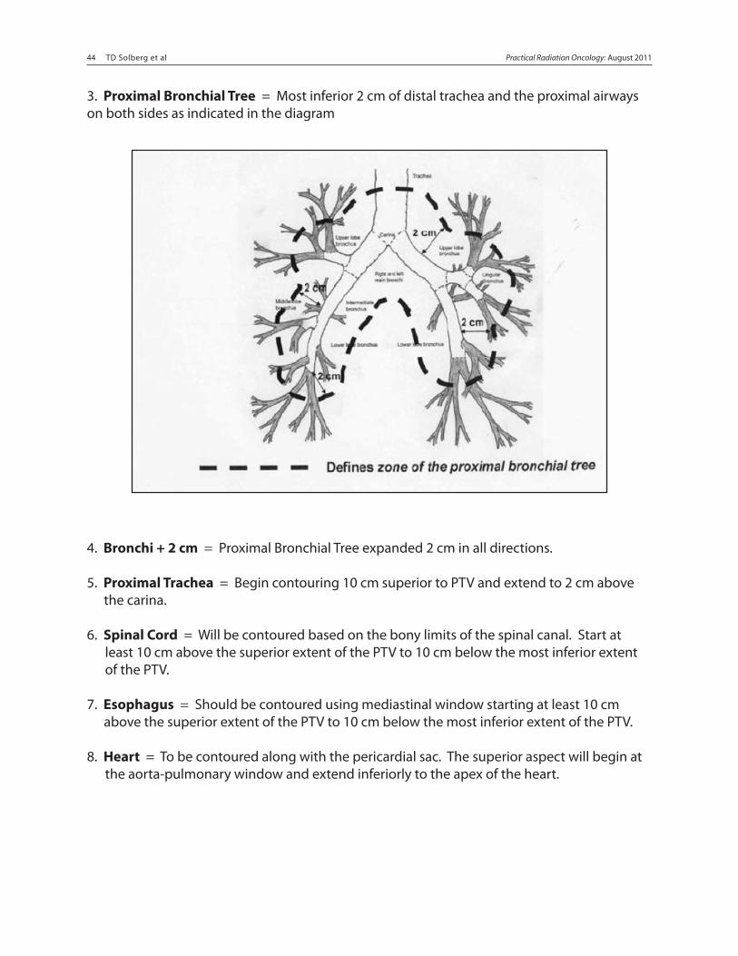
44 TD Solberg et al Safety Considerations for SRS and SBRT 44Practical Radiation Oncology: August 2011 Safety Considerations for SRS and SBRT 45
3. Proximal Bronchial Tree = Most inferior 2 cm of distal trachea and the proximal airways on both sides as indicated in the diagram
4. Bronchi + 2 cm = Proximal Bronchial Tree expanded 2 cm in all directions.
5. Proximal Trachea = Begin contouring 10 cm superior to PTV and extend to 2 cm above the carina.
6. Spinal Cord = Will be contoured based on the bony limits of the spinal canal. Start at least 10 cm above the superior extent of the PTV to 10 cm below the most inferior extent of the PTV.
7. Esophagus = Should be contoured using mediastinal window starting at least 10 cm above the superior extent of the PTV to 10 cm below the most inferior extent of the PTV.
8. Heart = To be contoured along with the pericardial sac. The superior aspect will begin at the aorta-pulmonary window and extend inferiorly to the apex of the heart.
Safety Considerations for SRS and SBRT 44 Safety Considerations for SRS and SBRT 45Practical Radiation Oncology: August 2011
9. Total Lung = Right and left lungs should be contoured as one structure. After contouring is complete, GTV will be subtracted from Total Lung for evaluation purposes.
10. Skin = The skin or body should be outlined beginning at least 12 cm above the superior extent of the PTV to 12 cm below the most inferior extent of the PTV.
11. Skin – PTV = PTV subtracted from the Skin contour.
12. PTV + 2 cm = PTV expanded 2 cm in all directions.
13. Skin – PTV + 2 cm = PTV + 2 cm subtracted from the Skin contour. This structure will be used to evaluate the dose at any point greater than 2 cm from the PTV.
14. Block Margin = PTV expanded 0.2 cm in sup/inf directions. Helps dose the most superior and inferior aspects of the PTV.
15. Brachial Plexus = Originates from the spinal nerves exiting C5 to T2 on the involved side. Extends along subclavian and axillary vessels to the level of the 2nd rib. Usually is contoured if tumor is near brachial plexus.
16. Bellows = assign density of ), not to be included in the calculations
Physician Contours: a. Outlines the ITV. b. Approves all other volumes before planning.
Dosimetrist Contours: a. All remaining contours including those for expansion and subtraction.
**Clean ROI’s before proceeding to planning
At this point if the isocenter has not been placed in the GTV using automatic “box” method, it must be done for the proceeding Hot Scripts to work.
46 TD Solberg et al Safety Considerations for SRS and SBRT 46Practical Radiation Oncology: August 2011 Safety Considerations for SRS and SBRT 47
Beam Configuration
1. Hot Scripts
a. ”Stereotactic Beams R Lung 1st” should be started after all contouring is completed. This is done first for both right or left lung lesions. If the ITV is in the right lung no further action is necessary. For left lung lesions, “Stereotactic Beams L Lung 2nd” should be used. This changes gantry and couch angles to the left sided configuration.
b. This script creates ten non – coplanar 6 MV beams found to be geometrically ideal for maximum falloff outside the PTV.
c. Beams are blocked to the “block margin” contour with no margin. MLC’s are turned on and clipped with leaves pushed in to the maximum setting. d. This hot script makes all beams homogeneous. Use heterogenous beams.
e. Creates prescription for 6000 cGy to 100% of dose at isocenter for 3 fractions (homogeneous) or 5400cGy for 3 fractions (hetereogeneity corrected). Beams are also equally weighted.
2. Manual Configuration
a. After beams are created, collimator angles can be changed to conform the MLC leaves to the PTV. This can be done be selecting a BEV window, turning on the PTV, and the manually adjusting the collimator angles.
b) Delete or modify angles on any beams that are incident on areas outside of the thorax (face, arms, etc.)
c) Dose grid must cover all contours and grid size changed to 0.3 cm by 0.3 cm by 0.2 cm. 2 mm spacing is used sup to inf for 2 mm CT spacing.
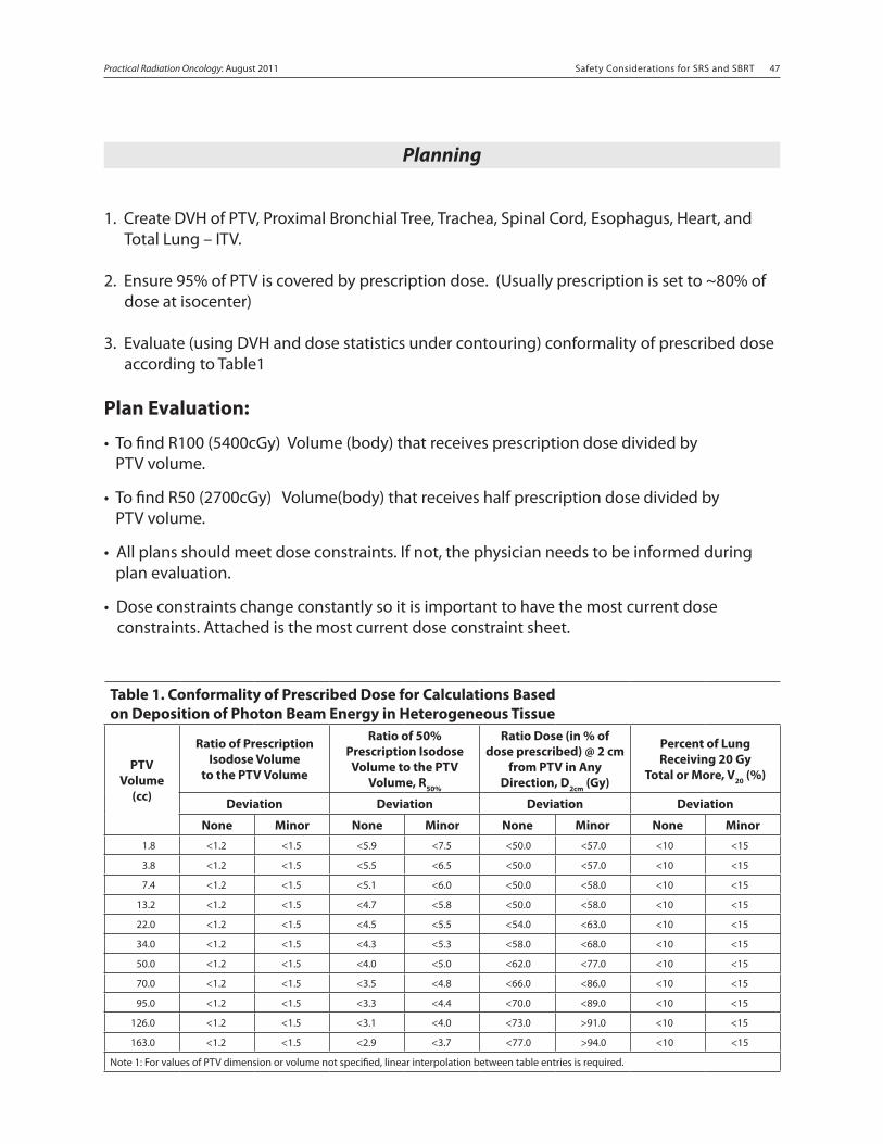
Safety Considerations for SRS and SBRT 46 Safety Considerations for SRS and SBRT 47Practical Radiation Oncology: August 2011
Planning
1. Create DVH of PTV, Proximal Bronchial Tree, Trachea, Spinal Cord, Esophagus, Heart, and Total Lung – ITV.
2. Ensure 95% of PTV is covered by prescription dose. (Usually prescription is set to ~80% of dose at isocenter)
3. Evaluate (using DVH and dose statistics under contouring) conformality of prescribed dose according to Table1
Plan Evaluation:
• To find R100 (5400cGy) Volume (body) that receives prescription dose divided by PTV volume.
• To find R50 (2700cGy) Volume(body) that receives half prescription dose divided by PTV volume.
• All plans should meet dose constraints. If not, the physician needs to be informed during plan evaluation.
• Dose constraints change constantly so it is important to have the most current dose constraints. Attached is the most current dose constraint sheet.
Table 1. Conformality of Prescribed Dose for Calculations Based on Deposition of Photon Beam Energy in Heterogeneous Tissue
PTV Volume
(cc)
Ratio of Prescription Isodose Volume
to the PTV Volume
Ratio of 50% Prescription Isodose
Volume to the PTV Volume, R50%
Ratio Dose (in % of dose prescribed) @ 2 cm
from PTV in Any Direction, D2cm (Gy)
Percent of Lung Receiving 20 Gy
Total or More, V20 (%)
Deviation Deviation Deviation Deviation
None Minor None Minor None Minor None Minor1.8 <1.2 <1.5 <5.9 <7.5 <50.0 <57.0 <10 <15
3.8 <1.2 <1.5 <5.5 <6.5 <50.0 <57.0 <10 <15
7.4 <1.2 <1.5 <5.1 <6.0 <50.0 <58.0 <10 <15
13.2 <1.2 <1.5 <4.7 <5.8 <50.0 <58.0 <10 <15
22.0 <1.2 <1.5 <4.5 <5.5 <54.0 <63.0 <10 <15
34.0 <1.2 <1.5 <4.3 <5.3 <58.0 <68.0 <10 <15
50.0 <1.2 <1.5 <4.0 <5.0 <62.0 <77.0 <10 <15
70.0 <1.2 <1.5 <3.5 <4.8 <66.0 <86.0 <10 <15
95.0 <1.2 <1.5 <3.3 <4.4 <70.0 <89.0 <10 <15
126.0 <1.2 <1.5 <3.1 <4.0 <73.0 >91.0 <10 <15
163.0 <1.2 <1.5 <2.9 <3.7 <77.0 >94.0 <10 <15
Note 1: For values of PTV dimension or volume not specified, linear interpolation between table entries is required.
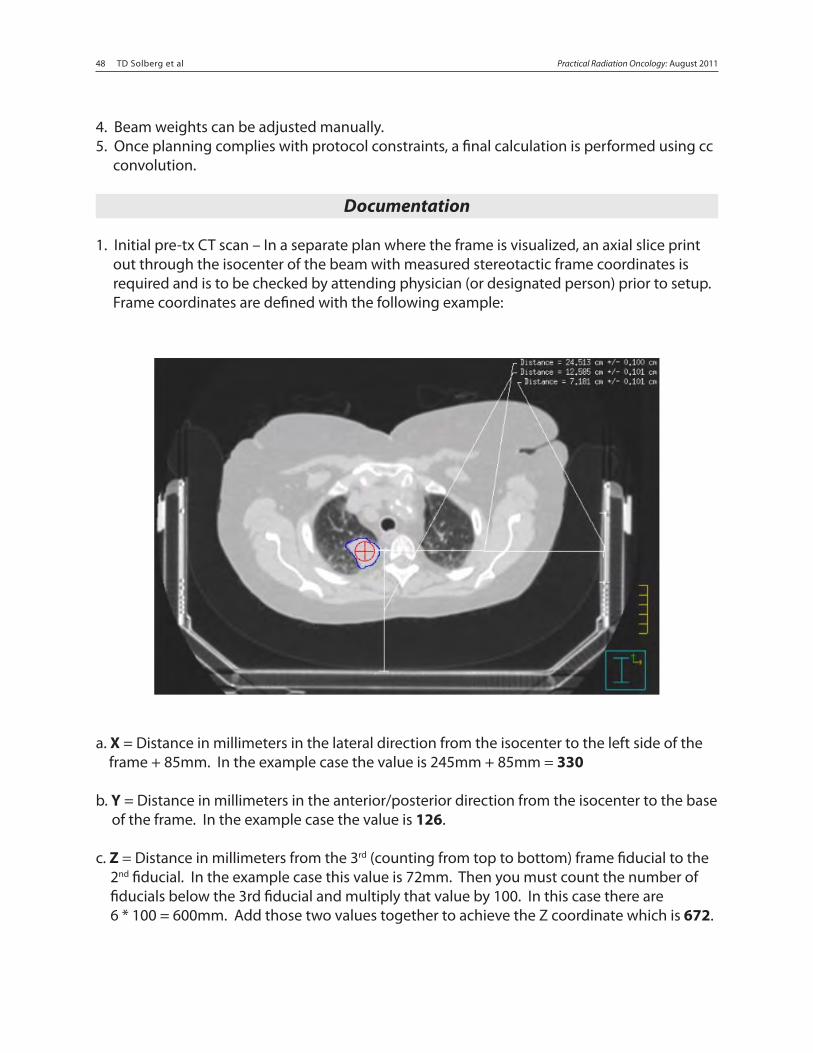
48 TD Solberg et al Safety Considerations for SRS and SBRT 48Practical Radiation Oncology: August 2011 Safety Considerations for SRS and SBRT 49
4. Beam weights can be adjusted manually.5. Once planning complies with protocol constraints, a final calculation is performed using cc convolution.
Documentation
1. Initial pre-tx CT scan – In a separate plan where the frame is visualized, an axial slice print out through the isocenter of the beam with measured stereotactic frame coordinates is required and is to be checked by attending physician (or designated person) prior to setup. Frame coordinates are defined with the following example:
a. X = Distance in millimeters in the lateral direction from the isocenter to the left side of the frame + 85mm. In the example case the value is 245mm + 85mm = 330
b. Y = Distance in millimeters in the anterior/posterior direction from the isocenter to the base of the frame. In the example case the value is 126.
c. Z = Distance in millimeters from the 3rd (counting from top to bottom) frame fiducial to the 2nd fiducial. In the example case this value is 72mm. Then you must count the number of fiducials below the 3rd fiducial and multiply that value by 100. In this case there are 6 * 100 = 600mm. Add those two values together to achieve the Z coordinate which is 672.
Safety Considerations for SRS and SBRT 48 Safety Considerations for SRS and SBRT 49Practical Radiation Oncology: August 2011
2. The plan documentation of the final converted plan includes in the following order from front to back:a. CAX transverse, sagittal and coronal views.b. BEV’s for each beam to document MLC treatment devices.c. Isodose cloud with critical structures visualized in a 3D window.d. Graph DVHe. Tabular DVHf. Plan printout (Physics Summary)
Charges
1. 77295 3-D Simulation2. 77300 Basic Calculation X number of treatment fields.3. 77334 Complex Treatment Device X number of treatment fields.
Cone Beam CT:• All SBRT patients are typically scheduled on the Synergy S• The data set that is sent to the treatment unit that is used for localization is the AVGF IP. • Any beams that traverse the couch needs to be corrected for couch attenuation.• If the plan “times out” when exporting to Synergy, close out the plan and copy without dose. Open the new plan (non computed) and delete all structure that are not used for planning. (ie. Large contours, really small contours, any contours drawn from sagittal and coronal images.) The plan export function should work now.