Embed Size (px)
Citation preview

Cell Host & Microbe, Volume 11
Supplemental Information
Zinc Sequestration by the Neutrophil Protein
Calprotectin Enhances Salmonella
Growth in the Inflamed Gut
Janet Z. Liu, Stefan Jellbauer, Adam Poe, Vivian Ton, Michele Pesciaroli, Thomas Kehl-Fie, Nicole
A. Restrepo, Martin Hosking, Robert A. Edwards, Andrea Battistoni, Paolo Pasquali, Thomas E.
Lane, Walter J. Chazin, Thomas Vogl, Johannes Roth, Eric P. Skaar, and Manuela Raffatellu

Figure S1, Related to Figure 1. Visual Representation of Colon Crypt Isolation
Colon crypts were isolated as described in the Supplemental Experimental Procedures. A
representative image of isolated colon crypts is shown.
Figure S2, Related to figure 3. Growth of S. Typhimurium in Rich and Minimal Media
S. Typhimurium wild-type, the znuA mutant ( znuA), or the znuA mutant complemented with
znuA expressed on a low-copy plasmid (pznuA) were grown in LB, M9, and M9 supplemented
with 5 M ZnSO4. (A and B) Growth in LB, M9 (A) and M9 supplemented with 5 M ZnSO4 (B)
was determined by reading the OD600 in a microplate reader at the indicated times. Data
represent the geometric mean of 4 replicates standard error. (C) Generation times in LB and

M9. Bars represent the geometric mean of 4 replicates standard deviation. * (P value ≤ 0.05)
and ** (P value ≤ 0.01).
Figure S3, Related to Figure 4.
(A) Histopathology scores of cecal samples four days after S. Typhimurium infection. Wild-type
mice were infected with either S. Typhimurium wild-type or the znuA mutant. Each stacked
column represents an individual mouse. A detailed scoring for the animals shown in Figure 4 is
provided.
(B–D) (B) Enumeration of S. Typhimurium in the colon content of mice. The colon content was
collected and plated 3 days after infection with either S. Typhimurium wild-type or the znuA
mutant (wild-type n=11, znuA mutant n=11). * P value ≤ 0.05. (C and D) Transcript levels of
S100a8 and S100a9 (C) and Il-17a, Il-22 and Cxcl-1 (D) were determined by real-time RT-PCR
in the cecum of mice infected with S. Typhimurium wild-type or the znuA mutant. Data are
expressed as fold increase over mock-infected wild-type mice. (B–D) Bars represent the
geometric mean of at least 5 replicates standard error.

Figure S4, Related to Figure 5.
(A) Histopathology scores of cecal samples four days after S. Typhimurium infection. Wild-type
and S100a9-/- mice were infected with mixtures of S. Typhimurium strains as indicated. Each
stacked column represents an individual mouse; n = no inflammation. A detailed scoring for the
animals shown in Figure 5 is provided.
(B and C) Transcript levels of Il-17a (B) and Il-22 (C), were determined in wild-type mice (white
bars), S100a9-/- mice (dark grey bars), and wild-type mice supplemented with zinc sulfate (light
grey bars). Mice were either mock-infected or infected with S. Typhimurium as indicated. Data
are expressed as fold increase over mock-infected wild-type mice. Bars represent the geometric
mean of at least 4 replicates standard error. Significant differences in gene expression in
comparison to wild-type infected C57BL/6 mice (first group) are indicated by ** (P value ≤ 0.01)
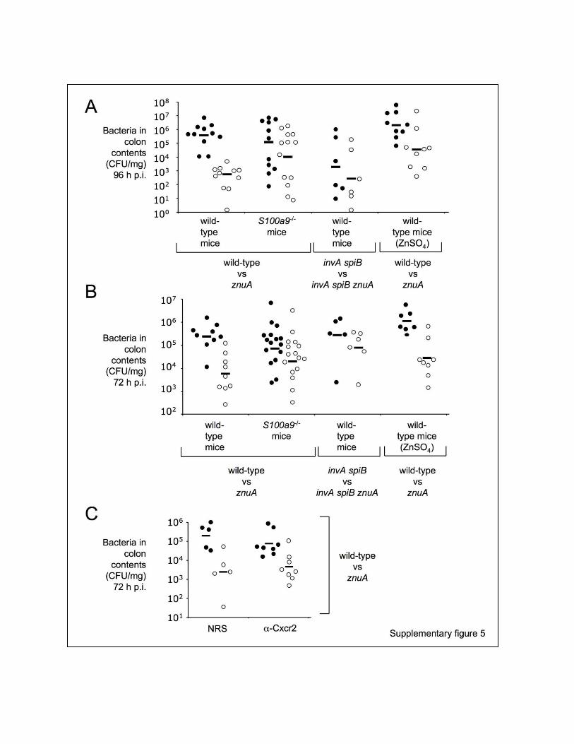

Figure S5, Related to Figure 7. Enumeration of S. Typhimurium in the Colon Content of
Mice
(A and B) Bacterial count of the mixture of S. Typhimurium strains in the colon contents of mice
(n≥6/group) was determined at four days (A) or three days (B) post infection.
(C) Total bacterial count of the mixture of S. Typhimurium strains in the colon contents of mice
treated with either normal rabbit serum (NRS) or a rabbit polyclonal antibody blocking the Cxcr2
receptor ( -Cxcr2) at 72 hours post-infection. Strain and mouse genotypes are indicated. Filled
circles indicate the strain with a wild-type znuA allele, while open circles indicate the strain with
a znuA mutation. Colonization of individual strains in each mouse (circles) and the averages
(bars) are indicated.

SUPPLEMENTAL EXPERIMENTAL PROCEDURES
Bacterial Strains and Growth Conditions
Bacterial strains and plasmids are listed in supplementary table 1. Cultures of S. Typhimurium
and E. coli were routinely incubated either aerobically at 37°C in Luria-Bertani (LB) broth (per
liter: 10 g tryptone, 5 g yeast extract, 10 g NaCl) or on LB agar plates (1.5% Difco agar)
overnight. Growth was also determined in M9 minimal media (3.75g Na2HPO4, 1.5g KH2PO4,
0.25g NaCl, 0.5g NH4Cl, 0.5M CaCl2, 1M MgSO4, 20% glucose) and M9 supplemented with
5 M zinc sulfate. Antibiotics and other chemicals were added at the following concentrations
(mg/l) as needed: carbenicillin (Carb), 100; chloramphenicol (Cm), 30; kanamycin (Km), 100;
nalidixic acid (Nal), 50; 5-bromo-4-choloro-3-indoyl-B-D-galactopyranoside (Xgal), 40.
Allelic Exchange Deletion of znuA
Primers used are detailed in supplementary table 2. Primers 7 and 8 were used to PCR
amplify the region upstream of znuA (flanking region 1) of S. Typhimurium. Primers 4 and 5
were used to PCR amplify the region downstream of znuA (flanking region 2). The PCR
products were ligated into pCR2.1 using the TOPO TA cloning kit (Invitrogen), heat shocked into
E. coli TOP10 and plated on LB+Km+Xgal. White colonies were screened by EcoRI digestion
for the appropriate length of linearized plasmid construct. Positive clones were sequence
confirmed by using M13 forward and M13 reverse universal primers. Accurate clones were
designated pJL2 (pCR2.1, znuA flanking region 1 cassette) and pJL1 (pCR2.1, znuA flanking
region 2 cassette). Flanking region 2 cassette was digested out of pJL1 using SacI and EcoRI
double-digestion, ligated into SacI and EcoRI double-digested pGP704 (Miller and Mekalanos,
1988), heat shocked into E. coli CC118 pir, and plated on LB+Carb, generating pJL3. Flanking
region 1 cassette was digested out of pJL2 using SalI and XbaI double-digestion, ligated into
SalI and XbaI double-digested pJL3, heat shocked into E. coli CC118 pir, and plated on
LB+Carb, generating pJL4. The chloramphenicol acetyl transferase (CAT) cassette from
pCMXX (Bäumler et al., 1997) was excised using SacI digestion and ligated into the compatible
SacI site of pJL4, generating pJL5 (pGP704, znuA::CAT). pJL5 was purified and heat shocked
into E. coli S17-1 pir, then conjugated separately into S. Typhimurium IR715. Transconjugants
were selected for using Nal and Cm and colonies with double crossover events were screened
for by loss of Carb resistance. Confirmation of mutant was performed using Southern blot
analysis. Primers 13 and 14 were used to PCR amplify a probe designed to flanking region 2.
The PCR product was ligated into pCR2.1, generating pLJ8. The probe encoded on pJL8 was
used to confirm the znuA::CAT mutant and the S. Typhimurium znuA::CAT strain was
designated JZL3. For complementation of JZL3, primers 10 and 11 were used to PCR amplify
znuA from S. Typhimurium IR715. The PCR product was ligated into pCR2.1 using the TOPO
TA system and heat shocked into E. coli TOP10 to generate pJL6. The znuA gene was excised
from pJL6 using XhoI and EcoRV double-digestion and ligated into XhoI and EcoRV digested
pWSK29 (Wang et al., 1991), heat shocked into E. coli, DH5α and plated on LB+Carb+Xgal,
generating pJL7. pJL7 was purified and electroporated into JZL3. The invA::tetRA
spiB::KSAC znuA::CAT triple mutant was generated by conjugating the pJL5 plasmid in E.
coli S17-1 pir with SPN452 (IR715 invA::tetRA spiB::KSAC) (Raffatellu et al, 2009). This
strain was confirmed by Southern blot with the same probe as before to flanking region 2 and
was designated JZL2.
Mouse Experiments
C57BL/6 mice and S100A9-/- mice were used in our study. C57BL/6 mice were purchased from
Taconic Farms and S100A9-/- mice were generated as previously described (Manitz et al.,
2003). Mice were gavaged with 0.1 ml of a 200 mg/ml streptomycin/sterile water solution one
day prior to mock-infection with LB or oral infection with 1 109 CFU of S. Typhimurium in LB.
For the zinc supplementation experiment, C57BL/6 mice were gavaged with 0.1 ml of a 10
mg/ml of zinc sulfate/sterile water solution starting a day before streptomycin treatment and
were gavaged once a day for the length of the experiment. The cecum was harvested for mRNA,
protein, and histopathology at 48-96h post-infection. The cecal contents were collected, serially
diluted, and plated on appropriate antibiotic LB agar plates to determine bacterial counts. In the
mixed infection experiments, data were normalized by dividing the output ratio (CFU of the wild-
type/CFU of the mutant) by the input ratio (CFU of the wild-type/CFU of the mutant). Groups of
5-11 mice were used for each experiment. In the mixed infections, competitive indices were
calculated by dividing the output ratio (CFU of wild-type / CFU of the mutant) by the input ratio
(CFU of wild-type / CFU of the mutant).
CXCR2 Antibody Blocking of Neutrophil Infiltration
A murine-specific CXCR2 blocking antibody was raised in rabbits following immunization with a
17 amino acid peptide (H2N-MGEFKVDKFNIEDFFSG-CO2H) corresponding to the amino
terminus of CXCR2 (Mehrad et al., 1999). We used the antibody to deplete neutrophils as
previously described (Walsh and Lane 2007). Mice were treated with anti-CXCR2 or normal
rabbit serum via intraperitoneal (i.p.) injection 24 hours prior to infection (i.e. at the same time as
streptomycin treatment).
Cell Isolation from Whole Blood
At 72 hours post infection, mice were sacrificed and 300 to 500 µl of whole blood was collected
in sodium heparin tubes (BD Vacutainer) by cardiac puncture using an insulin syringe (BD) and
stored on ice until further processing. Red blood cells were lysed by incubating whole blood with
5 ml of ACK Lysing Buffer (GIBCO) for 5 min at room temperature. To stop the lysis reaction, 7
ml of medium (RPMI-1640, 10% FCS, 1% Pen/Strep – GIBCO) was added and the suspension
was centrifuged at 1500 g for 7 min at room temperature. Thereafter the pellet was
resuspended in 150 µl medium and cells were transferred to a round bottom 96-well plate for
extracellular staining.
Extracellular Staining and Flow Cytometry Analysis
Blood neutrophils were detected with phycoerythrin (PE)-conjugated Ly6G (clone RB6-8C5,
eBioscience) and PE-Cy7-conjugated CD11b (eBioscience) monoclonal antibodies as described
previously (Hosking et al, 2009). Briefly, cells were incubated with purified anti-mouse
CD16/32 antibody (1 in 100 dilution, eBioscience) in FACS-staining buffer (phosphate-buffered
saline pH 7.45, 0.5% bovine serum albumin, and 0.02% sodium azide) for 30 min at room
temperature in order to block Fc gamma receptors. Subsequently, cells were stained for the
above mentioned surface markers for 30 min on ice in the dark (1 in 50 dilution). Cells were
washed, resuspended in staining buffer and fixed in 1% paraformaldehyde/phosphate-buffered
saline (pH 7.45). Data was acquired on a FACSCalibur (BD Biosciences, San Jose, CA) and
further analyzed with FlowJo software (TreeStar, Ashland, OR). Leucocytes were gated using
forward (FSC) and sight scatter (SSC) criteria of cells followed by identifying Ly6Ghigh CD11b+
neutrophils.
Measurement of Zinc in Fecal Samples by ICP-MS
C57BL/6 mice were treated with streptomycin and infected with S. Typhimurium wild-type or
mock as described. At 96 hours post-infection, fecal pellets were collected with plastic forceps
and placed in glass containers that were previously cleaned with nitric acid to remove metal
contamination. The fecal pellets were then autoclaved to kill all bacteria. Sample analysis was
performed by Applied Speciation (Bothell, WA) as previously described by Corbin et al (Corbin
et al, 2008). Briefly, the fecal samples from 4 infected and 4 uninfected mice were digested by
boiling in nitric acid and hydrochloric acid. The samples were then resuspended in water and

analyzed by inductively coupled plasma dynamic reaction cell mass spectrometry (ICP-DRC-
MS). Aliquots of each sample are introduced into a radio frequency (RF) plasma where energy-
transfer processes cause desolvation, atomization, and ionization. The ions were extracted from
the plasma through a differentially-pumped vacuum interface and traveled through a
pressurized chamber (DRC) containing a specific reactive gas which preferentially reacts with
interfering ions of the same target mass to charge ratios (m/z). A solid-state detector detected
ions transmitted through the mass analyzer, on the basis of their mass-to-charge ratio (m/z),
and the resulting current was processed by a data handling system. The results were reported
as mg of zinc per kg of dry weight.
Western Blots
Total protein was extracted from mouse cecum tissue using Tri-Reagent (Molecular Research
Center). 15 g of total protein were analyzed using 15% SDS-PAGE gels and transferred to
PVDF membranes. The membranes were blocked with 2% nonfat dried milk and incubated at
4°C with polyclonal goat anti-mouse S100A8, polyclonal goat anti-mouse S100A9 (R&D
Systems), polyclonal goat anti-human and mouse myeloperoxidase (R&D Systems) or
polyclonal rabbit anti-mouse α/β-tubulin (Cell Signaling Technology). After overnight incubation
the blots were washed then incubated with horse radish peroxidase conjugated anti-rabbit or
anti-goat secondary antibodies (Jackson Immuno Research) for 1 hour. After washing, bands
were developed using the Immobilon Western Luminol Reagent and Peroxide Solution (Millipore)
as per manufacturer’s instructions and visualized using the Fujifilm LAS 4000.
Quantitative Real-Time PCR
Total RNA was extracted from mouse cecal tissue using Tri-Reagent (Molecular Research
Center). Reverse transcription of 1 μg of total RNA was performed using the Transcriptor First
Strand cDNA Synthesis kit (Roche). Quantitative real-time PCR (qRT-PCR) for the expression

of β-actin, Il-17, Il-22, S100a8, S100a9, and Cxcl-1 (supplementary table 3) were performed
using the LightCycler 480 SYBR Green Master on the LightCycler 480 II (Roche). Conditions for
qRT-PCR were 95°C for 5 minutes, then 45 cycles of 95°C for 10 seconds 60°C for 10 seconds
and 72°C for 15 seconds. Gene expression was normalized to β-actin and fold changes in gene
expression was relative to uninfected controls and calculated using the ΔΔ Ct method.
Analysis of the Microbiota
Composition of the bacteria microbiota was analyzed as described earlier (Barman et al, 2008;
Winter et al., 2010). Briefly, the colon content was collected from mice 96 hours post-infection,
and snap frozen in liquid nitrogen. The DNA was subsequently extracted using the QIAamp
DNA stool kit (Qiagen). Two µl of extracted bacterial DNA was used as a template for the q-
PCR reaction with the primer pairs developed by Barman et al. (Barman et al, 2008) and
presented in supplementary table 3. The 16S gene copy numbers per µl of DNA from each
sample (one fecal pellet collected from each colon) was determined using the plasmids
described in supplementary table 1. To estimate the copy number of Enterobacteriaceae other
than Salmonella, for each sample the Salmonella 16S gene copy number was subtracted from
the total Enterobacteriaceae 16S gene copy number.
Isolation of Colon Crypts
Streptomycin-treated C57BL/6 mice were infected with S. Typhimurium IR715 as detailed above
and sacrificed at 72 hours post-infection. Crypt isolation from colon and cecum was performed
as described (Whitehead et al., 1993). Briefly, cecum plus colon tissue was collected, flushed,
and opened to expose mucosa. Tissue was then incubated at room temperature with a 3 mM
EDTA and 0.5 mM DTT solution. After 90 minutes of incubation the tissue was transferred into
PBS, shaken, and the detached crypts were decanted into a 15 ml falcon tube and spun down
at 4°C 200 g for 5 minutes (Fig. S1). RNA and protein were isolated using Tri-Reagent
(Molecular Research Center) as per the manufacturer’s instructions.
Growth in Media Supplemented with Calprotectin
Wild type and ΔznuA S. Typhimurium were grown overnight in M9 minimal media at 37°C with
agitation. OD600 was determined and used to calculate the volume of overnight culture needed to
obtain 1x10⁸ cells/ml. The calculated volume of culture was then spun down at 20,000g for 10
minutes in a tabletop centrifuge. The supernatant was discarded and the pellet resuspended in
1 ml of LB media. 1x10⁵ cells/ml was obtained with serial dilution and 10 μl were used to
inoculate the wells of a 96-well Nunclon Surface plate (Nunc). Each well contained a 62:28:10
ratio of calprotectin buffer (20 mM Tris pH 7.5, 100 mM NaCl, 10 mM BME, 3 mM CaCl2) to LB
media to inoculum. Recombinant calprotectin was produced as described elsewhere (Hunter
and Chazin, 1998). Two-fold dilutions of calprotectin stock were prepared, starting from 500
μg/ml and diluting down to 15.6 μg/ml. Triplicates were made for each concentration of
calprotectin. The 96-well plate was incubated inside a Tupperware container with a moistened
piece of paper towel and incubated at 37°C with 5% CO2. OD600 were taken at 0, 2, 4, 5, 6, 7, 8,
12, and 24 hours on a Microplate Reader (Bio Rad) and graphed on a semi-logarithmic scale.
The experiment was repeated four times.
Detection of Intestinal and Fecal Calprotectin Using ELISA
Streptomycin treated wild-type C57BL/6 mice were mock or S. Typhimurium IR715 infected. At
72 hours post-infection mice were sacrificed and the cecum, the colon and fecal samples were
collected. Cecum and colon tissue were placed in 3 ml sterile PBS and homogenized. The
homogenate was spun down at 4,000g at 4°C for 20 minutes; supernatant was then used for
Western blot and ELISA analysis. Extraction buffer (0.1 M Tris, 0.15 M NaCl, 1.0 M urea, 10 mM
CaCl2, 0.1 M citric acid monohydrate, 5 g/l BSA and 0.25 mM thimerosal (pH 8.0)) adopted from

Hycult Biotech’s H305 Human Calprotectin ELISA kit was added to fecal samples. The fecal
samples were incubated on ice for 30 minutes and were homogenized at 4°C. Samples were
then spun down at 10,000g at 4°C for 20 minutes and the supernatant was used for Western
blot and ELISA analyses. Murine S100A8/S100A9 was determined by an in-house established
ELISA as described (Vogl et al, 2007). Wells were coated with rabbit polyclonal anti-S100a8 (4
µg/ml). The detection antibody rabbit polyclonal anti-S100a9 (2 µg/ml) was coupled to biotin
according the manufactures’ instructions. Streptavidin coupled HRP and TMB as substrate was
used and absorbance at 405 nm was recorded using a MRX microplate reader (Dynex
Technologies; USA). Recombinant prepared murine S100a8/S100a9 heterodimer was used as
standard in the calibration curve. Data were normalized taking into account the weight of the
collected fecal samples and tissues and assuming a density of 1g/ml.

Table S1, Related to Figure 1. Bacterial Strains and Plasmids Used in This Study
Designation Genotype Source or Reference
Escherichia coli strains
CC118 pir F- araD139 (ara, leu)7697 ∆lacX74 phoA∆20 galE galK thi
rpsE rpoB argEam recA1 pir Herrero et al., 1990
DH5 MCR
F- mcrA (mrr-hsdRMS-mcrBC) 80dlacZ M15 (lacZYA-
argF)U169 deoR recA1 endA1 phoA supE44 - thi-1 gyrA96 relA1
Gibco BRL
S17-1 pir F- recA thi pro rK- mK+ RP4:2-Tc::MuKm Tn7 pir Herrero et al., 1990
Salmonella enterica serovar Typhimurium strains
IR715 ATCC 14028, NalR derivative Stojiljkovic et al., 1995
JZL3 IR715, ΔznuA::Cm This study
SPN452 IR715, ΔinvA::tetRA ΔspiB::KSAC Raffatellu et al., 2009
JZL2 IR715, ΔznuA::Cm ΔinvA::TetRA ΔspiB::KSAC This study
Plasmids:
pCR2.1 TOPO Cloning Vector (CarbR, KmR) Invitrogen
pJL1 pCR2.1::FR2 ZnuA This study
pJL2 pCR2.1::FR1 ZnuA This study
pJL3 pGP704::FR2 ZnuA This study
pJL4 pJL3::FR1 ZnuA This study
pJL5 pGP704::FR1 CAT FR2
pJL6 pCR2.1::znuA This study
pJL7 pWSK29::znuA This study
pJL8 pCR2.1::FR2 Southern probe This study
pSW191 pCR2.1::Eubacteria 16S rRNA Winter et al., 2010
pSW192 pCR2.1::Clostridiales 16S rRNA Winter et al., 2010
pSW193 pCR2.1::Lactobacillales 16S rRNA Winter et al., 2010
pSW194 pCR2.1::Bacteroidetes 16S rRNA Winter et al., 2010
pSW195 pCR2.1::Salmonella 16S rRNA Winter et al., 2010
pSW196 pCR2.1::Enterobacteriaceae 16S rRNA Winter et al., 2010

Table S2, Related to Figure 3. Primers Used in This Study—Cloning
Designation Purpose Sequence (5' to 3')
4 Flanking region 2 of ΔznuA construct
ATTGTCAGAGCTCTCTATCACTTCTGCAAGC
5 CGATTAGAATTCCGGGGTATCATAAAAGGC
7 Flanking region 1 of ΔznuA construct
TACATCGGTCGACCCCAATACTGTGGATAGCG
8 TAGCARGTCTAGATGCGAGCTGCCTGAAAGG
10 Clone the znuA gene
ATCTGCTCGAGGTAAGAGGGAACGAATCTCG
11 CTGATGATATCCCAGTGAAACTAAACTTGTCAT
13 Confirming ΔznuA deletion
mutant
TCTATCACTTCTGCAAGC
14 TCATCAAGTACCAGCAAC
M13 Forward Sequencing TGT AAA ACG ACG GCC AGT
M13 Reverse Sequencing CAG GAA ACA GCT ATG ACC
Bold: Extra 5’ DNA; Underlined: Restriction site utilized in cloning
Table S3, Related to Figure 4. Real-Time PCR Primers Used in This Study
Specie Target Primer Pairs
Mus musculus Actb 5’-GGCTGTATTCCCCTCCATCG-3’ 5’-CCAGTTGGTAACAATGCCATGT-3’
Mus musculus Il-17
5’-GCTCCAGAAGGCCCTCAGA-3’ 5’-AGCTTTCCCTCCGCATTGA-3’
Mus musculus Il-22
5’-GGCCAGCCTTGCAGATAACA-3’ 5’-GCTGATGTGACAGGAGCTGA-3’
Mus musculus S100a8 5’-TGTCCTCAGTTTGTGCAGAATATAAA-3’ 5’-TCACCATCGCAAGGAACTCC-3’

Mus musculus S100a9 5’-GGTGGAAGCACAGTTGGCA-3’ 5’-GTGTCCAGGTCCTCCATGATG-3’
Mus musculus Cxcl-1
5’-TGCACCCAAACCGAAGTCAT-3’ 5’-TTGTCAGAAGCCAGCGTTCAC-3’
Mus musculus Ly6g 5’-TGCGTTGCTCTGGAGATAGA-3’ 5’-CAGAGTAGTGGGGCAGATGG-3’
Eubacteria (Barman et al.)
16S rRNA
UniF340 5’-ACTCCTACGGGAGGCAGCAGT-3’
UniR514 5’-ATTACCGCGGCTGCTGGC-3’
Salmonella (Barman et al.)
16S rRNA
Sal454 5’-TGTTGTGGTTAATAACCGCA-3’
Uni785R 5’-GACTACCAGGGTATCTAATCC-3’
Firmicutes/ Clostridiales (Barman et al.)
16S rRNA
UniF338 5’- ACTCCTACGGGAGGCAGC -3’
C.cocR491 5’- GCTTCTTTAGTCAGGTACCGTCAT -3’
Firmicutes/ Lactobacillales (Barman et al.)
16S rRNA
LabF362 5’- AGCAGTAGGGAATCTTCCA -3’
LabR677 5’- CACCGCTACACATGGAG -3’
Bacteroidetes (Barman et al.)
16S rRNA
BactF285 5’- GGTTCTGAGAGGAGGTCCC -3’
UniR338 5’- GCTGCCTCCCGTAGGAGT -3’
Enterobacteriaceae (Barman et al.)
16S rRNA
Uni515F 5’- GTGCCAGCMGCCGGCGGTAA -3’
Ent826R 5’- GCCTCAAGGGCACAACCTCCAAG -3’

SUPPLEMENTAL REFERENCES
Miller, V.L. & Mekalanos, J.J. A Novel Suicide Vector and Its Use in Construction of Insertion
Mutations: Osmoregulation of Outer Membrane Proteins and Virulence Determinants in Vibrio
cholerae Requires toxR. J. Bacteriol. 170, 2575-2583 (1988).
Bäumler, A. J., Tsolis R.M., Valentine P.J., Ficht T.A., & Heffron F. Synergistic effect of
mutations in invA and lpfC on the ability of Salmonella typhimurium to cause murine typhoid.
Infect. Immun. 65, 2254–2259 (1997).
Wang, R.F., & Kushner, S.R. Construction of versatile low-copy-number vectors for cloning,
sequencing and gene expression in Escherichia coli. Gene. 100, 195-199 (1999).
Herrero, M., de Lorenzo, V., & Timmis, K.N. Transposon vectors containing nonantibiotic
resistance selection markers for cloning and stable chromosomal insertion of foreign genes in
gram-negative bacteria. J. Bacteriol. 172, 6557-6567 (1990).
Stojiljkovic, I., Bäumler, A.J., & Heffron, F. Ethanolamine utilization in Salmonella typhimurium:
nucleotide sequence, protein expression, and mutational analysis of the cchA cchB eutE eutJ
eutG eutH gene cluster. J. Bacteriol. 177, 1357-66 (1995).