Embed Size (px)
Citation preview

Nature and Science, 2(4), 2004, Supplement ISSN 1545-0740
Nature and Science
The Nature and Science is an international journal with a purpose to enhance our natural and scientific knowledge dissemination in the world under the free publication principle. Any valuable papers that describe natural phenomena and existence or any reports that convey scientific research and pursuit are welcome, including both natural and social sciences. Papers submitted could be reviews, objective descriptions, research reports, opinions/debates, news, letters, and other types of writings that are nature and science related. The journal is calling for papers and seeking co-operators and editors as well.
Editor-in-Chief: Hongbao Ma Associate Editors-in-Chief: Qiang Fu, Yongsheng Ma, Margaret Young Editors: George Chen, Mark Hansen, Mary Herbert, Wayne Jiang, Xuemei Liang, Tracy X. Qiao, Xiaofeng Ren, George Warren, Kerry Watson, Qing Xia, Yonggang Xie, Ding Xu, Lijian Yang, Tina X. Zhang, Ruanbao Zhou, Xulai Xu Web and Cover Design: Yan Young
Introductions to Authors
1. General Information (1) Goals: As an international journal published both in print and on internet, Nature and Science is dedicated to the dissemination of fundamental knowledge in all areas of nature and science. The main purpose of Nature and Science is to enhance our knowledge spreading in the world under the free opinion publishing principle. It publishes full-length papers (original contributions), reviews, rapid communications, and any debates and opinions as well in all the fields of nature and science. (2) What to Do: The Nature and Science provides a place for discussion of scientific news, research, theory, philosophy, profession and technology - that drive scientific progress. Research reports and regular manuscripts that contain new and significant information of general interest are welcome. (3) Who: All people are welcome to submit manuscripts in any fields of nature and science. (4) Publication Costs: US$30 per printed page of an article to defray costs of the publication will be paid by the authors when the submission or after the acceptance. Extra expense for color reproduction of figures will be paid by authors (estimate of cost will be provided by the publisher for the author’s approval). (5) Journal Copies to Authors: One hard copy of the journal will be provided free of charge for each author. (6) Additional Copies Bought by Authors: Additional hard copies and offprints could be purchased with the price of US$4/issue and US$0.2/page-offprint (mailing and handling cost included). The offprints must be ordered prior to printing of the journal. (7) Distributions: Web version of the journal is freely opened to the world without any payment or registration. The journal will be distributed to the selected libraries and institutions for free. US$5/issue is charged for the subscription of other readers. (8) Advertisements: The price will be calculated as US$400/page, i.e. US$200/a half page, US$100/a quarter page, etc. Any size of the advertisement is welcome.
2. Manuscripts Submission (1) Submission Methods: Electronic submission through email is encouraged and hard copies plus an IBM formatted computer diskette would also be accepted. (2) Software: The Microsoft Word file will be preferred. (3) Font: Normal, Times New Roman, 10 pt, single space. (4) Indent: Type 2 space in the beginning of each new paragraph. (5) Manuscript: Don’t use “Footnote” or “Header and Footer”. (6) Cover Page: Put detail information of authors and a short title in the cover page. (7) Title: Use Title Case in the title and subtitles, e.g. “Debt and Agency Costs”.
(8) Figure and Table: Use full word of figure and table, e.g. “Figure 1. Annul Income of Different Groups”, Table 1. Annual Increase of Investment”. (9) References: Cite references by “last name, year”, e.g. “(Smith, 2003)”. References should include all the authors’ last names and initials, title, journal, year, volume, issue, and pages etc.
Reference Examples: Journal Article: Hacker J, Hentschel U, Dobrindt U. Prokaryotic chromosomes and disease. Science 2003;301(34):790-3. Book: Berkowitz BA, Katzung BG. Basic and clinical evaluation of new drugs. In: Katzung BG, ed. Basic and clinical pharmacology. Appleton & Lance Publisher. Norwalk, Connecticut, USA. 1995:60-9.
(10) Submission Address: [email protected], Marsland Company, P.O. Box 753, East Lansing, Michigan 48826, The United States, 517-862-6881. (11) Reviewers: Authors are encouraged to suggest 2-5 competent reviewers with their email and mailing addresses. 2. Manuscript Preparation Each manuscript is suggested to include the following components but authors can do their own ways: (1) Title page: including the complete article title; each author’s full name that family name appears with uppercase; institution(s) with which each author is affiliated, with city, state/province, and zip code; and the name, complete mailing address, telephone number, facsimile number (if available), and e-mail address for all correspondence. (2) Abstract: including Background, Materials and Methods, Results, and Discussions. (3) Key Words. (4) Introduction. (5) Materials and Methods. (6) Results. (7) Discussions. (8) References. (9) Acknowledgments.
Journal Address: Marsland Company P.O. Box 21126 Lansing, Michigan 48909 The United States Telephone:(517) 980-4106 E-mail: [email protected] Homepage: http://www.sciencepub.org
2004 Marsland Company
Marsland Company, P.O. Box 21126, Lansing, Michigan 48909, The United States, (517) 980-4106 http://www.sciencepub.org [email protected]

Nature and Science, 2(4),2004 Contents ISSN 1545-0740
Nature and Science ISSN 1545-0740
(Quarterly, Started in 2003)
Volume 2 - Number 4, Supplement, December 15, 2004
Cover Page, Content, Introduction, Call for Papers, All in one file
CONTENTS
1. Recovering Extremely Low Frequency Signal from the Signal-Dependent Noise Background Hsien-Chiao Teng, Shen Cherng 1-3
2. The Non-Stationary Analysis of Osteoblast Cellular Response to the reaction of ELF magnetic Field
Hsien-Chiao Teng, Shen Cherng 4-7
3. Gene Transfer into Schistosome as a Therapy Tool Hongbao Ma, George Chen 8-16
4. Cholesterol and Human Health
Hongbao Ma 17-21 5. The Characteristic Frequencies in Cell Induced Magnetic Fluctuation Hsien-Chiao Teng, Chi-Cheng Lie, Shen Cherng 22-27
Marsland Company
P.O. Box 21126 East Lansing, Michigan 48909
The United States Telephone:(517) 980-4106
E-mail: [email protected] Homepage: http://www.sciencepub.org
http://www.sciencepub.org [email protected]
Nature and Science, 2(4), 2004,Supplement, Teng, Recovering ELF from the signal dependent background
http://www.sciencepub.org [email protected] ·1
Recovering Extremely Low Frequency Signal from the
Signal-Dependent Noise Background
1Hsien-Chiao Teng, 2Shen Cherng
1Department of Electrical Engineering, Chinese Military Academy, Fengshan, Kaohsiung, Taiwan 830, ROC
2Department of Electrical Engineering, Chengshiu University, Niaosong, Kaohsiung, Taiwan 833, ROC [email protected]
Abstract: We developed a signal dependent noise tensor, which can be used to describe the fluctuated geomagnetic field coupled with Extremely Low Frequency (ELF) signals for our further biological signal processing study. In order to isolate the coupled ELF signals from the signal dependent noise, we introduced Quantization (QT) decoding method to discrete the noise and recover the coupled signals from the background. The signal to noise ratio of the coupling ELF can be amplified by QT in the power density spectrum (PDS). [Nature and Science. 2004;2(4) (Supplement): 1-3].
Key words: Extremely Low Frequency (ELF); noise; power density spectrum (PDS); signal
Introduction
The signal dependent noise can be presented as a
noise tensor at time , in which index indicates
the i th sample at energy level j. The signal can be
shown as . Power density spectrum (PDS) analysis
for tensor ⊕ s can be used to identify the coupled
signals in . The intrinsic coupling oscillation can
be captured by probe and converted to electrical
voltages shown in oscilloscope.
ij
ij
ijn
n t
ijs
s
n
ij
n ij
ij
Theory and Methods
Set an AC ELF signal s as input to the
background , the output can be transformed to
electrical voltages shown to oscilloscope. By using
HP Benchlink, we collect the output data and
transform it to Microsoft Excel as text files. Matlab
and Fortran programs were performed to analyze the
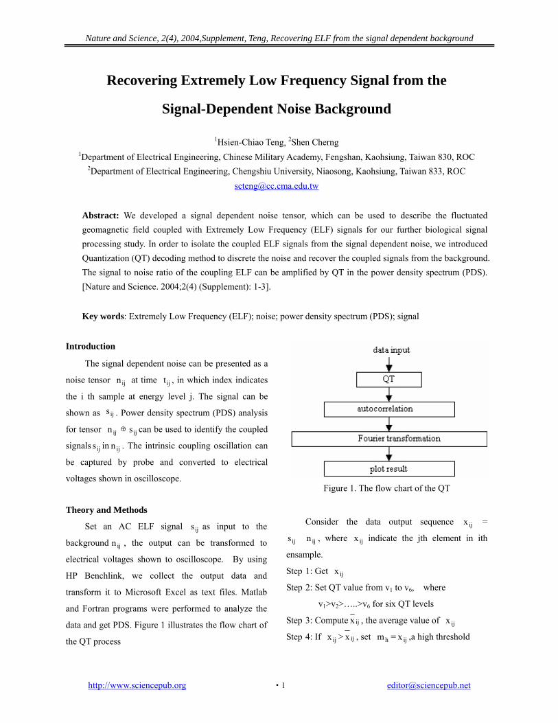
data and get PDS. Figure 1 illustrates the flow chart of
the QT process
ij
ij
Figure 1. The flow chart of the QT
Consider the data output sequence =
� , where indicate the jth element in ith
ensample.
ijx
ijs ijn ijx
ijxStep 1: Get
Step 2: Set QT value from v1 to v6, where
v1>v2>…..>v6 for six QT levels
Step 3: Compute ijx ijx
ijx
, the average value of
Step 4: If > ijx hm ij, set = x ,a high threshold
Nature and Science, 2(4), 2004,Supplement, Teng, Recovering ELF from the signal dependent background
value should be defined. hm
If < ijx ijx , set = , a low threshold
value should be defined. 1m ijx
1m
If > set = , a second high
threshold should be defined. ijx hm
hhmhhm ijx
Step 5: Set ijx = if m < 1hm ijx < hm
ijx = , if ml < h1m ijx < m
ijx = , if 11m ijx < , where 1m
hhm > > >m> > > . hm 1hm h1m 1m 11m
Step 6: If > mhh , set ijx ijx = v1
If < < , set = v2 h1m ijx hhm ijx
If < < , set 1hm ijx hm ijx = v3
If m < < , set = v4 ijx 1hm ijx
If < < m, set = v5 h1m ijx ijx
If < < , set = v6 1m ijx h1m ijx
It is defined amplitude signal to noise ratio as
ASNR =n
s
AA
, where As is the amplitude of the
coupled ELF signal and An is the amplitude of the
noise. In contrast, SNR =n
s
PP , where Ps is the power of
the output ELF signal calculated from PDS and Pn is the power of the noise. Noise can be defined as all unpredictable signals in PDS. Since both ASNR and SNR can be calculated, the plot of ASNR versus SNR produces a function curve showing the correlation between input amplitude and output power. Even through noise may have its own characteristic; we can calibrate the function curve with the help of adjusting trial signal’s amplitude to control the power difference by QT analysis. For instance, using a data sequence to simulate a sample function consisting of 2000 elements including a 15 Hz sinusoid signal,
, label index i is from 1 to
2000 for this sequence. The dimension of the noise tensor is 2000 × j. For simplicity, take j = 1, the power spectrum can be simply calculated. Note that the
identified ELF signal is supposed being occurred at 15 Hz. The frequency component of power density spectrum of the noise being illustrated will depend
upon the characteristic of .
)t( nk)t( s)t(x ijijij ×+=
ijn
In addition, Figure 1b showed the magnetic fluctuation very near the cell layer on the patch substrate.
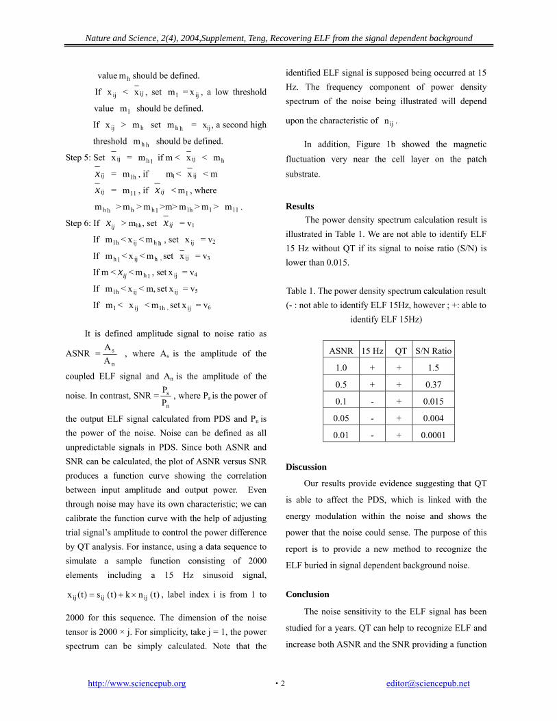
Results The power density spectrum calculation result is illustrated in Table 1. We are not able to identify ELF 15 Hz without QT if its signal to noise ratio (S/N) is lower than 0.015. Table 1. The power density spectrum calculation result (- : not able to identify ELF 15Hz, however ; +: able to
identify ELF 15Hz)
ASNR 15 Hz QT S/N Ratio
1.0 + + 1.5
0.5 + + 0.37
0.1 - + 0.015
0.05 - + 0.004
0.01 - + 0.0001
Discussion
Our results provide evidence suggesting that QT
is able to affect the PDS, which is linked with the
energy modulation within the noise and shows the
power that the noise could sense. The purpose of this
report is to provide a new method to recognize the
ELF buried in signal dependent background noise.
Conclusion
The noise sensitivity to the ELF signal has been
studied for a years. QT can help to recognize ELF and
increase both ASNR and the SNR providing a function
http://www.sciencepub.org [email protected] ·2
Nature and Science, 2(4), 2004,Supplement, Teng, Recovering ELF from the signal dependent background
http://www.sciencepub.org
curve to characterize the signal dependent noise. By
using this function curve, we can find the best estimate
signal-to-noise ratio of the coupled ELF. The
remaining question is how can we find the best
combination of the weights of QT in experiments.
Fuzzy and neuronet analysis may help for further noise
tensor characteristic studies.
2 0
frequency 0 1 0 2 0 3 0 40 5 0 6 0 7 0 8 0 9 0 1 0 0
0
5 0
10 0
15 0
20 0
25 0
30 0
F re q u e n c y H z
0 1 0 2 0 3 0 4 0 5 0 6 0 7 0 8 0 9 0 1 0 00
2 0
4 0
6 0
8 0
1 0 0
1 2 0
1 4 0
F re q u e n c y H z
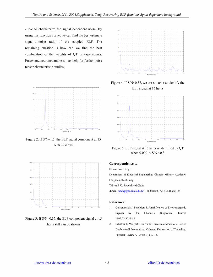
Figure 2. If S/N=1.5, the ELF signal component at 15 hertz is shown
Figure 3. If S/N=0.37, the ELF component signal at 15
hertz still can be shown
0 1 0 2 0 3 0 4 0 5 0 6 0 7 0 8 0 9 0 1 0 00
2
4
6
8
1 0
1 2
1 4
1 6
1 8
F re q u e n c y H z
Figure 4. If S/N<0.37, we are not able to identify the ELF signal at 15 hertz
3 0 0
0 1 0 2 0 3 0 4 0 5 0 6 0 7 0 8 0 9 0 1 000
5 0
1 0 0
1 5 0
2 0 0
2 5 0
F re q ue n c y H z
Figure 5. ELF signal at 15 hertz is identified by QT
when 0.0001< S/N <0.3 Correspondence to: Hsien-Chiao Teng,
Department of Electrical Engineering, Chinese Military Academy,
Fengshan, Kaohsiung,
Taiwan 830, Republic of China
,Email: [email protected]; Tel: 011886-7747-9510 ext 134
Reference: 1. Galvanovskis J, Sandblom J. Amplification of Electromagnetic
Signals by Ion Channels. Biophysical Journal
1997;73:3056-65.
2. Schatzer L, Weigert S. Solvable Three-state Model of a Driven
Double-Well Potential and Coherent Destruction of Tunneling.
Physical Review A 1998;57(1):57-78.
Nature and Science, 2(4), 2004, Supplement, Teng, Non-Stationary Analysis of Osteoblast Cellular Response
The Non-Stationary Analysis of Osteoblast Cellular Response to the reaction of ELF magnetic Field
1Hsien-Chiao Teng, 2Shen Cherng
1Department of Electrical Engineering, Chinese Military Academy, Fengshan, Taiwan 830, Republic of China
2Department of Electrical Engineering, Chengshiu University, Niaosong, Taiwan 833, Republic of China [email protected]; 011886-7747-9510 ext 134
Abstract: Cellular response to the external extremely low frequency (ELF) electromagnetic field is a non-stationary process. The time and frequency resolution problems and the background noise result the difficulty for analysis. Signal of cellular reaction to the ELF reaction can change with the time. All frequency components exist at all the times. Therefore, the signal where in time the spectral frequency components become visible is worth to investigate. This report provided the study of what cellular response signal frequency band and what existed cellular response signal time interval of osteoblast cell line system under the exposure of ELF electromagnetic field. Conclusively, 14±2Hz cellular response frequency band existed at the first 0.0005 second to elapse for 0.001 second in general and 20% gap junctional intracellular communication (GJIC) modulation within osteoblast cells was observed after 40 minutes exposure of ELF electromagnetic field. [Nature and Science. 2004;2(4) (Supplement): 4-7].
Key words: extremely low frequency (ELF); gap junctional intracellular communication (GJIC); non-stationary process
1. Introduction
We are in the environment of ELF Electromagnetic field. There have been of considerable discussion concerning the human cellular reaction to the external ELF signals. No clinical evidence has shown any human health effect and no mechanism can clearly explain every observed biological effect [Takebe et al., 1999]. This report describes the study of the Osteoblast cellular response to the reaction of external ELF magnetic field signal. Theoretically, four different types of cellular responding signals, deterministic, stochastic, fractal and chaotic signals are categorized for biological system [1,2]. A deterministic signal is one whose values in the future can be predicted if enough information about its past is known and stochastic signal is impossible to predict an exact future value even if one knows it’s entire past history. Fractal signals have the property that they look very similar at all levels of magnification, which is referred as scale-invariance. Chaotic signals are deterministic signals with sensitive dependence on some conditions that cannot be predicted exactly in the future. Experimentally, gap junctional intracellular communication (GJIC) within the cells may induce the signals from varying surface current [3]. In a cell, six connexin 43 subunits oligomerze in the Golgi apparatus into a connexon, called hemi channel and be transported to plasma membrane of the cell. Before pairing process, hemi channels are closed to avoid leakage of cellular contents and entry of
extra-cellular materials. During the pairing of connexons and aggregation into plaques at the plasma membrane, connexin 43 is phosphorylated at least twice and connexons are attracted to those located on the adjacent cells. Two connexons join in an end-to-end manner to form a complete channel. The channel aggregate into large gap junction plaques open to connect two cells for cell-to-cell communication and is called gap junctional intracellular communication (GJIC), which can be modulated by environmental factors, such as ELF signals. Since the function of the GJIC, cultured cells coupled together in vitro except the stem cells and cancer cells [2]. In this article, we will introduce a concept to recognize the non-stationary magnetic fluctuation process caused by the cellular response of the reaction of ELF magnetic field and clarify the correlation aspects for both time and frequency somewhat arbitrarily. For frequency aspects, we present one idea around the notion of local regularity. For time aspects, we present a list of domains. The magnetic field fluctuations created by the induced GJIC surface current of the osteoblast cell system is basically a non-stationary process. We also introduce the scrape loading dye transfer technique to identify the GJIC modulation by observing the diffusive range of the fluorescence [4,5]. The varied diffuse range of Lucifer yellow fluorescence expresses the cellular response under the exposure of external ELF magnetic field at intrinsic-resonance frequency ω. Since GJIC is affiliated with many pathological endpoints [4,5], GJIC modulation can be a good factor to evaluate the cellular response of the reaction of external ELF magnetic field.
http://www.sciencepub.org [email protected] ·4·

Nature and Science, 2(4), 2004, Supplement, Teng, Non-Stationary Analysis of Osteoblast Cellular Response
2. Theory Mathematically, the sequence V(t) can be written
as V(t) = { V1,V2,……….VN-1, VN }. We are able to calculate the SNR spectrum of V(t). Relying on surface electrical current distribution induced within osteoblast cells causees GJIC modulation [3]. Wavelet Mathematical transformations can be applied to V(t) to obtain further information from the process. In the following, we will use osteoblast cells induced magnetic fluctuation as V(t). Most of the signals in practice are time domain signals. In our case, signal information is hidden in the frequency content of V(t). The frequency spectrum of V(t) is basically the spectral components of the signal. The frequency spectrum of a (signal) process shows what frequencies exist in the process. However, if the process is not stationary, we have to know which signal corresponds to which frequency band, and if we put all of them together and plot them on a 3-D graph, we will have time in one axis, frequency in the second and amplitude in the third axis. Accordingly, we use『 wavelet transformation 』 to determine which frequencies exist at which time in osteoblast cells induced cellar responding magnetic fluctuation V(t) of the reaction of ELF magnetic field.
Let measured cellar responding magnetic
fluctuation x (t) be a V(t) andτbe a period of sample time, we can get
∫−
= )dtsτt(V(t)ψ
s1s),(Ψ *ψ
V τ and
We presume「Meyer Wavelet」as the good fit
mother wavelet for the transformation. Different mother wavelets may result shift of the location of the intrinsic signal in time domain V(t). 3. Cell Culture
The osteoblast cell line in vitro was obtained from
D.T. Yamaguchi, Research Service and Geriatrics Research, Education, and Clinical Center, VAMC, West Los Angeles, California, USA It was maintained in D-medium (Formula 78-5470EF, GIBCO, Grand Island, NY, USA), supplemented with 10% fetal bovine serum (GIBCO) and 50 µg/ml gentamicin (Quality Biological, Inc., Gaithersburg MD, USA). The cells were incubated at 37˚C in a humidified atmosphere containing 5% CO2 and 95% air and were fed or trypsinized every two to three days.
4. Bioassay of GJIC
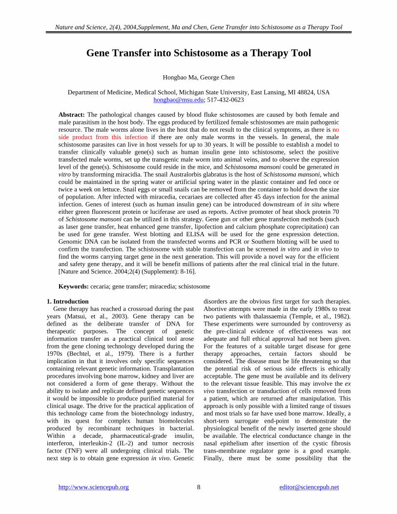
The scrape load/dye transfer (SL/DT) technique was used to measure the GJIC within cells. After exposure to ELF at intrinsic frequency, the cells were rinsed with phosphate buffered saline (PBS), and a PBS solution containing 4% concentration Lucifer yellow fluorescence dye is injected into the cells by a scrape using a scalpel blade. Afterwards the cells were incubated for 3 min and extra cellular dye was rinsed off and fixed with 5% formalin. We then measured the area of the dye migrated from the scrape line using digital images taken by an epifluorescent microscope and quantitated with Nucleotech image analysis software [3,4,5] for the GJIC images. Since GJIC is affiliated with many pathological endpoints [3,4], we use GJIC as a scale factor to evaluate the ELF reaction for cell system. Scrape loading dye transfer of Lucifer yellow is used to measure gap junction intracellular communication (GJIC) modulation under the exposure of ELF magnetic field. The intrinsic resonance detected in SNR spectrum of the mouse osteoblast cells system is very likely to be a chaotic signal, which is not fully predictable. 5. Results
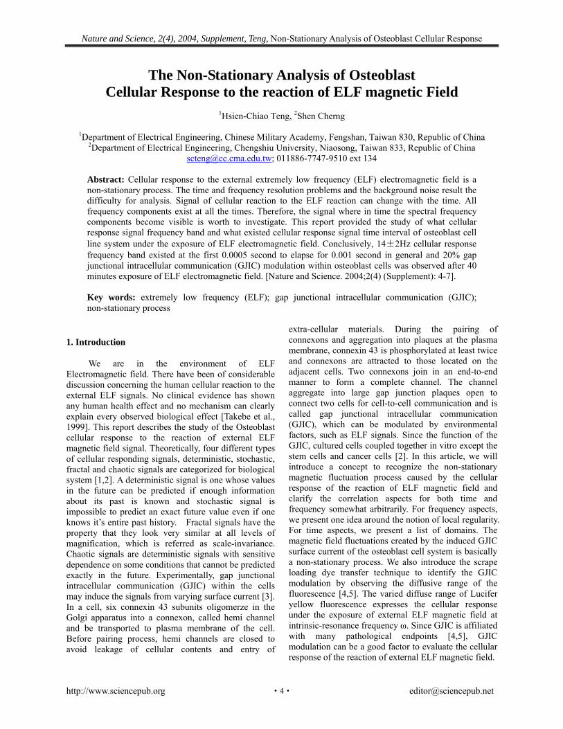
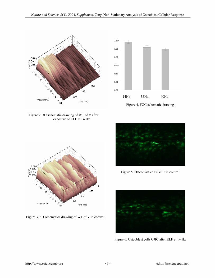
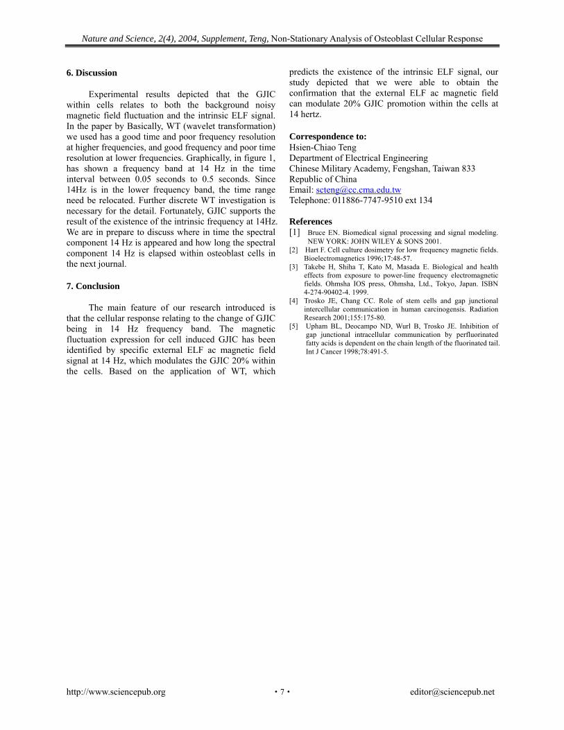
Figure 1 depicted the plot of V(t). Figure 2 depicted the 3D fitting curve of wavelet transformation such as to confirm the existence of intrinsic frequency situated. In contrary, 3D schematics drawing of WT are shown in Figure 2 and Figure 3. Figure 5 and Figure 6 show the GJIC fluorescent images. Since the GJIC of cells was quantified with the measurement of the average distance of dye migration, GJIC was reported in this article as a fraction of the control (FOC) in Figure 4. An FOC value equals to 1.0 indicates normal GJIC. The FOC value more than 1.0 indicates excitation.
0
0.005
0.01
0.015
0.02
0.025
0 0.005 0.01 0.015 0.02 0.025 0.03
sec
volt
Figure 1. V(t) schematic drawing
http://www.sciencepub.org [email protected] ·5·
Nature and Science, 2(4), 2004, Supplement, Teng, Non-Stationary Analysis of Osteoblast Cellular Response
Figure 2. 3D schematic drawing of WT of V after exposure of ELF at 14 Hz
Figure 3. 3D schematics drawing of WT of V in control
0.00
0.20
0.40
0.60
0.80
1.00
1.20
14Hz 35Hz 60Hz
Figure 4. FOC schematic drawing
Figure 5. Osteoblast cells GJIC in control
Figure 6. Osteoblast cells GJIC after ELF at 14 Hz
http://www.sciencepub.org [email protected] ·6·
Nature and Science, 2(4), 2004, Supplement, Teng, Non-Stationary Analysis of Osteoblast Cellular Response
http://www.sciencepub.org [email protected] ·7·
6. Discussion Experimental results depicted that the GJIC
within cells relates to both the background noisy magnetic field fluctuation and the intrinsic ELF signal. In the paper by Basically, WT (wavelet transformation) we used has a good time and poor frequency resolution at higher frequencies, and good frequency and poor time resolution at lower frequencies. Graphically, in figure 1, has shown a frequency band at 14 Hz in the time interval between 0.05 seconds to 0.5 seconds. Since 14Hz is in the lower frequency band, the time range need be relocated. Further discrete WT investigation is necessary for the detail. Fortunately, GJIC supports the result of the existence of the intrinsic frequency at 14Hz. We are in prepare to discuss where in time the spectral component 14 Hz is appeared and how long the spectral component 14 Hz is elapsed within osteoblast cells in the next journal.
7. Conclusion
The main feature of our research introduced is
that the cellular response relating to the change of GJIC being in 14 Hz frequency band. The magnetic fluctuation expression for cell induced GJIC has been identified by specific external ELF ac magnetic field signal at 14 Hz, which modulates the GJIC 20% within the cells. Based on the application of WT, which
predicts the existence of the intrinsic ELF signal, our study depicted that we were able to obtain the confirmation that the external ELF ac magnetic field can modulate 20% GJIC promotion within the cells at 14 hertz. Correspondence to: Hsien-Chiao Teng Department of Electrical Engineering Chinese Military Academy, Fengshan, Taiwan 833 Republic of China Email: [email protected] Telephone: 011886-7747-9510 ext 134 References [1] Bruce EN. Biomedical signal processing and signal modeling.
NEW YORK: JOHN WILEY & SONS 2001. [2] Hart F. Cell culture dosimetry for low frequency magnetic fields.
Bioelectromagnetics 1996;17:48-57. [3] Takebe H, Shiha T, Kato M, Masada E. Biological and health
effects from exposure to power-line frequency electromagnetic fields. Ohmsha IOS press, Ohmsha, Ltd., Tokyo, Japan. ISBN 4-274-90402-4. 1999.
[4] Trosko JE, Chang CC. Role of stem cells and gap junctional intercellular communication in human carcinogensis. Radiation Research 2001;155:175-80.
[5] Upham BL, Deocampo ND, Wurl B, Trosko JE. Inhibition of gap junctional intracellular communication by perfluorinated fatty acids is dependent on the chain length of the fluorinated tail. Int J Cancer 1998;78:491-5.
Nature and Science, 2(4), 2004,Supplement, Ma and Chen, Gene Transfer into Schistosome as a Therapy Tool
Gene Transfer into Schistosome as a Therapy Tool
Hongbao Ma, George Chen
Department of Medicine, Medical School, Michigan State University, East Lansing, MI 48824, USA [email protected]; 517-432-0623
Abstract: The pathological changes caused by blood fluke schistosomes are caused by both female and male parasitism in the host body. The eggs produced by fertilized female schistosomes are main pathogenic resource. The male worms alone lives in the host that do not result to the clinical symptoms, as there is no side product from this infection if there are only male worms in the vessels. In general, the male schistosome parasites can live in host vessels for up to 30 years. It will be possible to establish a model to transfer clinically valuable gene(s) such as human insulin gene into schistosome, select the positive transfected male worms, set up the transgenic male worm into animal veins, and to observe the expression level of the gene(s). Schistosome could reside in the mice, and Schistosoma mansoni could be generated in vitro by transforming miracidia. The snail Australorbis glabratus is the host of Schistosoma mansoni, which could be maintained in the spring water or artificial spring water in the plastic container and fed once or twice a week on lettuce. Snail eggs or small snails can be removed from the container to hold down the size of population. After infected with miracedia, cecariaes are collected after 45 days infection for the animal infection. Genes of interest (such as human insulin gene) can be introduced downstream of in situ where either green fluorescent protein or luciferase are used as reports. Active promoter of heat shock protein 70 of Schistosome mansoni can be utilized in this strategy. Gene gun or other gene transfection methods (such as laser gene transfer, heat enhanced gene transfer, lipofection and calcium phosphate coprecipitation) can be used for gene transfer. West blotting and ELISA will be used for the gene expression detection. Genomic DNA can be isolated from the transfected worms and PCR or Southern blotting will be used to confirm the transfection. The schistosome with stable transfection can be screened in vitro and in vivo to find the worms carrying target gene in the next generation. This will provide a novel way for the efficient and safety gene therapy, and it will be benefit millions of patients after the real clinical trial in the future. [Nature and Science. 2004;2(4) (Supplement): 8-16]. Keywords: cecaria; gene transfer; miracedia; schistosome
1. Introduction Gene therapy has reached a crossroad during the past years (Matsui, et al., 2003). Gene therapy can be defined as the deliberate transfer of DNA for therapeutic purposes. The concept of genetic information transfer as a practical clinical tool arose from the gene cloning technology developed during the 1970s (Bechtel, et al., 1979). There is a further implication in that it involves only specific sequences containing relevant genetic information. Transplantation procedures involving bone marrow, kidney and liver are not considered a form of gene therapy. Without the ability to isolate and replicate defined genetic sequences it would be impossible to produce purified material for clinical usage. The drive for the practical application of this technology came from the biotechnology industry, with its quest for complex human biomolecules produced by recombinant techniques in bacterial. Within a decade, pharmaceutical-grade insulin, interferon, interleukin-2 (IL-2) and tumor necrosis factor (TNF) were all undergoing clinical trials. The next step is to obtain gene expression in vivo. Genetic
disorders are the obvious first target for such therapies. Abortive attempts were made in the early 1980s to treat two patients with thalassaemia (Temple, et al., 1982). These experiments were surrounded by controversy as the pre-clinical evidence of effectiveness was not adequate and full ethical approval had not been given. For the features of a suitable target disease for gene therapy approaches, certain factors should be considered. The disease must be life threatening so that the potential risk of serious side effects is ethically acceptable. The gene must be available and its delivery to the relevant tissue feasible. This may involve the ex vivo transfection or transduction of cells removed from a patient, which are returned after manipulation. This approach is only possible with a limited range of tissues and most trials so far have used bone marrow. Ideally, a short-tern surrogate end-point to demonstrate the physiological benefit of the newly inserted gene should be available. The electrical conductance change in the nasal epithelium after insertion of the cystic fibrosis trans-membrane regulator gene is a good example. Finally, there must be some possibility that the
http://www.sciencepub.org [email protected] 8
Nature and Science, 2(4), 2004,Supplement, Ma and Chen, Gene Transfer into Schistosome as a Therapy Tool
disability caused by a disease is reversible. Some of the tragic mental and physical handicaps caused by genetic metabolic disorders may never be improved by somatic gene therapy, however successful with a gene transfer protocol. Gene transfer is one of the key factors in gene therapy. In this project, we will use gene gun or other methods (such as calcium phosphate coprecipitation, lipofection, laser or temperature enhancing gene transfer) as the tools to transfer insulin and other proper genes into schistosome. The blood fluke schistosomes are unusual trematodes that reside in the blood vessels of the definitive host. There are a number of species of schistosomes that can infect humans, but most human infections are caused by one of the three following species: Schistosoma mansoni; Schistosoma haematobium and Schistosoma japonicum. Even it is estimated that approximately 200,000,000 people are infected with schistosomes, resulting in 1,000,000 deaths each year (Parasites Research Group, 2004), the disease from schistosomes aroused by only the female and male together in the body, and male worms only do not arouse symptom. If there are only male schistosome parasites in animal vessels, they can live in animal vessels for up to 30 years without symptom, as there is no side product from this infection if there are only male worms in the vessels. The cultured sporocysts of schistosomes have been transiently transfected with pBluescript-based plasmids expressing green fluorescent protein (GFP) (Wippersteg, et al., 2002a; Wippersteg, et al., 2002b), demonstrating that it is possible in principle to generate transgenic parasites. However, it is not clear yet whether a transgenic line of schistosomes could be established from these transiently transfected sporocysts. At least two important obstacles on the path to the development of a reliable, tractable transgenesis system for schistosomes now need to be surmounted: (1) development of appropriate constructs to achieve stable integration into the schistosome genome and (2) a demonstration that the life cycle can be completed by genetically modified parasites. The life cycle of the schistosome includes two free-living, self-reliant, and mobile infective stages. These are the miracidium, which seeks out and directly infects the intermediate host snail, and the cercaria, which accomplishes infection of the mammalian, definitive host by direct penetration of host skin when a definitive host comes into contact with water contaminated with cercariae. These are the only two stages that could be reintroduced into the life cycle using a natural route of infection, thereby obviating the inefficient and laborious tasks of surgically implanting transformed sporocysts back into snails and/or surgically implanting adult schistosomes into the portal system blood vessels of mice (Cheever, et al., 1994; Jourdane, et al., 1985).
Here we subject miracidia of Schistosoma mansoni to particle bombardment and are able to show that miracidia bombarded with DNA-coated gold particles can directly and naturally infect the intermediate host snail Biomphalaria glabrata and establish as sporocysts in a natural fashion. Further we are able to demonstrate transgene transcription of enhanced GFP in adult worms infected with transformed sporocysts. Insulin gene will be considered as one of the targets in this project. Diabetes is a disease in which the body does not produce or properly use insulin. Insulin is a hormone that is needed to convert sugar, starches and other food into energy needed for daily life. There are 18.2 million people in the United States, or 6.3% of the population, who have diabetes (American Diabetes Association, 2004). The treatment of diabetes is one of the most important topics in the health science. The normal treatment of diabetes is to inject insulin to patients every day to keep the proper insulin level in the blood. It is high cost and plague. The genetic treatment has not been successfully. If it successfully transfers insulin gene into schistosome and the gene successfully expresses in the worm, and infect the male transtected worm in animal vessels, the worm can release suitable amount insulin into animal vessels. This will be a high efficient treatment method on the diabetes. The potential valuation of this project for diabetes treatment is to clone the insulin gene (with promoter) into schistosome and select male worm, then reside the cloned schistosome to diabetes patients’ vessels. 2. Specific Aims this Trial can Hit The scientific objective of this scientific trial is to establish an animal model by using transgenic schistosome as an alternative therapeutic material delivery vehicle for animal and eventually for human in the future. 2.1 To select one or a group of candidate genes, such as insulin, thyrotropin, growth factor and tumor necrosis factor genes, reconstruct the selected genes and transfect them into schistosome by the gene gun, temperature enhanced gene transfection, or laser enhanced gene transfection, etc. 2.2 To infect the transgenic schistosome in mice and valuate the transfected gene expression in the mice. 2.3 To establish the transgenic schistosome model and evaluate the medication capacity and possible expression level control. 2.4 Elongating knowledge from aims 1 – 3 for the future project, to explore the possibility of infecting the transgenic schistosome in human and evaluate the
http://www.sciencepub.org [email protected] 9
Nature and Science, 2(4), 2004,Supplement, Ma and Chen, Gene Transfer into Schistosome as a Therapy Tool
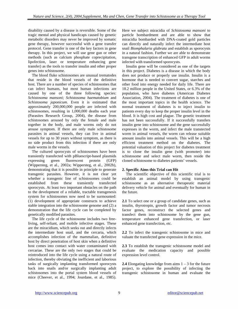
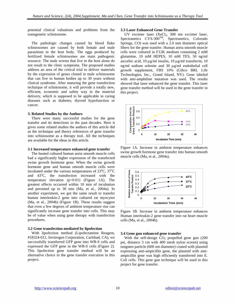
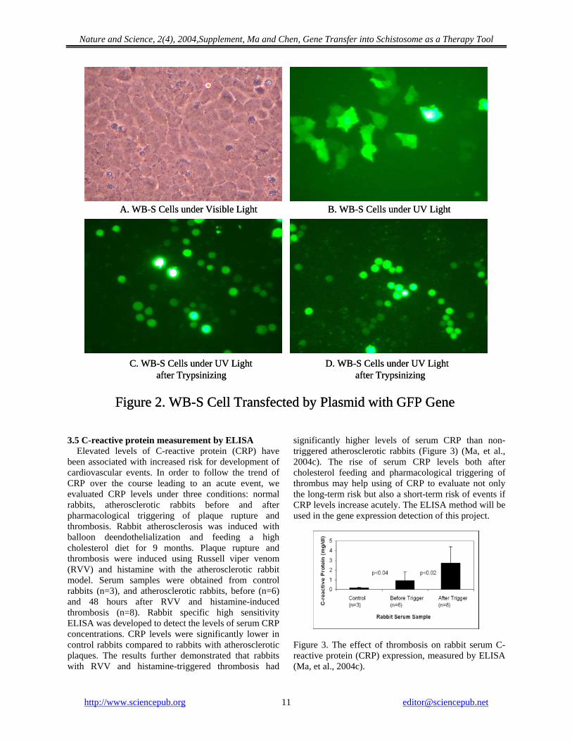
potential clinical valuations and problems from the transgenetic schistosome. The pathologic changes caused by blood fluke schistosomes are caused by both female and male parasitism in the host body. The eggs produced by fertilized female schistosomes are main pathogenic resource. The male worms that live in the host alone do not result to the clinic symptoms. The proposed studies address an area of the critical trial to deliver materials by the expression of genes cloned in male schistosome that can live in human bodies up to 30 years without clinical syndrome. After maturing the gene transfection technique of schistosome, it will provide a totally new, efficient, economic and safety way in the material delivery, which is supposed to be applicable in curing diseases such as diabetes, thyroid hypofunction or cancer. 3. Related Studies by the Authors There were many successful studies for the gene transfer and its detections in the past decades. Here it gives some related studies the authors of this article did as the technique and theory references of gene transfer into schistosome as a therapy tool. All the techniques are available for the ideas in this article. 3.1 Increased temperature enhanced gene transfer The heated cultured human aorta smooth muscle cells had a significantly higher expression of the transfected swine growth hormone gene. When the swine growth hormone gene and human smooth muscle cells were incubated under the various temperatures of 23oC, 37oC and 43oC, the transfection increased with the temperature elevation (p<0.01) (Figure 1A). The greatest effects occurred within 10 min of incubation and persisted up to 30 min (Ma, et al., 2004a). In another experiment, we got the same result to transfer human interleukin-2 gene into cultured rat myocytes (Ma, et al., 2004b) (Figure 1B). These results suggest that even a few degrees of ambient temperature rise can significantly increase gene transfer into cells. This may be of value when using gene therapy with transfection procedures. 3.2 Gene transfection mediated by lipofection With lipofection method (Lipofectamine Reagent, #18324-012, Invitrogen Corporation, Carlsbad, CA), we successfully transferred GFP gene into WB-S cells and expressed the GFP gene in the WB-S cells (Figure 2). This lipofection gene transfer method will be an alternative choice in the gene transfer execution in this project.
3.3 Laser Enhanced Gene Transfer UV excimer laser (XeCl2, 308 nm excimer laser, Spectranetics CVS-300TM, Spectranetics, Colorado Springs, CO) was used with a 2.0 mm diameter optical fibers for the gene transfer. Human aorta smooth muscle cells were cultured in F12K medium containing 2 mM glutamine, 10 mM HEPES, 10 mM TES, 50 ng/ml ascorbic acid, 10 μg/ml insulin, 10 μg/ml transferrin, 10 ng/ml sodium selenite and 30 μg/ml endothelial cell growth supplement, FBS 10% (Gibco BRL Life Technologies, Inc., Grand Island, NY). Gene labeled with anti-amphiline mutation was used. The results showed that laser enhanced the gene transfer. This laser gene transfer method will be used in the gene transfer in this project.
0
0.2
0.4
0.6
0 10 20 30 40
Incubation Time (min)
Com
para
tive
Uni
t for
G
ene
Tran
sfer
Pro
duct
430C
370C230C
0
0.2
0.4
0.6
0 10 20 30 40
Incubation Time (min)
Com
para
tive
Uni
t for
G
ene
Tran
sfer
Pro
duct
430C
370C230C
Figure 1A. Increase in ambient temperature enhances swine growth hormone gene transfer into human smooth muscle cells (Ma, et al., 2004a).
00.10.20.30.40.50.6
0 10 20 30 40
Incubation Time (min)
Rel
ativ
e A
mou
nt o
f Gen
e Tr
ansf
er P
rodu
ct 43oC
37oC
23oC
Figure 1B. Increase in ambient temperature enhances Human interleukin-2 gene transfer into rat heart muscle cells (Ma, et al., 2004b). 3.4 Gene gun enhanced gene transfer With the self-design CO2 propelled gene gun (200 psi, distance 3 cm with 400 mesh nylon screen) using tungsten particle (600 nm diameter) coated with plasmid expressing anti-ampicillin gene, the plasmid with anti-ampicillin gene was high efficiently transferred into E. Coli cells. This gene gun technique will be used in this project for gene transfer.
http://www.sciencepub.org [email protected] 10
Nature and Science, 2(4), 2004,Supplement, Ma and Chen, Gene Transfer into Schistosome as a Therapy Tool
http://www.sciencepub.org
A. WB-S Cells under Visible Light B. WB-S Cells under UV Light
C. WB-S Cells under UV Light D. WB-S Cells under UV Lightafter Trypsinizing after Trypsinizing
Figure 2. WB-S Cell Transfected by Plasmid with GFP Gene
A. WB-S Cells under Visible Light B. WB-S Cells under UV Light
C. WB-S Cells under UV Light D. WB-S Cells under UV Lightafter Trypsinizing after Trypsinizing
Figure 2. WB-S Cell Transfected by Plasmid with GFP Gene 3.5 C-reactive protein measurement by ELISA
Elevated levels of C-reactive protein (CRP) have been associated with increased risk for development of cardiovascular events. In order to follow the trend of CRP over the course leading to an acute event, we evaluated CRP levels under three conditions: normal rabbits, atherosclerotic rabbits before and after pharmacological triggering of plaque rupture and thrombosis. Rabbit atherosclerosis was induced with balloon deendothelialization and feeding a high cholesterol diet for 9 months. Plaque rupture and thrombosis were induced using Russell viper venom (RVV) and histamine with the atherosclerotic rabbit model. Serum samples were obtained from control rabbits (n=3), and atherosclerotic rabbits, before (n=6) and 48 hours after RVV and histamine-induced thrombosis (n=8). Rabbit specific high sensitivity ELISA was developed to detect the levels of serum CRP concentrations. CRP levels were significantly lower in control rabbits compared to rabbits with atherosclerotic plaques. The results further demonstrated that rabbits with RVV and histamine-triggered thrombosis had
significantly higher levels of serum CRP than non-triggered atherosclerotic rabbits (Figure 3) (Ma, et al., 2004c). The rise of serum CRP levels both after cholesterol feeding and pharmacological triggering of thrombus may help using of CRP to evaluate not only the long-term risk but also a short-term risk of events if CRP levels increase acutely. The ELISA method will be used in the gene expression detection of this project.
Figure 3. The effect of thrombosis on rabbit serum C-reactive protein (CRP) expression, measured by ELISA (Ma, et al., 2004c).
Nature and Science, 2(4), 2004,Supplement, Ma and Chen, Gene Transfer into Schistosome as a Therapy Tool
3.6 Heat shock protein 70 measured by Western blotting Transmyocardial laser revascularization (TMLR) has been shown to relieve symptomatic ischemia but laser tissue effects have potential complications. In order to define the mechanism of laser action, heat shock protein (hsp) expression was evaluated in rat hearts after TMLR. Under general anesthesia, hearts were removed from 10 rats and immediately placed in oxygenated physiologic buffered solution (PBS) at 0oC. After the various treatments, hearts were homogenized and hsp70 was measured with Western Blotting. Group 1 (n=3)
hearts were immediately homogenized; Group 2 (n=3) hearts were perfused with the PBS in a Langendorff setup for 6 h; Group 3 (n=3) hearts were lased (50 channels) using a Ho:Yag laser via a 0/600 mm core fiber at 3 Hz and 280 mJ/pulse and perfused up to 6 h. Group 4 (n=1) rat was heated to 42oC for 15 min then recovered at 23oC for 6h prior to hsp measurement. There was a significantly lower hsp70 expression in Group 3 (TMLR) and higher in Group 4 than that the control Groups 1, 2 (Table 1) (Ma, et al., 2004d). The Western Blotting technique will be used in the gene expression detection.
Table 1. Heat Shock Protein 70 Expressed in Rat Hearts
Groups Group 1* Group 2* Group 3** Group 4 Relative OD600nm 1 0.87±0.10 0.19±0.03 3.47
* to *: p=ns; * to **: p<0.003 4. Research Design and Methods The principle idea of this project is to insert target genes into the chromosome of schistosome eggs (single cell stage) to get stable gene transfection in the single egg cell. After then, raise the male transfected worms in animal and let the target gene be expressed in animals. This will provide an alternative treatment of genetic diseases with the transfected gene(s). 4.1 The brief description of this project steps is as the following: 4.1.1 To get target gene candidate(s) with promoter, plasmid, and expression control sequence. 4.1.2 To construct the expression plasmids with target genes (with suitable promoter). 4.1.3 To transform the expression plasmid(s) into schistosome eggs, miracidium or cercariae. 4.1.4 To detect the gene product if it is expressed in miracidium and cercariae. 4.1.5 To raise the transformed schistosome in mice. 4.1.6 To investigate the expression level of the target gene in the mice infected by transformed schistosome. 4.1.7 To screen the male schistosome transformed with target gene(s), and culture the male schistosome in animals. 4.1.8 To observe if the male schistosome can survive in the animal without hurting the animal and express the target protein. 4.1.9 To keep the schistosome transfected with target gene(s). To select the male schistosome transfected with target gene(s). 4.1.10 To raise the male schistosoma transfected with target gene(s) in animal.
4.1.11 To observe if the male schistosome can survive in the animal without hurting the animal and express the target protein. 4.1.12 Complete the model to transfer valuable genes into schistosome for the material delivery. Clinical discussion if it is possible to raise the schistosome with the target gene(s) in human in the future projects. There are three common methods of genetic engineering: the plasmid method, the vector method, and the biolistic method. Several techniques are currently used to transfer genes into various cells, tissues and organs. Although gene therapy is a potential therapeutic approach for arterial restenosis and angiogenesis, the efficiency of transfection is low regardless of the techniques used. To transfer gene efficiently, a novel method gene-gun will be used as the first choice. The other gene transfection methods including laser gene transfer, temperature enhanced gene transfer, electroporation, lipofection, calcium-phosphate-mediated coprecipitation, transfection mediated by DEAE-dextran, etc. will be considered. The transfected gene expression can be controlled by regulating the transgenic schistosome number, etc. Genes of interest (such as human insulin gene) can be introduced downstream of in situ where either green fluorescent protein or luciferase are used as reports. Active promoter of heat shock protein 70 of Schistosome mansoni (GenBank No. L02415) can be utilized in this strategy. Gene gun (Bio-Rad Laboratories) or other gene transfection methods (such as laser gene transfer, heat enhanced gene transfer, lipofection and calcium phosphate coprecipitation) can be used in this project for gene transfer. West blotting and ELISA can be used for the gene expression
http://www.sciencepub.org [email protected] 12
Nature and Science, 2(4), 2004,Supplement, Ma and Chen, Gene Transfer into Schistosome as a Therapy Tool
http://www.sciencepub.org [email protected] 13
detection. Genomic DNA can be isolated from the transfected worms and PCR or Southern blotting can be used to confirm the transfection. The schistosome with stable transfection can be screened in vitro and in vivo to find the worms carrying target gene in the next generation. 4.2 Maintenance of Schistosoma mansoni Schistosome will reside in mice. Schistosoma japonicum and Schistosoma mansoni sporocysts can be generated in vitro by transforming miracidia (Coustau, et al., 2000). The snail Australorbis glabratus is the host of Schistosoma mansoni, which is maintained in the spring water or artificial spring water in the food storage plastic container. Feed once or twice a week on lettuce. Temperature should be maintained above 25oC. Snail eggs or small snails can be removed from the container to hold down the size of population. Schistosoma mansoni adults are best maintained in mice or hamsters. 4.3 Obtaining miracidia Remove the livers of 1-4 infected animals. Homogenize the liver 20-30 seconds in 0.85% NaCl in blender. Pour homogenate into a flask and allow to settle for 10-20 min. Repeat above procedure 2-3 times until supernatant appears clear. Pour off final supernatant. Fill the flask nearly to the top with water. Place flask in bright light for 5 min to stimulate hatching of miracidia. Collect miracidia from the top of flask and transfer to a small dish for infecting snails. 4.4 Infecting snails Keep snails in small amount of spring water in small beakers. Add miracidia. For routine maintenance five miracidia per snail gives a high incidence of infection. Infected snails will usually be shedding cercariae in 30 days. 4.5 Obtaining cercariae To obtain large number of cercariae, place infected snails in the dark for 2-5 days before release cercariae. To check incidence of infection, isolate snails in separate beaker with spring water. Use rubber gloves and scoop. Place 10-20 snails in a beaker with 100-200 ml of spring water. Place beaker in strong light; cercariae will emerge in 5-60 min. Inspect beaker with a dissecting scope. 4.6 Infecting mice or hamster Wear gloves. Take 0.1 ml aliquots of the cercarial suspension, dilute the suspension with spring water to the desired number of cercaria in 0.2 ml spring water. Inject 50-200 cercariae into the abdomen of mouse or hamster. Mature worms develop in about 40 days. Feces may be examined for the presence of eggs to determine
presence and maturity of infection. Adult worms can be recovered by dissection from the mesenteric veins. 4.7 Gene construction To date, stable transformation has not been achieved in any schistomsome. While transient transfection provides a useful tool to study the effects of over expression of genes of interest. To effectively generate transformants, the appropriate vector/promoter combination must be chosen to ensure maximum expression. Active promoter of heat shock protein 70 (hsp70) of Schistosome mansoni (GenBank No. L02415) (Neumann, et al., 1992) will be utilized in this strategy. Genes of interest (such as insulin gene) will be introduced downstream of in situ where either green fluorescent protein or luciferase are used as reports. Therefore, we will use pUC19 plasmid to deliver our current combinations of genes into the candidate worms. Briefly, target genes will be constructed into gene-promoter-vector as the following: To engineer plasmid constructs for this transfection, polymerase chain reaction (PCR), TOBO (which is used for cloning after obtain a PCR product with a TA over hung) and Bluescript/plasmid (Invitrogen Corporation, Carlsbad, CA) is used for DNA fragment amplification as well as pUC19 plasmid backbone or a retroviral vector, G1Tk1SvNa vector (Sauce, et al., 2002) are used for gene transfection. According to sequence data, gene primers are designed to amplify parts of the gene by PCR. The restrictions sites are used to insert the gene fragment by ligating the fragment into pUC19 plasmid. The clones containing the promoter (such as CMV) enhanced green fluorescing protein as biological marker, and terminator regions will be inserted: 4.8 PCR and RT-PCR analysis Amplification reactions are performed in a total volume of 25 ml using 10 ng DNA isolated from worms for genotyping, 1 mM each primer gene forward and the gene reverse primer, 10 mM of each deoxynucleotide and 2.5 units Taq polymerase (Invitrogen Corporation, Carlsbad, CA). After an initial denaturation step at 95oC for 3 min, temperature cycling is performed at 93oC-45 seconds, 60oC-60 seconds, 70oC-45 seconds for 30 cycles. One fourth of the reaction volume is used for agarose gel electrophoresis (1.2%). Reverse transcriptase-polymerase chain reaction (RT-PCR) used
EGFP Promorter
Start
Gene
Nature and Science, 2(4), 2004,Supplement, Ma and Chen, Gene Transfer into Schistosome as a Therapy Tool
for gene expression evaluation and will be done stepwise in separate reactions. 4.9 Commercial gene Insulin gene cloned in E. Coli and promoter could be obtained from American Type Culture Collection (ATCC, Rockville, MD). This insulin gene and promoter will be used to transfer to schistosome. Other suitable genes for the transfection will be considered during the processing of this project. 4.10 Gene transfer To attain the maximal transformation efficiently, the gene gun method will be used as the first choice for the gene transfection. Bio-Rad gene gun and a self-design gene gun will be used for the gene transfer. Also, a rifle gun will be used as an alternative method for the gene transfection. Other gene transfer methods will be considered. 4.10.1 Gene gun transfection Schistosoma mansoni will be propagated in the laboratory as described (Copeland, et al., 2003). Snails are maintained at 26°C in aerated water and fed with lettuce leaves, in a room with a light/darkness cycle of 12 hours each. At 8 weeks after Schistosoma mansoni infection, mice are killed, adult worms are perfused from the mesenteric veins with PBS, and the livers are removed. Adult worms are washed three times in RPMI 1640 and maintained in vitro in RPMI 1640 + 10% FCS supplemented with penicillin/streptomycin at 37°C in 5% CO2. The infected livers are forced through a plastic mesh to release the schistosome eggs, after which the eggs are washed in cold sterile 1.8% NaCl to remove host tissues and debris. The 1.8% NaCl is replaced with sterile water to induce the eggs to hatch, miracidia are collected from the water, and concentrated by centrifugation at 500 rpm. For particle bombardment (biolistics), approximately 500 miracidia or 20 male, adult worms are evenly spread onto a polycarbonate membrane (Transwell, Costar), water or medium is removed and target schistosomes on the polycarbonate membranes are positioned in a Biolistic PDS-1000/HE Particle Delivery System (Bio-Rad Laboratories GmbH, München, Germany). Biolistic parameters are 15 in. Hg of chamber vacuum, target distance of 3 cm (stage 1), 900 psi (miracidia) or 1800 psi (adult worms) particle acceleration pressure, and 1.0 m gold microcarriers (Bio-Rad). Gold microcarriers are prepared, and circular plasmid DNA is precipitated onto the gold using methods recommended by Bio-Rad with the following modification; 0.6 mg of gold particles carrying ~5 g of plasmid DNA is used per bombardment. 4.10.2 Laser transfection
UV excimer laser (XeCl2, 308 nm) will be used in the gene transfection (5 min by a 0.7 0.9, 1.4 or 2.0 mm diameter fiber with fluence of 45 and 60 mj/mm2 - real laser energy 2.3, 5.9, 13.1, 32.0 mj/pulse, 25 Hz) (CVX-300 Excimer Laser System, The Spectranetics Corporation, Colorado Springs, CO, USA). Nd:Yag, Ho:Yag will be considered as other candidates. The laser equipments are available in PI’s lab. 4.10.3 Other transfection methods For the alternative, other transfection methods will be considered, such as lipofection (Young, et al., 2002), heat enhanced gene transfer (Ma, et al., 2004a; Ma, et al., 2004b), calcium phosphate coprecipitation methods will be considered (Sambrook, et al., 1989; Ausubel, et al., 1992), electroporation, and DEAE-dextran transfection (Puchalski and Fahl, 1992). 4.11 Screening and confirmation of transformed worms West blotting and ELISA will be used for the gene expression detection (Ma, et al., 2004c; Ma, et al., 2004d). Genomic DNA will be isolated from the transfected worms and PCR or Southern Blotting will be used to confirm the transfection (Spielmann, 2002). 4.12 Vertebrate animals Five hundred C57BL/6 mice (Charles River Laboratories, Inc., Wilmington, MA, USA) will be needed as the schistosome host in this project. Mice are housed according to NIH guidelines and the study is conducted according to Michigan State University’s Animal Care and Use Committee approved protocol. 4.13 Statistical analysis With Microsoft Office Excel and Jandel Scientific program SigmaStat (Sigma Chemical Co., St. Louis, Missouri) can used for data statistical analysis of transfected gene expression data. P<0.05 is considered statistically significant difference. Measured data are reported as mean±SD. The student t-test is used for comparison. 5. Discussions The Food and Drug Administration has not yet approved any human gene therapy product for sale. After get the matured method to transfer genes to schistosome and safety raise the transformed male schistosome in animal vessels, and get the expression product release of the transfected gene in the animals, this project will provide a totally new, efficient, economic and safety way in the material delivery and gene therapy to cure diseases such as diabetes, thyroid hypofunction or cancer, and it will be benefit millions of patients after the real clinical trial in the future. And, it will open a totally new way for the gene therapy.
http://www.sciencepub.org [email protected] 14
Nature and Science, 2(4), 2004,Supplement, Ma and Chen, Gene Transfer into Schistosome as a Therapy Tool
Also, the achievement of this project will be benefit in the veterinary application and agriculture applications. The proposed studies address an area of the critical important trial to cure diabetes with the human insulin gene cloned in male schistosoma that can live in human bodies up to 30 years and without physical hurt for human, or it can provide the treatment of other diseases with other suitable genes transfected. This project is supposed to get a safety and economic way to cure diabetes or other diseases efficiently. This is the first suppose trying to practice using schistosome as the gene therapy tool in the world. This can make a universal therapy tool and provide a more effective and safety way for gene therapy. It will improve both life science research and clinical practice. Correspondence to: Hongbao Ma B410 Clinical Center Michigan State University East Lansing, MI 48824, USA Telephone: (517) 432-0623 Email: [email protected] References American Diabetes Association. http://www.diabetes.org/about-diabetes.jsp. 2004. Ausubel FM, Brent R, Kingston RE, Moore DD, Seidman JG, Smith JA, Struhl K. Short Protocols in Molecular Biology, Second Edition, pp. 1.1-1.27, Greene Publishing Associates, New York 992. Bennett JL. Schistosomiasis vaccines: what parasitology can do for immunology. Parasitol Today. 2000;16(8):356-8. Bechtel J Jr, Boring JR 3rd. Antibiotic-resistance transfer in Yersinia enterocolitica. Am J Clin Pathol 1979;71:93-6. Botros S, William S, Ebeid F, Cioli D, Katz N, Day TA, Bennett JL. Lack of evidence for an antischistosomal activity of myrrh in experimental animals. Am J Trop Med Hyg 2004;71(2):206-10. Cheever AW, Macedonia JG, Mosimann JF, Cheever EA. Kinetics of egg production and egg excretion by Schistosoma mansoni and S. japonicum in mice infected with a single pair of worms. American Journal of Tropical Medicine and Hygiene 1994;50:281–95. Chen GZ, Bennett JL. Characterization of mevalonate-labeled lipids isolated from parasite proteins in Schistosoma mansoni. Mol Biochem Parasitol 1993;59(2):287-92. Chen GZ, Foster L, Bennett JL. Antischistosomal action of mevinolin: evidence that 3-hydroxy-methylglutaryl-coenzyme a reductase activity in Schistosoma mansoni is vital for parasite survival. Naunyn Schmiedebergs Arch Pharmacol 1990;342(4):477-82. Chen GZ, Foster L, Bennett JL. Purification and characterization of 3-hydroxymethylglutaryl-coenzyme A reductase of Schistosoma mansoni: regulation of parasite enzyme activity differs from mammalian host. Exp Parasitol 1991;73(1):82-92. Chen GZ, Huang ZY, Yao MY, Hu XB, Guo J. Purine metabolism in Schistosoma japonicum. Ji Sheng Chong Xue Yu Ji Sheng Chong Bing Za Zhi 1984;2(4):245-8. Copeland CS, Brindley PJ, Heyers O, Michael SF, Johnston DA, Williams DJ, Ivens A, Kalinna BH. Boudicca, a retrovirus-like, LTR retrotransposon from the genome of the human blood fluke, Schistosoma mansoni. Journal of Virology 2003;77:6153–66. Coustau C, Yoshino T. Flukes without snails: advances in the in vitro cultivation of intramolluscan stages of trematodes. Experimental Parasitology 2000;94:62–6.
Day TA, Chen GZ. The metalloprotease inhibitor 1,10-phenanthroline affects Schistosoma mansoni motor activity, egg laying and viability. Parasitology 1998;116 (Pt 4):319-25. Day TA, Chen GZ, Miller C, Tian M, Bennett JL, Pax RA. Cholinergic inhibition of muscle fibres isolated from Schistosoma mansoni (Trematoda: Digenea). Parasitology 1996;113 ( Pt 1):55-61. Foster LA, Chen GZ, VandeWaa EA, Pax RA, Bennett JL. Glutamine- vs glucose-supported motor activity in Schistosoma mansoni: physiological relevance of aerobic metabolism. Exp Parasitol 1989;69(1):44-53. Jourdane J, Liang YS, Bruce JI. Transplantation of Schistosoma japonicum daughter sporocysts in Oncomelania hupensis. Journal of Parasitology 1985;71:244–7. Kim E, Day TA, Marks NJ, Johnston RN, Halton DW, Shaw C, Chen GZ, Bennett JL, Pax RA. Immunohistochemical localization of a Shaker-related voltage-gated potassium channel protein in Schistosoma mansoni (Trematoda: Digenea). Exp Parasitol 1995;81(4):421-9. Ma H, Chi C, Abela GS. Increase in ambient temperature enhances gene transfer into human smooth muscle cells. FASEB Journal 2004a;18(8):C293. (The Annual Meeting of the American Society for Biochemistry and Molecular Biology and the 8th Conference of the International Union for Biochemistry and Molecular Biology. June 12-16, 2004 in Boston, Massachusetts, USA.) Ma H, Chi C, Abela GS. Abela. Increased ambient temperature enhances human interleukin-2 gene transfer into cultured myocytes. Journal of Investigative Medicine 2004b;52(2):S390. (2004 Annual Meeting of Clinical Research, April 17, 2004, Chicago, IL, USA. Abstract 79). Ma H, Claycombe K, Huang R, Abela GS. C-reactive protein rise is associated with the development of acute events in a model of plaque rupture and thrombosis. FASEB Journal 2004c;18(8):C193-194. (The Annual Meeting of the American Society for Biochemistry and Molecular Biology and the 8th Conference of the International Union for Biochemistry and Molecular Biology. June 12-16, 2004 in Boston, Massachusetts, USA.) Ma H, Huang R, Abela GS. Heat shock protein 70 expression is reduced in rat myocardium following transmyocardial laser revascularization. Journal of Investigative Medicine 2004d;52(2):S352. (2004 Combined Annual Meeting of CSCR/MWAFMR, April 15, 2004, Chicago, IL, USA. Abstract 36). Matsui T, Rosenzweig A. Targeting ischemic cardiac dysfunction through gene transfer. Curr Atheroscler Rep 2003;5:191-5. Parasites research group in Ohio State University. http://www.biosci.ohio-state.edu/~parasite/schistosoma.html. 2004. Pax RA, Chen GZ, Bennett JL. Schistosoma mansoni: measurement of Na+ ion activity in the tegument and the extracellular spaces using ion-selective microelectrodes. Exp Parasitol 1987;64(2):219-27. Puchalski RB, Fahl WE. Gene transfer by electroporation, lipofection, and DEAE-dextran transfection: compatibility with cell-sorting by flow cytometry. Cytometry 1992;13(1):23-30. Sambrook J, Fritsch EF, Maniatis T. Molecular Cloning, second edition, pp. 1.21-1.52 and 18.60-18.74, Cold Spring Harbor Laboratory Press, New York 1989. Sauce D, Bodinier M, Garin M, Petracca B, Tonnelier N, Duperrier A, Melo JV, Apperley JF, Ferrand C, Herve P, Lang F, Tiberghien P, Robinet E. Retrovirus-mediated gene transfer in primary T lymphocytes impairs their anti-Epstein-Barr virus potential through both culture-dependent and selection process-dependent mechanisms. Blood 2002;99(4):1165-73. Spielmann T. Southern blotting of parasite DNA. Methods Mol Med 2002;72:165-75. Temple GF, Dozy AM, Roy KL, Kan YW. Construction of a functional human suppressor tRNA gene: an approach to gene therapy for beta-thalassaemia. Nature 1982;296:537-40. Thompson DP, Chen GZ, Sample AK, Semeyn DR, Bennett JL. Calmodulin: biochemical, physiological, and morphological effects on Schistosoma mansoni. Am J Physiol 1986;251(6 Pt 2):R1051-1058. Vandewaa EA, Mills G, Chen GZ, Foster LA, Bennett JL. Physiological role of HMG-CoA reductase in regulating egg
http://www.sciencepub.org [email protected] 15

Nature and Science, 2(4), 2004,Supplement, Ma and Chen, Gene Transfer into Schistosome as a Therapy Tool
http://www.sciencepub.org [email protected] 16
production by Schistosoma mansoni. Am J Physiol 1989;257(3 Pt 2):R618-625. Wippersteg V, Kapp V, Kunz W, Jackstadt WP, Zahner H, Grevelding CG. HSP70-controlled GFP expression in transiently transformed schistosomes. Molecular and Biochemical Parasitology 2002a;120:141–50.
Wippersteg V, Kapp K, Kunz W, Grevelding CG. Characterisation of the cysteine protease ER60 in transgenic Schistosoma mansoni larvae. International Journal for Parasitology 2002b;32:1219–24. Young AT, Lakey JR, Murray AG, Moore RB. Gene therapy: a lipofection approach for gene transfer into primary endothelial cells. Cell Transplant 2002;11(6):573-82.
Nature and Science, 2(4), 2004, Supplement, Ma, Cholesterol and Human Health
Cholesterol and Human Health
Hongbao Ma
Department of Medicine, Michigan State University, East Lansing, Michigan, USA [email protected], (517) 432-0623
Abstract: Cholesterol plays a major role in human heart health and high cholesterol is a leading risk factor for human cardiovascular disease such as coronary heart disease and stroke. There are 102.3 million American adults who have total blood cholesterol values of 200 mg/dl and higher, and about 41.3 million. Cholesterol can be good (high-density lipoprotein) or bad (low-density lipoprotein) to the cardiovascular system. For the total cholesterol in blood: less than 200 mg/dl is desirable level, 200 to 239 mg/dl is the borderline high for heart disease, and 240 mg/dl and above is High blood cholesterol. A person with this level of 240 mg/dl or above has more than twice the risk of heart disease as someone whose cholesterol is below 200 mg/dl. Statin drugs are very effective for lowering LDL cholesterol levels and have few immediate short-term side effects. Some bacteria can change cholesterol in food to coprostanol that cannot be readily absorbed by the body and some oral bacteria such as Lactobacillus acidophilus have been commercial available for the cholesterol lowering. [Nature and Science. 2004;2(4) (Supplement): 17-21]. Keywords: cardiovascular; cholesterol; health; heart; lipoprotein
1. Introduction Cholesterol is a waxy substance made by animal liver and also supplied in diet through animal products such as meats, poultry, fish and dairy products. Cholesterol is needed in the body to insulate nerves, make cell membranes and produce certain hormones, and it is an important lipid in some membranes. However, the body makes enough cholesterol, so any dietary cholesterol isn't needed. Cholesterol plays a major role in human heart health. Cholesterol can be both good and bad. High-density lipoprotein (HDL) is good cholesterol and low-density lipoprotein (LDL) is bad cholesterol. High cholesterol in serum is a leading risk factor for human cardiovascular disease such as coronary heart disease and stroke - America's number one killer (Tabas, 2002). Excess cholesterol in the bloodstream can form plaque (a thick, hard deposit) in artery walls. The cholesterol or plaque build-up causes arteries to become thicker, harder and less flexible, slowing down and sometimes blocking blood flow to the heart. When blood flow is restricted, angina (chest pain) can result. A heart attack will result when blood flow to the heart is severely impaired and a clot stops blood flow completely. When there is too much LDL cholesterol in the blood, it is deposited inside the blood vessels, where it can build up to hard deposits and cause atherosclerosis, the disease process that underlies heart attacks. There are 102.3 million American adults who have total blood cholesterol values of 200 mg/dl and higher, and about 41.3 million American adults have levels of 240 mg/dl of cholesterol or above. Total blood cholesterol is the most common measurement of blood cholesterol. Cholesterol is measured in milligrams per
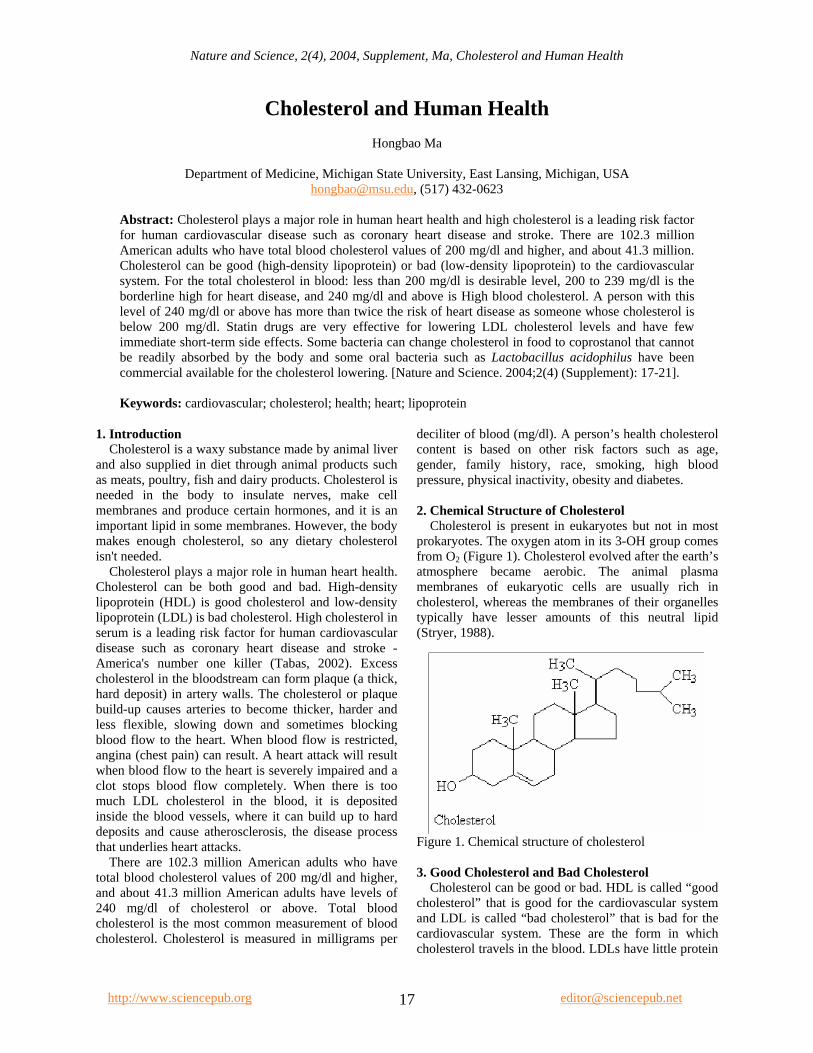
deciliter of blood (mg/dl). A person’s health cholesterol content is based on other risk factors such as age, gender, family history, race, smoking, high blood pressure, physical inactivity, obesity and diabetes. 2. Chemical Structure of Cholesterol Cholesterol is present in eukaryotes but not in most prokaryotes. The oxygen atom in its 3-OH group comes from O2 (Figure 1). Cholesterol evolved after the earth’s atmosphere became aerobic. The animal plasma membranes of eukaryotic cells are usually rich in cholesterol, whereas the membranes of their organelles typically have lesser amounts of this neutral lipid (Stryer, 1988).
Figure 1. Chemical structure of cholesterol 3. Good Cholesterol and Bad Cholesterol Cholesterol can be good or bad. HDL is called “good cholesterol” that is good for the cardiovascular system and LDL is called “bad cholesterol” that is bad for the cardiovascular system. These are the form in which cholesterol travels in the blood. LDLs have little protein
http://www.sciencepub.org [email protected] 17
Nature and Science, 2(4), 2004, Supplement, Ma, Cholesterol and Human Health
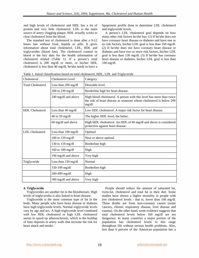
and high levels of cholesterol and HDL has a lot of protein and very little cholesterol. LDL is the main source of artery clogging plaque. HDL actually works to clear cholesterol from the blood. The standard test of cholesterol is done after a 9-12 hours fast without food, liquids or pills. It gives information about total cholesterol, LDL, HDL and triglycerides (blood fats). The cholesterol content in blood is the key data for the health information of cholesterol related (Table 1). If a person’s total cholesterol is 200 mg/dl or more, or his/her HDL cholesterol is less than 40 mg/dl, he/she needs to have a
lipoprotein profile done to determine LDL cholesterol and triglyceride levels. A person’s LDL cholesterol goal depends on how many other risk factors he/she has: (1) If he/she does not have coronary heart disease or diabetes and have one or no risk factors, his/her LDL goal is less than 160 mg/dl. (2) If he/she does not have coronary heart disease or diabetes and have two or more risk factors, his/her LDL goal is less than 130 mg/dl. (3) If he/she has coronary heart disease or diabetes, his/her LDL goal is less than 100 mg/dl.
Table 1. Initial classification based on total cholesterol, HDL, LDL and Triglyceride Cholesterol Cholesterol Level Category
Less than 200 mg/dl Desirable level.
200 to 239 mg/dl Borderline high for heart disease.
Total Cholesterol
240 mg/dl and above High blood cholesterol. A person with this level has more than twice the risk of heart disease as someone whose cholesterol is below 200 mg/dl.
Less than 40 mg/dl Low HDL cholesterol. A major risk factor for heart disease.
40 to 59 mg/dl The higher HDL level, the better.
HDL Cholesterol
60 mg/dl and above High HDL cholesterol. An HDL of 60 mg/dl and above is considered protective against heart disease.
Less than 100 mg/dl Optimal
100 to 129 mg/dl Near or above optimal
130 to 159 mg/dl Borderline high
160 to 189 mg/dl High
LDL Cholesterol
190 mg/dl and above Very high
Triglyceride Less than 150 mg/dl Normal
150-199 mg/dl Borderline high
200-499 mg/dl High
500 mg/dl and above Very high 4. Triglyceride Triglycerides are another fat in the bloodstream. High levels of triglycerides is also linked to heart disease. Triglyceride is the most common type of fat in the body. Many people who have heart disease or diabetes have high triglyceride levels. Normal triglyceride levels vary by age and sex. A high triglyceride level combined with low HDL cholesterol or high LDL cholesterol seems to speed up atherosclerosis, which is the buildup of fatty deposits in artery walls that increase the risk for heart attack and stroke.
People should reduce the amount of saturated fat, trans-fat, cholesterol and total fat in their diet. Some studies have shown a higher mortality in people with low cholesterol levels - that is, lower than 160 mg/dl. These deaths are from non-coronary causes (some cancers, chronic respiratory disease, liver disease and trauma). On the other hand, some evidence suggests that total cholesterol levels below 160 mg/dl are not dangerous. In many countries a major portion of the population has cholesterol levels in this range throughout life without serious health problems. Also, less than 6 percent of the American population has a
http://www.sciencepub.org [email protected] 18
Nature and Science, 2(4), 2004, Supplement, Ma, Cholesterol and Human Health
cholesterol level below 160 mg/dl. It's rarely necessary to lower total cholesterol below that. 5. Hyperlipidemia Hyperlipidemia is an elevation of lipids (fats) in the bloodstream. These lipids include cholesterol, cholesterol esters (compounds), phospholipids and triglycerides. They're transported in the blood as part of large molecules called lipoproteins. These are the five major families of blood (plasma) lipoproteins: (1) chylomicrons, (2) very low-density lipoproteins (VLDL), (3) intermediate-density lipoproteins (IDL), (4) low-density lipoproteins (LDL), (5) high-density lipoproteins (HDL). When hyperlipidemia is defined in terms of class or classes of elevated plasma lipoproteins, the term hyperlipoproteinemia is used. Hypercholesterolemia is the term for high cholesterol levels in the blood. Hypertriglyceridemia refers to high triglyceride levels in the blood. The average American man gets about 337 mg of cholesterol a day from food and the average woman gets about 217 mg. I suggest that a person should limit cholesterol from food to an average of no more than 300 mg per day. 6. Cholesterol from Foods Cholesterol from food is hard to get away from, even though one may be watching his/her diet. All foods of animal origin contain cholesterol, including eggs, red meat, and shrimp. Generally, foods that are high in saturated fats or trans fats should also be limited. These include foods you may not even think of, such as grilled-cheese sandwich, margarine, potato with butter and chicken pot pie, etc. As we eat, cholesterol from food is absorbed by our digestive tract. It then makes its way into our liver and can circulate through our body in the bloodstream. That’s one source. There’s also a little-known second source of cholesterol — human body. 7. Cholesterol Produced by Body Based on Genetics Like many people, one may not know that his/her body produces cholesterol naturally, based on family history genetically - despite the fact that it’s where more of one’s total cholesterol comes from. The liver makes cholesterol, as do other individual cells throughout the body. Once cholesterol is produced, it can make its way into the bloodstream. What does this process mean to people? Take the cholesterol the body makes and add it to the cholesterol one gets from food. Now one can see how easily cholesterol can build up in the bloodstream and how the overall cholesterol level can increase. 8. The Factors Affect Cholesterol Levels A variety of factors can affect your cholesterol levels. They include:
8.1 Diet. Saturated fat, trans fat, and cholesterol in the food you eat increase cholesterol levels. Reducing the amount of saturated fat and trans fats and cholesterol in your diet helps lower your blood cholesterol level. 8.2 Weight. In addition to being a risk factor for heart disease, being overweight can also increase your cholesterol. Losing weight can help lower your LDL, total cholesterol levels, and triglyceride levels, as well as raise your HDL. 8.3 Exercise. Regular exercise can lower LDL cholesterol and raise HDL cholesterol. You should try to be physically active for 30 minutes on most days. 8.4 Age and Gender. As we get older, cholesterol levels rise. Before menopause, women tend to have lower total cholesterol levels than men of the same age. After menopause, however, women's LDL levels tend to rise. 8.5 Heredity. Your genes partly determine how much cholesterol your body makes. High blood cholesterol can run in families. 8.6 Medical conditions. Occasionally a medical condition may cause an elevation of cholesterol levels in the blood. These include hypothyroidism (an underactive thyroid gland), liver disease and kidney disease. 8.7 Medications. Some medicines, like steroids and progestins may increase the "bad" cholesterol and decrease the "good" cholesterol. 9. Medications The main goal in lowering cholesterol is to lower your LDL and raise your HDL. To lower cholesterol, eat a heart-healthy diet, exercise regularly, and maintain a healthy weight. Some may also need to take cholesterol lowering medications. Cholesterol-lowering medicine is most effective when combined with a low-cholesterol diet and exercise program. The drugs of first choice for elevated LDL cholesterol are the HMG CoA reductase inhibitors, e.g. lovastatin, pravastatin and simvastatin. Statin drugs are very effective for lowering LDL cholesterol levels and have few immediate short-term side effects (Jongh, 2002). They are easy to administer, have high patient acceptance and have few drug-drug interactions. Patients, who are pregnant, have active or chronic liver disease, or those allergic to statins shouldn't use statin drugs. The most common side effects of statins are gastrointestinal, including constipation and abdominal pain and cramps. These symptoms are usually mild to severe and generally subside as therapy continues.
http://www.sciencepub.org [email protected] 19
Nature and Science, 2(4), 2004, Supplement, Ma, Cholesterol and Human Health
Another class of drugs for lowering LDL is the bile acid sequestrants - cholestyramine and colestipol - and nicotinic acid. These have been shown to reduce the risk for coronary heart disease in controlled clinical trials. Both classes of drugs appear to be free of serious side effects. But both can have troublesome side effects and require considerable patient education to achieve adherence. Nicotinic acid is preferred in patients with triglyceride levels exceeding 250 mg/dl because bile acid sequestrants tend to raise triglyceride levels. Other available drugs are gemfibrozil, probucol and clofibrate. Gemfibrozil and clofibrate are most effective for lowering elevated triglyceride levels. They moderately reduce LDL cholesterol levels in hypercholesterolemic patients, but the American FDA hasn't approved them for this purpose. Probucol also moderately lowers LDL levels and it has been received FDA approval for this purpose. If a patient doesn't respond adequately to single drug therapy, combined drug therapy should be considered to further lower LDL cholesterol levels. For patients with severe hypercholesterolemia, combining a bile acid sequestrant with either nicotinic acid or lovastatin has the potential to markedly lower LDL cholesterol. For hypercholesterolemic patients with elevated triglycerides, nicotinic acid or gemfibrozil should be considered as one agent for combined therapy. 9.1 Drugs There are a variety of medications available for lowering blood cholesterol levels. They may be prescribed individually or in combination with other drugs. Some of the common types of cholesterol-lowering drugs include (1) Clofibrate, (2) Gemfibrozil, (3) Nicotinic acid (niacin), (4) Resins, (5) Statins, (6) Fibric acid derivatives (7) Fibrates: 9.1.1 Clofibrate (Atromid-S). This drug raises the HDL cholesterol levels and lowers triglyceride levels. 9.1.2 Gemfibrozil (Lopid). This drug lowers blood fats and raises HDL cholesterol levels. 9.1.3 Nicotinic Acid (niacin). Niacin is a B-complex vitamin. It's found in food, but is also available at high doses by prescription. It lowers LDL cholesterol and raises HDL cholesterol. The main side effects are flushing, itching, tingling and headache. Niacin or nicotinic acid, includes the brand names Niacor, Niaspan, or Slo-niacin. Over-the-counter preparations include extended-release, timed-release, and controlled-release. Niacin found in dietary supplements should not be used to lower cholesterol. The doctor or lipid specialist will let patients know what type of niacin is best for them. This drug works in the liver by affecting the production of blood fats. It's used to lower
triglycerides, lower LDL cholesterol and raise HDL cholesterol. 9.1.4 Resins. Resins are also called bile acid-binding drugs. They work in the intestines by promoting increased disposal of cholesterol. There are three kinds of medications in this class: (1) Cholestryamine (Questran, Prevalite, Lo-Cholest), (2) Colestipol (Colestid), (3) Coleseveiam (WelChol). 9.1.5 Statins. Statins block the production of cholesterol in the liver itself. They lower LDL, the "bad" cholesterol, and triglycerides and have a mild effect in raising HDL, the "good" cholesterol. Statin drugs are very effective for lowering LDL cholesterol levels and have few immediate short-term side effects. They work by interrupting the formation of cholesterol from the circulating blood. These drugs are the first line of treatment for most people with high cholesterol. Side effects can include intestinal problems, liver damage, and in a few people, muscle tenderness or weakness. Examples of statins include: (1) Altocor, (2) Baycol (cerivastatin), (3) Crestor, (4) Lipitor (atorvastatin), (5) Lescol (Fluvastatin), (6) Mevacor (lovastatin), (7) Pravachol (pravastatin), (8) Zocor (simvastatin). Advicor is a combination of a statin and niacin. Caduet is a new drug that is a combination of a statin (Lipitor or atorvastatin) and a blood pressure-lowering drug called amlodipine (Norvasc). Commonly prescribed statins include: (1) Atorvastatin (Lipitor), (2) Cerivastatin (Baycol), (3) Fluvastatin (Lescol), (4) Lovastatin (Mevacor), (5) Pravastatin (Pravachol), (6) Simvastatin (Zocor). 9.1.6 Bile Acid Sequestrants. These drugs work inside the intestine, where they bind to bile and prevent it from being reabsorbed into the circulatory system. Bile is made largely from cholesterol, so these drugs work by reducing the body's supply of cholesterol,, thus lowering total and LDL cholesterol. The most common side effects are constipation, gas, and upset stomach. Examples of bile acid resins include: questran and questran light (cholestyramine), colestid (colestipol), WelChol (colesevelam). 9.1.7 Fibrates. Fibrates lower triglyceride levels and can increase HDL and lower LDL cholesterol. The mechanism of action is not clear but it is thought that fibrates enhance the breakdown of triglyceride-rich particles and decreases the secretion of certain lipoproteins. In addition, they induce the synthesis of HDL. Examples of fibrates include: tricor (fenofibrate), lopid (gemfibrozil), lofibra (fenofibrate). 9.1.8 Side Effects of Cholesterol-Lowering Drugs
http://www.sciencepub.org [email protected] 20
Nature and Science, 2(4), 2004, Supplement, Ma, Cholesterol and Human Health
http://www.sciencepub.org [email protected] 21
The side effects of cholesterol-lowering drugs include: (1) Muscle aches, (2) Abnormal liver function, (3) Allergic reaction (skin rashes), (4) Heartburn, (5) Dizziness. (6) Abdominal pain, (7) Constipation, (8) Decreased sexual desire. One newer drug is Zetia. This drug works by inhibiting the absorption of cholesterol from the blood. Side effects include back pain, joint pain, diarrhea and abdominal pain. As of August 8, 2001, Bayer Pharmaceutical Division voluntarily withdrew the drug Baycol (cerivastatin) from the U.S. market because of reports of fatal muscle damage caused by the drug. 9.2 Bacteria on Cholesterol Some bacteria can change cholesterol in food to coprostanol that cannot be readily absorbed by the body. These kinds of bacteria are called friendly bacteria. This helps recycle cholesterol to make hormones. Bifidobacterium and Lactobacillus acidophilus may play an important role in cholesterol metabolism of their host. Intestinal bacteria convert cholesterol into a less absorbable form coprostanol thus hampering its absorption from the intestinal tract (Lin, 2000). Lactic acid bacteria in intestine have the cholesterol lowering effect (Pereira, 2002). Some oral bacteria such as Lactobacillus acidophilus have been commercial available for the cholesterol lowering. Feeding friendly bacteria can do: (1) reduce the growth of unfriendly bacteria, (2) maintain regular bowel movements, (3)
maintain cholesterol and triglyceride levels, (4) maintain healthy blood sugar levels (Ma, 2004). Correspondence to: Hongbao Ma 138 Service Road Department of Medicine Michigan State University East Lansing, Michigan 48824 The United States Telephone: (517) 432-0623 Email: [email protected] References 1. De Jongh S, Ose L, Szamosi T, Gagne C, Lambert M, Scott R,
Perron P, Dobbelaere D, Saborio M, Tuohy MB, Stepanavage M, Sapre A, Gumbiner B, Mercuri M, Van Trotsenburg AS, Bakker HD, Kastelein JJ. Efficacy and Safety of Statin Therapy in Children With Familial Hypercholesterolemia: A Randomized, Double-Blind, Placebo-Controlled Trial With Simvastatin. Circulation 2002;106:2231-7.
2. Lin M, Chen T. Reduction of cholesterol by Lactobacillus Acidophilus in culture broth. Journal of Food and Drug Analysis. 2000;8:97-102.
3. Ma H. Concept and protocol to isolate cholesterol-reducing bacteria from carnivores. Nature and Science 2004;2(4):11-7.
4. Pereira DI, Gibson GR. Cholesterol assimilation by lactic acid bacteria and bifidobacteria isolated from the human gut. Appl Environ Microbiol 2002;68:4689-93.
5. Stryer L. Biochemistry. W. H. Freeman and Company. New York. 1988:284-7.
6. Tabas I. Cholesterol in health and disease. J Clin Invest 2002;110:583-90.
Nature and Science, 2(4), 2004, Supplement, Teng, The Characteristic Frequencies in Cell Induced Magnetic
The Characteristic Frequencies in Cell Induced Magnetic Fluctuation
1Hsien-Chiao Teng, 1Chi-Cheng Lie, 2Shen Cherng
1Department of Electrical Engineering, Chinese Military Academy, Fengsang, Taiwan 830, ROC
2Department of Electrical Engineering, Cheng Shiu University, Niau Sung, Taiwan 833, ROC [email protected]; [email protected]
Abstract: To exam the cellular responding frequencies buried in the cell-induced magnetic fluctuations (CIMF), we exposed the mouse osteoblast cell line system to the 50 Hz extremely low frequency (ELF) magnetic field and the rat liver epithelial cell line system to the 7Hz ELF magnetic field. CIMF can couple with geomagnetic fluctuation to create cell-induced geomagnetic fluctuation (CIGF). It was computed their power density spectrum of CIGF and revealed the characteristic frequencies in extremely low signal-to-noise ratio (SNR) circumstances. The gap junction intracellular communication (GJIC) within both the mouse osteoblast cells and rat liver epithelial cells under the exposure of different intrinsic ELF magnetic fields were 20% in difference to the contrary. Furthermore, it was found the 7Hz cellular responding frequency in CIGF may cause 20% GJIC in difference to the contrary if we exposed the rat liver epithelial cells into 2.4GHz electromagnetic field. In this article, we concluded the mouse osteoblast cell line system responded 50 Hz at intrinsic frequency in CIGF to the reaction of external 50 Hz ELF magnetic field but the rat liver epithelial cell line system responded 7Hz at intrinsic frequency in CIGF to the reaction of external 7Hz ELF magnetic field exposure. In contrast, the rat liver epithelial cells responded 7 Hz at intrinsic frequency in CIGF to the reaction of external 2.4GHz electromagnetic field exposure, which is the application of our novel antenna design suitable for the WLAN and IEEE 802.11 a/b protocol. The wireless communication design is requested application in 2.4GHz (2.4-2.484 GHz) and 5.2 GHz (5.15-5.35 GHz) bands to fit the Bluetooth IEEE 802.11/ a/b protocol, which may cause the cellular response in our study. [Nature and Science. 2004;2(4) (Supplement): 22-27]. Key words: extremely low frequency (ELF); gap junctional intracellular communication (GJIC)
INTRODUCTION
There are two major studies of the biological systems responded to the reaction of the electromagnetic field in the past twenty years. One is the cellular response and the other is genetic response. Since the energy provided by the electromagnetic field even is less than thermal energy [Adair,1991], scientists presume that the genetic response of the biological system to the reaction of electromagnetic field may indirectly associate with the genetic mutant and recombination [Aldinucci et al., 1998]. However, cellular response to the reaction of electromagnetic field may be reliant to the correlation between multi-enzyme complex and the pathways of cell-metabolism procedures. In physics point of view, surface electrical current of the cell monolayer in a culture dish can initiate cell-magnetic fluctuation. By using power density spectrum of the couple of geo-magnetic and cell-magnetic fluctuation for cell system, the signal to noise ratio (SNR) of characteristic frequencies can be computed [Teng, 2003]. The external extremely low frequency (ELF) magnetic field may perturb the surface current distribution and cause the cellular response. Gap junctional intracellular
communication (GJIC) is an indicator to express the cell’s response affiliated with many pathological endpoints [Upham et al., 1998; Trosko et al. 1990; 2001], such as cell aging, apoptosis, proliferation and differentiation, are associated with the modulation of GJIC. To measure GJIC modulation, scrape loading dye transfer of Lucifer yellow [Upham et al., 1998] was used. The degree of modulation of GJIC can then show the cellular response to the ELF reaction. MATERIAL AND METHOD
For the diameter of the culture dish used was 3.5 cm, we used a simple 5-cm radius helical coil, which is wrapped with 200 turns 0.45-mm diameter cooper string around a plastic cylinder tube connected to the function generator for input ELF signals. The whole system was placed in an incubator. The incubator controlled the environment at 5% CO2 at 98% relative humidity. Another sham field chamber was exactly same as the ELF incubator only with no exposure to the ELF. The cell culture dishes were placed perpendicular to the input ELF. The function generator generated the ELF signal through the solenoid applied to the mouse osteoblast
http://www.sciencepub.org [email protected] 22
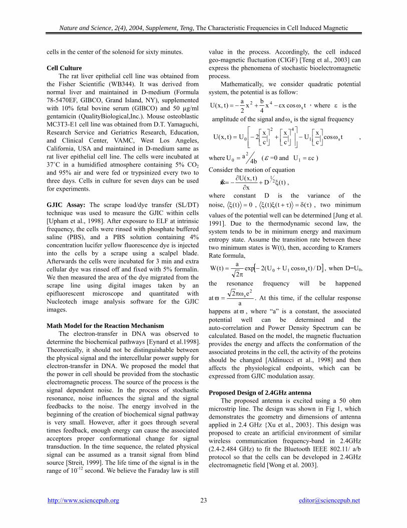
Nature and Science, 2(4), 2004, Supplement, Teng, The Characteristic Frequencies in Cell Induced Magnetic
cells in the center of the solenoid for sixty minutes. Cell Culture
The rat liver epithelial cell line was obtained from the Fisher Scientific (WB344). It was derived from normal liver and maintained in D-medium (Formula 78-5470EF, GIBCO, Grand Island, NY), supplemented with 10% fetal bovine serum (GIBCO) and 50 µg/ml gentamicin (QualityBiological,Inc.). Mouse osteoblastic MC3T3-E1 cell line was obtained from D.T. Yamaguchi, Research Service and Geriatrics Research, Education, and Clinical Center, VAMC, West Los Angeles, California, USA and maintained in D-medium same as rat liver epithelial cell line. The cells were incubated at 37˚C in a humidified atmosphere containing 5% CO2 and 95% air and were fed or trypsinized every two to three days. Cells in culture for seven days can be used for experiments. GJIC Assay: The scrape load/dye transfer (SL/DT) technique was used to measure the GJIC within cells [Upham et al., 1998]. After exposure to ELF at intrinsic frequency, the cells were rinsed with phosphate buffered saline (PBS), and a PBS solution containing 4% concentration lucifer yellow fluorescence dye is injected into the cells by a scrape using a scalpel blade. Afterwards the cells were incubated for 3 min and extra cellular dye was rinsed off and fixed with 5% formalin. We then measured the area of the dye migrated from the scrape line using digital images taken by an epifluorescent microscope and quantitated with Nucleotech image analysis software for the GJIC images. Math Model for the Reaction Mechanism
The electron-transfer in DNA was observed to determine the biochemical pathways [Eynard et al.1998]. Theoretically, it should not be distinguishable between the physical signal and the intercellular power supply for electron-transfer in DNA. We proposed the model that the power in cell should be provided from the stochastic electromagnetic process. The source of the process is the signal dependent noise. In the process of stochastic resonance, noise influences the signal and the signal feedbacks to the noise. The energy involved in the beginning of the creation of biochemical signal pathway is very small. However, after it goes through several times feedback, enough energy can cause the associated acceptors proper conformational change for signal transduction. In the time sequence, the related physical signal can be assumed as a transit signal from blind source [Streit, 1999]. The life time of the signal is in the range of 10-12 second. We believe the Faraday law is still
value in the process. Accordingly, the cell induced geo-magnetic fluctuation (CIGF) [Teng et al., 2003] can express the phenomena of stochastic bioelectromagnetic process.
Mathematically, we consider quadratic potential system, the potential is as follow:
tcosxx4bx
2a)t,x(U s
42 ωε−+−= ,where ε is the
amplitude of the signal and is the signal frequency sω
tcoscxU
cx
cx2U)t,x(U s1
42
0 ω⎥⎦⎤
⎢⎣⎡−
⎥⎥⎦
⎤
⎢⎢⎣
⎡⎥⎦⎤
⎢⎣⎡+⎥⎦
⎤⎢⎣⎡−= ,
where b4aU
20 = (ε =0 and ) cU1 ε=
Consider the motion of equation
)t(Dx
)t,x(Ux 21ξ+
∂∂
−=& ,
where constant D is the variance of the noise, 0)t( =ξ , )()t()t( τδ=τ+ξξ , two minimum values of the potential well can be determined [Jung et al. 1991]. Due to the thermodynamic second law, the system tends to be in minimum energy and maximum entropy state. Assume the transition rate between these two minimum states is W(t), then, according to Kramers Rate formula,
[ ]D/)tcosUU(2exp2a)t(W s10 ω+−π
= , when D=U0,
the resonance frequency will be happened
ata
e2 2sπω
=ϖ . At this time, if the cellular response
happens atϖ , where “a” is a constant, the associated potential well can be determined and the auto-correlation and Power Density Spectrum can be calculated. Based on the model, the magnetic fluctuation provides the energy and affects the conformation of the associated proteins in the cell, the activity of the proteins should be changed [Aldinucci et al., 1998] and then affects the physiological endpoints, which can be expressed from GJIC modulation assay. Proposed Design of 2.4GHz antenna
The proposed antenna is excited using a 50 ohm microstrip line. The design was shown in Fig 1, which demonstrates the geometry and dimensions of antenna applied in 2.4 GHz {Xu et al., 2003}. This design was proposed to create an artificial environment of similar wireless communication frequency-band in 2.4GHz (2.4-2.484 GHz) to fit the Bluetooth IEEE 802.11/ a/b protocol so that the cells can be developed in 2.4GHz electromagnetic field [Wong et al. 2003].
http://www.sciencepub.org [email protected] 23
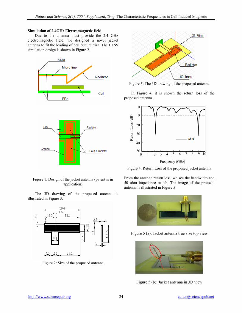
Nature and Science, 2(4), 2004, Supplement, Teng, The Characteristic Frequencies in Cell Induced Magnetic
Simulation of 2.4GHz Electromagnetic field Due to the antenna must provide the 2.4 GHz
electromagnetic field; we designed a novel jacket antenna to fit the loading of cell culture dish. The HFSS simulation design is shown in Figure 2.
Figure 1: Design of the jacket antenna (patent is in application)
The 3D drawing of the proposed antenna is
illustrated in Figure 3.
Figure 2: Size of the proposed antenna
Figure 3: The 3D drawing of the proposed antenna
In Figure 4, it is shown the return loss of the
proposed antenna.
Figure 4: Return Loss of the proposed jacket antenna
From the antenna return loss, we see the bandwidth and 50 ohm impedance match. The image of the protocol antenna is illustrated in Figure 5
Figure 5 (a): Jacket antenna true size top view
Figure 5 (b): Jacket antenna in 3D view
http://www.sciencepub.org [email protected] 24
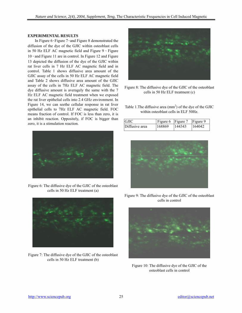
Nature and Science, 2(4), 2004, Supplement, Teng, The Characteristic Frequencies in Cell Induced Magnetic
EXPERIMENTAL RESULTS
In Figure 6、Figure 7、and Figure 8 demonstrated the diffusion of the dye of the GJIC within osteoblast cells in 50 Hz ELF AC magnetic field and Figure 9、Figure 10、and Figure 11 are in control. In Figure 12 and Figure 13 depicted the diffusion of the dye of the GJIC within rat liver cells in 7 Hz ELF AC magnetic field and in control. Table 1 shows diffusive area amount of the GJIC assay of the cells in 50 Hz ELF AC magnetic field and Table 2 shows diffusive area amount of the GJIC assay of the cells in 7Hz ELF AC magnetic field. The dye diffusive amount is averagely the same with the 7 Hz ELF AC magnetic field treatment when we exposed the rat liver epithelial cells into 2.4 GHz environment. In Figure 14, we can seethe cellular response in rat liver epithelial cells to 7Hz ELF AC magnetic field. FOC means fraction of control. If FOC is less than zero, it is an inhibit reaction. Oppositely, if FOC is bigger than zero, it is a stimulation reaction.
Figure 6: The diffusive dye of the GJIC of the osteoblast cells in 50 Hz ELF treatment (a)
Figure 7: The diffusive dye of the GJIC of the osteoblast cells in 50 Hz ELF treatment (b)
Figure 8: The diffusive dye of the GJIC of the osteoblast cells in 50 Hz ELF treatment (c)
Table 1.The diffusive area (mm2) of the dye of the GJIC within osteoblast cells in ELF 50Hz.
GJIC Figure 6 Figure 7 Figure 9 Diffusive area 168869 144343 164042
Figure 9: The diffusive dye of the GJIC of the osteoblast cells in control
Figure 10: The diffusive dye of the GJIC of the osteoblast cells in control
http://www.sciencepub.org [email protected] 25
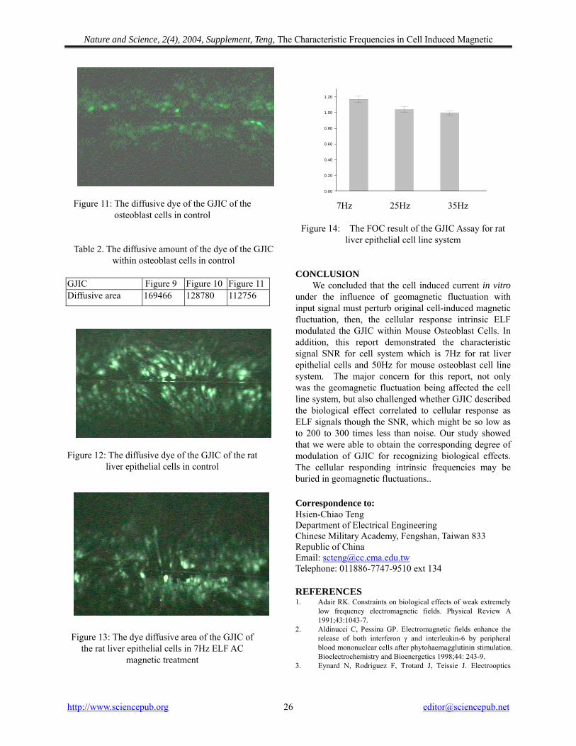
Nature and Science, 2(4), 2004, Supplement, Teng, The Characteristic Frequencies in Cell Induced Magnetic
Figure 11: The diffusive dye of the GJIC of the osteoblast cells in control
Table 2. The diffusive amount of the dye of the GJIC within osteoblast cells in control
GJIC Figure 9 Figure 10 Figure 11 Diffusive area 169466 128780 112756
Figure 12: The diffusive dye of the GJIC of the rat liver epithelial cells in control
Figure 13: The dye diffusive area of the GJIC of the rat liver epithelial cells in 7Hz ELF AC
magnetic treatment
0.00
0.20
0.40
0.60
0.80
1.00
1.20
7Hz 25Hz 35Hz
Figure 14: The FOC result of the GJIC Assay for rat
liver epithelial cell line system
CONCLUSION
We concluded that the cell induced current in vitro under the influence of geomagnetic fluctuation with input signal must perturb original cell-induced magnetic fluctuation, then, the cellular response intrinsic ELF modulated the GJIC within Mouse Osteoblast Cells. In addition, this report demonstrated the characteristic signal SNR for cell system which is 7Hz for rat liver epithelial cells and 50Hz for mouse osteoblast cell line system. The major concern for this report, not only was the geomagnetic fluctuation being affected the cell line system, but also challenged whether GJIC described the biological effect correlated to cellular response as ELF signals though the SNR, which might be so low as to 200 to 300 times less than noise. Our study showed that we were able to obtain the corresponding degree of modulation of GJIC for recognizing biological effects. The cellular responding intrinsic frequencies may be buried in geomagnetic fluctuations.. Correspondence to: Hsien-Chiao Teng Department of Electrical Engineering Chinese Military Academy, Fengshan, Taiwan 833 Republic of China Email: [email protected] Telephone: 011886-7747-9510 ext 134 REFERENCES 1. Adair RK. Constraints on biological effects of weak extremely
low frequency electromagnetic fields. Physical Review A 1991;43:1043-7.
2. Aldinucci C, Pessina GP. Electromagnetic fields enhance the release of both interferon γ and interleukin-6 by peripheral blood mononuclear cells after phytohaemagglutinin stimulation. Bioelectrochemistry and Bioenergetics 1998;44: 243-9.
3. Eynard N, Rodriguez F, Trotard J, Teissie J. Electrooptics
http://www.sciencepub.org [email protected] 26
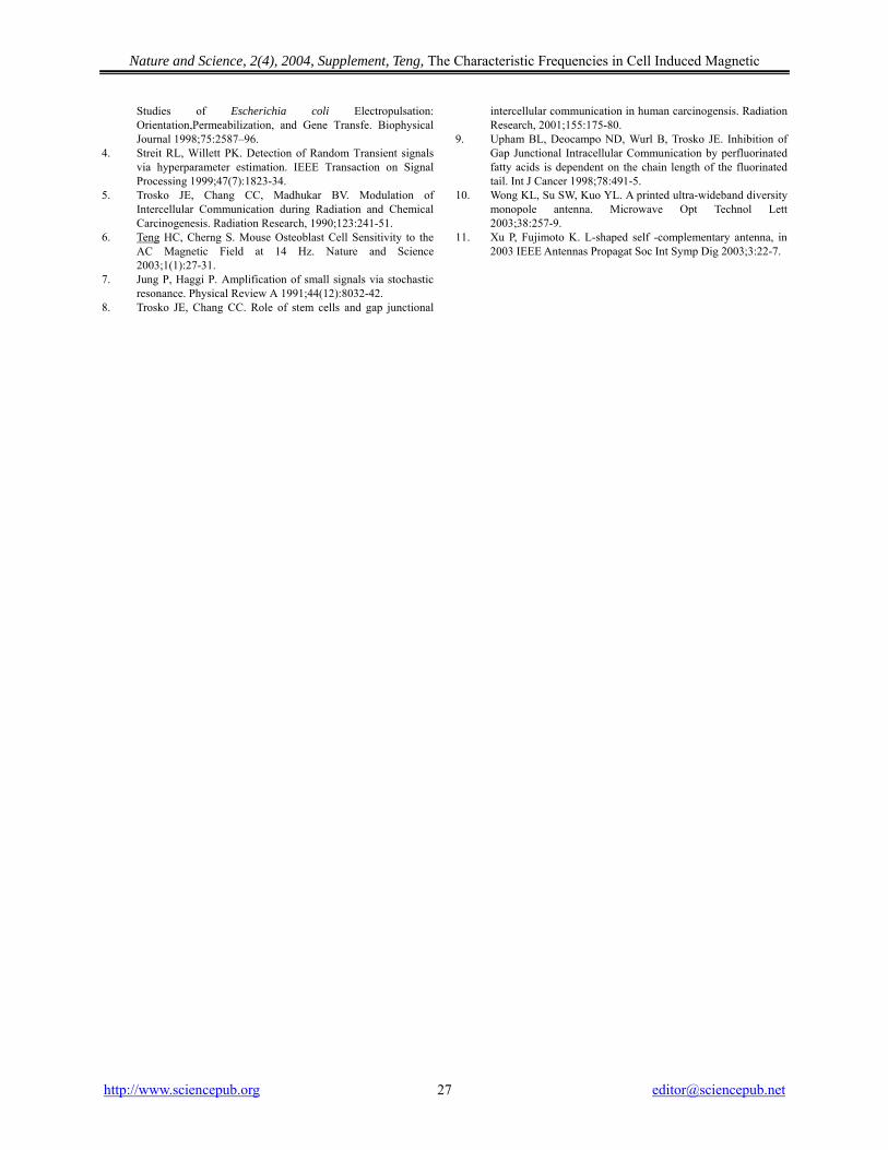
Nature and Science, 2(4), 2004, Supplement, Teng, The Characteristic Frequencies in Cell Induced Magnetic
http://www.sciencepub.org [email protected] 27
Studies of Escherichia coli Electropulsation: Orientation,Permeabilization, and Gene Transfe. Biophysical Journal 1998;75:2587–96.
4. Streit RL, Willett PK. Detection of Random Transient signals via hyperparameter estimation. IEEE Transaction on Signal Processing 1999;47(7):1823-34.
5. Trosko JE, Chang CC, Madhukar BV. Modulation of Intercellular Communication during Radiation and Chemical Carcinogenesis. Radiation Research, 1990;123:241-51.
6. Teng HC, Cherng S. Mouse Osteoblast Cell Sensitivity to the AC Magnetic Field at 14 Hz. Nature and Science 2003;1(1):27-31.
7. Jung P, Haggi P. Amplification of small signals via stochastic resonance. Physical Review A 1991;44(12):8032-42.
8. Trosko JE, Chang CC. Role of stem cells and gap junctional
intercellular communication in human carcinogensis. Radiation Research, 2001;155:175-80.
9. Upham BL, Deocampo ND, Wurl B, Trosko JE. Inhibition of Gap Junctional Intracellular Communication by perfluorinated fatty acids is dependent on the chain length of the fluorinated tail. Int J Cancer 1998;78:491-5.
10. Wong KL, Su SW, Kuo YL. A printed ultra-wideband diversity monopole antenna. Microwave Opt Technol Lett 2003;38:257-9.
11. Xu P, Fujimoto K. L-shaped self -complementary antenna, in 2003 IEEE Antennas Propagat Soc Int Symp Dig 2003;3:22-7.

Nature and Science, 2(4), 2004, Supplement ISSN 1545-0740
Nature and Science
Call for Papers
The new academic journal “Nature and Science” (ISSN: 1545-0740) is inviting you to publish your papers. A formal academic international journal, Nature and Science, is registered in the United States. For more information about the journal, please visit http://www.sciencepub.org. Please send your manuscript to [email protected].
The journal will be supported by manuscript contributors. To cover the printing cost, this journal will only charge authors the actual printing fee (about US$30 per printed page). At least one (1) hard copy of a printed journal will be given to each author free of charge. Here is a new avenue to publish your outstanding reports and reviews. Please also help spread this to your colleagues and friends and invite them to contribute papers to the journal. Let's work together to disseminate our research results and our opinions.
Papers in all the fields are welcome, including papers of natural science and social science.
Marsland Company P.O. Box 21126
East Lansing, Michigan 48909 The United States
Telephone:(517) 980-4106 E-mail: [email protected]
Homepage: http://www.sciencepub.org
Marsland Company, P.O. Box 21126, Lansing, Michigan 48909, The United States, (517) 980-4106
http://www.sciencepub.org [email protected]

![Researcher 2013;5(12) - Marsland Press · stored by hospital management systems [4]. • Manufacturing and Production: Process optimization ... applied statistics, machine learning](https://img.pdfslide.us/doc/110x75/5f112d3f9daf976bc6180123/researcher-2013512-marsland-stored-by-hospital-management-systems-4-a.jpg)


![Marsland Consulting Services [Honest] [Genuine] [Professional]](https://img.pdfslide.us/doc/110x75/55d177c3bb61eb0c788b4760/marsland-consulting-services-honest-genuine-professional.jpg)