Embed Size (px)
Citation preview

Supplemantal Figures
Figure S1. Representative image of Id4-Gfp expressing cells within seminiferous tubules from testes of adult mice. Arrows indicate cohorts of two Id4-Gfp+ cells and arrow heads indicate single Id4-Gfp+ cells. Bar is 50µm.

Figure S2. Representative images of co-immunofluorescent staining for the differentiating spermatogonial markers Stra8 and Kit (red) and Id4-Gfp (green) in cross-sections of testes from adult LT-11B6 mice. Stars indicate cells stained for Stra8 or Kit expression and arrows indicate cells stained for expression of Id4-Gfp.

Figure S3. Representative images depicting tubular/blood vessel, tubular/tubular, and tubular/interstitial localization of Id4-Gfp expressing cells within cross-sections of testes from adult LT-11B6 mice. Arrows indicate spermatogonia immunostained for expression of Id4-Gfp. Interstitial tissue, seminiferous tubules, and blood vessels are indicated by I, T, or BV, respectively.

Figure S4. Representative images of cross-sections from testes of W/Wv recipient mice three months after transplantation with Id4-Gfp+ spermatogonia isolated from testes of adult LT-11B6 donor mice using FACS. Stars indicate seminiferous tubules containing donor-derived spermatogenesis. Arrowheads indicate seminiferous tubules completely devoid of germ cells that is typical of W/Wv mice.
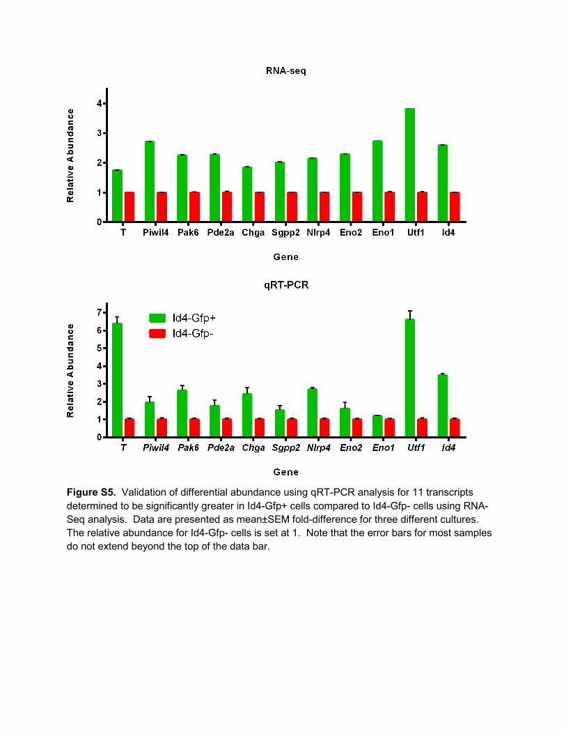
Figure S5. Validation of differential abundance using qRT-PCR analysis for 11 transcripts determined to be significantly greater in Id4-Gfp+ cells compared to Id4-Gfp- cells using RNA-Seq analysis. Data are presented as mean±SEM fold-difference for three different cultures. The relative abundance for Id4-Gfp- cells is set at 1. Note that the error bars for most samples do not extend beyond the top of the data bar.
SUPPLEMENTAL EXPERIMENTAL PROCEDURES Animals All animal procedures were approved by the Washington State University Institutional Animal
Care and Use Committee (IACUC). Id4-Gfp transgenic founder mice were produced on a FVB
background and then backcrossed onto a C57BL6/J background. Mice with ubiquitous
expression of a LacZ transgene from the Rosa26 locus (B6;129S-Gt (ROSA)26Sor/J) were
obtained from Jackson Laboratories. Recipients for spermatogonial transplantation were
C57BL/6J × 129S1/svlmJ F1 hybrids or W/Wv mutant mice.
Fluorescence activated cell sorting (FACS) Single cell suspensions generated from testes of adult LT-11B6 mice or primary cultures of
spermatogonia established from LT-11B6/RosaLacZ mice were subjected to sorting based on
Gfp intensity using a BD FACSAria instrument. Both Gfp+ and Gfp- populations were collected
followed by washing in mSFM using centrifugation at 600xg for 6 min. Cells were then
suspended in mSFM for transplantation into testes of recipient mice or processed for RNA
isolation in order to conduct RNA-Seq analysis.
Immunostaining of Testis Cross-Sections
Testes from LT-11B6 mice were fixed in Bouin’s solution for 2 hr (PD 0-6 donors) or overnight
(PD 10-35 donors) at 40C followed by dehydration in a graded series of ethanol washes and
embedded in paraffin. Cross-sections of 5-µm thickness were adhered to glass slides followed
by de-paraffinization and rehydration. Antigen retrieval was achieved by incubation in boiling
sodium citrate buffer (10mM citric acid, 0.05% Tween-20, pH 6.0) for 20 min followed by rinsing
in PBS. Non-specific antibody binding was blocked by incubation in PBS containing 0.5%
bovine serum albumin, 0.1% Triton X-100 and 10% normal serum from host species of the
secondary antibody for 1 hr at room temperature. Sections were then incubated with primary
antibodies diluted in binding buffer (PBS containing 0.5% BSA and 0.1% Triton X-100) for 2 hr
at room temperature followed by extensive washing in PBS and incubation with secondary
antibodies for 2 hr at room temperature. For colorimetric staining, sections were again washed
in PBS and developed with a horseradish peroxidase (HRP)-conjugated streptavidin kit (Vector
Labs) followed by hematoxylin counterstaining. For immunofluorescent staining, sections were
washed in PBS and coverslips mounted with Pro Long Gold antifade reagent containing DAPI
(Life Technologies, OR). Primary and secondary antibodies utilized in this study and working
dilutions are listed in Table S1.

Table S1. Antibodies used for immunostaining analyses.
Antibody Source Working Dilution Goat anti-mouse GFR alpha1 R&D Systems (AF560) 1:400 FITC conjugated goat anti-GFP Abcam (ab6662) 1:300 Rabbit anti-GFP Abcam (ab290) 1:400 Rabbit anti-mouse PLZF Santa Cruz (H-300) 1:50 Alexa Fluor 555 Donkey Anti-Goat IgG Life Technologies (A21432) 1:1000 Alexa Fluor 546 Donkey Anti-Rabbit IgG Life Technologies (A10040) 1:1000
Immunofluorescent Staining of Whole Mount Seminiferous Tubules
Testes from LT-11B6 mice were collected in ice cold PBS and the tunica albuginea removed
followed by gently teasing apart of seminiferous tubules with fine forceps. Samples were then
fixed in 4% PFA at 4oC for 2 hr, briefly dried at room temperature for 5 min, and washed in PBS
containing 0.2% NP40 for 20 min at 4oC. Tubules were then dehydrated in a graded series of
methanol washes for 5 min each at 4oC. For immunostaining, tubules were rehydrated in PBST
for 5min at 4oC and non-specific antibody binding was blocked by incubating in blocking buffer
(PBST containing 1% BSA and 10% normal serum of the host species for the secondary
antibody) for 1 hr at 4oC. Tubules were then incubated with primary antibodies diluted in
blocking buffer overnight at 4oC followed by washing extensive washing in PBST and incubated
with AlexaFluor-conjugated secondary antibodies diluted in blocking buffer overnight at 4oC.
Samples were washed again in PBST at 4oC before spreading on glass microscope slides and
mounting of coverslips with ProLong Gold antifade reagent containing DAPI. Primary and
secondary antibodies utilized in this study and working dilutions are listed in Table S1.
Microscopic Imaging Fluorescent microscopy and digital images were captured using an Olympus IX51 inverted
microscope and DP72 digital color microscope camera (Olympus Inc., USA). All images were
captured using CellSense acquisition software (Olympus Inc., USA). For quantification of cell
number within cross-sections of testes, at least 50 seminiferous tubules in at least 5 different
cross-sections were scored for various types of immunostained cells.
Primary Cultures of Undifferentiated Spermatogonia
Cultures of undifferentiated spermatogonia were established from LT11-B6 or LT-
11B/RosaLacZ donor mice at PD 6-8 as described previously (Kubota et al., 2004; Oatley and
Brinster, 2006). Briefly, the Thy1+ testis cell fraction was isolated using magnetic activated cell
sorting (MACS) and co-cultured with mitotically inactivated STO feeders cell monolayers.
Cultures were maintained in mSFM supplemented with 20 ng/ml recombinant human GDNF

(PeproTech, NJ) and 1 ng/ml recombinant human FGF2 (BD Biosciences) at 37oC in an
atmosphere of 5% CO2. At 7 day intervals, spermatogonial clumps were disassociated into
single cell suspensions and sub-cultured at a ratio of 1:2 or 1:3 onto fresh STO feeders. All
experiments utilizing primary cultures were conducted 1-3 months after initial establishment.
Quantitative RT-PCR Analysis Total cellular RNA from FACS isolated Id4-Gfp+ and Id4-Gfp- spermatogonial populations of
primary cultures was isolated using Trizol Reagent (Invitrogen, USA). Samples were subjected
to DNaseI treatment and reverse transcription was carried out using Superscript III reverse
transcriptase and oligo (d)T priming to generate cDNA. Both Taqman and Sybr green assays
were conducted using validated primers and probes for Id4, Utf1, Eno1, Eno2, Piwil4, Pde2a,
Pak6, Nlrp4, Sgpp2, Chga, T, and Rps2 (see Supplemental Table S2). All reactions were
performed with an ABI ABI 7500 Fast Sequence Detection system (Applied Biosystems).
Abundance of transcript for the constitutively expressed gene Rps2 was used for normalization
and to generate relative expression values for genes-of-interest in each sample using the 2-∆∆CT
formula as described previously (Oatley et al., 2011; Yang et al., 2013).
Table S2. Primers used for Sybr green based qRT-PCR analyses.
RNA-Seq Analyses
Total cellular RNA from FACS isolated Id4-Gfp+ and Id4-Gfp- spermatogonial populations of 3
different primary cultures was isolated using a combination of Trizol reagent (Invitrogen, USA)
Gene Amplicon Size
Forward Primer Reverse Primer
Utf1 135 TGTCCCGGTGACTACGTCT CCCAGAAGTAGCTCCGTCTCT Eno2 98 AGGTGGATCTCTATACTGCCAAA GTCCCCATCCCTTAGTTCCAG Piwil4
101 GGGAAAAGTTGACCCACGTAA CCAATCCTGGGGCAAATCTAAAT
Pde2a
128 TGGCGTTGTGGACGATGAG CGCGATAGAAAAGCGGATGG
Pak6
116 GTGGGGTGAAGGTTGCCAA TTCCTGGTTCTTAGGGCTAGG
Nlrp4 130 CAGCAAGTCGTTGAAGGTTCT ACTGAGGGAACAGTTGGACAG Sgpp2
135 TTCACCCACTGGAATATCGACC AAGTCTCACAACGGGAGGAAA
Chga
129 CCAAGGTGATGAAGTGCGTC GGTGTCGCAGGATAGAGAGGA
T 117 GCTTCAAGGAGCTAACTAACGAG CCAGCAAGAAAGAGTACATGGC Eno1
118 TGCGTCCACTGGCATCTAC CAGAGCAGGCGCAATAGTTTTA

and RNeasy columns (Qiagen, USA) as described previously (Oatley et al., 2006; Oatley et al.,
2009). Libraries of cDNA were then generated using oligo d(T) priming to represent the mRNA
transcriptome. The samples were then sent to Otogenetics Inc. for Illumina HiSeq2000 analysis
which generated ~24-30 million pair-end reads of 100 bp in length for each sample.
Sequencing reads were mapped against the mouse genome (mm9 build) using the Tophat
v2.0.5 program. Confidently mapped reads for each sample were analyzed by the
Cufflinks.Cuffdiff program for transcript assembly and generation of a Fragments Per Kilobase
per Million mapped (FPKM) value for each transcript which is directly proportional to
abundance. Significant differences in FPKM values for individual transcripts between Id4-Gfp+
and Id4-Gfp- populations was determined statistically using the Cuffdiff program. A q-value of
<0.05 was considered significantly different.
Spermatogonial Transplantation
To examine regenerative capacity of Id4-Gfp+ and Id4-Gfp- spermatogonial populations, cells
were transplanted into the seminiferous tubules of immunologically compatible recipient mice
utilizing methodology described previously (Oatley and Brinster, 2006). Briefly, single cell
suspensions were diluted in mSFM to a concentration of 1X10⁶ cells/ml and ~10 µl was
microinjected into the seminiferous tubules of adult recipient mice. Depending on the
experiment, W/Wv mutant recipient mice that lack endogenous germ cells or 129XC57 F1
hybrid mice pretreated with busulfan (60 mg/kg of body weight) to eliminate the germline were
used as recipients. For comparison of Id4-Gfp+ and Id4-Gfp- populations, one testis of each
recipient received Id4-Gfp+ cells and the contralateral testis received Id4-Gfp- cells. Testes of
recipient mice were then examined ~2-3 mo post-transplantation for colonies of donor-derived
spermatogenesis. For testes of W/Wv recipients that were transplanted with cells from LT-11B6
mice, donor-derived spermatogenesis was determined in cross-sections by hematoxylin and
eosin staining. For testes of 129XC57 mice that were transplanted with cells from LT-
11B6/RosaLacZ cells, donor-derived spermatogenesis was determined by staining with X-Gal.
A stereo zoom dissecting microscope was used to quantify LacZ stained donor colonies within
each recipient testis.
Statistical Analyses All quantitative data are presented as mean±SEM for at least 3 different biological replicate
samples. Differences between means for data other than that generated by RNA-Seq analysis
were determined using the general linear model one-way ANOVA function of SAS software.
Multiple comparisons analysis was conducted using Tukey post hoc test.

Transgenic Mouse Production A mouse Id4-Gfp reporter gene construct was generated using a yeast based recombineering
strategy described previously (Bentley et al., 2010). Briefly, a sequence comparison of human
and mouse Id4 locus was carried out using VISTA Plot analysis to identify conserved regions
surrounding the Id4 coding region. A 17 Kb genomic fragment containing all exons, introns, 5’
(7,512 bp) and 3’ (9,412 bp) flanking regions contained regions of high similarity (~80%). This
similarity tapered off on either side of the locus suggesting that major regulatory elements
controlling Id4 expression were present within this genomic fragment. A 192 Kb BAC clone
(RP32 344L21) containing mouse Id4 locus was purchased from the BACPAC consortium. To
capture 17 Kb of the Id4 genomic fragment, two 100 bp double stranded oligonucleotides were
designed as described previously (Bentley et al., 2010). Two 100 bp double stranded
oligonucleotides were designed to capture 17 Kb of the Id4 genomic fragment (Table S3). The
oligonucleotides contained regions of homology to linearized pClasperA vector and 5’ and 3’
regions of the 17 kb Id4 genomic fragment to be captured. Linearized pClasper, Id4-BAC, and
the oligonucleotides were mixed and transformed into Saccharomyces cerevisiae strain Y274
(gift, Mike Snyder, Stanford University). The colonies containing recombinant Id4-ClasperA
clones were selected on leucine dropout medium on SC agar. Genomic DNA from transformant
yeast colonies was isolated and tested for the presence of Id4 genomic locus by PCR using
primers specific to mouse Id4. DNA from two positive colonies were used to transform E. coli
DH10B strain by electroporation. Colonies containing pClasperA-Id4 clones were screened by
PCR. Plasmid DNA was then prepared from positive clones and digested with several
restriction endonucleases for analysis by gel electrophoresis. The restriction digestion pattern
was compared with the predicted pattern from known genomic sequence. Both experimentally
generated and predicted restriction digestion patterns were identical suggesting no significant
alteration in the sequences during recombineering procedures.
The clone pClasper-Id4 was further modified to insert an eGfp-Ura3 cassette in-frame of
the Id4 coding region by a similar recombineering method. The recombinogenic cassette
containing Gfp-Ura3 was amplified by PCR (Advantage, Clontec kit) using primers that
contained regions of homology to Id4 regions flanking the designed point of insertion (Table S4).
The yeast strain was transformed with pClasperA-Id4 and PCR generated recombinogenic Gfp-
Ura3 and colonies containing recombinant clones were identified by selection of SC agar
medium lacking in leucine and uracil. DNA from positive yeast colonies were shuttled to DH10B
as described above. Positive clones were extensively analyzed by restriction digestions and
sequencing across the point of insertion to confirm integrity of clones. DNA isolated from Id4-

Gfp pClasperA vector was completely digested with I-Sce I and the insert fragment isolated free
of the vector fragment using a sucrose density gradient method described previously
(Shashikant et al., 1995). The resulting Id4-Gfp genomic fragment (5ng/ul) was used to
generate transgenic mice in FVB strain as described previously (Shashikant et al., 1995). Mice
carrying the Id4-Gfp transgene were identified by Southern hybridization performed on genomic
DNA isolated from tail biopsy. Five founder lines carrying transgenes were identified and males
from F1 generation were examined for Gfp expression in the testis. All five lines showed clear,
bright Gfp activity and one was chosen for crossing with C57BL/6J females. Backcrossing of
hybrid animals containing the Id4-Gfp transgene with C57BL/6J mice was then carried out for
another 6 generations to generate a line denoted as LT-11B6.
Table S3. Oligonucleotide sequences used for capture an Id4 genomic fragment and insertion into the vector pClasperA.
1Red denotes Id4 genomic sequence and black denotes pClasperA sequence. Table S4. Primer sequences for eGfp-Ura3 cassette insertion into Id4 exon 1. Primer Sequence1 Id4-eGfp 5’CTCTACCGCTTGTCGCGGTCCTCTCGCGCAGGAAGCGCGCGATGAAGGCG
GTGCCCGGGATGGTGAGCAAGGGCGAGG3’ Id4-Ura3 5'CAGCGCCAGCTGCAGGTCCAGGATGTAGTCGATAACGTGCTGCAG
GATCTCCGCGGCCGCACCACAGCTTTTCAATTCAATTC3' 1Black denotes Id4 exon 1 genomic sequence, red denotes eGfp sequence, and blue denotes Ura3 sequence.
Region Nucleotide Sequence1 5’ Capture Oligo 1 5’TATCCCTAGG CCCTCGAGGC CGGCGCGCCA AGCTTGTCGA
CACGCGTTCGAATGTACTATTGTGTGAAAAGGGTAAAATTGGCCTGTCTCATTGACAACG3’
Oligo 2 5’CGTTGTCAATGATACATTCCAATTTTACCCTTTTCACACAATAGTACATTCGAACGCGTGTCGACAAGCTTGGCGCGCCGGCCTCGAGGGCC
TAGGGATA3’ 3’ Capture Oligo 1 5'CCTACCATGGGACTACTGAGTACTAACACACTAACCGTGTATAGAG
TTGGCGACCGCGGGGATCCGTTTAAACGCGGCCGCTTAATTAATTAGGGATAAC3’
Oligo 2 5’GTTATCCCTAATTAATTAAGCGGCCGCGTTTAAACGGATCCCCGCGGTCGCCAACTCTATACACGGTTAGTGTGTTAGTACTCAGTAGTCCCA
TGGTAGG3’

Supplemental References Bentley, K.L., Shashikant, C.S., Wang, W., Ruddle, N.H., and Ruddle, F.H. (2010). A yeast-based recombinogenic targeting toolset for transgenic analysis of human disease genes. Annals of the New York Academy of Sciences 1207 Suppl 1, E58-68. Kubota, H., Avarbock, M.R., and Brinster, R.L. (2004). Growth factors essential for self-renewal and expansion of mouse spermatogonial stem cells. Proceedings of the National Academy of Sciences of the United States of America 101, 16489-16494. Oatley, J.M., Avarbock, M.R., Telaranta, A.I., Fearon, D.T., and Brinster, R.L. (2006). Identifying genes important for spermatogonial stem cell self-renewal and survival. Proceedings of the National Academy of Sciences of the United States of America 103, 9524-9529. Oatley, J.M., and Brinster, R.L. (2006). Spermatogonial stem cells. Methods in enzymology 419, 259-282. Oatley, J.M., Oatley, M.J., Avarbock, M.R., Tobias, J.W., and Brinster, R.L. (2009). Colony stimulating factor 1 is an extrinsic stimulator of mouse spermatogonial stem cell self-renewal. Development 136, 1191-1199. Oatley, M.J., Kaucher, A.V., Racicot, K.E., and Oatley, J.M. (2011). Inhibitor of DNA binding 4 is expressed selectively by single spermatogonia in the male germline and regulates the self-renewal of spermatogonial stem cells in mice. Biology of reproduction 85, 347-356. Shashikant, C.S., Bieberich, C.J., Belting, H.G., Wang, J.C., Borbely, M.A., and Ruddle, F.H. (1995). Regulation of Hoxc-8 during mouse embryonic development: identification and characterization of critical elements involved in early neural tube expression. Development 121, 4339-4347.
Yang, Q.E., Racicot, K.E., Kaucher, A.V., Oatley, M.J., and Oatley, J.M. (2013). MicroRNAs 221 and 222 regulate the undifferentiated state in mammalian male germ cells. Development 140, 280-290.


![QRT Report [2001-2005]](https://img.pdfslide.us/doc/110x75/588c6afc1a28abbe218b82c4/qrt-report-2001-2005.jpg)