Embed Size (px)
Citation preview
Volumen 65 Suppl 1 Godina 2018.
REUMATIZAM
UDK 616-002.77 ISSN 0374-1338
CECR Central European Congress
of Rheumatology
Zagreb, hotel Westin, 6.–8. december 2018.
BOOK OF ABSTRACTS
1947
www.hrd-kongres.org

REUMATIZAMČasopis Hrvatskoga reumatološkog društva HLZ-a
Volumen 65, Suppl 1, 2018
Izdavač / PublisherHRVATSKO REUMATOLOŠKO DRUŠTVO HLZ-a, Zagreb
Glavni urednik / Editor-in-ChiefSimeon Grazio
[email protected] / Editor
Nadica Laktašić-Žerjavić [email protected]
Tajnica redakcije / SecretaryHana Skala-Kavanagh
[email protected] urednici / Editors-in-Chief
Drago Čop (1954.–1963.)Th eodor Dürrigl (1963.–1990.)
Ivo Jajić (1991.–1998.)Goran Ivanišević (1999.–2013.)
Simeon Grazio (2014.–)Članovi Uredničkog odbora / Editorial Board
Jasminka Ahić-Milas, Branimir Anić, Xenofon Baraliakos (Njemačka/Germany), Laszlo Czirjak (Mađarska/Hungary), Nada Čikeš, Marija Glasnović, Frane Grubišić, Iztok Holc (Slovenija/Slovenia), Marija Jelušić, Tatjana Kehler,
Ivan Malčić, Danijela Marasović-Krstulović, Marco Matucci Cerinic (Italija/Italy), Miroslav Mayer, Mevludin Mekić (Bosna i Hercegovina/Bosnia & Hercegovina), Joško Mitrović, Dušanka Martinović-Kaliterna, Jadranka Morović-Vergles, Srđan Novak, Porin Perić, Dijana Perković, Denis Poddubnyy (Njemačka/Germany),
Višnja Prus, Mislav Radić, Tea Schnurrer-Luke-Vrbanić, Zoltán Szekanecz (Mađarska/Hungary), Ladislav Šenolt (Češka/Czech Republic), Tonko Vlak
Urednički savjet / Editorial CouncilĐurđica Babić-Naglić, Božidar Ćurković, Th eodor Dürrigl, Zoja Gnjidić, Andrija Kaštelan,
Ladislav Krapac, Želimir Maštrović, Zmago Turk (Slovenija)Adresa uredništva / Editorial address
REUMATIZAMKlinika za reumatologiju, fi zikalnu medicinu i rehabilitaciju, KBC Sestre milosrdnice,
Vinogradska 29, 10000 Zagreb, HrvatskaLektor za hrvatski jezik / Croatian language editing
Branko ErdeljacLektor za engleski jezik / English language editing
Aleksandra Žmegač HorvatRješenje naslovne stranice / Front page design
Zvonimir BarišićKorektor / Proofreader
Danka StarčevićGrafi čki dizajn i slog / Graphic design and typesetting
Gredice, ZagrebTisak / Printing
Printera, Sveta NedeljaNaklada / Circulation
600Tiskanje dovršeno / Printed fi nished: prosinac / December 2018

Reumatizam 2018;65(Suppl 1) III
Content / Sadržaj
Welcome of the president / Uvodna riječ predsjednika VIII
Programme / Program
CECR Central European Congress of Rheumatology 2018 . . . . . . . . . . . . . . . . IX
Abstracts / Sažeci
Invited Plenary LectureUvodno predavanje . . . . . . . . . . . . . . . . . . . . . . . . . . . . . 1
Oral communications – Plenary lecturesOralna priopćenja – plenarna predavanja . . . . . . . . . . . . . . . . . . . . . . 3
Selected oral communicationsIzabrana oralna priopćenja . . . . . . . . . . . . . . . . . . . . . . . . . . . 15
Oral communications – Young rheumatologistsOralna priopćenja – mlada reumatologija . . . . . . . . . . . . . . . . . . . . . . 31
PostersPosteri . . . . . . . . . . . . . . . . . . . . . . . . . . . . . . . . . . 37
Author’s index / Kazalo autora 75
Instructions for authorsUpute autorima . . . . . . . . . . . . . . . . . . . . . . . . . . . . . . . 78
Publication ethics and publication malpractice statementIzjava o publicističkoj etici i publicističkoj zloupotrebi . . . . . . . . . . . . . . . . . 84
CECR Central European Congress
of Rheumatology 2018

CENTRAL EUROPEAN CONGRESS OF RHEUMATOLOGY 2018
Under the high auspices ofKolinda Grabar-Kitarović
President of the Republic of Croatia
Under the auspices ofCroatian Medical Association
OrganizerCroatian Society for Rheumatology
President of the CongressBranimir Anić
– on behalf of the Bord of the Central European Congress of Rheumatology and of the Croatian Society for Rheumatology
Scientifi c committeeRudolf Puchner (AUT)Branimir Anić (HRV)Jiří Vencovský (CZE)
Zoltán Szekanecz (HUN)Marek Brzosko (POL)
Žiga Rotar (SVN)Želmíra Macejová (SVK)
Local organizing committeeSimeon Grazio – president
Frane GrubišićNadica Laktašić Žerjavić
Nikolina Ljubičić MarkovićDušanka Martinović Kaliterna
Miroslav MayerJoško Mitrović
Jadranka Morović VerglesSrđan NovakVišnja Prus
Tea Schnurrer-Luke-VrbanićMirna SentićTonko Vlak
TECHNICAL ORGANIZERGlobtour Event d.o.o.Trg Nikole Šubića Zrinskog, Zagreb, CroatiaTel +385 1 488 11 00 | Fax +385 1 488 11 [email protected]
VENUETh e Westin Zagreb HotelIzidora Kršnjavog 1, Zagreb, Croatia
Th e Westin Zagreb Hotel is centrally located in the very heart of Zagreb. Th e hotel is situated in a leafy green area adjacent to the Mimara Museum and the world famous National Th eatre and Opera House, and is within easy walking distance to the central square, markets, the many trendy cafés, restaurants, designer boutiques, rich cultural attractions and capital city business destinations. Recognised for off ering discreet surroundings and professional, caring service, Th e Westin Zagreb is a regular host to high profi le local, national and international events.
Th e tram stop is right in front of the Hotel and the tram present the main mean of public transport towards and from the city centre. Trams no. 12, 13, 14 and 17 will take you to the hotel. Get off at the Vodnikova ulica (Vodnikova Street) stop.
REGISTRATION/INFORMATION DESKTh e conference registration/information desk is locat-ed on the ground fl oor of the Westin Hotel. All partici-pants should register at the Registration/Information Desk upon their fi rst arrival to the conference venue.
Registration/Information Desk opening hours:Th ursday, 6 December, 11:00 – 20:00Friday, 7 December, 07:30 – 20:00Saturday, 8 December, 08:00 – 20:00
THE CONFERENCE FEE INCLUDES:• Conference materials (bag, name tag, Final pro-
gramme and Book of abstracts)• Admission to all sessions of the scientifi c programme• Admission to the Welcome Reception• Coff ee breaks and lunch boxes at the conference venue
for the duration of the conference
NAME TAGSName tags will be issued when registering at the con-ference. For security purposes, the conference name tag must be worn at all times during the conference and social functions.
SOCIAL PROGRAMMEWelcome Reception: Th ursday, 6 December starting at 19:15 at the Hotel Westin, Ground fl oor and exhibition area).Zagreb Sightseeing Tour for accompanying persons: Friday, 7 December, at 12:00.Th e meeting point – Westin Hotel main entrance.Conference dinner: Friday, 7 December, starting at 20:00.Th e conference dinner will take place in the Vinodol restaurant, Teslina 10.Dress code for both programmes: CasualNote: Congress dinner is not included in registration fee for congress.Due to limited number of places please make your reg-istration on time. Registrations are based on the „fi rst come fi rst served“ principle.
PHOTOS AND RECORDINGSPhotos and videos will be taken during the CECR 2018.
INTERNET / WiFiFree WIFI is available throughout the congress centre and the hotel.
SPEAKERSA speaker preparation desk will be located at the REG-ISTRATION DESK.Presentations can be submitted to a technician in pdf or powerpoint format. Speakers are required to pro-vide the technician with their presentation as soon as possible and no later than half an hour before the start of the session.
EXHIBITIONAn exhibition will take place in the temporary struc-ture at Hotel Westin parking.
EXHIBITORSAbbvie d.o.o.Amgen d.o.o.Berlin-Chemie Menarini Hrvatska d.o.o.Celgene d.o.o.Celltrion HealthcareEli Lilly (Suisse) S.A.Ewopharma d.o.o.Medical CentarMedis Adria d.o.o.Merck Sharp & Dohme d.o.o.Mylan Hrvatska d.o.o.Novartis Hrvatska d.o.o.Oktal Pharma d.o.o.Pfi zer Croatia d.o.o.Pliva d.o.o.Roche d.o.o.Sandoz d.o.o.Sanofi -Aventis Croatia d.o.o.STADA
Reumatizam 2018;65(Suppl 1) VII
Dear rheumatologists, dear colleagues and friends,
a very warm welcome to the Central European Congress of Rheumatology (CECR), a traditional meeting of rheumatologists. Th e Congress is organized every other year by seven central European countries (Austria, Czech Republic, Hungary, Poland, Slovakia, Slovenia and Croatia).
Since its introduction, CECR became the perfect place for exchange of scientifi c and clinical information. Th is time CECR 2018 is organized by the Croatian Society for Rheumatology in Zagreb (Croatia).
Th is meeting is a great opportunity to present your results and to share your experience. It is also a great opportunity to meet your colleagues and friends, renew old and make new friendships, start new collaborations and plan new projects. Th e scientifi c programme of CECR usually covers a wide range of topics on basic, translational and clinical science. Scientifi c and organizing committees of CECR 2018 decided to continue with good practice and each organizing country chose a scientifi c/clinical topic and organized one session.
Th ere is going to be a separate session for young rheumatologists and poster session. Organizing committee is also planned aft er-congress workshop on scientifi c writing and publishing.
Beginning of December, during the Advent, is the perfect time to visit Zagreb, a beautiful central European town, capital of Croatia.
Welcome to CECR 2018, welcome to Zagreb!
Sincerely,Branimir Anić, PhD,President of the Croatian Society for Rheumatology

Reumatizam 2018;65(Suppl 1) IX
Programme / Program
Thursday, 6th December 2018
11.00 – 20.00 Registration
12.00 – 12.30 Pre-Congress refreshment
12.30 – 13.30 Opening ceremony
13.30 – 14.00 Invited plenary lecture
Maurizio Cutolo (ITA). Infl uence of Mediterranean diet on incidence and course of infl ammatory rheumatic diseases.
14.00 – 15.30 Section I – host country Croatia
Topic: Epidemiology of SLE in Central Europe Plenary lectures14.00 – 14.20 Ivan Padjen, Mislav Cerovec, Miroslav Mayer, Marko Barešić, Dubravka Bosnić, Mirna Sentić,
Marijan Erceg, Ranko Stevanović, Branimir Anić. Causes of early and late death and survival of SLE patients over a 10-year period: analysis from a Croatian tertiary center.
14.20 – 14.40 Felina Anić, Srđan Novak. Disease activity and damage index in 110 SLE patients.14.40 – 15.00 Ljiljana Smiljanić Tomičević, Darija Čubelić, Miroslav Mayer. Ultrasound evaluation of the ankle
joints and tendons in systemic lupus erythematosus. Selected oral communications15.00 – 15.10 Daniela Marasović Krstulović, Leona Žuvan, Dijana Perković, (HRV). Th e diff erences between
clinical manifestations and comorbidities between women and men with SLE treated in University Hospital of Split from January 2007 to December 2017.
15.10 – 15.20 Nastasia Čekada, Mario Šestan, Emilija Hostička, Maja Novoselec, Mateja Batnožić Varga, Ivan Padjen, Marijan Frković, Domagoj Kifer, Branimir Anić, Drago Batinić, Kristina Potočki, Ivan Malčić, Marija Jelušić, (HRV). Childhood-onset systemic lupus erythematosus over the last 25 years: predicting organ damage.
15.20 – 15.30 Veronika Balajková, Radka Moravcová, Marta Olejárová, (CZE). Cognitive dysfunction in systemic lupus erythematosus is more associated with non-infl ammatory mechanism than infl ammation.
15.30 – 15.45 Break
15.45 – 17.15 Section II – host country Austria
Topic: Rheumatoid arthritis – beyond the disease Plenary lectures15.45 – 16.05 Judith Sautner. Rheumatoid arthritis and female sexual dysfunction.16.05 – 16.25 Rudolf Puchner. Rheumatoid arthritis – psyche and depression.16.25 – 16.45 Helga Radner. Comorbidities in rheumatoid arthritis. Selected oral communications16.45 – 16.55 Joško Mitrović, Katarina Borić, Simeon Grazio, Frane Grubišić, Željka Kardum, Tatjana Kehler,
Nikolina Ljubičić Marković, Daniela Marasović-Krstulović, Ksenija Maštrović-Radončić, Sonja Milanović, Višnja Prus, Ivana Tomljanović Rudar, Jadranka Morović-Vergles, (HRV). Disease activity and treatment patterns in patients with rheumatoid arthritis in Croatia.
16.55 – 17.05 Krešimir Rukavina, Goran Šukara, Branimir Anić, (HRV). Diffi cult to treat – a patient with rheu-matoid arthritis and T-cell large granular lymphocyte leukemia (T-LGLL).
17.05 – 17.45 Break for refreshment
Industry sponsored symposia
17.45 – 18.45 Eli Lilly (Suisse) S.A.17.45 – 18.45 Sanofi – Aventis Hrvatska d.o.o.18.45 – 19.15 Break
19.15 – 22.00 Welcome reception
X Reumatizam 2018;65(Suppl 1)
Programme / Program
Friday, 7th December 2018
07.30 – 20.00 Registration
07.45 – 09.15 Section III – host country Slovenia
Topic: Giant Cell Arteritis Plenary lectures07.45 – 08.05 Alojzija Hočevar. Giant cell arteritis – an overview.08.05 – 08.25 Iztok Holc, Metka Koren Krajnc, Artur Pahor. Early diagnosis of giant cell arteritis – does it matter?08.25 – 08.45 Rok Ješe, Žiga Rotar, Matija Tomšič, Alojzija Hočevar. Colour doppler sonography of facial and
occipital arteries in patients with giant cell arteritis.
Selected oral communications08.45 – 08.55 Marcin Milchert, Marek Brzosko, (POL). Giant cell arteritis in Poland. How to increase diagnostic
rate?08.55 – 09.05 Ana Marija Lukinac, Željka Kardum, Ivana Kovačević, Jasminka Milas-Ahić, Višnja Prus, (HRV).
Th e occurrence of paraneoplastic syndromes in patients with polymyalgia rheumatica treated at the University Hospital Center Osijek.
09.05 – 09.15 Eugene J. Kucharz (POL). Diagnosis and management of patients with granulomatosis with poly-angiitis.
09.15 – 09.30 Break
Industry sponsored symposia
09.30 – 10.30 Celgene d.o.o.09.30 – 10.30 Merck Sharp & Dohme d.o.o.10.30 – 11.00 Break for refreshment
11.00 – 12.30 Section IV – host country Hungary
Topic: Advances in infl ammatory myopathies and systemic sclerosis Plenary lectures11.00 – 11.10 Jiří Vencovský, Kateřina Kubínová, (CZE). MRI in evaluation of infl ammatory myopathy.11.10 – 11.20 Zoltan Griger. Novel classifi cation of infl ammatory myopathies.11.20 – 11.30 Cecília Varjú, Katinka Gulyás, Tünde Minier, Tímea Berki, László Czirják, Endre Pál. Survival
and subset classifi cation analysis of 82 patients with infl ammatory myopathy.11.30 – 11.40 Attila Balog. Biomarkers and activity markers in scleroderma.11.40 – 11.50 Szilvia Szamosi. Osteoporosis in systematic sclerosis.11.50 – 12.00 Melinda Szabo. Novel factors associated with cyclophosphamide effi cacy in autoimmune diseases.
Selected oral communications12.00 – 12.10 Monika Chrzanowska, Magdalena Włoch-Targońska, Przemysław Kotyla, Eugeniusz Józef
Kucharz, (POL). Clinical course of systemic sclerosis patients.12.10 – 12.20 Hana Storkanova (CZE), Sabina Oreska (CZE), Maja Spiritovic (CZE), Barbora Hermankova
(CZE), Karel Pavelka (CZE), Jiri Vencovsky (CZE), Joerg HW Distler (GER), Ladislav Senolt (CZE), Radim Becvar (CZE), Michal Tomcik, (CZE). HSP90 plasma sevels are increased in patients with systemic sclerosis especially with interstitial lung involvement and skin fi brosis.
12.20 – 12.30 Barbora Heřmánková (CZE). Impaired sexual functioning in women with systemic sclerosis.
12.30 – 14.00 Poster viewing and poster tours (see list of posters at the end of programme)
12.30 – 14.00 Boxed lunch
Reumatizam 2018;65(Suppl 1) XI
Programme / Program
14.00 – 15.30 Section V – host country Poland
Topic: Specifi c aspects of SpA Plenary lectures
14.00 – 14.20 Marius Korkosz. Innate immune system in the pathogenesis of spondyloarthritis – monocytes involvement.
14.20 – 14.40 Hanna Przepiera-Bedzak, Marek Brzosko. Risk factors for extra-articular signs in spondyloarthritis.14.40 – 15.00 Hanna Przepiera-Bedzak, Marek Brzosko. SAPHO syndrome – clinical symptoms, imaging and
treatment – based on a group of Polish patients.
Selected oral communications
15.00 – 15.10 Alan Šućur, Zrinka Jajić, Marina Ikić Matijašević, Marinko Artuković, Darja Flegar, Tomislav Kelava, Nina Lukač, Antonio Markotić, Danka Grčević, (HRV). Abberancies of specifi c peripheral blood T-cell and monocyte subpopulations in ankylosing spondilitis correlate with disease activity parameters.
15.10 – 15.20 Hana Storkanova, Kristyna Bubova, Sabina Oreska, Maja Spiritovic, Barbora Hermankova, Mo-nika Gregova, Katerina Zegzulkova, Jana Horinkova, Karel Pavelka, Jiri Vencovsky, Jiri Stolfa, Marketa Husakova, Sarka Forejtova, Ladislav Senolt, Michal Tomcik, (CZE). Plasma levels of HSP90 are increased in axial spondyloarthritis and psoriatic arthritis patients with structural changes.
15.20 – 15.30 Frane Grubišić, Hana Skala Kavanagh, Ines Doko, Jure Aljinović, Tonko Vlak, Petra Kovačević, Simeon Grazio, (HRV). Which demographic disease related variables may be predictors of quality of life in psoriatic arthritis patients?
15.30 – 15.45 Break for refreshment
Industry sponsored symposia
15.45 – 16.45 Novartis Hrvatska d. o. o.15.45 – 16.45 Pfi zer Croatia d. o. o.16.45 – 17.00 Technical break
17.00 – 19.00 Young rheumatologists section
17.00 – 17.20 Katja Perdan-Pirkmajer, Rok Ješe, Alojzija Hočevar, Žiga Rotar, Sanja Markez, Milena Pavić--Nikolić, Matija Tomšič (SVN). Th e incidence rate and clinical characteristics of rheumatoid arthritis in Slovenia.
17.20 – 17.40 Marija Bakula, Nada Čikeš, Branimir Anić (HRV). Validation of SLICC-12 and ACR-97 classifi ca-tion criteria in a patient cohort with SLE treated in University Hospital Centre Zagreb.
17.40 – 18.00 Paul Studenic (AUT), Simon R. Stones (UK), Alessia Alunno (ITA), Valentin Ritschl (AUT), Elena Nikiphorou (UK). Social media use for health-related purposes by people with rheumatic and musculoskeletal diseases.
18.00 – 18.20 Klára Prajzlerová, Olga Kryštůfk ová, Petra Hánová, Hana Hulejová, Monika Gregová, Karel Pavelka, Jiří Vencovský, Ladislav Šenolt, Maria Filkova (CZE). Increase in non-classical subpopu-lations of monocytes and decrease in numbers of NK cells in the pre-clinical phase of rheumatoid arthritis.
18.20 – 18.40 Magdalena Wloch-Targonska (POL). Hemophagocytic lymphohistiocytosis.18.40 – 19.00 Veronika Lorand, Gabriella Nagy, Zsófi a Bálint, Dalma Komjáti, Balázs Németh, Tünde Minier,
Gábor Kumánovics, Nelli Farkas, László Czirják, Cecília Varjú (HUN). Responsiveness of articu-lar disease activity indices in patients with systemic sclerosis.
20.00 – 22.30 Gala dinner
XII Reumatizam 2018;65(Suppl 1)
Programme / Program
Saturday, 8th December 2018
08.00 – 20.00 Registration
08.00 – 09.30 Section VI – host country Slovakia
Topic: Imaging in Rheumatology Plenary lectures08.00 – 08.20 Martin Zlnay. Highlights and pitfalls of MRI imaging in axial spondyloarthritis patients.08.20 – 08.40 Zdenko Killinger. Importance of trabecular bone score in fracture risk prediction in rheumatoid
arthritis and ankylosing spondylitis.08.40 – 09.00 Tomáš Dallos, Juraj Lysý. Temporomandibular joint arthritis and the role of imaging. Oral presentations09.00 – 09.10 Mario Šestan, Natasia Čekada, Daniel Turudić, Mateja Batnožić Varga, Jagoda Stipić, Marko
Barešić, Marijan Frković, Domagoj Kifer, Marija Jelušić, (HRV). Comparison of computerized color telethermography and nailfold capillaroscopy in diagnostics of secondary Raynaud’s phenom-enon in children.
09.10 – 09.20 Simeon Grazio, Frane Grubišić, Hana Skala Kavanagh, Ines Doko, Rudolf Vukojević, Lora Bolić, Luciana Mijačika, Marija Punda, (HRV). Application of FRAX and trabecular bone score in patients with psoriatic arthritis.
09.20 – 09.30 Olga Sleglova, Olga Ruzickova, Karel Pavelka, Ladislav Šenolt, (CZE). Progression of pain, stiff -ness, function changes, and ultrasound detected synovitis and osteophyte formation in patients with hand osteoarthritis over three years.
09.30 – 09.45 Break for refreshment
Industry sponsored symposia
09.45 – 10.45 Oktal Pharma d.o.o. Celltrion Healthcare09.45 – 10.45 Mylan Hrvatska d.o.o.
11.00 – 12.30 Section VII – host country Czech Republic
Topic: Biomarkers in Rheumatology Plenary lectures11.00 – 11.20 Ladislav Šenolt. Biomarkers in rheumatology, what do we really know?11.20 – 11.40 Kristyna Bubova, Ladislav Šenolt. Biomarkers in axial spondyloarthritis.11.40 – 12.00 Maria Filkova. MiRNAs as biomarkers in autoimmune rheumatic diseases. Selected oral communications12.00 – 12.10 Klára Prajzlerová, Olga Kryštůfk ová, Petra Hánová, Hana Hulejová, Monika Gregová, Karel
Pavelka, Jiří Vencovský, Ladislav Šenolt, Mária Filková, (CZE). Expression of CXCL16 in periph-eral blood of individuals in the pre-clinical phase of rheumatoid arthritis.
12.10 – 12.20 Sara Sekelj (UK), Branimir Žarković (HRV), Miroslav Mayer (HRV), Renata Zadro (HRV), Marija Miloš, (HRV). Investigating the potential of mean lymphocyte volume and mean monocyte volume as biochemical markers for diagnosis and follow up of rheumatoid arthritis and ankylosing spondylitis.
12.20 – 12.30 Kristyna Bubova, Hana Storkanova, Sabina Oreska, Maja Spiritovic, Barbora Hermankova, Karel Pavelka, Jiri Vencovsky, Jindriska Gatterova, Ladislav Senolt, Michal Tomcik, (CZ). Plasma levels of HSP90 are increased in rheumatoid arthritis and osteoarthritis patients.
12.30 – 13.00 Final remarks and closing of the Congress
13.00 – 14.00 Boxed lunch

Reumatizam 2018;65(Suppl 1) XIII
Programme / Program
Posters
P1 Jozef Lukáč, Oľga Lukáčová, (SVK). Advances in management of systemic lupus erythematosus.
P2 Marta Skoczynska, Malgorzata Chowaniec, Agata Sebastian, Maria Misterska – Skora, Piotr Wiland, (POL). When a rheumatologic disease gets a head start. A case report of seronegative antiphospholipid syndrome.
P3 Antica Pasarić, Branimir Anić, Ivan Marković, Ivan Padjen, Jadranka Morović-Vergles, (HRV). Neutrophil to lymphocyte ratio (NLR) in patients with systemic lupus erythematosus (SLE) and rheumatoid arthritis (RA).
P4 Zbynek Hrncir, Doris Vokurkova, Marcela Drahosova, Tomas Soukup, (CZE). Defi ciency of marginal-zone-like B cell absolute values in peripheral blood in systemic lupus erythematosus – a twelve-month follow-up study.
P5 Frane Grubišić, Katarina Borić, Željka Kardum, Tatjana Kehler, Daniela Marasović Krstulović, Nikolina Ljubičić Marković, Sonja Milanović, Jadranka Morović Vergles, Joško Martinović, Višnja Prus, Ksenija Maštrović Radončić, Ivana Rudar Tomljanović, Simeon Grazio, (HRV). Effi cacy of biologic treatment on quality of life in rheumatoid arthritis patients in Croatia: results from a non-interventional, multicenter, cross-sectional study to estimate disease activity and treatment patterns in patients with rheumatoid arthritis.
P6 Ines Doko, Frane Grubišić, Hana Skala Kavanagh, Simeon Grazio, (HRV). Association between ICF brief core set for hand conditions and grip strength in rheumatoid arthritis patients.
P7 Vera Milić, Milka Grk, Biljana Jekić, Nela Maksimović, Ivana Maksimović, Nemanja Damjanov, (SRB). Association of rs17004921 ADORA2A gene polymorphism with effi cacy of methotrexate in patients with rheu-matoid arthritis.
P8 Ivana Ježić, Marko Barešić, Luka Simetić, Davorin Herceg, Branimir Anić, (HRV). Treatment options in patient with rheumatoid arthritis and history of malignancy – intracranial chondrosarcoma /osteochondroma.
P9 Vibeke Strand (USA), Nemanja Damjanov (SRB), Craig Scoville (USA), Namita Tundia (USA), Heidi Camp (USA), Kun Chen (USA), Jessica L Suboticki (USA), Ronald F van Vollenhoven (NLD), Orsolya Nagy (non-author presenter). Th e association between patient reported outcomes and clinical measures among rheumatoid arthritis patients: analyses using phase 3 clinical trials of upadacitinib.
P10 Ronald F van Vollenhoven (NLD), Tsutomu Takeuchi (JPN), Aileen L Pangan (USA), Mohamed-Elsam F Mohamed (USA), Su Chen (USA), Maureen Rischmueller (AUS), Ricardo Blanco (ESP), Alan Friedman (USA), RIcardo M Xavier (BRA), Vibeke Strand (USA), Orsolya Nagy (non-author presenter). A phase 3, randomized controlled trial comparing upadacitinib monotherapy to MTX monotherapy in MTX-naïve patients with active rheumatoid arthritis.
P11 Roy Fleischmann (USA), Aileen L Pangan (USA), Eduardo Mysler (ARG), Louis Bessette (CAN), Charles Peterfy (USA), Patrick Durez (BEL), Andrew Ostor (AUS), Yihan Li (USA), Yijie Zhou (USA), Ahmed A Othman (USA), Ih-Ho Song (USA), Mark C Genovese (USA), Orsolya Nagy (non-author presenter). A phase 3, randomized, double-blind study comparing upadacitinib to placebo and to adalimumab, in patients with active rheumatoid arthritis with inadequate response to methotrexate.
P12 Marija Ščepović Ljucović, Dubravka Bosnić, (HRV). Th e effi cacy of subcutaneous application of tocilizumab in a patient with long standing large vessel vasculitis relapse, a case report.
P13 Mateja Batnožić Varga, Natasia Čekada, Mario Šestan, Saša Sršen, Lucija Ružman, Maja Zaninović, Alek-sandar Ovuka, Ivana Oždanovac, Marija Pečnjak, Domagoj Kifer, Marijan Frković, Alenka Gagro, Marija Jelušić, (HRV). Childhood-onset of Henoch-Schönlein purpura nephritis in croatia: a study conducted in fi ve tertiary care centres over nine years.
P14 Radim Bečvář (CZE). Potential markers of skin fi brosis in systemic sclerosis.
XIV Reumatizam 2018;65(Suppl 1)
Programme / Program
P15 Andrea Smržová, Pavel Horák, Anna Petráčkova, Marketá Schubertová, Martina Skácelova, Tereza Dyšková, Regína Fillerová, Gabriela Gabčová, František Mrázek, Eva Kriegová (CZE). Infl ammation-related proteins as potential markers of disease activity in patients with systemic sclerosis.
P16 Dijana Perković, Marin Petrić, Ivona Božić, Daniela Marasović Krstulović, Katarina Borić, Dušanka Marti-nović Kaliterna, (HRV). Systemic sclerosis and immunoglobulin therapy: our experience in the last 5 years.
P17 Maja Spiritovic, Hana Smucrova, Sabina Oreska, Hana Storkanova, Barbora Hermankova, Petr Cesak, Adela Rathouska, Olga Ruzickova, Karel Pavelka, Ladislav Senolt, Jiri Vencovsky, Radim Becvar, Michal Tomcik, (CZE). Effi cacy of an intensive 24-week physical-occupational therapy program with subsequent 24-week follow-up in patients with systemic sclerosis – preliminary data from a single-center controlled study.
P18 Sabina Oreska, Maja Spiritovic, Petr Cesak, Michal Cesak, Hana, Storkanova, Hana Smucrova, Barbora Hermankova, Barbora Sumova, Olga Ruzickova, Herman Mann, Karel Pavelka, Ladislav Senolt, Jiri Vencovsky, Radim Becvar, Michal Tomcik, (CZE). Diff erences in body composition in scleroderma patients and healthy controls and association with disease activity, physical activity and serum levels of infl ammatory cytokines.
P19 Barbora Heřmánková (CZE). Impaired sexual functioning in women with idiopathic infl ammatory myopathies.
P20 Sabina Oreska, Maja Spiritovic, Petr Cesak, Ondrej Marecek, Hana Storkanova, Hana Smucrova, Barbora Hermankova, Katerina Kubinova, Martin Klein, Lucie Vernerova, Olga Ruzickova, Karel Pavelka, Ladislav Senolt, Herman Mann, Jiri Vencovsky, Michal Tomcik, (CZE). Negative changes of body composition in myositis patients compared to healthy controls and associations with myositis-related clinical manifestations.
P21 Maja Spiritovic, Sabina Oreska, Hana Storkanova, Barbora Hermankova, Petr Cesak, Adela Rathouska, Kate rina Kubinova, Martin Klein, Lucia Vernerova, Olga Ruzickova, Herman Mann, Karel Pavelka, Ladislav Senolt, Jiri Vencovsky, Michal Tomcik, (CZE). Effi cacy of an intensive 24-week specialized ADL exercise program with subsequent 24-week follow-up in patients with idiopathic infl ammatory myopathies – prelimi-nary data from a single-center controlled study.
P22 Ana Gudelj Gračanin, Joško Pavan, Ana Marija Valetić, Jadranka Morović Vergles, (HRV). Ocular manifes-tations in ankylosing spondylitis and rheumatoid arthritis.
P23 Mikel Jordhani, Dorina Ruci, (ALB). Th e relationship between HLA-B27 and ocular involvement in male Albanian patients with ankylosing spondylitis.
P24 Mirjam Szabo (HUN). Anti-JAK treatment and infection in a patient with spondyloarthritis.
P25 Lorena Petrač, Miroslav Mayer, (HRV). Spontaneous biceps femoris rupture hematoma with secondary infl ammation in a patient with psoriatic arthritis treated with secukinumab developing myelodysplastic syndrome (MDS) – case report.
P26 Marta Olejárová (CZE). Inhibition of IL-17 by secukinumab lead to the remission of severe psoriatic arthritis and symptoms of pemphigus vulgaris in a patient concidence of these conditions.
P27 Mislav Čaić, Miroslav Mayer, Ivana Knežević Štromar, (HRV). Apremilast as a treatment of choice for pso-riatic arthritis in a patient with diffi cult to treat autoimmune hepatitis.
P28 Pavel Horák (CZE). Innate immunity gene expression signature in patients with autoimmune diseases in active disease stage: RA, SLE, SSc.
P29 Lucie Andrés Cerezo (CZE), Hana Hulejová (non-author presenter). S100A11 (calgizzarin) induces infl am-mation via TLR-4 signalling and stimulates secretion of angiogenic factors IL-8 and VEGFs by mononuclear cells in rheumatoid arthritis.
P30 Jana Bohatá, Veronika Horváthová, Kateřina Pavelcová, Blanka Stibůrková, (CZE). Interaction of p.Q141K variant in ABCG2 gene with clinical data and cytokines levels in primary hyperuricemia and gout.
Reumatizam 2018;65(Suppl 1) XV
Programme / Program
P31 Felina Anić, Tatjana Kehler, Marija Rogoznica, Frane Grubišić, Simeon Grazio, Marta Žuvić, Srđan Novak, (HRV). Real life experience with golimumab in Croatia.
P32 Simeon Grazio, Dijana Perković, Ana Gudelj Gračanin, Nadica Laktašić Žerjavić, Marija Glasnović, Frane Grubišić, Jadranka Morović-Vergles, Porin Perić, Iva Žagar, Helena Mitrović, Petra Šimac, Željka Kolak, Ines Doko, (HRV). Effi cacy and safety of switching from oral to subcutaneous methotrexate in everyday clini-cal practice: results of the six-month observational prospective study in Croatia.
P33 Kristina Kovač Durmiš, Mislav Pap, Duje Birkić, Nadica Laktašić Žerjavić, Iva Žagar, Porin Perić, (HRV). Infections and malignancies in patients treated with biological disease modifying antirheumatic drugs- our experience.
P34 Tatjana Zekić, Ita Hadžisejdić, Srđan Novak, (HRV). Clinical outcomes of macrophage activation syndrome in University Hospital Centre.
P35 Agnė Petrulionienė, Daiva Radzišauskienė, Arvydas Ambrozaitis, Saulius Čaplinskas, Algimantas Paulaus-kas, Algirdas Venalis, (LTU). Lyme disease. Most frequent observed symptoms. Is arthralgia among them?
P36 Oľga Lukáčová, Jozef Lukáč, (SVK). TRAPS (Tumor necrosis factor receptor- associated periodic syndrome).
P37 Margarita Soloshenko, Ekaterina Alexeevna, Rina Denisovna, Tatyana Dvoryakovskaya, Ksenia Isaeva, Anna Mamutova, Nikolay Mayansky, Natalyaya Tkachenko, Irina Zubkova, Marina Fedoseenko, (RUS). Th e relationship between the duration of methotrexate / etanercept therapy and serum anti-SPP IgG antibodies in patients with JIA without systemic manifestations.
P38 Karel Pavelka (CZE). Structure modifi cation in OA and present and future perspectives.
P39 Sonja Golubović (SRB). Hypokalemic paralysis as a presenting manifestation of primary Sjögren’s syndrome: case report.
P40 Marta Skoczynska, Beata Maciazek – Chyra, Malgorzata Chowaniec, Piotr Wiland, (POL). A rheumato-logic disease or a paraneoplastic syndrome with a rheumatic manifestation? Case report.
P41 Dubravka Bosnić, Hrvoje Barić, Mirna Reihl Crnogaj, Ivan Padjen, Savko Dobrota, Dražen Jelašić, Brani-mir Anić, (HRV). Severe course of polyarteritis nodosa presenting with asynchronous spontaneous retroperi-toneal hemorrhage and spontaneous hepatic rupture: case report and literature review.
P42 Lorena Petrač, Miroslav Mayer, (HRV). Aseptic systemic abscesses syndrome – case report.
P43 Marina Barguil Macedo, Alexandre Lima Matos, Edgard Torres dos Reis Neto, (BRA). Arthritis of the ankles as a major manifestation of Poncet’s disease – case report.
P44 Željka Kardum, Marija Šola, Jasminka Milas Ahić, Marta Biljan, Ana Kovač, Ana Marija Lukinac, Ivana Kovačević, Kristina Stranski Kovačević, Višnja Prus, (HRV). Generalized telangiectasia misinterpreted as vasculitis – case report.
P45 Anna Kotulska-Kucharz (POL). Educational expectations of the Polish patients with rheumatic disorders treated with biopharmaceuticals.

Reumatizam 2018;65(Suppl 1):1–2 1
INVITED PLENARY LECTURE / UVODNO PREDAVANJE
INFLUENCE OF MEDITERRANEAN DIET ON INCIDENCE AND COURSE OF INFLAMMATORY RHEUMATIC DISEASES Maurizio Cutolo
Department of Internal Medicine – University of Genova Presenting author e-mail: [email protected]
Th e Greek “Father of Medicine” and physician Hippocrates, said around 400 B.C. “Let thy food be thy medicine and thy medicine be thy food” (Nikiphorou et al. 2018)Th erefore, over the last decades we become increasingly aware and concerned about how nutrition aff ects our health and the fi eld of nutrition have meet unprecedented interest and expansion.On the other hands, a number of dietary factors might act as environmental triggers in rheumatic and muskulo-skeletal diseases (RMDs) development. Overall, a ‘Western’ type diet rich in energy intake, total and saturated fat, an unbalanced ratio of n-3 to n-6 fatty acids, high in sugar and low in fi ber and antioxidants might increase the risk of RMDs both directly through increasing infl ammation (Minihane et al. 2015) and indirectly through increasing insulin resistance, obesity and associated co-morbidities, with obesity being a known risk factor for RMDs (Qin et al. 2015).In detail, high consumption of foods characteristic of the ‘Western-type’ diet such as red meat, meat and meat products combined, or total protein have been shown to increase the risk of infl ammatory polyarthritis suggest-ing a role of advanced glycation end products (AGEs) (Pattison et al. 2004).Th is is supported by fi ndings of regular consumption of sugar-sweetened soda, but not diet soda, being associated with an increased risk of seropositive rheumatoid arthritis (RA) in women (Hu et al. 2014), and of high-fructose corn-syrup sweetened soft drinks, fruit drinks and apple juice being associated with arthritis in young US adults (DeChristopher et al. 2016).It is hypothesized that regular consumption of excess free fructose and HFCS contributes to fructose reactivity in the gastrointestinal tract and intestinal in situ formation of enFruAGEs, which once absorbed, travel beyond the intestinal boundaries to other tissues and promote infl ammation (DeChristopher et al. 2016). Individual bio-markers of antioxidant intake have also been previously investigated in relation to RA with some evidence that low serum levels of selenium and alpha tocopherol (Knekt et al. 2000) and beta carotene (Comstock et al. 1997) are associated with an increased disease risk.Interestingly, a meta-analysis also suggests that coff ee consumption of ≥ four cups per day is associated with an elevated risk of seropositive RA but not seronegative RA (Lee et al. 2014). However, the results should be inter-preted with caution due to other potential confounders. Th e same meta-analysis found no association between tea consumption and risk of RA (Lee et al. 2014).On the contrary, consumption of long-chain omega-3 polyunsaturated fatty acids, derived from fi sh and fi sh oil, is associated with a reduced risk of infl ammatory RMD like RA (Di et al. 2014) probably due to their anti-infl am-matory properties.Th e Mediterranean diet (MD), rich in plant-based foods such as wholegrains, legumes, fruit, vegetables, extra-virgin olive oil and low in red meat consumption, might have the potential to reduce the risk of RA. It has been shown that greater adherence to the MD is associated with lower concentrations of infl ammatory biomarkers (Fung et al. 2005), while daily consumption of monounsaturated fatty acids from olive oil is thought to be the key factor in suppressing RA disease activity (Matsumoto et al. 2017).Other nutritional approaches like vegan, elemental or elimination diets did not showed any superiority to the MD (Ciccia et al 2018, Philippou et al. 2018) regarding the interference on RMDs.
2 Reumatizam 2018;65(Suppl 1):1–2
Abstracts / Sažeci Invited Plenary Lecture / Uvodno predavanje
In addition, recent evidences suggest the diet pattern, by modifying the composition of intestinal microbiome, might infl uence the activation of innate immune pathways such as infl ammasome and autophagy directly involved in the production of pro-infl ammatory cytokines such as IL-1b and IL-18 with eff ects on RMDsBased on current research evidence, it is concluded that adherence to the MD with an increased consumption of fatty fi sh, reduced consumption of sugar-sweetened drinks and maintenance of a normal body weight, contrib-utes to reducing the risk of RA.Interestingly, looking at the “chrononutrition” following the body circadian rhythms (Nobel Prize for Medicine 2017) it has been assessed that circadian misalignment, behavioral processes such as food intake or sleep occur-ring at inappropriate endogenous circadian times, commonly occurs during shift work (i.e. night shift workers) are associated with serious health problems over the time including RMDs (Cutolo 2018). In conclusion, both correct quality and timing in nutrition, are essential in prevention and/or co-management of RMDs.Disclosure: None
Reumatizam 2018;65(Suppl 1):3–13 3
ORAL COMMUNICATIONS – PLENARY LECTURES / ORALNA PRIOPĆENJA – PLENARNA PREDAVANJA
CAUSES OF EARLY AND LATE DEATH AND SURVIVAL OF SLE PATIENTS OVER A 10-YEAR PERIOD: ANALYSIS FROM A CROATIAN TERTIARY CENTER Ivan Padjen1, Mislav Cerovec1, Miroslav Mayer1, Marko Barešić1, Dubravka Bosnić1, Mirna Sentić1, Marijan Erceg2, Ranko Stevanović2, Branimir Anić1
1 Division of Clinical Immunology and Rheumatology, Department of Internal Medicine, University Hospital Centre Zagreb and University of Zagreb, School of Medicine, Zagreb, Croatia; 2 Croatian Institute of Public Health, Zagreb, Croatia Presenting author e-mail: [email protected]
Background: Causes of death (CODs) and survival serve as indicators of overall care of SLE patients. While most of the available data on CODs and survival originate from highly developed healthcare settings and dedicated lupus cohorts, data from Croatia and neighboring countries are still lacking. Objectives: Retrospective analysis of disease features and CODs of SLE patients deceased from 2002 to 2011; assessment of survival of patients diagnosed over the same period.Methods: We analyzed features of 90 patients followed-up at our center, who deceased over the 2002–2011 period. Early death (ED) was defi ned as death occuring within 5 (10) years following diagnosis, while late death (LD) was defi ned as death occuring thereaft er. An extensive set of variables was compared between the ED and LD groups: demographics, ACR classifi cation criteria, damage and causes of death. We also analyzed survival in a retrospec-tive cohort of 213 patients.Results: Among 90 deceased patients (68 females), mean age at death was 58±15 years. Th e most frequent clas-sifi cation criteria were antinuclear antibodies (96%), immunological (92%) and hematological disorder (83%), with no diff erence between the ED and LD groups. 85/90 (94%) patients accrued organ damage, most frequently in the musculoskeletal (59%), cardiovascular (51%) and neuropsychiatric (NP) (48%) domains. Th e most frequent CODs were cardiovascular diseases (40%), followed by infections (33%), active SLE (29%) and malignancies (17%). Th ere was no diff erence between the frequencies of CODs, except for stroke, which caused death exclu-sively ≥10 years aft er diagnosis. SLE was recorded in death certifi cates of 41/90 patients. Five- and ten-year sur-vival in the retrospective cohort (185 non-deceased, 28 deceased patients) was 91% and 80.5%, respectively. NP and renal disorder, serositis and later-onset disease were identifi ed as predictors of death.Conclusions: Five-year survival >90% is in line with survival rates observed in developed countries, while ten-year survival is lower. Th e contribution of SLE to death seems to be underrecognized in SLE patients’ death cer-tifi cates.References:1. Padjen I et al. Croat Med J. 2018;59:3–12.2. Calvo-Alen J et al. Rheumatology 2005;44:1186–9.
DISEASE ACTIVITY AND DAMAGE INDEX IN 110 SLE PATIENTS Felina Anić, Srđan Novak
Reumatology and Clinial Immunology Department, KBC Rijeka, University of Medicne Rijeka, Croatia Presenting author e-mail: [email protected]
Background: Assessment of disease activity and accumulated damage in systemic lupus erythematosus (SLE) patients is important for the successful treatment management. In 1996 Systemic Lupus International Collaborat-ing Clinics (SLICC)/ American College of Rheumatology (ACR) damage indeks (SDI) has been developed to
4 Reumatizam 2018;65(Suppl 1):3–13
Abstracts / Sažeci Oral communications – Plenary lectures / Oralna priopćenja – plenarna predavanja
assess irreversible damage in SLE patients, independently of its cause.Th e maximum possible score is 47. Th e Sys-temic Lupus Erythematosus Disease Activity Index (SLEDAI) is one of the standard scales used to assess the activ-ity of the disease. Th e maximum possible score of SELENA / SLEDAI index is 105. Th e fi rst classifi cation criteria for SLE were developed in 1971, revised in 1982, and adopted by ACR in 1997. Th ey were revised and validated by SLICC group in 2012. SLICC classifi cation criteria improved the clinical relevance of the ACR criteria. Objectives: 1) to determine SLICC/ACR damage index score in all patients, 2) to determine the prevalence and the correlation between both classifi cation criteria and activity of disease and 3) to determine the correlation between activity and damage index with duration of disease.Methods: We performed a cross-sectional analysis of 110 consecutive patients with SLE who were examined by physicians at our hospital center during the period of 3 months. SLEDAI index, SDI and the total number of ACR and SLICC classifi cation criteria were determined.Results: Median SLICC/ACR damage index score of all SLE patients was 2 (IQR 0-3). Th e most frequently observed organ systems were musculoskeletal, then neuropsychiatric, ocular, pulmonary, cardiovascular, renal and malignancy. Th e most frequently observed components of SLICC/ACR damage index were osteoporosis with fracture or vertebral collapse and cranial or peripheral neuropathy. Th e number of SLICC classifi cation criteria met per patient was signifi cantly higher than the number of ACR criteria (7[IQR 6-8] vs 5[IQR 4-6], P<0,001). Moderate corellation were detected between the number of SLICC classifi cation criteria and disease activity index, both in case of active (r=0.48, P=0.003) and inactive disease (r=0.43 P<0,001).Tthere was a good correla-tion between SLICC/ACR damage index and disease duration (r=0.63, P<0.001).Conclusions: Patients with longer duration of disease had a larger damage index score. SLICC classifi cation cri-teria correlate with disease activity because they capture more manifestations also included in the SLEDAI index.References:1. Lam GKV, Petri M. Assessment of systemic lupus erythematosus. Clin Exp Rheumatol 2005;23:120–32.2. Griffi ths B, Mosca M, Gordon C. Assessment of patients with systemic lupus erythematosus and the use of lupus disease activity indi-
ces. Best Pract Res Clin Rheumatol 2005;19:685–708.3. Gladman DD, Urowitz MB, Rahman P, Ibanez D, Tam LS. Accrual of organ damage over time in patients with systemic lupus erythe-
matosus. J Rheumatol. 2003;30:1955–9.4. Anić F, Žuvić-Butorac M, Štimac D, Novak S. New classifi cation criteria for systemic lupus erythematosus correlate with disease activ-
ity. Croat Med J. 2014;55:514–9.
ULTRASOUND EVALUATION OF THE ANKLE JOINTS AND TENDONS IN SYSTEMIC LUPUS ERYTHEMATOSUS Ljiljana Smiljanić Tomičević, Darija Čubelić, Miroslav Mayer
University Hospital Centre Zagreb, University of Zagreb School of Medicine, Department of Internal Medicine, Division of Clinical Immunology and Rheumatology, Zagreb Presenting author e-mail: [email protected]
Background: Systemic lupus erythematosus (SLE) is a chronic autoimmune disease with musculoskeletal involve-ment as one of the most common and earliest clinical manifestations which occur in 95% of patients. High-reso-lution ultrasound (US) already proved to be a useful diagnostic tool for the evaluation of pathological changes of the joints and tendons in the majority of infl ammatory rheumatic diseases. Th ere are no studies that evaluate the frequency of involvement of ankle joints in adult patients with SLE. Objectives: Th e aim of this study is to asses the frequency of ankle joints and tendons involvement in SLE patients using US and correlate the fi ndings with physical examination, laboratory tests, and disease activity scores. Here we will show preliminary results of the survey in the fi rst 10 out of 60 included patients. Methods: Ten consecutive SLE patients were enrolled in the study and underwent clinical evaluation, laboratory tests and bilateral high-resolution US on the same day. Gray-scale and power Doppler (PD) US were performed for imaging the talocrural (TC), subtalar joints (ST) and ankle tendons, then second and third MCP joints, sec-ond and third PIP joints, wrists and second and third MTP joints. Ankle infl ammatory US score and global infl ammatory US score were calculated. Results: Preliminary results in 10 patients show the US detected infl ammatory joint abnormalities in 7/10 (70%) patients and tendon involvement in 1/10 (10%). A total of 180 joints and 200 tendons were examined. Both of MTP and TC joints were aff ected in 60% patients, MCP joints in 50%, ST in 40%, wrists in 30% and PIP joints in 10% of patients. Th e most prevalent pathological US fi nding was joint eff usion, less frequently synovial hypertro-phy while positive PD signal was rarely presented. Only one patient had bony erosion detected. Eff usion in TC
Reumatizam 2018;65(Suppl 1):3–13 5
Oral communications – Plenary lectures / Oralna priopćenja – plenarna predavanja Abstracts / Sažeci
joints was present in 60% patients, synovial hypertrophy in 40% and positive PD in 10%. As many as 62,5% of patients without infl ammatory joint symptoms had pathological US fi ndings in ankle joints. Th e global US infl ammatory score had a mean value of 5,6, and ankle US infl ammatory mean value score 2,9. Conclusions: Results of the preliminary study show a high prevalence of US verifi ed infl ammatory joint changes in SLE patients. Surprisingly, the foot and ankle joints were most commonly aff ected and a great number of asymptomatic patients had pathological US fi ndings in ankle joints. References:1. Iagnocco A, Ceccarelli F, Rizzo C, Truglia S, Massaro L, Spinelli FR, et al. Ultrasound evaluation of hand, wrist and foot joint synovitis
in systemic lupus erythematosus. Rheumatology. 2014;53(3):465–72.2. Delle Sedie A, Riente L, Scire CA, Iagnocco A, Filippucci E, Meenagh G, et al. Ultrasound imaging for the rheumatologist XXIV.
Sonographic evaluation of wrist and hand joint and tendon involvement in systemic lupus erythematosus. Clin Exp Rheumatol. 2009;27(6):897–901.
3. Iagnocco A, Epis O, Delle Sedie A, Meenagh G, Filippucci E, Riente L, et al. Ultrasound imaging for the rheumatologist. XVII. Role of colour Doppler and power Doppler. Clin Exp Rheumatol. 2008 Oct;26(5):759–62.
4. Porta F, Radunovic G, Vlad V, Micu MC, Nestorova R, Petranova T, et al. Th e role of Doppler ultrasound in rheumatic diseases. Rheu-matology. 2012;51(6):976–82.
Disclosure: None
RHEUMATOID ARTHRITIS AND FEMALE SEXUAL DYSFUNCTION Judith Sautner
(Abstract not received)
RHEUMATOID ARTHRITIS – PSYCHE AND DEPRESSION Rudolf Puchner
(Abstract not received)
COMORBIDITIES IN RHEUMATOID ARTHRITIS Helga Radner
(Abstract not received)
GIANT CELL ARTERITIS – AN OVERVIEW Alojzija Hočevar
Department of Rheumatology, UMC Ljubljana, Ljubljana, Slovenia Presenting author e-mail: [email protected]
Giant cell arteritis (GCA) is the most common systemic vasculitis in adults over age of 50 years in Western coun-tries, aff ecting aorta and its primary branches (1). Th ough detailed etiopathogenesis of GCA is not completely understood, studies point to the breakage of immune privilegence of vessel wall, resulting in predominantly TH1 and TH17 mediated vascular infl ammation and damage (2). Symptoms and signs of GCA are heterogeneous and refl ect tissue and organ ischemia due to vessel wall infl ammation, stenosis and/or occlusion (eg. headache, jaw claudication, vision disturbances, arm claudication, etc.), and systemic infl ammation (constitutional symptoms, increased infl ammatory parameters, polymyalgia rheumatica, ect.) (3). Based on the location of infl amed arteries, GCA could be divided into “cranial limited” and “extracranial large vessel” GCA. Due to severe ischemic mani-festations (such as an irreversible vision loss or stroke), GCA represents a medical emergency. Major improve-ments in the diagnostic approach were reached in the last years, with the implementation of fast track clinics and imaging (mainly ultrasonography) into daily practice (4). Besides, European League Against Rheumatism recently published recommendations on imaging in large vessel vasculitides, acknowledging imaging result in clinically suspected GCA as suffi cient for diagnosing GCA and thus equivalent to the position of histology (i.e. temporal
6 Reumatizam 2018;65(Suppl 1):3–13
Abstracts / Sažeci Oral communications – Plenary lectures / Oralna priopćenja – plenarna predavanja
artery biopsy) (5). Furthermore, advances in the treatment of GCA have been made. Th e treatment goals are the prevention of ischemic complications and the achievement of sustained remission, with the minimum treatment related adverse events. Glucocorticoids have been for decades the standard therapy in GCA6. As prolonged glu-cocorticoid therapy could be associated with signifi cant adverse events, diff erent medications have been evalu-ated for the steroid sparing eff ect (6). Methotrexate was the most common conventional immunosuppressive drug used until very recently, when tocilizumab was approved for the GCA treatment, based on GiACTA trial (7). But therapeutic armamentarium is rapidly evolving and new medications (i.e. biologic and conventional tar-geted) for GCA are expected in the future.References:1. Pucelj NP, Hočevar A, Ješe R, et al. Th e incidence of giant cell arteritis in Slovenia. Clin Rheumatol. 2018 Jul 30. doi: 10.1007/s10067-
018-4236-6.2. Terrades-Garcia N, Cid MC. Pathogenesis of giant-cell arteritis: how targeted therapies are infl uencing our understanding of the
mechanisms involved. Rheumatology (Oxford) 2018; 57(suppl_2):ii51–62..3. Koster MJ, Matteson EL, Warrington KJ. Large-vessel giant cell arteritis: diagnosis, onitoring and management. Rheumatology
(Oxford) 2018;57(suppl_2):ii32–42.4. Hocevar A, Rotar Z, Jese R, et al. Do Early Diagnosis and Glucocorticoid Treatment Decrease the Risk of Permanent Visual Loss and
Early Relapses in Giant Cell Arteritis: A Prospective Longitudinal Study. Medicine (Baltimore). 2016 Apr;95(14):e32105. Dejaco C, Ramiro S, Duft ner C, et al. EULAR recommendations for the use of imaging in large vessel vasculitis in clinical practice.
Ann Rheum Dis 2018; 77:636–6436. Kermani TA, Dasgupta B. Current and emerging therapies in large-vessel vasculitis. Rheuma-tology (Oxford). 2018 Sep 1;57(9):1513–1524
7. Hočevar A, Ješe R, Rotar Ž, et al. Does lefl unomide have a role in giant cell arteritis? An open-label study. Clin Rheumatol. 2018 Aug 6. doi: 10.1007/s10067-018-4232-x
Disclosure: None
EARLY DIAGNOSIS OF GIANT CELL ARTERITIS – DOES IT MATTER? Iztok Holc, Metka Koren Krajnc, Artur Pahor
UMC Maribor, Division of Internal Medicine, Department of Rheumatology, Maribor, Slovenia Presenting author e-mail: [email protected]
Purpose: If untreated, giant cell arteritis can lead to blindness and stroke. Th e study objectives were to assess diagnostic procedures and treatment in early interventional clinic in University Clinical Centre Maribor in patients with temporal arteritis.Methods: Retrospective study (from 2012 to 2017) of patients diagnosed with temporal arteritis. We assessed epidemiological data, delay of diagnosis, and diagnostic procedures. Results were assessed with statistical meth-ods (SPSS 22.0).Th e main goal was to determinate the delay in days between symptom onset and admission to the interventional rheumatology clinic and to assess the causes of delay.Results: Fift y-three GCA (66 % female) patients with mean age 76.25 (from 63 – 89 years) years were included. Mean time duration of symptoms before admission to our early interventional clinic was 33.74 (0–180) days. Th e diagnostic procedure was completed in mean time of 2.04 days from the presentation at our interventional rheu-matology clinic. Th e median time to the temporal artery biopsy (TAB) performed in 52 /53 patients was 2 days, with the median 2 days to the preliminary histological results from admission. TAB was positive in 43 (81.1%) of cases. Th e median time from admittance to colour Doppler sonography (CDS) of aortic arch branches was 2 days and it was positive in all 19 (35.8%) performed cases. 16 (30.2%) patients had polymyalgia rheumatica, 35 (66%) patients had visual disturbances, permanent one eye blindness occurred in 12 (22.64%) patients, and 2 (2.8%) patients experienced permanent blindness on both eyes.Seventeen patients (32.1%) were initially treated with intravenous methylprednisolone pulse. Th e mean initial dose of oral methylprednisolone was 45.55 (+/– 15.54) mg. All patients received low dose Aspirin.Conclusions: Early diagnosis and treatment of giant cell arteritis are very important as miss- or non-diagnosed GCA can lead to permanent blindness of the patient.With better education and public awareness, better access and better professional education of primary care physicians, and early admission to secondary interventional clinics we might spare these patients from the devastating consequences of the GCA.Key words: giant cell arteritis, delayed admission, interventional clinicDisclosure: None
Reumatizam 2018;65(Suppl 1):3–13 7
Oral communications – Plenary lectures / Oralna priopćenja – plenarna predavanja Abstracts / Sažeci
COLOUR DOPPLER SONOGRAPHY OF FACIAL AND OCCIPITAL ARTERIES IN PATIENTS WITH GIANT CELL ARTERITIS Rok Ješe, Žiga Rotar, Matija Tomšič, Alojzija Hočevar
Department of Rheumatology, University Medical Centre Ljubljana, Slovenia Presenting author e-mail: [email protected]
Background: Giant cell arteritis (GCA) is the most common systemic large and medium size artery vasculitis in Western countries. Colour Doppler Sonography (CDS) allows the study of involvement of cranial arteries other than the temporal arteries, which are inconvenient to biopsy, such as the facial (FaA), and occipital (OcA) arteries.Objectives: We aimed to estimate the frequency of the FaA, and OcA involvement in GCA; and to explore the clinical characteristics of these subgroups of patients.Methods: From 1 January 2014 to 31 December 2016, we prospectively performed a CDS of the FaA, and OcA in addition to the temporal (TA), and the extracranial supra-aortic arteries in all newly diagnosed patients suspected of having GCA. We used a Philips IU22 with a 5–17.5 MHz multi-frequency linear probe from January 2014 to August 2016 and a Philips Epiq 7 with a 5–18 MHz multi-frequency linear probe from September 2016 to Decem-ber 2016. All the arteries were evaluated in two planes for the highly specifi c halo-sign.Results: During the 36-month observation period we performed a CDS of the cranial and extra-cranial arteries in 93 GCA (66.7% female) patients. Th e patients’ median (IQR) age was 73.7 (66.1–79.1) years, and they had a median (IQR) symptom duration of 30 (21–90) days. We observed the halo-sign on the FaA, and OcA in 38 (40.9%), and 29 (31.2%) cases, respectively. Th e FaA, and OcA were simultaneously aff ected in 18/93 (19.4%) cases. Either FaA, or OcA were aff ected in 4/22 (18.2%) patients with a negative TA CDS. FaA involvement sig-nifi cantly correlated with jaw claudication and with severe visual manifestations, including permanent visual loss. Patients with OcA involvement least commonly had extracranial large vessel disease.Conclusions: A fi ft h of patients with a negative CDS of the TAs had signs of vasculitis on the CDS of the FaA, or OcA. Th e addition of FaA and OcA CDS to the routine CDS of the TAs could identify 4.3% more patients and thus further improve the sensitivity of the CDS in the suspected GCA.References:1. Ješe R, Rotar Ž, Tomšič M, Hočevar A. Th e role of colour doppler ultrasonography of facial and occipital arteries in patients with giant
cell arteritis: A prospective study. European Journal of Radiology. 2017;95:9–12.
MRI IN EVALUATION OF INFLAMMATORY MYOPATHY Jiří Vencovský, Kateřina Kubínová
Institute of Rheumatology, Prague, Czech Republic Presenting author e-mail: [email protected]
Background: MRI of skeletal muscles has been widely used to assess several types of myopathies, including inherited and acquired muscle diseases.Objectives: To describe current possibilities in use of MRI in diagnostics and assessment of idiopathic infl amma-tory myopathies (IIMs).Methods: T2 weighted images with fat suppression (T2W/FS) or short tau inversion recovery (STIR) sequence with long time to echo (TE) and T1 weighted images were used to evaluate infl ammatory changes (STIR) and muscle atrophy or fat substitution (T1). Simple scoring system was used for correlative studies with histopatho-logical changes. New and more elaborate system for scoring of MR scans was developed and used to evaluate longitudinal images during the therapeutic study.Results: Muscle biopsy guided by positive MRI fi nding contains signifi cantly more infl ammatory cells than the biopsy taken from MRI identifi ed non-aff ected sites. However, even in parts of muscles, which look unaff ected on MR scan, important numbers of the infl ammatory cells can be found. It is mainly the signal intensity in MR scan, which is associated with disease activity in the acute presentation of IIMs. Longitudinal follow-up of patients with IIMs showed signifi cant reduction of signal intensity in number of muscles when using new detailed scoring method.Conclusions: Muscle MRI is a useful method to guide the biopsy site in IIMs. Scoring system that uses semiquan-titative assessment of individual muscles is sensitive for evaluation of improvement during the treatment. No universal scoring method has been validated and accepted so far for evaluation of infl ammation and atrophic
8 Reumatizam 2018;65(Suppl 1):3–13
Abstracts / Sažeci Oral communications – Plenary lectures / Oralna priopćenja – plenarna predavanja
changes during IIMs. Developmnet of standard recommendations for muscle MRI assessment in IIMs is very much needed.References:1. Tomasová Studýnková J, et al. Rheumatology (Oxford) 2007,46:1174–79.2. Kubínová K, et al. Curr Opin Rheumatol 2017;29:623–31.3. Kubínová K, et al. Clin Exp Rheumatol 2018;36 Suppl 114(5):74–81.
NOVEL CLASSIFICATION OF INFLAMMATORY MYOPATHIES Zoltan Griger
(Abstracts not received)
SURVIVAL AND SUBSET CLASSIFICATION ANALYSIS OF 82 PATIENTS WITH INFLAMMATORY MYOPATHY. Cecília Varjú1, Katinka Gulyás1, Tünde Minier1, Tímea Berki2, László Czirják1, Endre Pál3
1 Department of Rheumatology and Immunology, University of Pécs, Pécs, Hungary 2 Department of Immunology and Biotechnology, University of Pécs, Pécs, Hungary 3 Department of Neurology Medical School, University of Pécs, Pécs, Hungary Presenting author e-mail: [email protected]
Background: Idiopathic infl ammatory myopathies (IIM) are characterised by chronic muscle infl ammation, various organ involvements and the presence of certain specifi c autoantibodies. Objectives: We assessed survival and characterized subsets based on muscle biopsy and myositis specifi c autoan-tibodies (MSAs). Methods: Eighty-two patients with muscle biopsy proven IIM were included in the study. All cases had MSA and myositis associated antibody (MAA) tests (Jo-1, PL-7, PL-12, Mi-2, SRP, Pm-Scl, Ku, ribosomal, AMA-M2) using Western-blot kits. Survival analysis was performed by Kaplan Meier test. Results: Fift y-nine women and 23 men with a mean age of 49.3 ± 14.6 years and with 7.5 ± 4.5 years of mean follow-up time were included. Interstitial lung disease (ILD) (51.2%), arthritis (51.2%), Raynaud’s phenomenon (42.7%), skin symptoms (45.1%), dysphagia (24.4%) and signifi cant cardiac involvement (15.9%) were the most prevalent disease-manifestations. 15 cases were associated with malignancies.Myositis subsets were as follow: 26.8% (n=22) polymyositis /PM/, 30.5% (n=25) dermatomyositis/DM/, 1.2% (n=1) juvenile PM/DM, 8.5% (n=7) inclusion body myositis /IBM/, 22% (n=18) overlap myositis /OM/, and 11% (n=9) immune mediated necrotizing myopathy /IMNM/.Malignancy was most frequently associated with IMNM (7 out of 9 patients).Altogether 18 patients died from which 15 deaths can be connected to myositis related events. Eight patients died of malignancies, 5 patients due to cardiac events (heart failure, arrythmia), 2 due to lung fi brosis and 3 by unknown causes. Th e worst prognosis with a 10-year survival of 31 % was in the IMNM subgroup (p<0.01), followed by patients with PM (68%), IBM (84%) OM (85.1%) and DM (85.3%). Mi-2 positive patients had a favourable prog-nosis with a 10-year survival of 100%. Patients with IMNM had the worst prognosis (10-year survival of 31.1%), followed by PM (76%), DM and IBM (85.7% each). Patients with antisynthetase antibody-positivity had worse prognosis compared to patients with other antibodies or no identifi able antibodies (10-year survival of 55%, n=16) (p<0.05). Conclusions: Th e worst survivals were seen in the IMNM and PM groups, due to the high frequency of the underlying malignancies and cardiac manifestations. Although ILD was the most frequent involvement, it was not the main cause of death.Disclosure: None
BIOMARKERS AND ACTIVITY MARKERS IN SCLERODERMA Attila Balog
(Abstract not received)

Reumatizam 2018;65(Suppl 1):3–13 9
Oral communications – Plenary lectures / Oralna priopćenja – plenarna predavanja Abstracts / Sažeci
OSTEOPOROSIS IN SYSTEMATIC SCLEROSIS Szilvia Szamosi
(Abstract not received)
NOVEL FACTORS ASSOCIATED WITH CYCLOPHOSPHAMIDE EFFICACY IN AUTOIMMUNE DISEASES Melinda Szabo
(Abstract not received)
INNATE IMMUNE SYSTEM IN THE PATHOGENESIS OF SPONDYLOARTHRITIS – MONOCYTES INVOLVEMENT Marius Korkosz
(Abstract not received)
RISK FACTORS FOR EXTRA-ARTICULAR SIGNS IN SPONDYLOARTHRITIS Hanna Przepiera-Bedzak, Marek Brzosko
Department of Rheumatology, Internal Medicine and Geriatrics, Pomeranian Medical University in Szczecin, Poland Presenting author e-mail: [email protected]
Background: Th ere are data that development of diff erent extra-articular symptoms in seronegative spondyloar-thropathies (SpA) is connected with elevated levels of diff erent markers of infl ammatory processObjectives: Th e aim the study was to assess risk factors of diff erent extra-articular symptoms in SpA.Methods: We studied 287 SpA patients: 131 had AS, 110 had PsA, and 46 had SAPHO. We assessed extra-articu-lar symptoms in all cases. In 191 SpA patients, we measured serum interleukin–6 (IL-6), interleukin–18 (IL-18), interleukin–23 (IL-23), endothelin-1 (ET-1)Results: In SpA patients as compared to healthy controls:1. Increased serum levels of IL-6 (P=0.02), IL-23 (P=0.03), and Il-18 (P=0.0006) were associated with increased
risk of acute anterior uveitis (AAU).2. Increased serum levels IL-18 (P=0.03) were associated with an increased risk of infl ammatory bowel disease
(IBD).3. Increased serum levels of IL-18 (P=0.0002) and decreased serum levels of ET-1 (P=0.006) were associated with
increased risk of skin psoriasis.4. Increased serum levels of IL-18 (P=0.0002) and decreased serum levels of ET-1 (P=0.008) were associated with
increased risk of psoriatic onychopathy.5. Increased serum levels of IL-18 (P=0.01) was associated with increased risk of palmo-plantar pustulosis.SpA patients with AAU (P=0.0008) and IBD (P=0.03) had higher VAS. SpA patients with skin psoriasis (P=0.001) and psoriatic onychopathy(P=0.006) had lower VAS.Conclusions: In SpA patients, increased serum IL-18 and decreased serum ET-1 were associated with an increased risk of extra-articular symptoms. Increased VAS was connected with AAU and IBD, decreased VAS – with skin psoriasis and psoriatic onychopathy.
10 Reumatizam 2018;65(Suppl 1):3–13
Abstracts / Sažeci Oral communications – Plenary lectures / Oralna priopćenja – plenarna predavanja
SAPHO SYNDROME – CLINICAL SYMPTOMS, IMAGING AND TREATMENT – BASED ON A GROUP OF POLISH PATIENTS. Hanna Przepiera-Bedzak, Marek Brzosko
Department of Rheumatology, Internal Medicine and Geriatrics, Pomeranian Medical University in Szczecin, Poland Presenting author e-mail: [email protected]
Background: Synovitis, acne, pustulosis, hyperostosis and osteitis (SAPHO) syndrome is a very rare disease pre-senting as a constellation of skin and osteoarticular symptoms.Objectives: We studied clinical symptoms, imaging and treatment in 52 Polish SAPHO patients.Methods: Th e following data were recorded: age, sex, disease duration, type of joint involvement, type of skin changes, bone scintigraphy results, HLA-B27, rheumatoid factor (RF), comorbidities and treatment. Th e patient’s pain due to the disease was assessed using a visual analogue scale (VAS). We also assessed the Bath Ankylosing Spondylitis Disease Activity Index (BASDAI). Results: SAPHO syndrome was more common in women with the mean age at diagnosis 50.0 years. All patients had a negative RF. 25% of 23 assessed patients had a positive HLA B-27 antigen. 88.5% of patients had palmo-plantar pustulosis.Swelling and pain of sternoclavicular joints were the most common joint symptoms (present in 96.1 % of patients). Two patients (3.8%) had mandible involvement.Despite hypertension, the most prevalent comorbidities were hypothyroidism (9.8%), diabetes (9.8%) and depres-sion (5.9%).DMARDs and antibiotics were useful in treatment. Conclusions: Mandible involvement is a rare manifestation of SAPHO syndrome. Increased incidence of auto-immune diseases and depression was observed. DMARDs and antibiotics were useful in treatment.
HIGHLIGHTS AND PITFALLS OF MRI IMAGING IN AXIAL SPONDYLOARTHRITIS PATIENTS Martin Zlnay
National Institute of Rheumatic Diseases, Piestany, Slovakia Presenting author e-mail: [email protected]
Conventional radiography is still a cornerstone of diagnosis and classifi cation in ankylosing spondylitis (AS). However, it has limitations in early stages of the disease, because it can only visualize the consequences of infl am-mation. Magnetic resonance imaging (MRI) is superior to conventional radiography in early stages because of its ability to visualize active infl ammatory changes in sacroiliac joints when the pelvic radiographs are normal or equivocal. MRI of sacroiliac joints is also included in the Assessment of Axial Spondyloarthritis (ASAS) classifi -cation criteria for axial spondyloartritis (SpA). For classifi cation purposes a positive defi nition of MRI sacroiliitis was proposed as a clear presence of subchondral bone marrow edema (osteitis), which does not cross anatomical borders and is usually present on more consecutive slides. Besides quantitative defi nition of positive MRI signal (2 lesions on one slide or 1 lesion on two and more consecutive slides), the quality of MRI signal is maybe more important. Th ere are many lesions that can mimic infl ammation in the sacroiliac joints and the spine as well. Th e more intense the signal is on fl uid sensitive MRI sequences, the better it refl ects active infl ammation, because small focal bone marrow edema lesions may also occur in patients with mechanical back pain, as well as in healthy individuals. Th e presence of structural lesions such erosions and fatty metaplasia can enhance diagnostic utility of MRI in cases of not highly suggestive appearance of osteitis. When MRI fi ndings are not clear, an addi-tional MRI of the spine can be performed, especially of the area with the most pronounced complaints. Evidence of bone marrow edema in three or more vertebral edges is considered as highly suggestive of axial SpA, especially in patients of younger age, when degenerative changes are expected to play minor role for diff erential diagnosis. Th e author will present examples of MRI lesions typical for SpA, and especially the lesions that can mimic SpA, not suffi cient for making the diagnosis of axial SpA that we are dealing with in every day practice. Disclosure: None
Reumatizam 2018;65(Suppl 1):3–13 11
Oral communications – Plenary lectures / Oralna priopćenja – plenarna predavanja Abstracts / Sažeci
IMPORTANCE OF TRABECULAR BONE SCORE IN FRACTURE RISK PREDICTION IN RHEUMATOID ARTHRITIS AND ANKYLOSING SPONDYLITIS. Zdenko Killinger
Comenius University Faculty of Medicine, 5th Department of Internal Medicine, University Hospital Bratislava, Bratislava, Slovakia Presenting author e-mail: [email protected]
One of the most deleterious eff ects induced by the chronic infl ammation is bone loss.Fracture is one of the most common comorbidities in rheumatoid arthritis (RA) patients, especially patients using glucocorticoids. Bone mineral density (BMD) by dual-energy x-ray absorptiometry (DXA) is the gold standard of diagnosing and monitoring osteoporosis but does not entirely explain the fracture risk in patients suff ering from systemic infl ammatory diseases. A number of fractures are observed in patients with T-scores, which are not in the osteoporotic range. Th is discrepancy may be related to alterations of bone quality and measurements of bone mineral density are overestimated.A challenge in clinical practice is to detect patients with a risk of having fractures although their BMD is in osteo-penia.Th e trabecular bone score (TBS), novel texture parameter refl ects degradation of trabecular bone and therefore could be used as another bone measure to predict the risk of fragility fracture.Little is known about the importance of TBS in fracture risk prediction in systemic infl ammatory disease and about the infl uence of biologic treatment on TBS changes.Because the same cytokines are involved in local and systemic bone loss, it is rational to assume that biologics may infl uence bone turnover and systemic bone loss. Several new studies showed that therapies targeting specifi c cytokines and its signaling pathways with biologicDMARDs may protect the skeleton but outcomes in these clinical studies werebased mostly on bone turnover markers and BMD changes.We compared the eff ects of biological disease-modifying antirheumatic drugs (bDMARDs) and conventional synthetic (cs) DMARDs (methotrexate) on BMD, bone turnover markers (BTM) and trabecular bone score (TBS) in patients suff ering from active RA.Methods: A 12-month prospective trial in 105 active RA patients.Results: Treatment with bDMARDS led to increase of 1.7 % (p<0.05) in TBS but not on BMD. Th e greatest TBS increase (2.7%, p<0.05) was observed in premenopausal females treates with bDMARDs. No eff ect of csDMARDS on measured parameters was observed. Based on our observation and literature data TBS could contribute to fracture risk prediction especially in RA patients with osteopenia. Although several studies reported favorable actions of biologic therapies on bone protection, there are still unmet needs for studies regarding their actions on the risk of bone fractures. Disclosure: None
TEMPOROMANDIBULAR JOINT ARTHRITIS AND THE ROLE OF IMAGING Tomáš Dallos, Juraj Lysý
Department of Paediatrics, Comenius University Medical School in Bratislava, Slovak Republic Presenting author e-mail: [email protected]
Temporomandibular joints (TMJs) are complex ginglymoarthrodial articulations. TMJs comprise synovium and can be aff ected by chronic synovitis. TMJ involvement occurs in up to 50% of children with juvenile idiopathic arthritis (JIA) and can occur in rheumatoid arthritis (RA), too. TMJ arthritis and its sequelae have signifi cant impact on the function of TMJs and aff ect quality of life profoundly. In children, early onset TMJ arthritis causes growth disturbances of the mandible with severe cosmetic, dental and functional sequelae. Early diagnosis and treatment are thus essential to prevent permanent damage.Subjective complaints (pain, stiff ness, crepitation) have low sensitivity (26%) and even may not be reported by patients with TMJ arthritis. Objective fi ndings (tenderness, crepitation, limited mouth opening or deviation of the mandible) have a somewhat higher sensitivity (26–64%), but are not specifi c for TMJ arthritis. In children, sequential measurement of the inter-incisor distance and its assessment with age-specifi c reference values can be helpful. Ultrasound, despite its many advantages, has low sensitivity, but may be a useful screening method. X-rays and X-ray based assessments visualize mainly sequelae of arthritis (erosions, growth abnormalities).
12 Reumatizam 2018;65(Suppl 1):3–13
Abstracts / Sažeci Oral communications – Plenary lectures / Oralna priopćenja – plenarna predavanja
Among these, cone-beam CT provides the highest accuracy with a low exposure to radiation. Contrast enhanced MRI can visualize active synovitis in TMJs, diff erentiate active disease from its sequelae and thus determine the need for therapy, as well as its eff ectiveness.In summary, TMJ arthritis is frequent, has the potential to cause permanent damage and is diffi cult to diagnose clinically. Imaging techniques improve diagnostic accuracy, among these contrast-enhanced MRI a cone-beam CT are most helpful for identifi cation of active synovitis and its sequelae, respectively. References:1. Abramowicz S, Cheon JE, Kim S, et al. Magnetic resonance imaging of temporomandibular joints in children with arthritis. J Oral
Maxillofac Surg. 2011 Sep;69(9):2321–8. doi: 10.1016/j.joms.2010.12.0582. Rehan OM, Saleh HAK, Raff at HA, et al. Osseous changes in the temporomandibular joint in rheumatoid arthritis: A cone-beam
computed tomography study. Imaging Sci Dent. 2018 Mar;48(1):1–9. doi: 10.5624/isd.2018.48.1.1.3. Hsieh YJ, Darvann TA, Hermann NV, Larsen et al. Facial morphology in children and adolescents with juvenile idiopathic arthritis
and moderate to severe temporomandibular joint involvement. Am J Orthod Dentofacial Orthop. 2016 Feb;149(2):182–91. doi: 10.1016/j.ajodo.2015.07.033.
4. Caruso P, Buch K, Rincon S, et al. Optimization of Quantitative Dynamic Postgadolinium MRI Technique Using Normalized Ratios for the Evaluation of Temporomandibular Joint Synovitis in Patients with Juvenile Idiopathic Arthritis. AJNR Am J Neuroradiol. 2017 Dec;38(12):2344–2350. doi: 10.3174/ajnr.A5424.
Disclosure: None
BIOMARKERS IN RHEUMATOLOGY, WHAT DO WE REALLY KNOW? Ladislav Šenolt
Institute of Rheumatology and Department of Rheumatology, 1st Faculty of Medicine, Charles University, Prague, Czech Republic Presenting author e-mail: [email protected]
Th e diagnosis of rheumatic diseases is made mostly on the basis of clinical signs and symptoms. However, we rheumatologists very oft en rely on serological and proteomic biomarkers that help to our pure clinical judgement, which is a central element of medical profession. Identifying biomarkers that can contribute the diagnosis, effi cacy measurement, prognosis and treatment selection will be described in this paper. Personalized treatment strategy in the daily clinic using genomic, transcriptomic and proteomic screening is the era of the future medicine.However, heterogeneous manifestations of rheumatic diseases make interpreting of some confl icting results on biomarkers diffi cult. Th erefore, multibiomarker approach may prove useful.In this paper, several well-established and novel biomarkers that have already been incorporated to the routine clinical setting or that are just studied for diagnostic and prognostic purposes will be discussed. References:1. Jog NR, James JA. Biomarkers in connective tissue diseases. J Allergy Clin Immunol. 2017 Dec;140(6):1473–14832. Miossec P, Verweij CL, Klareskog L, et al; Group for Respect of Ethics and Excellence in Science (GREES). Biomarkers and person-
alised medicine in rheumatoid arthritis: a proposal for interactions between academia, industry and regulatory bodies. Ann Rheum Dis. 2011 Oct;70(10):1713–8.
3. Smolen JS, Aletaha D. Forget personalised medicine and focus on abating disease activity. Ann Rheum Dis. 2013 Jan;72(1):3–6. Disclosure: None
BIOMARKERS IN AXIAL SPONDYLOARTHRITIS Kristyna Bubova, Ladislav Senolt
Department of Rheumatology, 1st Faculty of Medicine, Charles University and Institute of Rheumatology Presenting author e-mail: [email protected]
Axial spondyloarthritis (axSpA) is a common chronic infl ammatory rheumatic disease aff ecting predominantly axial skeleton. Long-term duration of the disease causes bone erosions, new bone formation and can gradually lead to ankylosis of the joints. Despite new possibilities for detection of early disease, the delay from the occur-rence of the fi rst symptom(s) to the diagnosis is still striking. Recent studies have focused on biomarkers that would help to diagnose the disease earlier, to determine disease activity and to select patients with potential rapid progression.To this date the only widely used biomarker with some diagnostic value is HLA-B27 antigen. Other potential biomarkers can be found among acute-phase reactants. Some of them have been already well studied (calprotec-
Reumatizam 2018;65(Suppl 1):3–13 13
Oral communications – Plenary lectures / Oralna priopćenja – plenarna predavanja Abstracts / Sažeci
tin, IL-27), however some are newly discovered (defensin-2, lipocalin-2). Recently even relations between bio-markers of fat metabolism (triglycerides, glycerol) and fatty MRI lesions were studied for diagnostic utility. Sec-ond presented group could be entitled as disease activity biomarkers, where C-reactive protein (CRP) together with active magnetic resonance imaging (MRI) lesions are used as common indicators of disease activity. Other infl ammatory biomarkers are serum amyloid A, some interleukins and tissue turnover biomarkers (metallopro-teinases and their products of degradation). Th e last part of the presentation will be related to prognostic bio-markers. Except for already mentioned biomarkers (e.g. CRP or MMPs), vasoactive endothelial growth factor (VEGF) together with vimentin fragments, biomarkers of bone remodelling (DKK-1 and sclerostin) and some adipokines have been found to predict radiographic progression. Recently altered microRNAs (miRNAs) expres-sion and target gene dysregulation have been shown to potentially predict progression of the disease.Several biomarkers have been identifi ed as potential diagnostic candidates, disease activity refl ectors or markers of disease prognosis. Th e problem of inadequate sensitivity and specifi city of these biomarkers however still remains and therefore future studies are needed for further validation. References:1. Walter P. Maksymowych (2017) An update on biomarker discovery and use in axial spondyloarthritis,Expert Review of Molecular
Diagnostics, 17:11, 965–9742. Prajzlerova K, Grobelna K, Pavelka K, et al. An update on biomarkers in axial spondyloarthritis. Autoimmunity reviews 2016 doi:
10.1016/j.autrev.2016.02.0023. Mattey DL, Packham JC, Nixon NB, et al. Association of cytokine and matrix metalloproteinase profi les with disease activity and
function in ankylosing spondylitis. Arthritis Res Th er 2012; 14(3): R127.4. Taylan A, Sari I, Akinci B, et al. Biomarkers and cytokines of bone turnover: extensive evaluation in a cohort of patients with ankylosing
spondylitis. BMC Musculoskelet Disord 2012;13:191. doi: 10.1186/1471-2474-13-1915. Baraliakos X, Landewe R, Heijde DVD, et al. Th e Relationship of Biomarkers and Radiographic Progression in Patients with Ankylosing
Spondylitis. Arthritis and rheumatism 2010;62(10):105. Disclosure: None
MIRNAS AS BIOMARKERS IN AUTOIMMUNE RHEUMATIC DISEASES Maria Filkova
Institute of Rheumatology, Department of Rheumatology, First Faculty of Medicine, Charles University, Prague, Czech Republic Presenting author e-mail: fi [email protected]
MicroRNAs (miRNAs) are small non-coding single-stranded RNAs of about 22 nucleotides in length that act as post-transcriptional regulators of gene expression. Depending on the complementarity between miRNA and target mRNA, cleavage or destabilization of mRNA or translational suppression occurs within the RISC complex. As gene expression regulators, miRNAs are involved in a variety of biological functions. Dysregulation of miRNAs and their target genes contribute to pathophysiology of many disorders including autoimmune rheu-matic diseases. For example, dysregulation of miR-155, miR-146a or miR-203 have been known for a long time to contribute to aggressive behavior of synovial fi broblasts and infl ammatory milieu in rheumatoid arthritis. Dysregulation of miR-155 or miR-130b infl uence infl ammatory or resident renal tubular cells in systemic lupus erythematosus. MiR-29 appears a key regulator of collagen expression in systemic sclerosis. Many miRNAs have been shown to be of therapeutic potential in in vivo animal models.MiRNAs are also present extracellularly in body fl uids. Th eir incorporation into membrane vesicles or protein complexes with Ago2, HDL or nucleophosmin 1 protects them against RNases. Cell-free miRNAs can be deliv-ered to another cell in vitro and maintain their functional potential. Th erefore, miRNAs can be considered mediators of intercellular communication. Remarkable stability of cell-free miRNAs makes them accessible in body fl uids. However, their origin, target tissue/organ or mechanism of action at the targeted site remains to be elucidated.We aim to summarize growing pieces of evidence supporting diagnostic and prognostic potential of cell-free miRNAs in autoimmune rheumatic diseases such as rheumatoid arthritis, axial spondyloarthritis, systemic lupus erythematosus, systemic sclerosis, idiopathic infl ammatory myopathies or Sjögren´s syndrome.Acknowledgement: Projects MHCR 023728. References:1. Filková M, Jüngel A, Gay RE, Gay S. MicroRNAs in rheumatoid arthritis: potential role in diagnosis and therapy. BioDrugs. 2012 Jun
1;26(3):131–41.Disclosure: None

Reumatizam 2018;65(Suppl 1):15–30 15
SELECTED ORAL COMMUNICATIONS / IZABRANA ORALNA PRIOPĆENJA
THE DIFFERENCES BETWEEN CLINICAL MANIFESTATIONS AND COMORBIDITIES BETWEEN WOMEN AND MEN WITH SLE TREATED IN UNIVERSITY HOSPITAL OF SPLIT FROM JANUARY 2007 TO DECEMBER 2017 Daniela Marasović Krstulović1, Leona Žuvan2, Dijana Perković1
1 Department of rheumatology and clinical immunology, University Hospital Centre Split, Croatia 2 Split Medical School, University of Split, Croatia Presenting author e-mail: [email protected]
Background: SLE is chronic multisystem autoimmune disease with numerous clinical manifestations. Many comorbidities have signifi cant impact on clinical course of SLE. Th ey are partly mediated by the primary disease; some are partly caused by the treatment and some of the comorbidities are the result of genetic susceptibility, independently of the main disease.Objectives: Th e aim of this study was to determine the diff erences between clinical manifestations and comor-bidities in women and men with SLE treated in University Hospital Centre Split from January 2007 to December 2017.Methods: Th e study included 268 patients with SLE diagnosis from the beginning of 2007 to the end of 2017. Th e data were collected from outpatient clinics, stationers and daily hospital of the Department of Rheumatology and Clinical Immunology of the Clinic for Internal Diseases of University Hospital Centre Split. During the collection process, the data were included in the Microsoft Offi ce program package, or in Microsoft Excel, a program designed to create a table of budgets. For statistical analysis, SPSS 25 was used. We used χ² test and multivariate logistic regression. Results: Among 268 SLE patients, there were 26 (10%) males and 242 (90%) females. Th e median age of the patients was 52 years (min-max: 22–88 years; Q1-Q3: 41-62.75 years). We explored the association of individual clinical manifestations and comorbidities with gender with the χ² test. A statistically signifi cant association was obtained for Sjögren’s syndrome and associated neoplasms with female gender, and for antiphospholipid syn-drome (APS) and vasculitis with male gender. According the median age we divided our respondents into three groups. In the oldest age group >70 years there were no males, so we excluded patients > 70 years. Th e χ² test showed statistically signifi cant association between younger age and skin changes and lupus nephritis. In older patients, statistical signifi cant relation was found for dyslipidemia, hypertension, osteoporosis, gastritis and heart involvement. In multivariate logistic regression with the age and gender as independent variables, signifi cantly higher frequency of Sjogren’s syndrome (P = 0.04) and associated neoplasms (P = 0.004) were found in females, while vasculitides (P = 0.014) and APS (P = 0.003) were more frequent in males with SLE. Conclusions: Women with SLE are more frequently aff ected by Sjögren’s syndrome and associated neoplasms, while men with SLE suff er more frequently of vasculitis and APS. Lupus nephritis and skin changes usually occur in both sexes in younger patients. Dyslipidemia, hypertension, heart failure, osteoporosis and gastritis are more frequent in older patients than in younger patients with SLE. Disclosure: None declared
16 Reumatizam 2018;65(Suppl 1):15–30
Abstracts / Sažeci Selected oral communications / Izabrana oralna priopćenja
CHILDHOOD-ONSET SYSTEMIC LUPUS ERYTHEMATOSUS OVER THE LAST 25 YEARS: PREDICTING ORGAN DAMAGE Natasia Cekada1, Mario Sestan1, Emilija Hosticka1, Maja Novoselec1, Mateja Batnozic Varga2, Ivan Padjen3, Marijan Frkovic1, Domagoj Kifer4, Branimir Anic3, Drago Batinic5, Kristina Potocki6, Ivan Malcic1, Marija Jelusic1
1 Department of Paediatrics, University Hospital Centre Zagreb, University of Zagreb School of Medicine 2 Department of Paediatrics, University Hospital Centre Osijek 3 Department of Internal Medicine, University Hospital Centre Zagreb, University of Zagreb School of Medicine 4 Department of Biophysics, Faculty of Pharmacy and Biochemistry, University of Zagreb 5 Clinical Department of Laboratory Diagnosis, University Hospital Centre Zagreb, University of Zagreb School of Medicine 6 Diagnostic and Interventional Radiology Department, University Hospital Centre Zagreb, University of Zagreb School of Medicine, Zagreb, Croatia Presenting author e-mail: [email protected]
Background: Childhood-onset systemic lupus erythematosus (cSLE) is a chronic autoimmune disease with the course oft en more severe than in adults, and the activity of the disease widely being evaluated by SLE Disease Activity Index (SLEDAI 2K) and damage by the SLICC/ACR damage index (SDI).Objectives: To explore the correlation between the SLEDAI-2K disease activity index at the time of diagnosis and the SLICC/ACR damage index of patients at their last follow up and to predict the risk of organ damage occur-rence in time.Methods: Th e retrospective study included children treated for cSLE from January 1991 to September 2017 at Department of Pediatrics, UHC Zagreb. All children were diagnosed according to the ACR 1997 and SLICC 2012 criteria.Results: Th e disease development of 93 children (74 females) with cSLE was examined in this study. Th e median (range) follow up time was 7 (0.5–24) years and the median (range) age at diagnosis was 13 (5–19) years. Mean (SD) SLEDAI-2K was 18.3 (9.0) at the disease onset. 35 children (38 %) had organ damage at the last follow up with the median (range) SDI 0 (0–7). Th e fi rst organ systems damaged in aff ected patients were renal (28%), musculoskeletal (22%), ocular (19%), neuropsychiatric (17%), cardiovascular (11%) and peripheral vascular (2.8%). A statistically signifi cant positive correlation was found between SLEDAI-2K at the disease onset and SDI (τb = 0.252, p = 0.003). No signifi cant correlation was determined between the duration of the disease (τb = 0.042, p = 0.628) or follow up period (τb = 0.111, p = 0.191) and SDI, nor in SDI in regard to gender (p= 0.574). Using Kaplan-Meier method we estimated the decrease in ratio of patients without organ damage since diagnosis or the occurrence of the fi rst symptoms. Since the estimated ratio of patients with organ damage at the endpoint was less than 50%, we could not estimate the time required for damage development in 50% of patients. However, we are 95% sure that the damage is not happening in the fi rst 9 years aft er diagnosis.Conclusions: Th e high correlation detected between SLEDAI-2K and SDI indicated that the presentation of the cSLE at onset can be prognostic of the course and long-term prognosis of lupus. Our fi ndings suggest that it is unlikely that organ damage will occur in 50% of patients in the fi rst nine years of the disease course.References:1. Gladman DD, Ibanez D, Urowitz MB. Systemic lupus erythematosus disease activity index 2000. J Rheumatol. 2002; 29:288–91. Disclosure: None
COGNITIVE DYSFUNCTION IN SYSTEMIC LUPUS ERYTHEMATOSUS IS MORE ASSOCIATED WITH NON-INFLAMMATORY MECHANISM THAN INFLAMMATION Veronika Balajková, Radka Moravcová, Marta Olejárová
Institute of Rheumatology, 1st Medical facutly Charles University Prague, Czech Republic Presenting author e-mail: [email protected]
Introduction: Neuropsychiatric involvement in systemic lupus erythematosus (NPSLE) includes a heterogeneous variety of neurological and psychiatric syndromes involving central, peripheral and autonomic nervous system. Neuropsychiatric lupus is associated with increased morbidity and mortality, and NPSLE has been proven to have a profound eff ect on health-related quality-of-life. Cognitive impairment is one of the most common manifesta-tions of NPSLE. Th e aim of our study was to assess cognitive dysfunction and its association with an infl amma-tory and non-infl ammatory mechanism in a cohort of NPSLE patients.
Reumatizam 2018;65(Suppl 1):15–30 17
Selected oral communications / Izabrana oralna priopćenja Abstracts / Sažeci
Methods: One-hundred patients with the diagnosis of systemic lupus erythematosus (SLE) were enrolled in our study. All patients underwent clinical neuro-psychological and psychiatric examinations and based on the results the diagnosis of cognitive dysfunction was established according to the ACR classifi cation of NPSLE. In the study, the presence of serum autoantibodies and promising molecule tumour necrosis factor-like weak inducer of apop-tosis (TWEAK), which supposed to be involved in the pathogenesis of NPSLE, were evaluated.Results: Cognitive dysfunction (a moderate to severe degree of a cognitive defi cit) was found in fi ft y-seven per-cent of SLE patients. Of the examined biomarkers including TWEAK, none showed a signifi cant association with cognitive impairment. Th e only antibodies associated with cognitive dysfunction were antiphospholipid antibod-ies. Th e antiphospholipid antibodies were two times higher in a group with cognitive dysfunction than in the group without cognitive impairment and the prevalence of the antiphospholipid syndrome was signifi cantly higher in NPSLE patients (28.1% vs. 20.9%; p<0.05). Conclusion: Cognitive dysfunction signifi cantly decreased the mental performance of patients with SLE. Th e presence of antiphospholipid antibodies indicates that cogni-tive dysfunction is probably associated with non-infl ammatory mechanism rather than infl ammation. Supported by MHCR 023728. References:1. Moravcova R, Posmurova M et al. Cognitive dysfunction in the Czech population of patients with systemic lupus erythematosus.
CesRevmtol:2010;18;2;85–91.2. Fragoso-Loyo H, Atisha-Fregoso Y et al. Utility of TWEAK to assess neuropsychiatric disease activity in systemic lupus erythemato-
sus. Lupus:2016;25;364–369Disclosure: None
DISEASE ACTIVITY AND TREATMENT PATTERNS IN PATIENTS WITH RHEUMATOID ARTHRITIS IN CROATIA Joško Mitrović1, Katarina Borić2, Simeon Grazio3, Frane Grubišić3, Željka Kardum4, Tatjana Kehler5, Nikolina Ljubičić Marković6, Daniela Marasović-Krstulović2, Ksenija Maštrović-Radončić7, Sonja Milanović8, Višnja Prus4, Ivana Tomljanović Rudar9, Jadranka Morović-Vergles10
1 Division of Clinical Immunology, Allergology and Rheumatology, Department of Internal Medicine, School of Medicine University of Zagreb, University Hospital Dubrava, Zagreb, Croatia 2 Division of Rheumatology and Clinical Immunology, Department of Internal Medicine, Split University Hospital Center, Split, Croatia 3 Department of Rheumatology, Physical Medicine and Rehabilitation, School of Medicine University of Zagreb, Sestre Milosrdnice University Hospital Center, Zagreb, Croatia 4 Department of Rheumatology, Clinical Immunology and Allergology, Department of Internal Medicine, Clinical Hospital Center Osijek, Croatia 5 Department of Rheumatology, Th alassotherapia, Opatija, Croatia 6 Department of Physical Medicine and Rehabilitation, General Hospital Vukovar, Croatia 7 Department of Physical Medicine and Rehabilitation, Clinical Hospital “Sveti Duh”, Zagreb, Croatia 8 Department of Physical Medicine and Rehabilitation, General Hospital Karlovac, Croatia 9 Merck Sharp & Dohme, d.o.o., Zagreb, Croatia 10 Division of Clinical Immunology, Allergology and Rheumatology, Department of Internal Medicine, School of Medicine University of Zagreb, University Hospital Dubrava, Zagreb, Croatia Presenting author e-mail: [email protected]
Background: Access to biologic treatment diff er in EU countries and is infl uenced by diff erent factors. In CEE countries 1%–5% of all RA patients are treated with biological therapy. Factors infl uencing prescriptions of bio-logics in CEE are mostly macroeconomic conditions and restrictive treatment guidelines but also shortage of specialist prescribers, administrative hurdless and availability of care. [1]. Th ere are published data on access to biologic treatment in Central and Eastern EU countries but there currently is no data in Croatia. Objectives: Th e aim of this study was to assess and compare patient care and access to biologic therapy for rheu-matoid arthritis (RA) treated in secondary and tertiary institutions in Croatia.Methods: Non-interventional, multicenter study with retrospective chart review to collect demographics and clinical characteristics from patient’s history and a cross-sectional study on date of visit to the rheumatologist with evaluation of DAS28 score and therapeutic interventions taken at this cross-sectional visit. Study was con-ducted in 398 RA patients from Balkan region and this subanalysis is showing results on 130 RA patients from 8 sites in Croatia.
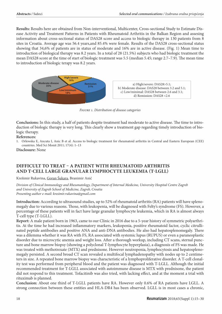
18 Reumatizam 2018;65(Suppl 1):15–30
Abstracts / Sažeci Selected oral communications / Izabrana oralna priopćenja
Results: Results here are obtained from Non-interventional, Multicenter, Cross-sectional Study to Estimate Dis-ease Activity and Treatment Patterns in Patients with Rheumatoid Arthritis in the Balkan Region and assesing information about cross-sectional status of DAS28 score and access to biologic therapy in 130 patients from 8 sites in Croatia. Average age was 56.4 years,and 85.4% were female. Results of the DAS28 cross-sectional status showing that 34,6% of patients are in status of moderate and 16% are in active disease. (Fig. 1) Mean time to introduction of biological therapy was 8.2 years. In a total of 28 (21.5%) subjects who had biologic treatment the mean DAS28 score at the time of start of biologic treatment was 5.5 (median 5.45; range 2.7–7.9). Th e mean time to introduction of biologic terapy was 8.2 years.
a) High/severe: DAS28>5.1; b) Moderate disease: DAS28 between 3.2 and 5.1;
c) Low/minimal: DAS28 between 2.6 and 3.1; d) Remission: DAS28 <2.6
Figure 1. Distribution of disease categories
Conclusions: In this study, a half of patients despite treatment had moderate to active disease. Th e time to intro-duction of biologic therapy is very long. Th is clearly show a treatment gap regarding timely introduction of bio-logic therapy. References:1. Orlewska E, Ancuta I, Anic B et al. Access to biologic treatment for rheumatoid arthritis in Central and Eastern European (CEE)
countries. Med Sci Monit 2011; 17(4): 1–13Disclosure: None
DIFFICULT TO TREAT – A PATIENT WITH RHEUMATOID ARTHRITIS AND T-CELL LARGE GRANULAR LYMPHOCYTE LEUKEMIA (T-LGLL) Krešimir Rukavina, Goran Šukara, Branimir Anić
Division of Clinical Immunology and Rheumatology, Department of Internal Medicine, University Hospital Centre Zagreb and University of Zagreb School of Medicine, Zagreb, Croatia Presenting author e-mail: [email protected]
Introduction: According to ultrasound studies, up to 52% of rheumatoid arthritis (RA) patients will have spleno-megaly due to various reasons. Th ose, with leukopenia, will be diagnosed with Felty’s syndrome (FS). However, a percentage of these patients will in fact have large granular lymphocyte leukemia, which in RA is almost always T-cell type (T-LGLL).Report: A male patient born in 1963, came to our Clinic in 2016 due to a 5-year history of symmetric polyarthri-tis. At the time he had increased infl ammatory markers, leukopenia, positive rheumatoid factor, cyclic citrulli-nated peptide antibodies and positive ANA and anti-DNA antibodies. He also had hepatosplenomegaly. Th ere was a dilemma whether it was RA with FS, RA associated with systemic lupus (RUPUS) or even a paraneoplastic disorder due to microcytic anemia and weight loss. Aft er a thorough workup, including CT scans, sternal punc-ture and bone marrow biopsy (showing a polyclonal T-lymphocyte hyperplasia), a diagnosis of FS was made. He was treated with methotrexate (MTX) and prednisone. However neutropenia, lymphocytosis and hepatospleno-megaly persisted. A second broad CT scan revealed a multifocal lymphadenopathy with nodes up to 2 centime-ters in size. A repeated bone marrow biopsy was characteristic of a lymphoproliferative disorder. A T-cell clonal-ity test was performed from peripheral blood and the patient was diagnosed with T-LGLL. Although the initial recommended treatment for T-LGLL associated with autoimmune disease is MTX with prednisone, the patient did not respond to this treatment. Tofacitinib was also tried, with lacking eff ect, and at the moment a trial with rituximab is planned.Conclusion: About one third of T-LGLL patients have RA. However only 0.6% of RA patients have LGLL. A strong connection between these entities and HLA-DR4 has been observed. LGLL is in most cases a chronic,
Reumatizam 2018;65(Suppl 1):15–30 19
Selected oral communications / Izabrana oralna priopćenja Abstracts / Sažeci
indolent disease, without a standardised treatment protocol. Symptomatic patients and patients with associated autoimmune diseases require therapy, most oft en MTX with prednisone as the inital treatment. Th is case is an example that all neutropenic RA patients with splenomegaly should be carefuly evaluated, especially if the triad of FS occurs early in the disease course, and also that thanks to clonality tests, a percentage of FS will most likely be reclassifi ed to LGLL.References:Annals of Rheumatic Disease, UpToDateDisclosure: None
GIANT CELL ARTERITIS IN POLAND. HOW TO INCREASE DIAGNOSTIC RATE? Marcin Milchert, Marek Brzosko
Department of Rheumatology, Internal Medicine and Geriatrics, Pomeranian Medical University, Poland Presenting author e-mail: [email protected]
Background: Th e incidence rate of giant cell arteritis (GCA) in Department of Rheumatology, Internal Medicine and Geriatrics was raised 10 folds between 2000 and 2016.Objectives: In this presentation a practical question: ”how to increase GCA diagnostic rate?” was addressed, and illustrated with some clinical cases.Methods: GCA referrals to Department of Rheumatology in Szczecin were analyzed. Additionally GCA epidemi-ology in Poland was analyzed based on national insurance registry and compared with distribution of fast track GCA clinics.Results: Increase of diagnostic rate was due to mainly 2 factors:1. Development of eff ective temporal / large vessels visualization techniques and introduction of fast track GCA
diagnostics.2. Better cooperation with ophthalmologist and other specialists (Table 1.)
Table 1. GCA (confi rmed cases) referrals to Department of Rheumatology in Szczecin, Poland (2000 to 2016).
Referral from: N=119 %general practitioners and emergency departments 31 26ophthalmologists 24 20internal medicine doctors 20 17rheumatologists 19 16neurologists 10 8infectious diseases specialists 5 4angiologists/vascular surgeons 4 3cardiologists 3 3other 3 3
Visualization techniques introduced were temporal/large arteries Doppler ultrasound (fast track) sometimes supported by large arteries computed tomography, occasionally by temporal artery biopsy and marginally by PET-CT. Based on national insurance registry, that we’ve analyzed, GCA diagnostic rate in Poland improved in recent years. Th is seems to correlate with increased interest in GCA in some sites that organized fast track GCA clinics. Conclusions: Increase of GCA diagnostic rate requires further eff orts.References:1. Atlas of ultrasound application in large vessel arteritis: giant cell arteritis and Takayasu arteritis., Milchert M, Diamantopoulos A,
Brzosko M; Wydawnictwo Pomorskiej Akademii Medycznej, Szczecin, 2015,
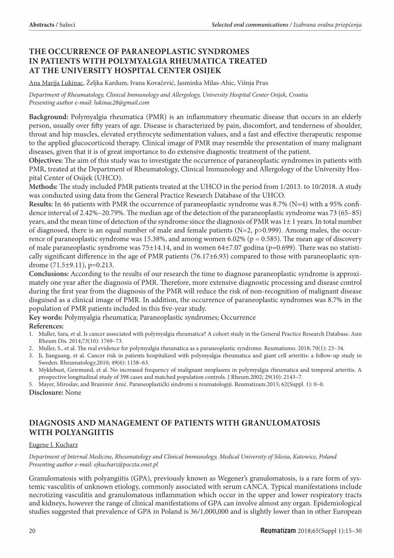
20 Reumatizam 2018;65(Suppl 1):15–30
Abstracts / Sažeci Selected oral communications / Izabrana oralna priopćenja
THE OCCURRENCE OF PARANEOPLASTIC SYNDROMES IN PATIENTS WITH POLYMYALGIA RHEUMATICA TREATED AT THE UNIVERSITY HOSPITAL CENTER OSIJEK Ana Marija Lukinac, Željka Kardum, Ivana Kovačević, Jasminka Milas-Ahic, Višnja Prus
Department of Rheumatology, Clinical Immunology and Allergology, University Hospital Center Osijek, Croatia Presenting author e-mail: [email protected]
Background: Polymyalgia rheumatica (PMR) is an infl ammatory rheumatic disease that occurs in an elderly person, usually over fi ft y years of age. Disease is characterized by pain, discomfort, and tenderness of shoulder, throat and hip muscles, elevated erythrocyte sedimentation values, and a fast and eff ective therapeutic response to the applied glucocorticoid therapy. Clinical image of PMR may resemble the presentation of many malignant diseases, given that it is of great importance to do extensive diagnostic treatment of the patient.Objectives: Th e aim of this study was to investigate the occurrence of paraneoplastic syndromes in patients with PMR, treated at the Department of Rheumatology, Clinical Immunology and Allergology of the University Hos-pital Center of Osijek (UHCO).Methods: Th e study included PMR patients treated at the UHCO in the period from 1/2013. to 10/2018. A study was conducted using data from the General Practice Research Database of the UHCO.Results: In 46 patients with PMR the occurrence of paraneoplastic syndrome was 8.7% (N=4) with a 95% confi -dence interval of 2.42%–20.79%. Th e median age of the detection of the paraneoplastic syndrome was 73 (65–85) years, and the mean time of detection of the syndrome since the diagnosis of PMR was 1± 1 years. In total number of diagnosed, there is an equal number of male and female patients (N=2, p>0.999). Among males, the occur-rence of paraneoplastic syndrome was 15.38%, and among women 6.02% (p = 0.585). Th e mean age of discovery of male paraneoplastic syndrome was 75±14.14, and in women 64±7.07 godina (p=0.699). Th ere was no statisti-cally signifi cant diff erence in the age of PMR patients (76.17±6.93) compared to those with paraneoplastic syn-drome (71.5±9.11), p=0.213. Conclusions: According to the results of our research the time to diagnose paraneoplastic syndrome is approxi-mately one year aft er the diagnosis of PMR. Th erefore, more extensive diagnostic processing and disease control during the fi rst year from the diagnosis of the PMR will reduce the risk of non-recognition of malignant disease disguised as a clinical image of PMR. In addition, the occurrence of paraneoplastic syndromes was 8.7% in the population of PMR patients included in this fi ve-year study.Key words: Polymyalgia rheumatica; Paraneoplastic syndromes; Occurrence References:1. Muller, Sara, et al. Is cancer associated with polymyalgia rheumatica? A cohort study in the General Practice Research Database. Ann
Rheum Dis. 2014;73(10): 1769–73.2. Muller, S., et al. Th e real evidence for polymyalgia rheumatica as a paraneoplastic syndrome. Reumatismo. 2018; 70(1): 23–34.3. Ji, Jianguang, et al. Cancer risk in patients hospitalized with polymyalgia rheumatica and giant cell arteritis: a follow-up study in
Sweden. Rheumatology.2010; 49(6): 1158–63.4. Myklebust, Geirmund, et al. No increased frequency of malignant neoplasms in polymyalgia rheumatica and temporal arteritis. A
prospective longitudinal study of 398 cases and matched population controls. J Rheum.2002; 29(10): 2143–7.5. Mayer, Miroslav, and Branimir Anić. Paraneoplastički sindromi u reumatologiji. Reumatizam.2015; 62(Suppl. 1): 0–0. Disclosure: None
DIAGNOSIS AND MANAGEMENT OF PATIENTS WITH GRANULOMATOSIS WITH POLYANGIITIS Eugene J. Kucharz
Department of Internal Medicine, Rheumatology and Clinical Immunology, Medical University of Silesia, Katowice, Poland Presenting author e-mail: [email protected]
Granulomatosis with polyangiitis (GPA), previously known as Wegener’s granulomatosis, is a rare form of sys-temic vasculitis of unknown etiology, commonly associated with serum cANCA. Typical manifestations include necrotizing vasculitis and granulomatous infl ammation which occur in the upper and lower respiratory tracts and kidneys, however the range of clinical manifestations of GPA can involve almost any organ. Epidemiological studies suggested that prevalence of GPA in Poland is 36/1,000,000 and is slightly lower than in other European
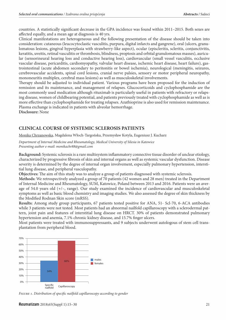
Reumatizam 2018;65(Suppl 1):15–30 21
Selected oral communications / Izabrana oralna priopćenja Abstracts / Sažeci
countries. A statistically signifi cant decrease in the GPA incidence was found within 2011–2015. Both sexes are aff ected equally, and a mean age at diagnosis is 40 yrs.Clinical manifestations are heterogeneous and the following presentation of the disease should be taken into consideration: cutaneous (leucocytoclastic vasculitis, purpura, digital infarcts and gangrene), oral (ulcers, granu-lomatous lesions, gingival hyperplasia with strawberry-like aspect), ocular (episcleritis, scleritis, conjunctivitis, keratitis, uveitis, retinal vasculitis or thrombosis, blindness, proptosis and orbital granulomatous masses), auricu-lar (sensorineural hearing loss and conductive hearing loss), cardiovascular (small vessel vasculitis, occlusive vascular disease, pericarditis, cardiomyopathy, valvular heart disease, ischemic heart disease, heart failure), gas-trointestinal (acute abdomen secondary to peritonitis or bowel ischemia), neurological (meningitis, seizures, cerebrovascular accidents, spinal cord lesions, cranial nerve palsies, sensory or motor peripheral neuropathy, mononeuritis multiplex, cerebral mass lesions) as well as musculoskeletal involvements.Th erapy should be adjusted to individual patient. Various programs have been proposed for the induction of remission and its maintenance, and management of relapses. Glucocorticoids and cyclophosphamide are the most commonly used medication although rituximab is particularly useful in patients with refractory or relaps-ing disease, women of childbearing potential, and patients previously treated with cyclophosphamide as well as is more eff ective than cyclophosphamide for treating relapses. Azathioprine is also used for remission maintenance. Plasma exchange is indicated in patients with alveolar hemorrhage. Disclosure: None
CLINICAL COURSE OF SYSTEMIC SCLEROSIS PATIENTS Monika Chrzanowska, Magdalena Włoch-Targońska, Przemysław Kotyla, Eugeniusz J. Kucharz
Department of Internal Medicine and Rheumatology, Medical University of Silesia in Katowice Presenting author e-mail: [email protected]
Background: Systemic sclerosis is a rare multisystem infl ammatory connective tissue disorder of unclear etiology, characterized by progressive fi brosis of skin and internal organs as well as systemic vascular dysfunction. Disease severity is determined by the degree of internal organ involvement, especially pulmonary hypertension, intersti-tial lung disease, and peripheral vasculopathy.Objectives: Th e aim of this study was to analyze a group of patients diagnosed with systemic sclerosis.Methods: We retrospectively analyzed a group of 70 patients (42 women and 28 men) treated in the Department of Internal Medicine and Rheumatology, SUM, Katowice, Poland between 2013 and 2016. Patients were an aver-age of 54.0 years old (+/–, range). Our study examined the incidence of cardiovascular and musculoskeletal symptoms as well as basic blood chemistry and imaging studies. We also assessed the degree of skin thickness by the Modifi ed Rodnan Skin score (mRSS).Results: Among study group participants, 67 patients tested positive for ANA, 51- Scl-70, 6-ACA antibodies while 3 patients were not tested. Most patients had an abnormal nailfold capillaroscopy with a sclerodermal pat-tern, joint pain and features of interstitial lung disease on HRCT. 30% of patients demonstrated pulmonary hypertension and anemia, 7.1% chronic kidney disease, and 15.7% fi nger ulcers.Most patients were treated with immunosuppressants, and 9 subjects underwent autologous of stem cell trans-plantation from peripheral blood.
Figure 1. Distribution of specifi c nailfold capillaroscopy according to gender
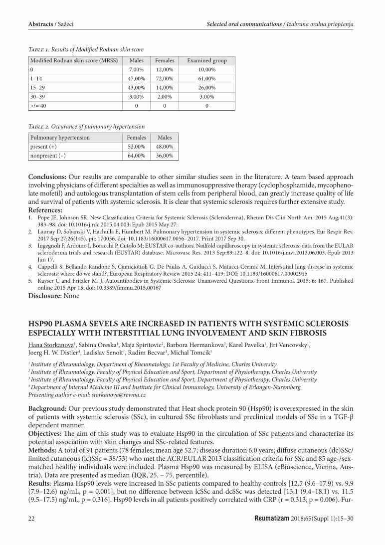
22 Reumatizam 2018;65(Suppl 1):15–30
Abstracts / Sažeci Selected oral communications / Izabrana oralna priopćenja
Conclusions: Our results are comparable to other similar studies seen in the literature. A team based approach involving physicians of diff erent specialties as well as immunosuppressive therapy (cyclophosphamide, mycopheno-late mofetil) and autologous transplantation of stem cells from peripheral blood, can greatly increase quality of life and survival of patients with systemic sclerosis. It is clear that systemic sclerosis requires further extensive study.References:1. Pope JE, Johnson SR. New Classifi cation Criteria for Systemic Sclerosis (Scleroderma), Rheum Dis Clin North Am. 2015 Aug;41(3):
383–98. doi: 10.1016/j.rdc.2015.04.003. Epub 2015 May 27.2. Launay D, Sobanski V, Hachulla E, Humbert M. Pulmonary hypertension in systemic sclerosis: diff erent phenotypes, Eur Respir Rev.
2017 Sep 27;26(145). pii: 170056. doi: 10.1183/16000617.0056–2017. Print 2017 Sep 30.3. Ingegnoli F, Ardoino I, Boracchi P, Cutolo M; EUSTAR co-authors. Nailfold capillaroscopy in systemic sclerosis: data from the EULAR
scleroderma trials and research (EUSTAR) database. Microvasc Res. 2013 Sep;89:122–8. doi: 10.1016/j.mvr.2013.06.003. Epub 2013 Jun 17.
4. Cappelli S, Bellando Randone S, Camiciottoli G, De Paulis A, Guiducci S, Matucci-Cerinic M. Interstitial lung disease in systemic sclerosis: where do we stand?, European Respiratory Review 2015 24: 411–419; DOI: 10.1183/16000617.00002915
5. Kayser C and Fritzler M. J. Autoantibodies in Systemic Sclerosis: Unanswered Questions, Front Immunol. 2015; 6: 167. Published online 2015 Apr 15. doi: 10.3389/fi mmu.2015.00167
Disclosure: None
HSP90 PLASMA SEVELS ARE INCREASED IN PATIENTS WITH SYSTEMIC SCLEROSIS ESPECIALLY WITH INTERSTITIAL LUNG INVOLVEMENT AND SKIN FIBROSIS Hana Storkanova1, Sabina Oreska1, Maja Spiritovic2, Barbora Hermankova3, Karel Pavelka1, Jiri Vencovsky1, Joerg H. W. Distler4, Ladislav Senolt1, Radim Becvar1, Michal Tomcik1
1 Institute of Rheumatology, Department of Rheumatology, 1st Faculty of Medicine, Charles University 2 Institute of Rheumatology, Faculty of Physical Education and Sport, Department of Physiotherapy, Charles University 3 Institute of Rheumatology, Faculty of Physical Education and Sport, Department of Physiotherapy, Charles University 4 Department of Internal Medicine III and Institute for Clinical Immunology, University of Erlangen-Nuremberg Presenting author e-mail: [email protected]
Background: Our previous study demonstrated that Heat shock protein 90 (Hsp90) is overexpressed in the skin of patients with systemic sclerosis (SSc), in cultured SSc fi broblasts and preclinical models of SSc in a TGF-β dependent manner.Objectives: Th e aim of this study was to evaluate Hsp90 in the circulation of SSc patients and characterize its potential association with skin changes and SSc-related features. Methods: A total of 91 patients (78 females; mean age 52.7; disease duration 6.0 years; diff use cutaneous (dc)SSc/limited cutaneous (lc)SSc = 38/53) who met the ACR/EULAR 2013 classifi cation criteria for SSc and 85 age-/sex- matched healthy individuals were included. Plasma Hsp90 was measured by ELISA (eBioscience, Vienna, Aus-tria). Data are presented as median (IQR, 25. – 75. percentile).Results: Plasma Hsp90 levels were increased in SSc patients compared to healthy controls [12.5 (9.6–17.9) vs. 9.9 (7.9–12.6) ng/mL, p = 0.001], but no diff erence between lcSSc and dcSSc was detected [13.1 (9.4–18.1) vs. 11.5 (9.5–17.5) ng/mL, p = 0.316]. Hsp90 levels in all patients positively correlated with CRP (r = 0.313, p = 0.006). Fur-
Table 2. Occurance of pulmonary hypertension
Pulmonary hypertension Females Malespresent (+) 52,00% 48,00%nonpresent (–) 64,00% 36,00%
Table 1. Results of Modifi ed Rodnan skin score
Modifi ed Rodnan skin score (MRSS) Males Females Examined group0 7,00% 12,00% 10,00%1–14 47,00% 72,00% 61,00%15–29 43,00% 14,00% 26,00%30–39 3,00% 2,00% 3,00%>/= 40 0 0 0
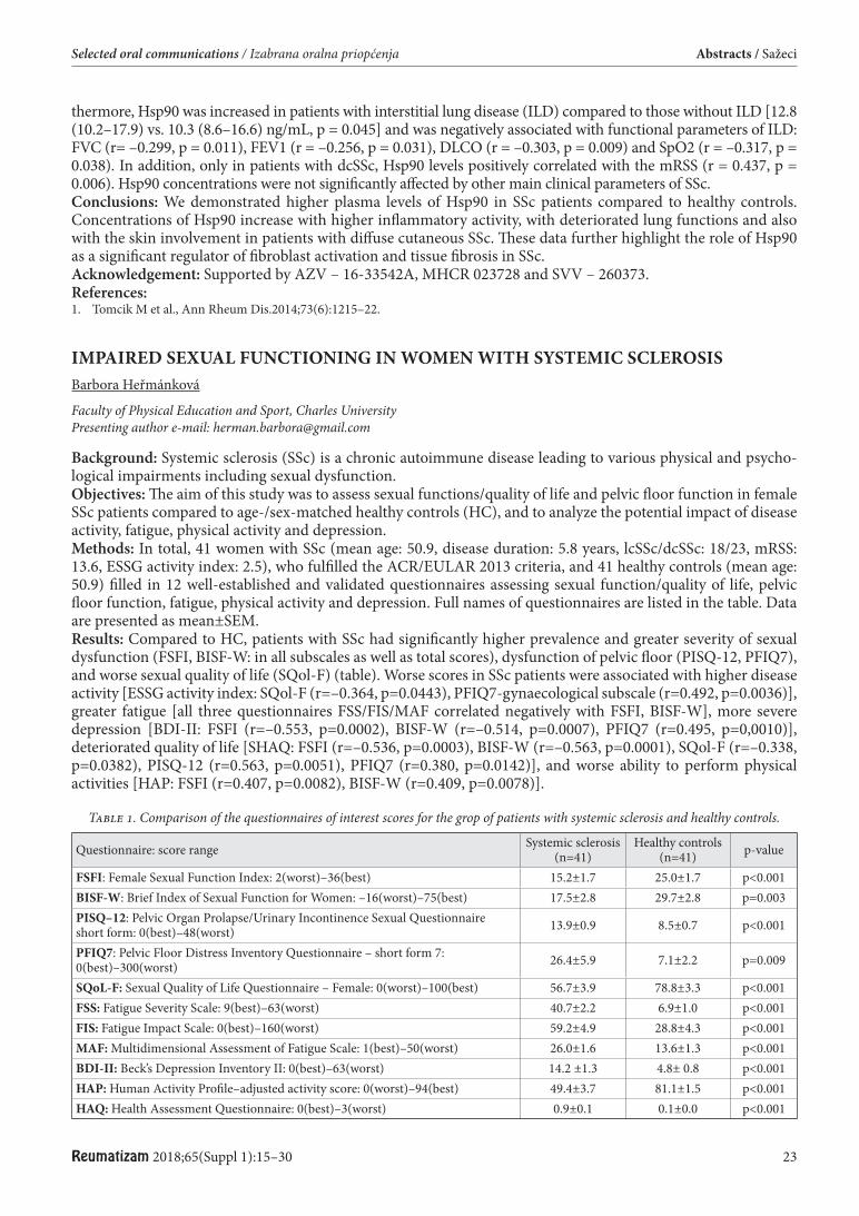
Reumatizam 2018;65(Suppl 1):15–30 23
Selected oral communications / Izabrana oralna priopćenja Abstracts / Sažeci
thermore, Hsp90 was increased in patients with interstitial lung disease (ILD) compared to those without ILD [12.8 (10.2–17.9) vs. 10.3 (8.6–16.6) ng/mL, p = 0.045] and was negatively associated with functional parameters of ILD: FVC (r= –0.299, p = 0.011), FEV1 (r = –0.256, p = 0.031), DLCO (r = –0.303, p = 0.009) and SpO2 (r = –0.317, p = 0.038). In addition, only in patients with dcSSc, Hsp90 levels positively correlated with the mRSS (r = 0.437, p = 0.006). Hsp90 concentrations were not signifi cantly aff ected by other main clinical parameters of SSc. Conclusions: We demonstrated higher plasma levels of Hsp90 in SSc patients compared to healthy controls. Concentrations of Hsp90 increase with higher infl ammatory activity, with deteriorated lung functions and also with the skin involvement in patients with diff use cutaneous SSc. Th ese data further highlight the role of Hsp90 as a signifi cant regulator of fi broblast activation and tissue fi brosis in SSc.Acknowledgement: Supported by AZV – 16-33542A, MHCR 023728 and SVV – 260373.References:1. Tomcik M et al., Ann Rheum Dis.2014;73(6):1215–22.
IMPAIRED SEXUAL FUNCTIONING IN WOMEN WITH SYSTEMIC SCLEROSIS Barbora Heřmánková
Faculty of Physical Education and Sport, Charles University Presenting author e-mail: [email protected]
Background: Systemic sclerosis (SSc) is a chronic autoimmune disease leading to various physical and psycho-logical impairments including sexual dysfunction. Objectives: Th e aim of this study was to assess sexual functions/quality of life and pelvic fl oor function in female SSc patients compared to age-/sex-matched healthy controls (HC), and to analyze the potential impact of disease activity, fatigue, physical activity and depression.Methods: In total, 41 women with SSc (mean age: 50.9, disease duration: 5.8 years, lcSSc/dcSSc: 18/23, mRSS: 13.6, ESSG activity index: 2.5), who fulfi lled the ACR/EULAR 2013 criteria, and 41 healthy controls (mean age: 50.9) fi lled in 12 well-established and validated questionnaires assessing sexual function/quality of life, pelvic fl oor function, fatigue, physical activity and depression. Full names of questionnaires are listed in the table. Data are presented as mean±SEM.Results: Compared to HC, patients with SSc had signifi cantly higher prevalence and greater severity of sexual dysfunction (FSFI, BISF-W: in all subscales as well as total scores), dysfunction of pelvic fl oor (PISQ-12, PFIQ7), and worse sexual quality of life (SQol-F) (table). Worse scores in SSc patients were associated with higher disease activity [ESSG activity index: SQol-F (r=–0.364, p=0.0443), PFIQ7-gynaecological subscale (r=0.492, p=0.0036)], greater fatigue [all three questionnaires FSS/FIS/MAF correlated negatively with FSFI, BISF-W], more severe depression [BDI-II: FSFI (r=–0.553, p=0.0002), BISF-W (r=–0.514, p=0.0007), PFIQ7 (r=0.495, p=0,0010)], deteriorated quality of life [SHAQ: FSFI (r=–0.536, p=0.0003), BISF-W (r=–0.563, p=0.0001), SQol-F (r=–0.338, p=0.0382), PISQ-12 (r=0.563, p=0.0051), PFIQ7 (r=0.380, p=0.0142)], and worse ability to perform physical activities [HAP: FSFI (r=0.407, p=0.0082), BISF-W (r=0.409, p=0.0078)].
Table 1. Comparison of the questionnaires of interest scores for the grop of patients with systemic sclerosis and healthy controls.
Questionnaire: score range Systemic sclerosis (n=41)
Healthy controls (n=41) p-value
FSFI: Female Sexual Function Index: 2(worst)–36(best) 15.2±1.7 25.0±1.7 p<0.001BISF-W: Brief Index of Sexual Function for Women: –16(worst)–75(best) 17.5±2.8 29.7±2.8 p=0.003PISQ–12: Pelvic Organ Prolapse/Urinary Incontinence Sexual Questionnaire short form: 0(best)–48(worst) 13.9±0.9 8.5±0.7 p<0.001
PFIQ7: Pelvic Floor Distress Inventory Questionnaire – short form 7: 0(best)–300(worst) 26.4±5.9 7.1±2.2 p=0.009
SQoL-F: Sexual Quality of Life Questionnaire – Female: 0(worst)–100(best) 56.7±3.9 78.8±3.3 p<0.001FSS: Fatigue Severity Scale: 9(best)–63(worst) 40.7±2.2 6.9±1.0 p<0.001FIS: Fatigue Impact Scale: 0(best)–160(worst) 59.2±4.9 28.8±4.3 p<0.001MAF: Multidimensional Assessment of Fatigue Scale: 1(best)–50(worst) 26.0±1.6 13.6±1.3 p<0.001BDI-II: Beck’s Depression Inventory II: 0(best)–63(worst) 14.2 ±1.3 4.8± 0.8 p<0.001HAP: Human Activity Profi le–adjusted activity score: 0(worst)–94(best) 49.4±3.7 81.1±1.5 p<0.001HAQ: Health Assessment Questionnaire: 0(best)–3(worst) 0.9±0.1 0.1±0.0 p<0.001
24 Reumatizam 2018;65(Suppl 1):15–30
Abstracts / Sažeci Selected oral communications / Izabrana oralna priopćenja
Conclusions: Women with SSc reported signifi cantly impaired sexual function, sexual quality of life and pelvic fl oor function than age-matched healthy controls. Worse scores in SSc were associated with disease activity, physical activity, fatigue, depression and quality of life.Acknowledgements: Supported by AZV-16-33574A, MHCR 023728, and SVV – 260373. Disclosure: None
ABBERANCIES OF SPECIFIC PERIPHERAL BLOOD T-CELL AND MONOCYTE SUBPOPULATIONS IN ANKYLOSING SPONDILITIS CORRELATE WITH DISEASE ACTIVITY PARAMETERS Alan Šućur1, Zrinka Jajić2, Marina Ikić Matijašević3, Marinko Artuković3, Darja Flegar1, Tomislav Kelava1, Nina Lukač1, Antonio Markotić1, Danka Grčević1
1 Croatian Institute for Brain Research, University of Zagreb School of Medicine 2 Department of Rheumatology, Physical Medicine and Rehabilitation, School of Medicine University of Zagreb, Sestre Milosrdnice University Hospital Center, Zagreb, Croatia 3 Department of clinical immunology and allergology, Sveti Duh University Hospital Presenting author e-mail: [email protected]
Background: Autoimmunity is major driving force in pathogenesis of rheumatic diseases, including ankylosing spondylitis (AS). AS is associated with abnormal immune cell functions. Several recent studies stressed the role of T-cells and monocyte subpopulations in AS, but these mechanisms are signifi cantly less understood compared to other rheumatic diseases (1). Objectives: Th e aim was to compare the frequency of T-cell and monocyte subpopulations in AS patients and controls, and to correlate them with disease activity. Methods: Mononuclear cells (MNC) were isolated from blood of healthy controls (n=110) and AS (n=65) patients. T-cell and monocyte phenotype of MNC was determined using fl ow cytometry for the following mark-ers: Th 1/2 (CD3+CD4+CCR6–), Th 17 (CD3+CD4+CCR4+CCR6+), Tfh (CD3+CD4+CXCR5+), Tc (CD3+CD8+), memory Tc (CD3+CD8+CCR4+) double-positive (dp) Tc (CD3+CD4+CD8+); and monocytes (CD3-CD19-CD56-CD11b+CD14+). On lymphocytes IL21R, CD25 and CD11b were analyzed; while on mono-cytes chemokine receptors CCR1, CCR2, CCR4, CXCR4 and RANK. Frequencies of lymphocyte and monocyte subpopulations were correlated with ASDAS, BASDAI, VAS of pain in spine and peripheral joints, stiff ness inten-sity and duration, ESR and CRP. Results: Results showed decrease of memory Tc (p=0.022) and Tfh in AS (p=0.038) which correlated negatively with physician’s assessment of disease activity. Th e frequency of dp Tc was unchanged, but negatively correlated with BASDAI and pain assessment. Th e frequency of monocytes was decreased in AS (p<0.001), while the sub-population expressing RANK was increased (p=0.007). In monocytes; CCR1 correlated with BASDAI and pain assessment, CCR2 correlated with stiff ness duration. CXCR4 expression was increased in AS (p<0.001) and cor-related with BASDAI and stiff ness intensity. Conclusions: Results show changes in T-cells and monocyte subpopulations induced in AS and indicate their possible importance for AS pathogenesis. Peripheral blood dp Tc cells may be of particular interest as further research targets for novel therapeutic approaches, since their frequency is associated with disease activity and patient’s pain assessment (2). References:1. Rezaiemanesh A, Abdolmaleki M, et al. Immune cells involved in the pathogenesis of ankylosing spondylitis. Biomed Pharm
2018;100:198–204.2. Overgaard NH, Jung JW, et al. CD4+/CD8+ double-positive T cells: more than just a developmental stage? J Leukoc Biol 2015;97:31–8. Disclosure: None
Reumatizam 2018;65(Suppl 1):15–30 25
Selected oral communications / Izabrana oralna priopćenja Abstracts / Sažeci
PLASMA LEVELS OF HSP90 ARE INCREASED IN AXIAL SPONDYLOARTHRITIS AND PSORIATIC ARTHRITIS PATIENTS WITH STRUCTURAL CHANGES Hana Storkanova1, Kristyna Bubova1, Sabina Oreska1, Maja Spiritovic2, Barbora Hermankova2, Monika Gregova1, Katerina Zegzulkova1, Jana Horinkova1, Karel Pavelka1, Jiri Vencovsky1, Jiri Stolfa1, Marketa Husakova1, Sarka Forejtova1, Ladislav Senolt1, Michal Tomcik1
1 Department of Rheumatology, 1st Faculty of Medicine, Charles University and Institute of Rheumatology 2 Institute of Rhumatology and Faculty of Physical Education and Sport, Department of Physiotherapy, Charles University Presenting author e-mail: [email protected]
Background: Hsp90 is required for the proper conformation and activation of a number of cellular proteins. It also regulates activation of innate immunity and the induction of proinfl ammatory cytokines and chemokines. Th ese properties predispose Hsp90 to its potential role in the pathogenesis of autoimmune infl ammatory rheu-matic diseases. Objectives: Th e aim of this study was to assess Hsp90 in the plasma of axial spondyloarthritis (axSpA) and pso-riatic arthritis (PsA) patients compared to healthy controls (HC) and to determine its potential associations with disease activity and clinical features.Methods: A total of 80 axSpA patients (37 females; mean age 37.0 years; disease duration 9.9 years; non-radio-graphic (nr-axSpA): 40; radiographic (r-axSpA): 40) and 21 PsA patients (9 females, mean age 52.1 years, disease duration 25.8 years) and age-/sex- matched healthy individuals (80 and 21 respectively) were included. Plasma Hsp90 levels were measured by ELISA (eBioscience, Vienna, Austria). Data are presented as median (IQR).Results: Plasma Hsp90 levels were signifi cantly increased in axSpA patients compared to healthy controls [15.7 (10.5–19.8) vs. 8.3 (6.6–11.7) ng/mL, p<0.001], but no diff erence between nr-axSpA and r-axSpA subsets was detected [14.8 (9.2 – 19.7) vs. 16.1 (10.5 – 21.6) ng/mL, p = 0.513]. Increased plasma levels of Hsp90 in PsA com-pared to HC did not reach statistical signifi cance [11.23 (8.15–16.20) vs 8.54 (6.32–11.73) ng/mL, p=0.066]. Hsp90 levels in r-axSpA patients positively correlated with the MRI presence of active infl ammatory lesions in sacroiliac joints (SPARCC MRI score for SI joints: r=0.594, p=0.020). Furthermore, increased Hsp90 levels in PsA patients were associated with the count of joint deformities (r=0.526, p=0.025). Plasma Hsp90 levels were neither signifi cantly associated with other main clinical features of axSpA and PsA nor with markers of disease activity (e.g. ESR, CRP, BASDAI, ASDAS, DAPSA). Conclusions: We demonstrated elevated plasma levels of Hsp90 in axSpA patients compared to healthy controls. In r-axSpA, Hsp90 may represent an independent marker of SI joint infl ammation, whereas in PsA, plasma Hsp90 correlates with joint deformities. Th ese data suggest that Hsp90 could become a potential biomarker of structural changes in SpA.Acknowledgement: Supported by AZV-16-33542A, MHCR 023728 and SVV – 260373.Disclosure: None
WHICH DEMOGRAPHIC DISEASE RELATED VARIABLES MAY BE PREDICTORS OF QUALITY OF LIFE IN PSORIATIC ARTHRITIS PATIENTS? Frane Grubišić1, Hana Skala Kavanagh1, Ines Doko1, Jure Aljinović2, Tonko Vlak2, Petra Kovačević1, Simeon Grazio1
1 Department of Rheumatology, Physical Medicine and Rehabilitation, School of Medicine University of Zagreb, Sestre Milosrdnice University Hospital Center, Zagreb, Croatia 2 Institute of physical medicine and rehabilitation with rheumatology, University Hospital Center Split, Croatia Presenting author e-mail: [email protected]
Psoriatic arthritis (PsA) patients may face challenges other than skin disease, arthralgias, synovitis or dactylitis.Th ese patients experience considerable disability and impaired quality of life, including sleep problems, depres-sion/anxiety and problems in intimate life.To assess if age at onset of PsA and psoriasis, duration of PsA and psoriasis severity of psoriasis, functional ability and disease activity are predictors of quality of life in PsA patients.Th e study was conducted in two visits with fi ve years period in between at the outpatient clinic of the rheumtol-ogy departments in two tertiary university hospitals, in Zagreb and Split. A total of 114 PsA patients (61 men, 53 women) were enrolled in the phase 1, while phase 2 included 104 patients (56 men, 48 women). Practising expe-rienced rheumatologists collected demographic data and history of diseases (age, gender, age of diagnosis, dura-tion of psoriasis and PsA). HAQ was used to assesses function and DAS28 for disease activity. For assessing qual-ity of life patients fi lled SF36. Methods of descriptive statistics and canonical regression were used in this research.
26 Reumatizam 2018;65(Suppl 1):15–30
Abstracts / Sažeci Selected oral communications / Izabrana oralna priopćenja
Mean age of patients in phase 1 was 57,3±11,1 years (men 57,81 years, women 56,83years) and mean age of patients in phase 2 was 60,1±11,3 years (men 60,1 years, women 60,1years). Mean duration of psoriasis in phase 1 was 211,9±147,1 months and in phase 2 was 251,6±153,9 months, while mean duration of PsA in phase 1 was 132,5±113,6 months and in phase 2 was 171,7±108,4 months. Canonical regression was performed in both phases and showed that age of PsA diganosis and duration of PsA were signifi cantly associated with worse functional ability, but not with the quality of life of PsA patients.Based on the result of this research, functional ability of PsA patients depends on age at diagnosis and disease duration which could be considered as the predictors of functional ability in PsA patients.References:1. Grubisic F. Quality of life in patients with psoriatic arthritis in comparison to rheumatoid arthritis and degenerative joint disease. PhD
Th esis. Zagreb. School of Medicine University of Zagreb. 2016.Disclosure: None
COMPARISON OF COMPUTERIZED COLOR TELETHERMOGRAPHY AND NAILFOLD CAPILLAROSCOPY IN DIAGNOSTICS OF SECONDARY RAYNAUD’S PHENOMENON IN CHILDREN Mario Sestan1, Natasia Cekada1, Daniel Turudic1, Mateja Batnozic Varga2, Jagoda Stipic3, Marko Baresic4, Marijan Frkovic5, Domagoj Kifer6, Marija Jelusic7
1 Department of Paediatrics, University Hospital Centre Zagreb 2 Department of Paediatrics, University Hospital Centre Osijek, University of Osijek School of Medicine 3 Department of Neurology, University Hospital Centre Zagreb 4 Department of Internal Medicine, Division of Clinical Immunology and Rheumatology, University Hospital Centre Zagreb 5 Department of Paediatrics, University Hospital Centre Zagreb; University of Zagreb School of Medicine 6 Department of Biophysics, Faculty of Pharmacy and Biochemistry, University of Zagreb 7 Department of Paediatrics, University Hospital Centre Zagreb; University of Zagreb School of Medicine Presenting author e-mail: [email protected]
Background: Raynaud’s phenomenon (RP) is a condition characterized by periodical vasospasm in response to cold temperatures or emotional stress exposure. To distinguish between primary and secondary RP, clinical examination, laboratory fi ndings, nailfold capillaroscopy (NC) and computerized color telethermography (CCTT) are necessary.Objectives: To analyze RP features in children in correlation with the most frequently associated laboratory tests, CCTT and NC.Methods: Th is study included children clinically recognized as RP in the period from 2011–2017 at the Referral Center for Pediatric and Adolescent Rheumatology Republic of Croatia. Laboratory data included serum level of IgG, C3, C4, CH50, RF, presence of ANA and ANCAs. Results: CCTT, performed in 188 patients, classifi cated 15 as primary RP, 57 as secondary RP, while in 47 no clas-sifi cation could be made. Among patients classifi cated as secondary RP on CCTT, the most of them, 14 (24.6%), were diagnosed with juvenile idiopathic arthritis (JIA). Th ere were 5 patients (8.8%) with systemic sclerosis (SSc), 2 (3.5%) with mixed connective tissue disease (MCTD), 1 (1.7%) with systemic lupus erythematosus, 11 (19.3%) with undiff erentiated connective tissue disease (UCTD), whilst 24 (42.1%) had no evident other disease. Th e appearance of abnormal capillaroscopic pattern was found in 17 out of 89 patients and nonspecifi c capillaro-scopic alterations were noticed in 27. Among patients with the appearance of abnormal capillaroscopic pattern, 5 (29.4%) were diagnosed with SSc, 3 (17.6%) with JIA, 2 (11.8%) with MCTD, 1 (5.9%) with dermatomyositis, 2 (11.8%) with UCTD, whilst 4 (23.5%) had no evident rheumatic disease. All patients with RP diagnosed with SSc and MCTD had both the appearance of abnormal capillaroscopic pattern and CCTT fi ndings consistent with secondary RP. No statistically signifi cant diff erence between NC and CCTT in predicting the diagnosis of second-ary RP was determined (McNemar’s test, χ2 = 0.042, p = 0.838) nor was there signifi cant diff erence between NC and CCTT in regard to the results of laboratory fi ndings (χ2 = 1.042, p = 0.307).Conclusions: We found that nailfold capillaroscopy and CCTT were equally eff ective in the diagnosis of second-ary RP in children. Th ere was no diff erence between them in regard to the results of immunological laboratory fi ndings distinctive with secondary RP.References:1. Wigley FM, Flavahan NA. Raynaud’s phenomenon. Th e New England Journal of Medicine 2016; 375: 556–565.Disclosure: None
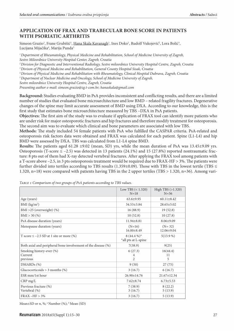
Reumatizam 2018;65(Suppl 1):15–30 27
Selected oral communications / Izabrana oralna priopćenja Abstracts / Sažeci
APPLICATION OF FRAX AND TRABECULAR BONE SCORE IN PATIENTS WITH PSORIATIC ARTHRITIS Simeon Grazio1, Frane Grubišić1, Hana Skala Kavanagh1, Ines Doko1, Rudolf Vukojević2, Lora Bolić3, Lucijana Mijačika4, Marija Punda5
1 Department of Rheumatology, Physical Medicine and Rehabilitation, School of Medicine University of Zagreb, Sestre Milosrdnice University Hospital Center, Zagreb, Croatia 2 Division for Diagnostic and Interventional Radiology, Sestre milosrdnice University Hospital Centre, Zagreb, Croatia 3 Divison of Physical Medicine and Rehabilitation, General County Hospital Sisak, Croatia 4 Divison of Physical Medicine and Rehabilitation with Rheumatology, Clinical Hospital Dubrava, Zagreb, Croatia 5 Department of Nuclear Medicine and Oncology, School of Medicine University of Zagreb, Sestre milosrdnice University Hospital Centre, Zagreb, Croatia Presenting author e-mail: [email protected]; [email protected]
Background: Studies evaluating BMD in PsA provides inconsistent and conflicting results, and there are a limited number of studies that evaluated bone microarchitecture and low BMD – related fragility fractures. Degenerative changes of the spine may limit accurate assessment of BMD using DXA. According to our knowledge, this is the fi rst study that estimates bone microarchitecture measured by TBS –DXA in PsA patients.Objectives: Th e fi rst aim of the study was to evaluate if application of FRAX tool can identify more patients who are under risk for major osteoporotic fractures and hip fractures and therefore modify treatment for osteoporosis. Th e second aim was to evaluate which clinical and bone parameters are associated with low TBS.Methods: Th e study included 54 female patients with PsA who fulfilled the CASPAR criteria. PsA-related and osteoporosis risk factors data were obtained and FRAX was calculated for each patient. Spine (L1-L4) and hip BMD were assessed by DXA. TBS was calculated from L1-L4 spine BMD.Results: Th e patients aged 61.28 ±9.02 (mean, SD) yrs, while the mean duration of PsA was 13.45±9.09 yrs. Osteoporosis (T-score ≤ –2.5) was detected in 13 patients (24.1%) and 15 (27.8%) reported nontraumatic frac-ture: 8 pts out of them had X-ray detected vertebral fractures. Aft er applying the FRAX tool among patients with a T-score above –2.5, in 3 pts osteoporosis treatment would be required due to FRAX-HF > 3%. Th e patients were further divided into tertiles according to TBS results (1.359±0.09). Th ose with TBS in the lowest tertile (TBS ≤ 1.320, n=18) were compared with patients having TBS in the 2 upper tertiles (TBS > 1.320, n=36). Among vari-
Table 1 Comparison of two groups of PsA patients according to TBS values.
Low TBS (< 1.320)N=18
High TBS (>1.320)N=36
Age (years) 63.6±9.95 60.11±8.42BMI (kg/m2) 34.53±3.84 28.65±5.02BMI >25 (overweight) (%) 16 (88.9) 19 (52.8)BMI > 30 (%) 10 (52.8) 10 (27.8)PsA disease duration (years) 11.94±8.81 8.06±9.09Menopause duration (years) (N=16)
16.00±8.49(N= 32)
12.06±9.04T score ≤ –2.5 SD at 1 site or more (%) 8 (44.4 %)*
*all pts at L-spine5(13.9 %)
Both axial and peripheral bone involvement of the disease (%) 7(38.9) 9(25)Smoking history-ever (%)Currentprevious
6 (27.3)42
16(44.4)115
DMARDs (%) 9 (50) 27 (75)Glucocorticoids > 3 months (%) 3 (16.7) 6 (16.7)ESR mm/1st hour 26.90±14.76 21.67±12.34CRP mg/L 7.62±8.74 6.73±5.53Previous fracture (%)Vertebral (%)
7 (38.9)3 (16.7)
8 (22.2)5 (13.9)
FRAX –HF > 3% 3 (16.7) 5 (13.9)
Mean±SD or n, %; a Number (%); b Mean (SD)
28 Reumatizam 2018;65(Suppl 1):15–30
Abstracts / Sažeci Selected oral communications / Izabrana oralna priopćenja
ables of interest patients with lower TBS (≤ 1.320) had more oft en osteoporosis (44.4% vs. 13.9%) and non-trau-matic fractures (38.9 % vs. 22.2%), longer duration of the disease (11.94±8.81 vs. 8.06±9.09 yrs), longer meno-pause duration (16.00±8.49 vs. 12.06±9.04 yrs) and higher body mass index (BMI) (34.53±3.84 vs. 28.65±5.02) (Table 1).Conclusions: According to results of our study application of FRAX tool would modify treatment decision in minority of patients with PsA. Th e TBS measurement enabled recognition of some clinical factors and parameters of the disease associated with lower TBS. Further studies involving larger number of PsA patients and healthy controls are required to investigate additional determinants of a low TBS and its importance in PsA.References:1. Chandran S, Aldei A, Johnson SR, Cheung AM, Salonen D, Gladman DD. Prevalence and risk factors of low bone mineral density in
psoriatic arthritis: A systematic review. Semin Arthritis Rheum. 2016;46(2):174–82.2. Pedreira PG, Pinheiro MM, Szejnfeld VL. Bone mineral density and body composition in postmenopausal women with psoriasis and
psoriatic arthritis. Arthritis Res Th er 2011;13(1):R16.3. Zhu TY1, Griffi th JF, Qin L, Hung VW, Fong TN, Au SK, Kwok AW, Leung PC, Li EK, Tam LS. Density, structure, and strength of
the distal radius in patients with psoriatic arthritis: the role of infl ammation and cardiovascular risk factors. Osteoporos Int. 2015;26(1):261–72.
4. Grazio S, Cvijetić S, Vlak T, Grubišić F, Matijević V, Nemčić T, Punda M, Kusić Z. Osteoporosis in psoriatic arthritis: Is there any? Wien Klin Wochenschr 2011;123(23–24): 743–50.
5. Krueger D, Fidler E, Libber J, Aubry-Rozier B, Hans D3, Binkley N. Spine trabecular bone score subsequent to bone mineral density improves fracture discrimination in women. J Clin Densitom. 2014 Jan–Mar;17(1):60–5.
Disclosure: None
PROGRESSION OF PAIN, STIFFNESS, FUNCTION CHANGES, AND ULTRASOUND DETECTED SYNOVITIS AND OSTEOPHYTE FORMATION IN PATIENTS WITH HAND OSTEOARTHRITIS OVER THREE YEARS Olga Sleglova, Olga Ruzickova, Karel Pavelka, Ladislav Šenolt
Rheumatology Institute and Department of Rheumatology, 1st Medical Faculty, Charles University Prague, Czech Republic Presenting author e-mail: [email protected]
Background: Hand osteoarthritis (HOA) is a common and frequent cause of pain. HOA is a heterogeneous group of disorders with two main subsets including non-erosive and erosive disease. Few studies demonstrated infl am-matory ultrasound changes and more severe clinical symptoms in patients with erosive compared with non-erosive disease, however the results are inconsistent. Objectives: Th e aim of this study was to evaluate progression of pain, stiff ness, physical impairment and ultra-sound features in patients with erosive and non-erosive HOA in a three years longitudinal study.Methods: Consecutive patients with symptomatic HOA fulfi lling the American College of Rheumatology (ACR) criteria were included in this study. Joint pain and swelling, the Australian/Canadian OA hand index (AUSCAN), radiographs and ultrasound. of both hands were assessed. Patients were examined at baseline and at the and third year of follow up.Results: Altogether, 97 patients (7 male) with symptomatic nodal HOA were included in this study and followed between April 2012 and January 2018. Out of these patients, 57 had erosive disease. Th e number of painful and clinically swollen joints (p<0.05) was signifi cantly higher in patients with erosive compared with non-erosive disease at baseline and still remains statistically higher (p<0.01) at the third year of follow up in patients with erosive disease. According to the AUSCAN, patients with erosive disease had more pain (p<0.05) and stiff ness (p<0.01) at baseline. Pain (p<0.01), stiff ness (p<0.05) and also function (p<0.01) worsened in patients with ero-sive disease at the third year of follow up. US-detected pathologies such as gray-scale synovitis (p<0.001), inten-sity of PDS (p<0.01) and number of osteophytes (p<0.01) were signifi cantly higher in patients with erosive disease at baseline. Patients with erosive disease also worsened aft er the third year of follow up (p<0.01). Conclusions: Th e fi ndings of this study show that pain and number of clinically swollen joints associated with US-detected synovial changes and osteophyte formation is more severe in patients with erosive HOA than in patients with non-erosive disease. In addition, osteophyte formation is more likely to progress independent of synovial infl ammation.Acknowledgement: Th is work was supported by the project (Ministry of Health, Czech Republic) for consensual development of research organization 023728.Disclosure: None
Reumatizam 2018;65(Suppl 1):15–30 29
Selected oral communications / Izabrana oralna priopćenja Abstracts / Sažeci
EXPRESSION OF CXCL16 IN PERIPHERAL BLOOD OF INDIVIDUALS IN THE PRE-CLINICAL PHASE OF RHEUMATOID ARTHRITIS Klára Prajzlerová1, Olga Kryštůfk ová1, Petra Hánová1, Hana Hulejová2, Monika Gregová1, Karel Pavelka1, Jiří Vencovský1, Ladislav Šenolt1, Mária Filková1
1 Department of Rheumatology, First Faculty of Medicine, Charles University and Rheumatology Institute, Prague 2 Rheumatology Institute, Prague Presenting author e-mail: [email protected]
Background: Individuals with arthralgia and positivity of anti-citrullinated protein antibodies (ACPA) are con-sidered at high risk of developing rheumatoid arthritis (RA). Th e altered expression of miRNAs is involved in the development and maintenance of autoimmune diseases.Objectives: We aimed to analyse miRNAs and expression of their target genes in individuals in the pre-clinical phase of RA. Methods: Th e initial study included 19 individuals with arthralgia (all were ACPA+) and 20 healthy controls (HC). Total RNA from peripheral blood mononuclear cells (PBMC) was isolated. A comprehensive analysis of miRNAs was performed using TaqMan® Low Density Array (TLDA) in 5 samples per group. Single assay analysis of miR-451a and CXCL16 at mRNA level was performed in remaining samples. dCt was used for relative quanti-fi cation. Th e expression of CXCL16 at protein level in diff erent subpopulations of leukocytes was analysed by fl ow cytometry in a validation cohort of 16 individuals with arthralgia and 13 HC.Results: TLDA analysis as well as single assay analysis revealed 2.43-3.19x higher levels of miR-451a in individu-als with arthralgia compared to HC. Levels of miR-451 positively correlated with DAS28-CRP and SDAI clinical activity scores. Online tools predicted CXCL16 as a direct target of miR-451a. Th e expression of CXCL16 at mRNA level was 1.51x higher in patients with arthralgia compared to HC and positively correlated with the expression of miR-451a. Th ere was no diff erence in plasma levels of CXCL16. Flow cytometry analysis of diff erent subpopulations of leukocytes revealed the expression of CXCL16 on the surface of granulocytes, monocytes and NK cells. Th e expression of CXCL16 was lower in patients with arthralgia compared to HC in granulocytes and monocytes while there was no diff erence in NK cells. Conclusions: MiR-451a is of higher expression in PBMC of individuals with arthralgia compared to healthy con-trols and positively correlates with increased expression of chemokine CXCL16 at transcriptional level. Impor-tantly, low protein expression of CXCL16 in granulocytes and monocytes in individuals with arthralgia is sugges-tive of direct regulation of CXCL16 by miR-451a. We therefore hypothesize a role of CXCL16 in the preclinical phase of RA.Acknowledgement: Project AZV-17-32612A. Disclosure: None
INVESTIGATING THE POTENTIAL OF MEAN LYMPHOCYTE VOLUME AND MEAN MONOCYTE VOLUME AS BIOCHEMICAL MARKERS FOR DIAGNOSIS AND FOLLOW UP OF RHEUMATOID ARTHRITIS AND ANKYLOSING SPONDYLITIS Sara Sekelj1, Branimir Žarković2, Miroslav Mayer3, Renata Zadro4, Marija Miloš4
1 Imperial College Health Partners, London, UK 2 Division of Rheumatology, Physical Medicine and Rehabilitation, General Hospital “Dr. Josip Benčević”, Slavonski Brod, Croatia 3 Division of Clinical Immunology and Rheumatology, Department of Internal medicine, University Hospital Center Zagreb and University of Zagreb School of Medicine, Zagreb, Croatia 4 Clinical Institute for Laboratory Diagnostics, University Hospital Centre Zagreb, Zagreb, Croatia Presenting author e-mail: [email protected]
Background: Th e evolution of automated haematological analysers provides insight into mean cell volumes of leukocyte subpopulations. Th e changes in cell volumes have already been investigated as markers for infl amma-tion response and sepsis. Th e focus of this research was set on rheumatoid arthritis (RA) and ankylosing spondy-litis (AS) since both conditions initiate an infl ammatory response and activate diff erent cell populations.Objectives: Th e aim of this research project was to determine the usefulness of mean monocyte volume (MCV Mo) and mean lymphocyte volume (MCV Ly) as biochemical markers for RA and AS, with the specifi c objectives:
30 Reumatizam 2018;65(Suppl 1):15–30
Abstracts / Sažeci Selected oral communications / Izabrana oralna priopćenja
1. To compare MCV Mo and MCV Ly between diagnosed patients and control group in active disease and remission
2. To investigate the relationship between these parameters and conventional biomarkers (CRP, SE, DAS28)3. To explore these parameters as predictors of therapy eff ectivenessMethods: Th is was a retrospective analysis from January 2016 to January 2017 which included a total of 183 blood samples from patients diagnosed with RA or AS at the Centre for Clinical Immunology and Rheumatology, Department for Internal Medicine at University Hospital Centre Zagreb, Croatia and 32 blood samples within the control group. Th e analysis was done on the automated haematology system Beckman Coulter DxH800. Data was collected and analysed in MedCalc Soft ware.Results: Th ere is a statistically signifi cant diff erence between MCV Mo and MCV Ly in patients with RA and AS compared to control group, as well as in active RA and remission. A signifi cant moderate positive correlation was found between MCV Ly and CRP in RA (r=0.43, p=0.001), MCV Ly and DAS28 in active RA (r=0.48, p=0.044), as well as MCV Mo and SE (r=0.58, p=0.000) and MCV Mo and CRP (r=0.53, p=0.000) in AS. Patients receiving anti-TNFa therapy had statistically higher values of both parameters. Diff erences are observed in MCV Mo of patients receiving over 5mg of corticosteroids compared to lower doses.Conclusions: Th e distribution and diff erences of investigated parameters could be used in longitudinal patient follow-up, thus enabling clinicians to track the disease state. Th e conventional biochemical markers give clini-cians a broad picture of the infl ammation process. Although investigated parameters cannot replace CRP, SE or DAS28 in RA and AS diagnosis, they could provide additional insight into the infl ammatory process.Disclosure: None
PLASMA LEVELS OF HSP90 ARE INCREASED IN RHEUMATOID ARTHRITIS AND OSTEOARTHRITIS PATIENTS Kristyna Bubova1, Hana Storkanova1, Sabina Oreska1, Maja Spiritovic2, Barbora Hermankova2, Karel Pavelka1, Jiri Vencovsky1, Jindriska Gatterova1, Ladislav Senolt1, Michal Tomcik1
1 Department of Rheumatology, 1st Faculty of Medicine, Charles University and Institute of Rheumatology 2 Institute of Rheumatology, Faculty of Physical Education and Sport, Department of Physiotherapy, Charles University Presenting author e-mail: [email protected]
Background: Hsp90 is required for the proper conformation and activation of a number of cellular proteins. It also regulates activation of innate immunity and the induction of proinfl ammatory cytokines and chemokines. Th ese properties predispose Hsp90 to its potential role in the pathogenesis of autoimmune infl ammatory rheu-matic diseases. Objectives: Th e aim of this study was to assess Hsp90 in plasma of rheumatoid arthritis (RA) patients and osteo-arthritis (OA) patients compared to healthy controls (HC). Secondly, potential associations of plasma Hsp90 levels with disease activity and clinical features were determined.Methods: A total of 35 RA patients (25 females; mean age 54.6 years; disease duration 13.0 years) and 36 OA patients (22 females, mean age 66.8 years, disease duration 6.7 years) and age-/sex- matched healthy individuals (35 and 36 respectively) were included. Plasma Hsp90 levels were measured by ELISA (eBioscience, Vienna, Aus-tria). Data are presented as median (IQR).Results: Plasma Hsp90 levels were signifi cantly increased in RA patients compared to HC [41.4 (22.1–68.9) vs. 9.3 (7.7–14.3) ng/mL, p<0.0001]. Furthermore, plasma Hsp90 levels correlated with serum low-density lipopro-teins (LDL), high-density lipoproteins (HDL) levels and atherogenic index (AI) (r=0.35, p=0.05; r=–0.35, p=0.05; r=0.45, p=0.009, respectively). Plasma Hsp90 levels were signifi cantly increased in OA patients compared to HC [45.6 (42.5–53.1) vs. 10.2 (7.8–14.4) ng/mL, p<0.0001]. Furthermore, Hsp90 plasma levels correlated with body mass index (BMI) (r=–0.71, p<0.0001). Concentrations of Hsp90 in plasma were neither signifi cantly associated with other main clinical parameters nor with infl ammatory/disease activity or RA/OA-related patient reported outcomes (e.g. ESR, CRP, DAS28, WOMAC, HAQ). Conclusions: We demonstrated elevated plasma concentrations of Hsp90 in RA and OA patients compared to HC. In RA, Hsp90 may have a potential role in the link between infl ammation and lipid metabolism, whereas in OA, Hsp90 may have a potential role in the link between obesity and progression of cartilage degradation.Acknowledgement: Supported by AZV-16-33542A, MHCR 023728 and SVV – 260373. Disclosure: None
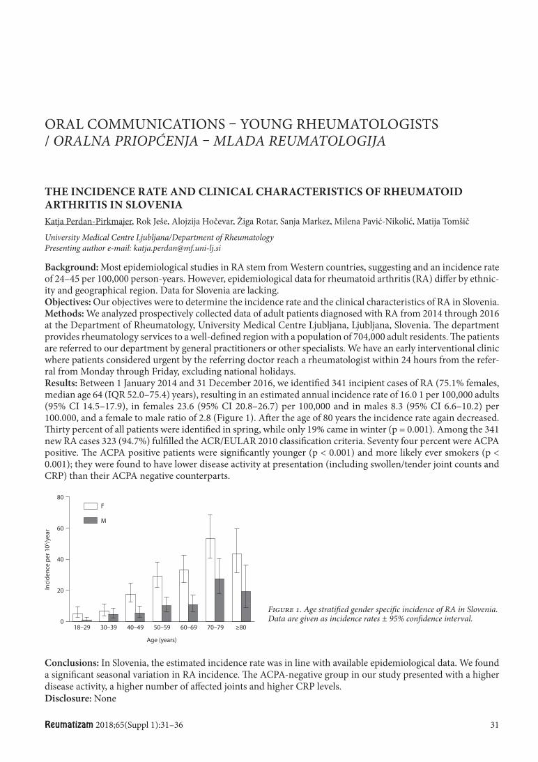
Reumatizam 2018;65(Suppl 1):31–36 31
ORAL COMMUNICATIONS – YOUNG RHEUMATOLOGISTS / ORALNA PRIOPĆENJA – MLADA REUMATOLOGIJA
THE INCIDENCE RATE AND CLINICAL CHARACTERISTICS OF RHEUMATOID ARTHRITIS IN SLOVENIA Katja Perdan-Pirkmajer, Rok Ješe, Alojzija Hočevar, Žiga Rotar, Sanja Markez, Milena Pavić-Nikolić, Matija Tomšič
University Medical Centre Ljubljana/Department of Rheumatology Presenting author e-mail: [email protected]
Background: Most epidemiological studies in RA stem from Western countries, suggesting and an incidence rate of 24–45 per 100,000 person-years. However, epidemiological data for rheumatoid arthritis (RA) diff er by ethnic-ity and geographical region. Data for Slovenia are lacking. Objectives: Our objectives were to determine the incidence rate and the clinical characteristics of RA in Slovenia.Methods: We analyzed prospectively collected data of adult patients diagnosed with RA from 2014 through 2016 at the Department of Rheumatology, University Medical Centre Ljubljana, Ljubljana, Slovenia. Th e department provides rheumatology services to a well-defi ned region with a population of 704,000 adult residents. Th e patients are referred to our department by general practitioners or other specialists. We have an early interventional clinic where patients considered urgent by the referring doctor reach a rheumatologist within 24 hours from the refer-ral from Monday through Friday, excluding national holidays.Results: Between 1 January 2014 and 31 December 2016, we identifi ed 341 incipient cases of RA (75.1% females, median age 64 (IQR 52.0–75.4) years), resulting in an estimated annual incidence rate of 16.0 1 per 100,000 adults (95% CI 14.5–17.9), in females 23.6 (95% CI 20.8–26.7) per 100,000 and in males 8.3 (95% CI 6.6–10.2) per 100.000, and a female to male ratio of 2.8 (Figure 1). Aft er the age of 80 years the incidence rate again decreased. Th irty percent of all patients were identifi ed in spring, while only 19% came in winter (p = 0.001). Among the 341 new RA cases 323 (94.7%) fulfi lled the ACR/EULAR 2010 classifi cation criteria. Seventy four percent were ACPA positive. Th e ACPA positive patients were signifi cantly younger (p < 0.001) and more likely ever smokers (p < 0.001); they were found to have lower disease activity at presentation (including swollen/tender joint counts and CRP) than their ACPA negative counterparts.
Figure 1. Age stratifi ed gender specifi c incidence of RA in Slovenia. Data are given as incidence rates ± 95% confi dence interval.
Conclusions: In Slovenia, the estimated incidence rate was in line with available epidemiological data. We found a signifi cant seasonal variation in RA incidence. Th e ACPA-negative group in our study presented with a higher disease activity, a higher number of aff ected joints and higher CRP levels. Disclosure: None
32 Reumatizam 2018;65(Suppl 1):31–36
Abstracts / Sažeci Oral communications – Young rheumatologists / Oralna priopćenja – mlada reumatologija
VALIDATION OF SLICC-12 AND ACR-97 CLASSIFICATION CRITERIA IN A PATIENT COHORT WITH SLE TREATED IN UNIVERSITY HOSPITAL CENTRE ZAGREB Marija Bakula, Nada Čikeš, Branimir Anić
Division of Clinical Immunology and Rheumatology, Department of Internal medicine, University Hospital Center Zagreb and University of Zagreb School of Medicine, Zagreb, Croatia Presenting author e-mail: [email protected]
Background: Systemic lupus erythematosus (SLE) is a chronic autoimmune disease of multi-organ nature, het-erogenuous clinical presentations and slow accumulation of typical symptoms, thus oft en hard to diagnose in early stages. Most widely used ACR classifi cation from 1997 (ACR-97) shows high specifi city and acceptable sensitivity in classifi ying patients with established SLE, but its value declines in early stages and in milder cases. Th e gold standard for diagnosing SLE and most rheumatic diseases is the experienced rheumatologist’s assess-ment. Eff orts are now put in validation and redefi ning current classifi cation rules. SLICC group has published the results of multiannual revision of the ACR-97 criteria in 2012. Th e new (SLICC-12) criteria have shown higher sensitivity, but lower specifi city than the ACR-97 criteria (97% vs 83% and 84% vs 96%, respectively).Objectives: Th e objective of this study was to validate SLICC-12 and ACR-97 classifi cation criteria on a patient cohort from UHC Zagreb. Overall sensitivity and specifi city, as well as sensitivity and specifi city according to disease duration were compared between the two sets of criteria. Th e value of every criterium and its contribution to diagnosing SLE was calculated.Methods: Th is retrospective observational study comprised 308 patients with SLE (n=146) and SLE-allied condi-tions (n=162). All patient-history charts were evaluated by expert rheumatologists to confi rm clinical diagnosis, regardless of the number of the ACR-97 criteria met. Value of the SLICC-12 and ACR-97 classifi cation criteria was analyzed and compared using logistic regression and ROC curves. Results: A clear distinction between SLICC-12 and ACR-97 criteria is observed. While sensitivity of the SLICC-12 criteria is signifi cantly higher with a tendency to rise as SLE progresses, ACR-97 criteria have shown higher specifi city. Specifi city of the SLICC-12 criteria is low and declines with disease progression. Analysing and com-paring the overall value of the new SLICC-12 classifi cation rule to the ACR-97 criteria, the new criteria show superiority in our patients, with areas under the ROC curve (AUC) 0,79 and 0,70, respectively. Conclusions: Although SLICC-12 criteria show superiority to ACR-97, and are more successful in diagnosing SLE in early stages, the specifi city in our population is too low. Our results contribute to the current initiative for developing new criteria for SLE.References:1. Aggarwal R, Ringold S, Khanna D, et al. Distinctions between diagnostic and clasifi cation criteria. Arthritis Care Res (Hoboken)
2015;67(7):891–72. Singh JA, Solomon DH, Dougados M, et al. Development of classifi cation and response criteria for rheumatic diseases. Arthritis
Rheum 2006;55(3):348–523. Yu C, Gershwin ME, Chang C. Diagnostic criteria for systemic lupus erythematosus: A critical review. J Autoimmun 2014;48–49:
10–134. Petri M, Orbai AM, Alarcón GS, et al. Derivation and validation of the Systemic Lupus International Collaborating Clinics classifi ca-
tion criteria for systemic lupus erythematosus. Arthritis Rheum 2012;64(8):2677–865. Aringer M, Dörner T, Leuchten N, Johnson SR. Toward new criteria for systemic lupus erythematosus-a standpoint. Lupus.
2016;25(8):805–11 Disclosure: None
Reumatizam 2018;65(Suppl 1):31–36 33
Oral communications – Young rheumatologists / Oralna priopćenja – mlada reumatologija Abstracts / Sažeci
SOCIAL MEDIA USE FOR HEALTH-RELATED PURPOSES BY PEOPLE WITH RHEUMATIC AND MUSCULOSKELETAL DISEASES Paul Studenic1, Simon R. Stones2, Alessia Alunno3, Valentin Ritschl4, Elena Nikiphorou5
1 Medical University of Vienna, Department of Internal Medicine 3, Division of Rheumatology, Vienna, Austria 2 University of Leeds, School of Healthcare, Leeds, United Kingdom 3 University of Perugia, Department of Medicine, Rheumatology Unit, Perugia, Italy 4 Medical University of Vienna, Section for Outcomes Research, Vienna, Austria 5 Kings College London, Academic Rheumatology Department, London, United Kingdom Presenting author e-mail: [email protected]
Background: Smartphone applications and social media (SM) transform the way in which people communicate, representing novel opportunities and challenges for people with rheumatic and musculoskeletal diseases (RMDs). Objectives: To explore the use of SM for health related purposes and perspectives of using SM. Methods: A questionnaire-based survey, co-designed in English by rheumatologists and patient research part-ners, translated into 6 languages, then distributed via patient organisations and SM platforms. We report on interims data analyses aft er 5 months of data collection.Results: A total of 809 participants were included in analyses from 57 countries. 40% rated to have good experi-ence in social media. More than half were between 35 and 54 years, 90% were female, 38% had >1 RMD, 61% of participants were multimorbid.96% were using SM and 75% reported using SM for health-related purposes, in particular to connect with other people living with the same condition (53%), via Facebook, YouTube and Google+ being the top 3 used platforms. Th e greatest advantage of SM is to acquire diff erent experiences and exchange knowledge with peers. More than half voiced concerns regarding confi dentiality.SM use and education was comparable between groups with diff erent levels of multimorbidity, although health was poorer (p=0.001), they were older (p=0.001) and more frequently considered that information provided by primary care physicians was inadequate (p=0.035).Conclusions: Th is study demonstrates that use of SM for health-related purposes is widespread among people with RMDs, mainly as a means to connect and exchange knowledge and experience. Despite concerns about confi dentiality the use of SM represents new avenues for health care.
INCREASE IN NON-CLASSICAL SUBPOPULATIONS OF MONOCYTES AND DECREASE IN NUMBERS OF NK CELLS IN THE PRE-CLINICAL PHASE OF RHEUMATOID ARTHRITIS Klára Prajzlerová1, Olga Kryštůfk ová1, Petra Hánová1, Hana Hulejová2, Monika Gregová1, Karel Pavelka1, Jiří Vencovský1, Ladislav Šenolt1, Mária Filková1
1 Department of Rheumatology, First Faculty of Medicine, Charles University and Rheumatology Institute, Prague 2 Rheumatology Institute, Prague Presenting author e-mail: [email protected]
Background: Anti-citrullinated protein antibodies (ACPA) are detectable long before the fi rst manifestation of rheumatoid arthritis (RA). EULAR defi ned a clinical characteristic of individuals with arthralgia suspicious for progression to RA (clinically suspect arthralgia, CSA). Th e alteration of monocyte subpopulations in patients with established RA has been described. Objectives: We aimed to study subpopulations of monocytes and lymphocytes in individuals in the preclinical phase of RA.Methods: Our study enrolled 34 individuals with arthralgia and 80 healthy controls (HC). Leukocytes from peripheral blood were analysed by fl ow cytometry. Lymphocyte subpopulations were defi ned as CD19+CD3–, CD3+CD4+, CD3+CD8+ and CD16/56+CD3– (NK) cells, monocytes were segregated into classical (CD14+CD16–), intermediate (CD14+CD16+/++) and non-classical (CD14–/dimCD16++) subsets. Data were analysed using t-test and Spearman’s correlation.Results: Out of 34 patients with arthralgia, 27 were ACPA+ and 17 met CSA defi nition (10 were ACPA+). Indi-viduals with arthralgia had higher %CD3 and %CD3CD8 T cells and lower %NK and absolute count of NK cells compared to HC. Th is was confi rmed in a subgroup of ACPA+ individuals. Th e count of tender joints correlated

34 Reumatizam 2018;65(Suppl 1):31–36
Abstracts / Sažeci Oral communications – Young rheumatologists / Oralna priopćenja – mlada reumatologija
positively with %CD3 and %CD3CD8 T cells and negatively with %NK cells. Five individuals developed RA within 3–9 months of follow up. Th ese individuals had higher baseline %CD3 cells with the trend for lower %NK cells. Expansion of intermediate and non-classical monocytes with reduction of classical monocytes were dem-onstrated in all individuals with arthralgia compared to HC. Importantly, ACPA+ individuals had higher non-classical and lower classical monocytes compared to ACPA-group. Similarly, ACPA+ individuals had higher intermediate or non-classical and lower classical monocytes compared to HC, while no diff erences were seen between ACPA- individuals and HC.Conclusions: We demonstrate lower NK cells, expansion of intermediate and non-classical monocyte subpopula-tion in individuals in the preclinical phase of RA, especially with ACPA positivity. Our data suggest their role even in early phases of RA development. Th ese subpopulations of leukocytes could be considered as prognostic biomarkers for further development of RA.Acknowledgement: Projects AZV-17-32612A. Disclosure: None
HEMOPHAGOCYTIC LYMPHOHISTIOCYTOSIS Magdalena Wloch-Targonska
Department of Internal and Rheumatic Diseases Medical University of Silesia in Katowice Presenting author e-mail: [email protected]
Hemophagocytic lymphohistiocytosis (HLH) is a rare disorder in which the immune response is dysregulated due to elevated levels of proinfl ammatory cytokines while the activity of NK-cells and cytotoxic T cells is impaired. Th e pathogenesis of HLH might be associated with genetic risk factors, infections, autoimmune disorders and neoplasms. A 24-year-old man with suspected Still’s disease was admitted to the Department of Internal and
Table 1. Laboratory results
End 2014 April/May 2015 May 2015 Start of treatment May / June 2015 August 2016
HGB [g/dl] 14.3 15.2 10.9 10.4 15.8PLT [x103/ul] 241 214 79 24 217Granulocytes rod cells 3%.
segments cells 58%rod cells 11%.
segments cells 74%segments cells 78% segments cells
69.8%Triglycerides[mg/dl] 78 155 778 568 340Fibrinogen [g/dl] 0.95 0.40 0.43Ferritin [ng/ml] 1404 705 >2 000 >40 000 1480WBC [x103/ul] 6.8 7.48 2.41 0.18 10.24ALT [U/l] 21.3 321 275 192 116AST [U/l] 19.3 472 224 636 54ALP [U/l] 63 341 416 444 59GGTP [U/l] 22.7 931 950 76Bilirubin [mg/dl] 0.31 1.06 2.5 7.96 0.86ESR [mm/h] 57 30/59 – – –CRP [mg/l] 73.42 35.3 10.8 6.03PCT [ng/ml] 0.06 0.09Na [mmol/l] 139 138 130 130 137LDH [U/l] 635.6 436 2402Creatinine [mg/dl] 0.82 0.80 1.1 1.79 0.89Uric acid [mg/dl] 6.7 6.6 – 7.1 7.5d-dimers[ng/ml] 860 1220.28 1330 3880 –PT 1.11 3.1 1.77 1.3 –APTT 30.2 44.9 –TSH [uIU/ml] 2.83 1.450 – 1.12 –
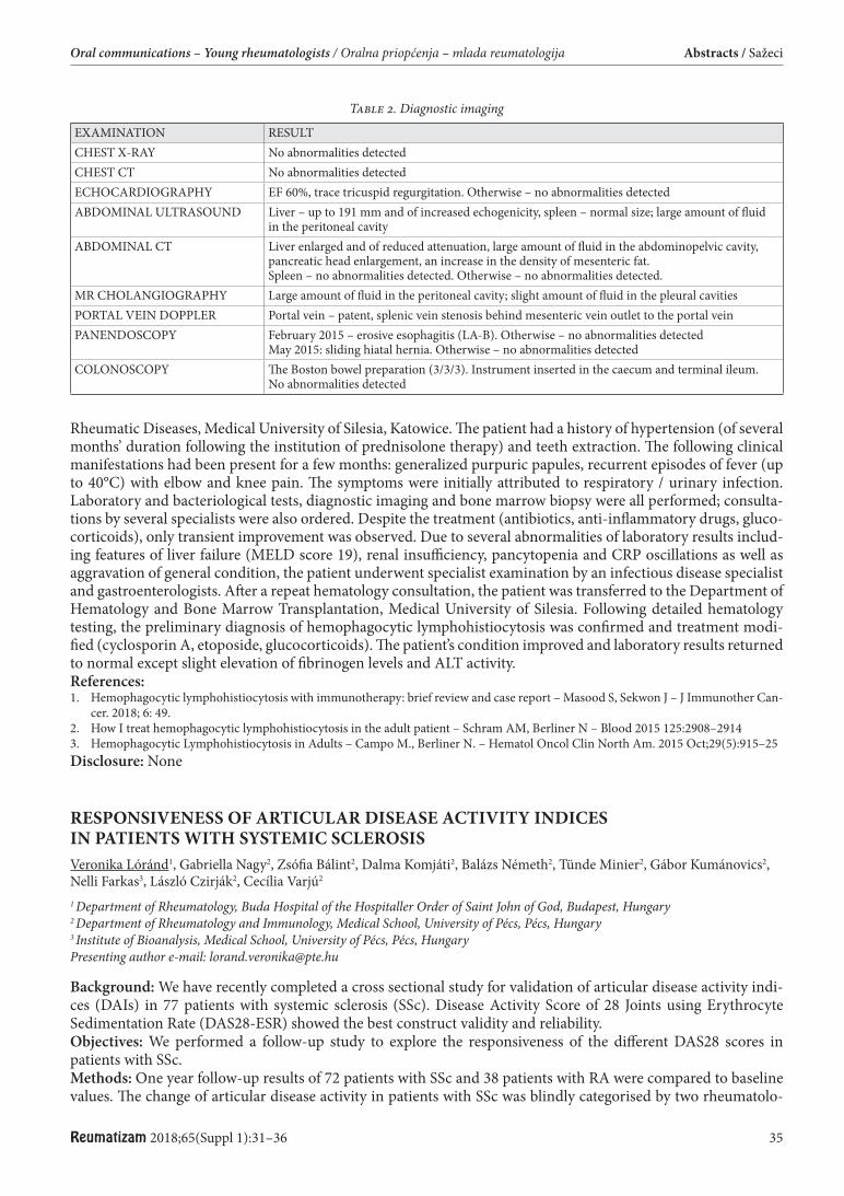
Reumatizam 2018;65(Suppl 1):31–36 35
Oral communications – Young rheumatologists / Oralna priopćenja – mlada reumatologija Abstracts / Sažeci
Rheumatic Diseases, Medical University of Silesia, Katowice. Th e patient had a history of hypertension (of several months’ duration following the institution of prednisolone therapy) and teeth extraction. Th e following clinical manifestations had been present for a few months: generalized purpuric papules, recurrent episodes of fever (up to 40°C) with elbow and knee pain. Th e symptoms were initially attributed to respiratory / urinary infection. Laboratory and bacteriological tests, diagnostic imaging and bone marrow biopsy were all performed; consulta-tions by several specialists were also ordered. Despite the treatment (antibiotics, anti-infl ammatory drugs, gluco-corticoids), only transient improvement was observed. Due to several abnormalities of laboratory results includ-ing features of liver failure (MELD score 19), renal insuffi ciency, pancytopenia and CRP oscillations as well as aggravation of general condition, the patient underwent specialist examination by an infectious disease specialist and gastroenterologists. Aft er a repeat hematology consultation, the patient was transferred to the Department of Hematology and Bone Marrow Transplantation, Medical University of Silesia. Following detailed hematology testing, the preliminary diagnosis of hemophagocytic lymphohistiocytosis was confi rmed and treatment modi-fi ed (cyclosporin A, etoposide, glucocorticoids). Th e patient’s condition improved and laboratory results returned to normal except slight elevation of fi brinogen levels and ALT activity.References:1. Hemophagocytic lymphohistiocytosis with immunotherapy: brief review and case report – Masood S, Sekwon J – J Immunother Can-
cer. 2018; 6: 49.2. How I treat hemophagocytic lymphohistiocytosis in the adult patient – Schram AM, Berliner N – Blood 2015 125:2908–29143. Hemophagocytic Lymphohistiocytosis in Adults – Campo M., Berliner N. – Hematol Oncol Clin North Am. 2015 Oct;29(5):915–25 Disclosure: None
RESPONSIVENESS OF ARTICULAR DISEASE ACTIVITY INDICES IN PATIENTS WITH SYSTEMIC SCLEROSIS Veronika Lóránd1, Gabriella Nagy2, Zsófi a Bálint2, Dalma Komjáti2, Balázs Németh2, Tünde Minier2, Gábor Kumánovics2, Nelli Farkas3, László Czirják2, Cecília Varjú2
1 Department of Rheumatology, Buda Hospital of the Hospitaller Order of Saint John of God, Budapest, Hungary 2 Department of Rheumatology and Immunology, Medical School, University of Pécs, Pécs, Hungary 3 Institute of Bioanalysis, Medical School, University of Pécs, Pécs, Hungary Presenting author e-mail: [email protected]
Background: We have recently completed a cross sectional study for validation of articular disease activity indi-ces (DAIs) in 77 patients with systemic sclerosis (SSc). Disease Activity Score of 28 Joints using Erythrocyte Sedimentation Rate (DAS28-ESR) showed the best construct validity and reliability.Objectives: We performed a follow-up study to explore the responsiveness of the diff erent DAS28 scores in patients with SSc.Methods: One year follow-up results of 72 patients with SSc and 38 patients with RA were compared to baseline values. Th e change of articular disease activity in patients with SSc was blindly categorised by two rheumatolo-
Table 2. Diagnostic imaging
EXAMINATION RESULTCHEST X-RAY No abnormalities detectedCHEST CT No abnormalities detectedECHOCARDIOGRAPHY EF 60%, trace tricuspid regurgitation. Otherwise – no abnormalities detectedABDOMINAL ULTRASOUND Liver – up to 191 mm and of increased echogenicity, spleen – normal size; large amount of fl uid
in the peritoneal cavity ABDOMINAL CT Liver enlarged and of reduced attenuation, large amount of fl uid in the abdominopelvic cavity,
pancreatic head enlargement, an increase in the density of mesenteric fat.Spleen – no abnormalities detected. Otherwise – no abnormalities detected.
MR CHOLANGIOGRAPHY Large amount of fl uid in the peritoneal cavity; slight amount of fl uid in the pleural cavitiesPORTAL VEIN DOPPLER Portal vein – patent, splenic vein stenosis behind mesenteric vein outlet to the portal vein PANENDOSCOPY February 2015 – erosive esophagitis (LA-B). Otherwise – no abnormalities detected
May 2015: sliding hiatal hernia. Otherwise – no abnormalities detected COLONOSCOPY Th e Boston bowel preparation (3/3/3). Instrument inserted in the caecum and terminal ileum.
No abnormalities detected
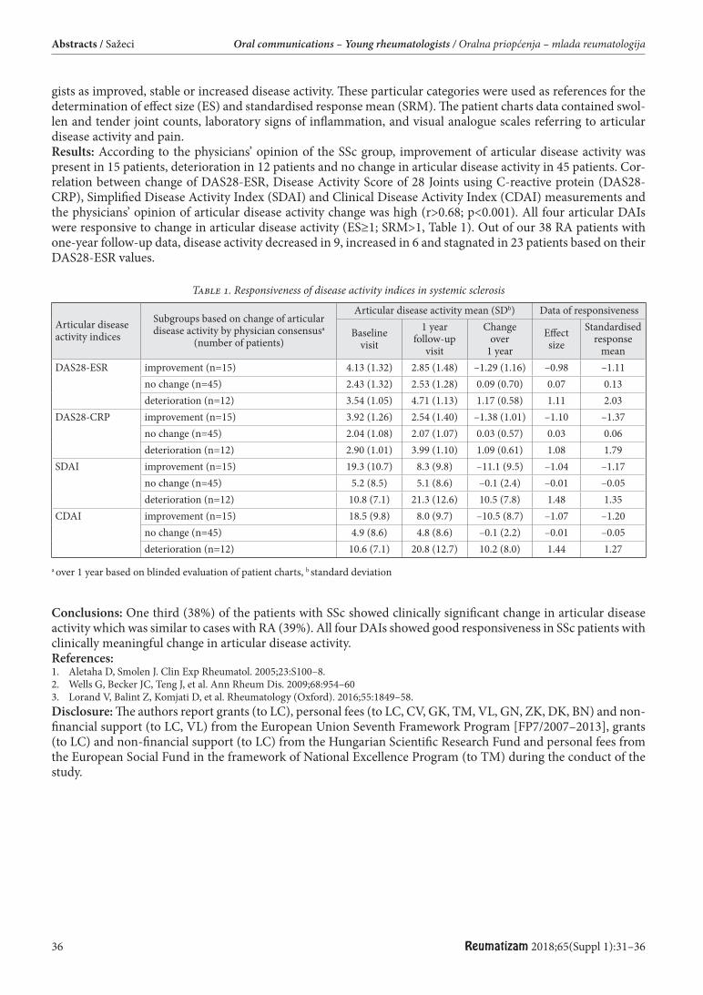
36 Reumatizam 2018;65(Suppl 1):31–36
Abstracts / Sažeci Oral communications – Young rheumatologists / Oralna priopćenja – mlada reumatologija
gists as improved, stable or increased disease activity. Th ese particular categories were used as references for the determination of eff ect size (ES) and standardised response mean (SRM). Th e patient charts data contained swol-len and tender joint counts, laboratory signs of infl ammation, and visual analogue scales referring to articular disease activity and pain.Results: According to the physicians’ opinion of the SSc group, improvement of articular disease activity was present in 15 patients, deterioration in 12 patients and no change in articular disease activity in 45 patients. Cor-relation between change of DAS28-ESR, Disease Activity Score of 28 Joints using C-reactive protein (DAS28-CRP), Simplifi ed Disease Activity Index (SDAI) and Clinical Disease Activity Index (CDAI) measurements and the physicians’ opinion of articular disease activity change was high (r>0.68; p<0.001). All four articular DAIs were responsive to change in articular disease activity (ES≥1; SRM>1, Table 1). Out of our 38 RA patients with one-year follow-up data, disease activity decreased in 9, increased in 6 and stagnated in 23 patients based on their DAS28-ESR values.
Table 1. Responsiveness of disease activity indices in systemic sclerosis
Articular disease activity indices
Subgroups based on change of articular disease activity by physician consensusa
(number of patients)
Articular disease activity mean (SDb) Data of responsiveness
Baseline visit
1 year follow-up
visit
Change over
1 yearEff ect size
Standardised response
meanDAS28-ESR improvement (n=15) 4.13 (1.32) 2.85 (1.48) –1.29 (1.16) –0.98 –1.11
no change (n=45) 2.43 (1.32) 2.53 (1.28) 0.09 (0.70) 0.07 0.13deterioration (n=12) 3.54 (1.05) 4.71 (1.13) 1.17 (0.58) 1.11 2.03
DAS28-CRP improvement (n=15) 3.92 (1.26) 2.54 (1.40) –1.38 (1.01) –1.10 –1.37no change (n=45) 2.04 (1.08) 2.07 (1.07) 0.03 (0.57) 0.03 0.06deterioration (n=12) 2.90 (1.01) 3.99 (1.10) 1.09 (0.61) 1.08 1.79
SDAI improvement (n=15) 19.3 (10.7) 8.3 (9.8) –11.1 (9.5) –1.04 –1.17no change (n=45) 5.2 (8.5) 5.1 (8.6) –0.1 (2.4) –0.01 –0.05deterioration (n=12) 10.8 (7.1) 21.3 (12.6) 10.5 (7.8) 1.48 1.35
CDAI improvement (n=15) 18.5 (9.8) 8.0 (9.7) –10.5 (8.7) –1.07 –1.20no change (n=45) 4.9 (8.6) 4.8 (8.6) –0.1 (2.2) –0.01 –0.05deterioration (n=12) 10.6 (7.1) 20.8 (12.7) 10.2 (8.0) 1.44 1.27
a over 1 year based on blinded evaluation of patient charts, b standard deviation
Conclusions: One third (38%) of the patients with SSc showed clinically signifi cant change in articular disease activity which was similar to cases with RA (39%). All four DAIs showed good responsiveness in SSc patients with clinically meaningful change in articular disease activity.References:1. Aletaha D, Smolen J. Clin Exp Rheumatol. 2005;23:S100–8.2. Wells G, Becker JC, Teng J, et al. Ann Rheum Dis. 2009;68:954–603. Lorand V, Balint Z, Komjati D, et al. Rheumatology (Oxford). 2016;55:1849–58.Disclosure: Th e authors report grants (to LC), personal fees (to LC, CV, GK, TM, VL, GN, ZK, DK, BN) and non-fi nancial support (to LC, VL) from the European Union Seventh Framework Program [FP7/2007–2013], grants (to LC) and non-fi nancial support (to LC) from the Hungarian Scientifi c Research Fund and personal fees from the European Social Fund in the framework of National Excellence Program (to TM) during the conduct of the study.
Reumatizam 2018;65(Suppl 1):37–74 37
POSTERS / POSTERI
ADVANCES IN MANAGEMENT OF SYSTEMIC LUPUS ERYTHEMATOSUS Jozef Lukáč1, Oľga Lukáčová2
1 Institute of Physiotherapy, Balneology and Medical Rehabilitation in Piešťany, UCM 2National Institute of Rheumatic Diseases, Piešťany, Slovak Republic 2 National Institute of rheumatic diseases, Piešťany, Slovak republic Presenting author e-mail: [email protected]
Survival and prognosis of patients with SLE was improved markedly. Progress is not only in new therapeutic modalities but also in early diagnostics, assessment of activity, prognosis and monitoring focused on eff ects and adverse eff ects of therapy, and comorbidities.For treatment of the most serious forms as neuropsychiatric lupus, lupus nephritis (LN), pulmonary alveolar haemorrhagy are indicated high doses of glucocorticoids (GC) or pulse therapy (PT) of metylprednisolone and cyclophosphamide. In refractory forms of lupus high doses of immunoglobulins, plasmapheresis, immunoad-sorption is indicated. Bone marrow ablative chemotherapy and transplantation with stem cell reconstruction is seldom used method of therapy. Biologic therapy with rituximab is the most frequent used biological therapy in severe forms of lupus. In maintenance phase is proved biological therapy with belimumab, mycophenolate mofetil, azatioprine, methotrexate and calcineurin inhibitors. Belimumab is used in moderate forms of SLE. Hydroxy-chlorochine is appropriate to combination with other therapy in all patients with SLE. New biologics are targeted to I IFNa, IL-12/23 and JAK-STAT inhibition. Aft er suppression of SLE activity is necessary to reduce of GC to the lowest eff ective dosages.Mobidity and mortality in late stages of lupus is caused by organ damages (estimated by SLICC Damage Index) and comorbidities. It is associated with age, duration of the disease, number of relapses and high doses of GC. Cardiovascular diseases are the most frequent complications of SLE. On them participate diabetes, hypertension, hypercholesterolemia, metabolic syndrome, nephropathy, hyperhomocyteinaemia and premature menopause. Statins are prevention for thromboses in patient with antiphospholipid antibodies ; statins aren´t risk factor for clinical worsening of SLE.Aim of SLE therapy is low activity or remission with as low as possible dosages of GC.Management of SLE requires monitoring activity, damage index, prevention and therapy cardiovascular compli-cations and comorbidities. Tight monitoring leads to stabilization of damage index and prognosis improvement. Disclosure: None
WHEN A RHEUMATOLOGIC DISEASE GETS A HEAD START. A CASE REPORT OF SERONEGATIVE ANTIPHOSPHOLIPID SYNDROME Marta Skoczynska1, Malgorzata Chowaniec2, Agata Sebastian3, Maria Misterska – Skora4, Piotr Wiland2
1 Department and Clinic of Rheumatology and Internal Diseases, Wroclaw Medical University, Poland 2 Department of Rheumatology and Internal Medicine, Wroclaw University Hospital, Department of Hygiene, Wroclaw Medical University, Poland 3 Department and Clinic of Rheumatology and Internal Diseases, Wroclaw Medical University, Poland 4 Department of Rheumatology and Internal Medicine, Wroclaw University Hospital, Poland Presenting author e-mail: [email protected]
We present a case report of a 56 year old woman with an established diagnosis of systemic lupus erythematosus and a history of multiple thrombotic events, i.e. two ischaemic strokes, myocardial infarction and recurrent deep
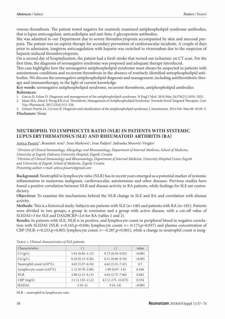
38 Reumatizam 2018;65(Suppl 1):37–74
Abstracts / Sažeci Posters / Posteri
venous thrombosis. Th e patient tested negative for routinely examined antiphospholipid syndrome antibodies, that is lupus anticoagulant, anticardiolipin and anti-beta-2-glycoprotein antibodies.She was admitted to our Department due to severe thrombocytopenia accompanied by skin and mucosal pur-pura. Th e patient was on aspirin therapy for secondary prevention of cardiovascular incidents. A couple of days prior to admission, longterm anticoagulation with heparin was switched to rivaroxaban due to the suspicion of heparin-induced thrombocytopenia.On a second day of hospitalization, the patient had a fresh stroke that turned out ischaemic on CT scan. For the fi rst time, the diagnosis of seronegative syndrome was proposed and adequate therapy introduced.Th is case highlights how the seronegative antiphospholipid syndrome must always be suspected in patients with autoimmune conditions and recurrent thrombosis in the absence of routinely identifi ed antiophospholipid anti-bodies. We discuss the seronegative antiphospholipid diagnosis and management, including antithrombotic ther-apy and immunotherapy, in the light of current knowledge.Key words: seronegative antiphospholipid syndrome, recurrent thrombosis, antiphospholipid antibodiesReferences:1. Garcia D, Erkan D. Diagnosis and management of the antiphospholipid syndrome. N Engl J Med. 2018 May 24;378(21):2010–2021.2. Islam MA, Alam F, Wong KK et al. Th rombotic Management of Antiphospholipid Syndrome: Towards Novel Targeted Th erapies. Curr
Vasc Pharmacol. 2017;15(4):313–326.3. Gómez-Puerta JA, Cervera R. Diagnosis and classifi cation of the antiphospholipid syndrome, J Autoimmun. 2014 Feb–Mar;48–49:20–5.Disclosure: None
NEUTROPHIL TO LYMPHOCYTE RATIO (NLR) IN PATIENTS WITH SYSTEMIC LUPUS ERYTHEMATOSUS (SLE) AND RHEUMATOID ARTHRITIS (RA) Antica Pasarić1, Branimir Anić2, Ivan Marković1, Ivan Padjen2, Jadranka Morović-Vergles1
1 Division of Clinical Immunology, Allergology and Rheumatology, Department of Internal Medicine, School of Medicine, University of Zagreb, Dubrava University Hospital, Zagreb, Croatia 2 Division of Clinical Immunology and Rheumatology, Department of Internal Medicine, University Hospital Centre Zagreb and University of Zagreb, School of Medicine, Zagreb, Croatia Presenting author e-mail: [email protected]
Background: Neutrophil to lymphocyte ratio (NLR) has in recent years emerged as a potential marker of systemic infl ammation in numerous malignant, cardiovascular, autoimmune and other diseases. Previous studies have found a positive correlation between NLR and disease activity in RA patients, while fi ndings for SLE are contra-dictory.Objectives: To examine the mechanisms behind the NLR change in SLE and RA and correlation with disease activity.Methods: Th is is a historical study. Subjects are patients with SLE (n=146) and patients with RA (n=181). Patients were divided in two groups, a group in remission and a group with active disease, with a cut-off value of SLEDAI=3 for SLE and DAS28CRP=2.6 for RA (tables 1 and 2).Results: In patients with SLE, NLR is in positive, and lymphocyte count in peripheral blood in negative correla-tion with SLEDAI (NLR: r=0.165,p=0.046; lymphocyte count: r=–0.173,p=0.037) and plasma concentration of CRP (NLR: r=0.233,p=0.005; lymphocyte count: r=–0.287,p=0.001), while a change in neutrophil count is insig-
Table 1. Clinical characteristics of SLE patients.
Characteristics ( ) ( ) valueC3 (g/L) 1.03 (0.82–1.13) 0.73 (0.54–0.92) <0.001C4 (g/L) 0.18 (0.13–0.26) 0.11 (0.06–0.19) <0.001Neutrophil count (x109/L) 4.63 (3.37–6.34) 4.62 (3.31–7.41) 0.7Lymphocyte count (x109/L) 1.12 (0.78–2.08) 1.09 (0.67–1.6) 0.166NLR 2.88 (2.15–6.15) 4.65 (2.75–7.06) 0.062CRP (mg/L) 3.1 (1.125–11.2) 4.5 (1.175–12.675) 0.534SLEDAI 2 (0–2) 9 (6–14) <0.001
NLR – neutrophil to lymphocyte ratio
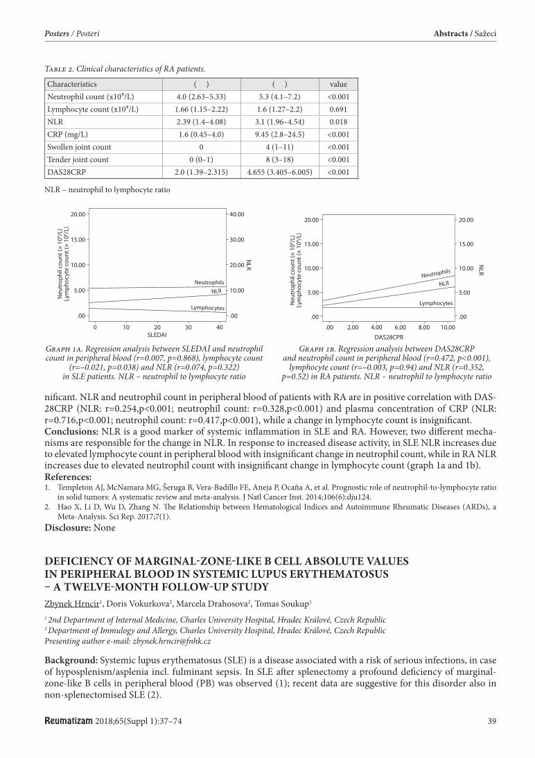
Reumatizam 2018;65(Suppl 1):37–74 39
Posters / Posteri Abstracts / Sažeci
nifi cant. NLR and neutrophil count in peripheral blood of patients with RA are in positive correlation with DAS-28CRP (NLR: r=0.254,p<0.001; neutrophil count: r=0.328,p<0.001) and plasma concentration of CRP (NLR: r=0.716,p<0.001; neutrophil count: r=0.417,p<0.001), while a change in lymphocyte count is insignifi cant. Conclusions: NLR is a good marker of systemic infl ammation in SLE and RA. However, two diff erent mecha-nisms are responsible for the change in NLR. In response to increased disease activity, in SLE NLR increases due to elevated lymphocyte count in peripheral blood with insignifi cant change in neutrophil count, while in RA NLR increases due to elevated neutrophil count with insignifi cant change in lymphocyte count (graph 1a and 1b).References:1. Templeton AJ, McNamara MG, Šeruga B, Vera-Badillo FE, Aneja P, Ocaña A, et al. Prognostic role of neutrophil-to-lymphocyte ratio
in solid tumors: A systematic review and meta-analysis. J Natl Cancer Inst. 2014;106(6):dju124.2. Hao X, Li D, Wu D, Zhang N. Th e Relationship between Hematological Indices and Autoimmune Rheumatic Diseases (ARDs), a
Meta-Analysis. Sci Rep. 2017;7(1). Disclosure: None
DEFICIENCY OF MARGINAL-ZONE-LIKE B CELL ABSOLUTE VALUES IN PERIPHERAL BLOOD IN SYSTEMIC LUPUS ERYTHEMATOSUS – A TWELVE-MONTH FOLLOW-UP STUDY Zbynek Hrncir1, Doris Vokurkova2, Marcela Drahosova2, Tomas Soukup1
1 2nd Department of Internal Medicine, Charles University Hospital, Hradec Králové, Czech Republic 2 Department of Immulogy and Allergy, Charles University Hospital, Hradec Králové, Czech Republic Presenting author e-mail: [email protected]
Background: Systemic lupus erythematosus (SLE) is a disease associated with a risk of serious infections, in case of hyposplenism/asplenia incl. fulminant sepsis. In SLE aft er splenectomy a profound defi ciency of marginal-zone-like B cells in peripheral blood (PB) was observed (1); recent data are suggestive for this disorder also in non-splenectomised SLE (2).
Graph 1a. Regression analysis between SLEDAI and neutrophil count in peripheral blood (r=0.007, p=0.868), lymphocyte count
(r=–0.021, p=0.038) and NLR (r=0.074, p=0.322) in SLE patients. NLR – neutrophil to lymphocyte ratio
Graph 1b. Regression analysis between DAS28CRP and neutrophil count in peripheral blood (r=0.472, p<0.001),
lymphocyte count (r=–0.003, p=0.94) and NLR (r=0.352, p=0.52) in RA patients. NLR – neutrophil to lymphocyte ratio
Table 2. Clinical characteristics of RA patients.
Characteristics ( ) ( ) valueNeutrophil count (x109/L) 4.0 (2.63–5.33) 5.3 (4.1–7.2) <0.001Lymphocyte count (x109/L) 1.66 (1.15–2.22) 1.6 (1.27–2.2) 0.691NLR 2.39 (1.4–4.08) 3.1 (1.96–4.54) 0.018CRP (mg/L) 1.6 (0.45–4.0) 9.45 (2.8–24.5) <0.001Swollen joint count 0 4 (1–11) <0.001Tender joint count 0 (0–1) 8 (3–18) <0.001DAS28CRP 2.0 (1.39–2.315) 4.655 (3.405–6.005) <0.001
NLR – neutrophil to lymphocyte ratio
40 Reumatizam 2018;65(Suppl 1):37–74
Abstracts / Sažeci Posters / Posteri
Objectives: Investigation of marginal-zone-like (CD19+ CD27+ IgM+) B cells in PB as a possible member of pathway associated with a risk of infection in non-splenectomised SLE.Methods: Sixty adult SLE (ACR/1982, updated 1997) pts at a level LLDAS (lupus low disease activity state; 3) and 10 age and sex-matched healthy controls (HC) were enrolled in month 0´, and 56 pts persistently at LLDAS also repeatedly aft er twelve-month-period, i. e. month 12´; overlap syndromes, infection, renal failure and monoclo-nal gammopathy in SLE under study were excluded. Th e DuraClone IM panel (Beckman Coulter) was used to identify CD19+ CD27+ IgM+ B cell subpopulation in PB samples by fl ow cytometry Navios (Beckman Coulter) with soft ware analysis using Kaluza version 1.2; data obtained were expressed not only in relative % of PB lym-phocytes, but also in absolute values x106 /L, and statistically processed using Medcalc-Statistical Soft ware pro-gramme.Results: Signifi cant diff erences (p<0.001) were obtained between absolute values of CD19+ CD27+ IgM+ B cells in HC (median 31.36, 95%CI:24.49-63.35) and SLE month 0´ (median 9.82, 95%CI:6.01-14.26) and also SLE month 12´(median 9.82,95%CI:7.12-14.42), but not between values obtained in SLE month 0´ and month 12´(p>0.05); not signifi cant diff rerences were found in analysis using relative % PB lymphocytes (p>0.05).Conclusions: Th e data obtained demonstrated signifi cant and persistent character of defi ciency of marginal-zone-like B cell absolute values in peripheral blood in LLDAS SLE. In this connection is suggested that the change observed should be a component of pathway associated with a risk of infections in SLE, but further studies are necessary.References:1. Hrncir Z et al.: Clin Exper Rheumatol 2016;34 (S99):S–63.2. Hrncir Z et al.: Lupus Sci Med 2018;5:S1/A83.2.3. Franklyn K et al.: Ann Rheum Dis 2016;75:1615–1621.Acknowledgement. Supported by the Charles University research project PROGRES Q40-15. Disclosure: None
EFFICACY OF BIOLOGIC TREATMENT ON QUALITY OF LIFE IN RHEUMATOID ARTHRITIS PATIENTS IN CROATIA: RESULTS FROM A NON-INTERVENTIONAL, MULTICENTER, CROSS-SECTIONAL STUDY TO ESTIMATE DISEASE ACTIVITY AND TREATMENT PATTERNS IN PATIENTS WITH RHEUMATOID ARTHRITIS Frane Grubišić1, Katarina Borić2, Željka Kardum3, Tatjana Kehler4, Danijela Krstulović Marasović2, Nikolina Ljubičić Marković5, Sonja Milanović6, Jadranka Morović Vergles7, Joško Mitrović7, Višnja Prus3, Ksenija Maštrović Radončić8, Ivana Rudar Tomljanović9, Simeon Grazio1
1 Department of Rheumatology, Physical Medicine and Rehabilitation, School of Medicine University of Zagreb, Sestre Milosrdnice University Hospital Center, Zagreb, Croatia 2 Division of Rheumatology and Clinical Immunology, University Hospital Center Split, Split, Croatia 3 Department of Rheumatology, Clinical Immunology and Allergology, Osijek University Hospital, Internal Clinic, Faculty of Medicine, University of Osijek, Osijek, Croatia 4 Rheumatology, Physical Medicine and Rehabilitation, Th allasotherapia Opatija, Opatija, Croatia 5 Department of Physical Medicine, Rehabilitation and Rheumatology, Vukovar General Hospital 6 Department of Physical Medicine and Rehabilitation, General Hospital Karlovac 7 Division of Clinical Immunology, Allergology and Rheumatology, Department of Internal Medicine, School of Medicine University of Zagreb, University Hospital Dubrava, Zagreb, Croatia 8 Department of Rheumatology, Clinical Hospital “Sveti Duh” 9 Merck Sharp & Dohme, d.o.o., Zagreb, Croatia Presenting author e-mail: [email protected]
Background: Achieving optimal QoL is the ultimate goal of any therapeutic intervention, including biologic therapy, in patients with RA. Objectives: To assess the effi cacy of biologic therapy on QoL in RA patients in Croatia. Methods: Th ere were 130 patients included (19 men, 111 women) with established diagnosis of RA based on 1984 ACR criteria. Patients were consequently selected from eight rheumatology centers across Croatia. Practising rheumatologist collected demographic information, information about medications (both conventional and bio-logic disease modifying antirheumatic drugs). DAS28 was used to assess disease activity. All the patients fi lled SF-12 questionnaire, as a measure of QoL.
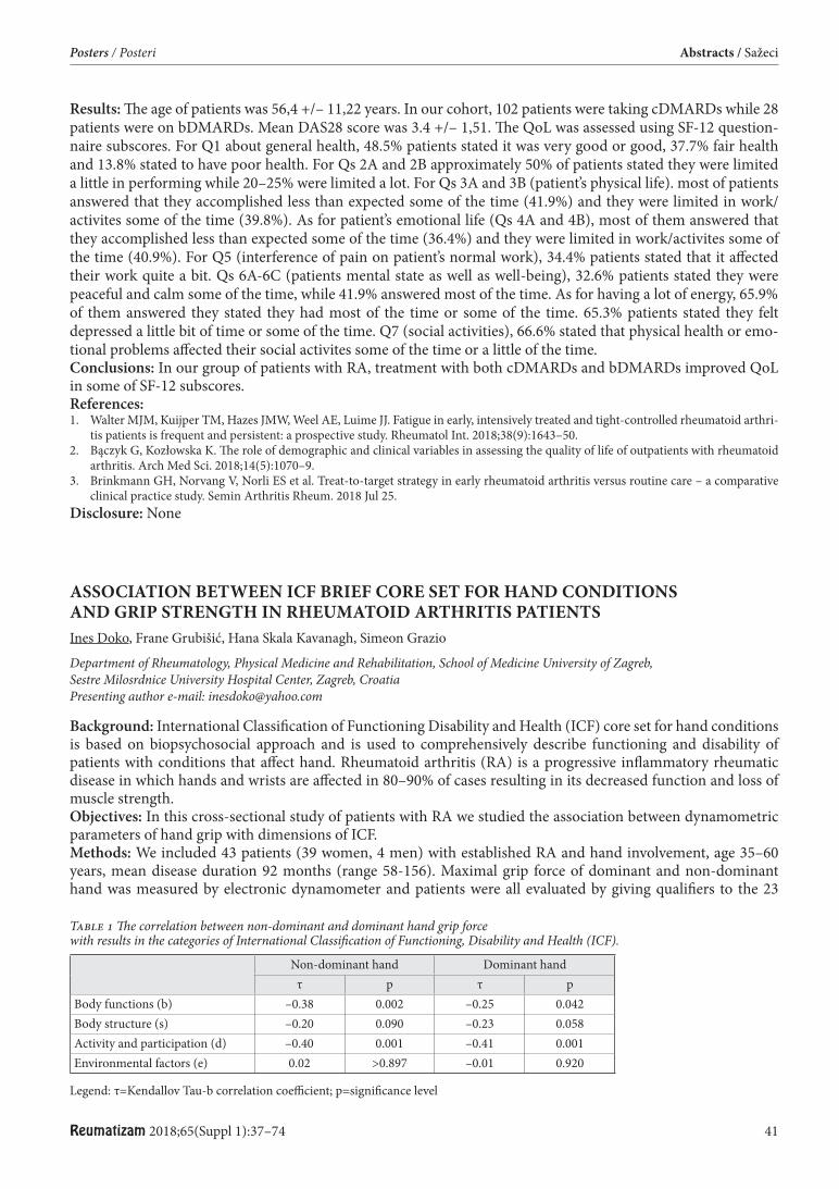
Reumatizam 2018;65(Suppl 1):37–74 41
Posters / Posteri Abstracts / Sažeci
Results: Th e age of patients was 56,4 +/– 11,22 years. In our cohort, 102 patients were taking cDMARDs while 28 patients were on bDMARDs. Mean DAS28 score was 3.4 +/– 1,51. Th e QoL was assessed using SF-12 question-naire subscores. For Q1 about general health, 48.5% patients stated it was very good or good, 37.7% fair health and 13.8% stated to have poor health. For Qs 2A and 2B approximately 50% of patients stated they were limited a little in performing while 20–25% were limited a lot. For Qs 3A and 3B (patient’s physical life). most of patients answered that they accomplished less than expected some of the time (41.9%) and they were limited in work/activites some of the time (39.8%). As for patient’s emotional life (Qs 4A and 4B), most of them answered that they accomplished less than expected some of the time (36.4%) and they were limited in work/activites some of the time (40.9%). For Q5 (interference of pain on patient’s normal work), 34.4% patients stated that it aff ected their work quite a bit. Qs 6A-6C (patients mental state as well as well-being), 32.6% patients stated they were peaceful and calm some of the time, while 41.9% answered most of the time. As for having a lot of energy, 65.9% of them answered they stated they had most of the time or some of the time. 65.3% patients stated they felt depressed a little bit of time or some of the time. Q7 (social activities), 66.6% stated that physical health or emo-tional problems aff ected their social activites some of the time or a little of the time.Conclusions: In our group of patients with RA, treatment with both cDMARDs and bDMARDs improved QoL in some of SF-12 subscores. References:1. Walter MJM, Kuijper TM, Hazes JMW, Weel AE, Luime JJ. Fatigue in early, intensively treated and tight-controlled rheumatoid arthri-
tis patients is frequent and persistent: a prospective study. Rheumatol Int. 2018;38(9):1643–50.2. Bączyk G, Kozłowska K. Th e role of demographic and clinical variables in assessing the quality of life of outpatients with rheumatoid
arthritis. Arch Med Sci. 2018;14(5):1070–9.3. Brinkmann GH, Norvang V, Norli ES et al. Treat-to-target strategy in early rheumatoid arthritis versus routine care – a comparative
clinical practice study. Semin Arthritis Rheum. 2018 Jul 25. Disclosure: None
ASSOCIATION BETWEEN ICF BRIEF CORE SET FOR HAND CONDITIONS AND GRIP STRENGTH IN RHEUMATOID ARTHRITIS PATIENTS Ines Doko, Frane Grubišić, Hana Skala Kavanagh, Simeon Grazio
Department of Rheumatology, Physical Medicine and Rehabilitation, School of Medicine University of Zagreb, Sestre Milosrdnice University Hospital Center, Zagreb, Croatia Presenting author e-mail: [email protected]
Background: International Classifi cation of Functioning Disability and Health (ICF) core set for hand conditions is based on biopsychosocial approach and is used to comprehensively describe functioning and disability of patients with conditions that aff ect hand. Rheumatoid arthritis (RA) is a progressive infl ammatory rheumatic disease in which hands and wrists are aff ected in 80–90% of cases resulting in its decreased function and loss of muscle strength.Objectives: In this cross-sectional study of patients with RA we studied the association between dynamometric parameters of hand grip with dimensions of ICF.Methods: We included 43 patients (39 women, 4 men) with established RA and hand involvement, age 35–60 years, mean disease duration 92 months (range 58-156). Maximal grip force of dominant and non-dominant hand was measured by electronic dynamometer and patients were all evaluated by giving qualifi ers to the 23
Table 1 Th e correlation between non-dominant and dominant hand grip force with results in the categories of International Classifi cation of Functioning, Disability and Health (ICF).
Non-dominant hand Dominant hand τ p τ p
Body functions (b) –0.38 0.002 –0.25 0.042Body structure (s) –0.20 0.090 –0.23 0.058Activity and participation (d) –0.40 0.001 –0.41 0.001Environmental factors (e) 0.02 >0.897 –0.01 0.920
Legend: τ=Kendallov Tau-b correlation coeffi cient; p=signifi cance level
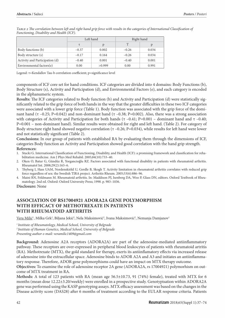
42 Reumatizam 2018;65(Suppl 1):37–74
Abstracts / Sažeci Posters / Posteri
components of ICF core set for hand conditions. ICF categories are divided into 4 domains: Body Functions (b), Body Structure (s), Activity and Participation (d), and Environmental Factors (e), and each category is encoded in the alphanumeric system.Results: Th e ICF categories related to Body function (b) and Activity and Participation (d) were statistically sig-nifi cantly related to the grip force of both hands in the way that the greater diffi culties in these two ICF categories were associated with a lower grip force (Table 1). Body function was associated with the grip force of the domi-nant hand (τ –0.25; P=0.042) and non-dominant hand (τ –0.38; P=0.002). Also, there was a strong association with categories of Activity and Participation for both hands (τ –0.41; P=0.001 – dominant hand and τ –0.40; P=0.001 – non-dominant hand). Similar results were obtained for right and left hand. (Table 2). For category of Body structure right hand showed negative correlation (τ –0.26; P=0.034), while results for left hand were lower and not statistically signifi cant (Table 2). Conclusions: In our group of patients with established RA by evaluating them through the dimensions of ICF, categories Body function an Activity and Participation showed good correlation with the hand grip strength.References:1. Stucki G. International Classifi cation of Functioning, Disability, and Health (ICF): a promising framework and classifi cation for reha-
bilitation medicine. Am J Phys Med Rehabil. 2005;84(10):733–40.2. Oken O, Batur G, Gündüz R, Yorgancioğlu RZ. Factors associated with functional disability in patients with rheumatoid arthritis.
Rheumatol Int. 2008;29(2):163–6.3. Th yberg I, Hass UAM, Nordenskiöld U, Gerdle B, Skogh T. Activity limitation in rheumatoid arthritis correlates with reduced grip
force regardless of sex: the Swedish TIRA project. Arthritis Rheum. 2005;53(6):886–96.4. Maini RN, Feldmann M. Rheumatoid arthritis. In: Maddison PJ, Isenberg DA, Woo P, Glass DN, editors. Oxford Textbook of Rheu-
matology. 2nd ed. Oxford: Oxford University Press; 1998. p. 983–1036.Disclosure: None
ASSOCIATION OF RS17004921 ADORA2A GENE POLYMORPHISM WITH EFFICACY OF METHOTREXATE IN PATIENTS WITH RHEUMATOID ARTHRITIS Vera Milić1, Milka Grk2, Biljana Jekić2, Nela Maksimović2, Ivana Maksimović2, Nemanja Damjanov1
1 Institute of Rheumatology, Medical School, University of Belgrade 2 Institute of Human Genetics, Medical School, University of Belgrade Presenting author e-mail: [email protected]
Background: Adenosine A2A receptors (ADORA2A) are part of the adenosine-mediated antiinfl ammatory pathway. Th ese receptors are over-expressed in peripheral blood leukocytes of patients with rheumatoid artritis (RA). Methotrexate (MTX), the gold standard for therapy, exerts its antiinfl amatory eff ects via increased release of adenosine into the extracellular space. Adenosine binds to ADOR A2A and A3 and initiates an antiinfl amma-tory response. Th erefore, ADOR gene polymorphisms could have an impact on MTX therapy outcome.Objectives: To examine the role of adenosine receptor 2A gene (ADORA2A, rs 17004921) polymorhism on out-come of MTX treatment in RA.Methods: A total of 123 patients with RA (mean age 56.5±10.73, 91 (74%) female), treated with MTX for 6 months (mean dose 12.22±3.20/weekly) were enrolled in a prospective study. Genotypisation within ADORA2A gene was performed using the KASP genotyping assays. MTX effi cacy assessment was based on the changes in the Disease activity score (DAS28) aft er 6 months of treatment according to the EULAR response criteria. Patients
Table 2 Th e correlation between left and right hand grip force with results in the categories of International Classifi cation of Functioning, Disability and Health (ICF).
Left hand Right hand τ p Τ p
Body functions (b) –0.37 0.002 –0.26 0.034Body structure (s) –0.17 0.164 –0.26 0.034Activity and Participation (d) –0.40 0.001 –0.40 0.001Environmental factors(e) 0.00 >0.999 0.00 0.991
Leg end: τ=Kendallov Tau-b correlation coeffi cient; p=signifi cance level
Reumatizam 2018;65(Suppl 1):37–74 43
Posters / Posteri Abstracts / Sažeci
with good and moderate response were classifi ed as „responders“, whereas patients with poor response were con-sidered „nonresponders“. Data of adverse eff ects were collected during this period. MTX effi cacy and toxicity were compared among patients with diff erent genotypes. Results: Median DAS 28 at the beginning of MTX treatment was 7.43±0.89. According to EULAR response cri-teria, 111 (90.2%) patients with RA were classifi ed as responders. Among all RA patients, 97 (78.9%) had CC, 20 (16.3%) CT and 6 (4.9%) TT genotype. Th e distribution of ADORA2 genotypes in responders (CC 77.5%, CT 17% and TT 5.4%) was not signifi cantly diff erent from nonresponders (CC 91.7%, CT 8.3%), p>0.05. Aft er 6 months, 26 carriers of T allele (CT+TT) had higher reduction DAS 28 (7.16 vs. 3.88) in comparison to other 97 patients (7.50 vs.4.64, p=0.013). Adverse eff ects were reported in 24 (19.5%) patients. Most of patients had hepa-totoxicity and nausea, 14 (58%) and 9 (37%) respectively. No statistically signifi cant association between ADORA2A genotype and side events has been observed (p>0.05). Conclusions: According to our results, T allele of ADORA2A rs17004921 polymorphism may have favourable infl uence on effi cacy in RA patients treated with methotexatate. References:1. Cronstein B, Sitkovsky M. Adenosine and adenosine receptors in the pathogenesis and treatment of rheumatic diseasess. www.nature.
com/nrrheum2. Hider SL, Th omson W, Mack LF et al. Polymorhisms within the adenosine receptor 2a gene are associated with adverse events in RA
patients treated with MTX. Rheumatology 2008;47:1156–1159. Disclosure: None
TREATMENT OPTIONS IN PATIENT WITH RHEUMATOID ARTHRITIS AND HISTORY OF MALIGNANCY – INTRACRANIAL CHONDROSARCOMA/OSTEOCHONDROMA Ivana Jezic1, Marko Baresic1, Luka Simetic2, Davorin Herceg2, Branimir Anic1
1 Division of Clinical Immunology and Rheumatology, Department of Internal medicine, University Hospital Center Zagreb and University of Zagreb School of Medicine, Zagreb, Croatia 2 Department of Oncology, University Hospital Center Zagreb, Croatia Presenting author e-mail: [email protected]
Background: Biologics are routinely being used as therapy for rheumatoid arthritis (RA), but there aren’t many available studies that include enough data about their safety in treatment of patients with prior malignancy (1). We are presenting a patient with RA and intracranial chondrosarcoma/osteochondroma treated with rituximab (RTX). Intracranial chondrosarcoma of the sellar region is an extremely rare tumor with only few cases described in literature (1).Methods: A 47-year old male patient with a positive family history of RA (mother) was diagnosed with a low grade malignancy chondrosarcoma with the elements of osteochondroma of the left parasellar region and cavern-ous sinus in 2006. He underwent the microneurosurgical procedure where tumor was reduced. Aft er that, he was treated with Gamma knife (18 Gy) and continued regular follow-ups. Two years aft er, he developed pain and swelling of small joints of his hands and was diagnosed with seropositive (RF and ACPA) erosive RA.Results: Patient was treated with NSAIDs, low dose prednisone and methotrexate (MTX). Sulphasalazine (SSZ) was added due to high disease activity (DAS28ESR=5.76). Although arthritis was controlled with high doses of prednisone (0.3–0-5 mg/kg), the disease remained active, and severe side-eff ects glucocorticoids started to occur. Because of the patient’s prior malignancy, the treatment was thoroughly discussed with the colleagues from the Oncology Department. In a 10 years’ follow-up period there was no relapse of malignancy, therefore due to high disease activity (DAS28ESR=6.1) it was decided that RA should be treated more aggressively. Th e patient started the treatment with RTX (1000 mg per application; two applications two weeks apart). His symptoms were improved and the disease activity decreased (DAS28ESR=2.51). Prednisone was excluded from the treatment, while he continued to use MTX and SSZ. On the last check-up the disease activity was low (DAS28ESR=1.9), the patient is without symptoms and there is no sign of malignancy.Conclusions: Treatment of RA in patients with prior malignancy is challenging and there is still not enough evi-dence to generally recommend one specifi c treatment strategy (1). We believe that close communication between the rheumatologist, oncologist, and the patient are necessary to fi nd the best individual therapeutic management. References:1. Annals of Rheumatic Disease, UpToDateDisclosure: None
44 Reumatizam 2018;65(Suppl 1):37–74
Abstracts / Sažeci Posters / Posteri
THE ASSOCIATION BETWEEN PATIENT REPORTED OUTCOMES AND CLINICAL MEASURES AMONG RHEUMATOID ARTHRITIS PATIENTS: ANALYSES USING PHASE 3 CLINICAL TRIALS OF UPADACITINIB Vibeke Strand1, Nemanja Damjanov2, Craig Scoville3, Namita Tundia4, Heidi Camp4, Kun Chen4, Jessica L. Suboticki4, Ronald F. van Vollenhoven5 (Orsolya Nagy, non-author presenter)1 Stanford University, Palo Alto, United States 2 Belgrade University School of Medicine, Belgrade, Serbia 3 Idaho Falls Arthritis Clinic, Idaho Falls, United States 4 AbbVie Inc., N Chicago, IL, USA 5 Amsterdam Rheumatology and Immunology Center ARC, Amsterdam, Netherlands Presenting non-author e-mail: [email protected]
Background: Patient-reported outcomes (PROs) in RA are important to evaluate total disease impact and may guide treatment decisions. Objectives: Th e purpose of this analysis was to determine the association between PROs and composite out-comes in RA patients receiving the JAK1-selective inhibitor, upadacitinib (UPA), with prior inadequate responses (IR) to conventional synthetic (cs) or biologic (b) DMARD(s).
Table 1. Association between PROs and clinical outcomesSELECT-NEXT (csDMARD-IR) SELECT-BEYOND (bDMARD-IR)
Substantial improvement
in pain (≥50%)Normative HAQ-DI
(≤0.25)Normative
FACIT-fatigue (≥43.6)
Substantial improvement
in pain (≥50%)Normative HAQ-DI
(≤0.25)
Improvement from baseline to wk 12, Odds ratio (95% CI)
TJC68 1.04(1.03–1.06)***
1.05(1.03–1.07)***
1.04(1.02–1.05)***
1.04(1.02–1.06)***
1.04(1.02–1.06)***
SJC66 1.04(1.02–1.06)***
1.05(1.02–1.07)***
1.04(1.01–1.06)**
1.04(1.02–1.07)***
1.05(1.02–1.08)**
MDGA 1.04(1.03–1.05)***
1.03(1.02–1.04)***
1.04(1.02–1.05)***
1.03(1.02–1.04)***
1.05(1.03–1.07)***
hsCRP 1.02(1.01–1.03)***
1.02(1.01–1.04)***
1.02(1.01–1.03)**
1.01(1.00–1.02)*
1.02(1.01–1.04)**
HAQ-DI 6.98(4.63–10.53)*** NE 6.56
(4.12–10.45)***8.76
(5.30–14.50)*** NE
PtGA 1.11(1.09–1.14)***
1.04(1.03–1.05)***
1.04(1.03–1.05)***
1.10(1.08–1.12)***
1.04(1.03–1.06)***
AM stiff ness duration 1.00(1.000–1.00)**
1.00(1.00–1.00)**
1.00(1.00–1.00)
1.00(1.00–1.01)***
1.00(1.00–1.00)*
AM stiff ness severity 1.56(1.43–1.70)***
1.34(1.22–1.48)***
1.36(1.24–1.48)***
1.66(1.50–1.84)***
1.69(1.45–1.97)***
Response at wk 12, odds ratio (95% CI)
ACR20 12.67(8.18–19.64)***
8.81(4.88–15.91)***
4.98(3.06–8.11)***
13.30(7.95–22.25)***
7.84(3.39–18.12)***
ACR50 38.57(22.94–64.86)***
8.52(5.02–14.46)***
5.22(3.31–8.23)***
60.15(30.64–118.06)***
12.18(5.79–25.65)***
ACR70 62.59(22.49–174.16)***
14.35(7.84–26.29)***
7.31(4.31–12.40)***
96.16(22.97–402.56)***
14.47(6.73–31.14)***
DAS28-CRP ≤3.2 11.27(7.56–16.78)***
6.75(4.07–11.18)***
3.95(2.53–6.15)***
9.43(5.90–15.07)***
7.09(3.52–14.30)***
DAS28-CRP <2.6 11.07(6.85–17.90)***
4.75(2.93–7.71)***
3.99(2.53–6.32)***
12.11(6.91–21.23)***
7.39(3.85–14.18)***
CDAI ≤10 12.36(8.09–18.87)***
4.75(2.97–7.58)***
3.61(2.34–5.57)***
12.99(7.83–21.53)***
8.76(4.44–17.29)***
CDAI ≤2.8 27.90(8.43–92.30)***
6.18(3.07–12.45)***
9.14(4.44–18.82)*** NE 6.76
(3.01–15.20)***Odds ratio, 95% CI, and P-values were calculated using univariate logistic regression model with treatment group, baseline value, and corresponding clinical outcome. ***, **, and * statistically signifi cant at <0.001, <0.01, and <0.05 levels, respectively. NE, estimate not possible.
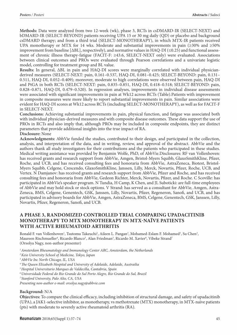
Reumatizam 2018;65(Suppl 1):37–74 45
Posters / Posteri Abstracts / Sažeci
Methods: Data were analyzed from two 12-week (wk), phase 3, RCTs in csDMARD-IR (SELECT-NEXT) and bDMARD-IR (SELECT-BEYOND) patients receiving UPA 15 or 30 mg daily (QD) or placebo and background csDMARD therapy; and from a third trial (SELECT-MONOTHERAPY), in which MTX-IR patients received UPA monotherapy or MTX for 14 wks. Moderate and substantial improvements in pain (≥30% and ≥50% improvement from baseline [ΔBL], respectively), and normative values in HAQ-DI (≤0.25) and functional assess-ment of chronic illness therapy-fatigue (FACIT-F: ≥43.6, SELECT-NEXT only) were evaluated. Associations between clinical outcomes and PROs were evaluated through Pearson correlations and a univariate logistic model, controlling for treatment group and BL value.Results: In general, ΔBL in pain and HAQ-DI scores were marginally correlated with individual physician-derived measures (SELECT-NEXT: pain, 0.161–0.537, HAQ-DI, 0.081–0.425; SELECT-BEYOND: pain, 0.131–0.511, HAQ-DI, 0.052–0.409); moreover, moderate to high correlations were observed between pain, HAQ-DI and PtGA in both RCTs (SELECT-NEXT: pain, 0.835–0.851, HAQ-DI, 0.418–0.518; SELECT-BEYOND: pain, 0.828–0.871, HAQ-DI, 0.479–0.520). In regression analyses, improvements in individual disease assessments were associated with signifi cant improvements in pain at Wk12 across RCTs (Table).Patients with improvement in composite measures were more likely to report substantial improvements in pain. Similar associations were evident for HAQ-DI scores at Wk12 across RCTs (including SELECT-MONOTHERAPY), as well as for FACIT-F in SELECT-NEXT.Conclusions: Achieving substantial improvements in pain, physical function, and fatigue was associated both with individual physician-derived measures and with composite disease outcomes. Th ese data support the use of PROs in RCTs and also imply that, although PROs may be included in composite endpoints, they are distinct parameters that provide additional insights into the true impact of RA.Disclosure: NoneAcknowledgments: AbbVie funded the studies, contributed to their design, and participated in the collection, analysis, and interpretation of the data, and in writing, review, and approval of the abstract. AbbVie and the authors thank all study investigators for their contributions and the patients who participated in these studies. Medical writing assistance was provided by Benjamin Wolfe, PhD, of AbbVie.Disclosures: RF van Vollenhoven: has received grants and research support from AbbVie, Amgen, Bristol-Myers Squibb, GlaxoSmithKline, Pfi zer, Roche, and UCB, and has received consulting fees and honoraria from AbbVie, AstraZeneca, Biotest, Bristol-Myers Squibb, Celgene, Crescendo, GlaxoSmithKline, Janssen, Lilly, Merck, Novartis, Pfi zer, Roche, UCB, and Vertex. N Damjanov: has received grants and research support from AbbVie, Pfi zer and Roche, and has received consulting fees and honoraria from AbbVie, Gedeon Richter, Merck, Novartis, Pfi zer, and Roche. C Scoville: has participated in AbbVie’s speaker program. N Tundia, H Camp, K Chen, and JL Suboticki: are full-time employees of AbbVie and may hold stock or stock options. V Strand: has served as a consultant for AbbVie, Amgen, Astra-Zeneca, BMS, Celgene, Genentech, GSK, Janssen, Lilly, Novartis, Pfi zer, Regeneron, Sanofi , and UCB, and has participated in advisory boards for AbbVie, Amgen, AstraZeneca, BMS, Celgene, Genentech, GSK, Janssen, Lilly, Novartis, Pfi zer, Regeneron, Sanofi , and UCB.
A PHASE 3, RANDOMIZED CONTROLLED TRIAL COMPARING UPADACITINIB MONOTHERAPY TO MTX MONOTHERAPY IN MTX-NAÏVE PATIENTS WITH ACTIVE RHEUMATOID ARTHRITIS Ronald F. van Vollenhoven1, Tsutomu Takeuchi2, Aileen L. Pangan3, Mohamed-Eslam F. Mohamed3, Su Chen3, Maureen Rischmueller4, Ricardo Blanco5, Alan Friedman3, Ricardo M. Xavier6, Vibeke Strand7 (Orsolya Nagy, non-author presenter)1 Amsterdam Rheumatology and Immunology Center ARC, Amsterdam, the Netherlands 2 Keio University School of Medicine, Tokyo, Japan 3 AbbVie Inc North Chicago, IL, USA 4 Th e Queen Elizabeth Hospital and University of Adelaide, Adelaide, Austrailia 5 Hospital Universitario Marques de Valdecilla, Cantabria, Spain 6 Universidade Federal do Rio Grande do Sul Porto Alegre, Rio Grande do Sul, Brazil 7 Stanford University, Palo Alto, CA, USA Presenting non-author e-mail: [email protected]
Background: N/AObjectives: To compare the clinical effi cacy, including inhibition of structural damage, and safety of upadacitinib (UPA), a JAK1-selective inhibitor, as monotherapy, vs methotrexate (MTX) monotherapy, in MTX-naïve patients (pts) with moderate to severely active rheumatoid arthritis (RA).
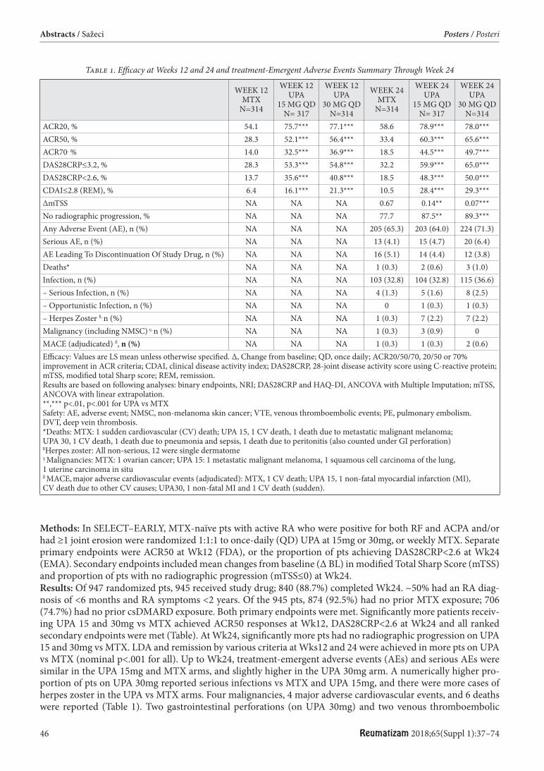
46 Reumatizam 2018;65(Suppl 1):37–74
Abstracts / Sažeci Posters / Posteri
Methods: In SELECT–EARLY, MTX-naïve pts with active RA who were positive for both RF and ACPA and/or had ≥1 joint erosion were randomized 1:1:1 to once-daily (QD) UPA at 15mg or 30mg, or weekly MTX. Separate primary endpoints were ACR50 at Wk12 (FDA), or the proportion of pts achieving DAS28CRP<2.6 at Wk24 (EMA). Secondary endpoints included mean changes from baseline (Δ BL) in modifi ed Total Sharp Score (mTSS) and proportion of pts with no radiographic progression (mTSS≤0) at Wk24.Results: Of 947 randomized pts, 945 received study drug; 840 (88.7%) completed Wk24. ~50% had an RA diag-nosis of <6 months and RA symptoms <2 years. Of the 945 pts, 874 (92.5%) had no prior MTX exposure; 706 (74.7%) had no prior csDMARD exposure. Both primary endpoints were met. Signifi cantly more patients receiv-ing UPA 15 and 30mg vs MTX achieved ACR50 responses at Wk12, DAS28CRP<2.6 at Wk24 and all ranked secondary endpoints were met (Table). At Wk24, signifi cantly more pts had no radiographic progression on UPA 15 and 30mg vs MTX. LDA and remission by various criteria at Wks12 and 24 were achieved in more pts on UPA vs MTX (nominal p<.001 for all). Up to Wk24, treatment-emergent adverse events (AEs) and serious AEs were similar in the UPA 15mg and MTX arms, and slightly higher in the UPA 30mg arm. A numerically higher pro-portion of pts on UPA 30mg reported serious infections vs MTX and UPA 15mg, and there were more cases of herpes zoster in the UPA vs MTX arms. Four malignancies, 4 major adverse cardiovascular events, and 6 deaths were reported (Table 1). Two gastrointestinal perforations (on UPA 30mg) and two venous thromboembolic
Table 1. Effi cacy at Weeks 12 and 24 and treatment-Emergent Adverse Events Summary Th rough Week 24
WEEK 12MTX
N=314
WEEK 12UPA
15 MG QDN= 317
WEEK 12UPA
30 MG QDN=314
WEEK 24MTX
N=314
WEEK 24UPA
15 MG QDN= 317
WEEK 24UPA
30 MG QDN=314
ACR20, % 54.1 75.7*** 77.1*** 58.6 78.9*** 78.0***ACR50, % 28.3 52.1*** 56.4*** 33.4 60.3*** 65.6***ACR70, % 14.0 32.5*** 36.9*** 18.5 44.5*** 49.7***DAS28CRP≤3.2, % 28.3 53.3*** 54.8*** 32.2 59.9*** 65.0***DAS28CRP<2.6, % 13.7 35.6*** 40.8*** 18.5 48.3*** 50.0***CDAI≤2.8 (REM), % 6.4 16.1*** 21.3*** 10.5 28.4*** 29.3***ΔmTSS NA NA NA 0.67 0.14** 0.07***No radiographic progression, % NA NA NA 77.7 87.5** 89.3***Any Adverse Event (AE), n (%) NA NA NA 205 (65.3) 203 (64.0) 224 (71.3)Serious AE, n (%) NA NA NA 13 (4.1) 15 (4.7) 20 (6.4)AE Leading To Discontinuation Of Study Drug, n (%) NA NA NA 16 (5.1) 14 (4.4) 12 (3.8)Deaths* NA NA NA 1 (0.3) 2 (0.6) 3 (1.0)Infection, n (%) NA NA NA 103 (32.8) 104 (32.8) 115 (36.6)– Serious Infection, n (%) NA NA NA 4 (1.3) 5 (1.6) 8 (2.5)– Opportunistic Infection, n (%) NA NA NA 0 1 (0.3) 1 (0.3)– Herpes Zoster ¥, n (%) NA NA NA 1 (0.3) 7 (2.2) 7 (2.2)Malignancy (including NMSC) γ, n (%) NA NA NA 1 (0.3) 3 (0.9) 0MACE (adjudicated) δ, n (%) NA NA NA 1 (0.3) 1 (0.3) 2 (0.6)Effi cacy: Values are LS mean unless otherwise specifi ed. Δ, Change from baseline; QD, once daily; ACR20/50/70, 20/50 or 70% improvement in ACR criteria; CDAI, clinical disease activity index; DAS28CRP, 28-joint disease activity score using C-reactive protein; mTSS, modifi ed total Sharp score; REM, remission.Results are based on following analyses: binary endpoints, NRI; DAS28CRP and HAQ-DI, ANCOVA with Multiple Imputation; mTSS, ANCOVA with linear extrapolation.**,*** p<.01, p<.001 for UPA vs MTXSafety: AE, adverse event; NMSC, non-melanoma skin cancer; VTE, venous thromboembolic events; PE, pulmonary embolism. DVT, deep vein thrombosis.*Deaths: MTX: 1 sudden cardiovascular (CV) death; UPA 15, 1 CV death, 1 death due to metastatic malignant melanoma; UPA 30, 1 CV death, 1 death due to pneumonia and sepsis, 1 death due to peritonitis (also counted under GI perforation)¥Herpes zoster: All non-serious, 12 were single dermatomeγ Malignancies: MTX: 1 ovarian cancer; UPA 15: 1 metastatic malignant melanoma, 1 squamous cell carcinoma of the lung, 1 uterine carcinoma in situδ MACE, major adverse cardiovascular events (adjudicated): MTX, 1 CV death; UPA 15, 1 non-fatal myocardial infarction (MI), CV death due to other CV causes; UPA30, 1 non-fatal MI and 1 CV death (sudden).
Reumatizam 2018;65(Suppl 1):37–74 47
Posters / Posteri Abstracts / Sažeci
events were reported (1 pulmonary embolism on MTX, 1 deep vein thrombosis on UPA 30mg). Laboratory abnormalities were consistent with other Phase 2 and 3 studies with UPA.Conclusions: In MTX-naïve pts, UPA 15 and 30mg QD demonstrated signifi cant and clinically meaningful improvements in RA signs & symptoms vs MTX. Radiographic progression was signifi cantly less with UPA vs MTX. Safety events were consistent with Phase 2 and 3 studies with UPA in RA to date.Disclosure: R von Vollenhoven: grants and research support from AbbVie, Amgen, Bristol-Myers Squibb, Glaxo-SmithKline, Pfi zer, Roche, and UCB; and consulting fees and honoraria from AbbVie, AstraZeneca, Biotest, Bris-tol-Myers Squibb, Celgene, Crescendo, GlaxoSmithKline, Janssen, Lilly, Merck, Novartis, Pfi zer, Roche, UCB, and Vertex. T Takeuchi: Honoraria (lecture fees); Mitsubishi-Tanabe Pharma Corporation, Janssen Pharmaceuti-cal KK, Chugai Pharmaceutical Co Ltd, Astellas Pharma Inc., AbbVie GK, Eisai Co., Ltd, Bristol-Myers Squibb Company, Daiichi Sankyo Company Ltd, Eli Lilly Japan KK, Pfi zer Japan Inc; Fees for promotional materials (e.g. manuscript fee); Astellas Pharma Inc. Consignment study without non-disclosure agreement; Chugai Pharma-ceutical Co Ltd, Mitsubishi-Tanabe Pharma Corporation; Grants; Pfi zer Japan Inc., Eisai Co., Ltd, Astellas Pharma Inc., AbbVie GK, Asahi Kasei Pharma Corporation, Nippon Kayaku Co., Ltd, Taisho Toyama Pharmaceutical Co., Ltd., Takeda Pharmaceutical Company Ltd, AYUMI Pharmaceutical Corporation, Takahashi Industrial and Economic Research Foundation Belong to lecture or course fi nancially maintained by private donation of com-pany; Astellas Pharma Inc., AbbVie GK, Eisai Co., Mitsubishi-Tanabe Pharma Corporation, Chugai Pharmaceu-tical Co Ltd, Bristol-Myers Squibb Company, UCB Japan Co., Ltd M Rischmueller: consultant for Abbvie, Bristol-Meyer-Squibb, Celgene, Glaxo Smith Kline, Hospira, Janssen Cilag, MSD, Novartis, Pfi zer, Roche, Sanofi , UCB R Blanco: received grants/research supports from Abbvie, MSD and Roche, and had consultation fees/participation in company sponsored speaker´s bureau from Abbvie, Pfi zer, Roche, Bristol-Myers, Janssen and MSD RM Xavier: consultant for Abbvie, Pfi zer, Novartis, Janssen, Lilly, Roche. V Strand: consultant for AbbVie, Amgen, Bayer, BMS, Boehringer Ingelheim, Celgene, Celltrion, CORRONA, Crescendo, EMD Serono, Genentech/Roche, GSK, Horizon, Inmedix, Janssen, Kezar, Lilly, Merck, Novartis, Pfi zer, Regeneron, Samsung, Sandoz, Sanofi , Servier, UCB. AL Pangan, A Friedman, MF Mohamed, S Chen: employees of AbbVie and may hold stock or options. Acknowledgements: AbbVie and the authors thank the patients, study sites and investigators who participated in this clinical trial. AbbVie, Inc was the study sponsor, contributed to study design, data collection, analysis & inter-pretation, and to writing, reviewing, and approval of fi nal version. Support for the study was provided by Stefania Zilli, statistical support was provided by Yijie Zhou, and medical writing support was provided by Naina Barretto, all employees of AbbVie, Inc.
A PHASE 3, RANDOMIZED, DOUBLE-BLIND STUDY COMPARING UPADACITINIB TO PLACEBO AND TO ADALIMUMAB, IN PATIENTS WITH ACTIVE RHEUMATOID ARTHRITIS WITH INADEQUATE RESPONSE TO METHOTREXATE Roy Fleischmann1, Aileen L. Pangan2, Eduardo Mysler3, Louis Bessette4, Charles Peterfy5, Patrick Durez6, Andrew Ostor7, Yihan Li2, Yijie Zhou2, Ahmed A. Othman2, In-Ho Song2, Mark C. Genovese8 (Orsolya Nagy, non-author presenter)1 University of Texas Southwestern, Metroplex Clinical Research Center, Dallas, Texas, USA 2 AbbVie Inc., North Chicago, Illinois, USA 3 Organización Medica de Investigación, Buenos Aires, Argentina 4 Laval University, Quebec, Canada 5 Spire Sciences Inc, Boca Raton, Florida, USA 6 Pôle de Recherche en Rhumatologie, Institut de Recherche Expérimentale et Clinique, UCL Saint-Luc, Brussels, Belgium 7 Cabrini Medical Center, Malvern, Australia 8 Division of Immunology & Rheumatology, Stanford University School of Medicine, Palo Alto, California, USA Presenting non-author e-mail: [email protected]
Background: N/AObjectives: To assess effi cacy, including inhibition of radiographic progression, and safety with upadacitinib (UPA), a JAK1- selective inhibitor, vs placebo (PBO) and active comparator, adalimumab (ADA), in patients (pts) with active rheumatoid arthritis (RA) continuing on prior methotrexate (MTX).Methods: In SELECT–COMPARE, pts with active RA despite MTX were randomized 2:2:1 to once-daily (QD) UPA 15mg, PBO, or ADA 40mg every other week (wk) in a double-blind manner, while continuing stable back-ground MTX. Primary endpoints were ACR20 and the proportion of pts achieving DAS28CRP<2.6 (NRI) at Wk12. Key secondary endpoints included non-inferiority (and superiority) of UPA vs ADA at Wk12 (for ACR50,
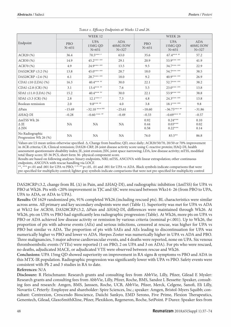
48 Reumatizam 2018;65(Suppl 1):37–74
Abstracts / Sažeci Posters / Posteri
DAS28CRP≤3.2, change from BL (Δ) in Pain, and ΔHAQ-DI), and radiographic inhibition (ΔmTSS) for UPA vs PBO at Wk26. Pts with <20% improvement in TJC and SJC were rescued between Wks14- 26 (from PBO to UPA, UPA to ADA, or ADA to UPA).Results: Of 1629 randomized pts, 91% completed Wk26.(including rescued pts). BL characteristics were similar across arms. All primary and key secondary endpoints were met (Table 1). Superiority was met for UPA vs ADA at Wk12 for ACR50, DAS28CRP≤3.2, ΔPain and ΔHAQ-DI, diff erences were maintained through Wk26. At Wk26, pts on UPA vs PBO had signifi cantly less radiographic progression (Table). At Wk26, more pts on UPA vs PBO or ADA achieved low disease activity or remission by various criteria (nominal p<.001). Up to Wk26, the proportion of pts with adverse events (AEs) and serious infections, censored at rescue, was higher for UPA vs PBO but similar vs ADA. Th e proportion of pts with SAEs and AEs leading to discontinuation for UPA was numerically higher vs PBO and lower vs ADA. Herpes Zoster was numerically higher in UPA vs ADA and PBO. Th ree malignancies, 5 major adverse cardiovascular events, and 4 deaths were reported, none on UPA. Six venous thromboembolic events (VTEs) were reported (1 on PBO, 2 on UPA and 3 on ADA). For pts who were rescued, no deaths, adjudicated MACE, or adjudicated VTE were observed between rescue and Wk26. Conclusions: UPA 15mg QD showed superiority on improvement in RA signs & symptoms vs PBO and ADA in this MTX-IR population. Radiographic progression was signifi cantly lower with UPA vs PBO. Safety events were consistent with Ph 2 and 3 studies in RA to date.References: N/ADisclosure: R Fleischmann: Research grants and consulting fees from AbbVie, Lilly, Pfi zer, Gilead E Mysler: Research grants and consulting fees from AbbVie, Lilly, Pfi zer, Roche, BMS, Sandoz L Bessette: Speaker, consult-ing fees and research: Amgen, BMS, Janssen, Roche, UCB, AbbVie, Pfi zer, Merck, Celgene, Sanofi , Eli Lilly, Novartis C Peterfy: Employee and shareholder: Spire Sciences, Inc.; speaker: Amgen, Bristol-Myers Squibb; con-sultant: Centrexion, Crescendo Bioscience, Daiichi Sankyo, EMD Serono, Five Prime, Flexion Th erapeutics, Genentech, Gilead, GlaxoSmithKline, Pfi zer, Plexikkon, Regeneron, Roche, SetPoint. P Durez: Speaker fees from
Table 1. Effi cacy Endpoints at Weeks 12 and 26.
Endpoint
WEEK 12 WEEK 26
PBON=651
UPA15MG QD
N=651
ADA40MG EOW
N=327PBO
N=651UPA
15MG QDN=651
ADA40MG EOW
N=327ACR20 (%) 36.4 70.5*** # 63.0 35.6 67.4*** ## 57.2ACR50 (%) 14.9 45.2*** ### 29.1 20.9 53.9*** ### 41.9ACR70 (%) 4.9 24.9*** ### 13.5 9.5 34.7*** ### 22.9DAS28CRP ≤3.2 (%) 13.8 45.0*** ### 28.7 18.0 54.7*** ### 38.5DAS28CRP <2.6 (%) 6.1 28.7*** ### 18.0 9.2 40.9*** ### 26.9CDAI ≤10 (LDA) (%) 16.3 40.4*** ## 30.0 22.1 52.7*** ### 38.2CDAI ≤2.8 (CR) (%) 3.1 13.4*** ## 7.6 5.5 23.0*** ### 13.8SDAI ≤11.0 (LDA) (%) 15.2 40.4*** ## 30.0 22.1 53.9*** ### 38.8SDAI ≤3.3 (CR) (%) 2.8 12.1*** # 7.3 4.8 24.3*** ### 13.8Boolean remission 2.0 9.8*** ## 4.0 3.8 18.1*** ### 9.8ΔPain –15.69 –32.10 ***
### –25.61 –18.60 –36.75*** ## –31.86
ΔHAQ-DI –0.28 –0.60 *** ## –0.49 –0.33 –0.69*** ## –0.57ΔmTSS Wk 26Δ JEΔ JSN
NA NA NA0.920.440.58
0.24***0.03***0.22***
0.100.020.14
No Radiographic Progression Wk 26 (%) NA NA NA 76.0 83.5** 86.8
Values are LS mean unless otherwise specifi ed. Δ, Change from baseline; QD, once daily; ACR20/50/70, 20/50 or 70% improvement in ACR criteria; CR, Clinical remission; DAS28-CRP, 28-joint disease activity score using C-reactive protein; HAQ-DI, health assessment questionnaire disability index; JE, joint erosion; JSN, joint space narrowing; LDA, low disease activity; mTSS, modifi ed total Sharp score; SF-36 PCS, short form 36- physical component score.Results are based on following analyses: binary endpoints, NRI; mTSS, ANCOVA with linear extrapolation; other continuous endpoints, ANCOVA with rescue handling via LOCF.**, *** p<.01 and .001 for UPA vs PBO; #, ##, ### p<.05, .01 and .001 for UPA vs ADA. Black symbols indicate comparisons that were pre-specifi ed for multiplicity control; lighter gray symbols indicate comparisons that were not pre-specifi ed for multiplicity control
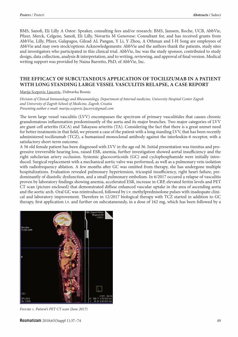
Reumatizam 2018;65(Suppl 1):37–74 49
Posters / Posteri Abstracts / Sažeci
BMS, Sanofi , Eli Lilly A Ostor: Speaker, consulting fees and/or research: BMS, Janssen, Roche, UCB, AbbVie, Pfi zer, Merck, Celgene, Sanofi , Eli Lilly, Novartis M Genovese: Consultant for, and has received grants from AbbVie, Lilly, Pfi zer, Galapagos, Gilead AL Pangan, Y Li, Y Zhou, A Othman and I-H Song are employees of AbbVie and may own stock/options Acknowledgements: AbbVie and the authors thank the patients, study sites and investigators who participated in this clinical trial. AbbVie, Inc was the study sponsor, contributed to study design, data collection, analysis & interpretation, and to writing, reviewing, and approval of fi nal version. Medical writing support was provided by Naina Barretto, PhD, of AbbVie, Inc.
THE EFFICACY OF SUBCUTANEOUS APPLICATION OF TOCILIZUMAB IN A PATIENT WITH LONG STANDING LARGE VESSEL VASCULITIS RELAPSE, A CASE REPORT Marija Scepovic Ljucovic, Dubravka Bosnic
Division of Clinical Immunology and Rheumatology, Department of Internal medicine, University Hospital Center Zagreb and University of Zagreb School of Medicine, Zagreb, Croatia Presenting author e-mail: [email protected]
Th e term large vessel vasculitis (LVV) encompasses the spectrum of primary vasculitides that causes chronic granulomatous infl ammation predominantly of the aorta and its major branches. Two major categories of LVV are giant cell arteritis (GCA) and Takayasu arteritis (TA). Considering the fact that there is a great unmet need for better treatments in that fi eld, we present a case of the patient with a long standing LVV, that has been recently administered tocilizumab (TCZ), a humanised monoclonal antibody against the interleukin-6 receptor, with a satisfactory short-term outcome.A 56 old female patient has been diagnosed with LVV in the age od 36. Initial presentation was tinnitus and pro-gressive irreversible hearing loss, raised ESR, anemia, further investigation showed aortal insuffi ciency and the right subclavian artery occlusion. Systemic glucocorticoids (GC) and cyclophosphamide were initially intro-duced. Surgical replacement with a mechanical aortic valve was performed, as well as a pulmonary vein isolation with radiofrequency ablation. A few months aft er GC was omitted from therapy, she has undergone multiple hospitalizations. Evaluation revealed pulmonary hypertension, tricuspid insuffi ciency, right heart failure, pre-dominantly of diastolic dysfunction, and a small pulmonary embolism. In 6/2017 occured a relapse of vasculitis proven by laboratory fi ndings showing anemia, accelerated ESR, increase in CRP, elevated feritin levels and PET CT scan (picture enclosed) that demonstrated diff use enhanced vascular uptake in the area of ascending aorta and the aortic arch. Oral GC was reintroduced, followed by i.v. methylprednisolone pulses with inadequate clini-cal and laboratory improvement. Th erefore in 12/2017 biological therapy with TCZ started in addition to GC therapy, fi rst application i.v. and further on subcutaneously, in a dose of 162 mg, which has been followed by a
Figure 1. Patient’s PET CT scan (June 2017)
50 Reumatizam 2018;65(Suppl 1):37–74
Abstracts / Sažeci Posters / Posteri
clinical and laboratory remission. Regarding LVV, IL-6 multiple activities include the induction of antibody pro-duction, activation of Th 17 cells, and increase of hepcidin, which is responsible for anemia (1). IL-6 is upregu-lated in infl ammed arteries of patients with GCA and TA (2). Th erefore it is to be expected that a treatment with TCZ is promising eff ective and safe solution for a refractory LVV, as it has been shown in available data obtained from limited clinical studies (3,4,5).References:1. Paroli M. Clinical use of biologics in vasculitis syndromes in Biologics: 2012; 6: 371–378.2. Unizony, S; Stone, JH; Stone, J. New treatment strategies in large-vessel vasculitis in Current Opinion in Rheumatology: January 2013
– Volume 25 – Issue 1 – p 3–9).3. Girelli F, Bezzi A, Galassi R, Moretti A, Tomassetti S, et al. (2017) Effi cacy of Subcutaneous Tocilizumab for the Treatment of Refrac-
tory Large Vessel Vasculitis in a Patient With Latent Tuberculosis Infection. SAJ Case Rep 4: 303.4. Evans J, Steel L, Borg F, et al. Long-term effi cacy and safety of tocilizumab in giant cell arteritis and large vessel vasculitis. RMD Open
2016;2:e000137.5. Salvarani C, Magnani L, Catanoso M, Pipitone N. et al. Tocilizumab: a novel therapy for patients with large-vessel vasculitis. Rheuma-
tology 2012;51:151–156. Disclosure: None
CHILDHOOD-ONSET OF HENOCH-SCHÖNLEIN PURPURA NEPHRITIS IN CROATIA: A STUDY CONDUCTED IN FIVE TERTIARY CARE CENTRES OVER NINE YEARS Mateja Batnozic Varga1, Natasia Cekada2, Mario Sestan2, Sasa Srsen3, Lucija Ruzman4, Maja Zaninovic4, Aleksandar Ovuka4, Ivana Ozdanovac1, Marija Pecnjak2, Domagoj Kifer5, Marijan Frkovic1, Alenka Gagro6, Marija Jelusic1
1 Department of Paediatrics, University Hospital Centre Osijek, University of Osijek School of Medicine, Osijek 2 Department of Paediatrics, University Hospital Centre Zagreb, University of Zagreb School of Medicine, Zagreb 3 Department of Paediatrics, University Hospital Centre Split, University of Split School of Medicine, Split 4 Department of Paediatrics, University Hospital Centre Rijeka, University of Rijeka School of Medicine, Rijeka 5 Faculty of Pharmacy and Biochemistry, University of Zagreb 6 Children’s Hospital Zagreb Presenting author e-mail: [email protected]
Background: Henoch-Schönlein purpura nephritis (HSPN) is the most severe complication of Henoch-Schön-lein purpura (HSP) that includes isolated microscopic/macroscopic hematuria, mild/heavy proteinuria with/without nephrotic syndrome, renal failure and hypertension. Objectives: Th e aim was to determine a possible prognostic factor for earlier HSPN onset, to explore indications for kidney biopsy according to urine analysis and the correlation between 24-h urinary protein levels and biopsy fi ndings, as well as biopsy fi ndings and patient outcome.Methods: Th e cross-sectional study included all children with HSPN diagnosed by EULAR/ PRES/PRINTO cri-teria from 2009 to 2017 at 5 tertiary care centres in Croatia.Results: Out of 522 patients diagnosed with HSP, 101 children developed HSPN (19.35%). Median (range) age of HSP diagnosis was 7.5 (2–17.5) years. Nephritis was present in 7.9% of cases at the HSP onset. No positive cor-relation was found between the time of HSP diagnosis to HSPN onset and gender, age, purpura distribution, joint and gastrointestinal involvement. Kidney biopsy was done in 30 patients: 3 had isolated persistent hematuria, 3 isolated proteinuria and 24 patients had both hematuria and proteinuria. Median (range) 24-h urinary protein levels in biopted patients was 1.11 (0–7.46) g/dU. Th e leading indication for biopsy was simultaneous hematuria and proteinuria (p<0.001. Biopsy fi ndings were graded with diff erent classifi cations (Oxford, Haas, ISKDC). No positive correlation was found between 24-h urinary protein levels and biopsy fi ndings. 60.4% of HSPN patients were treated with corticosteroids. Most patients had good outcome, with normal physical exam fi ndings and no signs of renal disease in laboratory tests (79.2%), or with microhematuria and proteinuria <1g/dU (15.8%), while only 4 patients had a more severe outcome with proteinuria >1g/dU. No positive correlation was found between biopsy fi ndings and patient outcome (p=0.017).Conclusions: Simultaneous hematuria and proteinuria was a statistically signifi cant factor for kidney biopsy. How-ever, while isolated proteinuria was not the sole determining factor, excessive levels of 24-h urinary proteins should be taken in consideration. Due to the small number of patients and no uniform classifi cation generally used in grad-ing biopsy fi ndings, a statistically signifi cant correlation in regard to outcome could not be confi rmed.References:1. Buscatti IM, Casella BB, Aikawa NE, Watanabe A, Farhat SCL, Campos LMA, Silva CA. Henoch-Schönlein purpura nephritis: initial
risk factors and outcomes in a Latin American tertiary center. Clin Rheumatol. 2018 May;37(5):1325.Disclosure: None
Reumatizam 2018;65(Suppl 1):37–74 51
Posters / Posteri Abstracts / Sažeci
POTENTIAL MARKERS OF SKIN FIBROSIS IN SYSTEMIC SCLEROSIS Radim Bečvář
Institute of Rheumatology Presenting author e-mail: [email protected]
Background: Skin fi brosis is a hallmark of systemic sclerosis (SSc). Th ere are no widely accepted biomarkers of skin involvement in this condition. Several serum or plasma markers have been studied in patients with SSc – monocyte chemoattractant protein-1 (MCP-1), chemokine (C-X-C motif) ligand 8 (CXCL8), interleukin-13 (IL-13), and some more recognized such as – platelet derived growth factor (PDGF), transforming growth factor-beta 1 (TGF-beta 1), epidermal growth factor (EGF) and basic fi broblast growth factor (bFGF). Objectives: Th e aim of this study was to assess several circulating biomarkers which may be relevant to the fi bros-ing process and further to correlate the obtained data with clinical indicators specifi c for SSc skin involvement. Methods: 59 SSc patients (M/F 9/50; mean age 52.1 years, mean disease duration 6.7 years, 36 patients with lim-ited cutaneous SSc and 23 with diff use cutaneous SSc. As a control group 36 healthy individuals matched to sex and age were examined.Serum concentrations of bFGF, granulocyte-colony stimulating factor (G-CSF), granulocyte-macrophage-colony stimulating factor (GM-CSF), MCP-1, PDGF, IL-8 and 13 were analysed using commercial multiplex kit. Th e following clinical examinations were performed: modifi ed Rodnan skin score (mRSS), Hand Mobility in Sclero-derma Test (assessing hand function) (HAMIS), Cochin Hand Function Scale (hand function) (CHFS), Delta Finger-to-Palm Distance (extension-fl exion) (dFTP), Inter-lip Distance (inter-lip), Inter-incisor Distance (inter-incisor), and Mouth Handicap in Systemic Sclerosis Scale (mouth opening) (MHISS). For statistical evaluation Spearman’s correlation coeffi cient was used. Results: When compared with healthy controls serum concentrations of bFGF (p<0.001), G-CSF (p<0.0001), GM-CSF (p<0.0001), MCP-1 (p<0.0001) IL-8 (p<0.0001), and IL-13 (p<0.001) were signifi cantly elevated in SSc cohort. PDGF levels were increased in SSc patients with only a lower signifi cance (p<0.01). bFGF, G-CSF, MCP-1 and IL-8 levels correlated signifi cantly (p<0.05) with mRSS and HAMIS. GM-CSF levels correlated with mRSS and HAMIS and there was only a trend for negative correlation with inter-incisor. Th e was no correlation of IL-13 and PDGF levels with the evaluated clinical data. Conclusions: Our results have shown that G-CSF, GM-CSF and IL-8 play a substantial role in SSc fi brosing pro-cess. Potential biomarkers as bFGF, G-CSF, MCP-1 and IL-8 correlated with a few clinical indices of SSc skin involvement.Acknowledgement: Study was supported by research grants AZV 16-33574A and AZV 16-33542A. References:1. Carulli MT, Handler C, Coghlan JG, Black CM, Denton CP. Can CCL2 serum levels be used in risk stratifi cation or to monitor treat-
ment response in systemic sclerosis? Ann Rheum Dis 2008;67:105–9.2. Chujo S, Shirasaki F, Kondo-Miyazaki M, Ikawa Y, Takehara K. Role of connective tissue growth factor and its interaction with basic
fi broblast growth factor and macrophage chemoattractant protein-1 in skin fi brosis. J Cell Physiol 2009;220:189–95.3. Fuschiotti P, Larregina AT, Ho J, Feghali-Bostwick C, Medsger TA Jr. Interleukin-13-producing CD8+ T cells mediate dermal fi brosis
in patients with systemic sclerosis. Arthritis Rheum 2013;65:236–46.4. Hasegawa M, Fujimoto M, Matsushita T et al. Serum chemokine and cytokine levels as indicators of disease activity in patients with
systemic sclerosis. Clin Rheumatol 2011;30:231–7. Disclosure: None
INFLAMMATION-RELATED PROTEINS AS POTENTIAL MARKERS OF DISEASE ACTIVITY IN PATIENTS WITH SYSTEMIC SCLEROSIS Andrea Smržová1, Pavel Horák1, Anna Petráčkova2, Marketá Schubertová1, Martina Skácelova1, Tereza Dyšková2, Regína Fillerová2, Gabriela Gabčová2, František Mrázek2, Eva Kriegová2
1 Department of Internal Medicine III – Nephrology, Rheumatology and Endocrinology, Faculty of Medicine and Dentistry, Palacky University Olomouc, University Hospital, Olomouc, Czech Republic 2 Department of Immunology, Faculty of Medicine and Dentistry, Palacky University Olomouc, Olomouc, Czech Republic Presenting author e-mail: [email protected]
Introduction: Systemic sclerosis (SSc) is a multiogran autoimmune connective tissue disease with varying mani-festations. Several serious clinical prognostic factors are investigated. Serum markers refl ecting clinical activity are not yet well known.
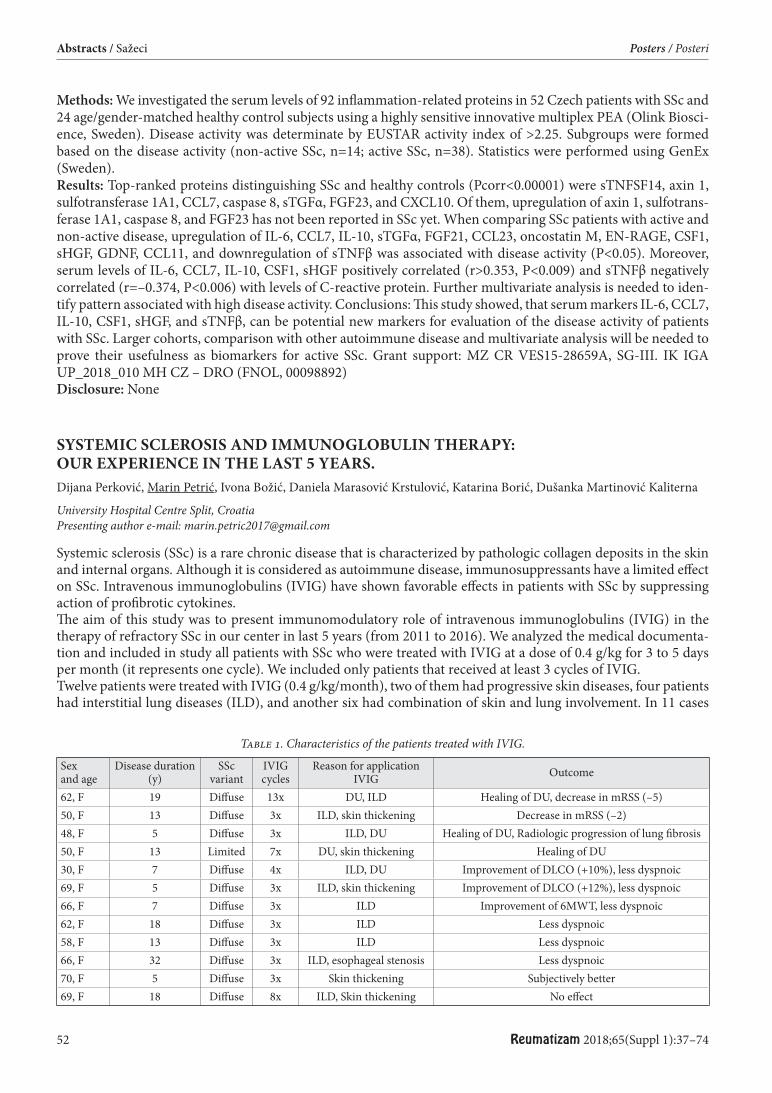
52 Reumatizam 2018;65(Suppl 1):37–74
Abstracts / Sažeci Posters / Posteri
Methods: We investigated the serum levels of 92 infl ammation-related proteins in 52 Czech patients with SSc and 24 age/gender-matched healthy control subjects using a highly sensitive innovative multiplex PEA (Olink Biosci-ence, Sweden). Disease activity was determinate by EUSTAR activity index of >2.25. Subgroups were formed based on the disease activity (non-active SSc, n=14; active SSc, n=38). Statistics were performed using GenEx (Sweden).Results: Top-ranked proteins distinguishing SSc and healthy controls (Pcorr<0.00001) were sTNFSF14, axin 1, sulfotransferase 1A1, CCL7, caspase 8, sTGFα, FGF23, and CXCL10. Of them, upregulation of axin 1, sulfotrans-ferase 1A1, caspase 8, and FGF23 has not been reported in SSc yet. When comparing SSc patients with active and non-active disease, upregulation of IL-6, CCL7, IL-10, sTGFα, FGF21, CCL23, oncostatin M, EN-RAGE, CSF1, sHGF, GDNF, CCL11, and downregulation of sTNFβ was associated with disease activity (P<0.05). Moreover, serum levels of IL-6, CCL7, IL-10, CSF1, sHGF positively correlated (r>0.353, P<0.009) and sTNFβ negatively correlated (r=–0.374, P<0.006) with levels of C-reactive protein. Further multivariate analysis is needed to iden-tify pattern associated with high disease activity. Conclusions: Th is study showed, that serum markers IL-6, CCL7, IL-10, CSF1, sHGF, and sTNFβ, can be potential new markers for evaluation of the disease activity of patients with SSc. Larger cohorts, comparison with other autoimmune disease and multivariate analysis will be needed to prove their usefulness as biomarkers for active SSc. Grant support: MZ CR VES15-28659A, SG-III. IK IGA UP_2018_010 MH CZ – DRO (FNOL, 00098892) Disclosure: None
SYSTEMIC SCLEROSIS AND IMMUNOGLOBULIN THERAPY: OUR EXPERIENCE IN THE LAST 5 YEARS. Dijana Perković, Marin Petrić, Ivona Božić, Daniela Marasović Krstulović, Katarina Borić, Dušanka Martinović Kaliterna
University Hospital Centre Split, Croatia Presenting author e-mail: [email protected]
Systemic sclerosis (SSc) is a rare chronic disease that is characterized by pathologic collagen deposits in the skin and internal organs. Although it is considered as autoimmune disease, immunosuppressants have a limited eff ect on SSc. Intravenous immunoglobulins (IVIG) have shown favorable eff ects in patients with SSc by suppressing action of profi brotic cytokines.Th e aim of this study was to present immunomodulatory role of intravenous immunoglobulins (IVIG) in the therapy of refractory SSc in our center in last 5 years (from 2011 to 2016). We analyzed the medical documenta-tion and included in study all patients with SSc who were treated with IVIG at a dose of 0.4 g/kg for 3 to 5 days per month (it represents one cycle). We included only patients that received at least 3 cycles of IVIG.Twelve patients were treated with IVIG (0.4 g/kg/month), two of them had progressive skin diseases, four patients had interstitial lung diseases (ILD), and another six had combination of skin and lung involvement. In 11 cases
Table 1. Characteristics of the patients treated with IVIG.
Sex and age
Disease duration (y)
SSc variant
IVIG cycles
Reason for application IVIG Outcome
62, F 19 Diff use 13x DU, ILD Healing of DU, decrease in mRSS (–5)50, F 13 Diff use 3x ILD, skin thickening Decrease in mRSS (–2)48, F 5 Diff use 3x ILD, DU Healing of DU, Radiologic progression of lung fi brosis 50, F 13 Limited 7x DU, skin thickening Healing of DU30, F 7 Diff use 4x ILD, DU Improvement of DLCO (+10%), less dyspnoic69, F 5 Diff use 3x ILD, skin thickening Improvement of DLCO (+12%), less dyspnoic66, F 7 Diff use 3x ILD Improvement of 6MWT, less dyspnoic62, F 18 Diff use 3x ILD Less dyspnoic58, F 13 Diff use 3x ILD Less dyspnoic66, F 32 Diff use 3x ILD, esophageal stenosis Less dyspnoic70, F 5 Diff use 3x Skin thickening Subjectively better69, F 18 Diff use 8x ILD, Skin thickening No eff ect
Reumatizam 2018;65(Suppl 1):37–74 53
Posters / Posteri Abstracts / Sažeci
(91.7%) some kind of improvement was documented. Th e best results were achieved in skin changes, where we recorded complete healing of digital ulcers (DU) in 3 cases (75%) and improvement of skin thickening in 4 cases (80%).Th is retrospective observational study suggests that IVIG may be an eff ective therapy option for refractory SSc in patients who have failed other therapies, but further studies on the exact role of IVIG in the treatment of SSc are required. References:1. Kowal-Bielecka O, Fransen J, Avouac J, Becker M, Kulak A, Allanore Y, et al. Update of EULAR recommendations for the treatment of
systemic sclerosis. Ann Rheum Dis. 2017;76(8):1327–39.2. Negi VS, Elluru S, Sibéril S, Graff -Dubois S, Mouthon L, Kazatchkine MD, et al. Intravenous immunoglobulin: an update on the
clinical use and mechanisms of action. J Clin Immunol. 2007;27(3):233–45.3. Poelman CL, Hummers LK, Wigley FM, Anderson C, Boin F, Shah AA. Intravenous immunoglobulin may be an eff ective therapy for
refractory, active diff use cutaneous systemic sclerosis. J Rheumatol. 2015;42(2):236–42.4. Sanges S, Rivière S, Mekinian A, Martin T, Le Quellec A, Chatelus E, et al. Intravenous immunoglobulins in systemic sclerosis: Data
from a French nationwide cohort of 46 patients and review of the literature. Autoimmun Rev. 2017;16(4):377–384.5. Takehara K, Ihn H, Sato S. A randomized, double-blind, placebo-controlled trial: intravenous immunoglobulin treatment in patients
with diff use cutaneous systemic sclerosis. Clin Exp Rheumatol. 2013;31(76):151–6. Disclosure: None
EFFICACY OF AN INTENSIVE 24-WEEK PHYSICAL-OCCUPATIONAL THERAPY PROGRAM WITH SUBSEQUENT 24-WEEK FOLLOW-UP IN PATIENTS WITH SYSTEMIC SCLEROSIS – PRELIMINARY DATA FROM A SINGLE-CENTER CONTROLLED STUDY Maja Spiritovic1, Hana Smucrova2, Sabina Oreska3, Hana Storkanova3, Barbora Hermankova1, Petr Cesak1, Adela Rathouska2, Olga Ruzickova3, Karel Pavelka3, Ladislav Senolt3, Jiri Vencovsky3, Radim Becvar3, Michal Tomcik3
1 Faculty of Physical Education and Sport, Charles University, Prague, Czech Republic 2 Institute of Rheumatology, Prague, Czech Republic 3 Institute of Rheumatology and Department of Rheumatology, 1st Faculty of Medicine, Charles University, Prague, Czech Republic Presenting author e-mail: [email protected]
Background: Involvement of the skin and musculoskeletal system in patients with systemic sclerosis (SSc) decreases their quality of life. Data on the eff ectiveness of non-pharmacological interventions in SSc are limited due to heterogeneity of studied interventions/outcomes.Objectives: Th e aim of our study was to minimalize the limitations of available studies and to determine the eff ect of our physical-occupational therapy (POT) program in cohorts with a substantial number of SSc patients.Methods: Patients were consecutively recruited into two groups, intervention (IG) and control (CG) group, they fulfi lled the ACR/EULAR classifi cation criteria from 2013 and had skin involvement of the hands and face. Both groups received educational material for home exercises, but only the IG underwent a 24-week intensive POT program with a subsequent 24-week follow-up. All patients were evaluated by a physician and physiotherapist blinded to intervention at 0, 12, 24 and 48 weeks. All of them also fi lled out patient reported outcomes/question-naires and provided blood for routine laboratory analysis and biobanking. Data analysis was performed between groups and within the group.Results: 25 patients were included in the IG and 30 into the CG. Compared to the observed statistically signifi cant deterioration in CG, we found a statistically signifi cant improvement in IG in objectively assessed function and strength of hand, distance between incisors and lips and also subjectively assessed functional ability (SHAQ). During the follow-up period there was a signifi cant deterioration or stagnation of the achieved results in the IG. Only numerical improvements in IG compared to numerical deterioration in CG, but not statistically signifi cant, were observed, during the intervention period, in subjectively evaluated parameters assessing hand/face func-tion, functional ability (HAQ), and some domains of (SF-36).Conclusions: Our program led to a signifi cant improvement in the observed parameters that were clinically rel-evant in a substantial proportion of patients with SSc (in the IG) and prevented the natural course of progressive deterioration in hand/face function (observed in the CG).Acknowledgments: Th e project was supported by AZV-16-33574A, MHCR 023728 and SVV for FTVS UK 2019-260466. Disclosure: None
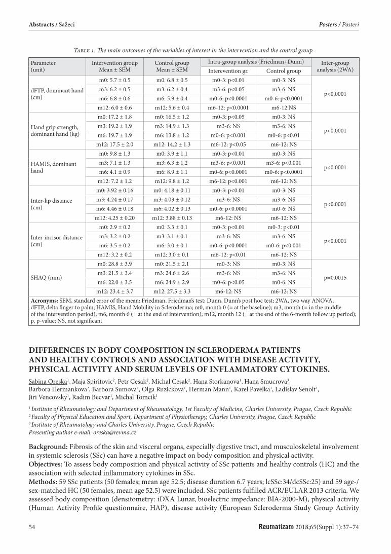
54 Reumatizam 2018;65(Suppl 1):37–74
Abstracts / Sažeci Posters / Posteri
Table 1. Th e main outcomes of the variables of interest in the intervention and the control group.
Parameter(unit)
Intervention groupMean ± SEM
Control groupMean ± SEM
Intra-group analysis (Friedman+Dunn) Inter-group analysis (2WA)Interevention gr. Control group
dFTP, dominant hand (cm)
m0: 5.7 ± 0.5 m0: 6.8 ± 0.5 m0-3: p<0.01 m0-3: NS
p<0.0001m3: 6.2 ± 0.5 m3: 6.2 ± 0.4 m3-6: p<0.05 m3-6: NSm6: 6.8 ± 0.6 m6: 5.9 ± 0.4 m0-6: p<0.0001 m0-6: p<0.0001
m12: 6.0 ± 0.6 m12: 5.6 ± 0.4 m6-12: p<0.0001 m6-12:NS
Hand grip strength, dominant hand (kg)
m0: 17.2 ± 1.8 m0: 16.5 ± 1.2 m0-3: p<0.05 m0-3: NS
p<0.0001m3: 19.2 ± 1.9 m3: 14.9 ± 1.3 m3-6: NS m3-6: NSm6: 19.7 ± 1.9 m6: 13.8 ± 1.2 m0-6: p<0.001 m0-6: p<0.01
m12: 17.5 ± 2.0 m12: 14.2 ± 1.3 m6-12: p<0.05 m6-12: NS
HAMIS, dominant hand
m0: 9.8 ± 1.3 m0: 3.9 ± 1.1 m0-3: p<0.01 m0-3: NS
p<0.0001m3: 7.1 ± 1.3 m3: 6.3 ± 1.2 m3-6: p<0.001 m3-6: p<0.001m6: 4.1 ± 0.9 m6: 8.9 ± 1.1 m0-6: p<0.0001 m0-6: p<0.0001
m12: 7.2 ± 1.2 m12: 9.8 ± 1.2 m6-12: p<0.001 m6-12: NS
Inter-lip distance (cm)
m0: 3.92 ± 0.16 m0: 4.18 ± 0.11 m0-3: p<0.01 m0-3: NS
p<0.0001m3: 4.24 ± 0.17 m3: 4.03 ± 0.12 m3-6: NS m3-6: NSm6: 4.46 ± 0.18 m6: 4.02 ± 0.13 m0-6: p<0.0001 m0-6: NS
m12: 4.25 ± 0.20 m12: 3.88 ± 0.13 m6-12: NS m6-12: NS
Inter-incisor distance (cm)
m0: 2.9 ± 0.2 m0: 3.3 ± 0.1 m0-3: p<0.01 m0-3: p<0.01
p<0.0001m3: 3.2 ± 0.2 m3: 3.1 ± 0.1 m3-6: NS m3-6: NSm6: 3.5 ± 0.2 m6: 3.0 ± 0.1 m0-6: p<0.0001 m0-6: p<0.001
m12: 3.2 ± 0.2 m12: 3.0 ± 0.1 m6-12: p<0.01 m6-12: NS
SHAQ (mm)
m0: 28.8 ± 3.9 m0: 21.5 ± 2.1 m0-3: NS m0-3: NS
p=0.0015m3: 21.5 ± 3.4 m3: 24.6 ± 2.6 m3-6: NS m3-6: NSm6: 22.0 ± 3.5 m6: 24.9 ± 2.9 m0-6: p<0.05 m0-6: NS
m12: 23.4 ± 3.7 m12: 27.5 ± 3.3 m6-12: NS m6-12: NSAcronyms: SEM, standard error of the mean; Friedman, Friedman’s test; Dunn, Dunn’s post hoc test; 2WA, two way ANOVA, dFTP, delta fi nger to palm; HAMIS, Hand Mobility in Scleroderma; m0, month 0 (= at the baseline); m3, month (= in the middle of the intervention period); m6, month 6 (= at the end of intervention); m12, month 12 (= at the end of the 6-month follow up period); p, p-value; NS, not signifi cant
DIFFERENCES IN BODY COMPOSITION IN SCLERODERMA PATIENTS AND HEALTHY CONTROLS AND ASSOCIATION WITH DISEASE ACTIVITY, PHYSICAL ACTIVITY AND SERUM LEVELS OF INFLAMMATORY CYTOKINES. Sabina Oreska1, Maja Spiritovic2, Petr Cesak2, Michal Cesak2, Hana Storkanova1, Hana Smucrova3, Barbora Hermankova2, Barbora Sumova1, Olga Ruzickova1, Herman Mann1, Karel Pavelka1, Ladislav Senolt1, Jiri Vencovsky1, Radim Becvar1, Michal Tomcik1
1 Institute of Rheumatology and Department of Rheumatology, 1st Faculty of Medicine, Charles University, Prague, Czech Republic 2 Faculty of Physical Education and Sport, Department of Physiotherapy, Charles University, Prague, Czech Republic 3 Institute of Rheumatology and Charles University, Prague, Czech Republic Presenting author e-mail: [email protected]
Background: Fibrosis of the skin and visceral organs, especially digestive tract, and musculoskeletal involvement in systemic sclerosis (SSc) can have a negative impact on body composition and physical activity. Objectives: To assess body composition and physical activity of SSc patients and healthy controls (HC) and the association with selected infl ammatory cytokines in SSc.Methods: 59 SSc patients (50 females; mean age 52.5; disease duration 6.7 years; lcSSc:34/dcSSc:25) and 59 age-/sex-matched HC (50 females, mean age 52.5) were included. SSc patients fulfi lled ACR/EULAR 2013 criteria. We assessed body composition (densitometry: iDXA Lunar, bioelectric impedance: BIA-2000-M), physical activity (Human Activity Profi le questionnaire, HAP), disease activity (European Scleroderma Study Group Activity
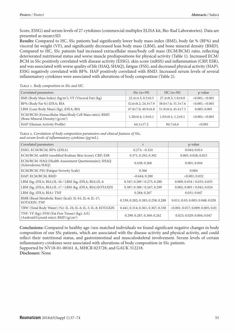
Reumatizam 2018;65(Suppl 1):37–74 55
Posters / Posteri Abstracts / Sažeci
Score, ESSG) and serum levels of 27 cytokines (commercial multiplex ELISA kit, Bio-Rad Laboratories). Data are presented as mean±SD. Results: Compared to HC, SSc patients had signifi cantly lower body mass index (BMI), body fat % (BF%) and visceral fat weight (VF), and signifi cantly decreased lean body mass (LBM), and bone mineral density (BMD). Compared to HC, SSc patients had increased extracellular mass/body cell mass (ECM/BCM) ratio, refl ecting deteriorated nutritional status and worse muscle predispositions for physical activity (Table 1). Increased ECM/BCM in SSc positively correlated with disease activity (ESSG), skin score (mRSS) and infl ammation (CRP, ESR), and was associated with worse quality of life (HAQ, SHAQ), fatigue (FSS), and decreased physical activity (HAP). ESSG negatively correlated with BF%. HAP positively correlated with BMD. Increased serum levels of several infl ammatory cytokines were associated with alterations of body composition (Table 2).
Table 1. Body composition in SSc and HC.
Correlated parameters SSc (n=59) HC (n=59) p-valueBMI (Body Mass Index) (kg/m2); VF (Visceral Fat) (kg) 22.4±4.3; 0.5±0.5 27.4±8.3; 1.0±0.8 <0.001; <0.001BF% (Body Fat %) iDXA; BIA 32.6±8.2; 24.3±7.9 38.0±7.6; 31.3±7.6 <0.001; <0.001LBM (Lean Body Mass) (kg), iDXA; BIA 47.8±7.0; 40.9±6.8 51.9±8.4; 45.4±7.3 0.005; 0.005ECM/BCM (Extracellular Mass/Body Cell Mass ratio); BMD (Bone Mineral Density) (g/cm2) 1.28±0.4; 1.0±0.1 1.03±0.1; 1.2±0.1 <0.001; <0.001
HAP (Human Activity Profi le) 64.1±17.2 84.7±6.6 <0.001
Table 2. Correlation of body composition parameters and clinical features of SSc, and serum levels of infl ammatory cytokines (pg/mL).
Correlated parameters r p-valueESSG: ECM/BCM; BF% (iDXA) 0.273; –0.324 0.044; 0.014ECM/BCM: mRSS (modifi ed Rodnan Skin Score); CRP; ESR 0.371; 0.292; 0.302 0.005; 0.028; 0.023ECM/BCM: HAQ (Health Assessment Questionnaire); SHAQ (Scleroderma HAQ) 0.438; 0.268 0.001; 0.044
ECM/BCM: FSS (Fatigue Severity Scale) 0.366 0.004HAP: ECM/BCM; BMD –0.644; 0.280 <0.001; 0.032LBM (kg, iDXA; BIA):IL-1b / LBM (kg, iDXA; BIA):IL-6 0.347; 0.289 / 0.275; 0.280 0.009; 0.034 / 0.035; 0.035LBM (kg, iDXA; BIA):IL-17 / LBM (kg, iDXA; BIA):EOTAXIN 0.387; 0.388 / 0.267; 0.299 0.002; 0.003 / 0.041; 0.024LBM (kg, iDXA; BIA): TNF 0.284; 0.267 0.031; 0.047BMR (Basal Metabolic Rate) (kcal): IL-b1; IL-6; IL-17; EOTAXIN; TNF 0.339; 0.282; 0.383; 0.258; 0.288 0.011; 0.03; 0.003; 0.048; 0.028
TBW (Total Body Water) (%): IL-1b; IL-6; IL-5; IL-8; EOTAXIN 0.441; 0.314; 0.361; 0.367; 0.338 <0.001; 0.017; 0.009; 0.005; 0.01TNF: VF (kg); FFM (Fat Free Tissue) (kg); A/G (Android/Gynoid ratio); BMD (g/cm2) 0.299; 0.287; 0.369; 0.262 0.023; 0.029; 0.004; 0.047
Conclusions: Compared to healthy age-/sex-matched individuals we found signifi cant negative changes in body composition of our SSc patients, which are associated with the disease activity and physical activity, and could refl ect their nutritional status, and gastrointestinal and musculoskeletal involvement. Serum levels of certain infl ammatory cytokines were associated with alterations of body composition in SSc patients.Supported by NV18-01-00161 A, MHCR 023728, and GAUK 312218. Disclosure: None
56 Reumatizam 2018;65(Suppl 1):37–74
Abstracts / Sažeci Posters / Posteri
IMPAIRED SEXUAL FUNCTIONING IN WOMEN WITH IDIOPATHIC INFLAMMATORY MYOPATHIES Barbora Heřmánková
Institute of Rheumatology Presenting author e-mail: [email protected]
Background: Idiopathic infl ammatory myopathies (IIM) are characterized by infl ammation and atrophy of skel-etal muscles, pulmonary and articular involvement, which leads to functional impairment, reduced quality of life including sexual life. Objectives: Th e aim of this study was to assess sexual functions/quality of life and pelvic fl oor function in female IIM patients compared to age-/sex-matched healthy controls (HC).Methods: In total, 22 women with IIM [mean age: 55.1, disease duration: 7.9 years, dermatomyositis (DM, 8)/ polymyositis (PM, 10)/ necrotizing myopathy (IMNM, 3)/ inclusion body myositis (IBM, 1)], who fulfi lled the Bohan/Peter 1975 criteria for DM/PM, and 22 healthy controls (mean age: 55.1 years) fi lled in 12 well-established and validated questionnaires assessing sexual function/quality of life, pelvic fl oor function, fatigue, physical activ-ity and depression. Data are presented as mean ±SEM.Results: Compared to HC, patients with IIM had signifi cantly higher prevalence and greater severity of sexual dysfunction (FSFI, BISF-W: in all subscales as well as total scores), dysfunction of pelvic fl oor (PISQ-12), and worse sexual quality of life (SQoL-F) (table). Worse scores in IIM patients were associated with elevated muscle enzyme levels [lactate dehydrogenase: FSFI (r=–0.524,p=0.0123), BISFW (r=–0.528,p=0.0115)], greater fatigue [FIS: FSFI (r=–0.434,p=0.0438), BISF-W (r=–0.488, p=0.0211), SQoL-F (r=–0.488,p=0.0070), PISQ-12 (r=0.643,p=0.0013)], more severe depression [BDI-II: PISQ-12 (r=0.474,p=0,0258)], deteriorated quality of life [HAQ: PISQ-12 (r=0.476,p=0.0252)], and worse ability to perform physical activities [HAP: FSFI (r=0.437,p=0.0417), BISF-W (r=0.451,p=0.0351), PISQ-12 (r=–0.494,p=0.0195)].Conclusions: Women with IIM reported signifi cantly impaired sexual function, sexual quality of life and pelvic fl oor function than age-matched healthy controls. Worse scores in IIM were associated with disease activity, physical activity, fatigue, depression and quality of life.Acknowledgements: Supported by AZV-16-33574A, MHCR 023728, and SVV – 260373.Disclosure: None
NEGATIVE CHANGES OF BODY COMPOSITION IN MYOSITIS PATIENTS COMPARED TO HEALTHY CONTROLS AND ASSOCIATIONS WITH MYOSITIS-RELATED CLINICAL MANIFESTATIONS Sabina Oreska1, Maja Spiritovic2, Petr Cesak2, Ondrej Marecek2, Hana Storkanova1, Hana Smucrova3, Barbora Hermankova2, Katerina Kubinova1, Martin Klein1, Lucie Vernerova1, Olga Ruzickova1, Karel Pavelka1, Ladislav Senolt1, Herman Mann1, Jiri Vencovsky1, Michal Tomcik1
1 Institute of Rheumatology and Department of Rheumatology, 1st Faculty of Medicine, Charles University, Prague, Czech Republic 2 Faculty of Physical Education and Sport, Department of Physiotherapy, Charles University, Prague, Czech Republic 3 Institute of Rheumatology, Charles University, Prague, Czech Republic Presenting author e-mail: [email protected]
Background: Skeletal muscle, pulmonary and articular involvement in idiopathic infl ammatory myopathies (IIM) limits the mobility/self-suffi ciency of patients, and can have a negative impact on body composition. Objectives: To assess body composition and physical activity of IIM patients and healthy controls (HC). Methods: 54 patients with IIM (45 females; mean age 57.7; disease duration 5.8 years; PM: 22 / DM: 25 / IMNM: 7) and 54 age-/sex-matched HC (45 females, mean age 57.7) were included. PM/DM patients fulfi lled Bohan/Peter criteria for PM/DM. Anthropometric parameters and body composition (by densitometry: iDXA Lunar, and by bioelectric impedance: BIA2000-M), and physical activity (by Human Activity Profi le questionnaire, HAP) were assessed. Routine biochemistry analysis was performed aft er 8 hours of fasting. Disease activity was evaluated by MITAX and MYOACT activity score, muscle involvement by manual muscle test (MMT-8) and functional index 2 (FI2). Data are presented as mean±SD.Results: Compared to HC, patients with IIM had increased body fat % (BF%) but signifi cantly decreased lean body mass (LBM), and increased extracellular mass/body cell mass (ECM/BCM) ratio, which refl ects worse
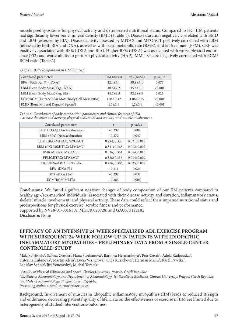
Reumatizam 2018;65(Suppl 1):37–74 57
Posters / Posteri Abstracts / Sažeci
muscle predispositions for physical activity and deteriorated nutritional status. Compared to HC, IIM patients had signifi cantly lower bone mineral density (BMD) (Table 1). Disease duration negatively correlated with BMD and LBM (assessed by BIA). Disease activity assessed by MITAX and MYOACT positively correlated with LBM (assessed by both BIA and DXA), as well as with basal metabolic rate (BMR), and fat free mass (FFM). CRP was positively associated with BF% (iDXA and BIA). Higher BF% (iDXA) was associated with worse physical endur-ance (FI2) and worse ability to perform physical activity (HAP). MMT-8 score negatively correlated with ECM/BCM ratio (Table 2).
Table 1. Body composition in IIM and HC.
Correlated parameters IIM (n=54) HC (n=54) p-valueBF% (Body Fat %) (iDXA) 42.4±7.1 39.9±7.1 0.077LBM (Lean Body Mass) (kg, iDXA) 40.6±7.2 45.6±8.1 <0.001LBM (Lean Body Mass) (kg, BIA) 48.7±9.5 52.6±8.8 0.023ECM/BCM (Extracellular Mass/Body Cell Mass ratio) 1.44±0.42 1.06±0.15 <0.001BMD (Bone Mineral Density) (g/cm2) 1.1±0.1 1.2±0.1 <0.001
Table 2. Correlation of body composition parameters and clinical features of IIM – disease duration and activity, physical endurance and activity, and muscle involvement.
Correlated parameters r p-valueBMD (iDXA):Disease duration –0.392 0.004
LBM (BIA):Disease duration –0.272 0.047LBM (BIA):MITAX; MYOACT 0.294; 0.335 0.031; 0.013
LBM (iDXA):MITAX; MYOACT 0.341; 0.368 0.012; 0.007BMR:MITAX; MYOACT 0.336; 0.351 0.014; 0.010FFM:MITAX; MYOACT 0.338; 0.356 0.014; 0.009
CRP: BF%-iDXA; BF%-BIA 0.276; 0.306 0.035; 0.025BF%-iDXA:FI2 –0.311 0.026
BF%-iDXA:HAP –0.292 0.032ECM/BCM:MMT8 –0.385 0.006
Conclusions: We found signifi cant negative changes of body composition of our IIM patients compared to healthy age-/sex-matched individuals, associated with their disease activity and duration, infl ammatory status, skeletal muscle involvement, and physical activity. Th ese data could refl ect their impaired nutritional status and predispositions for physical exercise, aerobic fi tness and performance.Supported by NV18-01-00161 A, MHCR 023728, and GAUK 312218.. Disclosure: None
EFFICACY OF AN INTENSIVE 24-WEEK SPECIALIZED ADL EXERCISE PROGRAM WITH SUBSEQUENT 24-WEEK FOLLOW-UP IN PATIENTS WITH IDIOPATHIC INFLAMMATORY MYOPATHIES – PRELIMINARY DATA FROM A SINGLE-CENTER CONTROLLED STUDY Maja Spiritovic1, Sabina Oreska2, Hana Storkanova2, Barbora Hermankova1, Petr Cesak1, Adela Rathouska3, Katerina Kubinova2, Martin Klein2, Lucia Vernerova2, Olga Ruzickova2, Herman Mann2, Karel Pavelka2, Ladislav Senolt2, Jiri Vencovsky2, Michal Tomcik2
1 Faculty of Physical Education and Sport, Charles University, Prague, Czech Republic 2 Institute of Rheumatology and Department of Rheumatology, 1st Faculty of Medicine, Charles University, Prague, Czech Republic 3 Institute of Rheumatology, Prague, Czech Republic Presenting author e-mail: [email protected]
Background: Involvement of muscles in idiopathic infl ammatory myopathies (IIM) leads to reduced strength and endurance, decreasing patients’ quality of life. Data on the eff ectiveness of exercise in IIM are limited due to heterogeneity of studied interventions/outcomes.
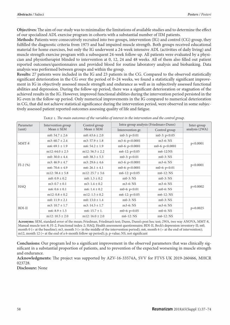
58 Reumatizam 2018;65(Suppl 1):37–74
Abstracts / Sažeci Posters / Posteri
Objectives: Th e aim of our study was to minimalize the limitations of available studies and to determine the eff ect of our specialized ADL exercise program in cohorts with a substantial number of IIM patients.Methods: Patients were consecutively recruited into two groups, intervention (IG) and control (CG) group, they fulfi lled the diagnostic criteria from 1975 and had impaired muscle strength. Both groups received educational material for home exercises, but only the IG underwent a 24-week intensive ADL (activities of daily living) and muscle strength exercise program with a subsequent 24-week follow-up. All patients were evaluated by a physi-cian and physiotherapist blinded to intervention at 0, 12, 24 and 48 weeks. All of them also fi lled out patient reported outcomes/questionnaires and provided blood for routine laboratory analysis and biobanking. Data analysis was performed between groups and within the group.Results: 27 patients were included in the IG and 23 patients in the CG. Compared to the observed statistically signifi cant deterioration in the CG over the period of 0–24 weeks, we found a statistically signifi cant improve-ment in IG in objectively assessed muscle strength and endurance as well as in subjectively assessed functional abilities and depression. During the follow-up period, there was a signifi cant deterioration or stagnation of the achieved results in the IG. However, improved functional abilities during the intervention period persisted in the IG even in the follow-up period. Only numerical improvements in the IG compared to numerical deterioration in CG, that did not achieve statistical signifi cance during the intervention period, were observed in some subjec-tively assessed patient reported outcomes assessing quality of life and fatigue.
Table 1. Th e main outcomes of the variables of interest in the intervention and the control group.
Parameter(unit)
Intervention groupMean ± SEM
Control groupMean ± SEM
Intra-group analysis (Friedman+Dunn) Inter-group analysis (2WA)Interevention gr. Control group
MMT-8
m0: 54.7 ± 2.6 m0: 63.6 ± 2.0 m0-3: p<0.01 m0-3: p<0.05
p<0.0001m3: 60.7 ± 2.4 m3: 57.9 ± 1.8 m3-6: p<0.0001 m3-6: NSm6: 69.1 ± 1.9 m6: 54.2 ± 1.9 m0-6: p<0.0001 m0-6: p<0.0001
m12: 64.0 ± 2.5 m12: 56.5 ± 2.2 m6-12: p<0.05 m6-12:NS
FI-2 (%)
m0: 30.0 ± 4.4 m0: 38.3 ± 5.3 m0-3: p<0.01 m0-3: NS
p<0.0001m3: 46.9 ± 4.7 m3: 29.6 ± 4.6 m3-6: p<0.0001 m3-6: NSm6: 70.6 ± 4.9 m6: 26.1 ± 4.1 m0-6: p<0.0001 m0-6: p<0.01
m12: 58.4 ± 5.8 m12: 25.7 ± 3.6 m6-12: p<0.05 m6-12: NS
HAQ
m0: 0.9 ± 0.2 m0: 1.3 ± 0.2 m0-3: NS m0-3: NS
p=0.0002m3: 0.7 ± 0.1 m3: 1.4 ± 0.2 m3-6: NS m3-6: NSm6: 0.6 ± 0.1 m6: 1.4 ± 0.2 m0-6: p<0.01 m0-6: NS
m12: 0.8 ± 0.2 m12: 1.5 ± 0.2 m6-12: p<0.05 m6-12: NS
BDI-II
m0: 11.9 ± 2.1 m0: 13.0 ± 1.4 m0-3: NS m0-3: NS
p=0.0025m3: 10.7 ± 1.7 m3: 14.3 ± 1.7 m3-6: NS m3-6: NSm6: 8.9 ± 1.5 m6: 15.7 ± 1. m0-6: p<0.05 m0-6: NS
m12: 10.5 ± 2.0 m12: 16.0 ± 2.0 m6-12: NS m6-12: NSAcronyms: SEM, standard error of the mean; Friedman, Friedman’s test; Dunn, Dunn’s post hoc test; 2WA, two way ANOVA, MMT-8, Manual muscle test-8; FI-2, Functional index-2; HAQ, Health assessment questionnaire; BDI-II, Beck’s depression inventory-II; m0, month 0 (= at the baseline); m3, month 3 (= in the middle of the intervention period); m6, month 6 (= at the end of intervention); m12, month 12 (= at the end of a 6-month follow up period); p, p-value; NS, not signifi cant
Conclusions: Our program led to a signifi cant improvement in the observed parameters that was clinically sig-nifi cant in a substantial proportion of patients, and to prevention of the expected worsening in muscle strength and endurance.Acknowledgments: Th e project was supported by AZV-16-33574A, SVV for FTVS UK 2019-260466, MHCR 023728.Disclosure: None
Reumatizam 2018;65(Suppl 1):37–74 59
Posters / Posteri Abstracts / Sažeci
OCULAR MANIFESTATIONS IN ANKYLOSING SPONDYLITIS AND RHEUMATOID ARTHRITIS Ana Gudelj Gračanin1, Joško Pavan1, Ana Marija Valetić2, Jadranka Morović Vergles1
1 Dubrava University Hospital, Zagreb, Croatia 2 Sestre milosrdnice University Hospital Centre Zagreb, Croatia Presenting author e-mail: [email protected]
Ocular manifestations occur in 10–50% of the patients with ankylosing spondylitis (AS)(1). Keratoconjuctivitis sicca occurs in 10 to 20 % patients with rheumatoid arthritis (RA)(2). It is known that the biomechanical proper-ties of the cornea may change in some infl ammatory diseases (3). Th e objective of the study was to evaluate cor-neal morphology and thickness in patients with AS and RA and to investigate the correlations between corneal alterations and disease activity.24 patients with AS and 28 patients with RA underwent ophthalmologic and physical examination. All patients were treated with biologic therapy. Corneal hysteresis(CH) and corneal resistance factor(CRF) were evaluated. Th e BASDAI for AS and DAS28CRP for RA were recorded. Signifi cant pathology changes of CH and CRF in both groups were not found. Moreover, correlations between disease activity and corneal changes were not found. Corneal morphology and thickness in patients with AS and RA could be changed, but the correlation between disease activity and the severity of changes has to be further investigated.References:1. Gouveia EB, et al.Ankylosing spondylitis and uveitis: overview. Rev Bras Reumatol.2012;52:742–756,2. Alpaslan M, et al.Assessment of Corneal Densitometry in Rheumatoid Arthritis Patients. Turk J Ophthalmol.2017;47:125–129,3. Caglayan M, et al.Biomechanical evaluation of cornea in patients with ankylosing spondylitis. Int Ophthalmol.2017;37:1017–1024.Disclosure: None
THE RELATIONSHIP BETWEEN HLA-B27 AND OCULAR INVOLVEMENT IN MALE ALBANIAN PATIENTS WITH ANKYLOSING SPONDYLITIS. Mikel Jordhani, Dorina Ruci
UHC Mother Teresa, Tirana, Albania Presenting author e-mail: [email protected]
Background: It is well-known the prevalence of uveitis and iritis in patients diagnosed with ankylosing spondy-litis. Patients with positive HLA-B27 are more susceptible to seronegative arthropathies and in particular mode to ankylosing spondylitis.Objectives: Th e aim of this study is to evaluate the relationship between ocular involvement and HLA-B27 in male albanian patients suff ering from ankylosing spondylitis.Methods: Patients included in this study were albanian males diagnosed with ankylosing spondylitis and ocular involvement All patients were completed with clinical, laboratory and imaging exams. Every patient was evalu-ated for HLA-B27 and consulted by an ophthalmologist for their ocular disorders. Results: Th irty-four patients (59.65%) were diagnosed with ocular involvement (iritis, uveitis). From those patients, 25 patients (73.53%) were found with positive HLA-B27. Aft er statistically analyzing all data, it was found a signifi cant relationship between ocular involvement and positive HLA-B27 (p <0.001).Conclusions: It was found that in patients with AS-related ocular involvement (iritis and uveitis), positive HLA-B27 was very prevalent. It was shown that there is a strong statistical relationship between ocular involvement and HLA-B27.References:Spondylarthropathies: Pathogenesis and Clinical Features pg 299; EULAR Textbook On Rheumatic DiseasesDisclosure: None
60 Reumatizam 2018;65(Suppl 1):37–74
Abstracts / Sažeci Posters / Posteri
ANTI-JAK TREATMENT AND INFECTION IN A PATIENT WITH SPONDYLOARTHRITIS Mirjam Szabo
ORFI Budapest, Hungary Presenting author e-mail: [email protected]
Ankylosing spondylitis (AS) is a chronic infl ammatory disease involving mainly the sacroiliac and axial joints.Th e current recommendations for the treatment considered the non-steroidal anti-infl ammatory drugs (NSAIDs), disease modifying antirheumatic drugs, biological agents, analgesics, surgical interventions. (1)Janus kinase (JAK) inhibitor is an alternative option for those patients who do not respond to classical treatment. By inhibition of multiple cytokines it might be a new potential strong strategy. (2)We describe a case of a 43 year-old patient with AS human leukocyte antigen B27-positive treated with Bari-citinib.Th e patient was admitted for painful walking. Previous year he underwent a total hip replacement of the left side due to bacteraemia.Examination revealed his hip range of motion restricted and scanner found an accumulation around the right femoral shaft .Th e diagnosis of infection of the right hip prothesis and a proximal femur osteomyelitis was confi rmed by aspira-tion.Th e patient went through a total hip replacement in two-steps, following with 3 months of antibiotherapy.JAK inhibitor was suspended during the investigation of the infection but persistence of infl ammatory syndrome made it necessary to resume the treatment.Th e effi cacy of JAK inhibitors is due to blocking the cytokine signaling, stability in laboratory parameters and long-term safety resulting in decrease of disease activity. (3) References:1. ASAS/EULAR recommendations for the management of ankylosing spondylitis. Ann Rheum Dis. 2006; 65(4):442-52 (ISSN: 0003-4967)2. Th e rationale for Janus kinase inhibitors for the treatment of spondyloarthritis. Veale DJ, McGonagle D, McInnes IB, Krueger JG,
Ritchlin CT, Elewaut D, Kanik KS, Hendrikx T, Berstein G, Hodge J, Telliez JB. Rheumatology (Oxford). 2018 Apr 3. doi: 10.1093/rheumatology/key070. [Epub ahead of print]
3. Wollenhaupt J, Silverfi eld J, Lee EB et al. Tofacitinib, an oral Janus kinase inhibitor, in the treatment of rheumatoid arthritis: safety and clinical and radiographic effi cacy in open-label, long-term extension studies over 7 years. Arthritis Rheumatol 2015;67:1645
Disclosure: None
SPONTANEOUS BICEPS FEMORIS RUPTURE HEMATOMA WITH SECONDARY INFLAMMATION IN A PATIENT WITH PSORIATIC ARTHRITIS TREATED WITH SECUKINUMAB DEVELOPING MYELODYSPLASTIC SYNDROME (MDS) – CASE REPORT Lorena Petrač1, Miroslav Mayer2
1 Special hospital for medical rehabilitation Krapinske Toplice, Gajeva 2, 49217 Krapinske Toplice, Croatia 2 Division of Clinical Immunology and Rheumatology, Department of Internal medicine, University Hospital Center Zagreb and University of Zagreb School of Medicine, Zagreb, Croatia Presenting author e-mail: [email protected]
Background: Psoriatic arthritis (PsA) is a chronic infl ammatory disease, a member of a spondiloarthritis (SpA) group associated with psoriasis. Treatment is based on non-steroidal anti-infl ammatory drugs (NSAID), gluco-corticoids, conventional and targeted disease modifying anti-rheumatic drugs (DMARDs) and biological agents. Arrival of biologics and targeted synthetic drugs has enabled more comprehensive treatment to target of all domains of the psoriatic disease. More agressive treatment approach of complex patients with many comorbidi-ties presents an additional adverse event risk.Case report: A sixty-fi ve year old diabetic male patient with longstanding severe psoriasis and psoriatic arthritis, treated sequentially with conventional DMARDs, ustekinumab and secukinumab, developed myelodysplastic syndrome and spontaneous partial rupture of the biceps femoris muscle complicated with infl ammed hematoma, caused by S.aureus infection.
Reumatizam 2018;65(Suppl 1):37–74 61
Posters / Posteri Abstracts / Sažeci
Conclusion: Th e aim of this paper is to present a patient in a long-lasting persistent remission induced and main-tained on biologics, developing rare serious side eff ect related to disease, comorbidities and the treatment itself. Further treatment options are discussed.References:1. Wright V, Moll JM. Psoriatic arthritis. Bull Rheum Dis. 1971; 21:627–32.2. Gossec L, et al. European League Against Rheumatism (EULAR) recommendations for the management of psoriatic arthritis with
pharmacological therapies:2015 update. Ann Rheum Dis 2015; 0:1–12.3. Frieder J, Kivelevitch D, Menter A. Secukinumab: a review of the anti IL-17A biologic for the treatment of psoriasis; Th er Adv Chron-
ic Dis. 2018 Jan; 9(1):5–21Disclosure: None
INHIBITION OF IL-17 BY SECUKINUMAB LEAD TO THE REMISSION OF SEVERE PSORIATIC ARTHRITIS AND SYMPTOMS OF PEMPHIGUS VULGARIS IN A PATIENT CONCIDENCE OF THESE CONDITIONS Marta Olejárová
Institute of Rheumatology, Prague, Czech Republic Presenting author e-mail: [email protected]
Background: IL-17 plays an important role in the pathogenesis of psoriasis and psoriatic arthritis (PsA) and its inhibition improves both, the skin and articular symptoms of the disease. Pemphigus vulgaris is a rare autoim-mune disease which may be also 1L-17 mediated based on preclinical data (1), but anti-IL17 agents have not been tested in pemphigus yet (2).. Coincidence of psoriasis and pemphigus vulgaris is exceptional. Objectives: To describe a rare case of severe psoriatic arthritis with preceding pemphigus vulgaris successfully treated with anti-IL17 therapy (secukinumab).Methods: We have observed a 43-years old man with a two years’ history of pemphigus vulgaris having been treated with continuous oral cyclophosphamide (CPM, 100 mg daily) and glucocorticoids (prednisone 10 mg daily) who developed psoriasis and severe psoriatic arthritis despite this treatment.Results: At the diagnosis of PsA CMP was stopped and replaced with methotrexate (MTX) up to the dose of 25 mg weekly, prednisone was increased up to 20 mg QD, several painful joints were injected with GC. Th ree months of this intensive treatment had only low impact on the activity of PsA and the patients was indicated for biological therapy. Because of the previous treatment with high cumulative dose of CPM and possible risk of malignancies of the combination of CPM and TNF inhibitors, we preferred secukinumab (150 mg weekly/ monthly) as the fi rst line biological. Th e treatment went promptly to the diminution of symptoms, decrease of infl ammatory markers (ESR, CRP) and aft er several weeks to the resolution of skin symptoms as well as arthritis. Symptoms of pemphi-gus vulgaris have not been observed more despite tapering prednisone off .Conclusions: In our patient, we have observed not only a successful treatment of psoriatic arthritis, but thanks to the coincidence with pemphigus vulgaris also for the very fi rst time the possible impact of this therapy on the course of pemphigus which was in remission throughout the durations of observation. In agreement with pre-clinical data our case report suggests the possible eff ect of anti-IL17 therapy also on pemphigus vulgaris. References:1. Timoteo RP, et al. Th 1/Th 17-related cytokines and chemokines and their implications in the pathogenesis of pemphigus vulgaris.
Mediators Infl amm 2017; 2017:7151285.2. Wasilewska A, Winiarska M, Olszewska M, Rudnicka L. Interleukin-17 inhibitors. A new era in treatment of psoriasis and other skin
diseases. Adv Dermatol Allergol 2016; XXXIII (4): 247–252. Disclosure: None

62 Reumatizam 2018;65(Suppl 1):37–74
Abstracts / Sažeci Posters / Posteri
APREMILAST AS A TREATMENT OF CHOICE FOR PSORIATIC ARTHRITIS IN A PATIENT WITH DIFFICULT TO TREAT AUTOIMMUNE HEPATITIS Mislav Čaić1, Miroslav Mayer2, Ivana Knežević Štromar3
1 University of Zagreb, School of Medicine 2 Division of Clinical Immunology and Rheumatology, Department of Internal medicine, University Hospital Center Zagreb and University of Zagreb School of Medicine, Zagreb, Croatia 3 Division of Gastroenterology and Hepatology, Department of Internal medicine, University Hospital Center Zagreb and University of Zagreb School of Medicine, Zagreb, Croatia Presenting author e-mail: [email protected]
Th e complexity of multiple coexisting illnesses, the interactions of several diff erent recommended treatment options and the attempts to avoid various side eff ects and contraindications, classify this case as one of the more challenging as well as interesting of the ones a physician may encounter. Although similar constellations of dis-eases are rather uncommon, the reason for presenting this case lies in its value as a teaching tool for specialists and non-specialists alike.To introduce an approach to a complex patient with a rheumatic disease and multiple comorbidities to a wider population of clinitians.A 56-year-old female patient suff ering from psoriasis, psoriatic arthritis (PsA) and autoimmune hepatitis (AIH) was referred to the rheumatology outpatient clinic at the University Hospital Centre Zagreb, Croatia. In spite of the remission of AIH due to the administered immunosuppressant therapy (tacrolimus, glucocorticoids), the patient experienced an exacerbation of PsA symptoms, including worsened arthralgia and dactylitis. Taking into account the referring gastroenterologist’s stance on contraindications regarding conventional PsA therapy, due to potential progression of hepatic fi brosis, hepatotoxicity, and severe drug interactions with tacrolimus, an alterna-tive treatment regime needed to be decided upon. Th e optimal treatment option, due to additional immunosup-pressive eff ects with fewest expected interactions with tacrolimus, was decided to be apremilast, a novel oral inhibitor of the enzyme phosphodiesterase-4 (PDE-4). At a follow-up two months since the beginning of treat-ment the patient’s state improved signifi cantly, in terms of alleviated joint pain and reduced psoriatic plaques.Th is case report highlights the importance of a multidisciplinary approach in the care for patients with rheumatic diseases. Apremilast signifi cantly mitigates the symptoms of enthesitis in addition to relieving pain, diminishing fatigue, and increasing work productivity in patients with active PsA.References:1. Keating GM. Apremilast: A Review in Psoriasis and Psoriatic Arthritis. Drugs. 2017;77(4):459–472. doi:10.1007/s40265-017-0709-12. Veale DJ, Fearon U. Th e pathogenesis of psoriatic arthritis. Th e Lancet. 2018;391(10136):2273–2284. doi:10.1016/S0140-6736(18)30830-4
INNATE IMMUNITY GENE EXPRESSION SIGNATURE IN PATIENTS WITH AUTOIMMUNE DISEASES IN ACTIVE DISEASE STAGE: RA, SLE, SSC Pavel Horák
Department of Internal Medicine III – Nephrology, Rheumatology and Endocrinology, Faculty of Medicine and Dentistry, Palacky University Olomouc, University Hospital, Olomouc, Czech Republic Presenting author e-mail: [email protected]
Background: Mounting evidence indicates that innate immunity, especially Toll-like-receptors (TLR) and inter-leukin (IL)-1/IL-1R families, play essential roles in the pathogenesis of autoimmune diseases. Th e diff erential innate expression pattern associated with disease activity in systemic lupus erythematosus (SLE), rheumatoid arthritis (RA), and systemic sclerosis (SSc) has not been established.Objective: To elucidate the underlying diff erences in innate immunity signatures associated with disease activity in major autoimmune diseases.Methods: We investigated gene expression of TLR1-10, 7 members of IL-1/IL-1R family, and interleukin 8 (IL-8/CXCL8) in peripheral blood mononuclear cells from patients with autoimmune disorders taken at time of active disease: SLE (n=28, SLEDAI>6), RA (n=36, DAS28≥3.2), and SSc (n=22, revised EUSTAR index>2.25) using high-throughput SmartChip Real-Time-qPCR system (WaferGen). Statistics were performed by R statistical soft -ware, P-value<0.05 was considered as signifi cant. Results: RA (a chronic joint infl ammatory disease) diff ered from SLE (a multisystem infl ammatory disease) and SSc (typical by tissue fi brosis of the skin and internal organs)
Reumatizam 2018;65(Suppl 1):37–74 63
Posters / Posteri Abstracts / Sažeci
by the upregulated expression of six genes (TLR2, TLR3, TLR5, SIGIRR, IL-1RAP, and IL-18R1; P<0.05). Active SLE and SSc showed high similarity in term of immunity gene expression signatures. In SSc, downregulated expression of IL-18R1 (P<0.05) was observed when compared to SLE and RA. In SLE, downregulated expression of IL-1R1 (P<0.05) was detected when compared to RA and upregulation of IL-18R1 (P<0.05) when compared to SSc. Conclusions: Innate immune gene expression signature in patients with autoimmune diseases in active dis-ease stage was identifi ed, showing high similarity between SLE and SSc. Grant support: MZ CR VES15-28659A, IGA UP_2018_010 References:1. Li M et al. Th e critical role of Toll-like receptor signaling pathways in the induction and progression of autoimmune diseases. Curr Mol
Med. 2009;9:365–74.2. Klonowska-Szymczyk A et al. Expression of toll-like receptors 3, 7, and 9 in peripheral blood mononuclear cells from patients with
systemic lupus erythematosus. Mediators Infl amm. 2014;2014:381418.3. Zhu YY et al. Th e largely normal response to Toll-like receptor 7 and 9 stimulation and the enhanced expression of SIGIRR by B cells
in systemic lupus erythematosus. PLoS One. 2012;7(8):e44131.)4. Rönnblom L et al..Th e interferon signature in autoimmune diseases.Curr Opin Rheumatol. 2013;25:248–53. Acknowledgement: Grant support: MZ CR VES15-28659A, IGA UP_2018_010
S100A11 (CALGIZZARIN) INDUCES INFLAMMATION VIA TLR-4 SIGNALLING AND STIMULATES SECRETION OF ANGIOGENIC FACTORS IL-8 AND VEGFS BY MONONUCLEAR CELLS IN RHEUMATOID ARTHRITIS Lucie Andrés Cerezo (Hana Hulejová, non-author presenter)
Institute of Rheumatology, Department of Rheumatology, First Faculty of Medicine, Charles University and Rheumatology Institute, Prague, Czech Republic Presenting non-author e-mail: [email protected]
Background: S100A11 (calgizzarin), S100 protein family member, is known to participate in oncogenesis via regulating number of biologic functions. It has also been described as infl ammatory mediator associated with rheumatoid arthritis (RA). Objectives: Given the fact that infl ammation evolves in parallel with angiogenesis, we aimed to assess the role of S100A11 in immune response and angiogenesis.Methods: For in vitro experiments, peripheral blood mononuclear cells (PBMCs) were obtained from patients with RA (n=6–12). Expression and protein synthesis were analysed by RT-PCR, ELISA and LUMINEX. Th e co-expression of S100A11 and TLR-4 receptor were analysed in synovial tissues from RA patients (n=6) by immu-nohistochemistry and immunofl uorescence. Receptor involvement was investigated using antibodies against RAGE, TLR-4, and inhibitor of MyD88. Intracellular signalling pathways were determined by use of inhibitors of NFkB, p38, erk1/erk2, and jnk.Results: PBMCs from patients with RA exposed to S100A11 increased the mRNA expression of pro-infl ammatory cytokines IL-6, TNFα and chemokine IL-8 (p<0.05, p<0.01 and p<0.01) as well as their secretion (p<0.01, p<0.001 and p<0.01). Moreover, S100A11 triggered the release of angiogenic factors VEGF-C and VEGF-D (p<0.05 and p<0.01) by PBMCs but did not modify their mRNA expression. No changes in IL-6 secretion by PBMCs were observed aft er blocking the receptor for advanced glycation products (RAGE) prior to S100A11 treatment. More importantly, PBMCs pre-treated with antibody against TLR-4 before S100A11 stimulation reduced the secretion of IL-6 by 92% (p<0.05). Inhibition of MyD88 adaptor protein also lead to a signifi cant decrease of IL-6 release by 55% (p<0.05). To support the implication of TLR-4 in S100A11 signalling, we demonstrated the co-expression of S100A11 and TLR-4 in the RA synovial tissue. Furthermore, S100A11 mediated IL-6 secretion was partially down-regulated by all selected inhibitors of cell signalling such as NFkB, erk1/2, jnk, p38 (p<0.05 for all).Conclusions: We are the fi rst to show that S100A11 may participate in regulation of angiogenesis and stimulates infl ammation via TLR-4 dependent pathway. Th ese fi ndings indicate more complex role of S100A11 in the patho-genesis of RA.Acknowledgement: Th is work received support from research project 15-34065A of the Agency for Healthcare Research of the Czech RepublicDisclosure: None
64 Reumatizam 2018;65(Suppl 1):37–74
Abstracts / Sažeci Posters / Posteri
INTERACTION OF P.Q141K VARIANT IN ABCG2 GENE WITH CLINICAL DATA AND CYTOKINES LEVELS IN PRIMARY HYPERURICEMIA AND GOUT Jana Bohatá1, Veronika Horváthová2, Kateřina Pavelcová1, Blanka Stibůrková3
1 Institute of Rheumatology, Prague, Czech Republic; First Faculty of Medicine, Charles University, Prague, Czech Republic 2 Institute of Rheumatology, Prague, Czech Republic; Faculty of Science, Charles University, Prague, Czech Republic 3 Institute of Rheumatology, Prague, Czech Republic; Department of Pediatrics and Adolescent Medicine, First Faculty of Medicine, Charles University, General University Hospital, Prague, Czech Republic Presenting author e-mail: [email protected]
Background: Gout is multifactorial disorder caused by many conditions including genetic background. One of the well-established genes linked to gout is ABCG2 and its common missense variant p.Q141K which has nega-tive infl uence on urate excretion (1). Long lasting hyperuricemia leads to formation of monosodium urate crys-tals in joints and tissues. Immune response to these crystals is triggered by NLRP3 infl ammasome and subse-quent production of proinfl ammatory cytokines which results in escalation of immune reaction.Objectives: Th e aim of our study was to analyse the possible diff erences in age, BMI, CRP and GFR as risk factors/symptoms for gout and immunological parameters between hyperuricemic, gouty and acute gouty arthritic patients who had known genetic background in risk allele p.Q141K.Methods: Gene ABCG2 was sequenced in cohort of 69 primary hyperuricemic and 177 gout patients. Variant p.Q141K was examined in 132 normouricemic controls using tetra primer ARMS-PCR method. Aft erwards we determined plasma levels of 27 cytokines using human multiple cytokine assay in subcohort of 42 patients with primary hyperuricemia, 131 gout patients including 17 acute gouty arthritis patients and 132 controls.Results: We have found diff erences between diagnostic groups for disease onset, BMI, CRP and GFR. In addition, homozygotes p.Q141K were signifi cantly younger (p = 0.019) and had earlier onset of disorder (p = 0.011) than wild-type and heterozygotes. Homozygotes p.Q141K tended to have lower BMI (p = 0.020) and CRP (p < 0.001). Heterozygous and mainly homozygous carriers of p.Q141K variant had signifi cantly higher GFR (p = 0.022). Levels of IL-1β, IL-1ra, IL-4, IL-6, IL-7, IL-8, IL-9, IL-13, IL-17, FGF basic, IFNγ and TNFα were signifi cantly increased in patients with acute gouty arthritis. Eotaxin, MCP-1 and RANTES were decreased in all patient groups compared to controls. We did not fi nd any impact of p.Q141K on cytokine amount. Conclusions: Variant p.Q141K is related to earlier onset, lower BMI, lower CRP and higher GFR at patients with primary hyperuricemia and gout. Levels of 19 cytokines were signifi cantly higher mainly in patients with acute gouty arthritis compared to other groups without correlation to presence of p.Q141K.Acknowledgement: Supported by AZV 15-26693A and RVO 00023728. References:1. Stiburkova B et al. Functional non-synonymous variants of ABCG2 and gout risk. Rheumatology. 2017;56(11):1982–92.Disclosure: None
REAL LIFE EXPERIENCE WITH GOLIMUMAB IN CROATIA Felina Anić1, Tatjana Kehler2, Marija Rogoznica2, Frane Grubišić3, Simeon Grazio3, Marta Žuvić4, Srđan Novak1
1 Department of Rheumatology and Clinical Immunology, Division of Internal Medicine, University Hospital Center Rijeka, Croatia 2 Department of Physical Medicine and Rheumatology Th alassotherapia Opatija, Croatia 3 Department of Rheumatology, Physical Medicine and Rehabilitation, School of Medicine University of Zagreb, Sestre Milosrdnice University Hospital Center, Zagreb, Croatia 4 Department of Biotechnology, University of Rijeka, Croatia Presenting author e-mail: [email protected]
Background: Golimumab (GLM) is a human monoclonal antibody indicated in the treatment of adults with active RA, PsA and AS who have had an inadequate response to conventional therapies. Th ere are some data that persistence with GLM in immune-mediated rheumatic disease may be higher than with other TNF-α agents and may be lower in biologic-experienced compared with biologic-naive patients. Effi cacy of golimumab through 3 and 5 years of treatment in patients with RA, PsA and AS was consistent with other TNF-α antagonist. Objectives: Th e aim of this study was to evaluate the effi cacy and the retention of golimumab (GLM) in treating patients with rheumatoid arthritis (RA), psoriatic arthritis (PsA) and ankylosing spondylitis (AS) in a real-world setting.
Reumatizam 2018;65(Suppl 1):37–74 65
Posters / Posteri Abstracts / Sažeci
Methods: Th is was a retrospective cohort study on patients with RA (11), PsA (20) and AS (23) who had active disease despite prior treatment with c DMARDs, NSAIDs s or biological drugs. Effi cacy of GLM was evaluated at weeks 12, 36 and 52 aft er starting GLM treatment. Golimumab drug retention rate at 2 and 5 years of treatment was determined for all patients and additionally for each indication (RA, PsA, AS) and according to TNFα naivety. Statistical signifi cance was determined at the level of 0.05. Kaplan-Meier analysis was used to assess drug retention.Results: GLM demonstrated signifi cant effi cacy in terms of decreasing disease activity in RA, PsA and AS patients already at week 12. Th e results were held even at week 36 and 52. Overall 2 and 5 years retention rate was 77% and 60%, the highest being in AS patients – 90% in 2 and 5 years. Two-year retention rate for GLM in PsA and RA patients was 75% and 55%. Five-year retention rate for GLM in PsA and RA patients was 57% and 37%. TNFα naïve patients had higher retention rate in comparison to non-naïve patients (66% vs 34%).Conclusions: In this retrospective study in a real-life setting it was demonstrated that GLM is an eff ective drug for RA, PsA and AS patients with a high retention rate. GLM retention rate was higher in AS patients compared to RA and PsA patients and in TNFα naïve patients compared to patients previously treated with TNFα inhibitors.References:1. Svedbom A, Storck C, Kachroo S, et al. Persistence with golimumab in immune-mediated rheumatic diseases: a systematic review of
real-world evidence in rheumatoid arthritis, axial spondyloarthritis, and psoriatic arthritis. Patient Prefer Adherence. 2017;11:719–729.
2. Kay J, Fleischmann R, Keystone E, et al. Five-year safety data from 5 clinical trials of subcutaneous golimumab in patients with rheumatoid arthritis, psoriatic arthritis and ankylosing spondylitis. J Rheumatol. 2016;43:2120–2130.
Disclosure: None
EFFICACY AND SAFETY OF SWITCHING FROM ORAL TO SUBCUTANEOUS METHOTREXATE IN EVERYDAY CLINICAL PRACTICE: RESULTS OF THE SIX-MONTH OBSERVATIONAL PROSPECTIVE STUDY IN CROATIA Simeon Grazio1, Dijana Perković2, Ana Gudelj Gračanin3, Nadica Laktašić Žerjavić4, Marija Glasnović5, Frane Grubišić1, Jadranka Morović Vergles3, Porin Perić4, Iva Žagar4, Helena Mitrović4, Petra Šimac2, Željka Kolak6, Ines Doko1
1 Department of Rheumatology, Physical Medicine and Rehabilitation, School of Medicine University of Zagreb, Sestre Milosrdnice University Hospital Center, Zagreb, Croatia 2 Department of Internal Medicine, School of Medicine University of Split, University Hospital Centre Split, Croatia 3 Department of Internal Medicine Dubrava, School of Medicine University of Zagreb, University Hospital Zagreb, Croatia 4 Department of Rheumatic Diseases and Rehabilitation, School of Medicine University of Zagreb, University Hospital Centre Zagreb, Croatia 5 Department of Internal Medicine, School of Medicine University of Osijek, University Hospital Centre Osijek, Croatia 6 Divison of Physical Medicine and Rehabilitation, General County Hospital Vinkovci, Croatia Presenting author e-mail: [email protected]
Background: Rheumatoid arthritis (RA) and psoriatic arthritis (PsA) are chronic debilitating infl ammatory rheumatic conditions with the major impact on quality of life (QoL). csDMARDs are the basic pharmacological option for these patients, and choice of DMARDs depends on many factors.Objectives: Th e objective of this study was to evaluate the changes in disease activity, QoL, adverse events when switching from oral to subcutaneous methotrexate (MTX) in patients with RA and PsA, addressing also the adherence to treatment.Methods: Th is was an observational prospective longitudinal study of patients with RA and PsA on csDMARDs who switched from oral to s.c. MTX. Consecutive patients with established diagnosis of RA and PsA were enrolled from the outpatient clinics of 6 centres in Croatia. Th ere were 48 patients (79.2% women), of median age 61 (range 39–79), and with the median of disease duration of 120 months. Data were collected at baseline (T0) including retrospective data collection for the previous 3 months (on oral MTX), at day 90 (±10 days) (T1) and at day 180 ((±10 days) (T2) for the previous periods (both of them during s.c. MTX treatment). Dose of MTX remained stable during the study. Domains of interest were: DAS28, level of pain, PtGHA and PhGHA, QoL (EQ-5Q) and physical function (HAQ-DI). Adherence to treatment and safety data were recorded, too.Results: In a comparison between T0 and T1/T2 there was more chance for patients having low to moderate dis-ease activity aft er 3 and 6 moths of s.c. MTX (8.0; 95%CI 1.9-32.0) and less chance to have high disease activity (0.25; 95%CI 0.11-0.48) in comparison to the baseline DAS 28 (on oral MTX). Adjusted mean values for intensity of pain, PtGHA and PhGHA showed signifi cant decrease for T1 and T2 versus T0 assessment (–1.46; 95%CI
66 Reumatizam 2018;65(Suppl 1):37–74
Abstracts / Sažeci Posters / Posteri
–1.55 to –0.35; –1.12; 95%CI –1.50 to –0.73; –1.15; 95%CI –1.50 to –0.80, respectively). Also, in the same com-parison EQ-5D global health showed signifi cant improvement (8.6; 95%CI 4.00 to 13.3), as well as HAQ-DI (–0.25; 95%CI –0.32 to –0.17). In the retrospective analysis there were more missed doses of oral compared to s.c. MTX, while on the other hand adverse events were less frequent with s.c. compared to oral MTX.Conclusions: In the group of patients with RA and PsA who switched from oral to s.c. MTX improvement was observed in multiple outcomes including disease activity, QoL and safety as well as better adherence.References:1. Li D, Yang Z, Kang P, Xie X. Subcutaneous administration of methotrexate at high doses makes a better performance in the treatment
of rheumatoid arthritis compared with oral administration of methotrexate: a systematic review and meta-analysis. Semin Arthritis Rheum. 2016;45(6):656–62.
2. Schiff MH, Jaff e JS, Freundlich B. Head-to-head, randomised, crossover study of oral versus subcutaneous methotrexate in patients with rheumatoid arthritis: drug-exposure limitations of oral methotrexate at doses 15 mg may be overcome with subcutaneous admin-istration. Ann Rheum Dis. 2014;73(8):1549–51.
3. Braun J, Kästner P, Flaxenberg P, Währisch J, Hanke P, Demary W, et al. Comparison of the clinical effi cacy and safety of subcutaneous versus oral administration of methotrexate in patients with active rheumatoid arthritis: results of a six-month, multicenter, random-ized, double-blind,controlled, phase IV trial. Arthritis Rheum. 2008;58(1):73–81.
4. Jay R. Methotrexate revisited: considerations for subcutaneous administration in RA. Clin Rheumatol. 2015;34(2):201–5.5. Bianchi G, Caporali R, Todoerti M, Mattana P. Methotrexate and rheumatoid arthritis: current evidence regarding subcutaneous ver-
sus oral routes of administration. Adv Th er. 2016;33(3):369–78. Disclosure: None
INFECTIONS AND MALIGNANCIES IN PATIENTS TREATED WITH BIOLOGICAL DISEASE MODIFYING ANTIRHEUMATIC DRUGS- OUR EXPERIENCE Kristina Kovač Durmiš, Mislav Pap, Duje Birkić, Nadica Laktašić Žerjavić, Iva Žagar, Porin Perić
Department of Rheumatology and Rehabilitation of School of Medicine, University Hospital Centre Zagreb, Croatia Presenting author e-mail: [email protected]
Background: Over the past century the treatment of infl ammatory rheumatic diseases has moved from an entirely empiric approach, based on the use of aspirin, gold salts and glucocorticoids, through evidence-based approaches with conventional synthetic disease modifying antirheumatic drugs (cs DMARDs), to mechanism-based regi-mens with biologic disease modifi ers. Biological disease-modifying antirheumatic drugs (bDMARDs) have become standard treatment option with a major safety issue related to increased risk of infections1.Objectives: Th e objective of the research was to determine prevalence of infections and malignancies in patients with infl ammatory rheumatism treated with bDMARDs in our Department.Methods: Data on one hundred seventeen (117) patients with rheumatoid arthritis (RA, N=50), psoriatic arthri-tis (PsA, N=18), ankylosing spondylitis (AS, N=42), undiff erentiated spondylarthritis (N=7) were analysed retro-spectively. Patients were mainly treated with tumour necrosis factor (TNF)-inhibitors (N=103) (adalimumab, certolizumab, etanercept, golimumab, infl iximab) and tocilizumab (N=14) from 2004 until September 2018. Descriptive statistical analysis was used.Results: Total number of patients experiencing any documented infection during treatment with bDMARDs was 27 (23,1%), mostly prevalent among patients with RA (N=16; 59,3%), less in patients with AS (N=7; 25,9%) and PsA (N=4; 14,8%). One patient was diagnosed with solid tissue malignancy (hepatocellular carcinoma).Conclusions: Infections were more oft en diagnosed among RA patients possibly due to nature of the disease itself and immunosuppressive eff ects of concomitant treatment. Only one patient was diagnosed with solid tissue car-cinoma which seems to be in concordance with published data on risk of malignancy during biologic treatment2. Th is study serves as a basis for further bDMARDs safety monitoring in our Department. It is necessary to increase the sample size and continue follow up to before making any defi nite conclusions.References:1. Singh JA et al. Risk of serious infection in biological treatment of patients with rheumatoid arthritis: a systematic review and meta-
analysis. Lancet. 2015 Jul 18;386(9990):258–65.2. Ramiro S et al. Safety of synthetic and biological DMARDs: a systematic literature review informing the 2016 update of the EULAR
recommendations for management of rheumatoid arthritis. Ann Rheum Dis. 2017 Jun;76(6):1101–1136.Disclosure: None
Reumatizam 2018;65(Suppl 1):37–74 67
Posters / Posteri Abstracts / Sažeci
CLINICAL OUTCOMES OF MACROPHAGE ACTIVATION SYNDROME IN UNIVERSITY HOSPITAL CENTRE Tatjana Zekić1, Ita Hadžisejdić2, Srđan Novak3
1 Division of Rheumatology and Clinical Immunology, Clinical Hospital Center Rijeka 2 Department of Pathology, University of Rijeka Medical school 3 Divison of Rheumatology and Clinical Immunology, University of Rijeka Medical school Presenting author e-mail: [email protected]
Background: Macrophage activation syndrome (MAS) is an infl ammatory, potentially life-threatening process that accompanies autoimmune diseases. It is more common in children than in adults. Outcome success depends on early diagnosis and prompt treatment.Objectives: To evaluate causes and clinical features of MAS, and to discuss clinical pitfalls, with respect to avail-able 2016 classifi cation criteria (EULAR/ACR PRINTO for MAS in JIA)1.Methods: Retrospective study (2012–2018) included 6 patients. Th e basic characteristics (age, gender, comor-bidities), laboratory fi ndings, biopsy (bone marrow, lymph node, liver) results, treatment, underlying disease, clinical course of illness and outcome were reported.Results: Four female and two male patients, age (26–71), were diagnosed with MAS. MAS was the fi rst manifesta-tion of autoimmune disease in one patient, Adult onset Still disease (AOSD), and appeared in previously known Rheumatoid arthritis (RA) and Undiff erentiated Connective Tissue Disease (UCTD). Triger infections were Cytomegalovirus (CMV) and Clamidophila pneumoniae. In one patient, antibodies to Syogren syndrome (SS) and Primary biliary cirrhosis occured aft er one year of follow up. Laboratory abnormalities included elevated liver enzymes and LDH and trombocytopenia. Th e hallmark was ferritin level above 5000 [ug/L]. Poorer out-comes were observed in cases of delayed diagnosis (≥20 days) and in elderly patients with comorbidites. All patients were treated with glucocorticoids and cyclosporin, and if necessary etoposide. One patient had refrac-tory disease without any evidence of infectious, malignant or autoimmune background despite additional therapy with IVIG, IL-1 and IL-6 inhibitors, and probably had an primary form.Th ree patients had a lethal outcome. More detailed information are shown in Table 1.Conclusions: A combination of symptoms and laboratory fi ndings of high fever, increased ferritin level, elevated liver function tests and trombocytopenia should always arouse suspicion of MAS in patient with known predis-posing factors. Recognising trigers and aggressive treatment could signifi cantly improve survival outcomes.References:1. Ravelli et al. 2016 Classifi cation Criteria for Macrophage Activation Syndrome Complicating Systemic Juvenile Idiopathic Arthritis,
ARTHRITIS & RHEUMATOLOGY, DOI 10.1002/ART.393322. Piero Ruscitti, Paola Cipriani, Paola Di Benedetto, Vasiliky Liakouli, Francesco Carubbi, Onorina Berardicurti, Francesco Ciccia,
Giuliana Guggino, Giovanni Triolo & Robert Giacomelli (2017): Advances in immunopathogenesis of macrophage activation syndrome during rheumatic infl ammatory diseases: toward new therapeutic targets?, Expert Review of Clinical Immunology, DOI: 10.1080/1744666X.2017.1372194
Disclosure: None
LYME DISEASE. MOST FREQUENT OBSERVED SYMPTOMS. IS ARTHRALGIA AMONG THEM? Agnė Petrulionienė1, Daiva Radzišauskienė2, Arvydas Ambrozaitis2, Saulius Čaplinskas3, Algimantas Paulauskas4, Algirdas Venalis5
1 Clinic of Rheumatology, Orthopaedics Traumatology and Reconstructive Surgery, Institute of Biomedical science, Faculty of Medicine, Vilnius University. Lithuania 2 Infectious Disease Center, Vilnius University. Lithuania 3 Center for Communicable Diseases and AIDS. Lithuania 4 Faculty of Natural Sciences, Vytautas Magnus University, Lithuania 5 Rheumatology center, Vilnius University, 5 State research Institute Centerfor Innovative Medicine, Vilnius, Lithuania Presenting author e-mail: [email protected]
Background: A Tick born Lyme disease is one of the reasons which can cause reactive arthritis. Lyme disease is very common disease in the world, It has spread into the whole of Europe, except for the warmest and dry areas in the south and the coldest areas in the north. Th e annual incidence of Lyme disease in the USA is 8/100 000 and
68 Reumatizam 2018;65(Suppl 1):37–74
Abstracts / Sažeci Posters / Posteri
in central Europe and in Scandinavia varies from 0.3 to 155/100 000 (Schnarr et al, 2006*), in Lithuania (central Europe) it’s frequency is 101.6/100 000 (2016 years). Infection usually begins with an expanding skin lesion, known as erythema migrans.Objectives: To fi nd out how oft en rheumatic symptoms appears in patients with Lyme disease in Lithuania.Methods: We have analyzed data from Center for Communicable Diseases and Aids of Lithuania, of persons who were diagnosed with Lyme disease in 2014–2016 years. Total number of cases is 7424 (2791 males, 4633 females).Results: 996 patients assumed to be symptomatic, the rest 6428 were asymptomatic either information about clinical disease manifestation is not known. Erythema migrans was the most frequently appeared symptom (n = 753, 75,6 %), arthralgia was the second one (220, 22,1 %), the following symptoms were headache (151, 15,2%), general weakness (124, 12,4%), fever (101, 10,1%), myalgia (78, 7,8%), head dizziness (64, 6,4%).Conclusions: In symptomatic patients rheumatic symptoms (arthralgia and myalgia) appears oft en and Lyme disease should be in rheumatologists minds, especially those who works in endemic zones.References:1. Sergamumo užkrečiamomis ligomis Lietuvoje apžvalga 2016. Užkrečiamų ligų ir AIDS centras. 96–95 pages. http://www.ulac.lt/up-
loads/downloads/leidiniai/Sergamumo_apzvalga_2016.pdf2. https://www.ncbi.nlm.nih.gov/pmc/articles/PMC5539539/3. EULAR on-line course on Rheumatic Diseases, 11 module, Infection and arthritis. Reactive arthritis – Lyme – Whipple – HIV – Viral
arthritis – Septic arthritis, Maxime Breban, Hill Gaston, Franco Schiavon, 2.5 Lyme arthritis.Disclosure: None
TRAPS (TUMOR NECROSIS FACTOR RECEPTOR – ASSOCIATED PERIODIC SYNDROME) Oľga Lukáčová1, Jozef Lukáč2
1 National Institute of rheumatic diseases, Piešťany, Slovak republic 2 National Institute of rheumatic diseases, Piešťany, Slovak republic, Institute of physiotherapy, balneology and medical rehabilitation, Piešťany, Slovak republic Presenting author e-mail: [email protected]
Periodic/recurrent fewer syndromes belong to group of autoinfl ammatory syndromes diseases. Familial Mediter-ranean Fever and Tumor necrosis factor receptor – associated periodic syndrome are most oft en from them, a less are : HIDS – Hyper IgD Syndrome, MKD – Mevalonate Kinase Defi ciency, CAPS: Cryopyrin-Associated Autoin-fl ammatory Syndromes, NALP12-associated periodic fewer.TRAPS is the second oft en hereditary syndrome of periodic fewer (aft er Familial Mediterranean Fever). Th ere are approximately 1000 peoples with TRAPS, mostly from Middle and South Europe, from northern countries, from America, Japan. Th ere are recurrent fewer episode with duration days to months, abdominal pain, myalgia, skin rush – mostly at limbs in clinical pictures. Sometimes there could appear arthralgia, ocular syndrome (periorbital oedema, conjunctivitis, uveitis, iritis). Th e amyloidosis could occur and lead to renal failure. During childhood disease could manifest as juvenile idiopatic arthritis, systemic form (fewer, rush, IBD).In TRAPS is mutation in gene TNFR SF1A, which insure interaction for TNF receptor 1. Aft er mutation incorrect confi guration of TNFR protein is producing, which could not bind at cell surface, could not bind to TNF.Th ere are use non-infl ammatory drugs, glucocorticoids, kolchicin in therapy of TRAPS. Aft er identifi cation of mutation of TNFRSFA1 gene biological therapy start to use: etanercept, anakinraCasuistic: Now 30 years old patient with juvenile idiopatic arthritis with systemic form from 1998 (from 11 years of age). Her mother had amyloidosis of kidneys and died as 43 years old. At year 2009 patient was diagnostic primary amyloidosis of heavy form. At 2012 was done biopsy, deposits of amyloid at vessel. Aft er chemotherapy the autologous transplantation of bone marrow was done. At 2014 was done rebiopsy with fi nding of AA amyloi-dosis. From 2015 therapy by etanercept was begin. In successive steps quantitative proteinuria was fall (tab.1).References:1. Dhanrajani A, Khubchandani RP, Indian Pediatric, 2014, 51.Disclosure: None
Reumatizam 2018;65(Suppl 1):37–74 69
Posters / Posteri Abstracts / Sažeci
THE RELATIONSHIP BETWEEN THE DURATION OF METHOTREXATE / ETANERCEPT THERAPY AND SERUM ANTI-SPP IGG ANTIBODIES IN PATIENTS WITH JIA WITHOUT SYSTEMIC MANIFESTATIONS. Margarita Soloshenko, Ekaterina Alexeevna, Rina Denisovna, Tatyana Dvoryakovskaya, Ksenia Isaeva, Anna Mamutova, Nikolay Mayansky, Natalyaya Tkachenko, Irina Zubkova, Marina Fedoseenko
National Medical Research Center of Children’s Health, Russia Presenting author e-mail: [email protected]
Background: Juvenile idiopathic arthritis (JIA) is a chronic disease with unknown etiology, a complex immuno-aggressive pathogenesis characterized by a steadily progressing course, the development of destruction of the connective tissue of the joints, a wide spectrum of severe extra-articular manifestations, leading to an early dis-ability of patients. Currently, the prevention of infectious diseases in children with juvenile idiopathic arthritis under conditions of immunosuppressive and biological therapy is of particular importance, since it requires resolving the issue of getting out of the vicious circle of prejudice to vaccination. Th e decision to immunize a child with JIA implies a special responsibility of pediatricians and children’s rheumatologists. Systematic reviews of the results of vaccination of patients with rheumatic diseases against infectious diseases summarize that most vac-cines are eff ective and safe even when patients receive immunosuppressive drugs.Objectives: To analyze the incidence of infectious complications, prescribing antibacterial drugs and adherence to antirheumatic therapy in patients with JIA without systemic manifestations before and aft er immunization of 13 PCV.Methods: During the prospective cohort study, fi ve groups were formed: children with JIA in remission phase with methotrexate (group 1) or etanercept (group 2), with JIA in the active phase prior to the appointment of methotrexate (group 3) or etanercept (group 4), control group (conditionally healthy children). 13-valent PCV was injected once in 0.5 ml subcutaneously against therapy in patients in the remission phase, or 3 weeks before the appointment of methotrexate or etanercept in patients in the active phase. In the course of the study, the fre-quency of acute infectious complications, the frequency of prescribing antibacterial drugs in patients with JIA, as well as adverse events against vaccination was assessed.Results: Before the vaccination of 13 PCV, the incidence of infectious complications, the morbidity rate and the frequency of administration of antibiotics in children with JIA who received methotrexate or etanercept (Ia and Ib group, respectively) were signifi cantly higher (p = 0.001) than in patients not treated with an immunosuppres-sant (IIa group), inhibitor of TNF-α (IIb group) and in children of the control group (Figure 2, 3). Aft er immuni-zation with 13 PCV, the incidence of infectious complications, the morbidity rate and the frequency of adminis-tration of antibiotics decreased (p = 0.001) and did not signifi cantly diff er from those of the control group.Conclusions: Th e severity of the vaccine response in patients in the phase of drug remission of JIA depends on the duration of antirheumatic therapy: the longer the children are treated with methotrexate (r = –0.876) and etanercept (r = –0.776), the lower the serum level of anti-SPP IgG antibodies aft er 4 weeks aft er vaccination 13 PCV. Reduction of the level of immune response is observed when using antirheumatic drugs for more than 8 months.References:1. Alexeeva E.I., Dvoryakovskaya T.M., Denisova R.V., Isaeva K.B., Soloshenko M.A., Mamutova A.V., Mayansky N.A., Tkachenko N.E.,
Zubkova I.V., Kaluzhnaya T.A., Shakhtakhtinskaya F.C., Gayvoronskaya A.G., Broeva M.I., Fedoseenko M.V. Immunization with a pneumococcal polysaccharide vaccine in children with juvenile idiopathic arthritis without systemic manifestations: a prospective study. Current Pediatrics. 2017;16(6):493–501. (In Russ.) https://doi.org/10.15690/vsp.v16i6.1822
Disclosure: E. Alexeeva Grant / Research Support from: Roche, Pfi zer, Centocor, Novartis, M. Soloshenko: None Declared, T. Dvoryakovskaya Grant / Research Support from: Roche, Pfi zer, R. Denisova: None Declared, K. Isaeva: None Declared, A. Mamutova: None Declared, N. Mayansky1: None Declared, N. Tkachenko: None Declared, I. Zubkova: None Declared, M. Fedoseenko: None Declared
70 Reumatizam 2018;65(Suppl 1):37–74
Abstracts / Sažeci Posters / Posteri
STRUCTURE MODIFICATION IN OA AND PRESENT AND FUTURE PERSPECTIVES Karel Pavelka
Institute of Rheumatology Prague Presenting author e-mail: [email protected]
Th erapy of OA has been considered as purely symptomatic at present. Despite of that, several RCT have been performed and published, which have documented positive eff ect of active drug compared to placebo. Th e evi-dence was shown in studies with glukosamine 1), chondroitin sulphate, combination of glucosamine and chon-droitin sulphate, diacerhein, piascledin, hyaluronic acid and tetracyklin. In 2 year controlled study, chondroitin sulphate against celecoxib, chondroitin documented less decrease of medial compartment cartilage volume when using special quantitative MRI methodology2)New therapeutic tested alternatives in OA are: autologous conditioned serum, platelet rich plasma (PRP), mesen-chymal stem cells, fi broblast growth factor 18 (FGF-18), inhibitors MMP, inhibitors of aggrecanase, K-inhibitors cathepsin, inhibitors MAP kinases, inhibitors nitric oxide synthase (i NOS).Recent metaanalysis didn´t confi rm structure modifi cation by bone marrow derived, mesenchymal stem cells.Methodological ant ethical problems in long-term studies in OA will be discussed. References:1. Pavelka et all. Glucosamine sulfate use and delay of progression of knee osteoarthritis. A 3-year, randomized, placebo-controlled,
double-blind study. Arch Intern Med 2002; 162: 2113–21232. Pelletier JP et all. Arthrit. Res Th er. 2016;18 (1): 256. Disclosure: None
HYPOKALEMIC PARALYSIS AS A PRESENTING MANIFESTATION OF PRIMARY SJÖGREN’S SYNDROME: CASE REPORT Sonja Golubovic
Department of Nephrology and Clinical Immunology, Clinical Center of Vojvodina, Novi Sad, Serbia Presenting author e-mail: [email protected]
Background: Primary Sjögren’s syndrome is a chronic autoimmune disease primarily characterized by a progres-sive lymphocytic infi ltration of the exocrine glands with varying degrees of systemic involvement. In case of renal involvement, the most common manifestation is renal tubular acidosis, resulting from tubular dysfunction due to tubulointerstitial nephritisObjectives: We present a case of a patient with hypokalemic quadriparesis due to tubulointerstital nephritis and renal tubular acidosis, as a manifestation of Sjögren’s syndromeMethods: A 41-year-old woman, was admitted to the emergency room due to weakness, muscle pain, diffi culties walking and moving all four limbs. Laboratory results showed extreme hypokalemia with metabolic acidosis, increased erythrocyte sedimentation rate, hyperchloremia and hypernatremia. Urin sample analysis revealed proteinuria semi-quantitative 2–3, ketonuria. Imunoserologic tests confi rmed the presence of speckled ANA with the presence of anti SSA and anti SSB antibodies. High values of muscle enzymes were identifi ed (CPK, Troponin, CK). In the left kidney, initial hydronephrosis with calcifi cation in the lower half of the size up to 4mm was foundResults: Hypokalemia and metabolic acidosis were treated with parenteral administration of sodium bicarbonate and thiazide diuretic. Immunofl uorescent microscopy of the renal biopsy sample revealed a densely infi ltrated hypercellular interstitial segment without presence of immunofl uorescent deposits within the glomerulus; the fi nding of light microscopic analysis fi ts into the clinical picture of tubulointerstitial nephritis.Th e patient was given pulse doses of corticosteroids with cyclophosphamide intravenously in monthly intervals and is on regular outpatient follow up, with a stable remission of the underlying diseaseConclusions: Th e presence of hypokalemia and consequent symptoms, such as quadriparesis, needs to be thor-oughly analyzed and further investigated for a possible causeReferences:1. Fox RI. Sjögren’s syndrome. A detailed review. Lancet 2005;366(9482):321–31. doi:10.1016/S0140-6736(05)66990-52. Mugundhan K, Mayan Vasif MC, Nidhin PD, Prakash G, Balamurugan N, Sivakumar KG, et al. Hypokalemic paralysis in Sjogren’s
syndrome secondary to renal tubular acidosis. J Assoc Phys India 2016;64:72. PMID: 27731561Disclosure: None
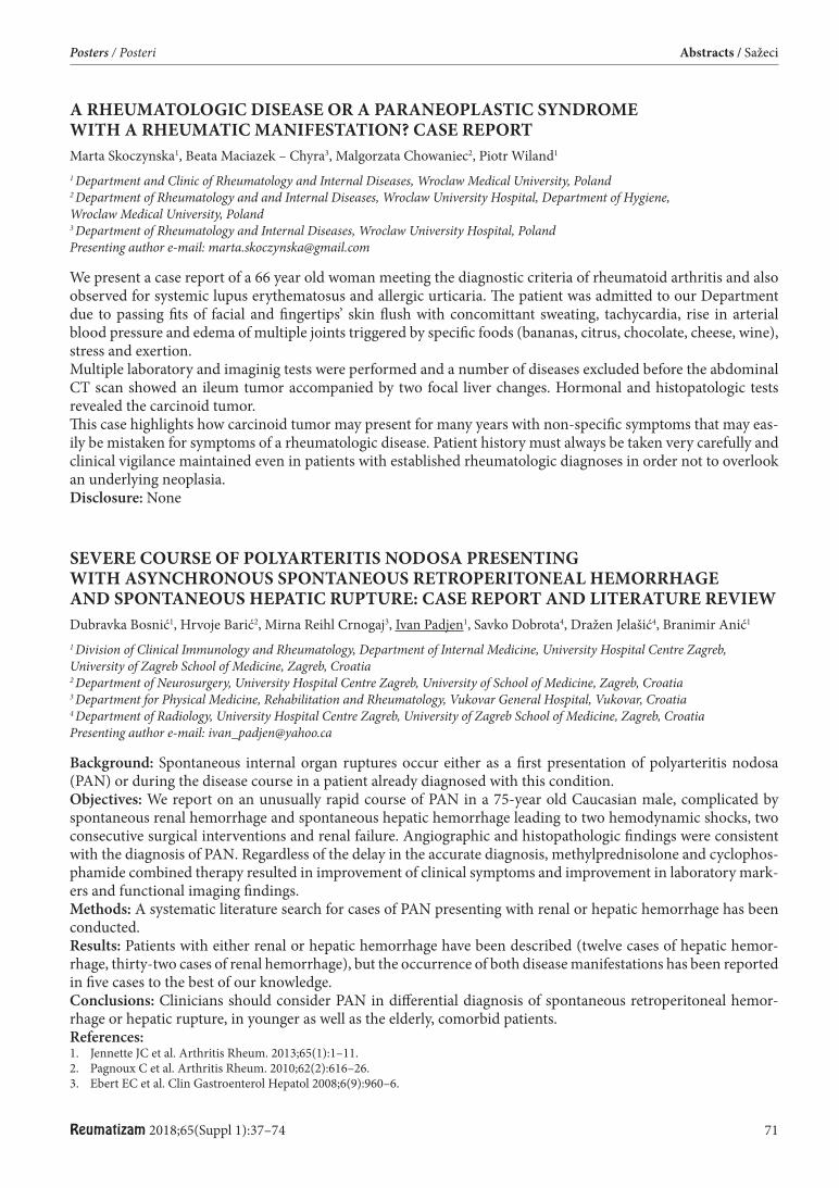
Reumatizam 2018;65(Suppl 1):37–74 71
Posters / Posteri Abstracts / Sažeci
A RHEUMATOLOGIC DISEASE OR A PARANEOPLASTIC SYNDROME WITH A RHEUMATIC MANIFESTATION? CASE REPORT Marta Skoczynska1, Beata Maciazek – Chyra3, Malgorzata Chowaniec2, Piotr Wiland1
1 Department and Clinic of Rheumatology and Internal Diseases, Wroclaw Medical University, Poland 2 Department of Rheumatology and and Internal Diseases, Wroclaw University Hospital, Department of Hygiene, Wroclaw Medical University, Poland 3 Department of Rheumatology and Internal Diseases, Wroclaw University Hospital, Poland Presenting author e-mail: [email protected]
We present a case report of a 66 year old woman meeting the diagnostic criteria of rheumatoid arthritis and also observed for systemic lupus erythematosus and allergic urticaria. Th e patient was admitted to our Department due to passing fi ts of facial and fi ngertips’ skin fl ush with concomittant sweating, tachycardia, rise in arterial blood pressure and edema of multiple joints triggered by specifi c foods (bananas, citrus, chocolate, cheese, wine), stress and exertion.Multiple laboratory and imaginig tests were performed and a number of diseases excluded before the abdominal CT scan showed an ileum tumor accompanied by two focal liver changes. Hormonal and histopatologic tests revealed the carcinoid tumor.Th is case highlights how carcinoid tumor may present for many years with non-specifi c symptoms that may eas-ily be mistaken for symptoms of a rheumatologic disease. Patient history must always be taken very carefully and clinical vigilance maintained even in patients with established rheumatologic diagnoses in order not to overlook an underlying neoplasia.Disclosure: None
SEVERE COURSE OF POLYARTERITIS NODOSA PRESENTING WITH ASYNCHRONOUS SPONTANEOUS RETROPERITONEAL HEMORRHAGE AND SPONTANEOUS HEPATIC RUPTURE: CASE REPORT AND LITERATURE REVIEW Dubravka Bosnić1, Hrvoje Barić2, Mirna Reihl Crnogaj3, Ivan Padjen1, Savko Dobrota4, Dražen Jelašić4, Branimir Anić1
1 Division of Clinical Immunology and Rheumatology, Department of Internal Medicine, University Hospital Centre Zagreb, University of Zagreb School of Medicine, Zagreb, Croatia 2 Department of Neurosurgery, University Hospital Centre Zagreb, University of School of Medicine, Zagreb, Croatia 3 Department for Physical Medicine, Rehabilitation and Rheumatology, Vukovar General Hospital, Vukovar, Croatia 4 Department of Radiology, University Hospital Centre Zagreb, University of Zagreb School of Medicine, Zagreb, Croatia Presenting author e-mail: [email protected]
Background: Spontaneous internal organ ruptures occur either as a fi rst presentation of polyarteritis nodosa (PAN) or during the disease course in a patient already diagnosed with this condition.Objectives: We report on an unusually rapid course of PAN in a 75-year old Caucasian male, complicated by spontaneous renal hemorrhage and spontaneous hepatic hemorrhage leading to two hemodynamic shocks, two consecutive surgical interventions and renal failure. Angiographic and histopathologic fi ndings were consistent with the diagnosis of PAN. Regardless of the delay in the accurate diagnosis, methylprednisolone and cyclophos-phamide combined therapy resulted in improvement of clinical symptoms and improvement in laboratory mark-ers and functional imaging fi ndings.Methods: A systematic literature search for cases of PAN presenting with renal or hepatic hemorrhage has been conducted.Results: Patients with either renal or hepatic hemorrhage have been described (twelve cases of hepatic hemor-rhage, thirty-two cases of renal hemorrhage), but the occurrence of both disease manifestations has been reported in fi ve cases to the best of our knowledge.Conclusions: Clinicians should consider PAN in diff erential diagnosis of spontaneous retroperitoneal hemor-rhage or hepatic rupture, in younger as well as the elderly, comorbid patients.References:1. Jennette JC et al. Arthritis Rheum. 2013;65(1):1–11.2. Pagnoux C et al. Arthritis Rheum. 2010;62(2):616–26.3. Ebert EC et al. Clin Gastroenterol Hepatol 2008;6(9):960–6.
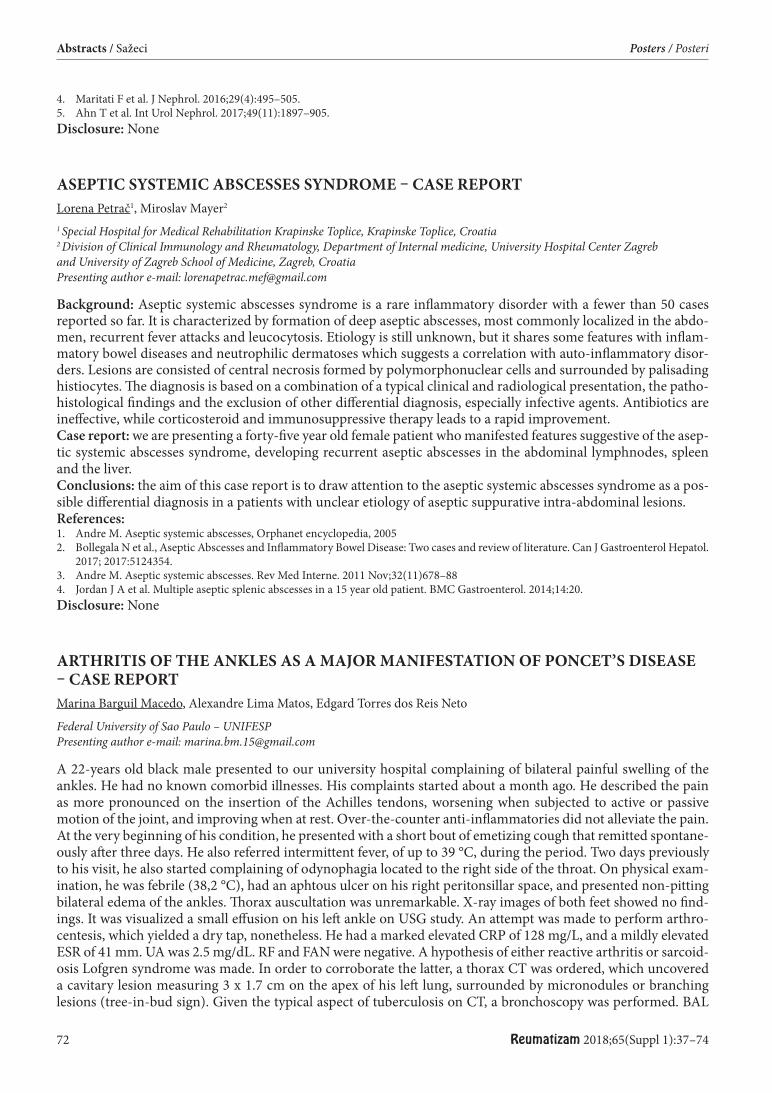
72 Reumatizam 2018;65(Suppl 1):37–74
Abstracts / Sažeci Posters / Posteri
4. Maritati F et al. J Nephrol. 2016;29(4):495–505.5. Ahn T et al. Int Urol Nephrol. 2017;49(11):1897–905.Disclosure: None
ASEPTIC SYSTEMIC ABSCESSES SYNDROME – CASE REPORT Lorena Petrač1, Miroslav Mayer2
1 Special Hospital for Medical Rehabilitation Krapinske Toplice, Krapinske Toplice, Croatia 2 Division of Clinical Immunology and Rheumatology, Department of Internal medicine, University Hospital Center Zagreb and University of Zagreb School of Medicine, Zagreb, Croatia Presenting author e-mail: [email protected]
Background: Aseptic systemic abscesses syndrome is a rare infl ammatory disorder with a fewer than 50 cases reported so far. It is characterized by formation of deep aseptic abscesses, most commonly localized in the abdo-men, recurrent fever attacks and leucocytosis. Etiology is still unknown, but it shares some features with infl am-matory bowel diseases and neutrophilic dermatoses which suggests a correlation with auto-infl ammatory disor-ders. Lesions are consisted of central necrosis formed by polymorphonuclear cells and surrounded by palisading histiocytes. Th e diagnosis is based on a combination of a typical clinical and radiological presentation, the patho-histological fi ndings and the exclusion of other diff erential diagnosis, especially infective agents. Antibiotics are ineff ective, while corticosteroid and immunosuppressive therapy leads to a rapid improvement.Case report: we are presenting a forty-fi ve year old female patient who manifested features suggestive of the asep-tic systemic abscesses syndrome, developing recurrent aseptic abscesses in the abdominal lymphnodes, spleen and the liver.Conclusions: the aim of this case report is to draw attention to the aseptic systemic abscesses syndrome as a pos-sible diff erential diagnosis in a patients with unclear etiology of aseptic suppurative intra-abdominal lesions.References:1. Andre M. Aseptic systemic abscesses, Orphanet encyclopedia, 20052. Bollegala N et al., Aseptic Abscesses and Infl ammatory Bowel Disease: Two cases and review of literature. Can J Gastroenterol Hepatol.
2017; 2017:5124354.3. Andre M. Aseptic systemic abscesses. Rev Med Interne. 2011 Nov;32(11)678–884. Jordan J A et al. Multiple aseptic splenic abscesses in a 15 year old patient. BMC Gastroenterol. 2014;14:20.Disclosure: None
ARTHRITIS OF THE ANKLES AS A MAJOR MANIFESTATION OF PONCET’S DISEASE – CASE REPORT Marina Barguil Macedo, Alexandre Lima Matos, Edgard Torres dos Reis Neto
Federal University of Sao Paulo – UNIFESP Presenting author e-mail: [email protected]
A 22-years old black male presented to our university hospital complaining of bilateral painful swelling of the ankles. He had no known comorbid illnesses. His complaints started about a month ago. He described the pain as more pronounced on the insertion of the Achilles tendons, worsening when subjected to active or passive motion of the joint, and improving when at rest. Over-the-counter anti-infl ammatories did not alleviate the pain. At the very beginning of his condition, he presented with a short bout of emetizing cough that remitted spontane-ously aft er three days. He also referred intermittent fever, of up to 39 °C, during the period. Two days previously to his visit, he also started complaining of odynophagia located to the right side of the throat. On physical exam-ination, he was febrile (38,2 °C), had an aphtous ulcer on his right peritonsillar space, and presented non-pitting bilateral edema of the ankles. Th orax auscultation was unremarkable. X-ray images of both feet showed no fi nd-ings. It was visualized a small eff usion on his left ankle on USG study. An attempt was made to perform arthro-centesis, which yielded a dry tap, nonetheless. He had a marked elevated CRP of 128 mg/L, and a mildly elevated ESR of 41 mm. UA was 2.5 mg/dL. RF and FAN were negative. A hypothesis of either reactive arthritis or sarcoid-osis Lofgren syndrome was made. In order to corroborate the latter, a thorax CT was ordered, which uncovered a cavitary lesion measuring 3 x 1.7 cm on the apex of his left lung, surrounded by micronodules or branching lesions (tree-in-bud sign). Given the typical aspect of tuberculosis on CT, a bronchoscopy was performed. BAL
Reumatizam 2018;65(Suppl 1):37–74 73
Posters / Posteri Abstracts / Sažeci
revealed a richly cellular fl uid, with predominance of lymphocytes (30%) and macrophages (58%). AFB testing resulted negative, but both RMT and culture for BK were positive. Th e patient was initiated on RIPE treatment, and improved of symptoms aft er 15 days. Th e typical presentation of bilateral ankle swelling and resolution of symptoms aft er RIPE treatment allowed the fi nal diagnosis of Poncet’s disease.References:1. Rueda, J.C., Crepy, MF. & Mantilla, R.D. Clinical features of Poncet’s disease. From the description of 198 cases found in the literature.
Clin Rheumatol (2013) 32: 929. doi:10.1007/s10067-013-2270-yDisclosure: None
GENERALIZED TELANGIECTASIA MISINTERPRETED AS VASCULITIS – CASE REPORT Zeljka Kardum1, Marija Sola2, Jasminka Milas Ahic1, Marta Biljan3, Ana Kovac1, Ana Marija Lukinac1, Ivana Kovacevic1, Kristina Stranski Kovacevic1, Visnja Prus1
1 Department of Rheumatology, Clinical Immunology and Allergology UHC Osijek, Croatia 2 Department of Dermatology UHC Osijek, Croatia 3 Department of Pulmonology UHC Osijek, Croatia Presenting author e-mail: [email protected]
Paraneoplastic syndromes (PNS) are defi ned as tumor-associ-ated symptoms and signs that are a result of tumor secretion of functional peptides, hormones, and cytokines or immune cross-reactivity between tumor and normal host tissues. Autoimmu-nity can be one of the presentations of PNS. Gastrointestinal stromal tumor (GIST) is rarely described as a cause of PNS. We present 81 – year old female patient with bizarre, generalized telangiectasia on her trunk and legs with subcutaneous indu-rated plaques of variable size (Figure) and positive immunolog-ical lab results (anti SSA, SSB, anti-centromeres, and anti-his-tones). Th e diagnosis of vasculitis and unknown connective tis-sue disease (UCTD) was made, although patient did not have other symptoms that would accompany collagenosis. Skin biopsy revealed isolated telangiectasia with no signs of infl am-mation and necrosis that would support the diagnosis of vascu-litis. Due to abrupt clinical start and the age of the patient para-neoplastic syndrome was suspected. Unfortunately, due to poor health and other comorbidities, the patient died. Th e autopsy revealed GIST in her intestines, as a cause of PNS.References:
Figure 1. In (A), the patient’s swollen ankles at presentation. In (B), typical lesion seen on chest CT.
A B
Figure 1. Clinical presentation of the patient.
74 Reumatizam 2018;65(Suppl 1):37–74
Abstracts / Sažeci Posters / Posteri
1. Chiu, M. L. S., and M. B. Y. Tang, “Generalized Granuloma Annulare Associated with Gastrointestinal Stromal Tumour: Case Report and Review of Clinical Features and Management,” Clinical and Experimental DermatologyVol. 33, No. 4, 2008, pp. 469–471.
2. Ozguroglu, E., N. Buyulbabani, M. Ozguroglu, and C. Baykal, “Generalized Telangiectasia as the Major Manifestation of Angiotropic (Intravascular) Lymphoma,” British Journal of DermatologyVol. 137, No. 3, 1997, pp. 422–425.
3. Tsikrikas, Spyridon, Spilios Manolakopoulos, Melanie Deutsch, Georgios Alexakis, Konstantinos Sialevris, Dimitrios Giannopoulos, Dimitrios Vassilopoulos, and Athanasios J. Archimandritis, “Unusual Combination of Paraneoplastic Manifestations in a Patient with Metastatic Gastrointestinal Stromal Tumor (GIST),” Scandinavian Journal of GastroenterologyVol. 43, No. 8, 2008, pp. 1012–1015.
4. Pelosof, Lorraine C., and David E. Gerber, “Paraneoplastic Syndromes: An Approach to Diagnosis and Treatment,” Mayo Clinic pro-ceedings vol. 85, No. 9, 2010, pp. 838–854.
5. András, C., Csiki, Z., Ponyi, A. et al. Rheumatol Int (2006) 26: 376. Disclosure: None
EDUCATIONAL EXPECTATIONS OF THE POLISH PATIENTS WITH RHEUMATIC DISORDERS TREATED WITH BIOPHARMACEUTICALS Anna Kotulska-Kucharz1, Eugene J. Kucharz2, Piotr Wiland2, Marzena Olesińska2, Anna Felis-Giemza2, Magdalena Kopeć-Mędrek2, Aleksandra Zoń-Giebel2, Wojciech Romanowski2, Lucyna Szymczak-Bartz2, Małgorzata Tłustochowicz2, Jolanta Lewandowicz2, Joanna Kowalska-Majka2, Jolanta Bucka2, Maria Majdan2, Zofi a Kiełbik2, Mariusz Korkosz2, Aneta Bielińska2, Piotr Leszczyński2, Katarzyna Pawlak-Buś2, Mariusz J. Puszczewicz2, Dominik Majewski2, Katarzyna Smolik2, Teresa Migas-Kukla2, Małgorzata Sochocka-Bykowska2, Maria Szarecka2, Bernadeta Luberda2, Małgorzata Falenta-Hitnarowicz2, Jadwiga Świkszcz-Gniadek2, Wanda Lepiarz-Rusek2, Grzegorz Rozwadowski2, Barbara Chara2, Jerzy Zajdel2, Zbigniew Zdrojewski2, Maria Maciejowska-Roge2, Irena Rosmus-Kuczia2
1 Department of Internal Medicine, Rheumatology and Clinical Immunology, Medical University of Silesia, Katowice, Poland and Research Group of the Polish Society for Rheumatology 2 Research Group of the Polish Society for Rheumatology Presenting author e-mail: [email protected]
Background: Biopharmaceuticals are still considered by patients as a new and partially enigmatic tool for man-agement of rheumatic diseases.Objectives: Th e study was designed to evaluate educational expectations and sources of knowledge in patients with rheumatic diseases treated with biologics.Methods: Anonymous questionnaires were distributed in 23 Polish rheumatological centers involved in the treat-ment. Responses were received from 606 patients with rheumatoid arthritis, 427 with ankylosing spondylitis, 117 psoriatic arthritis, and 62 adult patients with juvenile idiopathic arthritis (in whom administration of the drugs had been introduced before they were 18-year-old).Results: Almost all the patients had learnt for the fi rst time on biologics from their rheumatologist (93%). Few only patients had got such data from internet or from other patients. Likewise, most of the patients got majority of educational data on treatment with biologics from rheumatologist who was supervising the therapy (82%). Remaining sources included internet (8%) and other patients (5%). Relative low number of patients was educated by nurses (2%). Most of the patients (87%) were looking for more details on biological treatment. Th e patients with rheumatic disease lasting less than 10 yrs. were more interested in the management than those suff ering longer. Most of the patients (94%) considered their rheumatologist as the main person responsible for their edu-cation on biologics. Th ere was no diff erence between patients with various rheumatic diseases as well as no dif-ference was found between female and male patients. Biological treatment attracted more interest in younger than older patients.Conclusions: Education is still a challenge in patients receiving biopharmaceuticals. Most of the patients repre-sented traditional attitude to health education, expecting almost all educational data to be provided by their physician. We were surprised that role of the nurses was found to be rather low. An increase in role of nurses seems to be the future aim of the educational eff orts in Polish rheumatology. References:1. Kotulska A, Kucharz Ej, Wiland P et al.: Satisfaction and discontent of Polish patients with biological therapy of rheumatic diseases:
results of the multi-center questionnaire study. Reumatologia 2018.Disclosure: None
Reumatizam 2018;65(Suppl 1) 75
AUTHOR’S INDEX / KAZALO AUTORA
AAlexeevna E. 69Alunno A. 33Aljinović J. 25Ambrozaitis A. 67Andrés Cerezo L. 63Anić B. 3, 16, 18, 32, 38, 43, 71Anić F. 3, 64Artuković M. 24
BBakula M. 32Balajková V. 16Bálint Z. 35Balog A. 8Barešić M. 3, 26, 43Barguil Macedo M. 72Barić H. 71Batinić D. 16Batnozic Varga M. 16, 26, 50Bečvář R. 22, 51, 53, 54Berki T. 8Bessette L. 47Bielińska A. 74Biljan M. 73Birkić D. 66Blanco R. 45Bohatá J. 64Bolić L. 27Borić K. 17, 40, 52Bosnić D. 3, 49, 71Božić I. 52Brzosko M. 9, 10, 19Bubova K. 12, 25, 30Bucka J. 74
CCamp H. 44Cekada N. 16, 26, 50Cerovec M. 3Cesak M. 54Cesak P. 53, 54, 56, 57Chara B. 74Chen K. 44Chen S. 45Chowaniec M. 37, 71Chrzanowska M. 21
Cutolo M. 1Czirják L. 8, 35
ČČaić M. 62Čaplinskas S. 67Čikeš N. 32Čubelić D. 4
DDallos T. 11Damjanov N. 42, 44Denisovna R. 69Distler J. H. W. 22Dobrota S. 71Doko I. 25, 27, 41, 65Drahosova M. 39Durez P. 47Dvoryakovskaya T. 69Dyšková T. 51
EErceg M. 3
FFalenta-Hitnarowicz M. 74Farkas N. 35Fedoseenko M. 69Felis-Giemza A. 74Filková M. 13, 29, 33Fillerová R. 51Flegar D. 24Fleischmann R. 47Forejtova S. 25Friedman A. 45Frkovic M. 16, 26, 50
GGabčová G. 51Gagro A. 50Gatterova J. 30Genovese M. C. 47Glasnović M. 65Golubović S. 70Grazio S. 17, 25, 27, 40, 41,
64, 65Grčević D. 24Gregová M. 25, 29, 33
Griger Z. 8Grk M. 42Grubišić F. 17, 25, 27, 40, 41,
64, 65Gudelj Gračanin A. 59, 65Gulyás K. 8
HHadžisejdić I. 67Hánová P. 29, 33Herceg D. 43Heřmánková B. 22, 23, 25, 30, 53,
54, 56, 57Hočevar A. 5, 7, 31Holc I. 6Horák P. 51, 62Horinkova J. 25Horváthová V. 64Hosticka E. 16Hrncir Z. 39Hulejová H. 29, 33, 63Husakova M. 25
IIkić Matijašević M. 24Isaeva K. 69
JJajić Z. 24Jekić B. 42Jelašić D. 71Jelušić M. 16, 26, 50Ješe R. 7, 31Ježić I. 43Jordhani M. 59
KKardum Ž. 17, 20, 40, 73Kehler T. 17, 40, 64Kelava T. 24Kiełbik Z. 74Kifer D. 16, 26, 50Killinger Z. 11Klein M. 56, 57Knežević Štromar I. 62Kolak Ž. 65Komjáti D. 35Kopeć-Mędrek M. 74
76 Reumatizam 2018;65(Suppl 1)
Author’s Index / Kazalo autora
Koren Krajnc M. 6Korkosz M. 9, 74Kotulska-Kucharz A. 74Kotyla P. 21Kovačević I. 73Kovač Durmiš K. 66Kovačević I. 20, 73Kovačević P. 25Kowalska-Majka J. 74Kriegová E. 51Krstulović Marasović D. 40Kryštůfk ová O. 29, 33Kubínová K. 7, 56, 57Kucharz E. J. 20, 21, 74Kumánovics G. 35
LLaktašić Žerjavić N. 65, 66Lepiarz-Rusek W. 74Leszczyński P. 74Lewandowicz J. 74Lima Matos A. 72Li Y. 47Lóránd V. 35Luberda B. 74Lukáč J. 37, 68Lukač N. 24Lukáčová O. 37, 68Lukinac A. M. 20, 73Lysý J. 11
LjLjubičić Marković N. 17, 40
MMaciazek – Chyra B. 71Maciejowska-Roge M. 74Majdan M. 74Majewski D. 74Maksimović I. 42Maksimović N. 42Malčić I. 16Mamutova A. 69Mann H. 54, 56, 57Marasović Krstulović D.
15, 17, 52Marecek O. 56Markez S. 31Markotić A. 24Marković I. 38
Martinović Kaliterna D. 52Maštrović-Radončić K. 17, 40Mayansky N. 69Mayer M. 3, 4, 29, 60, 62, 72Migas-Kukla T. 74Mijačika L. 27Milanović S. 17, 40Milas-Ahić J. 20, 73Milchert M. 19Milić V. 42Miloš M. 29Minier T. 8, 35Misterska – Skora M. 37Mitrović H. 65Mitrović J. 17, 40Mohamed M.-E. F. 45Moravcová R. 16Morović-Vergles J. 17, 38, 40,
59, 65Mrázek F. 51Mysler E. 47
NNagy G. 35Nagy O. 44, 45, 47Németh B. 35Nikiphorou E. 33Novak S. 3, 64, 67Novoselec M. 16
OOlejárová M. 16, 61Olesińska M. 74Oreska S. 22, 25, 30, 53, 54,
56, 57Ostor A. 47Othman A. A. 47Ovuka A. 50Ozdanovac I. 50
PPadjen I. 3, 16, 38, 71Pahor A. 6Pál E. 8Pangan A. L. 45, 47Pap M. 66Pasarić A. 38Paulauskas A. 67Pavan J. 59Pavelcová K. 64Pavelka K. 22, 25, 28, 29, 30, 33,
53, 54, 56, 57, 70
Pavić-Nikolić M. 31Pawlak-Buś K. 74Pecnjak M. 50Perdan-Pirkmajer K. 31Perić P. 65, 66Perković D. 15, 52, 65Peterfy C. 47Petráčkova A. 51Petrač L. 60, 72Petrić M. 52Petrulionienė A. 67Potočki K. 16Prajzlerová K. 29, 33Prus V. 17, 20, 40, 73Przepiera-Bedzak H.
9, 10Puchner R. 5Punda M. 27Puszczewicz M. J. 74
RRadner H. 5Radzišauskienė D. 67Rathouska A. 53, 57Reihl Crnogaj M. 71Rischmueller M. 45Ritschl V. 33Rogoznica M. 64Romanowski W. 74Rosmus-Kuczia I. 74Rotar Ž. 7, 31Rozwadowski G. 74Ruci D. 59Rukavina K. 18Ruzickova O. 28, 53, 54,
56, 57Ruzman L. 50
SSautner J. 5Sčepović Ljucović M. 49Schubertová M. 51Scoville C. 44Sebastian A. 37Sekelj S. 29Sentić M. 3Sestan M. 16, 26, 50Šimetić L. 43Skácelova M. 51Skala Kavanagh H. 25, 27, 41Skoczynska M. 37, 71Sleglova O. 28Smiljanić Tomičević Lj. 4
Reumatizam 2018;65(Suppl 1) 77
Author’s Index / Kazalo autora
Smolik K. 74Smržová A. 51Smucrova H. 53, 54, 56Sochocka-Bykowska M.
74Šola M. 73Soloshenko M. 69Song I.-H. 47Soukup T. 39Spiritovic M. 22, 25, 30, 53, 54,
56, 57Sršen S. 50Stevanović R. 3Stibůrková B. 64Stipić J. 26Stolfa J. 25Stones S. R. 33Storkanova H. 22, 25, 30, 53, 54,
56, 57Strand V. 44, 45Stranski Kovacevic K. 73Studenic P. 33Suboticki J. L. 44Sumova B. 54Świkszcz-Gniadek J. 74Szabo M. 9, 60Szamosi S. 9Szarecka M. 74Szymczak-Bartz L. 74
ŠŠenolt L. 12, 22, 25, 28, 29, 30,
33, 53, 54, 56, 57Šimac P. 65Šućur A. 24Šukara G. 18
TTakeuchi T. 45Tkachenko N. 69Tłustochowicz M. 74Tomcik M. 22, 25, 30, 53, 54,
56, 57Tomljanović Rudar I. 17, 40Tomšič M. 7, 31Torres dos Reis Neto E. 72Tundia N. 44Turudić D. 26
VValetić A. M. 59van Vollenhoven R. F. 44, 45Varjú C. 8, 35Venalis A. 67Vencovský J. 7, 22, 25, 29, 30, 33,
53, 54, 56, 57Vernerova L. 56, 57
Vlak T. 25Vokurkova D. 39Vukojević R. 27
WWiland P. 37, 71, 74Włoch-Targońska M. 21, 34
XXavier R. M. 45
ZZadro R. 29Zajdel J. 74Zaninovic M. 50Zdrojewski Z. 74Zegzulkova K. 25Zekić T. 67Zhou Y. 47Zlnay M. 10Zoń-Giebel A. 74Zubkova I. 69
ŽŽagar I. 65, 66Žarković B. 29Žuvan L. 15Žuvić M. 64
78 Reumatizam 2018;65(Suppl 1)
Instructions for authors / Upute autorima
INSTRUCTIONS FOR AUTHORSABOUT THE JOURNAL
Reumatizam (Rheumatism) is the offi cial peer-reviewed journal of the Croatian Medical Association’s Society for Rheumatology. It appears twice a year and publishes editori-als, scientifi c and professional papers, short communications, review papers, preliminary reports, and case reports. It in-forms professionals in the fi eld of rheumatology on develop-ments in clinical and non-clinical aspects of their work. Ad-ditionally, supplements with abstracts or full texts presented at congresses or symposia are periodically published. Th e journal presents relevant information on diagnostic and ther-apeutic procedures, as well as on providing comprehensive care for individuals aff ected by rheumatic diseases and condi-tions. Th e papers are written in English or Croatian, and are published under the condition that they were not previously published in the same form. Reumatizam is indexed by MED-LINE/PubMed (Index Medicus) and Scopus.
Th e content of the journal Reumatizam may be used free of charge for educational and research purposes, with full reference to the source. Any other use is prohibited, except with explicit prior permission from the publisher.
PAPER SUBMISSION / MANUSCRIPT PUBLICATION
Articles are published in the Croatian (with title, abstract, keywords, table and fi gure titles and legends in English) or English languages (with title, abstract, keywords, table and fi gure titles and legends in Croatian). Instructions to Authors are in accordance with the instructions in the article: Interna-tional Committee of Medical Journal Editors (ICMJE) – Recom-mendations for the Conduct, Reporting, Editing, and Publica-tion of Scholarly Work in Medical Journals, available at: http://www.icmje.org/index.html. For texts in English, authors who are not native speakers are advised to seek professional assis-tance to ensure the accuracy and quality of the translation. Th e publisher can provide such service upon payment.
Manuscripts are submitted on paper (three identical com-puter printouts) accompanied by an electronic version writ-ten in Microsoft Word format on a CD, DVD, or USB stick, or by e-mail (upon previous agreement with the Editor-in-Chief) to: Reumatizam, Editorial Board, Klinika za reumatolo-giju, fi zikalnu medicinu i rehabilitaciju, Klinički bolnički cen-tar Sestre milosrdnice, Vinogradska 29, 10000 Zagreb, Croatia (e-mail: [email protected]).
Th e order in which papers are published does not corre-spond to the order by which manuscripts have reached the editorial board. Manuscripts and other submitted materials will not be returned.
AUTHORSHIPPersons designated as authors must qualify for author-
ship. Each author should have suffi ciently participated in creating the paper in order to be able to take public respon-sibility for the appropriate portion of its content, and all au-thors should take responsibility for the paper as a whole, from its inception to the published form. All others who have participated in the work but are not authors should be mentioned in the acknowledgments.
Manuscripts should be accompanied by a written declara-tion that the paper has not been previously published or sub-
mitted/accepted for publication elsewhere, and that it has been read and approved by all the authors, as well as by a declaration on absence of any fi nancial or other confl ict of interest. Additionally, the manuscript should be accompa-nied by a declaration of copyright transfer to the journal.
PREPARATION OF PAPER / MANUSCRIPTTh e text should be printed in 12-point sized letters on
white bond ISO A4 paper (210 × 297 mm), double spaced on one side only, including the title page, abstract, text, ac-knowledgments, declaration on confl ict of interest, refer-ences, tables, and legends. Th e left margin should be 35 mm wide, while the right, top, and bottom margins should all be 25 mm. All pages, including the title page, should be con-secutively numbered in the lower right-hand corner.
Th e text of a scientifi c or professional paper should con-tain: title page, abstract and keywords, introduction, ma-terials and methods, results, discussion, conclusions, ac-knowledgments (optional), declaration on confl ict of inter-est, references, tables, legends, and fi gures.
Review papers should not exceed 15 pages (including tables and fi gures), scientifi c and professional papers should not ex-ceed 12 pages (including tables and fi gures), and case reports should not exceed 8 pages (including tables and fi gures). Short communications and preliminary reports should not exceed 4 pages (including tables and fi gures) and 15 references.
TITLE PAGETh e title page should contain the title of the paper (which
must be concise, clear, and informative) in the Croatian and English languages, and the full name of each author. In the next line the institutional affi liation of the author(s) should be listed, with the full name of the institution, street, house number, city, and country. If the authors of the paper have diff erent institutional affi liations, aft er every name and sur-name, as well as before each affi liation, a corresponding number should be written in superscript.
Th is should be followed by the name, surname, and full address of the author responsible for correspondence, along with his/her phone number, fax number, and e-mail address.
ABSTRACT AND KEYWORDSTh e second page should contain the abstract in the Croa-
tian and English languages (up to 300 words), stating the purpose of the study or investigation, the basic procedures, main fi ndings, and principal conclusions.
Th e abstract should emphasize new and important as-pects of the study or observation. Below the abstract authors should list four to ten keywords or short phrases in Croatian and English, which will help indexers to cross-index the ar-ticle and may be published with the abstract. Terms from the Index Medicus Medical Subject Headings (MeSH) list should be used. General and plural terms, and multiple concepts (for example using “and”, “or”) should be avoided. Th e ab-stract must not contain references.
INTRODUCTIONTh e introduction section should state the purpose of the
paper and the aim of the study or observation. It is recom-mended to include relevant references only, without the data or conclusions from the paper.
Reumatizam 2018;65(Suppl 1) 79
Instructions for authors / Upute autorima
MATERIAL AND METHODSTh e selection criteria and all important characteristics of
the studied or observed human subjects or laboratory ani-mals should be stated in this section. Th e author(s) should specify the meaning of the descriptors in detail, explain how the data was collected, and identify methods, devices (with the manufacturer’s name in parentheses), and procedures in suffi cient detail to allow others to reproduce the results. For established methods references should be provided, while new or substantially modifi ed methods should be described in detail, alongside with the reasons for their use and esti-mates of their limitations.
For drugs and chemicals generic names must be used. All measurements should be expressed in SI units. In texts in Croatian decimal commas are to be used, and in texts in English decimal points.
ETHICS / ETHICAL STANDARDSIn papers dealing with experiments on human subjects it
should be clearly stated that all procedures were performed in accordance with the ethical standards of an institutional or regional committee responsible for human experimenta-tion, as well as the Helsinki Declaration of 1975, as revised in 1983. Th e subjects’ names and/or surnames must not be mentioned, especially in illustrative materials. Papers deal-ing with experiments on animals should state that institu-tional or national regulations for the care and use of labora-tory animals were complied with.
STATISTICSStatistical methods should be described with enough de-
tail to enable a knowledgeable reader with access to the orig-inal data to verify the reported results. Whenever possible, the fi ndings should be quantifi ed and presented with ap-propriate measurement error or uncertainty indicators. Th e computer program that was used should be specifi ed.
RESULTSTh e results are to be presented in logical sequence in the
text, tables, and illustrations. Not all the data from the tables or illustrations is to be reiterated in the text, only the impor-tant observations should be emphasized or summarized.
DISCUSSIONNew and important aspects of the study and the conclu-
sions that follow should be emphasized. Reiterating data or other material which was presented in the Introduction or the Results sections is discouraged. Th e discussion should elaborate the signifi cance of the fi ndings and their limita-tions, including the implications on future research, while avoiding statements and conclusions that are not completely supported by the data. Observations from the study should be compared with other relevant studies. When necessary, new hypotheses may be stated, clearly labelled as such.
CONCLUSIONSTh e conclusions are to be derived from the authors’ own
results, separately from the discussion.
ABBREVIATIONSOnly standard abbreviations should be used. Th e full term
for which an abbreviation stands must precede its fi rst use in the text, unless it is a standard abbreviation for a unit of measurement. Abbreviations in the title of the paper should be avoided.
SYMBOLSSymbols must be explained in the text. An extensive list of
symbols may be provided in the appendix.
TABLESEach table is to be double spaced and printed on a sepa-
rate page. Tables must not be submitted as photographs. Each table must have a title and be consecutively numbered in order of appearance in the text. Tables must be clear and simple. Any remarks should be written below the table, and referred to in the table by superscript lowercase letters. Ta-bles should not reiterate results presented elsewhere in the paper (e.g., in a diagram).
FIGURES/ILLUSTRATIONSAll illustrations should be professionally drawn or photo-
graphed. Letters, numbers, and symbols must be legible even when reduced in size for publication. Each photograph must be consecutively numbered in order of appearance in the text, list the author’s name, and have its top side marked. Each drawing must be consecutively numbered in order of appearance in the text, and have its top side marked. Draw-ings should be produced or printed in black ink on white bond paper. Color printouts or photocopies are not suitable for reproduction. Photocopies of photographs are not ac-ceptable. Photographs depicting people may be published only when accompanied by a written permission of the per-son in the photograph, or if the person is unrecognizable. Pictures and tables taken from other sources should be ac-companied by their publisher’s and author’s permission. When submitted in electronic form, fi gures/illustrations must be in TIFF or high quality JPEG format, with a mini-mum width of 1500 pixels. Illustrations in other formats might be accepted only with prior consent of the editorial board. Th e editorial board reserves the right not to publish illustrations that fail to meet these requirements.
ACKNOWLEDGMENTSIn the acknowledgments one should mention all associ-
ates who did not meet the criteria for authorship, such as individuals who provided technical writing assistance, or a department chair who provided general support. Financial and material support should also be mentioned.
DECLARATION ON CONFLICT OF INTERESTAuthors must declare whether or not there is a fi nancial
relationship between them and the organization/company that sponsored the research. Th is note must be added in a separate section preceding the references. When no confl ict of interest exists, authors should write: “Th e authors declare that there is no confl ict of interest.”
REFERENCESReferences are to be listed using the Vancouver reference
style which specifi es the numerical referencing system, ac-cording to the recommendations of the American National Library of Medicine. Th e most frequently used examples can be found in the article ICMJE Recommendations for the Con-duct, Reporting, Editing, and Publication of Scholarly Work in Medical Journals: Sample References (http://www.nlm.nih.gov/bsd/uniform_requirements.html). Detailed instructions can be found in the book Citing Medicine (http://www.ncbi.nlm.nih.gov/books/NBK7256).
References in the text, tables, and legends should be con-secutively numbered using Arabic numerals in parentheses,
80 Reumatizam 2018;65(Suppl 1)
Instructions for authors / Upute autorima
in order of appearance. If there is more than one number, they should be separated by commas.
In the list of references authors and/or editors are to be listed by their surname(s) and initials of their name(s). Aft er the initial(s) no period is added, except when the initial im-mediately precedes the title of the reference. Multiple au-thors/editors’ names are separated by commas. If there are more than six authors/editors, aft er the fi rst three names “et al.” should be written, and the others should be omitted. In the title capital letters are used only for the fi rst letter of the fi rst word in the title, and in the words that are commonly written with capital letters. When page numbers are includ-ed, identical initial digits should be omitted (e.g., 123-5 in-stead of 123-125). Each reference should end with a period, except when the reference ends with a URL.
In texts in the English language, when listing references published in other languages it is recommended to list the title in English (if it exists) or to translate it into English (in which case it should be put in square brackets), while at the end of the reference the original language is to be mentioned.
When listing papers which have been accepted but not yet published, “In press” should be added at the end. Authors should obtain written permission when citing such a paper, as well as confi rmation that the paper has been accepted for publication.
Journal articlesJournal titles should be listed by their usual abbreviations
(NLM Title Abbreviation), which can be found in the Na-tional Library of Medicine catalogue (http://www.ncbi.nlm.nih.gov/nlmcatalog/journals). Publishers of journals are not to be listed. It is obligatory to include the year of publication, volume, and page numbers. If the journal carries continuous pagination, the month and issue along with the parentheses may be omitted.
[Example] Article from a journal, more than six authors:1. Ćurković B, Babić-Naglić Đ, Morović-Vergles J, et al.
Proposal for biologic drugs therapy in rheumatoid ar-thritis. Reumatizam. 2010;57(1):29-35. Croatian.
[Example] Article from a journal, continuous pagination:2. Ritchlin CT. From skin to bone: translational perspec-
tives on psoriatic disease. J Rheumatol. 2008;35:1434-7.[Example] Article from a supplement:3. Gladman DD, Antoni C, Mease P, Clegg DO, Nash P.
Psoriatic arthritis: epidemiology, clinical features, course, and outcome. Ann Rheum Dis. 2005;64(Suppl 2):ii14–7.
BooksIt is obligatory to include the place of publication, pub-
lisher, and year of publication. Page numbers are to be in-cluded only when a part of the book is cited.
[Example] Book (authors):4. Walker JM, Helewa A. Physical rehabilitation in arthri-
tis. 2nd ed. St. Louis: Saunders; 2004.[Example] Book (editors):5. Isenberg DA, Maddison PJ, Woo P, Glass D, Breedveld
FC, editors. Oxford texbook of rheumatology. 3rd ed. New York: Oxford University Press; 2004.
[Example] Chapter in a book:6. Vasey FB, Espinoza LR. Psoriatic arthritis. In: Calin
A, editor. Spondyloarthropathies. Orlando: Grune and Stratton; 1984. p. 151–85.
Conference proceedingsIf the conference paper was published in a journal or a
supplement, the instructions for journals and supplements should be followed. If the conference paper was published in a book, aft er the book’s title the words “Proceedings of ” followed by the conference name, date(s), place, and country are to be added.
[Example] Conference paper, published in a supplement: 7. Matucci Cerinic M, Pignone A. Th e early diagnosis
of rheu matoid arthritis (RA). Reumatizam. 1997;44(Suppl):1.
[Example] Conference paper, published in a book: 8. Babić-Naglić Đ. Physical activity and exercises. In: Iva-
nišević G, editor. [Th alassotherapy, kinesitherapy and aromatherapy in Croatia]. Proceedings of the 14th Lo-šinj School of Natural Remedies; 2013 Sep 6-7; Veli Lošinj, Croatia. Zagreb: Hrvatski liječnički zbor; 2013. p. 49-55. Croatian.
[Example] Conference proceedings (book): 9. Gordon DA, editor. Immune reactions and experi-
mental models in rheumatic diseases. Proceedings of the Fourth Canadian Conference on Research in the Rheumatic Diseases; 1970 Oct 15-17; Toronto, Cana-da. Toronto: University of Toronto Press; 1972.
Web publicationsReferences of web publications should contain the date of
access and URL, except when the publication has a DOI.[Example] Article from a journal on the Internet:10. Mak A, Kow NY. Th e pathology of T cells in systemic
lupus erythematosus. J Immunol Res [Internet]. 2014 [cited 2014 May 25];2014:419029. Available from: http://www.ncbi.nlm.nih.gov/pmc/articles/PMC4017881
[Example] Article from a journal on the Internet, contains DOI:
11. Vivar N, Van Vollenhoven RF. Advances in the treat-ment of rheumatoid arthritis. F1000Prime Rep. 2014 May 6;6:31. doi: 10.12703/P6-31. PubMed PMID: 24860653; PubMed Central PMCID: PMC4017904.
[Example] Book/monograph on the Internet:12. Chen Q, editor. Osteoarthritis – diagnosis, treatment
and surgery [Internet]. Rijeka: InTech; 2012 [cited 2013 Oct 8]. Available from: http://www.intechopen.com/books/osteoarthritis-diagnosis-treatment-and-surgery
[Example] Web page:13. Croatian Society for Rheumatology [Internet]. Za-
greb: Croatian Society for Rheumatology of the CMA; c2014 [cited 2014 Apr 1]. Available from: http://www.reumatologija.org/engPocetna.aspx
REVIEW PROCESSTh e review process is conducted anonymously. Each paper
is reviewed by two reviewers, and a copy of their evaluation is sent anonymously to the author. When producing the fi nal version of the paper, the author should take into consideration the reviewers’ assessment or explain his/her standpoint based on fact.
Th e editorial board reserves the right to adapt the style of each paper to certain standards of uniformity.
Reumatizam 2018;65(Suppl 1) 81
Instructions for authors / Upute autorima
UPUTE AUTORIMAO ČASOPISU
„Reumatizam” je službeni recenzirani časopis Hrvatskoga reumatološkog društva Hrvatskoga liječničkog zbora. Izlazi dva puta na godinu. Objavljuje uvodnike, znanstvene radove, stručne radove, kratka priopćenja, pregledne radove, preli-minarna izvješća i prikaze bolesnika. Informira reumatologe o novostima u kliničkom i nekliničkom djelokrugu rada. Također, u časopisu se periodično kao suplement objavljuju sažetci ili cjeloviti radovi s kongresa i simpozija. Časopis čitatelju daje bitne informacije o dijagnostičkim i terapij-skim procedurama, odnosno pružanju sveobuhvatne skrbi osobama oboljelima od reumatskih bolesti i stanja. Radovi su napisani hrvatskim ili engleskim jezikom, a objavljeni su pod uvjetom da nisu prethodno publicirani u istom obliku. „Reumatizam” je indeksiran u bazama MEDLINE/PubMed (Index Medicus) i Scopusu.
Sadržaj iz časopisa „Reumatizam” smije se bez naknade rabiti u nastavne i istraživačke svrhe, uz potpuno navođenje izvora. Svaka druga uporaba zabranjena je bez izričitog dopuštenja izdavača.
PREDAJA RADA / OBJAVA RUKOPISAObjavljuju se članci na hrvatskom jeziku (s naslovom,
sažetkom, ključnim riječima te s naslovom i legendom tabli-ca i slika na engleskom jeziku) ili na engleskom jeziku (s na-slovom, sažetkom, ključnim riječima te s naslovom i legen-dom tablica i slika na hrvatskom jeziku). Upute autorima sukladne su uputama u članku: International Committee of Medical Journal Editors (ICMJE) – Recommendations for the Conduct, Reporting, Editing, and Publication of Scholarly work in Medical Journals (Preporuke za provođenje, prika-zivanje, uređivanje i objavljivanje radova u medicinskim časopisima) koje su dostupne na: http://www.icmje.org/index.html. Za tekstove na engleskom jeziku preporučuje se da autori koji nisu izvorni govornici engleskog jezika potra-že savjet stručnjaka radi točnog i kvalitetnog prijevoda. Iz-davač može osigurati takvu uslugu uz plaćanje.
Rukopisi se dostavljaju u papirnatom obliku (tri identič-na računalna ispisa), zajedno s elektroničkom verzijom napisanom u formatu Microsoft Word na CD-u, DVD-u, USB-sticku ili elektroničkom poštom (uz prethodni dogovor s glavnim urednikom) na adresu: „Reumatizam”, Ured-ništvo, Klinika za reumatologiju, fi zikalnu medicinu i reha-bilitaciju, Klinički bolnički centar Sestre milosrdnice, Vino-gradska 29, 10000 Zagreb, Hrvatska (e-adresa: [email protected]).
Radovi se ne objavljuju prema redoslijedu prispijeća ru-kopisa u uredništvo časopisa. Rukopisi i ostali dostavljeni materijali ne vraćaju se pošiljateljima.
AUTORSTVOOsobe određene kao autori moraju biti kvalifi cirane za au-
torstvo. Svaki autor treba dostatno sudjelovati u izradi rada kako bi preuzeo javnu odgovornost za odgovarajući dio sadr-žaja rada, a svi autori trebaju preuzeti odgovornost za cjeloku-pan rad od početka rada do njegove objave. Svi koji su sudje-lovali u radu, a nisu autori, trebaju biti spomenuti u zahvali.
Uz rukopis treba priložiti pisanu izjavu da rad prethodno nije bio objavljen ili ponuđen/prihvaćen za objavu u nekom drugom časopisu, da su ga pročitali i odobrili svi autori te izjavu da ne postoji fi nancijski ili bilo kakav drugi sukob in-teresa. Također, uz rukopis treba priložiti i izjavu o prijenosu autorskih prava na časopis.
PRIPREMA RADA / RUKOPISATekst treba biti otisnut slovima veličine 12 točaka na bije-
lom papiru formata A4 (210 × 297 mm) samo s jedne strane s dvostrukim proredom, uključujući i naslovnu stranicu, sažetak, tekst, zahvale, izjavu o sukobu interesa, referencije, tablice i legende. Lijeva margina treba biti široka 35 mm, a desna margina te gornji i donji rub 25 mm. Sve stranice, uključujući naslovnu, trebaju imati redni broj u donjem desnom kutu.
Tekst znanstvenog ili stručnog rada treba sadržavati: na-slovnu stranicu, sažetak i ključne riječi, uvod, materijal i me-tode, rezultate, raspravu, zaključke, zahvale (opcionalno), izjavu o sukobu interesa, referencije, tablice, legende i slike.
Pregledni radovi mogu biti opsega do 15 stranica (uklju-čujući tablice i slike), znanstveni i stručni radovi do 12 stra-nica (uključujući tablice i slike), prikazi bolesnika do 8 stra-nica (uključujući tablice i slike). Kratka priopćenja i preli-minarna izvješća opsega su do 4 stranice (uključujući tablice i slike) i do 15 referencija.
NASLOVNA STRANICANa naslovnoj stranici treba biti naslov rada (mora biti
sažet, jasan i informativan) na hrvatskom i engleskom jeziku te puno ime svakog od autora. U sljedećem retku treba nave-sti puni naziv ustanove, ulicu i broj, grad i državu autora. Ako su u izradi rada sudjelovali autori iz različitih ustanova, za svakog od njih poslije imena i prezimena te prije navoda ustanove treba napisati odgovarajući broj u superskriptu.
Slijedi ime i prezime te puna adresa autora za dopisivanje u vezi s radom, njegov/njezin telefonski broj, broj faksa i e-mail adresa.
SAŽETAK I KLJUČNE RIJEČIDruga stranica treba sadržavati sažetak na hrvatskom i
engleskom jeziku (do 300 riječi) u kojem su navedeni cilj studije ili istraživanja, osnovni postupci, najvažnija otkrića te osnovni zaključci.
U sažetku valja naglasiti nove i važne aspekte studije ili opservacije. Ispod sažetka autori trebaju navesti četiri do deset ključnih riječi ili kratkih pojmova na hrvatskom i engleskom jeziku koji će pomoći pri indeksiranju članka i mogu se objaviti uz sažetak. Za ključne riječi treba se kori-stiti pojmovima iz popisa Medical Subject Headings (MeSH) Index Medicusa. Općenite, pluralne i mnogostruke koncep-te (primjerice uz uporabu „i”, „ili”) treba izbjegavati. Sažetak ne smije sadržavati navode referencija.
UVODU uvodu se navode svrha rada i razlog provođenja studije
ili opservacije. Preporučuje se navesti samo relevantne refe-rencije, bez podataka ili zaključaka iz rada.
82 Reumatizam 2018;65(Suppl 1)
Instructions for authors / Upute autorima
MATERIJAL I METODENavode se odabir i sve važne karakteristike ispitanika ili
laboratorijskih životinja koje su studirane ili opservirane. Treba detaljno specifi cirati značenje deskriptora te objasniti kako su prikupljeni podatci, identifi cirati metode, aparate (s nazivom proizvođača u zagradi) te postupke s dovoljnim brojem detalja da bi se rezultati mogli reproducirati. Za me-tode treba navesti referencije ili detaljno opisati nove meto-de ili one metode koje su znatnije modifi cirane, navodeći razlog njihove primjene i procjene njihovih ograničenja.
Za lijekove i kemikalije moraju se rabiti generička imena. Sve veličine trebaju biti izražene u SI jedinicama. U tek-stovima na hrvatskom jeziku rabi se decimalni zarez, a u tekstovima na engleskom decimalna točka.
ETIKA / ETIČKI STANDARDIU radovima koji se bave eksperimentima na ljudima jasno
treba navesti da su postupci provedeni sukladno etičkim standardima institucijskog ili regionalnog odbora odgo-vornog za izvođenje eksperimenata na ljudima te u skladu s Helsinškom deklaracijom iz 1975. godine, revidiranom 1983. godine. Ne smije se navoditi ispitanikovo ime i/ili pre-zime, osobito u ilustrativnim materijalima. U radovima koji se bave eksperimentima na životinjama treba navesti da je poštovan institucionalni ili nacionalni pravilnik o brizi za laboratorijske životinje i njihovu upotrebu.
STATISTIČKA OBRADATreba iscrpno opisati statističke metode kako bi se obra-
zovanom čitatelju koji ima pristup originalnim podatcima omogućilo da potvrdi navedene rezultate. Gdje god je to moguće zaključke treba kvantifi cirati i prezentirati odgova-rajućim indikatorima pogrješke ili odstupanja od mjerenja. Treba navesti upotrijebljeni računalni program.
REZULTATIRezultati se izlažu logičnim slijedom u tekstu, tablicama i
ilustracijama. U tekstu se ne ponavljaju svi podatci iz tablica ili ilustracija već se naglašavaju ili sažimaju samo bitna opažanja.
RASPRAVATreba naglasiti nove i bitne aspekte studije te zaključke koji
iz nje proistječu. Ne preporučuje se detaljno ponavljati podat-ke ni bilo koje druge materijale koji su navedeni u uvodnom dijelu ili u dijelu s rezultatima. U dijelu za ras pravu treba obja-sniti važnost dobivenih rezultata i njihova ograničenja, uklju-čujući i implikacije vezane uz buduća istraživanja, ali uz izbje-gavanje izjava i zaključaka koji nisu potpuno potvrđeni dobi-venim podatcima. Opažanja iz ove studije treba usporediti s ostalim relevantnim studijama. Kad je potrebno, mogu se na-vesti nove hipoteze uz jasno naglašavanje da su nove.
ZAKLJUČCIZaključci se izvode na osnovi vlastitih rezultata, odvojeno
od rasprave.
KRATICETreba rabiti samo standardne kratice. Puni pojam za koji
se rabi kratica mora biti naveden pri prvoj uporabi kra tice u
tekstu, osim ako je riječ o standardnim kraticama mjernih jedinica. Kratice treba izbjegavati u naslovu rada.
SIMBOLIU tekstu se simboli moraju objasniti. U dodatku se može
navesti iscrpan popis simbola.
TABLICETablice se pišu s dvostrukim proredom na posebnoj stra-
nici. Tablice se ne smiju slati kao fotografi je. Svaka tablica mora imati naslov i redni broj prema redoslijedu pojavljiva-nja u tekstu. Tablica mora biti pregledna i jednostavna. Pri-mjedbe trebaju biti napisane ispod tablice, uz oznaku u tabli-ci malim slovima u superskriptu. Tablice ne bi trebale po-navljati rezultate koji su prezentirani bilo gdje drugdje u radu (npr., u grafi konu).
SLIKE/ILUSTRACIJESve ilustracije trebaju biti profesionalno nacrtane ili snim-
ljene. Slova, brojevi i simboli moraju biti čitki i u smanjenom obliku u kojem će se objaviti. Svaka fotografi ja mora imati broj prema redoslijedu pojavljivanja u tekstu, ime autora i označenu gornju stranu. Svaki crtež mora imati broj prema redoslijedu pojavljivanja u tekstu i označenu gornju stranu. Crteži trebaju biti izrađeni ili otisnuti crnom tintom na bije-lom papiru. Otisci u boji ili fotokopije nisu pogodni za re-produkciju. Fotokopije fotografi ja nisu prihvatljive. Fotogra-fi je osoba mogu se objavljivati samo uz pismeno dopuštenje osobe na fotografi ji ili osoba mora biti neprepoznat ljiva. Preuzete slike i tablice iz drugih izvora treba popratiti dopu-štenjem njihova izdavača i autora.
Ako se dostavljaju u elektroničkom obliku, slike/ilustraci-je moraju biti u formatu TIFF ili JPEG visoke kvalitete, naj-manje širine 1500 piksela. Ilustracije u ostalim formatima mogu biti prihvaćene samo uz prethodno odobrenje ured-ništva. Uredništvo pridržava pravo ne objaviti ilustracije koje ne zadovoljavaju ove uvjete.
ZAHVALAU zahvali treba navesti sve suradnike koji nisu zadovoljili
kriterije za autorstvo, poput osoba koje su pružile tehničku pomoć pri pisanju ili predstojnika koji je pružio opću pot-poru. Financijska i materijalna potpora također treba biti navedena.
IZJAVA O SUKOBU INTERESAAutori moraju izjaviti postoji li fi nancijski odnos između
njih i organizacije/tvrtke koja je sponzorirala istraživanje. Ova bilješka mora se dodati u odvojenom odjeljku prije po-pisa literature. Ako nema sukoba interesa, autori trebaju na-pisati: “Autori izjavljuju da nisu u sukobu interesa.”
LITERATURALiteratura se navodi primjenom Vancouverskih pravila koja
propisuju numerički način citiranja, prema preporukama američke National Library of Medicine. Najčešći primjeri mogu se naći u članku ICMJE Recommendations for the Con-duct, Reporting, Editing and Publication of Scholarly Work in Medical Journals: Sample References (http://www.nlm.nih.gov/bsd/uniform_requirements.html). Detaljne upute mogu
Reumatizam 2018;65(Suppl 1) 83
Instructions for authors / Upute autorima
se naći u knjizi Citing Medicine (http://www.ncbi.nlm.nih.gov/books/NBK7256).
Literaturu u tekstu, tablicama i legendama treba navoditi arapskim brojevima u zagradi, prema redoslijedu pojavljiva-nja. Ako brojeva ima više, odvajaju se zarezima.
U popisu literature autori i/ili urednici navode se prezi-menom/prezimenima i inicijalima imena. Iza inicijala ne stavlja se točka, osim ako je riječ o inicijalu neposredno prije naslova. Ako autora/urednika ima više, odvajaju se zare-zima. Ako ih ima više od šest, nakon prva tri treba napisati „i sur.”, a ostale ispustiti. U naslovu se velika slova rabe samo za početno slovo prve riječi u naslovu i u riječima koje se uobičajeno pišu velikim slovima. Kad se navode brojevi stra-nica, treba ispustiti iste početne znamenke stranica (npr. 123-125 postaje 123-5). Na kraju svake referencije stavlja se točka.
U tekstovima na engleskom jeziku pri navođenju radova objavljenih na drugim jezicima preporučuje se navesti na-slov na engleskom (ako postoji) ili ga prevesti na engleski (u tom slučaju treba ga staviti u uglate zagrade), a na kraju se navodi izvorni jezik rada.
Pri navođenju prihvaćenih, ali još neobjavljenih radova, na kraju treba dodati: „U tisku.” Autori trebaju dobiti pisme-no odobrenje za citiranje takvog rada zajedno s potvrdom da je rad prihvaćen za objavu.
Članak u časopisuNaslovi časopisa trebaju se navoditi uobičajenim kratica-
ma (NLM Title Abbreviation) koje se mogu naći u katalogu National Library of Medicine (http://www.ncbi.nlm.nih.gov/nlmcatalog/journals). Za časopise se ne navodi izdavač. Ob-vezatno se navode godište, volumen i stranice časopisa. Ako časopis ima kontinuiranu paginaciju, može se izostaviti mje-sec/broj u godištu časopisa i pripadajuća zagrada.
[Primjer] Članak iz časopisa, više od šest autora:1. Ćurković B, Babić-Naglić Đ, Morović-Vergles J, i sur.
Prijedlog primjene bioloških lijekova u reumatoidnom artritisu. Reumatizam. 2010;57(1):29-35.
[Primjer] Članak iz časopisa, kontinuirana paginacija:2. Ritchlin CT. From skin to bone: translational perspecti-
ves on psoriatic disease. J Rheumatol. 2008;35:1434-7.[Primjer] Članak iz suplementa:3. Gladman DD, Antoni C, Mease P, Clegg DO, Nash P.
Psoriatic arthritis: epidemiology, clinical features, cour-se, and outcome. Ann Rheum Dis. 2005;64(Suppl 2):ii14–7.
KnjigeObvezatno se navode mjesto izdanja, izdavač i godina izda-
nja. Brojevi stranica navode se samo kada se citira dio knjige.[Primjer] Knjiga (autori):4. Walker JM, Helewa A. Physical rehabilitation in arthri-
tis. 2. izd. St. Louis: Saunders; 2004.[Primjer] Knjiga (urednici):5. Isenberg DA, Maddison PJ, Woo P, Glass D, Breedveld
FC, urednici. Oxford texbook of rheumatology. 3. izd. New York: Oxford University Press; 2004.
[Primjer] Poglavlje u knjizi:6. Vasey FB, Espinoza LR. Psoriatic arthritis. U: Calin A,
urednik. Spondyloarthropathies. Orlando: Grune and Stratton; 1984. str. 151–85.
Izlaganje na znanstvenom skupuAko je izlaganje objavljeno u časopisu ili suplementu, tre-
ba slijediti upute za časopis ili suplement. Ako su izlaganja objavljena u knjizi, nakon naslova knjige dodaje se napome-na „Zbornik izlaganja na”, naziv skupa te vrijeme, mjesto i država održavanja.
[Primjer] Izlaganje na znanstvenom skupu, objavljeno u suplementu:
7. Matucci Cerinic M, Pignone A. Th e early diagnosis of rheumatoid arthritis (RA). Reumatizam. 1997;44(Suppl):1.
[Primjer] Izlaganje na znanstvenom skupu, objavljeno u knjizi:
8. Babić-Naglić Đ. Fizička aktivnost i vježbe. U: Ivani-šević G, urednik. Talasoterapija, kineziterapija i aro-matera pija u Hrvatskoj. Zbornik izlaganja na 14. lo-šinjskoj školi prirodnih ljekovitih činitelja; 2013 Ruj 6-7; Veli Lošinj, Hrvatska. Zagreb: Hrvatski liječnički zbor; 2013. str. 49-55.
[Primjer] Zbornik izlaganja na znanstvenom skupu (knjiga): 9. Gordon DA, urednik. Immune reactions and experi-
mental models in rheumatic diseases. Zbornik izlaga-nja na Četvrtoj kanadskoj konferenciji o istraživanju reumatskih bolesti; 1970 Lis 15-17; Toronto, Kanada. Toronto: University of Toronto Press; 1972.
Mrežne publikacijeCitati mrežnih publikacija trebaju uključivati URL i datum
pristupa, osim ako je riječ o publikaciji koja ima DOI.[Primjer] Članak iz časopisa na internetu:10. Mak A, Kow NY. Th e pathology of T cells in systemic
lupus erythematosus. J Immunol Res [Internet]. 2014;2014:419029. Dostupno na: http://www.ncbi.nlm.nih.gov/pmc/articles/PMC4017881. [Pristupljeno: 25. 5. 2014.].
[Primjer] Članak iz časopisa na internetu, sadrži DOI:11. Vivar N, Van Vollenhoven RF. Advances in the treat-
ment of rheumatoid arthritis. F1000Prime Rep. 2014 Svi 6;6:31. doi: 10.12703/P6-31. PubMed PMID: 24860653; PubMed Central PMCID: PMC4017904.
[Primjer] Knjiga/monografi ja na internetu:12. Chen Q, urednik. Osteoarthritis – diagnosis, treat-
ment and surgery [Internet]. Rijeka: InTech; 2012. Do-stupno na: http://www.intechopen.com/books/osteo-arthritis-diagnosis-treat ment-andsurgery. [Pristuplje-no: 8. 10. 2013.].
[Primjer] Mrežna stranica:13. Hrvatsko reumatološko društvo [Internet]. Zagreb: Hr-
vatsko reumatološko društvo HLZ-a; c2014. Dostupno na: http://www.reumatologija.org/Pocetna.aspx. [Pri-stupljeno: 1. 4. 2014.].
PROCES OCJENE RADAProces ocjene rada provodi se anonimno. Svaki rad šalje
se dvojici recenzenata, a preslik njihova mišljenja dostavlja se anonimno autoru. Autor treba uzeti u obzir mišljenja re-cenzenata pri izradi konačne verzije rada ili argumentirano obrazložiti svoje mišljenje.
Uredništvo zadržava pravo prilagoditi stil rada određenim standardima ujednačenosti.
84 Reumatizam 2018;65(Suppl 1)
Publication ethics and publication malpractice statement / Izjava o publicističkoj etici i publicističkoj zlouporabi
PUBLICATION ETHICS AND PUBLICATION MALPRACTICE STATEMENT
Journal Reumatizam and its publisher, the Croatian Society for Rheumatology (are members of the Committee on Publica-tion Ethics (COPE)). As such, this journal follows the COPE Code of Conduct and Best Practice Guidelines for Journal Editors and the Code of Conduct for Journal Publishers.
Duties of EditorsFair play and editorial independence
Editors evaluate submitted manuscripts exclusively on the basis of their academic merit (importance, originality, study’s validity, clarity) and its relevance to the journal’s scope, with-out regard to the authors’ race, gender, sexual orientation, eth-nic origin, citizenship, religious belief, political philosophy or institutional affi liation. Decisions to edit and publish are not determined by the policies of governments or any other agen-cies outside of the journal itself. Th e Editor-in-Chief has full authority over the entire editorial content of the journal and the timing of publication of that content.
Confi dentialityEditors and editorial staff will not disclose any informa-
tion about a submitted manuscript to anyone other than the corresponding author, reviewers, potential reviewers, other editorial advisers, and the publisher, as appropriate.
Disclosure and confl icts of interestEditors and editorial board members will not use unpub-
lished information disclosed in a submitted manuscript for their own research purposes without the authors’ explicit written consent. Privileged information or ideas obtained by editors as a result of handling the manuscript will be kept confi dential and not used for their personal advantage. Edi-tors will recuse themselves from considering manuscripts in which they have confl icts of interest resulting from competi-tive, collaborative, or other relationships/connections with any of the authors, companies or institutions connected to the papers; instead, they will ask another member of the editorial board to handle the manuscript.
Publication decisionsTh e editors ensure that all submitted manuscripts being
considered for publication undergo peer-review by at least two reviewers who are expert in the fi eld. Th e Editor-in-Chief is responsible for deciding which of the manuscripts submit-ted to the journal will be published, based on the validation of the work in question, its importance to researchers and read-ers, the reviewers’ comments, and such legal requirements as are currently in force regarding libel, copyright infringement and plagiarism. Th e Editor-in-Chief may confer with other editors or reviewers in making this decision.
Involvement and cooperation in investigationsEditors (in conjunction with the publisher and/or society)
will take responsive measures when ethical concerns are raised with regard to a submitted manuscript or published paper. Every reported act of unethical publishing behaviour will be looked into, even if it is discovered years aft er publi-
cation. AP-SMART editors follow the COPE Flowcharts when dealing with cases of suspected misconduct. If, on in-vestigation, the ethical concern is well-founded, a correc-tion, retraction, expression of concern or other note as may be relevant, will be published in the journal.
Duties of ReviewersContribution to editorial decisions
Peer review assists editors in making editorial decisions and, through editorial communications with authors, may assist authors in improving their manuscripts. Peer review is an essential component of formal scholarly communication and lies at the heart of scientifi c endeavour. AP-SMART shares the view of many that all scholars who wish to con-tribute to the scientifi c process have an obligation to do a fair share of reviewing.
PromptnessAny invited referee who feels unqualifi ed to review the
research reported in a manuscript or knows that its prompt review will be impossible should immediately notify the editors and decline the invitation to review so that alterna-tive reviewers can be contacted.
Confi dentialityAny manuscripts received for review are confi dential doc-
uments and must be treated as such; they must not be shown to or discussed with others except if authorized by the Edi-tor-in-Chief (who would only do so under exceptional and specifi c circumstances). Th is applies also to invited review-ers who decline the review invitation.
Standards of objectivityReviews should be conducted objectively and obser-
vations formulated clearly with supporting arguments so that authors can use them for improving the manuscript. Personal criticism of the authors is inappropriate.
Acknowledgement of sourcesReviewers should identify relevant published work that
has not been cited by the authors. Any statement that is an observation, derivation or argument that has been reported in previous publications should be accompanied by the rel-evant citation. A reviewer should also notify the editors of any substantial similarity or overlap between the manuscript under consideration and any other manuscript (published or unpublished) of which they have personal knowledge.
Disclosure and confl icts of interestAny invited referee who has confl icts of interest resulting
from competitive, collaborative, or other relationships or connections with any of the authors, companies or institu-tions connected to the manuscript and the work described therein should immediately notify the editors to declare their confl icts of interest and decline the invitation to review so that alternative reviewers can be contacted.
Reumatizam 2018;65(Suppl 1) 85
Publication ethics and publication malpractice statement / Izjava o publicističkoj etici i publicističkoj zlouporabi
Unpublished material disclosed in a submitted manu-script must not be used in a reviewer’s own research without the express written consent of the authors. Privileged infor-mation or ideas obtained through peer review must be kept confi dential and not used for the reviewer’s personal advan-tage. Th is applies also to invited reviewers who decline the review invitation.
Duties of AuthorsReporting standards
Authors of original research should present an accurate account of the work performed and the results, followed by an objective discussion of the signifi cance of the work. Th e manuscript should contain suffi cient detail and references to permit others to replicate the work. Review articles should be accurate, objective and comprehensive, while editorial ‘opinion’ or perspective pieces should be clearly identifi ed as such. Fraudulent or knowingly inaccurate statements consti-tute unethical behaviour and are unacceptable.
Data access and retentionAuthors may be asked to provide the raw data of their
study together with the manuscript for editorial review and should be prepared to make the data publicly available if practicable. In any event, authors should ensure accessibility of such data to other competent professionals for at least 10 years aft er publication (preferably via an institutional or subject-based data repository or other data centre), provid-ed that the confi dentiality of the participants can be protect-ed and legal rights concerning proprietary data do not pre-clude their release.
Originality and plagiarismAuthors should ensure that they have written and submit
only entirely original works, and if they have used the work and/or words of others, that this has been appropriately cit-ed. Publications that have been infl uential in determining the nature of the work reported in the manuscript should also be cited. Plagiarism takes many forms, from “passing off ” another’s paper as the author’s own, to copying or para-phrasing substantial parts of another’s paper (without attri-bution), to claiming results from research conducted by oth-ers. Plagiarism in all its forms constitutes unethical publish-ing behaviour and is unacceptable.
Multiple, duplicate, redundant or concurrent submission/publication
Papers describing essentially the same research should not be published in more than one journal or primary pub-lication. Hence, authors should not submit for consideration a manuscript that has already been published in another journal. Submission of a manuscript concurrently to more than one journal is unethical publishing behaviour and un-acceptable.
Th e publication of some kinds of articles (such as clinical guidelines, translations) in more than one journal is some-times justifi able, provided that certain conditions are met. Th e authors and editors of the journals concerned must agree to the secondary publication, which must refl ect the same data and interpretation of the primary document. Th e primary ref-erence must be cited in the secondary publication.
Authorship of the manuscriptOnly persons who meet these authorship criteria should
be listed as authors in the manuscript as they must be able to take public responsibility for the content: (i) made signifi -cant contributions to the conception, design, execution, data acquisition, or analysis/interpretation of the study; and (ii) draft ed the manuscript or revised it critically for important intellectual content; and (iii) have seen and approved the fi -nal version of the paper and agreed to its submission for publication. All persons who made substantial contributions to the work reported in the manuscript (such as technical help, writing and editing assistance, general support) but who do not meet the criteria for authorship must not be listed as an author, but should be acknowledged in the “Ac-knowledgements” section aft er their written permission to be named as been obtained. Th e corresponding author should ensure that all appropriate co-authors (according to the above defi nition) and no inappropriate co-authors are included in the author list and verify that all co-authors have seen and approved the fi nal version of the manuscript and agreed to its submission for publication.
Disclosure and confl icts of interestAuthors should – at the earliest stage possible (generally
by submitting a disclosure form at the time of submission and including a statement in the manuscript) – disclose any confl icts of interest that might be construed to infl uence the results or their interpretation in the manuscript. Examples of potential confl icts of interest that should be disclosed in-clude fi nancial ones such as honoraria, educational grants or other funding, participation in speakers’ bureaus, member-ship, employment, consultancies, stock ownership, or other equity interest, and paid expert testimony or patent-licens-ing arrangements, as well as non-fi nancial ones such as per-sonal or professional relationships, affi liations, knowledge or beliefs in the subject matter or materials discussed in the manuscript. All sources of fi nancial support for the work should be disclosed (including the grant number or other reference number if any).
Acknowledgement of sourcesAuthors should ensure that they have properly acknowl-
edged the work of others, and should also cite publications that have been infl uential in determining the nature of the reported work. Information obtained privately (from con-versation, correspondence or discussion with third parties) must not be used or reported without explicit, written per-mission from the source. Authors should not use informa-tion obtained in the course of providing confi dential ser-vices, such as refereeing manuscripts or grant applications, unless they have obtained the explicit written permission of the author(s) of the work involved in these services.
Hazards and human or animal subjectsIf the work involves chemicals, procedures or equipment
that have any unusual hazards inherent in their use, the au-thors must clearly identify these in the manuscript. If the work involves the use of animals or human participants, the authors should ensure that all procedures were performed in compliance with relevant laws and institutional guidelines
86 Reumatizam 2018;65(Suppl 1)
Publication ethics and publication malpractice statement / Izjava o publicističkoj etici i publicističkoj zlouporabi
and that the appropriate institutional committee(s) has ap-proved them; the manuscript should contain a statement to this eff ect. Authors should also include a statement in the manuscript that informed consent was obtained for experi-mentation with human participants. Th e privacy rights of human participants must always be observed.
Peer reviewAuthors are obliged to participate in the peer review pro-
cess and cooperate fully by responding promptly to editors’ requests for raw data, clarifi cations, and proof of ethics ap-proval, patient consents and copyright permissions. In the case of a fi rst decision of “revisions necessary”, authors should respond to the reviewers’ comments systematically, point by point, and in a timely manner, revising and re-submitting their manuscript to the journal by the deadline given.
Fundamental errors in published worksWhen authors discover signifi cant errors or inaccuracies
in their own published work, it is their obligation to prompt-ly notify the journal’s editors or publisher and cooperate with them to either correct the paper in the form of an er-ratum or to retract the paper. If the editors or publisher learns from a third party that a published work contains a signifi cant error or inaccuracy, then it is the authors’ obliga-tion to promptly correct or retract the paper or provide evi-
dence to the journal editors of the correctness of the paper. For guidelines on retracting or correcting articles, please click here: reumatizam.hlz.hr
Duties of the PublisherHandling of unethical publishing behaviour
In cases of alleged or proven scientifi c misconduct, fraud-ulent publication or plagiarism, the publisher, in close collaboration with the editors, will take all appropriate measures to clarify the situation and to amend the article in question. Th is includes the prompt publication of an erratum, clarifi cation or, in the most severe case, the retrac-tion of the aff ected work. Th e publisher, together with the editors, shall take reasonable steps to identify and prevent the publication of papers where research misconduct has occurred, and under no circumstances encourage such misconduct or knowingly allow such misconduct to take place.
Access to journal contentTh e publisher is committed to the permanent availability
and preservation of scholarly research and ensures accessi-bility by partnering with organizations and maintaining our own digital archive. For details please click here: www.reu-matizam.hlz.hr
Reumatizam 2018;65(Suppl 1) 87
Publication ethics and publication malpractice statement / Izjava o publicističkoj etici i publicističkoj zlouporabi
Časopis „Reumatizam“ i izdavač, Hrvatsko reumatološko društvo, članovi su Odbora publicističke etike (Committee on Publication Ethics – COPE). Stoga se ovaj časopis pridr-žava COPE-ova Zakonika provedbe i smjernica najbolje prakse urednika časopisa te Zakonika provedbe izdavanja časopisa.
Dužnosti urednikaKorektnost i urednička neovisnost
Urednici procjenjuju primljene rukopise samo temeljem njihovih akademskih vrlina (važnost, izvornost, vrijednost studije, jasnoća) i primjerenosti kompetencija časopisa bez osvrtanja na autorovu rasu, spol, seksualnu orijentaciju, et-ničko podrijetlo, građanstvo, religijsku ispovijest, politički svjetonazor i pripadnost instituciji. Odluke o uređenju i objavljivanju nisu određene politikom vlade ili bilo koje agencije izvan časopisa. Glavni urednik ima potpunu ovlast nad cijelim uredničkim sadržajem časopisa i vremenom iz-davanja sadržaja.
PovjerljivostUrednici i uredničko osoblje neće otkriti informacije
o primljenom rukopisu nikomu osim dopisnom autoru, recenzentima, potencijalnim recenzentima i drugim ured-ničkim stručnim savjetnicima te izdavaču.
Izjava o sukobu interesaIzdavač i članovi uredničkog odbora neće upotrebljavati
neobjavljene informacije koje se nalaze u primljenom ruko-pisu za njihova vlastita istraživanja bez autorova pisanoga jasnog pristanka. Povlaštene informacije ili ideje koje su urednici pribavili kao rezultat bavljenja rukopisom ostat će povjerljive i neće ih upotrebljavati za vlastitu prednost. Urednici će se izuzeti iz razmatranja rukopisa u kojima su u sukobu interesa zbog konkurentnog, suradničkog ili bilo kakvoga drugog odnosa/povezanosti s bilo kojim od autora, kompanija ili institucija koje su vezane uz rad, umjesto čega će zamoliti drugog člana uredničkog odbora da rukuje dotičnim rukopisom.
Odluke o objavljivanjuUrednici jamče da svi primljeni rukopisi koji se razmatra-
ju za objavljivanje prolaze ravnopravnu recenziju barem dvojice recenzenata koji su eksperti u području. Glavni urednik odgovoran je za odluku koji će od primljenih ruko-pisa biti objavljen na temelju provjere valjanosti danog rada, njegove važnosti za istraživače i čitaoce, komentara recenze-nata kao što su pravni zahtjevi koji su trenutačno na snazi, a vezano uz klevetu, povredu autorskog prava i plagijate. Glavni se urednik može savjetovati s drugim urednicima ili recenzentima pri donošenju odluke.
Uključenost i suradnja u istraživanjuUrednici (u sponi s izdavačem i/ili društvom) poduzet
će odgovarajuće mjere ako se javi etička briga vezana uz
primljeni rad ili objavljeni članak. Svaka prijava o neetič-kom ponašanju pri objavljivanju bit će istražena, čak i ako to bude otkriveno godinama nakon objave. Urednici-čla-novi AP-SMART-a (Th e Asia-Pacifi c Journal of Sports Me-dicine, Arthroscopy, Rehabilitation and Technology) slijede COPE-ov dijagram toka kad imaju posla sa slučajem sum-njiva nedoličnog ponašanja. Ako se istragom pokaže uteme-ljenost etičke brige, u časopisu će biti objavljen ispravak, povlačenje, izjava zabrinutosti ili kakva druga opaska koja bi mogla biti važna.
Obveze recenzenataDoprinos uredničkoj odluci
Ravnopravna recenzija pomaže urednicima pri donoše-nju uredničkih odluka i preko uredničkih komunikacija s autorima može pomoći autorima u poboljšanju njihova ru-kopisa. Ona je bitna sastavnica službene znanstvene komu-nikacije i središte je znanstvenog nastojanja. AP-SMART, kao i mnogi, smatra da svi znanstvenici koji žele pridonijeti znanstvenom procesu imaju obvezu nepristrane recenzije.
AžurnostRecenzent koji se smatra nekvalifi ciranim za recenziju
istraživanja u rukopisu ili koji zna da će njegova ažurnost biti nemoguća treba odmah izvijestiti izdavača i odbiti poziv na recenziju kako bi se mogli kontaktirati drugi recenzenti.
PovjerljivostBilo koji rukopis koji je primljen na recenziju povjerljiv je
dokument i kao takav treba biti tretiran: ne smije se poka zati niti se smije o njemu razgovarati s drugim osobama izuzev ako je to dopustio glavni urednik (koji bi to napravio u iz-nimnim i specifi čnim situacijama). To se odnosi i na pozva-ne recenzente koji su odbili poziv recenziranja.
Standardi objektivnostiRecenzije bi se trebale provoditi objektivno, a zapažanja
formulirati jasno s potpornim argumentima da bi ih autori mogli iskoristiti za ispravljanje rukopisa. Osobna kritika au-tora nije primjerena.
Objavljivanje izvoraRecenzenti bi trebali utvrditi relevantni objavljeni rad koji
autori nisu citirali. Svaka izjava koja je primjedba, izvedba ili argument, a bila je izrečena i već objavljena, trebala bi biti udružena s relevantnim citatom. Recenzenti bi također tre-bali upozoriti glavnog urednika o potencijalnoj sličnosti ili preklapanju rukopisa koji je u procecu recenzije s drugim rukopisom (objavljenim ili neobjavljenim), a o kojem imaju osobnu spozaju.
Priopćenje o sukobu interesaPozvani recenzent koji je u sukobu interesa zbog kompe-
titivnog, suradničkog ili drugog odnosa ili veze s bilo kojim od autora, kompanijama ili institucijama povezanima s ru-
IZJAVA O PUBLICISTIČKOJ ETICI I PUBLICISTIČKOJ ZLOUPOTREBI
88 Reumatizam 2018;65(Suppl 1)
Publication ethics and publication malpractice statement / Izjava o publicističkoj etici i publicističkoj zlouporabi
kopisom i radom opisanim u rukopisu odmah bi trebao oba-vijestiti urednika o sukobu interesa i odbiti poziv da recen-zira kako bi se mogli kontaktirati drugi recenzenti.
Neobjavljeni materijali uključeni u primljeni rukopis ne smiju se rabiti u recenzentovim vlastitim istraživanjima bez pisanog pristanka autora. Privilegirane informacije ili ideje dobivene preko ravnopravne recenzije moraju ostati tajne i ne smiju se iskoristiti za recenzentovu privatnu prednost.
To se odnosi i na pozvane recenzente koji su odbili poziv recenziranja.
Obveze autoraAutori originalnog istraživanja trebaju predstaviti točnu
količinu obavljenog rada i rezultate, koje slijedi objektivna rasprava o važnosti rada. Rukopis treba sadržavati dovoljno detalja i referencija kako bi omogućio drugima da ga po-nove. Recenzirani bi članci trebali biti točni, objektivni i iscrpni, dok bi uredničko „mišljenje“ i pregledni članci tre-bali biti jasno identifi cirani kao takvi. Prijevarne ili namjer-no netočne izjave neetično su ponašanje i nisu prihvatljive.
Pristup i čuvanje podatakaOd autora se može zatražiti da uredničkom recenziranju
prilože neobrađene podatke njihove studije zajedno s ruko-pisom i moraju biti spremni omogućiti da podatci budu jav-no dostupni ako je to izvedivo. U svakom slučaju autori moraju omogućiti dostupnost takvih podataka drugim kom petentnim profesionalcima barem 10 godina nakon objav ljivanja (preporučljivo preko institucijskih ili osobnih podatkovnih skladišta ili drugih podatkovnih centara) pod uvjetom da povjerljivost sudionika i prava vezana uz vlasničke podatke budu osigurani.
Originalnost i plagijatAutori trebaju biti sigurni da su napisali i poslali potpuno
originalan rad i ako su upotrijebili rad i/ili riječi drugih da su to primjereno citirali. Publikacije koje su utjecale na određivanje prirode rada također bi trebale biti citirane u rukopisu. Plagijat ima mnogo oblika, od „predstavljanja“ tuđeg rada kao vlastitoga, preko kopiranja ili parafraziranja znatnog dijela tuđeg rukopisa (bez pripisivanja), do polaga-nja prava na rezultate drugih radova provedenih od drugih. Plagijat u svim oblicima neetično je publicističko ponašanje i nije prihvatljiv.
Umnažanje, dupliciranje, prekomjerna prijava/publikacija
Radovi koji opisuju uglavnom isto istraživanje ne bi tre-bali biti objavljeni u više od jednog časopisa ili primarne pu-blikacije. Stoga autori ne bi trebali prijaviti na razmatranje rukopis koji je već bio objavljen u drugom časopisu. Isto-dobna prijava rukopisa u više od jednog časopisa nije etično publicističko ponašanje i neprihvatljivo je.
Objavljivanje neke vrste članka (kao što su kliničke smjer-nice, prijevodi) u više od jednog časopisa katkad je oprav-dano ako su prisutni određeni uvjeti. Autori i urednici takvih časopisa moraju se složiti oko sekundarnog objavlji-vanja koje mora imati iste podatke i interpretaciju kao i osnovni dokument. Primarna referencija mora biti citirana u sekundarnoj publikaciji.
Autorstvo rukopisaSamo osobe koje zadovoljavaju kriterije autorstva trebaju
biti nabrojene kao autori rukopisa jer moraju moći preuzeti javnu odgovornost za sadržaj: (i) da su dali znatan doprinos konceptu, dizajnu, provedbi, prikupljanju podataka ili anali-zi/interpretaciji studije; (ii) učinili nacrt ili kritički revidirali važan intelektualni sadržaj; (iii) vidjeli i odobrili završnu verziju rada i složili se da se pošalje na razmatranje za obja-vu. Sve osobe koje su znatno pridonijele radu u rukopisu (kao što su tehnička pomoć, pisanje i uređivanje, opća pot-pora), a nemaju kriterije za autorstvo ne smiju biti navedene kao autori, ali trebaju biti priznate u „zahvalama“ nakon pisanog pristanka. Odgovoran autor mora osigurati da su svi prikladni koautori (prema gornjoj defi niciji) navedeni na listi autora, a nijedan neprikladan koautor to nije, provjeriti jesu li svi koautori vidjeli i odobrili posljednju verziju ruko-pisa i složili se da se rad pošalje za objavu.
Priopćenje i sukob interesaAutori bi što prije (uglavnom prilikom slanja priopćenja,
uključujući izjavu u rukopisu) trebali izjaviti bilo koji sukob interesa što bi se mogao protumačiti da je utjecao na rezul-tate ili njihovu interpretaciju u rukopisu. Primjeri potenci-jalnih sukoba interesa koji bi se trebali priopćiti jesu fi nan-cijski kao honoriranje, obrazovne stipendije ili bilo koje drugo fi nanciranje, plaćena predavanja, članstvo, zaposlenje, konzultantstvo, posjedovanje dionica ili drugi kapitalni inte-res te plaćena ekspertna mišljenja ili ugovori s bolesnicima, ali i nefi nancijski kao što su osobni ili profesionalni odnosi, rođaštvo, znanja ili uvjerenja o temi ili materijalima o koji-ma se raspravlja u rukopisu. Svi izvori fi nancijske pomoći za rad trebali bi biti priopćeni (uključujući broj stipendije ili koji drugi referentni broj).
Potvrda izvoraAutori bi se morali uvjeriti da su pravilno priznali rad
drugih i trebali bi citirati publikacije koje su utjecale na određivanje prirode prijavljenog rada. Privatno dobivene in-formacije (razgovorima, dopisivanjem ili raspravom s tre-ćim stranama) ne bi smjele biti upotrijebljene ili napisane bez eksplicitnoga pisanog dopuštenja izvora. Autori ne bi smjeli iskoristiti informacije stečene putem povjerljivih usluga kao što su sudski spisi ili prijave za stipendije, izuzev ako su dobili eksplicitno pisano dopuštenje autora rada uključenih u tu uslugu.
Rizici te ljudski i životinjski subjektiAko rad uključuje kemikalije, procedure ili opremu koja
ima svojstvene, obično štetne učinke prilikom upotrebe, au-tori su to dužni jasno navesti u radu. Ako rad uključuje živo-tinje ili ljudske sudionike, autori trebaju osigurati da su sve procedure obavljene u skladu s valjanim zakonima i institu-cionalnim smjernicama i da su ih odobrili odgovarajući in-stitucionalni odbor(i): rukopis treba imati izjavu o tom učinku. Autori trebaju također u rukopis uključiti izjavu da je dobiven informirani pristanak za istraživanja koja uklju-čuju ljude. Uvijek se treba pridržavati prava privatnosti pri uključivanju ljudi u istraživanje.
Reumatizam 2018;65(Suppl 1) 89
Publication ethics and publication malpractice statement / Izjava o publicističkoj etici i publicističkoj zlouporabi
Ravnopravna recenzijaAutori su obvezatni prisustvovati procesu ravnopravne
recenzije i potpuno surađivati, odgovarajući brzo na uredni-kove zahtjeve za neobrađenim podatcima i objašnjenjima, kao i na povrede etičkog odobrenja, bolesnikovih pristanaka i dopuštenja autorskih prava. U slučaju prve odluke za „po-trebnim izmjenama“, autori trebaju na urednikove komenta-re odgovoriti sistematično, točku po točku, i ponovo predati rukopis časopisu u zadanom roku.
Osnovne pogrješke u publicističkom raduKad autori otkriju bitne pogrješke ili netočnosti u vlastito-
me publiciranom radu, njihova je obveza brzo to obzna niti uredniku časopisa ili izdavaču i surađivati s njima kako bi ispravili rad u smislu pogrješke pri pisanju ili povukli ruko-pis. Ako urednik ili izdavač sazna od treće strane da objavlje-ni rad ima bitne pogrješke ili netočnosti, tad je obveza autora brzo ispraviti ili povući rad ili dostaviti dokaze uredniku časopisa o točnosti rada. Za smjernice povlačenja ili isprav-ljanja članka molimo kliknuti ovdje: reumatizam.hlz.hr.
Dužnosti izdavačaPostupanje prilikom neetičnoga publicističkog ponašanja
U slučaju navodnog ili potvrđenog znanstveno nedoličnog ponašanja, nepoštenog objavljivanja ili plagiranja, izdavač će u uskoj suradnji s urednicima poduzeti potrebne mjere da bi razjasnio situaciju i ispravio upitni članak. To uključuje brzu objavu ispravka, objašnjenje ili, u najozbiljnijim slučajevima, povlačenje članka. Izdavač će zajedno s urednicima poduzeti razumne korake kako bi identifi cirao i prevenirao objavljiva-nje radova u kojima se dogodilo znanstveno loše postupanje i ni pod kojim okolnostima neće podupirati loše znanstveno vladanje ili svjesno dopustiti takvo ponašanje.
Pristup sadržaju časopisaIzdavač je obvezan omogućiti trajnu dostupnost i očuva-
nje znanstvenog rada, osigurati njegovu dostupnost u surad-nji s organizacijama i održavati vlastitu digitalnu arhivu. Za detalje molimo kliknuti ovdje: www.reumatizam.hlz.hr.