Embed Size (px)
Citation preview
JOHN FOSTER
CERTAIN OPERATIONS ON THE SUPERIOR OBLIQUE*BY
JOHN FOSTERLEEDS
IT is a far cry from the day when von Graefe could describe theoblique muscles as the " noli me tangere" of ophthalmic surgery.'9
Landolt's myectomy (tenectomy) of the inferior oblique hasbecome commonplace, while tenotomyl 20, recession8 and resection'7of this muscle are all performed with success. Terrien has evendivided its motor nerve'6 supply.
Operations on the superior oblique though less common, are stillwell known. Examples of this are Wegner's reconstruction of thetrochlea after injtiry'8, Hughes recession of the trochlea foroveraction'0, and transplantation of the superior oblique for IIIrdnerve paralysis' lS.On the other hand, recession of the muscle for overaction or
advancement for paresis appears to be relatively rare. Jackson190313, for example, treated such pareses by transplanting thesuperior rectus further backwards and ouitwards-a techniqueemployed as late as 1932 by Barri6re.'
While Wheeler'9 invented a technique of superior obliqueadvancement, and operated successfully by it on five cases in 1935,Gifford wrote six years later6 as follows:-"I have not been courageous enough to recess the inferior oblique
or to advance the superior oblique as Wheeler described. Onaccount of the extensive insertion of these muscles in areas far backon the globe these operations must impress many ophthalmologistsas exceedingly difficulty of execution."
Gifford, like Gibson, in the same year, advocated tenotomy of theipsolateral antagonist, and contralateral synergist, and employedthis technique in six cases of superior oblique paresis.
Since then, I have found only one recorded case, that of Borleyand Renaud2, though Professor Franceschetti told me during arecent visit to Geneva that he has several successful (unrecorded)cases.The relative rarity of direct operations on the superior oblique
tendon, has therefore prompted me to add three to their number.I am obliged to Mr. E. C. Pemberton for the synoptophore anddiplometer readings with comments, and to Mr. Claude Jellings forthe photographs of the patient, the operation, and the diplometercharts.
Received for publication, September 25, 1946.
-676
copyright. on January 10, 2020 by guest. P
rotected byhttp://bjo.bm
j.com/
Br J O
phthalmol: first published as 10.1136/bjo.30.11.676 on 1 N
ovember 1946. D
ownloaded from
CERTAIN OPERATIONS ON THE SUPERIOR OBLIQUE
Advancement of the superior oblique with tenotomyof the inferior oblique
Case 1.-The patient, a machine shop foreman, aged 36 years,developed concussion, in 1937, from a blow on the forehead in a motoraccident. As far as could be ascertained at a later date, the blowwas well above the orbital margin.From that time forward, a paresis of the left superior oblique
gave him vertical diplopia in the whole lower field, accentuated bylooking to the left. The central vision was normal in both eyes,and 14+ of left hyperphoria were present in the primary position.He accepted an 8A correcting prism which reduced the symptoms
slightly, and was given three courses of orthoptic exercises in thenext three years.
In January, 1943, he stated the diplopia bothered him incressingly,and therefore in July, 1943, a partial tenotomy of the right inferiorrectus was performed. I suspect that fear' that over-tenotomymight increase the diplopia in the important lower field, and therest-raint exercised by secondary attachments of the muscle to thelid and inferior oblique led to a disappointing result in this, as inone previous and one subsequent case in which I employed it.
Certainly no change occurred in the mieasured deviation, and fromthis date he could only work with the left eye occluded.
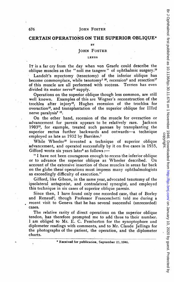
In January, 1946, the position was unaltered. He still had poormovement of the left eye down to the left as shown in the photo-graph Fig. 1. At the horizontal level he had 12° of L. ex-cyclophoriaat all angles, and the other synoptophore measurements were asgiven below: -
-Straight ahead=- 10'L/R 1 1200 to R. =-10 L/R 27A200 to L. =00 L/R 1A
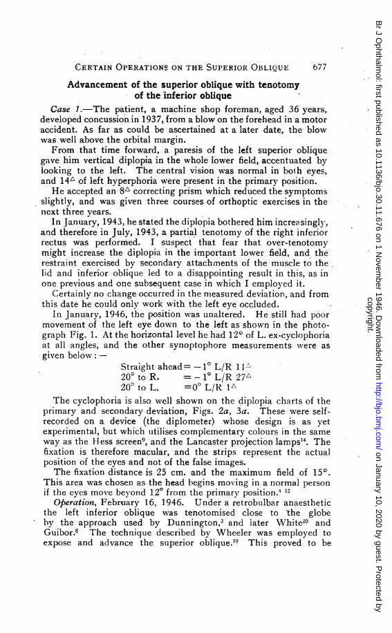
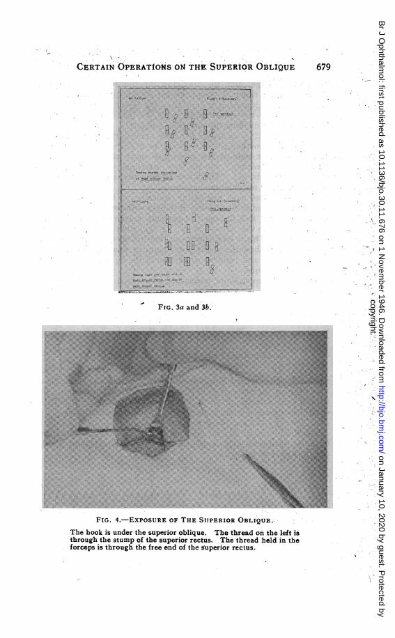
The cyclophoria is also well shown on the diplopia charts of theprimary and secondary deviation, Figs. 2a, 3a. These were self-recorded on a device (the diplometer) whose design is as yetexperimental, but which utilises complementary colours in the sameway as the Hess screen9, and the Lancaster projection lamps14. Thefixation is therefore macular, and the strips represent the actualposition of the eyes and not of the false images.The fixation distance is 25 cm. and the maximum field of 150.
This area was chosen as the head begins moving in a normal personif the eyes move beyond 120 from the primary position.4 12
Operation, February 16, 1946. Under a retrobulbar anaestheticthe left inferior oblique was tenotomised close to the globeby the approach used by Dunnington,3 and later White20 andGuibor.8 The technique described by Wheeler was employed toexpose and advance the superior oblique.19 This proved to be
677
copyright. on January 10, 2020 by guest. P
rotected byhttp://bjo.bm
j.com/
Br J O
phthalmol: first published as 10.1136/bjo.30.11.676 on 1 N
ovember 1946. D
ownloaded from
ws,r
.\ -, z .o
U10
,_ .
-zrn .srt . .
t ,,
9 .
/
; ''vA,,' ,'-
> ,..; , _s'_0S _
\;Ko o' S ..t - + . tA i wi .,wO F
..
{'s _9.e
V .r
,,.d. .. '. .0' 's* S .
, ;.e t.S
SV..
.--JojN- FOSTlElR
FIG. 1.
'''0 "tEl ElEWt years paralysis of the left superik obrcO u
Prior to operationNote failure of movment down to the right
] "tEl EllB El 18
Paralysis of superfior cf*ue after tenototy ofipsolateral fer ot ,iqueaadancem t ofipSofteral upero ObfiuNote spb# V*r*4eof kflt seior rectus csppWearce of cy.cc rie.
FIG. 2a and 2b.
copyright. on January 10, 2020 by guest. P
rotected byhttp://bjo.bm
j.com/
Br J O
phthalmol: first published as 10.1136/bjo.30.11.676 on 1 N
ovember 1946. D
ownloaded from
H ;,. f
CERTAIN OPERAT£ONS 09 THE SUPERIOR OBLIQUE\
... ....
PWt'" LEf *P4f)
. E-' ul 0 t.,t.Js,
. ~ ~~BS 84'S
<. MPgQ* Pse '(ttt
- FIG 3c an b
679
*:
a7;
\N ,
.
_ .s s
-
" . _B . . . , .9| | . | .;
| . || . || | . ....@ . |
E E N |@ | S --
889. | | | _.
Sk l | || | | || |.F | | ,.
i | | '.g 11 t sB. g . ,.z | 0'; ;'t:s | | .\w.P.....Fg |.,..9..0.o.:,Sl
*i.$. ._:.'2.''.FM a*',''-mw§,$
,.",.s...,o,> 9'. i.2 a X D.80s |! R .',:QQ '.i<Ka9°iXUXS
X .. .*'.S __.z<,.MeXX _'
: s'.X . ''.*8Sas .
: :
;. ......~~~~~~~~~~~~~~~~~~~......:...... :...::... ....:.-.
N.::.. : :.:.:::...... : ::..::.:.:...:::..::::::.:::::::::..:
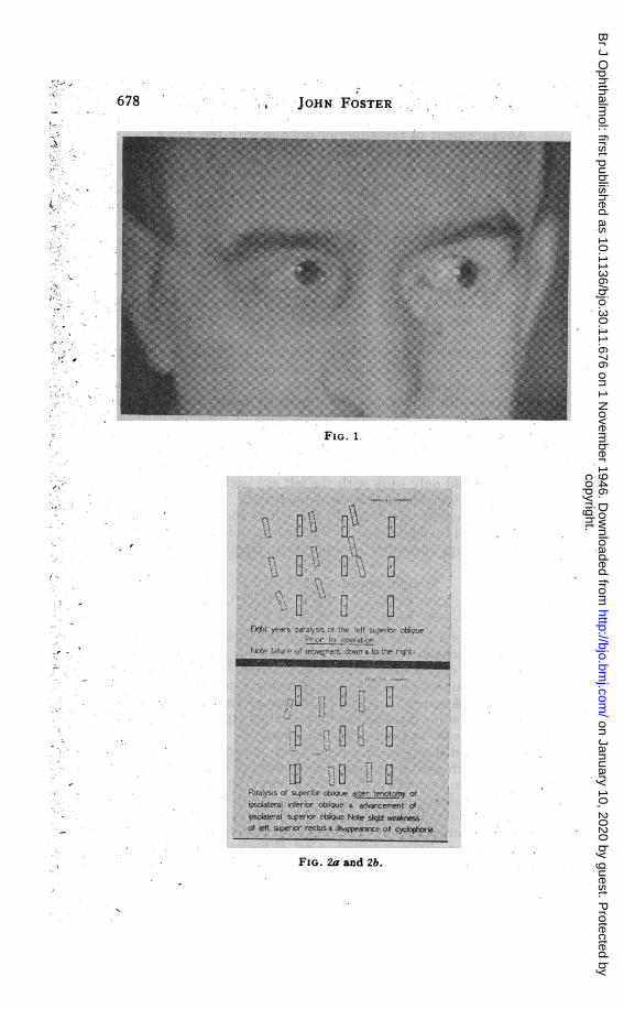
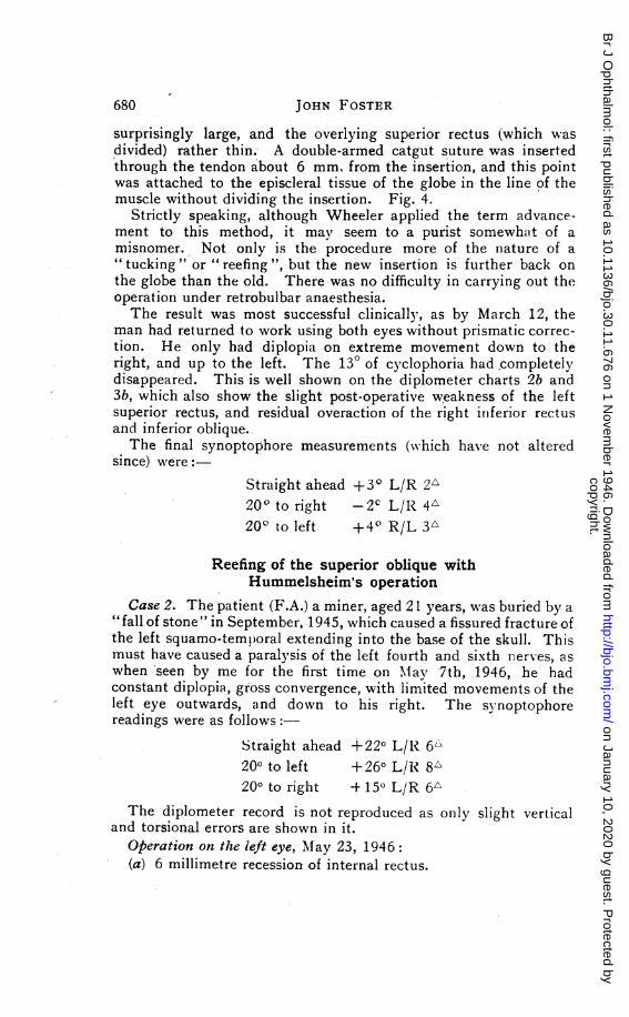
FIG. 4.-EXPOSURE OF THE SUPERIOR OBLIQUE.,
The hook is under the superior oblique. The thread on the left isthrough the stump of the superior rectus. The thread held in theforceps is through the free end of the superior rectus.
copyright. on January 10, 2020 by guest. P
rotected byhttp://bjo.bm
j.com/
Br J O
phthalmol: first published as 10.1136/bjo.30.11.676 on 1 N
ovember 1946. D
ownloaded from
JOHN FOSTER
surprisingly large, and the overlying superior rectus (which wasdivided) rather thin.- A double-armed catgut suture was insertedthrough the tendon about 6 mm, from the insertion, and this pointwas attached to the episcieral tissue of the globe in the line of themuscle without dividing the insertion. Fig. 4.
Strictly speaking, although Wheeler applied the term advance-ment to this method, it mavz seem to a purist somewhatt of amisnomer. Not only is the procedure more of the nature of a"tucking " or " reefing ", but the new insertion is further back onthe globe than the old. There was no difficulty in carrying out theoperation under retrobulbar anaesthesia.The result was most successful clinically, as by March 12, the
man had returned to work using both eyes without prismatic correc-tion. He only had diplopia on extreme movement down to theright, and up to the left. The 130 of cyclophoria had completelydisappeared. This is well shown on the diplometer charts 2b and3b, which also show the slight post-operative weakness of the leftsuperior rectus, and residual overaction of the right iniferior rectusand inferior oblique.The final synoptophore measurements (w\hich have not altered
since) were:-Straight ahead +30 L/R 2A20 ° to right -2c L/R 4A20° to left +40' R/L 3A
Reefing of the superior oblique withHummelsheim's operation
Case 2. The patient (F.A.) a miner, aged 21 years, was buried by a"fall of stone" in September, 1945, which caused a fissured fracture ofthe left squamo-temporal extending into the base of the skull. Thismust have caused a paralysis of the left fourth and sixth nerves, aswhen seen by me for the first time on Maay 7th, 1946, he hadconstant diplopia, gross convergence, with limited movements of theleft eye outwards, and down to his right. The synoptophorereadings were as follows:-
Straight ahead +220 L/R 6L200 to left +260 L/R 8A200 to right + 150 L/R 6
The diplometer record is not reproduced as only slight verticaland torsional errors are shown in it.
Operation on the left eye, May 23, 1946:(a) 6 millimetre recession of internal rectus.
680
copyright. on January 10, 2020 by guest. P
rotected byhttp://bjo.bm
j.com/
Br J O
phthalmol: first published as 10.1136/bjo.30.11.676 on 1 N
ovember 1946. D
ownloaded from
fQlk.X'IA1IL ErI\ft1JAJV'4b 'Lnk-1 Urr.KlUK JDJLl4Ur- UO1
(b) Suture of the outer half of the superior and inferior recti tothe insertion of the external rectus.The original Hummelsheim technique" was employed (it should be
noted that -variants, of this are described as O'Connor's or Temple-Smith's operation).
(c) The superior oblique tendon at a point 6 mm., from itsinsertion was sutured to the insertion itself, and a further sutureinserted to hold the reef firmly.
These procedures were carried out simultaneously under localanaesthesia. Only the outer half of the superior rectus was freedin this case.The result is still improving, the patient has simultaneous binocular
vision everywhere, except to th.e. right, though a slight tQrticolliscompensates a restriction of the internal rectus, when staring fixedlystraight ahead.The synoptophore readings on September 18, 1946:
0Straight ahead=0-0- Nil
020° to left =00 Nil
0
200 to right = 110 R/L 6In view of the continuing improvement it was decided not to
perform a partial re-advancement of the left internal rectus.
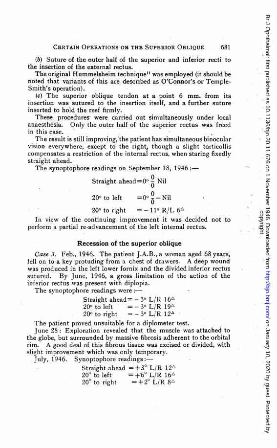
Recession of the superior obliqueCase 3. Feb., 1946. The patient J.A.13., a woman aged 68 years,
fell on to a key protuding from a chest of drawers. A deep woundwas produced in the left lower fornix and the divided inferior rectussutured. By June, 1946, a gross limitation of the action of theinferior rectus wvas present with diplopia.The synoptophore readings were:- -
Straight ahead - 30 L/R 16'20° to left 30 L/R 19A200 to right = - 30 L/R 12t
The patient proved unsuitable for a diplometer test.June 28: Exploration revealed that the muscle was attached to
the globe, but surrounded by massive fibrosis adherent to the orbitalrim. A good deal of this fibrous tissue was excised or divided, withslight improvement which was only temporary.
July, 1946. Synoptophore readings:-Straight ahead = +30 L/R 1V2200 to left =+60 L/R 16X'200 to right =+20 L/R 8"
rirr,p-rATM nlDT7'PA-rTCWQ'nM -rw-v C.TTOViDirrip nlD-T TnTYIM
copyright. on January 10, 2020 by guest. P
rotected byhttp://bjo.bm
j.com/
Br J O
phthalmol: first published as 10.1136/bjo.30.11.676 on 1 N
ovember 1946. D
ownloaded from
682 OTTO MEYER
Operation, August 1, 1946. An 8 mm. recession of the rightsuperior oblique was performed under local anaesthesia aftertemporary division of the superior rectus. This, of course, impliesa movement of the oblique insertion forwards and inwards. Afterthe operation it was decided that the inward movement was slightlygreater and the forward movement less than the ideal. The resultwas reasonably good, however, the patient obtaining single binocularvision without prisms everywhere except to the left, where thedivergence prevents it. Synoptophore readings on September 7,1946, were as follows:
Straight ahead=0° L/R 2',20 to left - 100 L/R 9A200 to right =00 R/L 3A
REFERENCES1. BARRIERE.-Ann. d'Ocul. 1932.2. BORLEY and RENAUD.-U.S.N. Med. Bull., Vol. XLV, p. 755. 1945.3. DUNNINGTON.-Trans. Amer. Ophthal. Soc., Vol. XXVII. p. 277. 1929.4. FRIEDENWALD.-Amer. Arch. Oohthal., Vol. XV, p. 283. 1936.5. GIBSON.-Amer. JIl. O,hthal., Vol. XXV, p. 565. 1941.6. GIFFORD.-;-Amer. JI. O,hthal., Vol. XXV, p. 761. 1941.7. - - Amer. JI. Ophthal., Vol. XXVIII, p. 882. 1942.8. GUIBOR.-Amer. Ji. Oththat., Vol. XXVII, P. 254. 1944.9. HEss.-Zeitschr. f. Augenheilk., Vol. XXXV, p. 201. 1916.
10. HUGHEs.-Amer. JI. OJhthal., Vol. XXVII, p. 1123. 1944.11. HuMMELSHEIM.-Ojhthal. Ges. Heidelberg, p. 248. 1907.12. JACKSON and O'RoURKE.-Amer. Arch. Oihthal., Vol. II, p. 756. 1929.13. JACKSON.-Ophthal. Rev., Vol. LXI, p. 22. 1903.14. LANCASTER.-Jl. Amer. Med. Assoc. Sect. Ophthal., p. 78. 1939.15. PETER.-Trans. Amer. Ophthal Soc., Vol. XXXI, p. 232. 1933.16. TERRIEN.-Chirurgie de l'oeil, Paris, p. 523. 1927.17. WAGMAN.-Amer. Ji. Ophth,al., Vol. XXVIII, p. 1226. 1945.18. WEGNER,-Klin. Monats. f. Augenheilk, Vol. C, p. 20. 1938.19. WHEELER.-Amer. JI. Ojbhthal., Vol. XVIII, p. 1. 1935.20. WHITE.-Amer. Ji. Ophthal., Vol. XXVI, p. 587. 1943.
INFLAMMATORY JUGULAR PHLEBOSTENOSIS ASTHE CAUSE OF GLAUCOMA EXOGENICUM*
BY
OTTO MEYERNEW YORK CITY
THE term "glaucoma exogenicum " is introduced to differentiatethis type of glaucoma with extra-ocular cause from " glaucomaendogenicum " that has an intra-ocular cause. Both types ofglaucoma are secondary in nature,
Just as retinitis albuminurica has its cause outside of the eye,exogenic glaucoma has an extra-ocular cause.
Received for publication, July 10, 1946.
copyright. on January 10, 2020 by guest. P
rotected byhttp://bjo.bm
j.com/
Br J O
phthalmol: first published as 10.1136/bjo.30.11.676 on 1 N
ovember 1946. D
ownloaded from